The investigation of EPR paramagnetic probe line width and shape temperature dependence in
EPR
1. ELEXSYS 系列
ELEXSYS是最新最完善的EPR波谱仪系列。它有连续波和脉冲傅立叶变换两种工作方式,工作频率 从1GHz到94GHz,代表了EPR仪器的最新发展。 至今为止,EPR波谱仪都是计算机辅助波谱仪。只有ELEXSYS系列提供的是软件辅助波谱仪。这种 新概念要求有一个卓越的软件战略思想和最新的硬件实施技术。ELEXSYS已具有能适应未来发展完善的网络功能。它的UNIX工作站具有前端处理器,采集服务器 和超高速的TRANSPUTER网络功能,执行各种应用程序。单用户和位于不同地方的多用户,可使用 这个系统。客户/服务器的结构,一方面解决了多用户/多任务之间的矛盾,另一方面又解决了实 时工作的矛盾。
ERP是Enterprise Resources Planning(企业资源计划)的缩写,
这一观念最初是由GartnerGroup公司在90年代初期提出的,并就其功能
标准给出了界定。
作为企业管理思想,它是一种新型的管理模式;而作为一种管理工具,
它同时又是一套先进的计算机管理系统。简单地说,EPR是用来对企业资源
MRPII的核心是MRP,但是他丰富了内涵,容入了企业整个的管理;ERP在MRPII的基础之上由于企业专业化的分工,强调了客户资源管理和供于MIS系统的范畴信息管理系统。
回答者: 5love_11 - 试用期 一级 5-7 12:48
EPR
进行优化配置,使企业运行更有效率。
它的前生是MRPII(制造资源计划),更前生是MRP(物料需求计划)
MRP主要用来判断计划中物料的缺料计划,然后生成采购计划(采购件)
和车间作业计划(自制件),但是他的基础建立在资源无限上的。
电子顺磁共振(ESR)在抗氧化活性研究中的应用

aN =14.3 G, aH β =11.7 G, aHγ =1.3 G
3. Singlet oxygen assay
23 mM TEMP
irradiation TEMPO + 18 µM rose bengal
OH
+
N
O
+ •OH
N
O
OOH
+
N
O
+ O2•¯
N
O
DMPO
DMPO(5,5-Dimethyl-1-pyrroline N-oxide 5,5-二甲基 吡咯林 氧化物)是研究氧 二甲基-1-吡咯林 氧化物) 二甲基 吡咯林-N-氧化物 自由基最常用的捕捉剂,具有较大的捕捉速率常数,易溶于多种溶剂, 自由基最常用的捕捉剂,具有较大的捕捉速率常数,易溶于多种溶剂,水溶液可以 达到0.1mM的浓度,对光不太敏感,与羟基自由基和超氧阴离子形成稳定的自选加 的浓度,对光不太敏感, 达到 的浓度 合物DMPO-OH和DMPO-OOH,而且具有特征的 和 波谱。 合物 ,而且具有特征的ESR波谱。 波谱
自旋捕捉技术
自旋捕捉剂是一类比较稳定的, 自旋捕捉剂是一类比较稳定的,可以与反应中产生的短寿命自由基结合产 生另一种较稳定的自由基即自旋加合物的化合物 一般来讲,捕捉剂通常有以下特性: 一般来讲,捕捉剂通常有以下特性: 在通常实验条件下比较稳定 在特定溶剂中有良好的溶解性 对短寿命自由基有较大的亲和力能迅速与之反应即有较高的捕捉效率, 对短寿命自由基有较大的亲和力能迅速与之反应即有较高的捕捉效率,所生成的 自旋加合物易溶解于特定溶剂中,且对光、热等外在条件呈惰性, 自旋加合物易溶解于特定溶剂中,且对光、热等外在条件呈惰性,尽可能保持较长 的稳定性 自旋加合物的ESR波谱特征尽可能多地反应出所捕捉的短寿命自由基的性质。 波谱特征尽可能多地反应出所捕捉的短寿命自由基的性质。 自旋加合物的 波谱特征尽可能多地反应出所捕捉的短寿命自由基的性质
EPR

CH3CH2OCCH3 1 2 3
=
O
NO的自旋捕获技术
自旋捕获剂: metal complex⎯Fe 2+ complex N-甲基葡萄糖胺-铁复合物 N-methyl-D-glucosamine,MGD (MGD)2-Fe2+-NO
(MGD)2-Fe2+-No
大鼠脂多糖引发脓毒性 休克6小时后皮下注射 (MGD)2-Fe2+,2小时后 测 EPR 谱
视紫红质 在黑暗 (蓝)状 态和光亮 (红)状 态的构象 变化和相 应的ESR 谱
自旋成像
血管舒张药物所产生的NO在体内的分布
思考题
1.为什么对称伸缩振动没有相应的红外吸收峰? 2.偶-偶核的自旋量子数为0,没有NMR谱,为什么? 3.偶-偶核是否没有自旋运动? 4.如何指认醋酸乙酯的COSY谱? 5.ESR谱有什么用处?
电子顺磁共振技术
洪远凯
Electron Paramagnetic Resonance,EPR Electron Spin Resonance, ESR
原子核
电子
Je=
I ( I + 1)η =
1 1 ( + 1)η = 2 2
3 η 2
e
μ= γe J
γe =
e 2meC
e
原子核
I=1 ms= -1,0,1
多电子物质的ESR?
电子在原子核外排布规则
1 首先占据能量较低的轨道 2 每个轨道最多允许2个自旋方向相反的电子 3 在同能量的轨道有多个(不止一个)时,电 子要首先分占不同的轨道,且自旋方向相同
外层价电子决定原子性质
. .N . ..
H2O
.. .O. ..
HeartSine samaritan PAD 450P CPR Rate Advisor 产品说明

CPR Rate Advisor ™ ICG technologyBrief summary of indications and important safety information on page 3.Tech NoteOverviewWhen CPR treatment is provided to a victim of sudden cardiac arrest, it is vital the chest compressions are of a good quality. If the quality of the CPR provided is good, the chances of successfully resuscitating a patient are greatly increased 1.Research has demonstrated that non-professional responders regularly provide ineffective CPR due to inexperience [2-3].The HeartSine samaritan PAD 450P (SAM 450P) with CPR Rate Advisor provides real-time feedback to the rescuer on the rate and fraction of the CPR they are providing to the victim during a sudden cardiac arrest (SCA) resuscitation. The SAM 450P uses both audible and visual feedback to provide instructions to the rescuer.CPR Rate Advisor provides feedback to the rescuer on the rate ofcompressions the rescuer is providing to the victim via the defibrillator electrodes, without the addition of accelerometers (or pucks).Figure 1. HeartSine’s defibrillator detects changes in patient impedance.How CPR Rate Advisor worksWhen a patient collapses and a rescuer performs CPR, the compressionsapplied by the rescuer cause the patient’s chest to change shape and result in a change to the patient’s ICG (impedance cardiogram) waveform 4. CPR Rate Advisor captures the change in the ICGwaveform which it uses to count the number of compressions a rescuer administers.By counting deflections in the ICG waveform, CPR Rate Advisor determines the compression rate and advises the rescuer to “Push faster” if the compression per minute (CPM) rate is below thatrecommended by the AHA/ERC guidelines (see Figure 2). Likewise, if the rescuer’s CPM rate is greater than that recommended by the AHA/ERC guidelines, CPR Rate Advisor will tell the rescuer to “Push slower” (see Figure 3).The AHA and ERC also recognize the need to keep interruptions to a minimum during CPR. The SAM 450P uses the signals detected through the electrode pads to determine if CPR is being performed when itshould be and, if not, will prompt the rescuer to “Begin CPR.” The SAM 450P also will detect when compressions have stalled between shock decision cycles and give feedback to the rescuer to ensure that CPR interruptions are minimized to maximize hands-on time (see Figure 4).I C G A Figure 4. No movement detected in the ICG waveform. In an effort to maximize CPR compression time by the rescuer, the SAM 450P will issue the audible prompt “Begin CPR” repeatedly until CPR is started.This real-time feedback is important as even though most trained rescuers understand the need to push hard and push fast, rescuer fatigue may set in after as little as one minute, resulting in slowercompression rates 5,6. The SAM 450P provides compression rate feedback to the rescuer via both visual indicators on the SAM 450P user interface and audible voice prompts (see Figure 5).No CPR being performed/“Begin CPR”“Push faster”“Good speed”“Push slower”Figure 5. Visual indicators and audible feedback tell the rescuer if the rate of CPR is in line with the AHA/ERC guidelines.Improved CPR rate efficacyUsability study results showed that, without compromisingcompression depth, the percentage of users achieving good compression speed was higher with CPR Rate Advisor when compared to a device without CPR feedback 7.Studies have shown that effectiveness of CPR is most likely limited by poor performance in any of its components and that inadequate rate, even in the presence of sufficient depth and technique, likely reduces the effectiveness of CPR compressions 8. Evidence suggests that even healthcare professionals do not always achieve the correct CPR compression rates according to AHA/ERC guidelines 8,9 and that chest compression rate is associated with the return of spontaneous circulation (ROSC)10.References1. Christenson J, Andrusiek D, Everson-Stewart S, et al. Chest compression fraction determines survival in patients with out-of-hospital ventricular fibrillation.Circulation . 2009;120:1241-1247. 2. Gyllenborg T, Granfeldt A, Lippert F , et al. Quality of bystander cardiopulmonary resuscitation during real-life out-of-hospital cardiac arrest. Resuscitation.2017;120:63-70.3. White AE, Ng H, Ng W , et al. Measuring the effectiveness of a novel CPRcard feedback device during simulated chest compressions by non-healthcare workers.Singapore Med J . 2017;58:438-445.4. Howe A, O’Hare P , Crawford P , et al. An investigation of thrust, depth and the impedance cardiogram as measures of cardiopulmonary resuscitation efficacy in aporcine model of cardiac arrest. Resuscitation. 2015;96:114–120. 5. Heidenreich JW , Berg RA, Higdon TA, et al. Rescuer fatigue: standard versus continuous chest-compression cardiopulmonary resuscitation. Academic EmergencyMedicine . 2006;13(10):1020–1026.6. Ochoa FJ, Ramalle-Gómara E, Lisa V , Saralegui I. The effect of rescuer fatigue on the quality of chest compressions. Resuscitation . 1998;37:149-152.7. Torney H et al. A Usability Study of a Critical Man–Machine Interface: Can layperson responders perform optimal compression rates when using a public accessdefibrillator with automated real-time feedback during cardiopulmonary resuscitation. IEEE Transactions on Human-Machine Systems . 2016;vol.PP:no.99:1-6. 8. Abella B et al. Chest compression rates during cardiopulmonary resuscitation are suboptimal. Circulation . 2005;111:428-434.9. Milander MM, Hiscok PS, Sanders AB, et al. Chest compression and ventilation rates during cardiopulmonary resuscitation: the effects of audible tone guidance. AcadEmerg Med . 1995;2:708-713.10. Idris A et al. Relationship between chest compression rates and outcomes from cardiac arrest. Circulation . 2012;125:3004-3012.11. Meaney PA, Bobrow BJ, Mancini ME, et al. Written on behalf of the CPR Quality Summit Investigators, the American Heart Association Emergency CardiovascularCare Committee, and the Council on Cardiopulmonary, Critical Care, Perioperative and Resuscitation. CPR quality: improving cardiac resuscitation outcomes both inside and outside the hospital: a consensus statement from the American Heart Association. Circulation . 2013;128:1-19.CPR Rate Advisor Tech NoteEffective CPR, provided alone or together with a lifesaving shock,can increase the chance of survival 11. CPR Rate Advisor, in conjunction with the metronome, is intended to help rescuers perform CPR in line with the AHA/ERC guidelines by monitoring their real-time CPR performance and helping to guide them toward the correct rate of compressions.Integrated CPR Rate Advisor helps improve compliance with CPR rate and CPR fraction guidelines. And because CPR Rate Advisor is integrated within HeartSine SAM 450P , a lifesaving shock can be delivered if needed.HeartSine ® samaritan ® PAD Automated External Defibrillators (AEDs)BRIEF SUMMARY OF INDICATIONS AND IMPORTANT SAFETY INFORMATIONINDICATIONS FOR USE: The HeartSine samaritan PAD SAM 350P (SAM 350P), HeartSine samaritan PAD SAM 360P (SAM 360P) and HeartSine samaritan PAD SAM 450P (SAM 450P) are indicated for use on victims of cardiac arrest who are exhibiting the following signs: unconscious, not breathing, without circulation (without a pulse). The devices are intended for use by personnel who have been trained in their operation. Users should have received training in basic life support/AED, advanced life support or a physician-authorized emergency medical response training program. The devices are indicated for use on patients greater than 8 years old or over 55 lb (25 kg) when used with the adult Pad-Pak (Pad-Pak-01 or Pad-Pak-07). They are indicated for use on children between 1 and 8 years of age or up to 55 lb (25 kg) when used with the Pediatric-Pak (Pad-Pak-02).CONTRAINDICATION: If the patient is responsive or conscious, do not use the HeartSine samaritan PAD to provide treatment.WARNINGS: AEDs: • The HeartSine samaritan PAD delivers therapeutic electrical shocks that can cause serious harm to either users or bystanders. Take care to ensure that no one touches the patient when a shock is to be delivered. • Touching the patient during the analysis phase of treatment can cause interference with the diagnostic process. Avoid contact with the patient while the HeartSine samaritan PAD is analyzing the patient. The device will instruct you when it is safe to touch the patient. • Do not delay treatment trying to find out the patient’s exact age and weight. If a Pediatric-Pak or an alternative suitable defibrillator is not available, you may use an adult Pad-Pak. • The SAM 360P is a fully automatic defibrillator. When required, it will deliver a shock to the patient WITHOUT user intervention. • The SAM 450P CPR Rate Advisor is currently only intended to provide feedback on adult patients. If you treat a pediatric patient with the SAM 450P and an adult Pad-Pak, ignore any voice prompts regarding the rate of CPR. • Do NOT use the HeartSine samaritan PAD in the vicinity of explosive gases, including flammable anesthetics or concentrated oxygen. • Do NOT open or repair the device under any circumstances as there could be danger of electric shock. If damage is suspected, immediately replace the HeartSine samaritan PAD. Pad-Paks: • Do not use if the gel is dry. • The Pediatric Pad-Pak is not for use on patients under 1 year old. For use with children up to the age of 8 years or up to 55 lb (25 kg). DO NOT DELAY THERAPY IF YOU ARE NOT SURE OF EXACT AGE OR WEIGHT . • Only HeartSine samaritan PADs with the label are suitable for use with the Pediatric-Pak. If the HeartSine samaritan PAD you are using does not have this label, use the adult Pad-Pak if no alternatives are available. • The use of the Pediatric-Pak will enable delivery of 50J shocks to the pediatric patient. • The Pediatric-Pak contains a magnetic component (surface strength 6500 gauss). Avoid storage next to magnetically sensitive storage media. It is advised that Pediatric-Paks are stored separately when not in use. • Never charge, short circuit, puncture, deform, incinerate, heat above 85o C or expose contents of TSO (Aviation) Pad-Pak to water. Remove when discharged.PRECAUTIONS: AEDs: • Proper placement of the HeartSine samaritan PAD electrode pads is critical. Electrode pads must be at least 1 in (2.5 cm) apart and should never touch one another. • Do not use electrode pads if pouch is not sealed. • Check the device periodically in accordance with the service and maintenance instructions provided in the User Manual. • Operate the HeartSine samaritan PAD at least 6 feet (2 meters) away from all radio frequency devices or switch off any equipment causing interference. • Use of the device outside the operating and storage ranges specified in the User Manual may cause the device to malfunction or reduce the shelf life of the Pad-Pak. • Do not immerse any part of the HeartSine samaritan PAD in water or any type of fluid. • Do not turn on the device unnecessarily as this may reduce the standby life of the device. • Do not use any unauthorized accessories with the device as the HeartSine samaritan PAD may malfunction if non-approved accessories are used. • Dispose of the device in accordance with national or local regulations. • Check with the relevant local government health department for information about any requirements associated with ownership and use of a defibrillator in the region where it is to be used. Pad-Paks: • Check expiration date. Saver EVO Software: • Download the complete HeartSine samaritan PAD memory prior to erasing it. This information should be stored safely for future reference. Ensure that only the events you want to delete have been selected prior to deleting. Once deleted from your computer’s memory, events cannot be regenerated and all information will be lost.POTENTIAL ADVERSE EFFECTS: The potential adverse effects (e.g., complications) associated with the use of an automated external defibrillator include, but are not limited to, the following: • Failure to identify shockable arrhythmia. • Failure to deliver a defibrillation shock in the presence of VF or pulseless VT , which may result in death or permanent injury. • Inappropriate energy which could cause failed defibrillation or post-shock dysfunction. • Myocardial damage. • Fire hazard in the presence of high oxygen concentration or flammable anesthetic agents. • Incorrectly shocking a pulse-sustaining rhythm and inducing VF or cardiac arrest. • Bystander shock from patient contact during defibrillation shock. • Interaction with pacemakers. • Skin burns around the electrode placement area. • Allergic dermatitis due to sensitivity to materials used in electrode construction. • Minor skin rash.CAUTION: U.S. Federal law restricts this device to sale by or on the order of a physician.Please consult the user manual at for the complete list of indications, contraindications, warnings, precautions, potential adverse events, safety and effectiveness data, instructions for use and other important information.All claims valid as of July 2021.For further information, please contact your Stryker representative or visit our web site at Emergency Care Public AccessStryker’s AEDs require a prescription in the U.S. Please consult your physician. AED users should be trained in CPR and in the use of the AED. Although not everyone can be saved, studies show that early defibrillation can dramatically improve survival rates. AEDs are indicated foruse on adults and children. AEDs may be used on children weighing less than 25 kg (55 lb) but some models require separate defibrillation electrodes.The information presented is intended to demonstrate Stryker’s product offerings. Refer to operating instructions for complete directions for use indications, contraindications, warnings, cautions, and potential adverse events, before using any of Stryker’s products. Products may not be available in all markets because product availability is subject to the regulatory and/or medical practices in individual markets. Please contact your representative if you have questions about the availability of Stryker’s products in your area. Specifications subject to change without notice.Stryker or its affiliated entities own, use, or have applied for the following trademarks or service marks: CPR Rate Advisor, HeartSine,Pad-Pak, Pediatric-Pak, samaritan, Saver EVO, Stryker. All other trademarks are trademarks of their respective owners or holders.The absence of a product, feature, or service name, or logo from this list does not constitute a waiver of Stryker’s trademark or other intellectual property rights concerning that name or logo.UL Classified. See complete marking on product.Date of Issue: 07/2021Made in U.K.H009-020-008-AF EN-USHeartSine SAM 450P is not available for sale outside of the U.S. or Japan.Copyright © 2021 Stryker.Manufactured by: HeartSine Technologies Ltd. 207 Airport Road West Belfast, BT3 9EDNorthern IrelandUnited KingdomTel +44 28 9093 9400Fax +44 28 9093 9401**************************** Distributed in U.S. by: Stryker Emergency Care 11811 Willows Road NE Redmond, WA, 98052 U.S.A. Toll free 800 442 1142 。
PHZS:Cu2+的EPR谱和局域结构研究

PHZS:Cu2+的EPR谱和局域结构研究张华明【摘要】采用Cu2+离子正交对称电子顺磁共振(EPR)参量的高阶微扰公式计算K2Zn(SO4)2·6H2O:Cu2+的EPR参量g因子(gx,gy,gz)和超精细结构常数(Ax,Ay,Az).研究结果表明,K2Zn(SO4)2·6H2O中[Cu(H2O)6]2+基团的Cu2+-H2O键长分别为Rx≈0.197 nm,Ry≈0.213 nm,Rz≈0.224 nm;中心金属离子基态波函数混合系数分别为α ≈0.978和β ≈0.209.所得EPR参量理论值与实验符合很好.【期刊名称】《电子科技大学学报》【年(卷),期】2018(047)005【总页数】4页(P766-769)【关键词】Cu2+离子;电子顺磁共振谱;局域结构;晶体PHZS【作者】张华明【作者单位】南昌航空大学光电检测工程技术实验室南昌 330063;南昌航空大学教育部无损检测重点实验室南昌 330063【正文语种】中文【中图分类】O737晶体K2Zn(SO4)2·6H2O (PHZS)属于单斜晶系,空间群为(P121/a1),每个晶胞含2个Zn原子,晶格常数[1]a ≈ 0.903 4 nm,b ≈ 1.218 4 nm,c ≈0.614 8nm,δ =104.8°,该晶体由一系列不规则的KO8多面体和Zn(H2O)6八面体通过氢键与SO4相连接而构成网格状结构。
其中Zn2+与周围6个H2O分子构成[Zn(H2O)6]2+基团,该位置属正交(D2)点群对称。
众所周知,晶体的光学、磁学等性能与掺杂离子的局域结构密切相关,而掺杂离子所处的局域环境往往不同于母体位置[2-3]。
因此,研究晶体中掺杂离子的局域结构对理解掺杂离子影响材料性能的微观机理非常重要。
电子顺磁共振(EPR)谱强烈依赖于顺磁离子所处局域环境,并可通过分析作为其实验结果的EPR参量定量地确定掺杂离子周围的局部结构[4]。
英语调查报告作文开头结尾

英语调查报告作文开头结尾英文回答:Introduction.The purpose of this investigation was to explore the attitudes and beliefs of native English speakers towards the use of English in international communication. The investigation sought to answer the following research questions:1. What are the attitudes of native English speakers towards the use of English as a lingua franca?2. What are the beliefs of native English speakers about the impact of English on other languages?3. What are the implications of the findings for the future of English as a global language?Methodology.The investigation was conducted using a mixed-methods approach, which included both qualitative and quantitative data collection methods. The qualitative data werecollected through interviews with 20 native English speakers from a variety of backgrounds. The interviews were semi-structured and explored the participants' attitudes and beliefs about the use of English in international communication. The quantitative data were collected through a survey of 100 native English speakers. The survey measured the participants' attitudes towards the use of English as a lingua franca and their beliefs about the impact of English on other languages.Results.The findings of the investigation revealed that native English speakers have a range of attitudes and beliefs about the use of English in international communication. Some participants expressed positive attitudes towards the use of English as a lingua franca, while others expressedmore negative attitudes. The majority of participants believed that English has a positive impact on other languages, while a minority believed that it has a negative impact. The findings also suggested that the future of English as a global language is uncertain.Conclusion.The findings of this investigation provide someinsights into the attitudes and beliefs of native English speakers towards the use of English in international communication. The findings suggest that there is a range of perspectives on this issue and that the future of English as a global language is uncertain.中文回答:引言。
酶在接近无水有机介质中的生物催化特性
生物化工与催化收稿日期:2004-12-01;修订日期:2005-05-20 基金项目:国家自然科学基金资助项目(20176019)作者简介:张 娜(1980-),女,青岛科技大学生物化工专业在读硕士研究生。
通讯联系人:刘均洪,博士生导师,教授。
E 2mail :qdht2004@酶在接近无水有机介质中的生物催化特性张 娜,刘均洪(青岛科技大学化工学院,山东青岛266042)摘 要:介绍了接近无水条件下以悬浮状态存在的酶以及固定化酶生物催化特性。
采用电子顺磁共振(EPR )及核磁共振(NMR )研究了水与酶之间的关系。
在有机溶剂中加水和不加水情况下,处于悬浮状态的酶及固定化酶结构和功能存在差别。
研究发现,尽管以悬浮状态存在的酶和固定化酶结构明显不同,但水含量、酶活性、酶柔性以及活性位点极性之间存在着密切的关系。
结果表明,在含水量很少的条件下,水对酶活性位点的生物催化起着关键作用。
关键词:非水酶学;电子顺磁共振(EPR );核磁共振(NMR )中图分类号:Q55;TQ033 文献标识码:A 文章编号:100821143(2005)0720048204Biocatalytic properties of enzymes in nearly anhydrous organic solventsZHA N G N a ,L IU J un 2hong(College of Chemical Engineering ,Qingdao University of Science and Technology ,Qingdao 266042,China )Abstract :The biocatalytic properties of enzymes in nearly anhydrous organic solvents were reviewed.Water 2protein interactions and the involvement of water in enzyme structure and function were investi 2gated by electron paramagnetic resonance (EPR )and multinuclear nuclear magnetic resonance (NMR )studies of enzymes suspended and immobilized in organic solvents with and without addition of water.In these systems ,correlations were observed between water content and enzyme activity ,flexibility ,and active 2site polarity ,despite the remarkable difference between structural properties of suspended and immobilized enzymes.The results indicated that water in very small amount plays a key role in the biocatalysis at the enzymic active sites.K ey w ords :non 2aqueous enzymology ;electron paramagnetic resonance (EPR );nuclear magnetic reso 2nance (NMR )C LC number :Q55;TQ033 Document code :A Article ID :100821143(2005)0720048204 酶作为生物催化剂,具有专一性、高效性、反应条件温和等优点,受到人们的普遍关注。
EPR电子顺磁共振教程

5、三重态分子
这类化合物分子轨道上也有两个未偶电子,但其与双基 不同,这两个电子彼此相距很近,有很强的相互作用。有两类: 1、激发三重态;如:萘激发三重态;
2、基态就是三重态分子如:氧分子。
6、过渡金属和稀土元素
EPR—研究对象三
EPR—研究对象三
过渡金属、稀土元素具有未充满的3d,4d,5d及 4f壳层,核外有一个或一个以上的未成对电子。
Purcell
1955年 1966年 1981年 1989年 W.E. Lamb, P. Kusch A. Kastler N. Bloembergen N.F. Ramsey 1964年 1977年 1983年 1991年 C.H. Townes J.H. Van Vleck H. Taube R.R. Ernst
Raymond snubbed by Nobel Committee because he is beloved by God? Godless science shits on a saint.
I don't know. Perhaps Raymond deserves the prize. MRI was first attempted by him but his results were pretty much useless. His original paper has been discredited by follow up research. I personally see no conflict between science and spiritualism, but putting a Christian God as the head of the universe is tacky in my opinion. Teaching that there has been no evolution, that Genesis contains a literal cosmology, well that isn't science in any sense I understand.
玉米叶绿体铁氧还蛋白1的原核表达及部分性质分析

核农学报2011,25(1):0062 0066 26Journal of Nuclear Agricultural Sciences文章编号:1000-8551(2011)01-0062-05玉米叶绿体铁氧还蛋白1的原核表达及部分性质分析冯爱花陈宗梅李静程备久范军(安徽农业大学生命科学学院/安徽省作物生物学重点实验室,安徽合肥230036)摘要:叶绿体铁氧还蛋白(Fd)通过活性中心的铁硫簇传递还原力,在各种氧化还原途径中起重要作用。
本研究中,氨基酸序列比对显示玉米中5种Fd的叶绿体导肽同源性很低,而去除导肽的成熟蛋白氨基酸序列具有很高的同源性。
采用RT-PCR技术从玉米幼叶总RNA中克隆了编码成熟Fd1的基因,并分别插入pQE80和p28SUMO表达载体,转化大肠杆菌BL21(DE3),表达的N端含有组氨酸标签的Fd1(His-Fd1),和含有组氨酸标签的小类泛素修饰蛋白(SUMO)的Fd1(HisSUMO-Fd1),用Ni-NTA层析介质亲和纯化,SDS-PAGE显示纯化的His-Fd1为一条带,亚基分子量约12kD。
纯化的HisSUMO-Fd1用专一性蛋白酶Ulp除去HisSUMO,获得纯化的Fd1。
紫外可见光谱扫描显示纯化的HisSUMO-Fd1溶液在315nm、415nm和459nm有特异吸收峰,电子顺磁共振波谱分析表明Fd1中存在[2Fe-2S]。
SDS-PAGE显示多种玉米幼叶可溶性蛋白被固定化的His-Fd1吸附。
关键词:铁氧还蛋白(Fd);Fd1;玉米;重组表达;纯化;铁硫簇PROKARYOTIC EXPRESSION AND PARTIAL CHARACTERIZATION OFMAIZE CHLOROPLAST FERREDOXIN1FENG Ai-hua CHEN Zong-mei LI Jing CHENG Bei-jiu FAN Jun(Provincial Key Lab of Crop Science,School of Life Science,Anhui Agricultural University,Hefei,Anhui230036)Abstract:The ferredoxin(Fd)proteins in plant chloroplasts play important roles in cellular metabolism by delivering reducing equivalents through the[2Fe-2S]cluster in the active site to various essential oxido-reductive pathways.In this study,the chloroplast leading peptides from five Fd proteins of maize share the low homogeneity,whereas the mature Fd proteins deleted the leading peptides are high homogeneous,as displayed by the amino acid sequence alignments. The gene encoding mature maize ferredoxin1(Fd1)was cloned by RT-PCR using the total RNA from young leaves as the template.The cloned gene was inserted into pQE80and p28SUMO plasmid,and transformed into Escherchia coli BL21(DE3)respectively.The expressed Fd1fused with the histidine-tag(His-Fd1)or HisSUMO tag(HisSUMO-Fd1)at N terminus,was purified by Ni-NTA affinity chromatography independently.The recombinant Fd1protein was obtained by removing the HisSUMO using the specific protease Ulp.SDS-PAGE analysis showed that purified His-Fd1 has a molecular mass of about12kD.The purified HisSUMO-Fd1has the absorption peaks at315,415and459nm identified by UV-visible spectra scanning,and the[2Fe-2S]cluster determined by electron paramagnetic resonance (EPR)experiments.Several proteins from soluble extracts of young maize leaves were bound by the immobilized His-Fd1,as shown by SDS-PAGE analysis.Key words:ferredoxin protein;Fd1;maize(Zea mays);prokaryotic expression;purification;[2Fe-2S]cluster收稿日期:2010-06-23接受日期:2010-10-18基金项目:转基因生物新品种培育重大专项(No.2009ZX0810-002B),国家自然科学基金(No.30840018),安徽省攻关课题(No.0701302137)作者简介:冯爱花(1983-),女,山东临沂人,硕士,研究方向为植物生物化学。
纳米材料的表征方法

纳米材料的表征及其催化效果评价方式纳米材料的表征主要目的是确定纳米材料的一些物理化学特性如形貌、尺寸、粒径、等电点、化学组成、晶型结构、禁带宽度和吸光特性等。
纳米材料催化效果评价方式主要是在光照(紫外、可见光、红外光或者太阳光)条件下纳米材料对一些污染物质(甲基橙、罗丹明B、亚甲基蓝和Cr6+等)的降解或者对一些物质的转化(用于选择性的合成过程)。
评价指标为污染物质的去除效率、物质的转化效率以及反应的一级动力学常数k的大小。
1 、结构表征XRD,ED,FT-IR, Raman,DLS2 、成份分析AAS,ICP-AES,XPS,EDS3 、形貌表征TEM,SEM,AFM4 、性质表征-光、电、磁、热、力等UV-Vis,PL,Photocurrent1. TEMTEM为透射电子显微镜,分辨率为0.1~0.2nm,放大倍数为几万~百万倍,用于观察超微结构,即小于0.2微米、光学显微镜下无法看清的结构。
TEM是一种对纳米材料形貌、粒径和尺寸进行表征的常规仪器,一般纳米材料的文献中都会用到。
The morphologies of the samples were studied by a Shimadzu SSX-550 field-emission scanning electron microscopy (SEM) system, and a JEOL JEM-2010 transmission electron microscopy (TEM)[1].一般情况下,TEM还会装配High-Resolution TEM(高分辨率透射电子显微镜)、EDX(能量弥散X射线谱)和SAED(选区电子衍射)。
High-Resolution TEM用于观察纳米材料的晶面参数,推断出纳米材料的晶型;EDX一般用于分析样品里面含有的元素,以及元素所占的比率;SAED用于实现晶体样品的形貌特征与晶体学性质的原位分析。
2. SEMSEM 表示扫描电子显微镜,可以获取被测样品本身的各种物理、化学性质的信息,如形貌、组成、晶体结构和电子结构等等。
布鲁克器EMX-Plus X带电子参磁共振(EPR)光学仪器操作指南说明书
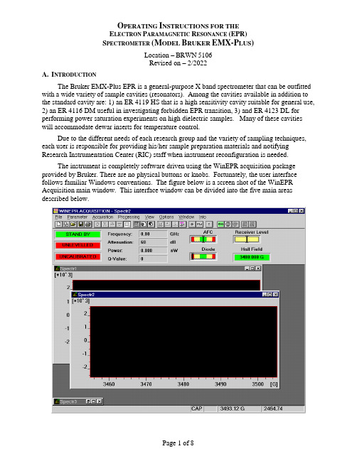
O PERATING I NSTRUCTIONS FOR THEE LECTRON P ARAMAGNETIC R ESONANCE (EPR)S PECTROMETER(M ODEL B RUKER EMX-P LUS)Location –BRWN5106Revised on – 2/2022A.I NTRODUCTIONThe Bruker EMX-Plus EPR is a general-purpose X band spectrometer that can be outfitted with a wide variety of sample cavities (resonators). Among the cavities available in addition to the standard cavity are: 1) an ER 4119 HS that is a high sensitivity cavity suitable for general use, 2) an ER 4116 DM useful in investigating forbidden EPR transition, 3) and ER 4123 DL for performing power saturation experiments on high dielectric samples. Many of these cavities will accommodate dewar inserts for temperature control.Due to the different needs of each research group and the variety of sampling techniques, each user is responsible for providing his/her sample preparation materials and notifying Research Instrumentation Center (RIC) staff when instrument reconfiguration is needed.The instrument is completely software driven using the WinEPR acquisition package provided by Bruker. There are no physical buttons or knobs. Fortunately, the user interface follows familiar Windows conventions. The figure below is a screen shot of the WinEPR Acquisition main window. This interface window can be divided into the five main areas described below.Menu Bar – Like all Windows programs, there is a Menu Bar at the top of the window used to access all other program features. All of the features listed in the other areas of the interface are directly accessible via the menu bar.Button Bar – This bar is located just below the Menu and can be toggled visible/invisible using the View option in the Menu bar. It is highly recommended to leave it visible since all spectrometer functions needed for data acquisitions are most easily accessed from the tool pallet. The table below shows each icon and lists its “tip strip”. The specif ic functions of many are discussed in the relevant sections below.New Spectrum Duplicate Spectrum Open Spectrum Save Spectrum Print Spectrum ExperimentalParametersExperimental OptionsComment ParametersRun / Stop ScriptRun AcquisitionStop AcquisitionStop Acquisition atEnd ScanTimes 2 Divide by 2 Expand Contract Change Center Fieldand Sweep WidthChange Center FieldChange Static FieldInteractive ReceiverLevelSend Spectrum toWinEPRSend Spectrum toSimfoniaReset Display Rectangular Zooming Cursor Moving Microwave Fine Tune Microwave Bridge ControlInteractive Spectrometer ControlInstrument Monitor – This area is located just below the Button Bar and shows the status of the microwave bridge and signal channel. The displays can be toggled visible/invisible by using the View option in the Menu bar.Data Area – This area fills the center of the screen and is populated by various windows. The windows can be any combination of spectra, parameters, and instrument control windows. Status Line – This is located at the bottom of the window and displays current instrument status and messages.B.O PERATION:S TARTUPFollow the procedure below to insure proper instrument operation. Although spectra can be obtained immediately after startup, waiting at least an hour for the magnet and electronic components to thermally stabilize before taking spectra is recommended.EMX S TARTUP1.Nitrogen Purge – Open the gas supply valve on the liquid nitrogen dewar. This will purgethe EPR cavity, waveguide, and microwave bridge of water vapor which absorbsmicrowave energy and oxygen which gives a signal since it is paramagnetic2.Cooling water – Open the two chilled water valves on the wall to the right of the computerdesk.3.Heat Exchanger –Use the rotary switch labeled “Heat Exchanger” located on the controlbox above the water filter. Be careful to not mistakenly push the Main power button.4.Magnet power supply – The magnet power supply not only controls current in the electromagnet but powers the console and microwave bridge as well. It is powered on in atwo-step process as detailed below.a.Press the ELECT. ON (Electronics) button in far left, upper corner. Wait for the fivered LED-warning lights on far right to extinguish before continuing. If the LEDs donot extinguish, confirm that the cooling water is on and repeat this step.b.Press POWER ON, which is located to the immediate right of the ELECT. ON switch.5.Console Power – Press the round button located in the lower, center of the cabinet. Thisalso provides power for the Microwave Bridge located on the top, left corner of the shelf above the electro magnet. The console houses: 1) Signal Channel – A phase sensitivedetector measuring the EPR signal by comparison with a signal of known frequency.2) Magnetic Field Controller – Controls both the magnitude and sweep rate of themagnetic field during a scan. 3) Modulation Amplifier – This unit provides the modulated reference signal for detection in the signal channel.NOTE –The console houses ~$150,000 of electronics.DO NOT set samples or your coffee on itC OMPUTER L OGONThe computer is usually left on and can be awakened by pressing any key on the keyboard. If the screen does not respond within 30 seconds of pressing a key, press the power button on the front of the case. At the Logon prompt e nter your “User Name” and “Password”. The “Domain” entry should be set to BoilerAD.Once logged on, WinEPR shortcuts are visible in the upper right-hand corner of the desktop. “WinEPR Acquisition” is the application used to run the spectrometer and obtain data. “WinEPR Processing” and “WinEPR SimFonia” are for analysis and simulation, respectively. This training document focuses only on the acquisition software and basic instrument operation.Launch the WinEPR Acquisition by double clicking its icon. It takes about 90 seconds for the software to fully launch. Once the window opens, click the Interactive Spectrometer Control button and click the “Calibrated” check box in the upper right of the window that opens.C. O PERATION:L OADING S AMPLESInserting / Changing Samples(Room Temperature)1.If the Microwave Bridge Controller window is not open, click on the Microwave Bridgebutton on the tool bar.2.Select the STANDBY or TUNE mode. See the image below.3.To avoid contaminating the cavity, clean the outside of the sample tube with a ChemWipe.4.Center the sample tube in the collet (loosen the collet ring if necessary) and gently slide itdown until either the sample is centered in the cavity or the tube is resting on the pedestal.Tighten the collet ring just enough to prevent the sample tube from moving.(Helium Variable temperature)1.If the Microwave Bridge Controller window is not open, click on the Microwave Bridgebutton on the tool bar.2.Select the STANDBY or TUNE mode. See the image below.3.Position the sample tube in the top-hat using the cavity gauge drawing to center the samplein the cavity. Tighten the top-hat ring so that the sample tube is snug.Notes – Do not over loosen or remove the upper portion of the top-hat.– The sample should be located in the center of the cavity for the best response.– Samples must be frozen prior to inserting them in the cavity!4.To avoid contaminating the cavity, clean the outside of the sample tube with a ChemWipe. Be sure to remove any frost.5.In as smooth and rapid a motion as possible remove the top-hat currently on the VT cavityand replace it with the new sample by carefully sliding the sample tube straight down into the cavity. Once the top-hat contacts the vent tube, apply gentle but firm pressure until the top-hat snaps into place.6.Tune the cavity and set parameters to the instrument as usual.7.Wait for the sample to thermally equilibrate (~10 minutes).Tuning the Microwave Cavity and Bridge1.If the Microwave Bridge Controller window is not open, click on the Microwave Bridgebutton on the tool bar.2.Select the TUNE mode. See the image below.e the pairs of up/down arrow keys set the attenuation to 25dB. The leftmost pairchanges in units of 10dB and the rightmost pair changes in 1dB units.e the right/left arrow buttons of the Frequency slider to center the tuning dip as shownabove. The system takes long fractions of a second to respond so do not click too fast.Use the right/left arrow buttons on the Signal Phase slider to adjust phase until the tuning dip is symmetrical as shown above. The system takes long fractions of a second torespond so do not click too fast.5.Either click the Up or Down arrow to start the cavity auto-tune process. The autotuneroutine will then adjust the frequency, phase, bias, and iris coupling for optimalperformance. Wait for this process to complete as indicated by the three green indicators in the left of the Instrument Monitor window.Note – If auto-tune fails, follow the procedure in Appendix C.6.Click the Microwave Bridge button to close the window.D. O PERATION:T AKING S PECTRASetting ParametersInstrument parameters may be loaded from disk by opening an existing spectrum, or set manually as described below. Whichever method is chosen it is important to verify that the cavity calibration file is loaded and is being used.1.Setting parameters manuallya.Click the New Spectrum button to ensure that a previously open spectrum is notaccidentally overwritten.b.Click the Experimental Parameters button and enter the desired parameters.Notesa)If looking for radicals centered around g = 2, check the box just above the “CenterField” numeric to automatically set the “Center Field” parameter.b)Microwave power – The range is 1 to 200 mW. Usual values are 1 to 20 mW.c)Modulation Amplitude – The range is 0.1 to ~20 (dependent on cavity) gauss. 5 to10 gauss is typical. The amplitude should be no higher than the width of thenarrowest line (in gauss) in the spectrum. See page 2-19 in the Bruker WinEPRacquisition manual for a discussion of over modulation.d)Receiver Gain – Typical range 100 to 5000.e)Conversion Time – This is the time allotted for the A/D process and directlyinfluences resolution in the Y-axis. This value multiplied by Resolution in X (# ofpoints collected across spectrum) yields the sweep time for the spectrum.f)Time constant – This value should be less than one tenth the time needed to scanthe narrowest line in the spectrum. See page 2-20 in the Bruker WinEPRacquisition manual for a discussion of using an excessively long time constant.g)If a single scan does not yield spectra with reasonable signal-to-noise, either of thetwo options below can help.a.Repetitive scanb.Increased time constant with either increased conversion time and/or Xresolution to slow the scan through the signal2.Loading/Verified cavity calibrationa.Click the Interactive Spectrometer Control button.b.Verify that the “Calibrated” check box in the upper right is checked.c.Click the “SCT Options” button at the lower right to open the Signal Channel Optio nsdialog shown below.d.Verify that the loaded calibration file in use matches the name of the cavity currentlyinstalled. See the table below for the calibration file names. If necessary, click the“Change File” button and select the proper file.Resonator Name Calibration FileER 4102 ST ST 0203.calER 4103 TM TM 9304.calER 4119 HS HS 0716.calER 4116 DM DM 0708.calER 4123 D D 0247.calAcquiring DataAfter setting the scan parameters, click the Run button to initiate data collection. Acquisition can be terminated immediately using the Stop Acquisition button or at the end of the current scan via the button. This latter operation is useful during averaging operations if the desired spectral quality has been achieved.E. I NSTRUMENT O PERATION:S HUTDOWN1.Set Microwave Bridge controller to STANDBYa.If the Microwave Bridge Controller window is not open, click on the MicrowaveBridge button on the tool bar.b.Select the STANDBY mode.2.Exit the WinEPR software.3.“Sign Out …” - Right click on your name at the top of the Windows “Start” menu.4.Remove the sample from the cavity and replace the cap.5.Power down in this order.a.Turn off the console by pressing the lit button in the center of the unit.b.Turn off the magnet power supply (PWR first and then ELECT).6.Turn off the Heat Exchanger – Be careful not to mistakenly push the Main power button.7.Close the chilled water valves.8.Close the nitrogen gas valve.9.Sign the logbook.F.C ONTACTSAdvance Methods Consultation Training and ServiceDr. Michael Everly Dr. Hartmut HedderichAmy Faculty, Director Snr. Instrumentation SpecialistDepartment of Chemistry Department of ChemistryOffice: BRWN 4151 Office: BRWN 4151Phone: 49-45232 Phone: 49-46543E-mail : ******************E-mail : *******************Appendix A – V ARIABLE T EMPERATURE O PERATION (C OLD E DGE) Using high purity helium as a coolant, sample temperature can be varied from 150 K to~5 K. This is done by flowing helium gas at the desired temperature through a dewar assembly installed within the cavity that surrounds the sample. The cold gas is generated by passing99.999% helium at room temperature through a heat exchanger connected to a Sumitomo closed-loop compressor system. This eliminates the need for using liquid helium and reduces the cost of operation by a factor of 10.Due to the extreme low temperatures, great care must be taken when inserting samples not to thermally stress the sample/cavity, contaminate the cavity with room temperature air, or crush the heater and thermocouple that sit just below the sample. To reduce the thermal shock, which usually results in broken sample tubes, all samples MUST be frozen in liquid nitrogen before placing them in the cavity. Prefreezing samples also greatly reduces the time needed for temperature equilibration. Sample tubes MUST be rigorously cleaned to avoid cavity contamination. Wash your sample tubes between uses with an appropriate solvent and wipe them off with a ChemWipe before inserting them in the cavity. System maintenance due to contamination by a carless user may be billed to the PI at a cost of $60/hr with a 4-hour minimum!P ROCEDURES–The procedures below are intended to be used as needed and are not listed sequentially.S TARTUP,C OOL-D OWN,S HUTDOWNDue to the complexity of the ColdEdge VT system, center staff will perform these operations. Users will need to coordinate startup and shutdown times with center staff.S ETTING T EMPERATUREThe Oxford Instruments Cryostat and LakeShore controller combination uses a heater to warm the flowing cold helium gas to the desired temperature. To set the temperature press the “Setpoint” button and enter the desired temperature and hit “Enter”. If you find that you have entered an incorrect value or menu that you didn’t want, simpl y press the cancel button.C HANGING/I NSERTING S AMPLES1.Place the Microwave Bridge Controller in STANDBY mode.2.With one hand, remove the sample/cap from the cavity. With the other hand, insert thenew sample.Notes – This must be done as quickly as possible to prevent room temperature air from entering the cavity.– Samples must be frozen prior to inserting them in the cavity!3.Tune the cavity and set parameters to the instrument as usual.4.Wait for the sample to thermally equilibrate (~10 minutes) and fine-tune the cavity.Appendix B – M ANUAL T UNING THE M ICROWAVE C AVITY In case the automatic tuning operation (in Part V. of the training outline) fails to properly tune the cavity and bridge, a message indicating this failure will be shown in the mode indicator in the center of the microwave bridge controller display.Some samples may be somewhat "lossy," i.e., the sample or solvent changes the conditions in the cavity to decrease the cavity absorption dip. If this effect is only minimal, it may still be possible to manually tune the cavity and bridge by the following procedure.If the sample is very "lossy," it may be impossible to observe a sufficient cavity dip for lock-on by the control system. If the control system cannot be stabilized as indicated by inability to center the LOCK OFFSET meter or the DIODE CURRENT meter, then a different (aqueous) cavity will need to be installed. Contact RIC staff to make this change.Manually Tuning Procedure (Summarized from section 5.1of the WinEPR acquisition manual)1.If the Microwave Bridge Controller window is not open, click on the Microwave Bridgebutton on the tool bar.2.Select the TUNE mode. See the image below.e the pairs of up/down arrow keys set the attenuation to 25dB. The leftmost pairchanges in units of 10dB and the rightmost pair changes in 1dB units.e the right/left arrow buttons of the Frequency slider to center the tuning dip as shownabove. The system takes long fractions of a second to respond so do not click too fast.5.The dip should cover come about 2/3 of the way to the baseline. If the dip is too small orlarge, decrease or increase, respectively the attenuation in 1dB steps.e the right/left arrow buttons of the Signal Phase slider to adjust phase until the tuningdip is symmetrical as shown above and is as deep as possible. If the dip points up, the phase is 180 degrees off. If it has positive and negative lobes, it is ~90 degrees off. The system takes long fractions of a second to respond so do not click too fast.7.Select the Operate mode and then fine-tune the Frequency to center the AFC indicator inthe Instrument Monitor window. Readjust as needed if the AFC drifts during subsequent steps.8.Set attenuation to 50dB and use the right/left arrow buttons of the Bias slider to center theDiode Current meter (200 µA) in the Instrument Monitor Window9.The steps below adjusting the iris (critical coupling) are iterative in nature.a.Lower the attestation by 10dB.e the Up/Down keys of the Iris, bring the diode current back to center.c.Repeat the steps above until reaching 10dB.Note – If the AFC lock drifts, center it by adjusting the frequency.10.While at 10dB of attenuation, adjust phase to achieve maximum Diode current.11.Cycle through 10, 20, 30, 40, 50 dB to verify that the Diode current remains constant. Ifnot, repeat the tuning process.12.Click the Microwave Bridge button to close the window.Page B2。
EPR技术在化学分析中的应用

EPR技术在化学分析中的应用电子顺磁共振(Electron Paramagnetic Resonance,EPR)技术是一种非常优秀的技术,在化学分析中有着广泛的应用。
除了物理和生物学领域中的研究外,EPR技术同样被一个越来越多的化学家所借助,用于定量和定性表征有机和无机物质,同时还在环境化学和食品科学中广泛使用。
一般来说,EPR技术通过测量待分析样品产生的电子的微波辐射信号来进行分析。
待测样品的特定属性影响到电子的辐射,使它有一个独特的谱,从而提供有关样品的信息。
这种技术广泛应用在研究化学反应动力学,分析电子的传输和催化。
在接下来的段落中,我们将深入了解EPR技术在化学分析中的应用。
定性分析EPR是分析某些物质的性质时一种重要的方法,通过测量辐射的谱线给出物质的自由基或金属离子的参数。
简单来说,自由基是分子内存在未成对电子的剩余电子。
通过EPR技术,我们可以非常容易地检测到自由基并定量表征它们。
自由基在化学反应中有着广泛的应用,例如在辐射损伤修复机制、发动机和涡轮机的燃烧中等。
此外,金属的离子也可以借助EPR技术进行定性分析。
金属离子对于化学反应过程至关重要。
使用EPR技术,金属离子可以被直接测量到,从而获得如氧化还原状态、配位化学、磁性等信息。
这对于合成金属配合物、研究金属催化反应或者了解生物体中的金属离子也有很大的帮助。
定量分析EPR技术在定量分析中同样拥有重要作用。
对于某些光敏和放射性物质而言,使用传统的化学检测技术是不可能的,因为它们会破坏样品。
而通过利用EPR技术可以避免这种情况,从而进行更精确的定量分析。
此外,EPR也可以在催化和电化学反应定量分析中应用。
比如,在电化学反应中,EPR技术可以帮助确定催化剂中的金属离子的浓度和配位环境。
这对于催化剂的性能和催化反应机理的研究都有非常重要的意义。
研究氧化还原反应机理EPR技术在研究氧化还原反应机理中也有着广泛的应用。
化学反应过程中的氧化还原过程是非常重要的。
电子磁共振技术在催化研究中的应用EPR.

洛仑兹线型和高斯线型
电子磁共振的基本原理
过渡金属离子的EMR谱
晶体场理论 八面体的“四方畸变”和“三角畸变” 晶场分裂:三重态能级t2g和二重态能级eg 八面晶体场中的电子填充:高自旋与低自旋
等效自旋S/能级在外
磁场作用下发生分裂。在
取向体系中,当H平行于Z
轴,S≥1,自旋哈密顿算
符为:
H geHSZ A·S·I A·S·I 为超精细作用项。
gYY为中间的g因子。一般,g∥>g⊥。若 分子的对称性更低,则gXX≠ gYY≠gZZ, 必须用三个主值表示。
三g值的EMR一次微商谱
电子磁共振的基本原理
超精细结构
处在外磁场中的顺磁样品的电子可能会与相邻近的磁性核 的自旋磁矩发生相互作用。
磁性核在外磁场的作用下沿Z方向的分量是量子化的。核 的自旋态MI可以有从-I、-I+1…… I-1直到I的共(2I+1) 个取值。
未偶电子除了处在 外磁场(H外)中,还处 在邻近磁性核的磁场中。 这相当于有一个局部磁 场(H局)以矢量方式叠 加在H外上而成为一个等 效磁场。 H等效=H外+H局
电子磁共振的基本原理
∴EMR谱线由原来的单一谱线分裂成(2I+1)条谱线,即 得到“超精细结构”谱(hfs)。
一个未偶电子(S=1/2)与一个核(I=1/2)发生相互作 用,并在外磁场作用下发生能级分裂,在一级近似情况下各能 级的能量分别为: EⅠ=E1+hA/4 EⅡ=E1-hA/4 EⅢ=E2+hA/4 EⅣ=E2-hA/4 A称做“各向同性超精细偶合常数”(单位为磁场单位mT)。
根据量子力学原理,能级间跃迁的选择规则为ΔMS=±1和 ΔMI=0是允许跃迁,跃迁的能量为:
ΔEⅠ-Ⅳ=gβeH+hA/2 ΔEⅡ-Ⅲ=gβeH-hA/2 ΔEⅠ-Ⅳ-ΔEⅡ-Ⅲ=hA
电子顺磁共振波谱EPRESR概论

一、 电子顺磁共振的基本原理
1、概述
电子自旋的磁特性
Joseph John Thomson (英国)
The Nobel Prize in Physics 1906
• In 1891, the Irish physicist, George Stoney, believed that electricity should have a fundamental unit. He called this unit the electron.
• The electron was discovered by J.J. Thomson in 1897. • The electron was the first sub-atomic particle ever found. It
was also the first fundamental particle discovered. • The concept of electron spin was discovered by S.A.
电子的磁矩主要来自自旋磁矩(> 99%)的贡献。
若轨道中所有的电子都已成对,则它们 的自旋磁矩就完全抵消,导致分子无顺磁性;
若至少有一个电子未成对,其自旋就会产生 自旋磁矩。
因此,EPR研究的对象必须具有未偶电子。
H =0时,每个自旋磁矩的方向是随机的,并处于同一个平均能态。
H≠0时,自旋磁矩 就有规则 地排列起 来 (平行 外磁场 — 对 应能级的能量较低,或反平行于外磁场—对应能级 的能量较高)。
• 顺磁性 (B’>0,即B’与B0同向) • 铁磁性 (B’>0,即B’与B0同向, B’随B0增大而急
剧增加, 但当B0 消失而本身磁性并不消失) • 反磁性(B’<0,即B’与B0反向) (逆、抗)
epr定量测定煤中自由基的方法及煤液化机理的研究

《epr定量测定煤中自由基的方法及煤液化机理的研究》1. 研究背景在当今能源日益紧缺的环境下,煤作为一种重要的化石能源备受关注。
煤液化作为一种重要的煤化工技术,对于提高能源利用效率具有重要的意义。
而煤中的自由基含量,作为反映煤液化机理的重要参数,一直以来备受关注。
如何准确测定煤中的自由基含量,以及研究煤液化的机理就显得尤为重要。
2. 自由基的概念自由基是指分子中的一个或多个原子带有未成对电子而呈现出的不稳定活泼态的分子。
在煤中,自由基的存在对煤的反应性、热稳定性以及裂解性能都有着重要的影响。
3. epr定量测定方法电子顺磁共振(electron paramagnetic resonance, EPR)是一种测定自由基含量的重要方法。
在煤中,通过EPR技术,可以准确测定煤中的自由基含量,并对其进行定量分析。
该方法具有准确性高、灵敏度高等特点,是目前研究煤中自由基含量的主要手段之一。
4. 煤液化机理的研究煤液化机理是指在煤液化过程中,煤分子发生裂解、重组和生成液态产物的过程。
在煤液化过程中,自由基的含量和性质对于反应过程以及产物的性质都具有重要影响。
研究煤中自由基的含量和性质,对于深入理解煤液化机理具有重要的意义。
5. 个人观点和理解我认为,研究煤中自由基的含量和煤液化机理是一项具有深远意义的工作。
通过准确测定自由基的含量,并研究其与煤液化机理之间的关系,不仅可以加深对于煤化学反应的理解,还可以为煤液化工艺的改进和优化提供重要的参考依据。
总结通过epr定量测定煤中自由基的方法及煤液化机理的研究,可以得到对煤中自由基含量的精确测定,并且揭示了煤液化的重要机理,这对于煤化工技术的发展具有重要的意义。
相信随着研究的不断深入,这一领域的成果将为煤炭资源的高效利用、清洁利用提供重要的支持。
通过以上深入的讨论,相信您对于epr定量测定煤中自由基的方法及煤液化机理的研究已经有了更加深入的理解。
希望我的文章对您有所帮助。
顺磁共振电子顺磁共振(ElectronParamagneticResonance简称EPR)或
顺磁共振电子顺磁共振(Electron Paramagnetic Resonance 简称EPR )或称电子自旋共振(Electron Spin Resonance 简称ESR )是探测物质中未耦电子以及它们与周围原子相互作用的非常重要的现代分析方法,它具有很高的灵敏度和分辨率,并且具有在测量过程中不破坏样品结构的优点。
自从1944年物理学家扎伏伊斯基(Zavoisky )发现电子顺磁共振现象至今已有五十多年的历史,在半个多世纪中,EPR 理论、实验技术、仪器结构性能等方面都有了很大的发展,尤其是电子计算机技术和固体器件的使用,使EPR 谱仪的灵敏度、分辨率均有了数量级的提高,从而进一步拓展了EPR 的研究和应用范围。
这一现代分析方法在物理学、化学、生物学、医学、生命科学、材料学、地矿学和年代学等领域内获得了越来越广泛的应用。
本实验的目的是在了解电子自旋共振原理的基础上,学习用射频或微波频段检测电子自旋共振信号的检测方法,并测定DPPH 中电子的g 因子和共振线宽。
一 实验原理原子的磁性来源于原子磁矩。
由于原子核的磁矩很小,可以略去不计,所以原子的总磁矩由原子中个电子的轨道磁矩和自旋磁矩所决定。
在本单元的基础知识中已经谈到,原子的总磁矩μJ 与P J 总角动量之间满足如下关系:J J BJ P P g γμμ=-= (1-6-1) 式中μB 为波尔磁子,ћ为约化普朗克常量。
由上式可知,回磁比Bg μγ-= (1-6-2) 按照量子理论,电子的L -S 耦合结果,朗得因子)1(2)1()1()1(1++-++++=J J L L S S J J g (1-6-3) 由此可见,若原子的磁矩完全由电子自旋磁矩贡献(L =0,J =S ),则g =2。
反之,若磁矩完全由电子的轨道磁矩所贡献(S=0,J=1),则g =1。
若自旋和轨道磁矩两者都有贡献,则g 的值介乎1与2之间。
因此,精确测定g 的值便可判断电子运动的影响,从而有助于了解原子的结构。
1996年年度简报-沈阳材料科学国家(联合)实验室
1996年年度简报-沈阳材料科学国家(联合)实验室中国科学院国际材料物理中⼼(ICMP)1996年⼯作简报⼀. 发表论⽂情况1996年本中⼼成员及其合作者共发表学术论⽂135篇,其中发表在中⼼指定刊物上的有24篇,发表在虽⾮中⼼指定但有影响的国际性刊物上的有65篇。
1996年中⼼成员为72⼈,⼈均为1.88篇。
⼈均篇数⽐1995年的1.96篇略少,⽐1994年的1.53篇略多。
原因之⼀是1996年投稿在⾮国际性刊物上的论⽂不再统计。
1996年中⼼成员向中⼼提交已发表论⽂主要情况如下:1. 中⼼成员为第⼀作者,在指定刊物上发表论⽂的有:秦国刚, 郑⽂琛(2),魏国柱,魏宝华,郑⼤,赵平波(1)。
2. 中⼼成员和中⼼单位均为第⼀位,在重要国际性刊物上发表论⽂的有:龙期威(2),王崇愚,姜健,李世红,隋国鑫,杨⾦龙(1)。
3. 中⼼成员为第⼀位,在重要的国际性刊物上发表2篇论⽂以上的有:郑⽂琛(6),程天⼀(4),李世红(3),周本濂,郑⾥平(2)。
4. 在中⼼指定刊物上,中⼼成员⾮第⼀作者的有:梁敬魁(7),⽥德诚(3),秦国刚,姚凯伦(2),周先意,魏宝华(1)。
5. 提供论⽂数量较多的有:梁敬魁(15),周本濂(12),姚凯伦(9),郑⽂琛(8),龙期威(6),刘伍明(6),⽥德诚(5),程天⼀(5)。
另外,按国家教委信息中⼼提供的1995年SCI收录的论⽂统计结果排名,中⼼成员郑⽂琛教授个⼈论⽂数是全国第三名。
⼆. 部分成员在科研⼯作中取得了显著成就1996年中⼼成员报来的获奖成果有(详情见附件⼆):国家教委或省部级科技进步⼆等奖三项(何开元,杜懋陆,谢和平),辽宁省教委科技进步⼀等奖⼀项(魏国柱),国家教委或省部级科技进步三等奖⼆项(吴翔,张邦维);获其它奖的有:周本濂获⾸届桥⼝隆吉基⾦会奖;刘有延获⼴东省“⼋五”基础性研究⼗⼤成果奖;张邦维获湖南省⾃然科学优秀论⽂⼀等奖;韦钦获中南⼯业⼤学“⼋五科研⼯作突出贡献”奖。
电子顺磁共振技术应用及进展

第32卷第5期2013年5月实验室研究与探索RESEARCH AND EXPLORATION IN LABORATORYVol.32No.5May 2013·实验技术·电子顺磁共振技术应用及进展王翠平,叶柳,谢安建,李广,李爱侠,张子云,张惠(安徽大学物理与材料科学学院,安徽省信息材料与器件重点实验室,安徽合肥230039)摘要:电子顺磁共振(EPR )波谱技术是一种新的检测方法,用于检测顺磁性离子、自由基及顺磁性配合物分子的结构。
近几年又发展成为一种操控自旋电子材料内部原子核外单电子自旋状态手段,用于单电子自旋相干态的制备,实现量子运算和信息传输。
目前文献中报道EPR 在化学、物理、生物和医药领域的应用很多。
针对当前EPR 在不同领域的应用,综述了EPR 技术的应用原理和进展,为更好地将EPR 技术应用在量子物理、配合物化学、自由基生物学、医学、药学等领域提供参考和借鉴。
关键词:电子顺磁共振;电子自旋相干态;自由基捕捉;自选标记中图分类号:O 4-33文献标志码:A 文章编号:1006-7167(2013)05-0005-03Progress and Applications of Electron ParamagneticResonance SpectroscopyWANG Cui-ping ,YE Liu ,XIE An-jian ,LI Guang ,LI Ai-xia ,ZHANG Zi-yun ,ZHANG Hui(School of Physics and Materials Science ,Anhui University ,Hefei 230039,China )Abstract :In this paper ,in view of the technical application of electron paramagnetic resonance (EPR ),the application principle and development of EPR technology were summarized to provide reference for its applications in quantum physics ,chemistry ,free radicals-biology ,medicine ,and archaeological and materials science fields.Key words :electron paramagnetic resonance ;electron spin coherence ;free radical trap ;spin label收稿日期:2012-10-09基金项目:国家自然科学基金资助项目(50973001,2117300);安徽大学2012校级教学研究项目资助(JYXM201238,JYXM201231)作者简介:王翠平(1971-),女,安徽蒙城人,博士,高级实验师,主要研究方向为有机/无机复合材料制备和磁共振波谱研究。
植物中活性氧的检测方法_祁艳
生物技术通报BIOTECHNOLOGYBULLETIN·技术与方法·2007年第5期收稿日期:2007-03-30基金项目:国家自然科学基金资助项目(No.30671244);植物生理学与生物化学国家重点实验室开放课题资助项目(No.PPB04006);河北省自然科学基金资助项目(No.303180;No.C2005000220)作者简介:祁艳(1981-),女,山西人,硕士,研究方向:植物抗病生理通讯作者:王冬梅,E-mail:dongmeiwang63@hotmail.com由于活性氧(reactiveoxygenspecies,ROS)具有很高的反应活性,它们可以与许多不同的化合物发生反应,根据反应生成物或者反应物的变化程度可以间接地对ROS进行定量或者定性分析。
通常采用的方法有化学发光法、紫外-可见吸收分光光度法、荧光染色法及DAB组织染色法等,电子显微技术检测法也受到研究者的青睐。
活体中ROS的直接检测对于解释生命机体的各种生理反应有着重要的意义。
目前能够用于解决该问题的方法比较有限,已报道的有电子顺磁共振技术(electronparamagneticresonance,EPR),又称电子自旋共振技术(electronspinresonance,ESR)。
EPR是一种检测自由基的波谱学方法,可以直接检测O2-·,·OH等活性氧自由基及参与其形成的过渡金属离子的化学变化[1,2],因此该法受到植物界工作者的广泛关注。
下面对目前实验室中所用到的检测植物体内ROS的方法逐一进行介绍。
1植物体内ROS的检测方法1.1化学发光法化学发光法是活性氧检测中常用的实验技术,其原理是化学发光试剂与活性氧反应生成激发态的产物,产物中的电子经非辐射性跃迁回到基态而放出光子。
常用的化学发光试剂有鲁米诺(luminol)、光泽精(lucigenin)、甲壳动物荧光素(如海萤荧光素)等。
scientific reports
Synergistic effect on the visible light activity of Ti 31doped TiO 2nanorods/boron doped graphene compositeMingyang Xing 1*,Xiao Li 1*&Jinlong Zhang 1,21Key Lab for Advanced Materials and Institute of Fine Chemicals,East China University of Science and Technology,Shanghai,200237,China,2Department of Chemistry,Tsinghua University,Beijing 100084,P.R.China.TiO 2/graphene (TiO 2-x /GR)composites,which are Ti 31self-doped TiO 2nanorods decorated on boron doped graphene sheets,were synthesized via a simple one-step hydrothermal method using low-cost NaBH 4as both a reducing agent and a boron dopant on graphene.The resulting TiO 2nanorods were about 200nm in length with exposed (100)and (010)facets.The samples were characterized by X-ray diffraction (XRD),UV-visible diffuse reflectance spectroscopy,X-band electron paramagnetic resonance (EPR),X-ray photoelectron spectra (XPS),transmission electron microscope (TEM),Raman,and Fourier-transform infrared spectroscopy (FTIR).The XRD results suggest that the prepared samples have an anatase crystalline structure.All of the composites tested exhibited improved photocatalytic activities as measured by the degradation of methylene blue and phenol under visible light irradiation.This improvement was attributed to the synergistic effect of Ti 31self-doping on TiO 2nanorods and boron doping on graphene.Since Fujishima and Honda discovered the phenomenon of photocatalytic splitting of water on TiO 2electrodes in 19721–3,titanium dioxide (TiO 2)has emerged as one of the most promising oxide semi-conductors and has been employed in diverse applications including air and waste water purifiers,solar energy cells and sensors 4,5.However,the wide band gap and fast recombination of the photoexcited electron-holes of TiO 2restrict its use in many practical applications.Therefore,TiO 2modification is necessary for improving the optical sensitivity and activity of TiO 2in the presence of visible light.Such modifications might include impurity ion doping,noble metal loading,and others 6,7.Among these,impurity doping is an efficient technology for improving the response of TiO 2to visible light.However,impurity doping could result in crystal or thermal instability and increased carrier recombination centers 8.Owing to its relatively high surface area and special photoelectrochemical properties compared to powder catalysts,many studies on TiO 2nanorods have been previously reported.Jun et al.9varied the ratio of a non-selective and a surface selective surfactant (trioctylphosphine oxide and lauric acid,respectively)in dioctyl ether to induce the transformation of TiO 2nanoparticles into nanorods dissolved in dioctyl ether.Additionally,Li et al.10synthesized tetragonal faceted-nanorods of single-crystalline anatase TiO 2with a large percentage of higher-energy (100)facets.Generally,the previous nanorods were prepared in organic solvent,increasing the tediousness of operational processes and subsequently reducing the working efficiency.In addition,modifica-tions such as doping and controlling the morphology,reduced the amount of TiO 2containing Ti 31or an oxygen vacancy and has also been confirmed to exhibit high photocatalytic activity 11.Our group has previously reported studies on Ti 31.For example,Xing et al.8,12successfully synthesized Ti 31self-doped TiO 2with either NaBH 4as the reducing agent or using a vacuum-activated procedure.Both samples exhibited high photo-degradation of organic pollutants.In spite of the research progress achieved on Ti 31and vacancy,there are still some contro-versies concerning especially the theoretical research on this topic.Rusu et al.13concluded that the photocatalytic activity of rutile increased by vacuum pretreatment through the production of a large amount of anion on the (110)faces.Nevertheless,Hoffmann et al.4ascribed this phenomenon to the reinforced crystallinity achieved via high temperature activation.Meanwhile,Sato et al.14found that heating the material to 500u C induced desorption of surface oxygen and produced many oxygen defects resulting in improved photo-oxidation capacity.On the contrary,Yu et al.15attributed the high photocatalytic activity of TiO 2to the existence of Ti 31surface states.This was due to the ability of TiO 2to capture photogenerated electrons prior to transferring the electrons to the O 2adsorbed on the active sites of surface Ti 31,thus reducing the recombination of photogenerated electrons and holes.OPENSUBJECT AREAS:POLLUTION REMEDIATION PHOTOCATALYSISReceived 1May 2014Accepted 11June 2014Published 30June 2014Correspondence and requests for materials should be addressed to J.Z.(jlzhang@ecust.)*These authors contributed equally tothis work.On the other hand,after the discovery of an atomic sheet of sp 2-bonded carbon atoms by Geim et al.16,17in 2004,graphene has attracted great interest from both theoretical and experimental scien-tists.Graphene nanosheets,as two-dimensional (2D)conductors and monolayers of carbon atoms arranged into honeycomb network formations,have attracted attention as a consequence of their unique properties such as elasticity,low density,excellent electrical conduc-tivity,chemical stability and their large surface area 18,19.Additionally,graphene can also potentially act as a support material,allowing semiconductor particles (such as TiO 2nanoparticles)to anchor themselves to the surface 20.Because of this feature,the surface prop-erties of graphene can be widely adjusted by chemical modifications to form composites 7,bining TiO 2and graphene into compo-sites is a promising approach to facilitate the effective photodegrada-tion of pollutants under visible light irradiation.Recently,the fabrication of hybrid materials,such as TiO 2loaded onto graphene,has been a popular topic of study.Zhang et al.17synthesized a chemically bonded TiO 2(P25)/graphene nanocompo-site using a facile,one-step hydrothermal method,affording impress-ive methylene blue degradation activity.Choi et al.22reported the fabrication of TiO 2/GR nanocomposites via a facile electrostatic attraction mbert et al.23obtained TiO 2/GR hybrid mate-rials by mixing graphene oxide (GO)and TiF 4followed by ultraso-nication and heating before reduction by hydrazine hydrate (HHA)and hydrothermal processing for heightened stability.All of the reported composite hybrids have superior photocatalytic activities compared to other TiO 2materials used for the degradation of dyes.Yet,many open problem remain;for example,this process usually gives rise to TiO 2aggregation while loading P25onto GO 24.While HHA has been widely used in the reduction of GO,it is recognized,however,as an environmental pollutant.Additionally,solvothermal treatment is selective for the epoxy group of GO,leaving the hydroxyl and carboxyl groups unreduced.To mediate these problems,there is strong demand for environmentally friendly reducing agents and novel reduction processes.Additionally,some doping modifications of graphene in order to improve its electronic properties have attracted a great deal of atten-tion.Tran Van Khai et al.25prepared boron-doped graphene oxides by means of annealing the films at 1100u C.The modified GOs wereobtained from suspensions of GO and H 3BO 3in a solution of N,N -dimethylformamide (DMF).Similarly,Niu et al.26prepared boron-doped graphene through pyrolysis of graphene oxide with H 3BO 3in an argon atmosphere at 900u C.Each of these experiments adopted high-temperature processes,increasing the economic cost of these methods.Theoretical studies on graphene nanoribbons doped with boron have demonstrated that edge-type as well as substitutional doping can induce half-metallic behavior and that the band gap can be tuned by doping 27,thus highlighting the potential application of boron-doped graphene (B-GR)in photocatalysis.Here,we report the preparation of TiO 2nanorods in deionized water via a simple one-step hydrothermal method.First,we exposed nanorods with (100)and (010)facets of about 200nm in length.Next,the composite,consisting of Ti 31self-doped TiO 2nanorods were loaded onto the boron-doped graphene sheets.This was suc-cessfully achieved using NaBH 4as the reducing agent as well as the boron source.The photocatalytic activity of Ti 31-TiO 2/B-graphene composites will also be discussed.ResultsThe FESEM and TEM images of TiO 2nanorods are presented in Figure 1.The prepared TiO 2nanoparticles are shaped like nanorods with lengths in the range of 50–200nm.It is obvious from the cross-section of the FESEM image (Figure 1a)that the angle between two adjacent sides is 90u .For increased clarification,we set up a structural modeling image.From this image,it is obvious that the nanorod exists with the (100)and (010)facets exposed and at an angle of 90u ,which is in agreement with the above result.To further char-acterize the exposure of the (100)facet,TEM (Figure 1b)and fast-Fourier transform (FFT)(Figure 1c)were performed.The axis direction of the nanorod is parallel to the (002)facet,as determined by FFT,confirming that the nanorod is extended along the (001)direction.Considering the observation of the (200)facet perpendic-ular to the (002)facet in the FFT image,it can be concluded that the prepared TiO 2nanorod exposes the (100)facet.Theoretical studies demonstrated that anatase (100)facets are more active and accord-ingly exhibit higher catalytic activity than (001)or (101)facets 10.The mechanism of formation of TiO 2nanorods can be explained in the kinetic growth region 9,shown in Figure 2.The structure ofanataseFigure 1|(a)FESEM and (b)TEM images of TiO 2nanorods,and (c)the corresponding fast-Fourier transform (FFT).The inset of (a)is the amplified image of TiO 2nanorods and a structural modulingimage.Figure 2|The formation mechanism of TiO 2nanorods.TiO 2is tetragonal with the (101)and (001)facets exposed.The added ammonia results in the growth of the (001)facet,resulting in a change in growth velocity,namely,n (001).n (101),ultimately resulting in the formation of nanorods.The loading of TiO 2nanorods on graphene sheets was character-ized by TEM.Images of pure graphene and TiO 2-x /GR composites are shown in Figure 3.Figure 3a demonstrates that the prepared sheet-like graphene oxide was a transparent,smooth,and 2D-layered material well suited for the addition of TiO 2.We intended to load the TiO 2nanorods on the wrinkled or edged areas of the GO where carboxyl functional group are likely to be abundant 14(Figure 3b–d).Accordingly,the TiO 2nanoparticles were covalent bonded to GO,forming a composite favoring the separation of electron-hole pairs (Figure 3d).To further characterize the composition of the as-prepared sam-ples,we performed Raman spectroscopy (Supplementary Figure S1).The samples exhibited strong peaks at g 51.978and g 51.959,char-acteristic of Ti 3128,29.The peak corresponding to surface Ti 31is dif-ficult to observe at room temperature due to its instability but it can be inferred that the signal arising from paramagnetic Ti 31centers belongs to bulk Ti 31.Additionally,we observed no peaks indicative of surface Ti 31(g 52.02–2.03)further confirming the interaction between Ti 31and O 2to form O 2230,31.Thus,it can be concluded that sufficient amounts of Ti 31exist in the bulk under conditions using NaBH 4as the reducing agent during hydrothermal processing.XRD patterns of TiO 2-x /GR composites prepared using different amounts of NaBH 4are shown in Figure 4.Well-defined diffraction peaks of the anatase phase structure of TiO 2are clearly visible.Diffraction peaks are located at 25.3u ,37.8u ,48.0u ,53.9u ,54.9u ,62.9u and 68.8u ,corresponding to the (101),(004),(200),(105),(201),(204)and (116)facets of anatase TiO 2,respectively (JCPDS No.21-1272).It can be observed for all composites that increasing amounts of NaBH 4do not alter the polymorph of TiO 2.In all cases,the polymorph can be described as fine anatase crystallites,confirm-ing that the graphene supports are not affecting the phase or struc-ture of TiO pared to pure TiO 2in Figure 4b,the crystallinity of samples prepared with NaBH 4is weakened.This is likely because a large amount of hydrogen gas was evolved during the reaction,resulting in the reduction of Ti 41on the surface to Ti 31and oxygen vacancies during the hydrothermal treatment.These defects inhib-ited the growth of TiO 2nanoparticles,decreasing the crystallinity.The average crystal size and d-spacing of different samples were determined by XRD using the Scherrer equation as shown in Supplementary Table S1.It can be seen that Ti 31self-doping does not change the phase,however,there is a slight increase in particle size after reduction.It has been reported that boron doping into the lattice tends to lead to lattice distortion 32,suppressing crystal growth and thereby diminishing the particle size of the catalyst 5.Therefore,it can be inferred that boron is not introduced into the TiO 2lattice here by using NaBH 4as the reducing agent.Additionally,‘‘d’’space values are similarly unchanged,implying that the doping modification does not change the dimensions of the average unit cell.XPS techniques were adopted in order to detect the different chemical states present in TiO 2/GO and the interaction between GO and TiO 2.In the C1s core level spectrum (Figure 5),there are six main peaks corresponding to TiO 2-x /GR composites 7,33,34,includ-ing:(i)the C of the Ti-O-C corresponding to the interaction of TiO 2and graphene (283.1eV);(ii)sp 2C bonds of the graphene skeleton (283.5eV);(iii)adventitious carbon impurities adsorbed on the sur-face of sample (284.6eV);(iv)the C of the C-OH bonds (285.9eV);(v)the C of the epoxy group (C-O-C,287.3eV);and (vi)the C of the carboxyl group (O 5C-OH,288.3eV).There are large changes in the low field peaks of C1s and the appearance of a new peak at 283.9eV assigned to the sp 2B-C bond 35.These results indicate that NaBH 4was introduced as a reducing agent as well as a boron dopant in the graphene.The results of the high resolution B1s XPS spectra of the 0.1-TiO 2-x /GR composite are displayed in Figure 5b,further confirming that the boron has been doped into the lattice of graphene rather than into TiO 2.The peak at 187.3eV can be associated with a boron carbide such as C 3B with boron atoms substituting carbon atoms in the graphene structure 26.Additionally,there is another new peak at 189.4eV attributed to C-B bonds resulting from boron supplant-ing hydroxyl groups on the edges of graphene.It is noteworthy that no peak corresponding to Ti-B bonds appears between186.0–Figure 3|TEM images of (a)graphene oxied,(b)pure TiO 2nanorods,and (c,d)0.1-TiO 2-x /GRcomposite.Figure 4|XRD patterns of (a)n-TiO 2-x /GR samples with adding different amount of NaBH 4and (b)0.1-TiO 2-x /GR and control blank samples.187.0eV,demonstrating the absence of boron doping into TiO2.The above result is consistent with our previous work12.In order to investigate the presence of Ti31in TiO2after the addi-tion of NaBH4,we performed room-temperature electron paramag-netic resonance(EPR)on NaBH4reduced samples(see Supple-mentary Figure S2).Strong peaks were observed at g51.978and g51.959,characteristic of Ti3128,29.The peak of surface Ti31does not appear at room temperature because of its instability,therefore it can be inferred that the signal of the paramagnetic Ti31centers belongs to bulk Ti31.In addition,there is no signal peak at g52.02–2.03indicative of surface Ti31,further confirming the inter-action between Ti31and O2to form O2230,31.It can be concluded that a large amount of Ti31exists in the bulk when NaBH4is used as the reducing agent during the hydrothermal process. Supplementary Figure S3represents the FTIR spectra of TiO2/GO and TiO2before and after addition NaBH4.The peak at around 3400cm21can be assigned to the vibration of the O-H groups of adsorbed water and Ti-OH groups on the catalyst surface36.The intensity of this band is obviously enhanced after the addition of NaBH4.The release of hydrogen from NaBH4gives rise to oxygen defects on the TiO2surface during the solvothermal process,helping absorb-OH and H2O and thus concentrating hydroxyl groups at the catalyst surface.We also observe peaks corresponding to carbon impurities including saturated and unsaturated C-H and C5O bonds in the range of2300–3300cm21.These impurities likely result from solvents present on the sample surface arriving there during the solvothermal process12.The band appearing at about1600cm21of the FTIR spectrum of GO and GR(Figure S3b)can be attributed to the skeletal vibration of the GR sheets33,confirming the reduction of GO to GR.By compar-ison,after reduction,no obvious signals characteristic of oxygen-containing functional groups such as C-O alkoxy,O5C-O carboxyl or-OH hydroxyl can be observed for GR.The peak in the range of 2500–3700cm21is sharper and broader for GO compared to GR, likely resulting from residual unreduced-OH and adsorbed water molecules.The curve of GO shows two sharp absorption bands in the range of1500–2000cm21corresponding to the stretching vibration of C5O(1750cm21)and the bending vibration of O-H (1620cm21),respectively,but they are not obvious for GR.This indicates that hydrothermal treatment in the presence of NaBH4 can effectively result in the reduction of carboxyl and hydroxyl groups and thus the reduction of GO to GR.The UV-visible diffuse reflectance spectra of pure TiO2,0.1-TiO2-x,TiO2/GO and TiO2-x/GR composites with varying amounts of boron doping demonstrate that the absorption intensity of sam-ples in the visible region modified with Ti31self-doping is clearly enhanced in comparison to that of pure TiO2(Supplementary Figure S4).This result agrees with our previous work12which also demon-strated that the conversion of Ti31into TiO2using the vacuum-activated process or NaBH4increased the absorption intensity in the visible region.It should be noted there is an obvious red shift to longer wavelengths in the UV-vis absorption spectra.Considering that band gap narrowing can allow more absorption of visible light and more efficient photogenerated electron transfer,the prepared TiO2-x/GR composites are expected to have enhanced photocatalytic performance under visible light irradiation.DiscussionThe photocatalytic activity of catalysts in the visible light specturm was investigated for the purpose of demonstrating potential applica-tions.Figure6a shows the concentration of methylene blue(MB) solution after reaching the adsorption-desorption equilibrium in the dark.Note that the catalyst containing graphene exhibited improved MB adsorption compared to pure TiO2.This is likely due to the large p-conjugation system and2D planar structure of graphene34,36. Interestingly,with inceased amounts of NaBH4,the adsorption capa-city of composites was enhanced accordingly.When boron was incorporated into the graphene lattice,the negative surface charge was increased,resulting in a different isoelectric point.This effect is likely to enhance the adsorption of cationic dye molecules.The photocatalytic activities of pure TiO2and composites with different weight ratios of NaBH4were explored by photodegration of20mg/L of MB under visible light irradiation(Figure6b).The photocatalytic activity of TiO2nanorods anchored to B-GR nanosheets was greater than pure TiO2.Because of the hydrothermal reduction,TiO2inter-acted with the graphene surface–OH hydroxyl groups to form Ti-O-C bonds,ultimately resulting incovalently bound TiO2-x/GR composites17.Several reports found that MB is not appropriate as a model compound for testing visible light induced photocatalytic activity37,38.In order to fully understand the photocatlytic activity of Ti31-TiO2nanorods/B-graphene composite,the photo-degrada-tion of colorless phenol was measured under simulated solar light irradiation(using an AM1.5air mass filter).The results are shown in Figure6c,d.Unlike the adsorption of MB,the0.10-TiO2-x/GR com-posite cannot enhance the adsorption of phenol in absense of light (Figure6c).In addition to the conjugated structure,the surface charges may be another important factor affecting the adsorption of organic molecules on the GR.The phenol’s absence of surface charges may explain the poor adsorption onto the surface of TiO2-x/GR composites.Recently,many efforts have been made towards the exploitation of TiO2-based photocatalysts under intense simulated solar light conditions for industrial purposes.Exmaples include black hydrogen-doped TiO239,yellow-vacuumed TiO240,and TiO2/graphene aerogels41.Here,the solar light photocatalytic activ-ities of Ti31-TiO2nanorods/B-graphene composites are investigated to further confirm their photocatalytic performance(Figure6d).The solar light photocatalytic activity of0.10-TiO2-x/GR for the degrada-tion of phenol is greater than other similar catalysts such as TiO2and TiO2/GO.In fact,0.10-TiO2-x/GR can completely decompose of phenol in100min under solar light irradiation;a far faster ratethan Figure5|XPS analysis.(a)C1s for0.1-TiO2-x/GR and TiO2/GO,and(b)B1s for0.1-TiO2-x/GR composites.other catalysts.The impurity level between TiO 2and GR narrows the band gap and is responsible for the enhanced photocatalytic activity of the Ti 31-TiO 2nanorods/B-graphene composites.Additionally,the oxygen vacancy and Ti 31produced in the TiO 2bulk also decrease the bandgap of TiO 2.However,graphene has excellent electron accepting and trans-porting properties and effectively allows for the transfer of photo-generated electrons from TiO 2to the graphene surface.Additionally,since boron atoms have three valence electrons 26,boron-doped gra-phene,a kind of p-type semiconductor,could produce abundant photogenerated vacancies for the capture of more electrons and would exhibit clearly improved reduction effectiveness.It can be concluded that the photocatalytic activities of composites depend on the amount of NaBH 4added and that the dopant at 0.10g is optimal.However,when a gross excess of NaBH 4was added,the surfaces of TiO 2and GR became covered with boron oxide,thus decreasing the number of available active sites on the surface 12.We also explored the mechanism of the photocatalytic activity described here.The impurities introduced by Ti 31self-doping enabled TiO 2to respond to visible-light,as shown in Figure 7.As previoulsy mentioned,NaBH 4was used as a boron dopant on gra-phene and the unique p-type semiconductor properties of B-GR enhance hole transfer and effective charge separation.Upon solar-light irradiation,the composite exhibited a significant synergistic effect between Ti 31doping on TiO 2and boron doping on graphene.That is,the photogenerated electrons were transfered from the val-ence band of TiO 2to the Ti 31impurity level,narrowing the bandgap of TiO 2.Also,given that the surface energy of the exposed (100)or (001)facet is relatively high compared to that of the (101)facet,the electrons have the tendency to transfer from (100)or (001)facettoFigure 6|Photocatalytic activities of different catalysts.(a)Adsorption changes in the dark and (b)photocatalytic degradation of MB under visible light of different catalysts.(c)Adsorption changes in the dark and (d)photocatalytic degradation of phenol under the simulated solar light (with an AM 1.5air mass filter)of differentcatalysts.Figure 7|Structural model of energy states.Schematic diagram of the charge transfer of TiO 2-x /GR composite.the(101)facet.Finally,the electrons of the nanorod transfered to the graphene surface via the Ti-O-C bonds.Meanwhile,the incremental holes on the B-GR surface were transfered to the valence band, resulting in the effective separation of electron-hole pairs.As a result, electrons were left lying on the graphene sheet and holes on TiO2 surface.The electrons can be scavenged by O2,in turn producing the superoxide O22while the positive holes can be trapped by OH2or H2O species to produce reactive hydroxyl radicals42.All of the react-ive radicals are induced by the synergistic effects of Ti31doping on TiO2and boron doping on graphene,resulting in powerful oxidizing agents for the degradation of dyes and phenols.In summary,TiO2-x/GR composite photocatalysts consisting of Ti31self-doped TiO2nanorods decorated on boron doped graphene sheets were successfully synthesized via a simple,one-step,hydro-thermal method in which low-cost NaBH4was introduced as a redu-cing agent while simultaneously affording boron as a dopant.The prepared TiO2nanorods were about200nm in length with exposed (100)and(010)facets.The prepared catalysts were anatase crystal-lites with high photocatalytic activity under visible or solar light irradiation.The samples containing0.10g NaBH4exhibited better MB adsorption and displayed the overall greatest efficiency in the degradation of MB and phenol.The high solar light-dependant activ-ity was attributed to the synergistic effect between Ti31self-doped TiO2and boron-doped graphene.MethodsPreparation of graphene oxide(GO).GO was synthesized from natural graphite flakes by a modified Hummers method43,44.The synthesis method is as follows:Flake graphite(1g)and NaNO3(0.5g)were added into cold(0u C)concentrated H2SO4 (23mL)in a flask.3g of KMnO4was slowly added to the flask under vigorous stirring and the temperature was kept below20u C.The mixture was stirred at35u C for30min and then diluted with de-ionized water(40mL),causing a gradual increase in tem-perature to98u C.The suspension was kept at98u C for15min.Subsequently,140mL of deionized water and10mL of30wt%H2O2solution were slowly added into the mixture,after which the suspension turned bright yellow and evolved bubbles.The mixture was filtered and washed several times with5%HCl solution to remove residual salt and impurities45–47.The resulting solid was dried in vacuo at60u C overnight and finally ground into powdered GO.Preparation of TiO2nanorods.40mL of deionized water was added to2.0g of titanium sulfate in a cylindrical vessel and stirred for30min before the slow addition of20mL of ammonia under vigorous stirring.Stirring was continued for1h.Then the cylindrical vessel was sealed in a Teflon-lined autoclave and hydrothermally treated at180u C for24h.As the autoclave cooled to room temperature under ambient conditions,the resulting suspension was centrifuged before being washed with deionized water for five times.TiO2nanorods were obtained by drying at60u C in a vacuum oven.In order to remove impurities,the TiO2powder was calcinated at500u C for60min with a heating rate of2u C/min and the final sample was denoted as Pure TiO2. Preparation of Ti31doped TiO2nanorods/boron doped graphene composite photocatalyst.0.03g of GO was mixed with70mL of deionized water before ultra-sonic dispersion for1h.Before adding specific and different amounts of NaBH4, 0.5g of pure TiO2was added and the suspension was stirred for2h.Subsequently, the mixture was hydrothermally treated at150u C for12h.After it cooled to the room temperature,the precipitate was collected by centrifugation for40min before the addition of50mL hydrochloric acid(1M),followed by stirring for an additional for 3h.The HCl solution was used to remove the by-products of boron oxides12.The resulting solution was washed with deionized water five times and the solid was dried in vacuo at60u C for12h.The final sample was denoted as n-TiO2-x/GR,where n is the weight of NaBH4,chosen as0.01g,0.05g,0.075g,0.1g,0.125g and0.15g. For comparison,control samples were prepared in the absence of NaBH4or GO according to the above procedure.These were denoted as TiO2/GO and0.1-TiO2-x (‘‘0.1’’denoted the weight of NaBH4),respectively.Characterization.X-ray diffraction(XRD)measurements were performed with a Rigaku Ultima IV(Cu K a radiation,l51.5406A˚)in the range of10–80u(2h).The morphologies were characterized by transmission electron microscopy(TEM, JEM2000EX)and scanning electron microscopy(SEM,JEOL JSM-6360LV).The instrument employed for X-ray photoelectron spectroscopy(XPS)studies was a Perkin-Elmer PHI5000C ESCA system with Al K a radiation.The shift of the binding energy was referenced to the C1s level at284.6eV as an internal standard.The X-band EPR spectra were recorded at room temperature(Varian E-112).The Fourier transform infrared(FTIR)spectra were recorded with KBr disks containing the powder sample with an FTIR spectrometer(Nicolet Magna550).Raman spectra measurements were recorded with an inVia Reflex Raman spectrometer with 524.5nm laser excitation.UV-vis diffuse reflectance spectra(DRS)were obtained with a SHIMADZU UV-2450spectroscope equipped with an integrating sphere assembly and using BaSO4as reflectance sample.Photocatalytic Measurements.The visible light photocatalytic activity was mea-sured by analyzing the degradation of methyl blue(MB)(20mg/L).Solar light photocatalytic activity was measured by analyzing the degradation of phenol(10mg/ L).0.06g of prepared sample was added into a100mL quartz photoreactor con-taining60mL of MB/phenol solution.After ultrasonication for1min,the suspen-sion was stirred in the dark for an hour to achieve adsorption-desorption equilibrium on the catalyst surface.A500W tungsten halogen lamp equipped with a UV cutoff filter(l.420nm)was used as a visible light source and the distance between the light and the reaction tube was fixed at10cm.The lamp was cooled with flowing water in a quartz cylindrical jacket around the lamp,and the ambient temperature was main-tained during the photocatalytic reaction.A300W Xe lamp with an AM1.5air mass filter was used as a simulated solar light source.The mixture was stirred for60min in the dark in order to reach the adsorption–desorption equilibrium.At regular irra-diation intervals,the dispersion was sampled(ca.5mL),centrifuged,and subse-quently filtered to remove the photocatalyst.The resulting solution was analyzed by checking the maximum absorbance of the residual MB/phenol solution with a UV-vis spectrophotometer(Varian Cary100)at660/270nm.1.Fujishima,A.&Honda,K.Electrochemical Photolysis of Water at aSemiconductor Electrode.Nature238,37–38(1972).2.Tryk,D.A.,Fujishima,A.&Honda,K.Recent topics in photoelectrochemistry:achievements and future prospects.Electrochim.Acta45,2363–2376(2000). 3.Chen,X.&Mao,S.S.Titanium dioxide nanomaterials:synthesis,properties,modifications,and applications.Chem.Rev.107,2891–2959(2007).4.Hoffmann,M.R.,Martin,S.T.,Choi,W.&Bahnemann,D.W.EnvironmentalApplications of Semiconductor Photocatalysis.Chem.Rev.95,69–96(1995). 5.Xing,M.,Wu,Y.,Zhang,J.&Chen,F.Effect of synergy on the visible light activityof B,N and Fe co-doped TiO2for the degradation of MO.Nanoscale2,1233–1239 (2010).6.Wu,X.-F.,Song,H.-Y.,Yoon,J.-M.,Yu,Y.-T.&Chen,Y.-F.Synthesis ofCore2Shell Au@TiO2Nanoparticles with Truncated Wedge-ShapedMorphology and Their Photocatalytic ngmuir25,6438–6447(2009).7.Khalid,N.R.,Ahmed,E.,Hong,Z.,Sana,L.&Ahmed,M.Enhancedphotocatalytic activity of graphene–TiO2composite under visible lightirradiation.Curr.Appl.Phys.13,659–663(2013).8.Xing,M.,Zhang,J.,Chen,F.&Tian,B.An economic method to prepare vacuumactivated photocatalysts with high photo-activities and photosensitivities.Chem.Commun.47,4947–4949(2011).9.Jun,Y.-W.et al.Surfactant-assisted elimination of a high energy facet as a meansof controlling the shapes of TiO2nanocrystals.J.Am.Chem.Soc.125,15981–15985(2003).10.Li,J.&Xu,D.Tetragonal faceted-nanorods of anatase TiO2single crystals with alarge percentage of active{100}mun.46,2301–2303(2010).11.Sasikala,R.et al.Highly dispersed phase of SnO2on TiO2nanoparticlessynthesized by polyol-mediated route:Photocatalytic activity for hydrogengeneration.Int.J.Hydrogen.Energ34,3621–3630(2009).12.Xing,M.et al.Self-doped Ti31-enhanced TiO2nanoparticles with a high-performance photocatalysis.J.Catal.297,236–243(2013).13.Rusu,C.N.&Yates,J.T.Defect Sites on TiO2(110).Detection by O2ngmuir13,4311–4316(1997).14.Sato,S.Photocatalytic activity of NO x-doped TiO2in the visible light region.Chem.Phys.Lett.123,126–128(1986).15.Yu,J.,Zhao,X.&Zhao,Q.Photocatalytic activity of nanometer TiO2thin filmsprepared by the sol–gel method.Mater.Chem.Phys.69,25–29(2001).16.Novoselov,K.S.et al.Electric Field Effect in Atomically Thin Carbon Films.Science306,666–669(2004).17.Zhang,H.,Lv,X.,Li,Y.,Wang,Y.&Li,J.P25-Graphene Composite as a HighPerformance Photocatalyst.ACS Nano4,380–386(2009).18.Hu,H.,Zhao,Z.,Wan,W.,Gogotsi,Y.&Qiu,J.Ultralight and HighlyCompressible Graphene Aerogels.Adv.Mater.25,2219–2223(2013).19.Lee,J.S.,You,K.H.&Park,C.B.Highly Photoactive,Low Bandgap TiO2Nanoparticles Wrapped by Graphene.Adv.Mater.24,1084–1088(2012). 20.Williams,G.,Seger,B.&Kamat,P.V.TiO2-Graphene Nanocomposites.UV-Assisted Photocatalytic Reduction of Graphene Oxide.ACS Nano2,1487–1491 (2008).21.Bekyarova,E.et al.Chemical Modification of Epitaxial Graphene:SpontaneousGrafting of Aryl Groups.J.Am.Chem.Soc.131,1336–1337(2009).22.Zhang,W.L.&Choi,H.J.Fast and facile fabrication of a graphene oxide/titaniananocomposite and its electro-responsive mun.47, 12286–12288(2011).mbert,T.N.et al.Synthesis and Characterization of Titania2GrapheneNanocomposites.J.Phys.Chem.C113,19812–19823(2009).24.Liu,S.,Sun,H.,Liu,S.&Wang,S.Graphene facilitated visible lightphotodegradation of methylene blue over titanium dioxide photocatalysts.Chem.Eng.J.214,298–303(2013).25.Khai,T.V.et parison study of structural and optical properties of boron-doped and undoped graphene oxide films.Chem.Eng.J.211–212,369–377 (2012).26.Niu,L.et al.Pyrolytic synthesis of boron-doped graphene and its application aselectrode material for supercapacitors.Electrochim.Acta108,666–673(2013).。
- 1、下载文档前请自行甄别文档内容的完整性,平台不提供额外的编辑、内容补充、找答案等附加服务。
- 2、"仅部分预览"的文档,不可在线预览部分如存在完整性等问题,可反馈申请退款(可完整预览的文档不适用该条件!)。
- 3、如文档侵犯您的权益,请联系客服反馈,我们会尽快为您处理(人工客服工作时间:9:00-18:30)。
The investigation of EPR paramagnetic probe line width and shape temperature dependence in high-temperature superconductors of Bi-Pb-Sr-Ca-Cu-O system.J.G.Chigvinadze a, J.V.Acrivos b, I.G.Akhvlediani a, M.I.Chubabria c, T.L.Kalabegishvili a, T.I.Sanadze d.a –Andronikashvili Institute of Physics, 0177 Tbilisi, Georgia, Tamarashvili st. 6, Tel.:995 32 397924, Fax:995 32 391494, jaba@iphac.ge.b – San Jose State University, San Jose CA 95192-0101, USA, Tel.:408 924 4972, Fax: 408 924 4045,jacrivos@.c – Institute of Cybernetics, 0186 Tbilisi, Georgia.d – Javakhishvili State University, 0178, Tbilisi, Georgia.PACS: 74.72.-h, 76.30.-VAbstractThe work is related with the finding out of magnetic phases in strongly anisotropic high-temperature superconductor Bi1,7Pb0,3Sr2Ca2Cu3O10-δ in the temperature region where the superconductor is in the normal state. It was studied the temperature dependence of the paramagnetic probe EPR line width. In the normal state at T>T c near 175 K it was revealed a pick in the temperature dependence of line width. In this region it was observed the time increase of the line width with the characteristic time ~ 17 min. This shows the possibility of magnetic phase formation in this material.1. IntroductionThe Bi-Pb-Sr-Ca-Cu-O system is one of the most perspective material from the point of view of technical application of high-temperature superconductivity (HTSC) [1]. It is characterized by a high critical temperature of superconductive transition T c=107K and a high upper critical magnetic field H c2 of the order of 150T [2]. The contemporary technology of superconductor’s synthesis makes it possible the changing of their critical current density J c [3]. Its high value is necessary also for applications of high-temperature superconductors in one of the most perspective direction of contemporary technique – strong-current energetics [4].The Bi-Pb-Sr-Ca-Cu-O system is characterized by a such high critical temperature of superconducting transition T c that it remains superconductive also at temperatures when thermal fluctuations play significant role because their energy becomes comparative with the elastic energy of vortices and also with the pinning energy [5]. This creates prerequisites for phase transitions. Due to the layered crystal structure and anisotropy of HTSC it appears conditions for creation of different phases in them [6-17], as example, in the Bi-Pb-Sr-Ca-Cu-O(2223) system the three-dimensional 3D Abrikosov vortices experience a phase transition in the quasi-two-dimensional 2D vortices, so-called “pancake”-ones, at increasing the magnetic field(in fields of the order of several kOe).At investigations of dissipations processes in HTSC it was revealed that the energy absorption of low-frequency oscillations was sharply reduced and fell down approximately on two orders of value [14,15]. The reason for a such sharp reducing of energy absorption of low-frequency oscillation is a step-wise increase of pinning force anticipated by the American theoreticians [16] and experimentally observed in work [17]. It is known that the phase transition (3D-2D) in a high-temperature vortex matter is caused by its layered crystal structure and strong anisotropy (the anisotropy factor of this superconducting system is of the order of 3000) [14]. Another example of phase transition in the vortex matter of HTSC is the Abrikosov vortex lattice melting near T c. During the Abrikosov vortex lattice melting it is sharply changed the character of relaxation phenomena.At temperatures much lower T c it is observed the slow logarithmical reduction of captured magnetic flux [18-20] what is explained by the Anderson creep [21]. Near T c in the region of Abrikosov vortex melting temperature the logarithmic character of relaxation is changed by the power dependence with exponent 2/3 [22].Consequently, the investigation of phase transitions in HTSC is important for the understandingof processes taking place in these materials.It is important also the finding out of new HTSC phases with higher transitions temperatures T c in the superconducting state. It should be noted that for the understanding of processes observed in HTSC it could be decisive the study of processes taking place in superconducting samples in their normal state, i.e. at temperatures higher then T c.2. ExperimentFor investigations of superconductive transitions [23] and phase transitions in the vortex matter, and also for the study of magnetic flux structure [24,25], one could use the highly sensitive and informative electron paramagnetic resonance method. For this aim it is necessary to coat the surface of sample by a paramagnetic probe, as example, by the diphenylpicrylhydrazil (DPPH) and to record the EPR line width and amplitude of this radical at the temperature change both above and below the critical temperature of superconducting transition.A great importance could be also the microwave nonresonant absorption using an EPR spectrometer.For the first time the EPR probe investigations of superconductors was presented in work by B.Rakvin et al. [26] in 1989. It is based on the earlier work by P.Pincus et al. [27] where using the NMR method it was observed the line broadening at superconducting transition in type II superconductors. The line broadening take place due to a magnetic field inhomogeneity at appearance of Abrikosov vortices [28] in the mixed state, when H o is in the range between the first and the second critical magnetic fields: H c1<H o <H c2.In the case of electron paramagnetic resonance it is changed the EPR line width of paramagnetic DPPH probe coating on the surface of a sample. The magnetic field inhomogeneity caused by the Abrikosov vortex lattice does not depend on the field in the range of H c1<H o <H c2 and the second moment of a field distribution is expressed by [27]:4320216λπΦ=∆cH (1) where Φo is the flux quantum (Φo =2.07·10-7 G·cm 2) and λ is the field penetration depth. If the second moment of the EPR line observed just above T c is denoted by 2n H ∆, the total measured peak-to-peak line width is given by222()pp nsH H H ∆=∆+∆1/2(2) The 2n H ∆is determined by the spin interactions of DPPH radicals coating the surface and does not change when the sample becomes superconducting.Investigating the temperature dependence of EPR paramagnetic probe line width one could observe different magnetic transitions [29]. For this aim our Bi 1,7Pb 0,3Sr 2Ca 2Cu 3O 10-δ sample was coated by a stable DPPH radical. The DPPH probe was coated on the surface of a sample. The latter was immersed in the 10-2M solution of DPPH in acetone and then dried.For the temperature measurement of EPR line width, the double quartz ampule with a sample, heating unit and thermocouple were placed into quartz cryostat with liquid nitrogen.The EPR measurements were carried out on the RE1306 spectrometer operating at 9,4 GHz (X-range) frequency and 100 kHz modulation. The amplitude and level of microwave power were chosen in the way to exclude the broadening and saturation of the EPR line.The EPR installation was operated in the temperature range 77-300K.The heating unit was wound on the inner ampule so that to exclude the reduction of resonator’s quality factor [30]. Changing the current value through the heating unit one could reach necessary temperature in a sample. The temperature stability was ~0,2K.3. Results and their discussions.The presented work is devoted to the finding out the magnetic phases in HTSC in temperature range above T c =107K up to 300K, i.e. in the temperatureregion where the high-temperature sample is in the normal state.Temperature measurements were carried out in the following way: a sample, placed in a magnetic field was quickly cooled down to 77K transforming it into superconducting state. Then the line width and line amplitude was measured, firstly at increasing temperature from 77K up to 300K, and then decreasing it from 300K down to 77K.Fig. 1. The EPR line width dependence on temperature:- for DPPH adsorbed on the surface of Bi 1,7Pb 0,3Sr 2Ca 2Cu 3O 10-δ.U - for DPPH adsorbed on the surface of copperª - for the powder-like DPPH.In Fig.1 it is presented a temperature dependence of EPR line width of the paramagnetic probe coated on the surface of HTSC Bi 1,7Pb 0,3Sr 2Ca 2Cu 3O 10-δ(2223). As it is seen from the figure, that in the region of 175K temperature it is observed the clear-cut pick of the EPR line width.On the right side of the observed pick up to room temperature and on the left side from the pick in the temperature interval from 150K to 107K the EPR line width does not depend on temperature, meanwhile at the pick temperature (Fig.2) the line width changes in time and is described by the expression:)/exp(10t t A H H pp pp −−∆=∆ (3)where = 4.2 ± 0.08G; A = 1.18 ± 0.07G; t 0pp H ∆1 = 17 ± 2.6 min. ,i.e. at this temperature, from our point of view, it takes place the formation of magnetic phase with a characteristic time ~ 17 min.∆H , GTime, minFig. 2. The time dependence of the EPR paramagnetic probe line width of the Bi 1,7Pb 0,3Sr 2Ca 2Cu 3O 10-δ sample at 175 K temperature. – experiment;solid curve – the approximation by exponent.The increase of line width, by the way essential, is observed below T c what is connected with the transition of a sample in the superconductive state during which the sample is penetrated by Abrikosov vortices [28] changing sharply the magnetic field homogeneity what is related with the EPR line width increase.The test experiments carried out on a powder-like DPPH and also on a DPPH coating a normal metal copper sample the same size and shape as the Bi 1,7Pb 0,3Sr 2Ca 2Cu 3O 10-δ sample under investigation, showed the absence of the EPR line width change in the whole temperature interval 77-300K (Fig.1). So, it is seen that the pick at 175K is related only with the HTSC Bi 1,7Pb 0,3Sr 2Ca 2Cu 3O 10-δ sample being in the normal state.To clear out the fact whether this pick is related with superconductivity we transformed our sample in non-superconductive state. For this aim we heated it up to the melting temperature and then quickly cooled it down to room temperature. After such treatment the sample was again coated by the DPPH layer. The experiment showed the absence of the EPR line broadening below 107K, i.e. below the critical temperature of superconducting transition at the decrease of temperature, the EPR line broadening was not observed but the pick in the high-temperature region remained, although it was decreased in the value (Fig.3)∆H , GT, KFig.3. The temperature dependence of EPR paramagnetic probe line width of the Bi 1,7Pb 0,3Sr 2Ca 2Cu 3O 10-δ sample,1 - before a thermal treatment2 - melted and quickly cooled down to room temperature.The EPR line width pick of high-temperature Bi-Pb-Sr-Ca-Cu-O(2223) superconductor observed by us is apparently connected with the essential change of material’s properties (as example, with the formation of a new magnetic phase with completely unusual characteristics), because along with the EPR line width pick appearance with the maximum at 175K in this temperature region, it is observed an unusual temperature dependence of the EPR signal amplitude (Fig.4).∆H , GT, KFig.4. The temperature dependence of the EPR line amplitude of theBi 1,7Pb 0,3Sr 2Ca 2Cu 3O 10-δ system.It is seen from the figure that in the temperature interval where the sample is in the superconducting state with the decrease of EPR line width the signal amplitude increases what should be observed. But after the transition in the normal state the line width in the temperature interval 107-150 K remains constant (see Fig.1) but the amplitude decreases more sharply then it is expected from the Curie law. The observed change of signal amplitude could be caused by the change of signal shape. It should be noted that the signal amplitude minimum is reached at temperature where it is observed the EPR line width maximum (175K).And, finally, it is should be noted the hysteresis phenomena observed in the temperature dependence of the EPR line width.∆H , GT, KFig.5.The hysteresis curve of the EPR line width temperature dependence of the paramagnetic probe in high-temperature Bi 1,7Pb 0,3Sr 2Ca 2Cu 3O 10-δ superconductor.As it is seen from Fig.5, at the decrease of temperature (300→77K) at 175K the line width pick is not observed. So, in the formation of magnetic phase the important role is played by the sample prehistory, i.e. whether the 175K temperature is approached from the superconductive or the normal states.4. Conclusions.1. It was observed the pick in the EPR line width temperature dependence in high-temperature samples of Bi1,7Pb0,3Sr2Ca2Cu3O10-δ system in the normal state, in the region of 175K temperature.2. It was shown that in the temperature region of pick (T~ 175 K) the EPR line width of paramagnetic probe increases with time and is described by the exponent with characteristic time ~ 17 min.3. It was observed the anomaly in the temperature dependence of EPR line amplitude: the temperature of the line amplitude minimum coincides with the one of the line width maximum. The observed anomaly in the temperature dependence is apparently caused by the change of the EPR line shape.4. It was observed the hysteresis phenomena in the temperature dependence of the EPR line width.The results obtained make it possible to conclude that in the region of temperature near 175 K it is apparently formed a new magnetic phase.AcknowledgementsThe work is supported by the grants of International Science and Technology Center (ISTC)G-389 and G-593.References.1. J.G. Bednorz, K.A.Müller, Z.Phys., B. 64, 189(1986).2. G. Blatter, M.V. Feigelman, V.B. Ceshkenbein, A.I. larkin, V.M. Vinokur, Rev. mod. Phys., 6, p. 1125 (1994).3. Yu.A. Bashkirov, L.S. Fleishman,Superconductivity: phys., chem.., techn., v.5, N8,p.1951 (1992).4. “Critical currents in superconductors” by A.M.Campbell and J.E. Events, Department of Metallurgy and materials Science, University of Cambridge,Taylor and Francis LTD, LONDON, 1972.5. V.M. Pan, A.V. Pan, Low Temperature Physics,v.27, 732, (2001).6. E.H. Brandt, O.P. Esquinazi, W.C.Weiss, Phys.Rev.Lett. 62, 2330 (1991)7. Y. Xu, M. Suenaga, Phys. Rev., 43, 5516 (1991).Y. Kopelevich, P. Esquinazi, Cond-mat.0002019.8. E. Koshelev, V.M. Vinokur, Phys.Rev. Lett. 73,3580 (1994).9. E.W. Carlson, A.H. Castro Neto, D.K. Campbell,Phys.Rev. Lett. 90, 087001 (2003). 10. D.E. Farrell, J.P. Rice, D.M. Ginsberg, Phys.Rev. Lett. 67,1165 (1991).11. S.M. Ashimov, J.G. Chigvinadze, arXiv: cond-mat/0306118.12. V.M. Vinokur, P.S. Kes, A.E. Koshelev, Physica C 168,29 (1990).13. M.V. Feigelman, V.B. Geshkenbein, A.I. Larkin, Physica C 167, 29 (1990).14. J.G. Chigvinadze, A.A. Iashvili, T.V. Machaidze, Phys. Lett. A300, 524 (2002).15. J.G. Chigvinadze, A.A.Iashvili, T.V.Machaidze,Phys. Lett. A300, 311 (2002).16. C.J. Olson, G.T. Zimanyi, A.B. Kolton, N.Gronbech-Jensen, Phys. Rev. Lett. 85, 5416 (2000).C.J. Olson, C. Reichbardt, R.T. Scalettar, G.T.Zimanyi, arXiv: cond-mat/0008350.17. S.M. Ashimov, J.G. Chigvinadze, Phys. Lett. A313, 238 (2003).18. K.A. Muller, M. Tokashige, J.G. Bednorz,Phys.Rev.Lett. 58, 1143, (1987).19. M. Touminen, A.M. Goldman, M.L. McCartney, Phys.Rev. 37, 548 (1998).20. A.G. Klimenko, A.G. Blinov, Yu.I. Vesin, M.A.Starikov, Pisma Zh.Eksp. Teor. Fiz. 46(suppl), 196 (1987).21. P.W. Anderson, Phys. Rev.Lett. 9 303, (1962).22. A.A. Iashvili, T.V. Machaidze, L.T. Paniashvili,J.G. Chigvinadze, Phys. Chem. Techn. 7(2),297(1994).23. B. Rakvin, T.A. Mahl, A.S. Bhalla, Z.Z. Sheng and N.S. Dalal, Phys. Rev. B 41(1990) 769 (1990). 24. A.A. Romaniucha, Yu.N. Shvachko, V.V. Ustinov, UFN, 161(10), 37 (1991).25. P. Monthoux, D. Pines, and G.G. Lonzarich,Nature, 450, 1177 (2007).26. B. Rakvin, M. Pozek and A. Dulcic. Solid StateCommunications, 72, 199 (1989).27. P. Pinkus, A.C. Gossard, V. Jaccarino and J.H.Vernick, Phys. Lett, 13, 21 (1964).28. A.A. Abrikosov, Zh.Eksp.Teor. Fiz. 32, 1142(1957) [Sov.Phys.JETP, 5, 1174 (1957)].29. A. M. Suvarna, C.S. Sunandana, Physica C, 276, 65 (1997).30. T. Kalabegishvili, J. Aneli, I. Akhvlediani,J. Chigvinadze. ”Method and Construction ofSample Thermostabilization in ElectronParamagnetic Resonance Spectrometer”. Author Certificate GE P 2005 3440 B, GE, Tbilisi 2005. (In Georgian).。
