Biofuels_An_Unfolding_Catastrophe_February2010
海洋黏细菌防治甘蔗梢腐病的筛选评价

南方农业学报 Journal of Southern Agriculture 2023,54(11):3265-3276ISSN 2095-1191; CODEN NNXAABDOI:10.3969/j.issn.2095-1191.2023.11.014海洋黏细菌防治甘蔗梢腐病的筛选评价官佳松1,2,卢天梅2,邹承武1,蒙姣荣1,马仲辉1,苏志维2*(1广西大学农学院/广西甘蔗生物学重点实验室,广西南宁530004;2广西中医药大学海洋药物研究院/广西海洋药物重点实验室,广西南宁530200)摘要:【目的】明确来源于广西北部湾的海洋黏细菌对甘蔗梢腐病病原真菌的生物防治潜力,为研发有效防治甘蔗梢腐病的生物农药提供科学依据和物质基础。
【方法】以来源于广西北部湾不同海洋生境的12种62株黏细菌为研究对象,以2株广西蔗区甘蔗梢腐病优势病原菌甘蔗镰孢菌(Fusarium sacchari)9DF-3-2和FS-2-1为靶标,通过平板对峙培养法评价黏细菌对甘蔗镰孢菌的拮抗作用,初步筛选出潜力活性菌株;利用透析膜对峙培养法、菌丝裂解法和菌丝生长速率法等从拮抗作用方式、对病原菌菌丝生长的影响等方面评价潜力活性菌株对甘蔗镰孢菌的生物防治潜力。
【结果】通过平板对峙培养法从62株黏细菌中发现47株黏细菌对2株甘蔗镰孢菌9DF-3-2和FS-2-1具有明显的抑制作用,其活性菌株率达75.8%,有18株黏细菌对2株甘蔗镰孢菌的抑菌直径大于1.000 cm,其中以叶柄黏球菌(Myxococ‐cus stipitatus)T163和蜂窝囊菌(Melittangium sp.)T269的抑制作用最强。
黏细菌胞外发酵液对甘蔗镰孢菌菌丝裂解试验结果显示,黏细菌T163和T269的胞外发酵液对2株甘蔗镰孢菌的菌丝抑制作用最明显;透析膜平板隔离试验结果显示,黏细菌T269、T163和T071受透析膜隔离后对甘蔗镰孢菌仍具有很好的抑制作用;黏细菌发酵提取物抗菌活性测定结果显示,黏细菌T163和T269的发酵提取物对2株甘蔗镰孢菌均具有较好的抑制作用,其中浓度为100.0 μg/mL)分别为13.97和84.58 μg/mL。
安福定产品知识

(2)
CR 90.9% 79.3%
01/05/06
A
27
临床应用-头颈癌
氨磷汀有效减轻头颈癌放疗的毒性(口腔干燥症 和粘膜炎),提高了放射剂量
54例头颈部癌皮下注射(SC)安磷汀,与另一III期静注比较。安磷汀皮下注射耐 受良好。≥75%的病人接受了计划剂量。恶心、呕吐和低血压的严重程度比注 射低,但皮下注射皮肤毒性比较多。
80 %
50
10
146例NSCLC接受55-60Gy常 规放疗,随机分配接受 340mg/m2氨磷汀预处理。急 性和晚期毒性用RTOG 分级系 统评估为0~4。三期临床试验 (n=97)。
75% 76%
4% 42%
28% 53%
16% 43%
20% 43%
01/05/06
A
24
临床应用-非小细胞肺癌
入射光子
HO H
反冲电子
H+ OH
自由基与周围分子反 应为链锁级能放大。
OO
OO
散射光子
核 苷 类 自 由
脂 质 自 由 基
基
蛋 白 质 自 由 基
01/05/06
A
6
活性氧组分(ROS)
•O2•OH ROO• H2O2 1O2 NO• ONOOHOCl
超氧自由基 氢氧自由基 过氧化自由基 过氧化氢 单态氧 氧化氮 过氧化氮 次氯酸
•拓扑替康低剂量(1.25 mg/m2 x 5 天), 氨磷汀(500 mg/m2/d, d15)预处理。
01/05/06
A
21
临床应用-非小细胞肺癌
氨磷汀减少NSCLC依托泊苷、顺铂化疗和放疗所致 的急性食道、肺和血液毒性。
蝴蝶的生命周期 英语作文
The Life Cycle of a ButterflyThe life cycle of a butterfly is a fascinating and intricate process that showcases the wonders of nature and the incredible transformations that occur in the insect world.This process,known as metamorphosis, involves four distinct stages:egg,larva(caterpillar),pupa(chrysalis),and adult butterfly.Each stage is crucial for the development and survival of the butterfly,and together they form a remarkable journey from a tiny egg to a beautiful,winged insect.The first stage of a butterfly's life cycle begins with the egg.Female butterflies lay their eggs on the leaves of specific host plants,which will later serve as food for the emerging caterpillars.The eggs are often tiny and can vary in shape and color depending on the species.The duration of the egg stage can range from a few days to several weeks,depending on environmental conditions such as temperature and humidity.Once the egg hatches,the second stage of the life cycle begins:the larva, commonly known as a caterpillar.Caterpillars are voracious eaters and spend most of their time consuming the leaves of their host plants.This stage is characterized by rapid growth,and as the caterpillar grows,it undergoes a series of molts,shedding its exoskeleton to accommodate its increasing size.Caterpillars can have a wide variety of colors,patterns, and shapes,which often serve as camouflage or a warning to predators. The larval stage is critical for accumulating the energy and nutrients needed for the next stage of development.After reaching a certain size and undergoing several molts,the caterpillar enters the third stage of the life cycle:the pupa,or chrysalis. During this stage,the caterpillar undergoes a remarkable transformation. It finds a suitable spot to attach itself,often using silk,and then sheds its final larval skin to reveal the chrysalis.Inside the chrysalis,the caterpillar's body undergoes a complete reorganization through a process called histolysis,where larval tissues break down,and histogenesis,where adult structures form.This transformation can take anywhere from a few weeks to several months,depending on the species and environmental factors.The final stage of the butterfly's life cycle is the adult butterfly.When the transformation within the chrysalis is complete,the adult butterflyemerges.This process,known as eclosion,involves the butterfly breaking free from the chrysalis and expanding its wings.Initially,the wings are soft and crumpled,but within a few hours,they harden and become strong enough for flight.The adult butterfly's primary focus is on reproduction and feeding.Butterflies are known for their striking colors and patterns,which serve various purposes,including attracting mates,camouflage,and warning predators.Adult butterflies feed on nectar from flowers,using their long proboscis to reach deep into the blossoms.They play a vital role in pollination, transferring pollen from one flower to another as they feed.This mutualistic relationship benefits both the butterflies and the plants, contributing to the health and diversity of ecosystems.In conclusion,the life cycle of a butterfly is a remarkable journey that highlights the beauty and complexity of nature.From the tiny egg to the vibrant adult butterfly,each stage of metamorphosis is a testament to the adaptability and resilience of these incredible insects.Understanding and appreciating the life cycle of butterflies not only deepens our connection to the natural world but also underscores the importance of conserving their habitats and ensuring the survival of these delicate and enchanting creatures for future generations to enjoy.。
未折叠蛋白反应英语

未折叠蛋白反应英语The Unfolded Protein Response.The unfolded protein response (UPR) is a cellular signaling pathway that is activated when the endoplasmic reticulum (ER) experiences a buildup of unfolded or misfolded proteins. This response is crucial for maintaining cellular homeostasis and preventing the accumulation of potentially harmful proteins. The UPR serves to restore ER function by enhancing protein folding capacity, reducing protein translation, and promoting the degradation of damaged proteins.The ER is a crucial organelle responsible for protein synthesis, folding, and trafficking. When the ER is unable to cope with the demand for protein folding, it triggers the UPR to address the imbalance. This imbalance can be caused by various factors such as changes in cellular metabolism, environmental stress, or mutations that affect protein folding.The UPR is initiated by three ER-resident transmembrane proteins: protein kinase RNA-like endoplasmic reticulum kinase (PERK), inositol-requiring enzyme 1α (IRE1α), and activating transcription factor 6 (ATF6). Under normal conditions, these proteins are bound to the ER chaperone BiP/GRP78, which inhibits their activation. However, when unfolded proteins accumulate in the ER, BiP/GRP78 dissociates from these sensors, allowing them to initiate the UPR.PERK activation leads to the phosphorylation of the eukaryotic translation initiation factor 2α (eIF2α), which attenuates global protein synthesis. This reductionin protein synthesis reduces the load on the ER, allowingit to focus on folding the existing proteins. Additionally, phosphorylated eIF2α promotes the translation of specific mRNAs, including those encoding transcription factors such as ATF4, which induce the expression of genes involved in amino acid metabolism, oxidative stress resistance, and chaperone synthesis.IRE1α activation leads to its endonuclease activity, which splices the mRNA of the transcription factor XBP1. This splicing event converts XBP1 from an inactive form to an active form that can regulate the expression of genes involved in ER expansion, lipid metabolism, and protein degradation.ATF6 activation leads to its translocation to the Golgi apparatus, where it is cleaved to release its cytosolic domain. This cleaved ATF6 fragment then enters the nucleus and activates the expression of genes encoding chaperones, ER-associated degradation (ERAD) components, and other proteins that enhance ER function.Collectively, these UPR signaling branches aim to restore ER homeostasis by enhancing protein folding capacity, reducing protein synthesis, and promoting the degradation of damaged proteins. If the ER stress persists despite these adaptive responses, the UPR can also trigger apoptotic signaling, leading to cell death.The UPR plays a crucial role in maintaining cellularprotein homeostasis and preventing the accumulation of potentially harmful proteins. Its activation is a highly conserved mechanism across different cell types and organisms, indicating its importance in maintainingcellular function and survival.In summary, the unfolded protein response is a complex cellular signaling pathway that is activated in response to ER stress. It involves the activation of three ER-resident sensors, PERK, IRE1α, and ATF6, which trigger adaptive responses to restore ER homeostasis. These responses include enhancing protein folding capacity, reducing protein synthesis, and promoting the degradation of damaged proteins. The UPR is crucial for maintaining cellular protein homeostasis and preventing the accumulation of potentially harmful proteins.。
Enterobacteriaceae

Enteropathogenic E. coli
1.
fever infant diarrhea vomiting nausea non-bloody stools Destruction of surface microvilli loose attachment mediated by bundle forming pili (Bfp); 2. Stimulation of intracellular calcium level; 3. rearrangement of intracellular actin,
Enteroinvasive E. coli (EIEC) Dysentery - resembles shigellosis - elder children and adult diarrhea
E.coli-c. Enteropathogenic (EPEC)
Malaise and low grade fever diarrhea, vomiting,
鞭毛抗原(H)
K或Vi抗原
菌体抗原(O)
Opportunistic diseases -Enterobacteriaceae
– – – – septicemia, pneumonia, meningitis urinary tract infections
Citrobacter Enterobacter Escherichia Hafnia Morganella Providencia Serratia
E.coli-Meningitis and Sepsis
Neonatal meningitis – is the leading cause of neonatal meningitis and septicemia with a high mortality rate. Usually caused by strains with the K1 capsular antigen.
Interaction between arbuscular mycorrhizal fungi and Trichoderma harzianum

Applied Soil Ecology 47 (2011) 98–105Contents lists available at ScienceDirectApplied SoilEcologyj o u r n a l h o m e p a g e :w w w.e l s e v i e r.c o m /l o c a t e /a p s o ilInteraction between arbuscular mycorrhizal fungi and Trichoderma harzianum under conventional and low input fertilization field condition in melon crops:Growth response and Fusarium wilt biocontrolAinhoa Martínez-Medina ∗,Antonio Roldán,Jose A.PascualDepartment of Soil and Water Conservation and Organic Waste Management,CSIC-Centro de Edafología y Biología Aplicada del Segura.Campus Universitario de Espinardo,E-30100Espinardo,Murcia,Spaina r t i c l e i n f o Article history:Received 16April 2010Received in revised form 12November 2010Accepted 17November 2010Keywords:Biocontrol FertilizerFusarium oxysporum Glomus sp.MelonTrichoderma harzianuma b s t r a c tThe objective of this work was to evaluate the interactions between four arbuscular mycorrhizal fungi (AMF)(Glomus intraradices ,Glomus mosseae ,Glomus claroideum ,and Glomus constrictum )and the ben-eficial fungus Trichoderma harzianum ,inoculated in a greenhouse nursery,with regard to their effects on melon crop growth under conventional and integrated-system field conditions,and the biocontrol effect against Fusarium wilt.A synergistic effect on AM root colonization due to the interaction between T.harzianum and G.constrictum or G.intraradices ,was observed under a reduced fertilizer dosage,while no significant effect was observed for G.claroideum or G.mosseae.With the reduced fertilizer input,AMF-inoculated plants and T.harzianum -inoculated plants had improved shoot weight and nutritional status,but the combined inoculation of AMF and T.harzianum did not result in an additive effect.Under the con-ventional fertilizer dosage,plant growth was not influenced by AM formation;however,it was increased significantly in T.harzianum -inoculated plants.The AMF-inoculated plants were effective in controlling Fusarium wilt,G.mosseae -inoculated plants showing the greatest capacity for reduction of disease inci-dence.The T.harzianum -inoculated plants were more effective than AMF-inoculated plants with regard to suppressing disease incidence.Co-inoculation of plants with the AMF and T.harzianum produced a more effective control of Fusarium wilt than each AMF inoculated alone,but with an effectiveness similar to that of T.harzianum -inoculated plants.© 2010 Elsevier B.V. All rights reserved.1.IntroductionIn recent years,low-input agricultural systems have gained increasing importance in many industrialized countries,for reduc-tion of environmental degradation (Mäder et al.,2002).Integrated farming systems with reduced inputs of fertilizers and pesticides have been developed.It is under these conditions that plants are expected to be particularly dependent on beneficial rhizosphere microorganisms (Smith et al.,1997).Arbuscular mycorrhizal fungi (AMF)are key components of soil microbiota and form symbiotic relationships with the roots of most terrestrial plants,improving the nutritional status of their host and protecting it against several soil-borne plant pathogens (Smith et al.,1997;Harrison,1999;Bi et al.,2007).The incidence and the effect of root colonization vary depending on the plant species and the AMF (Jeffries and Barea,2001);they are influenced by soil microorganisms and environmental factors (Azcón-Aguilar∗Corresponding author.Tel.:+34968396339;fax:+34968396213.E-mail address:ammedina@cebas.csic.es (A.Martínez-Medina).and Barea,1992;Bowen and Rovira,1999).Trichoderma sp.is a common component of rhizosphere soil and has been reported to suppress a great number of plant diseases (Chet,1987;Harman and Lumsden,1990;De Meyer et al.,1998;Elad,2000;Howell,2003).Some strains,also,have been reported to colonize the root sur-face,enhancing root growth and development,crop productivity,resistance to abiotic stresses,and the uptake and use of nutrients (Ousley et al.,1994;Björkman et al.,1998;Harman and Björkman,1998;Rabeendran et al.,2000;Harman et al.,2004).Several reports have demonstrated that the interaction of these two groups of microorganisms may be beneficial for both plant growth and plant disease control (Linderman,1992;Barea et al.,1997;Saldajeno et al.,2008;Martínez-Medina et al.,2009a ).A syn-ergistic effect of some saprophytic fungi on AMF spore germination and colonization has been confirmed (Calvet et al.,1993;McAllister et al.,1996;Fracchia et al.,1998).For example,it has been reported that some Trichoderma strains may influence AMF activity (Calvet et al.,1992,1993;Brimner and Boland,2003;Martinez et al.,2004;Martínez-Medina et al.,2009a ).Volatile and soluble exudates pro-duced by saprophytic fungi are involved in these effects (McAllister et al.,1994,1995;Fracchia et al.,1998).Nevertheless,the results0929-1393/$–see front matter © 2010 Elsevier B.V. All rights reserved.doi:10.1016/j.apsoil.2010.11.010A.Martínez-Medina et al./Applied Soil Ecology47 (2011) 98–10599of research on the interactions between soil saprophytic and AM fungi differ widely,even when the same species of saprophytic fungi are involved.For example,Trichoderma harzianum has been found to have antagonistic,neutral,and stimulating effects on AMF (Rousseau et al.,1996;Siddiqui and Mahmood,1996;Fracchia et al., 1998;Godeas et al.,1999;Green et al.,1999;Martínez-Medina et al., 2009a).Little is known about the interactions between AMF and beneficial saprophytic fungi,and the few studies published on this topic do not provide any conclusivefindings(Green et al.,1999; Vázquez et al.,2000).Even more,the beneficial effect attributable to these interactions under controlled experimental conditions may not be reflected infield experiments(Calvet et al.,1992;McAllister et al.,1997;Fracchia et al.,1998;Vázquez et al.,2000;Martinez et al.,2004).The aim of this work was to evaluate the effect of T.harzianum and four AMF,previously inoculated in a greenhouse nursery, with regard to melon plant growth and their potential biocontrol of Fusarium wilt,under different soil fertilization conditions.To achieve this aim,dual inoculation in a greenhouse nursery with four mycorrhizal fungi from the genus Glomus(G.constrictum,G. mosseae,G.claroideum and Glomus intraradices)and the fungus T. harzianum was evaluated in twofield experiments(under conven-tional conditions and with reduced fertilizer dosage,and under F. oxysporum pressure)for its effect on melon crops,with regard to(1)plant growth and(2)biocontrol of Fusarium wilt.2.Material and methods2.1.Host plant and fungal inoculaMelon plants(Cucumis melo L.,cv.“Giotto”)were used as the host plants.Plants were inoculated with T.harzianum and four dif-ferent AMF from the genus Glomus(G.constrictum,G.mosseae,G. claroideum,and G.intraradices)in a greenhouse nursery(Martínez-Medina et al.,2009a).Here,the AM inocula were mixed at a rate of20g kg−1of peat,while T.harzianum was added to reach a population density of1×106conidia g−1of peat,according to Martínez-Medina et al.(2009a).The AM fungal inoculum density was found to be35infective propagules per gram of inoculum. The isolate of T.harzianum,deposited in the Spanish Type Culture Collection(isolate CECT20714)by Centro de Edafología y Biologia Aplicada del Segura-CSIC(Spain),was chosen for this study owed to its high biocontrol capacity against F.oxysporum(Martínez-Medina et al.,2009a).T.harzianum inoculum was produced using a specific solid medium,prepared by mixing commercial oats,bentonite and vermiculite(1:2.5:5,w:v:v)according to Martínez-Medina et al. (2009b).The plants were grown in a peat-vermiculite mixture under nat-ural conditions,forfive weeks.They were irrigated manually,as necessary,during this period.Five weeks after planting,the melon plants were transplanted to thefield,at the Estación Experimen-tal Cuatro Caminos(Spain)(38◦11 N;1◦03 W),where they were arranged in a randomized design.Monoconidial Fusarium oxysporum f.sp.melonis was isolated from infected melon plants from a greenhouse nursery.For the production of inocula,the pathogen was cultivated for5days on potato dextrose broth(Scharlau Chemie,Barcelona,Spain),at28◦C in darkness,on a shaker at120rpm.After the incubation period,the fungal culture was centrifuged at193×g,10min,re-suspended in sterilized water,and re-centrifuged.The fungal suspension con-tained1×108conidia mL−1.2.2.Experimental design and growth conditionsTwo experiments,using a completely randomized design,were conducted separately.Thefirst experiment had three factors,thefirst factor withfive levels:non-inoculation and inoculation with four AMF(G.constrictum,G.mosseae,G.claroideum,or G. intraradices)in the greenhouse nursery.The second factor had two levels:non-inoculation and inoculation with T.harzianum,in the greenhouse nursery.The third factor had two levels:conventional and reduced fertilization dosage.To assess the effect of the fertilization on the interactions between the AMF and T.harzianum,half of the experiment was fertirrigated with a conventional fertilization dose for melon plants in the Mediterranean area:0.51g L−1NH4NO3and0.51g L−1 NH4H2PO4.The other half of the experiment was fertirrigated with 1/3of this dose.Eight replicates were established for each of the20 treatments.The second experiment had three factors,thefirst factor with five levels:non-inoculation and inoculation in the greenhouse nursery with four AMF(G.constrictum,G.mosseae,G.claroideum,or G.intraradices).The second factor had two levels:non-inoculation and inoculation in the greenhouse nursery with T.harzianum.The third factor had two levels:non-inoculation and inoculation with F.oxysporum.To assess the effect of the interactions between the AMF and T.harzianum on the potential biocontrol of Fusarium wilt, four weeks after planting,half of the melon plants were infected by F.oxysporum to reach afinal concentration of1×104conidia g−1 in the rhizosphere,while the other half was maintained as a control.Eight replicates were established for each of the20treat-ments.For both experiments,plants were planted1m apart,at a depth of10cm,in rows.The soil had a pH of8.04(1:1soil:water ratio), the NaHCO3-extractable P was26g g−1,total N was1mg g−1, and extractable K was289g g−1.The soil texture was39g kg−1 coarse sand,502g kg−1fine sand,301g kg−1silt,and158g kg−1 clay.Plants were grown for eleven weeks in thefield under natural conditions(the climate is semi-arid Mediterranean with an aver-age annual rainfall of300mm and a mean annual temperature of 19.2◦C;the potential evapo-transpiration reaches1000mm/year). Plants were fertirrigated automatically for10min every12h with 2.5L h−1water drippers.The fertilizer was added in fertigation at the following doses:0.13g L−1NH4NO3(total nitrogen:35.5%, nitric nitrogen:16.9%,ammoniacal nitrogen:17.6%)and0.13g L−1 NH4H2PO4(ammoniacal nitrogen:12%,soluble P2O5in neutral ammonium citrate:60%).Eleven weeks after planting,plants were harvested and rhi-zosphere samples were taken and stored at4◦C for biological and biochemical analyses.In thefirst experiment,the shoot fresh weight and the nitrogen,phosphorus,and potassium contents were recorded as well as the fruit production.In the second experiment, further F.oxysporum-infected plants were determined.2.3.Plant analysesPlant samples for nutrient content analysis were digested by a microwave technique,using a Milestone Ethos I microwave diges-tion instrument.A standard aliquot(0.1g)of dry,finely ground plant material was digested with concentrated nitric acid(HNO3) (8mL)and hydrogen peroxide(H2O2)(2mL).Subsequently,the phosphorus and potassium contents were analyzed using ICP(Iris intrepid II XD2Thermo).Plant nitrogen content was determined using a Flash1112series EA carbon/nitrogen analyzer.Roots were softened with10%KOH in water bath and stained with0.05%trypan blue(Phillips and Hayman,1970).The per-centage root length colonized by AMF was calculated by the line intersect method(Giovannetti and Mosse,1980).Positive counts for AM colonization included the presence of vesicles,arbuscules, or typical mycelium within the roots.To determine the F.oxysporum colonization of inoculated plants, stem segments(∼1.5cm)from inoculated plants were cut imme-100 A.Martínez-Medina et al./Applied Soil Ecology47 (2011) 98–105 diately above crowns,surface-sterilized by soaking in1%sodiumhypochlorite for5min,and rinsed with sterilized water.Thesegments were incubated on PDA at28◦C for6days,and theappearance of F.oxysporum colonies was considered to be indica-tive of infected plants.The percentage of infected plants was usedto determine the disease incidence.2.4.Soil biological analysesSerial dilutions of1g of rhizosphere soil from the top0.3m,insterile,quarter-strength Ringer solution,were used for quantify-ing the T.harzianum colony forming units(CFU)by a plate counttechnique using PDA(Scharlau Chemie,Barcelona,Spain)amendedwith50mg L−1rose bengale and100mg L−1streptomycin sulfate.The plates were incubated at28◦C for5days.After the incubationperiod,CFUs were counted.Komada medium(Komada,1975)wasused for quantification of F.oxysporum.2.5.Statistical analysisThe data were subjected to analysis of variance(ANOVA)usingSPSS software(SPSS system for Windows,version15.0,SPSS Inc,Chicago,II).The statistical significance of the results was deter-mined by performing Duncan’s multiple-range test(P<0.05).3.Results3.1.Experiment I3.1.1.Plant shoot fresh weightIn treatments involving reduced fertilization,plants inoculatedwith T.harzianum alone had significantly increased shoot freshweight relative to the non-inoculated plants(Table1).The AMF-inoculated plants also showed increases in fresh weight,with nodifferences among the AMF.Plants co-inoculated with T.harzianumand AMF showed fresh weights similar to those of AMF-inoculatedplants.Conventionally fertilized plants had a higher fresh weightthan plants receiving a reduced dosage of fertilizer,the factor fer-tilization being highly significant(P<0.001)(Tables1and4).T.harzianum-inoculated plants receiving the conventional fertilizerdosage had an increased fresh weight compared with the non-inoculated plants,while the fresh weight of AMF-inoculated plantsdid not differ from that of the non-inoculated plants.Co-inoculated(T.harzianum-AMF)plants which were fertilized conventionallyexhibited fresh weights similar to those of AMF-inoculated plants(Table1).Table1Fresh shoot weight(g)of plants inoculated or not with Trichoderma harzianumand/or Glomus constrictum,Glomus mosseae,Glomus claroideum,or Glomusintraradices,under conventional and reduced fertilization dosages.Treatment Reduced fertilizerdose Conventional fertilizer doseNon-inoculated772±28c1156±27cG.constrictum890±64b1160±22cG.mosseae854±69b1291±56abc G.claroideum881±22b1154±45cG.intraradices884±47b1257±19abc T.harzianum1057±35a1378±80abG.constrictum×T.harzianum885±9b1166±77cG.mosseae×T.harzianum978±42ab1307±75abc G.claroideum×T.harzianum818±19b1233±121bc G.intraradices×T.harzianum911±14ab1288±55abcData are means±standard error of eight replicates.Values in the same column with the same letters,represent no significant difference between treatments according to Duncan’s multiple range test(P≤0.05),n=8.3.1.2.Nutrient contentWith the low fertilizer dosage,total plant nitrogen was increased(P<0.001)in AMF-inoculated plants(Tables2and4). Inoculation of plants with T.harzianum increased the nitrogen content significantly.No additive effect for nitrogen content was observed in plants co-inoculated with T.harzianum and AMF;even negative effect could be observed in the case of plants co-inoculated with G.constrictum and T.harzianum.The plants co-inoculated with T.harzianum and G.mosseae showed higher nitrogen contents than plants co-inoculated with any other AMF.The conventional fer-tilization dose increased the plant nitrogen concentration in all treatments(P<0.001)(Tables3and4).The nitrogen content was increased in T.harzianum-inoculated plants,while no differences in nitrogen content were found in AMF-inoculated plants,compared to the non-inoculated plants.Co-inoculation with T.harzianum and AMF gave nitrogen values similar to those of plants inoculated with the AMF alone.Under reduced fertilizer dosage,the phosphorus concentration was increased(P<0.001)in plants which had been inoculated in the greenhouse nursery with G.mosseae,G.claroideum,or G.intraradices alone,but it was unaffected in G.constrictum-inoculated plants(Tables2and4).The phosphorus content of T.harzianum-inoculated plants at the reduced fertilizer dosage was increased with respect to the non-inoculated plants.The plants co-inoculated with T.harzianum and AMF showed phos-phorus levels similar to those of plants inoculated with the AMF alone.The conventional fertilizer application increased(P<0.001) the shoot phosphorus level in all the treatments compared with the lower dose(Tables3and4).The phosphorus content of T. harzianum-inoculated plants was not altered with respect to non-inoculated plants.A decreased phosphorus level was observed in AMF-inoculated plants,and in plants co-inoculated with AMF and T. harzianum,relative to non-inoculated plants.Co-inoculated plants showed lower phosphorus contents than T.harzianum-inoculated plants.With the low fertilizer dosage,the plant potassium con-tent was increased in plants which had been inoculated in the greenhouse nursery with the AMF(Table2).The potas-sium content of T.harzianum-inoculated plants was increased with respect to the non-inoculated plants,at the reduced fertil-izer dosage.The potassium contents of plants co-inoculated with T.harzianum and AMF were similar to those of plants inocu-lated with each AMF alone,with no differences among them or with respect to T.harzianum-inoculated plants.The factor fer-tilization was not significant for the plant potassium content (Table4).At the conventional fertilizer dosage,no differences in plant potassium content were found among the treatments (Table3).3.1.3.AM root colonizationInoculation in the greenhouse nursery with the different AMF produced a significant increase(P<0.001)in the AM root coloniza-tion underfield conditions(Fig.1).Under low fertilizer dosage,G. constrictum-and G.intraradices-inoculated plants showed higher percentages of AM root colonization than any other AMF tested. The lowest percentage of AM colonization was observed in G. claroideum-inoculated plants.Under the reduced fertilizer dosage, AM root colonization by G.constrictum or G.intraradices was increased(P<0.001)in plants which were also co-inoculated with T.harzianum,with respect to plants inoculated with AMF alone,but it was unaffected in plants co-inoculated with G. mosseae or G.claroideum and T.harzianum.The conventional fer-tilizer dosage produced,in general,a decreased percentage of AM root colonization(P<0.001)compared with the reduced fertilizer dose.A.Martínez-Medina et al./Applied Soil Ecology47 (2011) 98–105101Table2Shoot nitrogen,phosphorus,and potassium contents(g per plant)of melon plants inoculated or not with Trichoderma harzianum and/or Glomus constrictum,Glomus mosseae, Glomus claroideum,or Glomus intraradices,under reduced fertilization dosage.Treatment Nitrogen Phosphorous PotassiumNon-inoculated 1.90±0.01c0.37±0.02c 1.18±0.09cG.constrictum 2.11±0.01b0.36±0.02c 1.56±0.16abG.mosseae 2.49±0.01ab0.62±0.17ab 1.73±0.40aG.claroideum 2.08±0.16b0.47±0.05b 1.51±0.16abG.intraradices 2.05±0.05b0.45±0.02b 1.34±0.21bT.harzianum 2.42±0.06ab0.88±0.09a 1.51±0.13abG.constrictum×T.harzianum 1.82±0.29c0.39±0.09bc 1.52±0.05abG.mosseae×T.harzianum 2.63±0.07a0.48±0.05b 1.66±0.44abG.claroideum×T.harzianum 1.94±0.11bc0.49±0.19b 1.29±0.59bG.intraradices×T.harzianum 2.07±0.23b0.41±0.01bc 1.33±0.46bData are means±standard error of eight replicates.Values in the same column with the same letters,represent no significant difference between treatments according to Duncan’s multiple range test(P≤0.05),n=8.Table3Shoot nitrogen,phosphorus,and potassium contents(g per plant)of melon plants inoculated or not with Trichoderma harzianum and/or Glomus constrictum,Glomus mosseae, Glomus claroideum,or Glomus intraradices,under conventional fertilization dosage.Treatment Nitrogen Phosphorous PotassiumNon-inoculated 4.74±0.01bcd 1.49±0.15a 1.20±0.14abG.constrictum 4.39±0.05bcde 1.11±0.40bc 1.56±0.49abG.mosseae 4.80±0.49bcd 1.16±0.21bc 1.55±0.46abG.claroideum 4.19±0.34cde0.95±0.06c 1.19±0.28abG.intraradices 5.46±0.27ab 1.21±0.09bc 1.70±0.62aT.harzianum 5.72±0.81a 1.59±0.57a 1.78±0.84aG.constrictum×T.harzianum 3.73±0.32de0.98±0.42c 1.00±0.21abG.mosseae×T.harzianum 5.02±0.64abc 1.10±0.36bc 1.47±0.35abG.claroideum×T.harzianum 3.56±0.04e0.99±0.14c0.99±0.03bG.intraradices×T.harzianum 4.98±0.17abc0.93±0.02c 1.77±0.03aData are means±standard error of eight replicates.Values in the same column with the same letters,represent no significant difference between treatments according to Duncan’s multiple range test(P≤0.05),n=8.3.1.4.T.harzianum populationT.harzianum was detected in the rhizosphere,reaching values around1×104CFU g−1and showing similar CFU values in all the treatments which included inoculation with T.harzianum;in non-inoculated treatments,its density was below1×102CFU g−1(data not shown).3.1.5.Number of fruitsWith the reduced fertilizer dosage,AMF-inoculated plants had an increased number of fruits(P<0.01)compared with non-inoculated plants(Fig.2).T.harzianum-inoculated plants did not differ in their fruit number relative to non-inoculated plants. At the reduced fertilizer dose,and compared with the AMF-inoculated plants,the number of fruits was decreased significantly by T.harzianum–G.constrictum or T.harzianum–G.intraradices co-inoculation,whereas T.harzianum–G.mosseae co-inoculation significantly increased the number of fruits.The conventional fertilization dose reduced significantly the number of fruits(P<0.001),compared with the lower dose(Fig.2). No.significant differences in fruit number were produced by AMF or T.harzianum inoculation,alone or in combination,at the con-ventional fertilizer dosage,compared to non-inoculated plants. 3.2.Experiment II3.2.1.T.harzianum populationT.harzianum was detected in the rhizosphere,reaching values around1×104CFU g−1and showing similar CFU values in all the treatments which included inoculation with T.harzianum;in non-inoculated treatments,its density was below1×102CFU g−1(data not shown).3.2.2.Disease incidenceThe disease incidence in AMF-inoculated plants was reduced by up25–50%,G.mosseae-inoculated plants showing the low-est percentage of infection(Fig.3).The disease incidence in T. harzianum-inoculated plants was reduced by60%with respect to non-inoculated plants.Plants co-inoculated with T.harzianum and AMF showed a lower percentage infection than AMF-inoculated plants.Table4The three-factor ANOVA(arbuscular mycorrhizal fungi(AMF)inoculation,Trichoderma harzianum inoculation,and fertilization(F)level)for all parameters studied.P significant values.Parameters studied AMFinoculation AM T.harzianuminoculation ThFertilization F InteractionAM×ThInteractionAM×FInteractionTh×FInteractionAM×Th×FShoot fresh weight0.0290.008<0.0010.0200.005NS NS Nitrogen content<0.0010.05<0.001<0.001<0.001NS NS Phosphorus content<0.0010.045<0.001NS NS NS NS Potassium content0.030.05NS0.045NS NS NS AM root colonization<0.001NS<0.001<0.001<0.001<0.001NS Fruit number0.0060.027<0.0010.017NS NS NS T.harzianum population NS<0.001NS<0.001NS<0.001NSNS:non-significant.102 A.Martínez-Medina et al./Applied Soil Ecology47 (2011) 98–105Fig.1.Percentage of root length colonized by Glomus constrictum ,Glomus mosseae ,Glomus claroideum ,and Glomus intraradices in melon plants receiving conventional or reduced fertilization and co-inoculated or not with Trichoderma harzianum .Bars indicate standard error of eight replicates.Values with the same letter do not differ significantly according to Duncan’s multiple range test (P ≤0.05),n =8.4.DiscussionThe results show a synergistic effect on AM root colonization due to the interaction between T.harzianum and G.constrictum or G.intraradices ,while no significant effect was observed for G.claroideum and G.mosseae .Although saprophytic fungi have been reported to influence AM colonization and host plant response (Fracchia et al.,2000),the effects of the saprophytic fungi on AM formation differ depending on the inherent characteristic of both agents (Martinez et al.,2004;Saldajeno et al.,2008;Martínez-Medina et al.,2009a ).A synergistic interaction between T.aureoviride and G.mosseae has been reported for AM root col-onization (Calvet et al.,1993).Fracchia et al.(1998)found that T.harzianum did not affect the percentage of soybean root length col-onized by G.mosseae ,whereas T.pseudokoningii increased it.Calvet et al.(1992)reported a stimulation of G.mosseae spore germination by T.harzianum and T.aureoviride .The synergistic effect produced by the interaction between T.harzianum and G.constrictum or G.intraradices in our experiment could have been caused by a direct beneficial action of soluble exudates and volatile compounds pro-duced by the saprophytic fungus (Calvet et al.,1992).No negative interaction was observed in our results,in contrast to previous results (McAllister et al.,1996;Green et al.,1999;Martinez et al.,2004).Our results further demonstrate that,under reduced fertilizer dosage,AMF and T.harzianum inoculation resulted in an improve-ment in shoot weight and nutritional status.Soil microorganisms and their activities play important roles in thetransformationFig.2.Number of fruits produced by plants inoculated with Trichoderma harzianum and/or Glomus constrictum ,Glomus mosseae ,Glomus claroideum ,or Glomus intraradices ,under conventional and reduced fertilization doses.Bars indicate standard error of eight replicates.Values with the same letter do not differ significantly according to Duncan’s multiple range test (P ≤0.05),n =8.A.Martínez-Medina et al./Applied Soil Ecology47 (2011) 98–105103Fig.3.Disease incidence(%)in plants inoculated with Trichoderma harzianum and/or Glomus constrictum,Glomus mosseae,Glomus claroideum,or Glomus intraradices, seven weeks after pathogen inoculation.Bars indicate standard error of eight repli-cates.Values with the same letter do not differ significantly according to Duncan’s multiple range test(P≤0.05),n=8.of plant nutrients from unavailable to available forms and the improvement of soil fertility(Adesemoye and Kloepper,2009).The capacity of AMF to promote plant growth and enhance phospho-rous availability and uptake has been widely reported over the years(Ames et al.,1983;Smith et al.,1997;Barea et al.,2002; Tawaraya et al.,2006).Several investigations indicated as well,that plant interaction with Trichoderma sp.correlates with improved phosphorous availability and plant growth(Harman and Björkman, 1998;Altomare et al.,1999).However,the combined inoculation of AMF and T.harzianum did not result in an additive effect.In general, for co-inoculated plants,both growth and nutrient uptake were maintained at values similar to those of plants inoculated with the AMF alone.In contrast to our results,Haggag and Abd-El latif(2001) found that the combined inoculation of G.mosseae and T.harzianum enhanced growth of geranium plants.Similarly,combined inocu-lation of T.aureoviride and G.mosseae had a synergistic effect on the growth of marigold plants(Calvet et al.,1993).However,root and shoot weights of soybean were decreased by co-inoculation with T.pseudokoningii and Gigaspora rosea(Martinez et al.,2004). The interaction between AMF and T.harzianum and its effect on plant growth may vary depending on the inherent characteristics of the AMF and the T.harzianum strain(Saldajeno et al.,2008).In our experiment,this interaction was in fact negative in the case of plants co-inoculated with G.constrictum and T.harzianum,which showed a decrease in nitrogen content relative to plants inocu-lated with the AMF or T.harzianum alone.However,an increase in the plant nitrogen content was observed in plants which had been co-inoculated with T.harzianum and G.mosseae in the greenhouse nursery,relative to plants inoculated with the saprophyte alone. Co-inoculation with T.harzianum and G.mosseae was more effec-tive than any other combination tested with regard to increases in the uptake of nitrogen.Under the conventional fertilizer dose,plant growth was not influenced by AM formation,but it was significantly increased when T.harzianum was inoculated alone.However,the growth pro-motion mediated by T.harzianum was decreased at this fertilizer rate.Rabeendran et al.(2000)hypothesized that when plants are grown under optimal conditions growth promotion by Trichoderma is unlikely,whereas under suboptimal conditions enhanced growth can be achieved.Our results show that differences in growth pro-motion by T.harzianum and AMF are related to differences in growing conditions,being more pronounced in soils relatively poor in nutrients.It is noteworthy that plants co-inoculated with the AMF and T.harzianum had growth which was similar to that of non-inoculated plants under these conditions.The negative impact of high N and P levels on mycorrhizal root colonization has been reported(Rubio et al.,2002;Kohler et al.,2006).In our experiment,under the higher fertilization dose, the beneficial effect of the AMF disappeared and the effect was even negative in the case of phosphorus uptake.The suppression of extraradical mycelium development,which occurs in soil fol-lowing a high fertilizer application(Azcón et al.,2003),could not explain ourfindings,since under this condition no effect on plant growth should be expected.This negative effect may be explained by an alteration in the rhizosphere microbial population due to the nutrient supply(Liu et al.,2000;Rengel and Marschner,2005). Stimulation of the rhizospheric population may increase competi-tion between plant roots and the microbial population,which has particular nutrient requirements(Germida et al.,1998;Griffiths et al.,1999),microorganisms being,in many cases,superior com-petitors(Kaye and Hart,1997;Hodge et al.,2000).Similar results have been reported by Azcón et al.(2003),who observed that a higher application of nitrogen and phosphorus to the soil reduced the nutrient uptake in AM-compared with non-AM-lettuce plants. Ourfindings may indicate not only the lack of mycorrhizal ben-efit at these high fertilizer doses,but also a negative influence of AMF on mechanisms associated with the mineral nutrition of plants when grown in a highly fertilized soil.These results suggest that the beneficial mycorrhizal effect on plant nutrition is only evident under lower fertility levels and that fertilizer application can reduce or even eliminate it.Interestingly,a decrease in fruit number was observed due to an increase in the fertilizer dose.Imbalanced fer-tilizer use in soil has been reported to cause yield decline(Manna et al.,2005).In our experiment,T.harzianum was able to increase plant nitrogen uptake even at the higher fertilizer level.Ourfind-ings indicate an improvement in plant active-uptake mechanisms, and an increase in the effectiveness of nitrogen-containing fertil-izer.These results contrast markedly with the absence of effects observed in tomato plants under fertilized conditions:no effects on plant nutritional status occurred following inoculation with Tri-choderma(Inbar et al.,1994).The AMF-inoculated plants showed a significant decrease in Fusarium wilt incidence,G.mosseae-inoculated plants showing the greatest reduction.AM symbiosis has been shown to reduce the damage caused by soil-borne plant pathogens(Azcón-Aguilar and Barea,1996;Bi et al.,2007).Com-petition for host photosynthates or sites,microbial changes in the mycorrhizosphere due to AM,and induction of local and systemic defense responses have been proposed(Azcón-Aguilar and Bago, 1994;Caron,1989;Liu et al.,2007).With regard to suppress-ing disease incidence,T.harzianum was more effective than the AMF.Several studies report the biocontrol capacity of Trichoderma sp.(Chet,1987;Chet et al.,1997;De Meyer et al.,1998;Yedidia et al.,1999;Harman,2000;Howell et al.,2000;Yedidia et al., 2003;Shoresh et al.,2005;Martínez-Medina et al.,2009b).Various mechanisms of biocontrol have been reported,such as mycopara-sitism,antibiotic production,competition,or induction of local and systemic defense responses(Howell,2003;Yedidia et al.,2003; Harman et al.,2004).Co-inoculated plants showed disease sup-pression similar to that of T.harzianum-inoculated plants.Datnoff et al.(1995)reported a higher suppressive effect against Fusarium crown and root rot of tomato with the combination of T.harzianum and G.intraradices than with each biological agent applied alone. However,there are several examples of combinations of different biocontrol agents providing no better or,in some cases,worse bio-control than the isolates used singly(Larkin and Fravel,1998;de Boer et al.,1999).。
蝶豆提取物杀虫剂技术指南说明书

Safe for bees,pollinators and predatorsAPVMA registeredForManagement of VerticilliumWilt in CottonPage 1VERTICILLIUM WILT TECHNICAL GUIDEPage 1LABEL CLAIM:For the control or suppression of a range of insect pests, including green mirids, silver leaf white fly (biotype b),heliothis, diamondback moth and two spotted spider mite, in cotton, lucerne, brassicas, cucurbits and tomatoes as specified in the Directions for Use table.A lso for use in cotton for the reduction in formation of the microsclerotia of Verticillium dahliae assisting in the management of Verticillium wilt.APVMA Approval no: 81070/129496Innovate Ag Pty Ltd77a Rose Street, Wee Waa, NSW, 2388(02)****************************.au.au400 g/L Clitoria Ternatea EXTRACTGilding et. al. New Phytologist (2016) 210: doi:10.1111/nph.13789SERO-X: Why it works, the power of PeptidesThe active constituent in Sero-X is an extract of Clitoria ternatea(Butterfly Pea). The bioactive compounds in Butterfly Pea are a group of ultra stable cyclic peptides called cyclotides.Proteins and peptides are the working molecules of life, they make up the fundamental machinery that runs most biological processes.The ARC Centre of Excellence for Innovations in Peptide and Protein Science (CIPPS) is a national research centre.Their vision is to discover new proteins and peptides from Australia's diverse flora and fauna, decode their biological functions, and develop new proteins and peptides to address challenges in health, agriculture and industry. Innovate Ag are a proud industry partner and together with international collaborators we are working to unleash the power of peptides and proteins for the benefit of humankind.Over 70 bioactive peptides have been identified in Butterfly Pea and they are chemically diverse depending on which part of the plant they come from e.g. leaves, stems or seeds.They are compounds that have distinct biological activities. Butterfly pea produces them for defence against pests and diseases and they all play different roles.Research is evolving with these exciting compounds and in collaboration with University of Queensland's Institute for Molecular Bioscience we will continue to define and characterise the various modes of action and activities of these defensive compounds.Mode of action in phytophagous insects of cylcotides involves membrane interactionElectron micrographs show gut damage to insects after feeding trials.Barbeta et. al.PNAS 2008, 105, 1221Anti-feedant: Resulting in reduced plant damage and leading to starvation and decreased viability of the pest. Direct Mortality: The specific active peptide will disrupt the membrane wall in the cells of the pest Ovipositing deterrent: Altering pest behaviour to adversely affect egg lay.Sero-X is registered in cotton against Helicoverpa spp. Silverleaf whitefly (biotype b) (Bemisia tabaci) and Green mirid (Creontiades dilutus). It has three distinct modes of action that provide control;Laboratory Trials - 2016-1018Replicated laboratory assays began in 2016-17 by Dr Karen Kirkby and her team. Dr Kirkby has worked for NSW Department Primary Industries, as a plant pathologist based at the Australian Cotton Research Institute Narrabri since 2010 specialising in pathogens of agronomically important crops.SERO-X vs VERTICILLIUM WILT: The path to registrationThis is the survival structure Contain food reserves for extended survival (>14 years)Resistant to harsh conditions Terminology ExplainedPathogen - Verticillium dahliae Inoculum - included all parts of the pathogen (conidia, hyphae or microsclerotia)Microsclerotia – mass of melanised cells (propagule).PPG - propagules per gram of dry soil pgDNA/gm -picograms of DNA per gram of soil.These assays demonstrated suppression of microsclerotia at all rates, whether applied to plates as a spray or drench.SERO-X vs INSECTS:SERO-X vs VERTICILLIUM WILT: Field TrialsReplicated field trials commenced 2017-18Field trials were conducted by Dr Karen Kirkby and the team at NSW DPI over 2 seasons and across 2 valleys. Application & trial design was targeted to prevent microsclerotia developing in infected plant tissue.Over 3,200 soil samples were taken from each trial area. The data presented here is a testament and summary of their hard work.December or when majority of plants are between first square and first flower. February or when majority of plants are between mid to late flowering. With first defoliation.Pre-planting of the cotton (Pre Season) During the growing seasonPost-harvest Post incorporation of the plant material into the soil (Post Season)Applications and method:Foliar sprays were applied at intervals by a ground rig or by aircraft to foliage 1.2.3.Inoculum levels in the soil (propagules per gram of soil) were measured in treated and untreated, and replicated across 3Time points1.2.3.The effect of Sero-X was measured by the comparative differences between the inoculum in the soil pre plant and post incorporation of the soil.SERO-X vs VERTICILLIUM WILT: ResultsN1: Increase in PPG in both treated and untreated blocks however the rise in the treated area was significantly lower.Results 2017-18: Season where conditions saw an increasingpopulation of InoculumM2: Reduced PPG, though not significant, in the treatedblocks whilst the untreated control significantly increased.N11: in a season where the PPG was decreasing naturallythere was an increased reduction.M4: In a season where the PPG was reducing naturally there was an increase in that reduction though in this case not significantly.Results 2018-19: Season where conditions saw a decreasing population of InoculumPage 6SERO-X vs VERTICILLIUM WILT: What does this mean?In seasons where Inoculum in the soil increases Sero-X will limit the increase - or reduce the levels of Innoculum in the soil.In season where there would be a natural reduction, Sero-X can speed up the reduction.1.2.APVMA ConclusionsField and laboratory trial data confirm that Sero-X Pesticide containing 400g/L Clitora ternatea extract, provides effective suppression of microsclerotia of Verticillium dahliae in cotton and would assist in management of verticillium wilt as an alternative to crop rotation.AcknowledgementsInnovate Ag thanks Dr Karen Kirkby, Sharlene Roser, the late Peter Lonergan and NSW DPI for going above and beyond in these trials.DNA Abundance Assessment PREDICTA®B and Crown Analytics ServicesMeasuring Propagules per gram of soil was used to assess the impact of Sero-X for APVMA registration. It is very labour intensive and not commercially availablePREDICTA®B - Crown Analytical Services (CAS) partnered with SARDI in 2011 to service their PREDICTA® B soil and stubble borne disease DNA test in the northern growing regionMeasuring the pathogen verticillium Dahliae , including the DNA of conidia, hyphae and microsclerotia.Aim of trials to see if CAS and PREDICTA® B DNA measurement could assess the impact of Sero-X in a commercially scalable way, providing industry with a tool to make management decisions on use of Sero-X.PREDICTA® B gives a result from a sample of soil in Picograms of DNA per Gram of soil (pgDNA/gm/sample). It provides a simple and efficient testing process, where one soil sample can be analysed for multiple diseases.SERO-X vs VERTICILLIUM WILT: Current workSERO-X vs VERTICILLIUM WILT: 2020-21Large plot Pilot Trial – 2020-21Aim: Monitor the change over time of DNA Abundance in the soil, looking to either 1. Limit the increase,2. Reverse the build up or3. Speed up the decreasing levels depending on the season.Site 1Example Site 1Two sites were chosen and two single application treatments made in March and at defoliation.With thanks to CSD for allowing use of their trial sites and to the growers for their ongoing co-operation.Results: Site 1The change in DNA Levelsbetween picking andincorporatingindicate that the defoliationapplication may have been themost effective treatmentas it showed the highest level ofDNA reduction.Example Site 1Site 1: Factor increase in Verticillium dahliae in pg/DNA/gm of soilA single 2lt/ha application of Sero-X at both site 1 and site 2 was shown to reduce the increase of levels ofVerticillium dahliae DNA in the soil at the end of the season.Analysis limited by the number of data pointsA randomised, replicated field trial with more data points required to assess the appropriateness of thismeasurement technique and thus the efficacy of a single application and/or lower rates to compare its effectiveness to the current label use pattern.SUMMARY OF 2020-21Determine rate and timing response of Sero-X in inhibiting microsclerotia.Assess the appropriateness of DNA abundance measurement to Sero-X's mode of actionProvide enough data points from replicated & randomised treatments for statistical analysis.Minimise the effect of the spatial variability across the paddock.3 replicates of 0.96 ha of each treatment.Randomised placement in the middle of the field.4 GPS located sites and 1 broad scale sampled and tested for Verticillium dahliae (pg/DNA/g soil) in each replicate.2021-22 DNA ABUNDANCE TRIAL Trial designed to:1.2.3.4.Treatment ListTreatment 1 - Current program low rate Treatment 2 - DefoliationTreatment 3 - Current ProgramTreatment 4 - Current Program Half rate Treatment 5 - ControlApplications by air @ 30lt/ha (thanks to Exact Aviation)Sampling Time points (TP)TP1 Pre plant - 18/10/21. - To give our starting point for each Data pointTP2 Post Pick – 01/06/22. Prior to incorporation of the plants material into the soil we are not expecting any treatment effect to be evidenced.TP3 Post incorporation – Nov 22.Click here or scanQR code fortrial resultsSERO-X vs VERTICILLIUM WILT: Current workWith thanks to the grower and to CAS, a field with prior history of Verticillium wilt incidence was identified and trial laid out.THE FUTURE AND YOUR SUPPORT:In partnership with The University of Queensland Institute for Molecular Bioscience, we at Innovate Ag and our sister company Growth Agriculture are investing heavily inunderstanding more about the nature of and role these cyclotides will play in agricultural production here and around the world.It is only with the support of growers and the industry as a whole that we can invest in this type of unique, world first research and we thank you for your support.。
一种从大熊猫粪便中提取DNA的改进方法
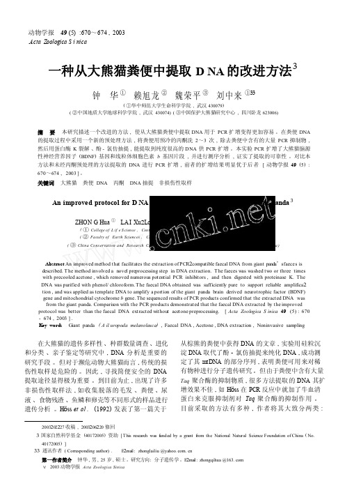
动物学报49 (5) :670~674 , 2003Acta Zoologica S i nica一种从大熊猫粪便中提取D NA 的改进方法3钟华①赖旭龙②魏荣平③刘中来①33( ①华中师范大学生命科学学院, 武汉430079)( ②中国地质大学地球科学学院, 武汉430074) ( ③中国保护大熊猫研究中心, 四川卧龙623006)摘要本研究描述一个改进的方法, 使从大熊猫粪便中提取DNA 用于PCR 扩增变得更加容易。
在粪便DNA 的提取过程中采用一个新的预处理方法, 将粪便用预冷的丙酮洗2~3 次, 除去粪便中含有的大量PCR 抑制物, 然后用蛋白酶K 裂解、酚- 氯仿抽提, 能提取到纯度很高的DNA 供PCR 扩增。
本实验PCR 扩增了大熊猫脑源性神经营养因子(BDNF) 基因和线粒体细胞色素 b 基因片段, 并进行测序分析, 证实了提取的可靠性。
对比本方法和未经丙酮预处理的方法提取的DNA 进行PCR 扩增, 前者的扩增结果明显优于后者[ 动物学报49 (5) : 670~674 , 2003 ] 。
关键词大熊猫粪便DNA 丙酮DNA 抽提非损伤性取样An improved protocol for D NA extraction from the faeces of the giant panda 3 ZHON G Hua ① LA I Xu2Long ② W EI Rong2Ping ③ L IU Zhong2Lai ①33( ①College of L if e S cience , Cent ral China Nor mal U niversit y , W uh an430079 , China)( ②Faculty of Earth Sciences , China U niversit y of Geosciences , W uhan430074 , China)( ③China Cons ervation and Res earch Center f or th e Gi ant Pan da , W olong623006 , S ichuan , China) Abstract An improved method that facilitates the extraction of PCR2compatible faecal DNA from giant pand a’s faeces is described. The method involved a novel preprocessing step in DNA extraction. The faeces was washed two or three timeswith precooled acetone , which removed numerous potential PCR inhibitors , and then digested with proteinase K. The DNA was purified with phenol/ chloroform. The faecal DNA obtained was sufficiently pure to support reliable amplifica2 tion , and was applied as template DNA to amplify a portion of the giant panda brain derived neurotrophic factor (BDNF) gene and mitochondrial cytochrome b gene. The sequenced results of PCR products confirmed that the extracted DNA was from the giant panda. Comparison with the PCR products demonstrated that the faecal DNA extracted b y the improved protocol was better than the faecal DNA extracted without acetone preprocessing. [ Acta Zoologica S inica 49 (5) : 670 - 674 , 2003 ] .K ey words Giant panda ( A il uropoda melanoleuca) , Faecal DNA , Acetone , DNA extraction , Noninvasive sampling在大熊猫的遗传多样性、种群数量调查、进化和分类、亲子鉴定等研究中, DNA 分析是重要的研究手段。
《纳米抗体研究进展综述》3300字

纳米抗体研究进展综述摘要:单域抗体因其独特的优势,如水溶性好、分子量小、稳定性好、免疫原性小等一系列特点,在生物研究和医学领域中的作用愈发广泛。
在疾病诊断、病原检测、癌症疾病治疗、药物残留检测分析,坏境检测,用作sdAbs分子探针、分子诊断和显影等等领域具有广阔的应用前景。
纳米抗体因其优势,可实现重组表达,从而使得生产周期和生产成本均可大幅下降,是目前国内外研发的热点。
作者重点介绍了纳米抗体的特点,然后简述了纳米抗体的制备流程,简述了纳米抗体在疾病诊断、疾病治疗、食品安全和环境监测等领域的应用,最后对纳米抗体的应用前景进行了分析和展望。
1 介绍自1890年,第一种抗体——抗毒素,这是在血清中发现的第一种抗体[1]。
这是一种可中和外毒素的物质,1975年,杂交瘤技术的诞生开始了抗体研究和应用快速发展的时代。
由于抗体可特异性识别和结合抗原的特性,使其在疾病诊断、疾病治疗、药物运载、病原、毒素和小分子化合物检测等领域具有广泛的应用[2]。
但通过单克隆抗体技术制备的传统单克隆抗体有其不可忽视的缺点:生产耗时长、成本高、在组织和肿瘤中穿透力差、长期使用会引起机体免疫排斥反应以及动物道德问题等。
相比于传统抗体,纳米抗体具备传统抗体不具备的分子质量小和穿透性强的优势而成为现在抗体研究的主要方向之一。
单链抗体(single chain antibody fragment,scFv)就是新型小分子抗体的一种,其穿透力更强、生产成本更低,但scFv抗体存在溶解度低、稳定性较差、表达量低、易聚合和亲和力低的缺点[3]。
1989年,比利时免疫学家Hamers-Casterman 在骆驼血清中的偶然发现一种天然缺失轻链的重链抗体(HcAbs)可以解决scFv所存在的问题,重链抗体只包含2个常规的CH2与CH3区和1个重链可变区(VHH),重链可变区具有与原重链抗体相当的结构稳定性以及与抗原的结合活性,是已知的可结合目标抗原的最小单位,其分子质量只有单克隆抗体的1/10,是迄今为止获得的结构稳定且具有抗原结合活性的最小抗体单位,因此也被称作纳米抗体(nanobody,Nb)[4]。
产细菌素苏云金芽孢杆菌的鉴定及其所产抗菌物质性质
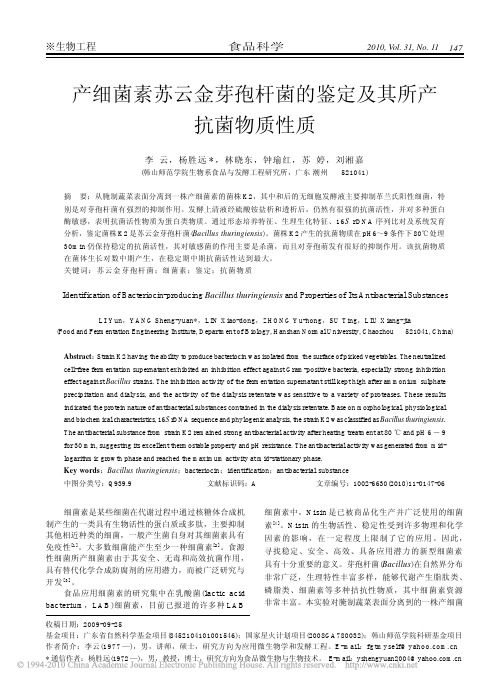
※生物工程食品科学2010, Vol. 31, No. 11147产细菌素苏云金芽孢杆菌的鉴定及其所产 抗菌物质性质李 云,杨胜远 * ,林晓东,钟瑜红,苏 婷,刘湘嘉(韩山师范学院生物系食品与发酵工程研究所,广东 潮州 摘 521041)要:从腌制蔬菜表面分离到一株产细菌素的菌株 K2,其中和后的无细胞发酵液主要抑制革兰氏阳性细菌,特别是对芽孢杆菌有强烈的抑制作用。
发酵上清液经硫酸铵盐析和透析后,仍然有很强的抗菌活性,并对多种蛋白 酶敏感,表明抗菌活性物质为蛋白类物质。
通过形态培养特征、生理生化特征、16S rDNA 序列比对及系统发育 分析,鉴定菌株 K2 是苏云金芽孢杆菌(Bacillus thuringiensis)。
菌株 K2 产生的抗菌物质在 pH6~9 条件下 80℃处理 30min 仍保持稳定的抗菌活性,其对敏感菌的作用主要是杀菌,而且对芽孢萌发有很好的抑制作用。
该抗菌物质 在菌体生长对数中期产生,在稳定期中期抗菌活性达到最大。
关键词:苏 云 金 芽 孢 杆 菌;细菌素;鉴定;抗 菌 物 质Identification of Bacteriocin-producing Bacillus thuringiensis and Properties of Its Antibacterial SubstancesLI Yun,YANG Sheng-yuan*,LIN Xiao-dong,ZHONG Yu-hong,SU Ting,LIU Xiang-jia (Food and Fermentation Engineering Institute, Department of Biology, Hanshan Normal University, Chaozhou 521041, China)Abstract: Strain K2 having the ability to produce bacteriocin was isolated from the surface of picked vegetables. The neutralized cell-free fermentation supernatant exhibited an inhibition effect against Gram-positive bacteria, especially strong inhibition effect against Bacillus strains. The inhibition activity of the fermentation supernatant still kept high after ammonium sulphate precipitation and dialysis, and the activity of the dialysis retentate was sensitive to a variety of proteases. These results indicated the protein nature of antibacterial substances contained in the dialysis retentate. Base on morphological, physiological and biochemical characteristics, 16S rDNA sequence and phylogenic analysis, the strain K2 was classified as Bacillus thuringiensis. The antibacterial substance from strain K2 remained strong antibacterial activity after heating treatment at 80 ℃ and pH 6 - 9 for 30 min, suggesting its excellent thermostable property and pH resistance. The antibacterial activity was generated from midlogarithmic growth phase and reached the maximum activity at mid-stationary phase. Key words:Bacillus thuringiensis;bacteriocin;identification;antibacterial substance 中图分类号:Q939.9 文献标识码:A 文章编号:1002-6630(2010)11-0147-06细菌素是某些细菌在代谢过程中通过核糖体合成机 制产生的一类具有生物活性的蛋白质或多肽,主要抑制 其他相近种类的细菌,一般产生菌自身对其细菌素具有 免疫性[ 1 ] 。
片段化生境中极小种群野生植物种子传播研究进展

至于无法将携带的种子散播到更远的生境ꎮ 如ꎬ Car ̄
法ꎬ 种子 数 量 普 查ꎬ 种 子 捕 捉 器ꎬ 利 用 遗 传 学 进 行
DNA 分子技术进行追踪ꎬ 无线电追踪ꎬ 放射性同位素
信号追踪ꎬ 动物活动模型ꎬ 遗传标记等ꎮ
ine Emer 等利用元网络的方法在 16 个新热带森林片段
中探索了 鸟 类 种 子 传 播 ( Bird Seed Dispersalꎬ BSD)
碎化ꎬ 是指原本自然状态下连续的生境在受到人为干
键的生态过程ꎬ 决定了植物的更新模式ꎬ 并影响之后
扰或者自然干扰的情况下ꎬ 生境逐渐由完整发展为支
幼苗的更新生存ꎬ 因此其在生态系统和生物多样性的
离破碎的较小面积生境ꎮ 随着人类社会的不断发展ꎬ
对植物种群的持续性至关重要
[1]
持久中发挥着至关重要的作用 [2] ꎮ 其中极小种群野生
(1 西南林业大学园林园艺学院ꎬ 云南 昆明 650224ꎻ 2 云南省农业科学院生物技术与种质资源研究所ꎬ 云南 昆明 650205)
摘 要: 全球生境碎片化趋势逐渐加深ꎬ 使在碎片化生境中的野生植物受到生存威胁ꎬ 其中包含一些亟需保护的
极小种群野生植物ꎮ 本文主要通过综述片段化生境中植物种子传播过程的研究ꎬ 如片段化生境对种子传播者的影
据营养繁殖物种扩散受限ꎬ 依靠动物传播种子和依靠
程中受到了怎样的影响ꎬ 遇到了哪些困难ꎮ 一起探明
昆虫传播花粉的植物也将受到影响ꎬ 进一步对于植物
物种传播过程中的关键问题ꎬ 为保护极小种群野生植
种群的信息流、 基因流产生一定的影响ꎬ 最终影响到
物提供理论基础ꎮ
物种更新延续和发展 [5] ꎮ
收稿日期: 2023-10-30
蛞蝓提取物对肺癌细胞周期的影响
内蒙古 中医药
蛞蝓 提取 物对 肺癌 细胞 周 期 的影 响△
韦金 育 曾振 东 曾翠 琼 俸 曙光 杨 增艳 摘 要: 目的 : 了解 蛞蝓提 取 物对肺 癌 细胞 周期 的影 响 。 法 : 流式 细胞 仪 分析蛞 蝓提 取物 对A 4 N胞周 期 的影响 。结果 : 方 用 59 蛞蝓 提取 物 能使 细胞 的S ( N 合 成期 ) 降。结论 : 蝓提取 物 可能通 过诱 导A 4 N胞 周期 阻滞从 而发挥 抗癌 作 用。 期 DA 下 蛞 59
中 图分类 号 : 63 R 8. 2
1 材料 与方 法
文献标 识码 : B
文章编 号 :0 6 07 (0 2— 09 0 10— 992 1)4 03— 2 1
神 经与 椎体 外缘 的夹 角 。 1 标本制备 : . 1 尽量不损伤神经血管 , 暴露 T L 的椎体 、 弓根、 22 1 椎 .左右 两侧 椎 间孔外 口处 神经 血 管 的排列 走行 是 : 自上 而下为 椎 间孑 、 神 经 、 管等 结构 。 L脊 血 椎 间静 脉 、 神经 、 动 脉 即节段 动脉 的中 间支 ( 1; 脊 根 图 )出椎 间孔
21年 1 01 2月
3 9
其 抑制作 用 主要是 通过 抑 制细 胞 内 D A的合 成 而实 现 的 。本实 嘲. N 中国 中西 医结合 杂志, 9 , ()2 . 1 91 94 3 9 9 : 验 结果 也许 提示这 可 能是 蛞蝓 抗肿 瘤 作用 的 机理 之一 。 [ 甄永 苏. 瘤 药物 研 究与 开发『 ] 3 】 抗肿 M. 北京: 工 出版社. 0 . 1 化 2 41 . 0 0
蛞蝓 提取 物对 人肺 癌 的作 用 良好 、 毒性 较小 、 资源 丰 富 , 得 【 郭岳峰 , 守章, 民, . 蝓胶 囊治疗 晚期 非 小细胞肺 癌 3 值 4 ] 王 焦智 等 蛞 2 进一 步研 究 。 例 疗 效观察 中医研 无 19 ,3: . 947 ) 4 ( 2 参 考文献 [] 金魁 漕 建 国. 蝓 对人 肺鳞 癌 、 5 谢 蛞 肺腺 癌 细胞抑 瘤作 用初 探 『. J 1 [ 李仪 奎. 1 ] 中药药理 实验 方 法 学【 . 1 . 海: 海科 学技 术 出 M】 第 版上 上 肿 瘤 防治研 究, 9 , () 4 . 1 7 46: 4 9 2 3
植物维生素B1生物合成及生物强化的研究进展

SUN Ya⁃li, TANG Jia⁃qi, MAO Xin⁃chen et al ( Agricultural College of Yangzhou University / Jiangsu Key Laboratory of Crop Genetics
也会增加[19] 。 对向日葵根部进行外源施加维生素 B1 ,其可
剂
[4-5]
缺乏症。 若严重缺乏维生素 B1 ,则会干扰中枢神经和循环系
以高碳水化合物为主食的国家中普遍存在[8] 。
维生素 B1 在植物的生长发育、非生物和生物胁迫的响
应中发挥着重要的作用
[9]
。 维生素 B1 参与许多细胞代谢途
加[17-18] ,维生素 B1 生物合成途径关键酶的 mRNA 转录水平
酸合成酶(thiamine phosphate synthase,TH1) 催化 HMP -P 而
完成
[22]
。
噻唑 部 分 的 生 物 合 成 是 通 过 噻 唑 合 成 酶 ( HEP - T
synthase,THI1)催化底物形成腺苷二磷酸-5-( β-乙基) -4-
催化,耦联形成 TMP。 TMP 在原核生物中可以直接转化为
5-β-羟乙基噻唑)和嘧啶环(4-氨基-5-羟甲基嘧啶)2 个部
分组成。 2 个部分在质体中单独合成,然后结合在一起,最终
形成 TPP 的形式(图 2)。
嘧啶是通过嘧啶合成酶(HMP -P synthase,THIC) 催化底
6
安徽农业科学 2024 年
世界卫生组织儿童标准处方集
WHO Model Formulary for ChildrenBased on the Second Model List of Essential Medicines for Children 2009世界卫生组织儿童标准处方集基于2009年儿童基本用药的第二个标准目录WHO Library Cataloguing-in-Publication Data:WHO model formulary for children 2010.Based on the second model list of essential medicines for children 2009.1.Essential drugs.2.Formularies.3.Pharmaceutical preparations.4.Child.5.Drug utilization. I.World Health Organization.ISBN 978 92 4 159932 0 (NLM classification: QV 55)世界卫生组织实验室出版数据目录:世界卫生组织儿童标准处方集基于2009年儿童基本用药的第二个标准处方集1.基本药物 2.处方一览表 3.药品制备 4儿童 5.药物ISBN 978 92 4 159932 0 (美国国立医学图书馆分类:QV55)World Health Organization 2010All rights reserved. Publications of the World Health Organization can be obtained fromWHO Press, World Health Organization, 20 Avenue Appia, 1211 Geneva 27, Switzerland (tel.: +41 22 791 3264; fax: +41 22 791 4857; e-mail: ******************). Requests for permission to reproduce or translate WHO publications – whether for sale or for noncommercial distribution – should be addressed to WHO Press, at the aboveaddress(fax:+41227914806;e-mail:*******************).世界卫生组织2010版权所有。
leptospira interrogans ss. icterohaemorrhagiae-感染的
LEPTOSPIRA INTERROGANS SSP.Aetiology Epidemiology Diagnosis Prevention and ControlPotential Impacts of Disease Agent Beyond Clinical Illness References AETIOLOGYClassification of the causative agentLeptospira interrogans, the causative agent of leptospirosis, consists of numerable serotypes capable of causing a variety of disease manifestations in a wide range of hosts. This motile and flagellated spirochete bacterium is recognizable via microscopy and earned its name from a characteristic hooked appearance that resembles a question mark. The taxonomic classifications within the Leptospira genus have been reorganized many times in accordance with new antigenic, genomic, and pathologic data. What was once over 250 serovars grouped into two Leptospira species is now 21 genomospecies of Leptospira with reclassified serovars. While the new taxonomic names are typically used in the scientific literature, historical names still circulate on product labels and in common use.Not all serovars of Leptospira are pathogenic, and many are associated with a reservoir species in which little disease is apparent. Many serovars are highly prevalent within maintenance host populations and persist in the kidneys or genital tract. Small antibody responses and low tissue burdens are typical in these animals. Incidental hosts, however, typically develop serious disease with high tissue burdens and robust antibody responses. These classifications are not always entirely distinct and some overlap between presentations can exist.Leptospirosis is a zoonotic disease but is typically associated with at-risk occupations (veterinarian, livestock owners, dairy workers, etc.) and exposure to contaminated water.Resistance to physical and chemical actionTemperature: Generally fails to persist at <10°C or >34°C;pasteurization and moist heat at 121°C/15 minutes are effective methodsof killing leptospirespH: Prefers neutral to slightly alkaline conditionsChemicals/Disinfectants: Inactivated by 1% sodium hypochlorite, 70%ethanol, formaldehyde, detergents, quaternary ammonium compounds,iodine based compounds, glutaraldehyde, and hydrogen peroxideSurvival: Warm, moist conditions greatly enhance survival; maypersist up to 6 weeks under favorable conditions; freezing, dehydration,and UV radiation inactivate leptospiresEPIDEMIOLOGYHosts●Virtually all mammals are vulnerable to pathogenic Leptospira serovars to varying degrees○Many serovars have specific maintenance hosts while others are more promiscuous●Leptospira serovars known to cause disease in mammals have been isolated from amphibians○Some serovars have also been isolated from invertebrates, reptiles, and birdsProminent host-serovar associations●Armadillos (Dasypus novemcinctus, Euphractus sexcinctus) - Autumnalis, Cynopteri, Hebdomadis,Pomona●Bandicoots (Isoodon macrourus, Perameles spp.) - numerous serovars have been associated withthese species●Brazilian tapir (Tapirus terrestris) - Pomona●Canids (Canis latrans, C. familiaris, C. lupus) - Bratislava, Canicola, Grippotyphosa, Hardjo,Icterohaemorrhagiae, Pomona●Cattle (Bos taurus, Syncerus caffer) - Hardjo and others●Cervids - Bratislava, Canicola, Grippotyphosa, Hardjo, Icterohaemorrhagiae, Pomona●European hedgehog (Erinaceus europaeus)●Felids (Felis silvestris silvestris, F. silvestris catus, Lynx spp.)●Flying foxes (Pteropus spp.) - a multitude of Leptospira serovars have been identified in variousspecies of bat●Foxes (Vulpes lagopus, V. vulpes, Urocyon cinereoargenteus, Lycalopex griseus) - Bratislava,Canicola, Grippotyphosa●Giant anteater (Myrmecophaga tridactyla) - Djasiman●Horses (Equus ferus) - Bratislava●Lagomorphs (Lepus europaeus, L. timidus, Oryctolagus cuniculus) - Grippotyphosa●Marine mammals (Eubalaena australis,Trichechus manatus ) - Australis, Manaua○Sea lions (Zalophus californianus, Z. wollebaeki) and seals (Callorhinus ursinus, Phoca vitulina, Mirounga angustirostris, Arctocephalus forsteri) - Canicola, Hardjo, Pomona ○There have been multiple mass-mortality events attributed to leptospirosis in California sea lions●Marsupials - Australis, Autumnalis, Ballum, Bataviae, Celledoni, Cynopteri, Djasiman,Grippotyphosa, Hardjo, Hebdomadis, Icterohaemorrhagiae, Javanica, Mini, Panama, Pomona, Pyrogenes, Sejroe, Tarassov, Topaz●Mongooses (Herpestes auropunctatus, Mungos mungo, Paracynictis selousi) - Bratislava, Hardjo●Mustelids (Meles meles, Martes fiona, M. martes, Mustela putorius, M. nivalis, M. ermine, Lutra lutra)●Platypus (Ornithorhynchus anatinus) - Autumnalis, Hardjo, Grippotyphosa●Raccoons (Procyon itor) and skunks (Mephitis mephitis) - Bratislava, Canicola, Grippotyphosa,Hardjo, Icterohaemorrhagiae, Pomona●Rodents and insectivores - Arborea, Australis, Ballum, Bindjei, Broomi, Canicola, Celledoni,Gryppotyphosa, Icterohaemorrhagiae, Javanica, Mini, Pomona, Pyrogenes, Sejroe, Tarrasovi, Zanoni○Rats are well-appreciated hosts for serotype Icterohaemorrhagiae●Swine (Sus scrofa and other spp.) - Bratislava, Hardjo, Pomona●Vervet monkey (Cercopithecus aethiops sabaeus) - Australis, Grippotyphosa, Javanica●Multiple snake, turtle, toad, and frog species have also been identified as PCR and/or serologicallypositiveTransmission●Ingestion●Contact with mucous membranes or wet, abraded skin●Some serovars can be transmitted venerally or transplacentallySources●Urine●Contaminated soil and water●Placental fluids●Genital secretions●Milk●BloodOccurrenceMany wildlife species are reservoirs for Leptospira and subsequently maintain host-bacterium interactions that do not negatively impact the animal. Over time, selection pressures on the leptospires many change and drive reservoir species relationships to shift accordingly. Therefore, it is important to assess the epidemiology of leptospirosis on a more local level to understand transmission risks and disease impacts.Leptospira is globally enzootic, but disease is more frequently seen in warm and moist environments. This may be seasonal (temperate zones) or more constant (tropical regions). Rainfall encourages persistence of the organism in the environment. Additionally, some serotypes are much more geographically dispersed and others are found in more limited regions.For more recent, detailed information on the occurrence of this disease worldwide, see the OIE World Animal Health Information System - Wild (WAHIS-Wild) Interface [http://www.oie.int/wahis_2/public/wahidwild.php/Index].DIAGNOSISAfter invasion of mucous membranes or damaged skin, there is a 4-20 day incubation period followed by a 7-10 day period of circulation in the bloodstream. Clinical signs of acute leptospirosis depend on the tissues colonized during this period of bacteraemia, the host species, and infecting serovar. A robust antibody response follows and is associated with a declining bacteraemia. Tissues may recover slowly or not at all depending on the degree of damage. Death is possible in severe cases.Incidental hosts maintain the bacterium in renal tubules for days to weeks and shed the organism in urine. Maintenance hosts, however, maintain the bacterium in the renal tubules, genital tract, and/or eyes and shed it in urine or genital secretions for months to years after infection.Clinical diagnosisLeptospirosis is a highly variable, systemic infection and the presentation depends on the infecting serovar, the host species, and the host’s general health and immune status. Maintenance hosts typically do not develop significant clinical disease. Incidental hosts typically experience severe, acute disease secondary to bacterial toxins and inflammatory responses generated by the immune system. Initially, animals may be febrile and anorectic. They may quickly develop signs of haemorrhage and haemolytic anaemia secondary to endothelial damage such as mucosal petechiation, icterus, haemoglobinuria/haematuria, dehydration, vomiting, and colic. Acute renal injury develops rapidly and is a significant contributor to mortality. Pneumonia, meningitis, uveitis, corneal opacification, photosensitization, myalgia, and pancreatitis are also possible.Reproductive disease is often characterised by abortion/stillbirth, mummified fetuses, infertility, blood in milk, or a cessation of milk production. If not aborted, neonates infected transplacentally are typically weak. Maintenance hosts do not develop reproductive disease acutely like incidental hosts, but instead remain subclinical for weeks to months.Lesions●Renal tubular necrosis and suppurative nephritis○Pale, oedematous parenchyma +/- pitting of the serosal surface and capsular adhesions○Subcapsular haemorrhage○Inflammation initially characterised by neutrophils but becomes lymphoplasmacytic○Mixed inflammatory processes are associated with higher mortality rates●Hepatomegaly +/- necrotizing hepatitis○The liver is often friable and discolored in a lobular pattern●Pulmonary haemorrhage●Petechiae and ecchymoses on mucous membranes and internal organs●Horses may develop uveitis with conjunctivitis, corneal oedema, synechia, or cataractsDifferential diagnoses●Ocular disease○Equine recurrent uveitis○Traumatic uveitis/reflex uveitis○Infectious conjunctivitis●Kidney disease○Toxin exposure (e.g., ethylene glycol)○Infectious nephritis, pyelonephritis, glomerulonephritis○Renal tubular acidosis○Nematodes (Stephanurus dentatus, Dioctophyma renale)●Reproductive failure or compromise○Brucellosis○Bovine viral diarrhea virus (BVDV)○Porcine reproductive and respiratory syndrome (PRRS)○Q-Fever (Coxiella burnetii)○Neospora spp.○Tritrichomonas foetus○Mastitis, metritis●Liver disease, icterus, and haemolytic anaemia○Viral hepatitis○Toxin exposure (e.g., heavy metals, anticoagulant rodenticides)○Rickettsial infection○Clostridium haemolyticum, C. perfringens A○Neonatal isoerythrolysis●Bacterial septicaemiaLaboratory diagnosisSamplesFor isolation of agent●Kidney●Blood●Urine●Other grossly affected tissue such as liverSerological tests●Serum●Whole bloodProceduresIdentification of the agent●Silver-stained histopathology slides allow for direct visualization of the organism in renal tubules●Immunohistochemistry (IHC)●Bacterial culture○Because the organism is low-growing, this may take 12-26 weeks○Best available method to determine infecting serovar●Polymerase chain reaction (PCR)○Widely variable protocols○Does not provide serovar-specific resultsSerological tests●Microscopic agglutination test (MAT)○Uses live, regionally common serovars of Leptospira○Requires diagnostic laboratory to maintain live cultures of serovars○Provides quantitative titre level●Antibody capture enzyme-linked immunosorbent assay (ELISA)○Currently used for domestic canines; detects antibodies to LipL32 protein○Results are qualitative (positive/negative) and may yield false positives in the event of prior vaccination●Immunofluorescence assay (IFA)●There is not yet a consensus on what a diagnostic titre for Leptospira should be, therefore pairedacute and convalescent sera are recommended for testing●Caution should be taken when interpreting serology data; antibody titre does not always correspondwith disease stateFor more detailed information regarding laboratory diagnostic methodologies, please refer to Chapter 3.1.12 Leptospirosis in the latest edition of the OIE Manual of Diagnostic Tests and Vaccines for Terrestrial Animals.PREVENTION AND CONTROLSanitary prophylaxis●Extra precautions should be taken when cleaning areas frequented by potential Leptospira hosts.Wear gowns, shoe covers, and gloves to prevent contamination of personal clothing. Face shields are recommended to protect mucous membranes from aerosols.Medical prophylaxis●There are a variety of Leptospira vaccines available for domestic animals, including livestock○Vaccine intent may vary from prevention of infection to reduction of renal colonization and urine shedding○Read vaccine labels to determine which serovars are targeted, as immunity is believed to be serovar-specificPOTENTIAL IMPACTS OF DISEASE AGENT BEYOND CLINICAL ILLNESS Risks to public health●Leptospirosis is a zoonotic disease. Because clinical signs can be vague and maintenance hostscan be asymptomatic carriers, basic protective measures are suggested for at-risk populations (veterinarians, livestock owners, dairy workers, etc.): protect eyes with safety glasses, wear gloves especially if there are openings in the skin, thoroughly wash hands after interacting with animals of unknown status and before consuming food or water, etc.○Pregnant individuals within at-risk populations are particularly advised to utilise protective measures.●Many domestic animal species, including dogs, horses, and livestock, are susceptible to leptospirosisand could potentially transmit it to humans. Individuals should speak with local veterinarians to determine risk and appropriate prevention strategies, including animal vaccines.Risks to agriculture●If livestock or working animals (horses, dogs) develop clinical disease due to Leptospira, decreasedthrift and reproductive compromise can significantly impact production. Working animals may not beable to do their jobs as efficiently, and livestock may demand increased resources for treatment while producing less.REFERENCES AND OTHER INFORMATION●Atherstone, C., Picozzi, K., & Kalema-Zikusoka, G. (2014). Seroprevalence of Leptospira hardjo incattle and African buffalos in southwestern Uganda. The American Journal of Tropical Medicine and Hygiene, 90(2), 288–290.●Ayral, F., Djelouadji, Z., Raton, V., Zilber, A. L., Gasqui, P., et al. (2016). Hedgehogs and mustelidspecies: major carriers of pathogenic Leptospira, a survey in 28 animal species in France (20122015). PloS One, 11(9), e0162549.●Biscola, N. P., Fornazari, F., Saad, E., Richini-Pereira, V. B., Campagner, M. V., et al. (2011).Serological investigation and PCR in detection of pathogenic leptospires in snakes. Pesquisa Veterinária Brasileira, 31(9), 806-811.●Buhnerkempe, M. G., Pragger, K. C., Strelloff, C. C., Greig, D. J., Laake, J. L., et al. (2017). Detectingsignals of chronic shedding to explain pathogen persistence: Leptospira interrogans in California sea lions. Journal of Animal Ecology, 86(3), 460-472.●Denkinger, J., Guevara, N., Ayala, S., Murillo, J. C., Hirschfeld, M., et al. (2017). Pup mortality andevidence for pathogen exposure in Galapagos sea lions (Zalophus wollebaeki) on San Cristobal Island, Galapagos, Ecuador. Journal of Wildlife Disease, 53(3), 491-498.●Gravekamp, C., Korver, H., Montgomery, J., Everard, C. O. R., Carrington, D., et al. (1991).Leptospires isolated from toads and frogs on the island of Barbados. Zentralblatt für Bakteriologie, 275(3), 403-411.●Jobbins, S. E., Sanderson, S. E., & Alexander, K. A. (2014). Leptospira interrogans at the human-wildlife interface in northern Botswana: a newly identified public health threat. Zoonoses and Public Health, 61, 113-123.●Karesh, W. B., Hart, J. A., Hart, T. B., House, C., Torres, A., et al. (1995). Health evaluation of fivesympatric duiker species (Cephalophus spp). Journal of Zoo and Wildlife Medicine, 26(4), 485-502.●Leighton, F. A. & Kuiken, T. (2001). Leptospirosis. In E. S. Williams and I. K. Barker (Eds.), InfectiousDiseases of Wild Mammals (3rd ed., pp. 498-502). Iowa State Press.●Loffler, G. S., Rago, V., Martinez, M., Uhart, M., Florin-Christensen, M., et al. (2015). Isolation of aseawater tolerant Leptospira spp. from a southern right whale (Eubalaena australis). PLoS One, 10(12), e0144974.●Lunn, K. F. (2018). Overview of leptospirosis. Merck Veterinary Manual.Accessed 2020:https:///generalized-conditions/leptospirosis/overview-of-leptospirosis?query=leptospira●Pedersen, K., Anderson, T. D., Maison, R. M., Wiscomb, G. W., Pipas, M. J., et al. (2018). Leptospiraantibodies detected in wildlife in the USA and the US Virgin Islands. Journal of Wildlife Diseases, 54(3), 450-459.●Spickler, A. R. & Leedom, L. K. R. (2013). Leptospirosis. Accessed 2020:/Factsheets/pdfs/leptospirosis.pdf●The World Organisation for Animal Health (2018). Leptospirosis. Accessed 2020:https://www.oie.int/fileadmin/Home/eng/Health_standards/tahm/3.01.12_LEPTO.pdf●Vieira, A. S., Pinto, P. S, & Lillenbaum, W. (2018). A systematic review of leptospirosis on wildanimals in Latin America. Tropical Animal Health and Production, 50(2), 229-238.●Wildlife Health Australia (2018). Leptospira infection in Australian mammals. Accessed 2020:https://.au/FactSheets.aspx●Wildlife Health Australia (2011). Leptospira infection in Australian seals. Accessed 2020:https://.au/FactSheets.aspx** *。
毕赤酵母翻译

Glycoengineered(糖基化工程) 毕赤 pastoris(毕赤酵母) 中表达的重组单克隆抗体的纯化工艺开发摘要一个强健的和可扩展的净化过程被开发快速从 glycoengineered 毕赤酵母发酵生成抗体的纯度高、数量充足。
蛋白 A 亲和层析用于捕获从发酵液抗体。
PH 梯度洗脱已应用到要防止抗体沉淀在低 pH 的蛋白 A 列。
从蛋白抗体色谱法所载一些产品的相关杂质,被劈重链、重链和轻链的 misassembling。
它也有一些处理相关的杂质,包括蛋白 A 残留、 endotoxin(内毒素)、宿主细胞 DNA 和蛋白质。
Ph 值为 4.5-6.0 的最优氯化钠渐变阳离子交换色谱法有效去除这些产品和过程相关的杂质。
从 glycoengineered P.酵母抗体是其 heterotetramer 折叠、物理稳定性和约束力的亲和性的商业同行相媲美。
介绍单克隆抗体(Mab)正迅速成为关键产品的生物医药产业。
大多数商业治疗性抗体是原封 IgG 分子与 IgG1、 IgG2 正在共同的子类。
这些 IgGs 由调解抗原和效应器函数之间的联系,在免疫过程中发挥中心作用。
对目标细胞的特异性抗原的 igg 抗体可变域的绑定将抗体依赖的细胞毒作用 (ADCC) 及补体依赖性细胞毒性 (CDC) 定向杀害的目标单元格。
ADCC 触发由交联体 Fc 域的 IgG 抗体(Fc(Rs),尤其是 Fc (RI 和Fc (制证机对免疫效应细胞。
可能调解 ADCC 效应细胞包括自然杀手细胞、巨噬细胞和嗜中性粒细胞。
疾病预防控制中心是由补充组件 C1q 绑定到 IgG,触发蛋白水解的级联,以激活补充 fc 功能区启动。
IgG 型抗体有两重链和两条轻链由分子内的二硫键形成 heterotetramer 在一起。
重链是通过共价键固定在天冬酰胺 297 (Asn-297) 低聚糖的糖基化。
人免疫球蛋白的主要N-聚糖复杂 biantennary (二分支的) 类型,有 '核心' heptasaccharide(七庚糖),GlcNac2Man3GlcNac2,被称为 G0 在我们的工作和文学。
地衣芽孢杆菌对雪茄烟叶发酵产香及菌群演替的影响

引用格式:宋 雯,陈 曦,余 君,等. 地衣芽孢杆菌对雪茄烟叶发酵产香及菌群演替的影响[J]. 湖南农业科学,2023(8):69-75. DOI:DOI:10.16498/ki.hnnykx.2023.008.0152021年国产手工雪茄销量超过2 000万支[1],但国产雪茄烟叶香气不够浓郁、化学成分不协调[2],因此需要通过微生物、酶及一些化学作用共同完成雪茄烟叶的发酵以提升烟叶品质。
利用生物发酵技术改善雪茄烟叶品质成为了一大研究热点[3]。
微生物的生长代谢使得烟叶中的木质素、蛋白质等生物大分子降解或转化,形成一系列的挥发性香气物质,同时降低烟叶中的青杂气,进而提升发酵后烟叶品质[4-5]。
迟建国[6]为了降低烟叶中木质素含量,从废弃烟草中筛选出一株白腐菌并用于烟叶发酵,使得发酵后烟叶木质素含量降低30%,并显著提升了烟叶品质;蔡文等[7]为了降低烟叶蛋白质含量,采用源自烟叶的高斯芽孢杆菌进行发酵,降低了烟叶总氮含量,且提高了烟叶中β-紫罗兰酮、E-大马士酮等类胡萝卜素降解产物的含量。
张倩颖等[8]使用冬虫夏草菌株发酵烟叶,提高了发酵后烟叶中茄酮等西柏烷类降解产物的香气含量,且感官质量评价明显提升。
地衣芽孢杆菌作为一种遗传背景清楚的益生菌[9],被广泛应用于食品发酵等[10-11],许多发酵食品特征性风味化合物与地衣芽孢杆菌代谢特征关系密切[12-13]。
目前地衣芽孢杆菌在烟草领域主要作为根际促生菌用于育苗过程[14-15]。
雪茄发酵过程中菌群演替规律对科学可控地设计雪茄发酵工艺具有重要意义[16]。
由于传统分离培地衣芽孢杆菌对雪茄烟叶发酵产香及菌群演替的影响宋 雯1,陈 曦1,余 君2,胡路路1,陈 雄1,王 志1(1. 发酵工程教育部重点实验室,湖北工业大学,湖北武汉 430068;2. 湖北省烟草科学研究院,湖北武汉 430030)摘 要:为揭示施加地衣芽孢杆菌对雪茄烟叶发酵的影响,结合宏基因组学技术对雪茄烟叶发酵后香气物质生成、菌群演替及其功能多样性进行了分析,探讨了各菌属在香气物质形成中的作用,揭示了菌群演替特征与代谢功能变化。
- 1、下载文档前请自行甄别文档内容的完整性,平台不提供额外的编辑、内容补充、找答案等附加服务。
- 2、"仅部分预览"的文档,不可在线预览部分如存在完整性等问题,可反馈申请退款(可完整预览的文档不适用该条件!)。
- 3、如文档侵犯您的权益,请联系客服反馈,我们会尽快为您处理(人工客服工作时间:9:00-18:30)。
Corn for ethanol, US
Palm oil, Malaysia
30% reductions?
How can the EU claim 'greenhouse gas savings' from biofuels?
Step 1 Ignore all carbon emissions from indirect land-use change. If a plantation company in Malaysia sells palm oil from an older plantation (for which they destroyed rainforest in the past) for biofuels, claim that no land has been deforested for biofuels – even if the same company burns and cuts down more rainforest for a new plantation to meet existing demands for food and cosmetics.
Tree Plantation in South Africa:
photo: Wally Menne
Risks of large-scale ecosystem collapse
African Palm in Indonesia
GHGs from deforestation contribute 20% global GHG emissions (IPCC AR4)
FALSE vs REAL Solutions
(Symptomatic vs Fundamental)
Economy
Bailouts, stimulus packages
Energy security
?, ROCs
Climate
Climate Bill, Creative GHG accounting!
Brazil: Eviction on 120 families for sugar cane ethanol /patzek/ BiofuelQA/Brazil/brazil.htm
Colombia: Military and paramilitary repression of Afro-Colombian communities in the palm oil areas of the Bajo Atrato Photo: Intercelestial Commission for Justivce and Peace
And even rapeseed oil…
Paul Crutzen, Oct 2007
Nobel Prize winning chemist
Scientific research of Rapeseed Oil and Maize biofuels: Life-cycles emissions of Rapeseed Oil and Maize produce up to 70% and 50% more greenhouse gases respectively than fossil fuels. This contrasts with the Low Carbon Vehicle Partnership figures of a 45-65% carbon emission saving for oil seed rape and 25-40% saving for corn ethanol.
70,000 fires burn in Amazon, September 2007. NASA
IPCC AR4: Deforestation = 20% GHG
Energy balances…and how the EU claims that biofuels deliver GHG reductions?
Peak Oil
Association for the Study of Peak Oil and Gas, ASPO
Converging Crises
Damian Carrington New scientist environmental blog
FINANCIAL CRASH
So convinced by their own creative accounting…
“By driving, you will be saving the planet. And the more you drive, the more you prevent catastrophic climate change.” Biopact ( 29th October 2007)
Underlying assumption; sustaining our consumption patterns is our prerogative Infinite growth, Malthus?
Agrofuels: where 4 concerns intersect
Agrofuel Impacts
Food Poverty
Lester Brown Food price spikes linked to hunger and civil unrest 2008 wheat and other grains 2009 (recession) 2010 Sugar, 128% above 2009 prices – 29 yr high 2007: 860m living in food poverty 2009: Over 1 billion
Biodiversity Losses
Deforestation for palm oil
Sarawak, Malaysia Friends of the Earth Europe
Papua New Guinea
Colombia Klaus Schenck, Salva la Selga
Bugala Island, Uganda
Step 2: Allow companies to provide all the information themselves and to pay their own auditors. Also allow for bilateral agreements. This means the EU can sign an agreement for example with Brazil under which all biofuels from Brazil have to be classed as climate-friendly – never mind the destruction of the Amazon and other ecosystems. Step 3: Claim there are positive indirect emission savings. Waste-products from biofuel refining can be fed to cattle. Hence, assume that the more biofuels we produce, the less soya we need to grow to feed our cattle and the less land-use change and agricultural emissions there will be overall.
Joseph Fargione, scientist for the Nature Conservancy
“All the biofuels we use now cause clearing of natural ecosystems for agriculture. Adding energy production to our current and growing demand for food production inevitably requires more land to be converted to agriculture, whether or not the biofuel is grown directly on that land. So biofuels either directly or indirectly cause land clearing, which releases carbon to the atmosphere and contributes to global warming. This is the biofuel carbon debt.”
(Leaked 2008 report from WB indicated that over 70% of driving force is agrofuels)
RSPO?, Rapeseed / wheat / corn?
Evictions anቤተ መጻሕፍቲ ባይዱ human rights abuses are no bar to meeting the EU's definition of 'sustainable biofuels'
Biofuels: An Unfolding Catastrophe
Deepak Rughani, Biofuelwatch. UCL Feb 2010
/blog/
Climate Change
University of Illinois at Urbana–Champaign (UIUC) 1979 to 2007 reduction is around 1.2 million square km of ice (UK is approx 245,000 sq km)
