BaySpec Raman Spectrometer Portfolio-Feb 2011
BioSpectrum凝胶成像分析系统

仪器功能介绍:
BioSpectrum凝胶成像系统可用于1D 电泳凝胶分析、Dot blot 分析、
活体动物及植物分析、菌落计算、分子量定量、GFP 表达分析、蛋白定量分析、PCR 基因表达、PCR定量、TLC 分析、Western blot 密度分析。
仪器主要技术参数:
◆多功能的暗室,光密性非常好,给化学发光成像提供了最佳条件
◆全自动变焦镜头F1.2 12.5-75mm ,通过软件可完全自动控制镜头的光圈,缩放以及聚焦
◆binning功能:有效整合像素,提高灵敏度
◆软件控制光源选择,包括顶置反射白光、紫外365nm、蓝光光源460-470nm
◆软件控制五位滤光轮设计标配三个滤光片
EtBr溴化乙锭570 - 640nm
SYBR Green 515 - 570nm波长
SYBR Gold 485 - 655nm
◆标配有超薄自发光白光屏,可折叠且高度可调
◆抽屉式紫外透照台,可选配:单波长、双波长、三波长透照台
◆防紫外观察窗,无须开启暗箱就可以观察到样品情况,更直观,更安全◆VisionWorksLS专业图像分析软件,同时可对系统进行全自动控制
仪器使用注意事项:
使用完毕后请将载物台清理干净。
土耳其比尔肯特大学研制出一种全硫属化物可变红外滤光片
● 藿
5
( c )
图 1 () a 斜蒸 发 几何 结 构 示 意 图, () 间可 变多 b空 层 滤光 片 结构 ,() 0层 A 2 3G l s S i e C2 s 一 es 2 es 4 S A 方 法 的研 究 [. 外 与 毫 米 J红 1
波学报 , 0 6 2 () 4 4 . 2 0 , 5 5:32 34
t soy 【 . e a C e sr, 00 7 () 15 r cp J C r l hmi y 20 , 72: 5— o 】 e t
哥召开 的 “ 红外 系统与光 电子技 术” S I PE专题
会议上 , 土耳其 比尔肯特大 学的研 究人员 HE a .s t
IF A E M N HY/ O . , o7 U 00 N R R D( O T L )V L 1 N .,J L2 1 3
图 2 该 红 外光 谱 仪 实验 装 置 的草 图
【 8 】Do el . iee t tn i eu n o —i e u w lFE D f rni ig t o s dn nvt o s a vr a r
d r u um e t k r e s b sn e ri fa e p e wh a e n l y u i g n a —n r r d s e — ・ -
1 . 58
[ ] i b O 6 . ai a d nndsrci 1 Tg u M, dnP C R pd n o-et t e 2 a u v
a a y i fv g u fPi u a u a s e s u i g sn l n l ss o i o r o n s p t l e d sn i g e s e e r i fa e r n m it n e s c r n u — e d n a n r r d t a s t a c pe t a a d m l
显微镜光学配置图说明书

Supplementary MethodsOptical configuration:A diagram of the optical configuration used for the photobleaching experiments is shown insupplemental figure 1 below.Supplemental Figure 1. Diagram of the optical configuration of the side-port for photobleaching.e. he d or the YFP bleaching experiment shown in figure 1, the microscope filter cube used for bleaching rom 0DCSXscanning mirrorsView is from above, and the scanning mirrors are at the rear of a Nikon TE-300 inverted microscop The dichroic mirror near the tube lens is a long-pass extended reflection mirror (650 DCXRU). The light from an argon-ion laser (Coherent Sabre) was coupled into an optical fiber and connected to the side-port at the connector shown at the right side of the figure. To preview the fluorescence and select cells, an in-house developed fiber-optic coupled high-power blue light emitting diode (LED) source (patent pending) was connected in place of the laser coupling fiber. Depending on the experiment a band-pass filter was sometimes included before coupling the LED emission to the fiber. In some experiments an additional dichroic mirror, 440 nm long-pass (440DCLP), was included between t fiber-optic coupler and the divergence-correcting lens to allow the additional coupling of a liquid-fille light guide (Oriel) to permit the use of a xenon arc lamp (Cairn).F contained a 545nm long-pass dichroic (545DCLP) and a 540-600nm band-pass emission filter(HQ570/30). A portion of the YFP emission could be monitored visually while scattered light f the 514.5 nm laser excitation was blocked. For bleaching experiments using laser lines shorter than 514.5, a filter cube containing a 510 long pass dichroic and a 510-560 band-pass emission filter (510DCLP and HQ535/50 respectively) was used. The filter cube for the 2-photon excitationcontained a 700 nm short-pass, UV reflecting, dichroic and a 710 short pass emission filter (70and E710SP respectively).YFP photoconversion revisited: confirmation of the CFP-like speciesMichael T Kirber, Kai Chen & John F Keaney JrIn experiments to check if the fluorescent decay product was visible with arc lamp illumination, a filter experiments to check if bleaching using arc lamp illumination produced the fluorescent decay ht , and en rlabs. ther than observing that the fluorescent decay product of YFP could be excited using a configuration ell culture and plasmid transfection: cube containing a 405-445 band-pass excitation filter, a 455 long-pass, extended reflection dichroic, and a 460-500 nm band-pass emission filter (D425/40X, 455DCXRU, and D480/40M respectively) was used. Infrared light was removed from the arc lamp output using a short wavelength visible and UV reflecting cold mirror (Thorlabs).In product, the fiber coupler was removed from the side port and the output from the liquid-filled lig guide and a collimating lens were coupled directly in place of the fiber-optic coupler. The 440DCLP dichroic was left in place. The filter cube used for bleaching contained, a 460-500nm band passemission filter inserted in reverse direction in the excitation position, a 510 nm long-pass dichroic a 510-560 nm band-pass emission filter ( HQ480/40M, 510DCLP, and HQ535/50M respectively) Bleaching YFP about 50% took 2 hours and when the 440DCLP mirror was removed and the CFP filter cube (D425/40X, 455DCXRU, and D480/40M) selected, the fluorescent decay product was se with the arc lamp illumination. All filters and dichroic mirrors were purchased from ChromaTechnology. Optical fiber, lenses, and optomechanical components were purchased from ThoO appropriate for exciting CFP using single photon (arc lamp) or 2-photon excitation, we did not try to determine the excitation spectrum of this byproduct. Such experiments might well be worthwhile but we are not presently set up to carry them out.Cvitrogen) and transfection with overexpression plasmids was ) was age acquisition and display:COS-7 cells were cultured in DMEM (In carried out using Fugene 6 (Roche) in cells at 70% confluency according to the manufacturer’sinstructions. PTP1B trapping mutant expression vector pcDNA6.2/YFP-PTP1B (D181A/Q262A constructed by using PCR subcloning technique with pcDNA6.2/N-YFP vector (Invitrogen) and pJ3H-PTP1B (D181A/Q262A)(kindly provided by Dr. Zhong-Yin Zhang, Indiana University). Cells were grown on glass cover slides and fixed with 4% paraformaldehyde.Imas designed and built in-house in collaboration with the laboratory of inal irror d between the number of counted photons and the brightness of the display.The 2-photon microscope used w Dr. Peter So at the Massachusetts Institute of Technology. The optical microscope portion of the system was a Nikon TE300 and the objective used was a 100X, 1.3 NA, oil-immersion type. Orig images obtained with 2-photon excitation (800 or 916 nm excitation) were 384x384 pixels with each pixel imaging 130 by 130 nm in the sample, which is beyond the resolution capability of the system. The dwell time at each pixel was 2 ms. Images shown in figure 1 are cropped from the originals and are 280 by 280 pixels. The fluorescence at each channel was measured with photon counting photomultiplier tubes (Hamamatsu R4700P-01 for the green channel and R4700P for the bluechannel). The green channel was separated from the red using an extended reflection dichroic m 565 nm long-pass (565DCXR) and the blue and green channels were separated using a 500nm long-pass extended reflection dichroic mirror (500DCXR). The pass-bands of the filters are given in the body of the text. The number of counts at each location in the sample was stored as a 16 bit unsigne integer. Images were imported into ImageJ as raw data. The colors for images in figure 1a-f were chosen to approximate the actual color of the fluorescence. The scale bars indicate the relationshipColocalization:In quantitatively assessing the degree of colocalization of two distinctly fluorescently labeled (or the different colored images each as a sample of a e or expressed) compounds in a cell, it is useful to treat random process which accurately represents it statistically (ergodicity). The normalized covarianc correlation coefficient between 2 sets of data, which in this case are images E and F which are N i x N j in size and contain elements e ij and f ij respectively, can be written as}Where E{ } denotes the “expected value” and E ¯ is the arithmetic mean of the pixel values in image E nd F ¯ is the mean of image F. This calculation has been applied to quantitatively assess colocalization E ()()ij ij e E f F ρ−−a (Vereb et al .). Because expectation is a linear operator, this expression can be rearranged so that the contribution at each pixel pair to the correlation coefficient is clear.ρ==We can then display an image G* with pixel values g*ij , the sum of all the pixels being the correlationoefficient. c*ij ij ij e f E F N N g −=or simplicity we normalize this so that we display images where the mean of all of the pixels, G ¯, is e correlation coefficient.F thij g =ll of the values can be easily calculated using ImageJ (Wayne Rasband, NIH).this manner we can easily compare different size sets of images and not lose the quantitative ove comparing the degree of colocalization under different experimental conditions some caution is oth ference:atko J, Vamosi G, Ibrahim SM, Magyar E, Varga S, Szollosi J, Jenei A, Gaspar R Jr, AIn information. Areas where the normalized covariance image is positive indicate colocalization ab that predicted by random uniform distributions of the 2 fluorescent species. Additionally, there are generally some regions where the pixels have a negative value. These regions correspond to areas where the colocalization of the 2 labeled compounds is less than would be predicted by a uniform random distribution.In needed. For example the regions for which the computation is performed should be similar under b sets of conditions (i.e. ratio of area of background to area of cell interior).re Vereb G, M Waldmann TA, Damjanovich S. Proc Natl Acad Sci U S A. 2000 May 23;97(11):6013-8.。
英文版原子物理课件

1.1 Introduction
The origins of atomic physics :quantum mechanics Bohr model of the H This introductory chapter surveys some of the early ideas: Spectrum of atomic H and Bohr Theory Einstein's treatment of interaction of atom with light the Zeeman effect Rutherford scattering And so on
Shanxi University Atomic Physics
1.2 Spectrum of atomic hydrogen_3
Wavenumbers may seem rather old-fashioned but they are very useful in atomic physics
the characteristic spectrum for atoms is composed of discrete lines that are the ‘fingerprint' of the element.
In 1888, the Swedish professor J. Rydberg found that the spectral lines in hydrogen obey the following mathematical formula:
Shanxi University Atomic Physics
Lyman series: n’ = 2; 3; 4; … n = 1. Balmer (n = 2), Paschen series: (n = 3), Brackett (n = 4) and Pfund (n = 5)
固相萃取-_高效液相色谱-_串联质谱法同时测定海产品中微囊藻毒素和鱼腥藻毒素

分析检测固相萃取-高效液相色谱-串联质谱法同时测定海产品中微囊藻毒素和鱼腥藻毒素吕晓静,鞠光秀,曲 欣,汪 勇,于红卫*(1.青岛市疾病预防控制中心/青岛市预防医学研究院,山东青岛 266033;2.岛津企业管理(中国)有限公司,北京 100020)摘 要:目的:建立固相萃取-高效液相色谱-串联质谱法同时测定海产品中7种微囊藻毒素和2种鱼腥藻毒素的方法。
方法:样品经80%乙腈提取,HLB小柱净化后,采用MRM模式进行分析,外标法定量。
结果:7种微囊藻毒素和2种鱼腥藻毒素在0.5~50.0 μg·L-1范围内线性关系良好,检出限为0.3 μg·kg-1,回收率为75.5%~98.8%,相对标准偏差在1.5%~5.4%。
结论:该方法重现性较好、灵敏度高、成本低,可以实现海产品中的鱼腥藻毒素和微囊藻毒素的同时检测。
关键词:微囊藻毒素;鱼腥藻毒素;固相萃取(SPE);高效液相色谱-串联质谱法(HPLC-MS/MS)Simultaneous Determination of Microcystins and Anatoxins in Marine Products by High Performance Liquid Chromatography-Tandem Mass Spectrometry with SolidPhase ExtractionLYU Xiaojing, JU Guangxiu, QU Xin, WANG Yong, YU Hongwei*(1.Qingdao Municipal Center For Disease Control & Prevention/Qingdao Institute of Preventive Medicine, Qingdao266033, China; 2.Shimadzu (China) Co., Ltd., Beijing Branch, Beijing 100020, China) Abstract: Objective: A method for simultaneous determination of 7 microcystins (MCs) and 2 Anatoxins (AnTXs) in marine products was achieved by solid phase extraction (SPE)-high performance liquid chromatography tandem mass spectrometry (HPLC-MS/MS). Method: The sample was extracted with 80% acetonitrile, purified by HLB small column, analyzed using MRM mode, and quantified using external standard method. Result: The linear ranges for 7 MCs and 2 AnTXs were 0.5~50.0 μg·L-1. The limits of detection were 0.3 μg·kg-1. The recoveries of the 7 MCs and 2 AnTXs spiked in blank marine products ranged from 75.5% to 98.8% with the relative deviations of 1.5%~5.4%. Conclusion: The method has the advantages of good reproducibility, high sensitivity and low cost, and can achieve simultaneous detection of fishy algae toxins and microcystins in seafood.Keywords: microcystin; anatoxin; solid phase extraction (SPE); high performance liquid chromatography-tandem mass spectrometry (HPLC-MS/MS)微囊藻毒素(Microcystins,MCs)和鱼腥藻毒素(Anatoxins,AnTXs)是两种典型的蓝细菌毒素[1]。
分离转移性癌细胞的方法与组合物,及其在检测癌转移性上的应用[发明专利]
![分离转移性癌细胞的方法与组合物,及其在检测癌转移性上的应用[发明专利]](https://img.taocdn.com/s3/m/d98cbf17f705cc1754270940.png)
专利名称:分离转移性癌细胞的方法与组合物,及其在检测癌转移性上的应用
专利类型:发明专利
发明人:W·T·陈
申请号:CN01817353.5
申请日:20010828
公开号:CN1484709A
公开日:
20040324
专利内容由知识产权出版社提供
摘要:本发明是关于检测、分离具有转移性的癌细胞的新方法与新组合物。
本发明还涉及检测此类癌细胞转移性的方法,以及鉴定抗转移活性药物的筛选方法。
本发明还提供了通过调节转移性癌细胞表面所表达丝氨酸膜内在蛋白酶[SIMP,包括seprase,二肽基肽酶IV(DPPIV)]的活性来抑制癌细胞转移的方法及组合物。
申请人:纽约州立大学研究基金会
地址:美国纽约州
国籍:US
代理机构:上海专利商标事务所
代理人:余颖
更多信息请下载全文后查看。
Yb∶Ca3(NbGa)5O12晶体的坩埚下降法生长及光学性能研究
第53卷第4期2024年4月人㊀工㊀晶㊀体㊀学㊀报JOURNAL OF SYNTHETIC CRYSTALS Vol.53㊀No.4April,2024YbʒCa 3(NbGa )5O 12晶体的坩埚下降法生长及光学性能研究赵㊀涛,艾㊀蕾,梁团结,钱慧宇,孙志刚,潘建国(宁波大学材料科学与化学工程学院,浙江省光电探测材料及器件重点实验室,宁波㊀315211)摘要:使用坩埚下降法成功生长出了镱离子掺杂钙铌镓石榴石晶体(YbʒCa 3(NbGa)5O 12)㊂通过XRD 测试分析了晶体的结构,该晶体为立方晶系,晶胞参数a =b =c =12.471Å㊂对该晶体进行了拉曼光谱㊁透过光谱㊁吸收和发射光谱㊁荧光寿命等测试,计算了该晶体的吸收截面㊁发射截面㊁增益截面等㊂研究了在空气中退火对该晶体吸收光谱㊁发射光谱㊁荧光寿命的影响,退火前在935nm 处吸收截面为1.82ˑ10-20cm 2,退火后降低为1.40ˑ10-20cm 2,退火前在1031nm 处的发射截面为0.56ˑ10-20cm 2,退火后降低为0.40ˑ10-20cm 2,退火前荧光衰减时间为1.42ms,退火后为1.32ms㊂结果表明,YbʒCa 3(NbGa)5O 12单晶在空气中退火会对晶体的激光性能造成不利影响㊂关键词:YbʒCa 3(NbGa)5O 12晶体;坩埚下降法;吸收光谱;发射光谱;荧光衰减;退火中图分类号:O782㊀㊀文献标志码:A ㊀㊀文章编号:1000-985X (2024)04-0620-07Growth and Optical Properties of YbʒCa 3(NbGa )5O 12Crystals by Bridgman MethodZHAO Tao ,AI Lei ,LIANG Tuanjie ,QIAN Huiyu ,SUN Zhigang ,PAN Jianguo(Key Laboratory of Photoelectric Detection Materials and Devices of Zhejiang Province,School of Materials Science and Chemical Engineering,Ningbo University,Ningbo 315211,China)Abstract :Ytterbium ion doped calcium niobium gallium garnet crystal (Yb ʒCa 3(NbGa)5O 12)was successfully grown by Bridgman method.The structure of the crystal was analyzed by XRD.The crystal is cubic crystal system,and the unit cell parameter a =b =c =12.471Å.The crystal was tested by Raman spectroscopy,transmission spectroscopy,absorption and emission spectroscopy,and fluorescence lifetime.The absorption cross section,emission cross section,and gain cross section of the crystal were calculated.The effects of annealing in air on the absorption spectrum,emission spectrum and fluorescence lifetime of the crystal were studied.The absorption cross section at 935nm before annealing is 1.82ˑ10-20cm 2,and it decreases to 1.40ˑ10-20cm 2after annealing.The emission cross section at 1031nm before annealing is 0.56ˑ10-20cm 2,and it decreases to 0.40ˑ10-20cm 2after annealing.The fluorescence decay time before annealing is 1.42ms,and it is 1.32ms after annealing.The results demonstrate that the annealing of YbʒCa 3(NbGa)5O 12single crystal in air will adversely affect the laser performance of the crystal.Key words :YbʒCa 3(NbGa)5O 12crystal;Bridgman method;absorption spectrum;emission spectrum;fluorescence decay;annealing㊀㊀㊀收稿日期:2023-12-08㊀㊀基金项目:国家自然科学基金(51832009,512302300)㊀㊀作者简介:赵㊀涛(1997 ),男,山西省人,硕士研究生㊂E-mail:1254983331@ ㊀㊀通信作者:孙志刚,博士,助理研究员㊂E-mail:sunzhigang@0㊀引㊀㊀言钙铌镓石榴石(CNGG)晶体是一类无序激光晶体,结构介于激光玻璃的无序结构和激光晶体的有序结构之间㊂无序结构的激光玻璃,是一类典型的非均匀加宽的激光增益介质,但玻璃具有长程无序结构,限制㊀第4期赵㊀涛等:YbʒCa3(NbGa)5O12晶体的坩埚下降法生长及光学性能研究621㊀了声子的平均自由程,导致其热学性能相对较差,限制了高效㊁高功率密度激光的获得[1]㊂而传统的激光晶体如钇铝石榴石(YAG)晶体,具有很好的热学性质,但长程有序的特点使其具有相对单一的激活离子取代位置,导致其配位单一,激活离子的光谱较窄[2]㊂无序的钙铌镓石榴石晶体兼具两者的优点,具有光谱的非均匀加宽特性和较高的热导率,使得其在激光领域中具有潜在的应用价值㊂NdʒCNGG晶体的具有较宽的吸收与发射光谱,Pan等[3]采用直拉法生长了无序的NdʒCNGG晶体,InGaAs LD泵浦的峰值吸收截面约为4.1ˑ10-20cm2,在808nm LD激发的发射荧光谱中,4F3/2ң4I11/2的半峰全宽(full width at half maximum, FWHM)为15nm,4F3/2ң4I13/2半峰全宽为27nm,在超快激光脉冲产生方面展示出巨大的潜力㊂目前,研究人员对NdʒCNGG晶体的连续波㊁调Q及锁模超短脉冲激光特性已做了大量㊁系统的研究[4-6]㊂20世纪90年代初,随着体积小㊁效率高㊁寿命长的LD泵浦源的出现,Yb3+作为激光基质激活离子的研究迅猛发展㊂Yb3+具有最简单的能级结构,与Nd3+相比,具有本征量子缺陷低,辐射量子效率高,能级寿命长,吸收和发射光谱宽等特点㊂特别是Yb3+的吸收峰位于900~1000nm,能与目前商用的InGaAs半导体激光二极管泵浦源有效耦合,并且不需要严格控制温度㊂YbʒCa3(NbGa)5O12晶体(YbʒCNGG)已有相关报道,可获得连续激光输出,并通过锁模和调Q获得脉冲激光输出[7-9],证明了YbʒCNGG在激光领域的潜在价值㊂目前报道的YbʒCNGG晶体都是使用提拉法生长,该晶体的坩埚下降法生长还没有报道㊂坩埚下降法生长晶体是在密闭环境中进行,能有效防止原料Ga2O3的挥发;此外,与提拉炉相比较,坩埚下降炉价格低廉,设备维护简单,使用坩埚下降法生长晶体能够极大地降低生产成本,因此YbʒCNGG晶体可能更适合使用坩埚下降法生长㊂本文成功使用坩埚下降法生长出较大尺寸的YbʒCNGG晶体,并开展了其光学性能研究㊂1㊀实㊀㊀验1.1㊀原料制备和晶体生长YbʒCNGG晶体在1450ħ左右一致熔融,但在高温下Ga2O3原料会挥发,因此本实验采用坩埚下降法,在密闭环境中生长该晶体㊂使用的原料为Yb2O3(纯99.99%),CaCO3(纯99.99%),Nb2O5(纯99.99%), Ga2O3(纯99.999%),采用Ca3Nb1.6875Ga3.1875O12成分配比,按照以下的化学反应式进行多晶料的合成㊂2.892CaCO3+0.813375Nb2O5+1.626375Ga2O3+0.054Yb2O3=0.964Ca3Nb1.6875Ga3.1875O12㊃0.036Yb3Ga5O12+2.892CO2(1)按上述配比称量原料,进行充分研磨,放入混料机中混合24h,再进行液压机压块,随后放入马弗炉进行第一次烧结,烧结温度1000ħ,保温10h;取出后再次研磨㊁压块,进行第二次烧结,烧结温度1250ħ,保温时间30h,得到YbʒCNGG的多晶料㊂将多晶料放进装有YAG[111]籽晶的铂金坩埚,放入坩埚下降炉中进行晶体生长㊂接种温度为1450ħ,下降速度8mm/d㊂晶体生长结束后,以20ħ/h左右的速率使炉温降至室温,以消除晶体生长过程中所产生的热应力㊂众所周知,激光晶体在高温环境中工作一段时间后,性能会有所降低㊂在高温㊁富氧或贫氧环境中工作一段时间后某些单晶会改变颜色,导致其光学吸收带发生变化,这种现象已经在硅酸铋[10]㊁铌酸盐[11-12]㊁磷酸盐[13]和碱金属钼酸盐[14-16]等氧化物中发现㊂因此,本文在空气中对YbʒCNGG晶体进行了热退火,以此来探究高温环境工作后晶体的光学性能变化㊂将加工好的一块晶片切成两块,其中一块放进马弗炉中,在空气氛围下进行退火,退火温度为1000ħ,保温时间10h㊂1.2㊀性能测试使用德国Bruker XRD D8Advance型X射线粉末衍射仪对YbʒCNGG晶体的粉末样品进行XRD测试,辐射源为Cu靶X射线管,工作电压和电流分别为40kV和40mA,扫描范围10ʎ~70ʎ,步幅为0.02ʎ㊂使用DXR3Raman Microscope光谱仪记录了晶体在295K下的拉曼光谱,激发源为532nm波长的激光㊂使用美国Lambda950型紫外可见近红外分光光度计测量了晶体的吸收和透过光谱㊂使用法国FL3-111型荧光光谱仪测试了晶体的发射光谱,激发源为980nm激光㊂采用英国FLS980荧光光谱仪测试了晶体的荧光衰减曲线,激发波长980nm,监测波长1031nm㊂622㊀研究论文人工晶体学报㊀㊀㊀㊀㊀㊀第53卷2㊀结果与讨论2.1㊀晶体生长图1(a)为采用坩埚下降法生长得到的YbʒCNGG晶体,晶体直径为25mm,接种后生长部分长度约为80mm,其中偏析层部分约为25mm㊂晶体呈现咖啡色,透明,内部有少量裂纹,晶体开裂与晶体自身性质以及生长工艺有关㊂图1(b)为加工后的YbʒCNGG晶片,晶片直径25mm,厚度为1mm,属于(111)晶面,晶片中横向裂纹是加工所致㊂图1㊀坩埚下降法生长的YbʒCNGG晶体Fig.1㊀YbʒCNGG crystals grown by Bridgman method2.2㊀XRD分析图2为YbʒCNGG晶体单晶部分和顶部偏析层部分的粉末XRD图谱,将单晶部分的XRD数据导入Jade 中,通过拟合得出该晶体是Ia3d空间群,属于立方晶系,晶胞参数a=b=c=12.471Å,α=β=γ=90ʎ,比已报道的CNGG晶体晶胞参数(12.51Å)略小[17],原因是掺杂的Yb3+半径小于被取代的Ca2+半径,导致晶体晶格收缩㊂通过Jade分析,顶部偏析层的杂质成分大部分是立方焦火成岩(Ca2Nb2O7),这与文献[18]中得出结论一致,原因是掺入Yb3+后,生成了镱镓石榴石(Yb3Ga5O12),导致Ca2+与Nb5+的过量,从而生成了不属于石榴石相的Ca2Nb2O7㊂2.3㊀拉曼光谱图3是室温下YbʒCNGG退火前后晶体样品的拉曼图谱对比,孤立金属氧四面体基团[MO4](M代表Ga 和Nb)在700~900cm-1存在对称伸缩振动,这些[MO4]基团是石榴石晶格的结构单元,M阳离子进入到石榴石结构的d位[19]㊂在700~900cm-1看到两个密集的振动峰C1和C3,分别是[GaO4]和[NbO4]基团群的对称伸缩振动造成的,C1和C2峰下降明显,C3和C4变化较小的可能原因是晶体中部分Ga3+挥发,改变了晶体的结构和振动特性,影响了振动模式的活性㊂Ga3+挥发会对晶体中[GaO4]基团的对称伸缩振动产生影响㊂通常情况下,Ga O键连接可能会中断或减弱,这种情况可能导致对称伸缩振动变弱,在拉曼光谱中可能会表现为C1和C2峰强度下降㊂C2和C4分别是C1和C3的伴峰,此处出现峰,则代表[GaO4]和[NbO4]附近出现阳离子空位,峰强度越高,则代表阳离子空位浓度越高㊂从图中可以看出,退火后C2和C4处都出现了微弱的伴峰,表明在退火后的晶体中,阳离子空位浓度增加了,主要原因是高温退火后晶体表面的Ga3+浓度降低,但是幅度较小[20]㊂2.4㊀透过和吸收光谱退火前后晶体样品的透过图谱如图4(a)所示,600~2500nm的整体透过率接近80%,说明晶体质量较高,退火后晶体颜色变化不明显㊂图4(b)是YbʒCNGG晶体的吸收截面图,吸收峰对应Yb3+的2F7/2(基态)ң2F5/2(激发态)跃迁㊂基态2F7/2和激发态2F5/2分别被晶体场劈裂为4个和3个Stark能级,从基态多重态的几个Stark能级到激发态多重态2F7/2(0㊁1㊁2㊁3)ң2F5/2(0ᶄ㊁1ᶄ㊁2ᶄ)的电子跃迁大多数是声子辅助的,从而产生了相当宽的谱带㊂晶体退火前在935nm处吸收截面为1.82ˑ10-20cm2,退火后为1.40ˑ10-20cm2;退火前在971nm处吸收截面为1.22ˑ10-20cm2,退火后为1.03ˑ10-20cm2,退火后吸收截面明显降低㊂此外,㊀第4期赵㊀涛等:YbʒCa 3(NbGa)5O 12晶体的坩埚下降法生长及光学性能研究623㊀从图4(c)和4(d)可以计算得出,晶体退火前在935nm 处FWHM 为47.46nm,退火后为44.60nm;退火前在971nm 处FWHM 为23.47nm,退火后为23.86nm㊂退火后在935nm 处的FWHM 比退火前小了2.86nm㊂图2㊀YbʒCNGG 晶体中部单晶部分及顶部偏析层部分的粉末XRD 图谱Fig.2㊀Powder XRD patterns of the middle single crystal part and the top segregation layer of YbʒCNGGcrystal 图3㊀室温下退火前后YbʒCNGG 晶体样品的拉曼图谱Fig.3㊀Raman spectra of YbʒCNGG crystal samples before and post annealing at roomtemperature图4㊀室温下退火前后YbʒCNGG 晶体样品的性能测试㊂(a)透过光谱;(b)吸收光谱;(c)退火前晶体样品吸收光谱的高斯拟合图;(d)退火后晶体样品吸收光谱的高斯拟合图Fig.4㊀Performance testing of YbʒCNGG crystal samples before and post annealing at room temperature.(a)Transmission spectra;(b)absorption spectra;(c)Gaussian fitting of absorption spectra of crystal sample before annealing;(d)Gaussian fitting of the absorption spectrum of crystal sample post annealing 2.5㊀发射光谱关于YbʒCNGG 晶体的发射截面σem (λ)计算,本文使用互易法(reciprocity method),用下列公式进行计算㊂624㊀研究论文人工晶体学报㊀㊀㊀㊀㊀㊀第53卷σem (λ)=σαbsZ l Z u exp E zl -hc λkT ()(2)式中:σabs 为吸收截面,h 为普朗克常数,k 为玻耳兹曼常数,c 为光速,λ为波长,T 为实验温度,Z l /Z u 为下㊁上能级的配分函数比,E zl 为零声子线㊂如图5(a)所示,计算得出退火前975nm 处的发射截面为1.28ˑ10-20cm 2,退火后为1.11ˑ10-20cm 2,退火前1031nm 处的发射截面为0.56ˑ10-20cm 2,退火后为0.40ˑ10-20cm 2㊂退火后975㊁1031nm 处的发射截面均低于退火前㊂图5(b)是在980nm 激光激发下得到的发射光谱,发射峰位于1031nm 处,在相同测试条件下,退火后该晶体的发射强度明显低于退火前,这与计算得出的结果相一致,表明YbʒCNGG 晶体在空气中退火后,对其激光性能有不利影响㊂原因是空气中的高温退火可能会对材料的物理和化学性质产生影响,包括晶格结构的变化和缺陷的生成㊂退火过程中晶格结构的变化和缺陷的形成可能对透过谱和发射谱性能产生影响㊂晶格结构变化:高温退火可能引起晶格结构的重新排列㊂在退火过程中,原子或分子在晶体中重新定位以达到更低的能量状态㊂这可能导致晶格略微变化,晶格参数可能发生微小的变化,如晶胞参数㊁晶体取向等㊂这种微小的结构变化可能会影响透过谱和发射谱的特性㊂缺陷的生成:高温退火也可能导致缺陷的生成㊂例如,点缺陷(Ga 3+的挥发)㊁位错或晶界等缺陷的产生㊂这些缺陷可能导致电子状态的变化㊁局部晶格畸变或者在晶体中引入能级㊂这些缺陷可能会影响材料的光学性质,包括透过谱和发射谱㊂图5㊀室温下退火前后YbʒCNGG 晶体样品的发射截面曲线(a)和980nm 激光激发下得到的发射光谱(b)Fig.5㊀Emission cross-section curves (a)and emission spectra at 980nm excitation (b)of YbʒCNGG crystal samples before and post annealing at room temperature2.6㊀增益截面根据上述吸收和发射截面光谱,增益截面σg (λ)可由下式计算:σg (λ)=βσem (λ)-(1-β)σabs (λ)(3)式中:β为激发态离子反转分数㊂图6所示为退火前后的YbʒCNGG 晶体样品在不同β值(0,0.25,0.50,0.75,1.00)下的增益截面曲线㊂如图6(a)所示,在1010~1040nm 处,当布居反转分数达到25%时,增益截面变为正值㊂如此低的反转比例意味着1031nm 波长的YbʒCNGG 激光器将具有较低的泵浦阈值,这表明YbʒCNGG 晶体是1031nm 激光器的理想候选材料㊂在高抽运情况下,增益截面谱也较宽,表现出良好的可协调性㊂而退火后该晶体增益截面曲线如图6(b)所示,并且在布居反转比例达到50%时,在1031nm 附近的增益带宽明显低于退火前,因此理论上通过被动锁模达到最小脉冲也将会受到影响[21],也就是说,在高温下工作会对该晶体超快激光的产生造成不利影响㊂2.7㊀荧光衰减室温下对退火前后的YbʒCNGG 晶体样品进行荧光衰减测试㊂如图7所示,激发波长980nm,监测波长1031nm,采用单指数函数拟合,如公式(4)所示㊂y =A 1e -x t +y 0(4)㊀第4期赵㊀涛等:YbʒCa3(NbGa)5O12晶体的坩埚下降法生长及光学性能研究625㊀式中:A1为前因子,y0为初始强度,t为时间,x㊁y为测试的横纵坐标,对应波长㊁强度㊂通过拟合得到退火前的荧光衰减时间为1.42ms,退火后的荧光衰减时间为1.32ms,观察到退火后Yb3+的寿命减少,表明这种退火在晶体中引入了进一步的缺陷,很可能是由表面Ga3+的挥发造成的,与文献中采用提拉法生长的YbʒCNGG晶体τ=816μs相比较,结果相差很大,可能是该晶体有很强的重吸收,造成直接测量荧光寿命不准确,但是与文献中退火后Yb3+的寿命会减少的结论是一致的[20]㊂图6㊀室温下退火前后YbʒCNGG晶体样品增益截面曲线Fig.6㊀YbʒCNGG crystal samples gain cross-section curves before and post annealing at room temperature图7㊀室温下退火前后YbʒCNGG晶体样品荧光衰减曲线Fig.7㊀YbʒCNGG crystal samples fluorescence decay curves before and post annealing at room temperature3㊀结㊀㊀论采用坩埚下降法,生长出尺寸为ϕ25mmˑ80mm的YbʒCNGG透明单晶,通过XRD粉末衍射,得出了偏析层的主要杂质成分为Ca2Nb2O7㊂通过透过和吸收光谱得出该晶体退火前在935和971nm处有很宽的吸收带宽,分别为47.46和23.47nm,退火后935nm处吸收带宽变窄㊂尽管常规情况下退火有助于提高晶体的均匀性和激光性能,但在本文中通过对YbʒCNGG晶体退火前后晶体发射截面和增益截面的计算,以及发射光谱和荧光衰减的测量,发现采用高温退火可能会引入缺陷并导致激光性能下降㊂这可能暗示着退火温度需要重新评估或者退火周期需要调整以更好地维持晶体性能,后续本团队会继续研究不同退火条件对YbʒCNGG晶体激光性能的影响㊂参考文献[1]㊀于浩海,潘忠奔,张怀金,等.无序激光晶体及其超快激光研究进展[J].人工晶体学报,2021,50(4):648-668+583.YU H H,PAN Z B,ZHANG H J,et al.Development of disordered laser crystals and their ultrafast lasers[J].Journal of Synthetic Crystals, 2021,50(4):648-668+583(in Chinese).[2]㊀KANCHANAVALEERAT E,COCHET-MUCHY D,KOKTA M,et al.Crystal growth of high doped NdʒYAG[J].Optical Materials,2004,26626㊀研究论文人工晶体学报㊀㊀㊀㊀㊀㊀第53卷(4):337-341.[3]㊀PAN H,PAN Z B,CHU H W,et al.GaAs Q-switched NdʒCNGG lasers:operating at4F3/2ң2I11/2and4F3/2ң2I13/2transitions[J].OpticsExpress,2019,27(11):15426-15432.[4]㊀SHI Z B,FANG X,ZHANG H J,et al.Continuous-wave laser operation at1.33μm of NdʒCNGG and NdʒCLNGG crystals[J].Laser PhysicsLetters,2008,5(3):177-180.[5]㊀LI Q N,FENG B H,ZHANG D X,et al.Q-switched935nm NdʒCNGG laser[J].Applied Optics,2009,48(10):1898-1903.[6]㊀XIE G Q,TANG D Y,LUO H,et al.Dual-wavelength synchronously mode-locked NdʒCNGG laser[J].Optics Letters,2008,33(16):1872.[7]㊀SCHMIDT A,GRIEBNER U,ZHANG H J,et al.Passive mode-locking of the YbʒCNGG laser[J].Optics Communications,2010,283(4):567-569.[8]㊀LIU J H,WAN Y,ZHOU Z C,et parative study on the laser performance of two Yb-doped disordered garnet crystals:YbʒCNGG andYbʒCLNGG[J].Applied Physics B,2012,109(2):183-188.[9]㊀SI W,MA Y J,WANG L S,et al.Acousto-optically Q-switched operation of YbʒCNGG disordered crystal laser[J].Chinese Physics Letters,2017,34(12):124201.[10]㊀COYA C,FIERRO J L G,ZALDO C.Thermal reduction of sillenite and eulite single crystals[J].Journal of Physics and Chemistry of Solids,1997,58(9):1461-1467.[11]㊀ZALDO C,MARTIN M J,COYA C,et al.Optical properties of MgNb2O6single crystals:a comparison with LiNbO3[J].Journal of Physics:Condensed Matter,1995,7(11):2249-2257.[12]㊀GARCÍA-CABAES A,SANZ-GARCÍA J A,CABRERA J M,et al.Influence of stoichiometry on defect-related phenomena in LiNbO3[J].Physical Review B,Condensed Matter,1988,37(11):6085-6091.[13]㊀MARTÍN M J,BRAVO D,SOLÉR,et al.Thermal reduction of KTiOPO4single crystals[J].Journal of Applied Physics,1994,76(11):7510-7518.[14]㊀SCHMIDT A,RIVIER S,PETROV V,et al.Continuous-wave tunable and femtosecond mode-locked laser operation of YbʒNaY(MoO4)2[J].JOSA B,2008,25(8):1341-1349.[15]㊀MÉNDEZ-BLAS A,RICO M,VOLKOV V,et al.Optical spectroscopy of Pr3+in M+Bi(XO4)2,M+=Li or Na and X=W or Mo,locallydisordered single crystals[J].Journal of Physics:Condensed Matter,2004,16(12):2139-2160.[16]㊀VOLKOV V,RICO M,MÉNDEZ-BLAS A,et al.Preparation and properties of disordered NaBi(X O4)2,X=W or Mo,crystals doped with rareearths[J].Journal of Physics and Chemistry of Solids,2002,63(1):95-105.[17]㊀SHIMAMURA K,TIMOSHECHKIN M,SASAKI T,et al.Growth and characterization of calcium niobium gallium garnet(CNGG)singlecrystals for laser applications[J].Journal of Crystal Growth,1993,128(1/2/3/4):1021-1024.[18]㊀CASTELLANO-HERNÁNDEZ E,SERRANO M D,JIMÉNEZ RIOBÓO R J,et al.Na modification of lanthanide doped Ca3Nb1.5Ga3.5O12-typelaser garnets:Czochralski crystal growth and characterization[J].Crystal Growth&Design,2016,16(3):1480-1491.[19]㊀VORONKO Y K,SOBOL A A,KARASIK A Y,et al.Calcium niobium gallium and calcium lithium niobium gallium garnets doped with rareearth ions-effective laser media[J].Optical Materials,2002,20(3):197-209.[20]㊀ÁLVAREZ-PÉREZ J O,CANO-TORRES J M,RUIZ A,et al.A roadmap for laser optimization of YbʒCa3(NbGa)5O12-CNGG-type singlecrystal garnets[J].Journal of Materials Chemistry C,2021,9(13):4628-4642.[21]㊀SU L B,XU J,XUE Y H,et al.Low-threshold diode-pumped Yb3+,Na+ʒCaF2self-Q-switched laser[J].Optics Express,2005,13(15):5635-5640.。
Crystal Structure and the Paraelectric-to-Ferroelectric Phase Transition of Nanoscale BaTiO3

Crystal Structure and the Paraelectric-to-Ferroelectric PhaseTransition of Nanoscale BaTiO3Millicent B.Smith,†Katharine Page,‡Theo Siegrist,§Peter L.Redmond,†Erich C.Walter,†Ram Seshadri,‡Louis E.Brus,†and Michael L.Steigerwald*,†Department of Chemistry,Columbia Uni V ersity,3000Broadway,New York,New York10027,Materials Department and Materials Research Laboratory,Uni V ersity of California,Santa Barbara,California93106,and Bell Laboratories,600Mountain A V enue,Murray Hill,New Jersey07974Received August3,2007;E-mail:mls2064@Abstract:We have investigated the paraelectric-to-ferroelectric phase transition of various sizes ofnanocrystalline barium titanate(BaTiO3)by using temperature-dependent Raman spectroscopy and powderX-ray diffraction(XRD).Synchrotron X-ray scattering has been used to elucidate the room temperaturestructures of particles of different sizes by using both Rietveld refinement and pair distribution function(PDF)analysis.We observe the ferroelectric tetragonal phase even for the smallest particles at26nm.Byusing temperature-dependent Raman spectroscopy and XRD,wefind that the phase transition is diffusein temperature for the smaller particles,in contrast to the sharp transition that is found for the bulk sample.However,the actual transition temperature is almost unchanged.Rietveld and PDF analyses suggestincreased distortions with decreasing particle size,albeit in conjunction with a tendency to a cubic averagestructure.These results suggest that although structural distortions are robust to changes in particle size,what is affected is the coherency of the distortions,which is decreased in the smaller particles.IntroductionBarium titanate(BaTiO3)is a ferroelectric oxide that under-goes a transition from a ferroelectric tetragonal phase to aparaelectric cubic phase upon heating above130°C.In cubicperovskite BaTiO3,the structure of which is displayed in Figure1a,titanium atoms are octahedrally coordinated by six oxygenatoms.Ferroelectricity in tetragonal BaTiO3is due to an averagerelative displacement along the c-axis of titanium from itscentrosymmetric position in the unit cell and consequently thecreation of a permanent electric dipole.The tetragonal unit cellis shown in Figure1b.The elongation of the unit cell along thec-axis and consequently the deviation of the c/a ratio from unityare used as an indication of the presence of the ferroelectricphase.1–3Ferroelectric properties and a high dielectric constant make BaTiO3useful in an array of applications such as multilayer ceramic capacitors,4,5gate dielectrics,6waveguide modulators,7,8IR detectors,9and holographic memory.10The dielectric and ferroelectric properties of BaTiO3are known to correlate with size,and the technological trend toward decreasing dimensions makes it of interest to examine this correlation when sizes are at the nanoscale.11–16†Columbia University.‡University of California.§Bell Laboratories.(1)Jaffe,B.;Cook,W.R.;Jaffe,H.Piezoelectric Ceramics,Vol.3;Academic Press:New York,1971.(2)Lines,M.E.;Glass,A.M.Principles and Applications of Ferroelec-trics and Related Materials;Clarendon Press:Oxford,1977.(3)Strukov,B.A.;Levanyuk,A.P.Ferroelectric Phenomena in Crystals;Springer-Verlag:Berlin,1998.(4)Wang,S.F.;Dayton,G.O.J.Am.Ceram.Soc.1999,82,2677–2682.(5)Hennings,D.;Klee,M.;Waser,R.Ad V.Mater.1991,3,334–340.(6)Yildirim,F.A.;Ucurum,C.;Schliewe,R.R.;Bauhofer,W.;Meixner,R.M.;Goebel,H.;Krautschneider,W.Appl.Phys.Lett.2007,90, 083501/1–083501/3.(7)Tang,P.;Towner,D.J.;Meier,A.L.;Wessels,B.W.IEEE PhotonicTech.Lett.2004,16,1837–1839.(8)Petraru,A.;Schubert,J.;Schmid,M.;Buchal,C.Appl.Phys.Lett.2002,81,1375–1377.(9)Pevtsov,E.P.;Elkin,E.G.;Pospelova,M.A.Proc.SPIE-Int.Soc.Opt.Am.,1997,3200,179-182.(10)Funakoshi,H.;Okamoto,A.;Sato,K.J.Mod.Opt.2005,52,1511–1527.(11)Shaw,T.M.;Trolier-McKinstry,S.;McIntyre,P.C.Annu.Re V.Mater.Sci.2000,30,263–298.(12)Frey,M.H.;Payne,D.A.Phys.Re V.B1996,54,3158–3168.(13)Zhao,Z.;Buscaglia,V.;Vivani,M.;Buscaglia,M.T.;Mitoseriu,L.;Testino,A.;Nygren,M.;Johnsson,M.;Nanni,P.Phys.Re V.B2004, 70,024107.(14)Buscaglia,V.;Buscaglia,M.T.;Vivani,M.;Mitoseriu,L.;Nanni,P.;Terfiletti,V.;Piaggio,P.;Gregora,I.;Ostapchuk,T.;Pokorny,J.;Petzelt,J.J.Eur.Ceram.Soc.2006,26,2889–2898.Figure1.Unit cell of BaTiO3in both the(a)cubic Pm-3m structure and (b)tetragonal P4mm structure.In the tetragonal unit cell,atoms are displaced in the z-direction,and the cell is elongated along the c-axis.Atom positions: Ba at(0,0,0);Ti at(1/2,1/2,z);O1at(1/2,1/2,z);and O2at(1/2,0,z). Displacements have been exaggerated forclarity.Published on Web05/08/200810.1021/ja0758436CCC:$40.75 2008American Chemical Society J.AM.CHEM.SOC.2008,130,6955–696396955Many experimental and theoretical17–25studies have indicated that the phase-transition temperature of BaTiO3is size-depend-ent,with the ferroelectric phase becoming unstable at room temperature when particle diameter decreases below a critical size.However,both theoretical and experimental reports of this critical size encompass a broad range of sizes.The experimental discrepancies may arise because of intrinsic differences between ferroelectric samples,because the transition is sensitive to conditions such as compositional variation,26lattice defects,12 strain,27or surface charges.20Furthermore,the differences in cell parameters between the two phases are small compared to other sources of broadening in diffraction data,likely leading to an overestimation of the critical size.Recent work by Fong et al.on perovskite(PbTiO3)thinfilms indicates that ferroelec-tric behavior persists down to a thickness of only three unit cells,25a value significantly less than that suggested by previous experimental studies.Several theoretical studies have been particularly useful in furthering the understanding of the observed behavior of ferroelectrics at small sizes.17However,ferroelectrics are particularly sensitive to surface effects,making modeling increasingly complicated as dimensions are reduced.Many models based on Landau theory18overestimate critical sizes;it has been suggested that this overestimation has resulted from the use of material parameters in the free-energy expression that were derived from the bulk material.19Spanier et al.have found by theoretical modeling that certain surface termination of thin films can stabilize polarization down to a thickness of only several unit cells.20Their calculations,which take into account experimentally determined nanoscale material parameters,es-timate the critical size for a BaTiO3sphere to be4.2nm.Other theoretical treatments,such as effective Hamiltonian and ab initio calculations,have predicted the presence of ferroelectricity in perovskitefilms as thin as three unit cells.23,24Various experimental probes of the structure of BaTiO3have revealed a complex and sometimes controversial picture.In the study of bulk material,structural transformations have been explained by averaging domains that are locally rhombo-hedral.28,29For the tetragonal phase,the titanium atoms are distorted in the〈111〉directions and oriented with a net displacement in the c-direction.A number of studies have reported evidence of disorder within BaTiO3above the transition temperature,supporting the existence of distortions within the cubic phase.30–32X-ray diffraction(XRD)studies produce data that are consistent with an increasingly cubic structure at smaller particle sizes,not distinguishing between average and local structure.12,33In contrast,Raman results have supported the existence of tetragonal symmetry at small dimensions,even though it was not discernible by XRD.34The disagreement between Raman and diffraction studies suggests that the phase transition in bulk BaTiO3is complex,with order-disorder as well as displacive character.12,35,36Extended X-ray absorptionfine structure(EXAFS)and X-ray absorption near-edge structure(XANES)studies of bulk BaTiO3 have supported a dominant order-disorder component to the structural phase transitions.29In EXAFS and XANES analysis of10,35,and70nm BaTiO3particles,37Frenkel et al.find titanium displacements for all samples studied,in contrast to their cubic macroscopic crystal structures from laboratory XRD. Petkov et al.38have recently demonstrated the use of the pair distribution function(PDF)to understand local structure distor-tions and polar behavior in Ba x Sr1-x TiO3(x)1,0.5,0) nanocrystals.They found that locally,refining over thefirst15Å,the tetragonal model was the bestfit to the experimental PDF;however,over longer distances(15-28Å),the cubic model was the bestfit.Their conclusion was that5nm BaTiO3 is on average cubic,but that tetragonal-type distortions in the Ti-O distances are present within the cubic structure.They did not,however,find the distortions to be inherent to small particles because they were not present in the perovskite SrTiO3. Several preparation strategies have been reported in recent years for high-quality,well-defined BaTiO3nanocrystalline samples.Hydrothermal or solvothermal methods have been systematically used to make nanocrystalline BaTiO3.39–42O’Brien et al.43and Urban et al.21,44have produced BaTiO3particles and rods,respectively,from the reaction of a bimetallic alkoxide precursor with hydrogen peroxide.Niederberger et al.report a solvothermal preparation of5nm particles of BaTiO3and(15)Hoshina,T.;Kakemoto,H.;Tsurumi,T.;Wada,S.;Yashima,M.J.Appl.Phys.2006,99,054311–054318.(16)Yashima,M.;Hoshina,T.;Ishimura,D.;Kobayashi,S.;Nakamura,W.;Tsurumi,T.;Wada,S.J.Appl.Phys.2005,98,014313. (17)Duan,W.;Liu,Z.-R.Curr.Opin.Solid State Mater.Sci.2006,10,40–51.(18)Wang,C.L.;Smith,S.R.P.J.Phys.:Condens.Matter1995,7163–7171.(19)Akdogan,E.K.;Safari,A.J.Appl.Phys.2007,101,064114.(20)Spanier,J.E.;Kolpak,A.M.;Urban,J.J.;Grinberg,I.;Ouyang,L.;Yun,W.S.;Rappe,A.M.;Park,H.Nano Lett.2006,6,735–739.(21)Urban,J.J.;Spanier,J.E.;Lian,O.Y.;Yun,W.S.;Park,H.Ad V.Mater.2003,15,423–426.(22)Urban,J.J.Synthesis and Characterization of Transition Metal Oxideand Chalcogenide Nanostructures.Ph.D.Dissertation,Harvard Uni-versity,Cambridge,MA,2004.(23)Ghosez,P.;Rabe,K.M.Appl.Phys.Lett.2000,76,2767–2769.(24)Meyer,B.;Vanderbilt,D.Phys.Re V.B2001,63,205426.(25)Fong,D.D.;Stephenson,G.B.;Streiffer,S.K.;Eastman,J.A.;Auciello,O.;Fuoss,P.H.;Thompson,C.Science2004,304,1650–1653.(26)Lee,S.;Liu,Z.-K.;Randall,C.A.J.Appl.Phys.2007,101,054119.(27)Choi,K.J.;Biegalski,M.;Li,Y.L.;Sharan,A.;Schubert,J.;Uecker,R.;Reiche,P.;Chen,Y.B.;Pan,X.Q.;Gopalan,V.;Chen,L.-Q.;Schlom,D.G.;Eom,C.B.Science2004,306,1005–1009. (28)Kwei,G.H.;Lawson,A.C.;Billinge,S.J.L.;Chong,S.-W.J.Phys.Chem.1993,97,2368–2377.(29)Ravel,B.;Stern,E.A.;Vedrinskii,R.I.;Kraizman,V.Ferroelectrics1998,206–207,407–430.(30)Zalar,B.;Laguta Valentin,V.;Blinc,R.Phys.Re V.Lett.2003,90,037601.(31)Lambert,M.;Comes,R.Solid State Commun.1968,6,715–719.(32)Comes,R.;Lambert,M.;Guinier,A.Acta Crystallogr.,Sect.A:Cryst.Phys.,Diffr.,Theor.Gen.Crystallogr.1970,26,244–254.(33)Wada,S.;Tsurumi,T.;Chikamori,H.;Noma,T.;Suzuki,T.J.Cryst.Growth2001,229,433–439.(34)El Marssi,M.;Le Marrec,F.;Lukyanchuk,I.A.;Karkut,M.G.J.Appl.Phys.2003,94,3307–3312.(35)Wada,S.;Suzuki,T.;Osada,M.;Kakihana,M.;Noma,T.Jpn.J.Appl.Phys.1998,37,5385–5393.(36)Noma,T.;Wada,S.;Yano,M.;Suzuki,T.Jpn.J.Appl.Phys.1996,80,5223–5233.(37)Frenkel,A.I.;Frey,M.H.;Payne,D.A.J.Synchrotron Radiat.1999,6,515–517.(38)Petkov,V.;Gateshki,M.;Niederberger,M.;Ren,Y.Chem.Mater.2006,18,814–821.(39)Jung,Y.-J.;Lim,D.-Y.;Nho,J.-S.;Cho,S.-B.;Riman,R.E.;Lee,B.W.J.Cryst.Growth2005,274,638–652.(40)Yosenick,T.;Miller,D.;Kumar,R.;Nelson,J.;Randall,C.;Adair,J.J.Mater.Res.2005,20,837–843.(41)Guangneng,F.;Lixia,H.;Xueguang,H.J.Cryst.Growth2005,279,489–493.(42)Joshi,U.A.;Yoon,S.;Baik,S.;Lee,J.S.J.Phys.Chem.B2006,110,12249–12256.(43)O’Brien,S.;Brus,L.;Murray,C.B.J.Am.Chem.Soc.2001,123,12085–12086.(44)Urban,J.J.;Yun,W.S.;Gu,Q.;Park,H.J.Am.Chem.Soc.2002,124,1186–1187.6956J.AM.CHEM.SOC.9VOL.130,NO.22,2008A R T I C L E S Smith et al.SrTiO3from titanium isopropoxide and metallic barium or strontium in benzyl alcohol.45Here,we describe the use of a bimetallic alkoxide precursor in conjunction with solvothermal techniques to produce high-quality nanoparticles of BaTiO3with controllable sizes.We have studied particles with average sizes of26,45,and70nm by temperature-dependent Raman spectroscopy and XRD and with room temperature Rietveld and atomic PDF analysis of high-energy,high momentum-transfer synchrotron X-ray diffraction data.The sample particles are unstrained,because they are not thin-film samples and are compositionally homogeneous with, in particular,no discernible OH impurities that are known to plague many low-temperature solution preparations of ferro-electric oxides.12,33,36The complementary structural methods we employ provide information on different time and length scales.Raman spectra reflect the local symmetry around the scattering sites and are averaged over different parts of the sample.The X-ray techniques both allow an average depiction of the structure (through pattern matching and Rietveld analysis)and provide information on the near-neighbor length scale through PDF. The outcomes of the current study are consistent between the different techniques and are somewhat surprising.Raman spectroscopy indicates that the small particles undergo a more diffuse phase transition than in the bulk,although the T C remains nearly unchanged.Careful temperature-dependent XRD studies show that all sizes of particles are tetragonal until close to the bulk T C,and yet the smaller particles seem more cubic by using the c/a ratio as the metric.Average(Rietveld)and local(PDF) structure analyses of X-ray synchrotron data show that as the particle size is reduced,there is a clear and surprising trend toward increasing structural distortion.The increase in the off-centering of the titanium cation as particle size decreases in conjunction with the decrease in the c/a ratios is consistent with diminished structural coherence in smaller particles. Experimental SectionPreparation of BaTiO3Nanoparticles.Anhydrous benzene, isopropanol,dendritic barium(99.99%),and titanium isopropoxide (99.999%)were obtained from Aldrich Chemical Co.and used as received.Sintered pieces of BaTiO3were also purchased from Aldrich for use as a bulk standard.The bimetallic precursor BaTi[OC3H7]6was prepared according to Urban et al.44Parr acid digestion bombs with23mL Teflon liners were used for the solvothermal reaction.In a typical synthesis,10mmol(5.4g)of the precursor,BaTi[OC3H7]6,was added to the Teflon liner of a digestion bomb under an inert atmosphere.A total of10mL of solvent was added to the precursor underflowing argon according to the water and isopropanol ratios in Table1.In none of the solvents used did the precursor dissolve,but rather it formed a thick white suspension.The Teflon liner was tightly sealed inside the acid digestion bomb,and the mixture was heated in an oven at 220°C for18h.The resulting white precipitate was collected by centrifugation,washed with ethanol,and allowed to dry underambient conditions.A white powder suitable for powder XRD andRaman measurements was produced with a typical yield of1.93g.Transmission electron microscope(TEM)images were taken on aJEOL100CX instrument by using an accelerating voltage of100kV.Raman Spectroscopy.Raman spectroscopy was performed in air by using a backscattering micro-Raman spectrometer withhelium-neon laser(633nm)excitation.A home-built thermoelec-tric heating stage was used for temperature-dependent measure-ments.Spectra were taken at temperatures ranging from roomtemperature to above150°C.The300cm-1peak35wasfit to aLorentzian line shape on a sloping baseline,and from thisfit,thescaled peak area and linewidth were determined.Differential Scanning Calorimetry.Differential scanning cal-orimetry(DSC)was performed on a Perkin-Elmer Pyris1DSC.For each scan,3-4mg of sample was used.The heating profileconsisted of two cycles of heating from0to150°C at a rate of10°C/min and then cooling from150to0°C at that same rate. Thermodiffraction.X-ray diffraction data were obtained by using a Rigaku rotating anode together with a custom-built four-circle diffractometer.Graphite monochromated Cu K radiation(1.39217Å),together with a matched graphite analyzer,was usedin Bragg-Brentano geometry.In this way,a well-defined powderdiffraction profile was obtained for all reflections,allowing adetailed analysis of the profile changes associated with theparaelectric-to-ferroelectric phase transition.The intensities werenormalized to the incident beam to eliminate drift over the dataacquisition time.A home-built heating stage was used to reachtemperatures up to150°C.X-ray patterns above143°C werecollected to obtain a cubic reference for the expected increase inthe peak widths with2θ.Full pattern refinements were executedin the program Winprep46by using the profile parameters obtainedfrom the cubic phase above143°C.Synchrotron X-ray Diffraction.Synchrotron powder diffrac-tion data were collected in transmission mode at beamline11-ID-B of the Advanced Photon Source,Argonne National Laboratory,by utilizing high-energy X-rays(∼90kV)at room temperature.The use of high-energy X-rays enables measure-ments at longer wavevectors,Q)4πsin(θ/λ),which is important for the application of the PDF technique.Samples were loaded in Kapton tubes,and scattering data were collected on an image plate system(amorphous silicon detector from General Electric Healthcare)with sample-to-detector distances of660 mm for Rietveld refinement data and150mm for PDF data. The raw data sets were processed to one-dimensional X-ray diffraction data by using the program FIT2D.47A bulk internal standard was used to calibrate the processed data,to supply an effective wavelength ofλ)0.13648Åfor refinements.Rietveld refinement of the synchrotron data was carried out in the XND program.48Lattice parameters,atomic positions,and atomic displacement parameters were refined.The PDF,G(r))4πr[F(r)-F],was extracted from the processed scattering data asdescribed by Chupas et al.49with a maximum momentum transferof Q)24Å-1by using the program PDFGETX2.50In thisequation,F(r)is the local atomic number density,F0is theaverage atomic number density,and r is the radial distance.Fullstructure profile refinements were carried out in the programsPDFfit2and PDFgui.51The scale factor,lattice parameters,(45)Niederberger,M.;Garnweitner,G.;Pinna,N.;Antonietti,M.J.Am.Chem.Soc.2004,126,9120–9126.(46)Stahl,K.Winprep;Lyngby,Denmark.(47)Hammersley,A.P.;Svensson,S.O.;Hanfland,M.;Fitch,A.N.;Hausermann,D.High Pressure Res.1996,14,235–248.(48)Bèrar,J.F.;Garnier,P.NIST Spec.Publ.1992,846,212.(49)Chupas,P.J.;Qui,X.;Hanson,J.C.;Lee,P.L.;Grey,C.P.;Billinge,S.J.L.J.Appl.Crystallogr.2003,36,1342–1347.(50)Qiu,Y.;Wu,C.Q.;Nasu,K.Phys.Re V.B2005,72,224105-1–224105-7.(51)Farrow,C.L.;Thompson,J.W.;Billinge,S.J.L.J.Appl.Crystallogr.2004,37,678.Table1.Particle Size Dependence on Solvent Compositionwater:isopropanol(v:v)particle size(nm)1:070(1040:6060(1030:7045(920:8026(50:1∼10J.AM.CHEM.SOC.9VOL.130,NO.22,20086957 Paraelectric-to-Ferroelectric Phase Transition of Nanoscale BaTiO3A R T I C L E Satomic displacement parameters,and atomic positions as well as broadening from the sample and the instrument resolution were refined.Results and DiscussionPreparation of BaTiO 3Nanoparticles.We explored the effectsof reaction conditions such as temperature,precursor concentra-tion,solvent composition,and addition of surfactants in the preparation of BaTiO 3nanoparticles.We found that the composition of the solvent played a critical role in determining the size of the particles,pure water producing the largest sizes and pure isopropanol producing the smallest.A TEM was used to determine the particle size and morphology,and typical images are shown in Figure 2,with histograms of the particle-size distributions displayed as insets.The particles were nearly spherical in shape with average sizes of 70,45,and 26nm.Table 1gives the average particle size obtained with each solvent mixture as determined by TEM;the given error is plusor minus one standard deviation.Scherrer analysis 52of the laboratory XRD (111)peak at room temperature gave X-ray coherence lengths (grain sizes)of 33,29,and 21nm for the 70,45and 26nm particles,respectively.The instrumental line width limits the determination of particle size to a maximum of 35nm,preventing any conclusions about the single crystal-linity (grain size)of the 70nm particles.However,for the two smaller sizes,the individual particles are likely single crystals.The final size of the particles is determined by the balance between particle nucleation and growth.In order to form BaTiO 3from the alkoxide precursor,M -O -M bonds must be formed from M -OR species (M )Ti,Ba;R )-OC 3H 7).In the mixed solvent system,it is likely that several mechanisms are in competition with one another,determining the reaction pathway.In pure water,the pH of the solvent -precursor solution was 13,suggesting the partial hydrolysis of the precursor to Ba(OH)2.This M -OH species can react with a second M -OH or with an M -OR to form the M -O -M bonds and water or isopro-panol,respectively.M -O -M bonds might also form through a -hydride elimination and the reaction of the metal hydride with an M -OR.An additional effect of the solvent composition is that the isopropyl group is a better capping group than the hydroxide because -OC 3H 7is less reactive than -OH.Isopro-poxy moieties on the surface of a particle passivate the surface,inhibiting particle growth and leading to smaller particle sizes.Raman Spectroscopy.Tetragonal BaTiO 3has 10Raman-active modes.When splitting of transverse and longitudinal optical modes,as well as splitting due to differing polarizability in each unit cell direction is considered,18Raman-active phonons result.53Symmetry demands that cubic BaTiO 3should be completely Raman-inactive.However,broad peaks centered at 260and 530cm -1are still observed above the cubic-to-tetragonal phase-transition temperature.34The Raman activity of the cubic phase has been generally attributed in the literature to disorder of titanium in the nominally cubic phase.53Figure 3shows the Raman spectrum of (a)bulk,(b)70nm,(c)45nm,and (d)26nm BaTiO 3over a range of temperatures between 25and 150°C.The assignments given to the Raman modes at the top of Figure 3are those reported in the literature.34Below 200cm -1,we find some weak scattering in the nanoparticle samples due to a BaCO 3impurity.As seen by others,the BaTiO 3Raman spectra have the broad features characteristic of titanium disorder in the unit cell at all temperatures and at all sizes.In the bulk BaTiO 3spectra in Figure 3a,the intensities of the E (LO +TO),B 1peaks at ∼300cm -1and E (LO),A 1(LO)peaks at ∼715cm -1decrease rapidly as the temperature increases through the bulk T C ,an observation consistent with prior reports.35We interpret the disappearance of the 300cm -1peak as an indicator of the tetragonal phase and use two characteristics as an indication of the phase transition.The first is an increase in peak width at the phase-transition temperature similar to that reported by Hoshina et al.,15and the second is the loss of peak intensity with increasing temperature.These values are given in Figure 4a -d.For all samples,the linewidth for the E (LO +TO),B 1peak increases both with increasing temperature and with decreasing particle size.The much larger linewidths of the Raman peaks of the nanoparticles suggest that the tetragonality present is accompanied by a significantly decreased structural coherence.(52)Cullity,B.D.;Stock,S.R.Elements of X-ray Diffraction ,3rd ed.;Prentice Hall:Upper Saddle River,NJ,2001.(53)DiDomenico,M.;Wemple,S.H.;Porto,S.P.S.Phys.Re V .1968,174,522–530.Figure 2.TEM images of BaTiO 3nanoparticles.Histograms of individualparticle sizes,shown as insets,correspond to (a)70(10nm,(b)45(9nm,and (c)26(5nm.The 200nm scale bar is common to all three micrographs.6958J.AM.CHEM.SOC.9VOL.130,NO.22,2008A R T I C L E S Smith et al.It is interesting to note that bulk BaTiO 3near the cubic-to-tetragonal phase transition displays a Raman linewidth that is similar to the line width displayed by the 26nm particles at all temperatures.The linewidth analysis is complemented by the analysis of scaled peak area.Figure 4shows that near the expected phase-transition temperature of 130°C,there is a sharp drop in the Raman intensity of the 300cm -1peak for the bulk sample but a more gradual decrease in intensity over the entire temperature range for the 70and 45nm particles.In contrast,the peak area of the 26nm particles in Figure 4d is nearly constant over the entire temperature range.These results indicate a phase transition that becomes increasingly diffuse in temperature as the particle size decreases.The lack of a sharply defined phase transition in nanosized samples is also observed by using DSC.For bulk BaTiO 3,the DSC trace exhibits a peak near 130°C,indicative of the phase transition.Similar features are not observed in the DSC of nanoparticle samples.Together with the Raman results,these findings support the idea that the phase transition is distributed over a wide range of temperatures in the nanoparticles,although it is sharply defined in the bulk material.Thermodiffraction.The splitting of the X-ray diffraction peaks is well defined in terms of symmetry,allowing analysis of systematic changes for different (hkl )indices.Figure 5shows diffraction data for 70nm BaTiO 3at room temperature and at 148°C over a small 2θrange.In the high-symmetry cubic phase,no reflections are split.In the tetragonal phase,(222)remains a single peak whereas the (400)reflection is divided into (400/040)and (004)peaks with an intensity ratio of 2:1.Because the c /a ratio is larger than 1,the (004)reflection shifts to a lower 2θvalue,and the (400/040)reflection correspondingly shifts to a higher 2θvalue.In spite of changes in symmetry,the cubic-to-tetragonal phase transition is usually not well resolved in diffraction studies of nanosized BaTiO 3because of inherent line broadening due to small particle size.In our study,the phase evolution of BaTiO 3particles was determined by pattern matching to the laboratory X-raydif-Figure 3.Raman spectra at different temperatures for (a)bulk BaTiO 3,(b)70nm particles,(c)45nm particles,and (d)26nm particles.Temperatures increase from top to bottom in each panel.Temperatures are specified to be within a range of up to (3°C.The locations of Raman modes are indicated at the top of the figure.The features below 200cm -1are due to a trace BaCO 3impurity,and these are not found in the bulksample.Figure 4.Results from fits to the Raman data.Filled circles show variationof the linewidth of the 300cm -1Raman signal as a function of temperature.Open squares are intensities of the 300cm -1Raman signal normalized to the intensity at 280cm -1.Displayed for (a)bulk powder,(b)70nm particles,(c)45nm particles,and (d)26nmparticles.Figure 5.70nm BaTiO 3particle laboratory XRD data shown over a small 2θrange.(a)Recorded at room temperature.(b)Recorded at 148°C.Reflections have been labeled for the cubic phase in panel b.The (222)peak does not split in the tetragonal phase,and consequently,the peak width is constant with temperature.Peaks which are degenerate in the cubic phase but not in the tetragonal phase,for example cubic (400),widen and lose intensity upon cooling.J.AM.CHEM.SOC.9VOL.130,NO.22,20086959Paraelectric-to-Ferroelectric Phase Transition of Nanoscale BaTiO 3A R T I C L E S。
杰尼奥公司的Raman光谱仪使用培训课程说明书

6Who should attendFrom Monday 9 am to Wednesday 5:30 pmDates: February 11-13, 2019 May 13-15, 2019 June 24-26, 2019 October 7-9, 2019November 18-20, 2019Users of HORIBA Scientific Raman spectrometers • A cquire theoretical and practical knowledge on Raman spectrometers • L earn how to use the software • L earn methodology for method development and major analytical parameters • H ow to set up an analytical strategy with an unknown sample • H ow to interpret results• L earn how to follow the performances of theRaman spectrometer over the time.Day 1• The theory of the Raman principle • R aman Instrumentation • P ractical session – System and software presentation, Acquisition Parameters: - L abSpec 6 presentation and environment: useraccounts, file handling, display of data, basic functions - S et up of acquisition parameters and singlespectra measurement - Templates & ReportsDay 2• Analysis of Raman spectra • P ractical session: Raman spectrum measurement and Database Search - O ptimization of the parameters: how to chosethe laser, the grating, the confocal hole, the laser power- How to use the polarization options - Library Search using KnowItAll software - How to create databasesRaman imaging • H ow to make a Raman image (1D, 2D and 3D) • D ata evaluation: cursors, CLS fitting, peakfitting•Image rendering, 3D datasets •Fast mapping using SWIFT XSDay 3Data processing• Processing on single spectra and datasets • Baseline correction • Smoothing • Normalization• Spectra subtraction, averaging • Data reduction • Methods• Practical exercisesCustomer samples: Bring your own samples!Duration: 3 daysReference: RAM1Raman Microscopy for Beginners7Acquire technical skills on DuoScan, Ultra Low Frequency (ULF), Particle Finder or TERS.Users of HORIBA Scientific Raman spectrometers who already understand the fundamentals of Raman spectroscopy and know how to use HORIBA Raman system and LabSpec Software. It is advised to participate in the basic Raman training first (RAM1).Introduction to DuoScan• Principle and hardwareDuoScan Macrospot• Practical examplesDuoScan MacroMapping• Practical examplesDuoScan Stepping Mode• Practical examplesCustomer samples: Bring your own samples!Presentation of the ULF kit• Principle and requirements • Application examplesInstallation of the ULF kitIntroduction to Particle Finder• Principle and requirementsPractical session• Demo with known sample• Customer samples: Bring your own samples!Practical session• Demo with known samplesCustomers samples: Bring your own samples! Presentation of the TERS technique• Principle and requirements • Application examplesDemo TERS• Presentation of the different tips and SPM modes • Laser alignment on the tip • T ERS spectra and TERS imaging on known samplesPractical session• Hands-on on demo samples (AFM mode)• Laser alignment on the tip • T ERS spectra and TERS imaging on known samplesRaman Options: DuoScan, Ultra Low Frequency, Particle Finder, TERS8Users of HORIBA Scientific Raman spectrometers who already understand the fundamentals of Raman spectroscopy and know how to use HORIBA Raman system and labSpec Software. It is adviced to participate in the basic Raman training first.Who should attendDates: February 14, 2019 June 27, 2019November 21, 2019Duration: 1 dayReference: RAM2From 9 am to 5:30 pm• Acquire theoretical and practical knowledge on SERS (Surface Enhanced Raman Spectroscopy)• Know how to select your substrate • Interpret resultsRaman SERSIntroduction to SERSPresentation of the SERS technique • Introduction: Why SERS?• What is SERS?• Surface Enhanced Raman basics • SERS substratesIntroduction to the SERS applications• Examples of SERS applications • Practical advice • SERS limitsDemo on known samplesCustomer samples: Bring your own samples!Raman Multivariate Analysis9Users of HORIBA Scientific Raman spectrometerswho already understand the fundamentals of Ramanspectroscopy and know how to use HORIBA Ramansystem and LabSpec Software. It is advised toparticipate in the basic Raman training first (RAM1).• Understand the Multivariate Analysis module• Learn how to use Multivariate Analysis for data treatment• Perform real case examples of data analysis on demo and customer dataIntroduction to Multivariate Analysis• Univariate vs. Multivariate analysis• Introduction to the main algorithms: decomposition (PCA and MCR), classification and quantification (PLS)Practical work on known datasets (mapping)• CLS, PCA, MCRIntroduction to classification• HCA, k-means• Demo with known datasetsIntroduction to Solo+MIA• Presentation of Solo+MIA Array• Demo with known datasetsData evaluation: cursors, CLS fitting, peak fitting• Fast mapping using SWIFT XSObjective: Being able to select the good parameters for Raman imaging and to perform data processScanning Probe Microscopy (SPM)• Instrumentation• T he different modes (AFM, STM, Tuning Fork) and signals (Topography, Phase, KPFM, C-AFM, MFM,PFM)Practical session• Tips and sample installation• Molecular resolution in AFM tapping mode• M easurements in AC mode, contact mode, I-top mode, KPFM• P resentation of the dedicated tips and additional equipment• O bjective: Being able to use the main AFM modes and optimize the parametersimaging)Practical session• Hands-on on demo samples (AFM mode)• Laser alignment on the tip• T ERS spectra and TERS imaging on known sample Day 3TERS Hands-on• T ERS measurements, from AFM-TERS tip installation to TERS mapping.• TERS measurements on end users samples.• Bring your own samples!28Practical informationCourses range from basic to advanced levels and are taught by application experts. The theoretical sessions aim to provide a thorough background in the basic principles and techniques. The practical sessions are directed at giving you hands-on experience and instructions concerning the use of your instrument, data analysis and software. We encourage users to raise any issues specific to their application. At the end of each course a certificate of participation is awarded.Standard, customized and on-site training courses are available in France, G ermany, USA and also at your location.Dates mentionned here are only available for HORIBA France training center.RegistrationFill in the form and:• Emailitto:***********************• Or Fax it to: +33 (0)1 69 09 07 21• More information: Tel: +33 (0)1 69 74 72 00General InformationThe invoice is sent at the end of the training.A certificate of participation is also given at the end of the training.We can help you book hotel accommodations. Following your registration, you will receive a package including training details and course venue map. We will help with invitation letters for visas, but HORIBA FRANCE is not responsible for any visa refusal. PricingRefreshments, lunches during training and handbook are included.Hotel transportation, accommodation and evening meals are not included.LocationDepending on the technique, there are three locations: Longjumeau (France, 20 km from Paris), Palaiseau (France, 26 km from Paris), Villeneuve d’Ascq (France 220 km from Paris) or at your facility for on-site training courses. Training courses can also take place in subsidiaries in Germany or in the USA.Access to HORIBA FRANCE, Longjumeau HORIBA FRANCE SAS16 - 18 rue du canal91165 Longjumeau - FRANCEDepending on your means of transport, some useful information:- if you are arriving by car, we are situated near the highways A6 and A10 and the main road N20- if you are arriving by plane or train, you can take the train RER B or RER C that will take you not far from our offices. (Around 15 €, 150 € by taxi from Charles de Gaulle airport, 50 € from Orly airport).We remain at your disposal for any information to access to your training place. You can also have a look at our web site at the following link:/scientific/contact-us/france/visi-tors-guide/Access to HORIBA FRANCE, Palaiseau HORIBA FRANCE SASPassage Jobin Yvon, Avenue de la Vauve,91120 Palaiseau - FRANCEFrom Roissy Charles de Gaulle Airport By Train • T ake the train called RER B (direction Saint RemyLes Chevreuse) and stop at Massy-Palaiseaustation• A t Massy-Palaiseau station, take the Bus 91-06C or 91-10 and stop at Fresnel• T he company is a 5 minute walk from the station,on your left, turn around the traffic circle and youwill see the HORIBA building29 Practical InformationAround 150 € by taxi from Charles de Gaulle airport. From Orly Airport By Train• A t Orly airport, take the ORLYVAL, which is ametro line that links the Orly airport to the AntonyRER station• A t Antony station, take the RER B (direction StRemy Les Chevreuse) and stops at Massy-Palai-seau station• A t Massy-Palaiseau station, take the Bus 91-06C, 91-06 B or 91-10 stop at Fresnel• T he company is 5 minutes walk from the station,on your left, turn around the traffic circle and youwill see the HORIBA building• O r at Orly take the Bus 91-10 stop at Fresnel.The company is 5 minutes walk from the station,on your left, turn around the traffic circle and youwill see the HORIBA building. We remain at yourdisposal for any information to access to your trainingplace. You can also have a look at our web site at thefollowing link:/scientific/contact-us/france/visi-tors-guide/Around 50 € by taxi from Orly airport.Access to HORIBA FRANCE, Villeneuve d’Ascq HORIBA Jobin Yvon SAS231 rue de Lille,59650 Villeneuve d’Ascq - FRANCEBy Road from ParisWhen entering Lille, after the exit «Aéroport de Lequin», take the direction «Bruxelles, Gand, Roubaix». Immmediatly take the direction «Gand / Roubaix» (N227) and No «Bruxelles» (A27) Nor «Valenciennes» (A23).You will then arrive on the ringroad around Villeneuve d’Ascq. Take the third exit «Pont de Bois».At the traffic light turn right and follow the road around, (the road will bend left then right). About 20m further on you will see the company on the right hand side where you can enter the car park.By Road from Belgium (GAND - GENT)Once in France, follow the motorway towards Lille. After «Tourcoing / Marcq-en-Baroeul», follow on the right hand side for Villeneuve d’Ascq. Take the exit «Flers Chateau» (This is marked exit 6 and later exit 5 - but it is the same exit). (You will now be following a road parallel to the mo-torway) Stay in the middle lane and go past two sets of traffic lights; at the third set of lighte, move into the left hand lane to turn under the motorway.At the traffic lights under the motorway go straight, (the road shall bend left then right). About 20 m further you shall see the company on the right hand side where you can enter the car park.AeroplaneFrom the airport Charles de Gaulle take the direction ‘Ter-minal 2’ which is also marked TGV (high speed train); where you can take the train to ‘Lille Europe’.Train - SNCFThere are two train stations in Lille - Lille Europe or Lille Flandres. Once you have arrived at the station in Lille you can take a taxi for HORIBA Jobin Yvon S.A.S., or you can take the underground. Please note both train stations have stations for the underground.Follow the signs:1. From the station «Lille Flandres», take line 1, direction «4 Cantons» and get off at the station «Pont de bois».2. From the station «Lille Europe», take line 2, direction «St Philibert» and get off at the following station «Gare Lille Flandres» then take line 1, direction «4 Cantons» and get off at the station «Pont de Bois».BusBus n°43, direction «Hôtel de Ville de Villeneuve d’Ascq», arrêt «Baudoin IX».InformationRegistration: Fill inthe form and send it back by FAX or Email four weeks before beginning of the training.Registration fees: the registration fees include the training courses and documentation. Hotel, transportation and living expenses are not included except lunches which are taken in the HORIBA Scientific Restaurant during the training.Your contact: HORIBA FRANCE SAS, 16-18 rue du Canal, 91165 Longjumeau, FRANCE Tel: + 33 1 64 74 72 00Fax: + 33 1 69 09 07 21E-Mail:***********************Siret Number: 837 150 366 00024Certified ISO 14001 in 2009, HORIBA Scientific is engaged in the monitoring of the environmental impact of its activitiesduring the development, manufacture, sales, installation and service of scientific instruments and optical components. Trainingcourses include safety and environmental precautions for the use of the instrumentsHORIBA Scientific continues contributing to the preservation of theglobal environment through analysis and measuring technologymentisnotcontractuallybindingunderanycircumstances-PrintedinFrance-©HORIBAJobinYvon1/219。
用于体外和体内成像和检测的取代的硅杂蒽阳离子红至近红外荧光染

专利名称:用于体外和体内成像和检测的取代的硅杂蒽阳离子红至近红外荧光染料
专利类型:发明专利
发明人:凯文·格罗夫斯,莱恩·巴福
申请号:CN201480016182.X
申请日:20140314
公开号:CN105073761A
公开日:
20151118
专利内容由知识产权出版社提供
摘要:本发明提供了一类荧光化合物。
所述化合物是取代的硅杂蒽阳离子化合物,其可以化学连接至一个以上的生物分子,诸如蛋白质、核酸和治疗性小分子。
所述化合物可以用于在各种医疗、生物和诊断应用中成像。
所述染料在各种体外、体内和离体成像应用中是特别有用的。
申请人:文森医学公司
地址:美国马塞诸塞州
国籍:US
代理机构:北京泛诚知识产权代理有限公司
更多信息请下载全文后查看。
氧含量及附着面积对海洋底栖硅藻生长和胞外多聚物的影响

氧含量及附着面积对海洋底栖硅藻生长和胞外多聚物的影响陈琪;武福源;郑纪勇;杨靖亚;蔺存国【摘要】The extracellular polymeric substance (EPS) was secreted when marine benthic diatom attached on submerged surfaces,the environment factors had a directly impact on the composition and secretion action of EPS.The effects of the oxygen content and the attachment area on EPS were studied.The EPS from marine benthic diatoms were extracted by hot solvent method.The phenol-sulfuric acid method was employed to measure the content of polysaccharide and the Coomassie Brilliant Blue G-250 method was employed to measure the content of protein in the extractions.The cell number of diatoms was measured through the method of blood cell counting plate and UV spectrophotometry.The results show that the growth rate of diatoms is fast and the secretion amounts of EPS in a single cell are great under the higher oxygen content and larger attachment area conditions.The oxygen content has a great impact on the content of water-soluble polysaccharide as the main composition in EPS,while it has little impact on the else pared to the oxygen content,the attachment area has little impact on the attachment and growth of diatoms.%胞外多聚物(extracellular polymeric substance,EPS)是海洋底栖硅藻在水下表面附着时分泌的物质,环境因素对胞外多聚物的组成和分泌行为有直接影响,本文重点探讨了氧含量和附着面积对胞外多聚物的影响.采用热溶剂浸提法提取硅藻胞外多聚物,利用苯酚-硫酸法定量其中的多糖成分,利用考马斯亮蓝G-250法定量蛋白质成分,通过血球计数板法和紫外分光光度法计算细胞数量.结果表明,在氧含量高、可附着面积大的条件下,硅藻的生长繁殖速率快,且单个细胞的胞外多聚物分泌量大.氧含量对胞外多聚物主要成分中的水溶性多糖含量影响大,而对其余成分含量影响小.可附着面积对硅藻附着和生长的影响比氧含量影响小.【期刊名称】《海洋学研究》【年(卷),期】2017(035)001【总页数】7页(P66-72)【关键词】海洋底栖硅藻;氧含量;附着面积;胞外多聚物【作者】陈琪;武福源;郑纪勇;杨靖亚;蔺存国【作者单位】海洋腐蚀与防护重点实验室,山东青岛266101;上海海洋大学食品学院,上海201306;中国船舶重工集团公司第七二五研究所,河南洛阳371000;海洋腐蚀与防护重点实验室,山东青岛266101;中国船舶重工集团公司第七二五研究所,河南洛阳371000;上海海洋大学食品学院,上海201306;海洋腐蚀与防护重点实验室,山东青岛266101;中国船舶重工集团公司第七二五研究所,河南洛阳371000【正文语种】中文【中图分类】U672.7+2海洋底栖硅藻是海洋生态系统中最重要的一类微生物,在海洋初级生产者中,其贡献超过45%[1],在全球范围的硅循环、碳循环中发挥了重要作用[2]。
用于均相FRET检测的纳米稀土荧光生物标记材料的研制项目通过评审

在 国家 9 3项 目、国家 自然科 学 7 基金委重大研究计划 、中科 院 “ 引进 海
外 杰 出 人 才 ” 百 人 计 划 等 项 目的 支 持
下, 由合肥物质科 学研 究院智能所刘 锦 淮研 究员和 黄行 九研究 员领 导的课 题
组 首 次 制 备 了 具 有 蛋 形 水 母 状 的
土 荧光生物 标记材 料 的制备 、表面修
饰 、 物 偶 联 、 谱 性 质 以及 生ቤተ መጻሕፍቲ ባይዱ 应 用 生 光
将原本三 维 ( D)或二维 (D)无序 3 2 的导 电聚合物 结构降 为一 维 ( D)有 1
检测等方面 的研究 , 取得 了系列创新性
研究成果 : ( ) 制 了一 类 新 颖 的稀 土 发 光 I研 纳米材料 , 巧妙 地 将 时 间分 辨 技 术 和 荧
具 有 简 单 器 件 工 艺 的 高 性 能 双 侧 栅 晶
体 、 比优化聚合环境 , 功实现 了吡 参 成 咯 单体在 含纳米 孔 的金 属有机 框架 中 的高度有序聚合 。 该 小组在世界上 率先使用含有 仅
lm 大 小 一 维 孔 道 的金 属 有 机 框 架 为 n
用 水 中,只 要微量 浓度 即产生 毒性 效
读者 服 务 卡 编 号 0 2 1 2 1 -
件、 纳米传感器 、分子机器 等器件 中获
得应用 。
读者服务卡编 号 03 2 口
yAI O 勃姆石) i /eO4 - O H( @SO2 3 空心 磁 F
性 微 球 ,能 够 高 效 地 去 除水 中 的 P 2、 b+
cu2 、 Hg2 + +
元研究员主持完成 。 审委 员会 一致认 评
为 , 项 目研 究 处 于 国 际前 沿 领 域 , 该 项
南海典型海区浮游植物吸收光谱特征及遥感反演产品的精度评估

南海典型海区浮游植物吸收光谱特征及遥感反演产品的精度评估赵文静;曹文熙;胡水波;王桂芬;刘振宇;徐敏【摘要】Using remote sensing to accurately estimate phytoplankton absorption coefficient aph(λ) can provide basic data and useful method to distinguish different functions of phytoplankton species for long time and large spatial scale. In this paper, the characteristics of aph(λ) spectral are compared and analyzed in four typical areas of the South China Sea (SCS), east area of Qiongdong (QD), Guangdong Coastal area (GD), and the Pearl River Estuary (PE) by using field data collected during2003-2012.Then, the phytoplankton population structure differences are preliminarily identified. Furthermore, the performances of MODIS-Aqua aph(λ) products derived from the semi-analytical algorithm QAA and empirical algorithm PL by using MODIS-Aqua remote sensing reflectance Rrs(λ) products are compared in the SCS and QD waters based on the relaxed match-ups between MODIS-Aqua products and field data. The results show the differences of aph(λ) spectral features are obvious among the clear water represented by the SCS and QD and turbid waters represented by GD and PE. In the clear waters, the aph(λ) value is small but in a dominant position of particle absorption, while in the GD and PE areas, the aph(λ) value is relatively large but not in a dominant position. The aph(λ) coefficient have obvious spatial differences, and the possible causes are pigment packaging effect and the variation of pigment composition andconcentration. MODIS-Aqua aph(λ) products derived from the empi rical algorithm PL perform better than those from the semi-analytical algorithm QAA. The algorithm QAA-derived aph(λ) products underestimate the results compared to the field data, while the algorithm PL overestimate the results, with the average relative error (APD)less than 22% for both algorithms. There is a great improvement in the accuracy of the PL algorithm by using the Chl-a products derived from the optimized algorithm of OCI (named algorithm NOCI), with the APD less than 14%. In summary, there are strong application prospects to discuss different functions of ocean phytoplankton species by using remote sensing products.%基于遥感手段准确估算浮游植物吸收系数aph(λ), 可为长时间、大尺度范围识别浮游植物功能种群提供有力的数据和方法支撑.利用2003至2012年获取自南海、琼东、广东近岸和珠江口各典型海区的实测aph(λ)数据, 对比分析表层光谱特征, 初步判断浮游植物种群结构差异; 基于MODIS-Aqua二级遥感反射率产品, 分别采用经验算法PL和半分析算法 QAA 对aph(λ)遥感产品进行精度评估.结果表明, 以南海、琼东为代表的清洁海域和以广东沿岸、珠江口为代表的浑浊海域表层aph(λ)光谱差异明显; aph(λ)在清洁海域量值较小但在颗粒物吸收中居于主导, 而在浑浊海域并不占优;浮游植物单位吸收系数aph*(λ)存在明显的空间差异, 色素打包效应以及色素组成是造成差异的可能原因.经验算法 PL较之于半分析算法QAA反演得到的aph(λ)(λ=412, 443, 490)遥感产品精度更高, 平均相对误差APD 小于22%; 采用区域优化算法NOCI获得的Chl-a产品作为输入参数, 算法PL所得的aph(λ)遥感产品APD不超过14%.结果表明, 基于水色遥感产品进行aph(λ)遥感产品精度评估和探讨不同海区浮游植物功能种群具有较强应用前景.【期刊名称】《热带海洋学报》【年(卷),期】2018(037)003【总页数】10页(P35-44)【关键词】南海;浮游植物吸收系数;光谱特征;遥感产品【作者】赵文静;曹文熙;胡水波;王桂芬;刘振宇;徐敏【作者单位】环境保护部华南环境科学研究所,广东广州 510535;热带海洋环境国家重点实验室(中国科学院南海海洋研究所),广东广州 510301;海岸带地理环境监测国家测绘地理信息局重点实验室,空间信息智能感知与服务深圳市重点实验室,深圳大学,广东深圳 518060;热带海洋环境国家重点实验室(中国科学院南海海洋研究所),广东广州 510301;中南民族大学资环学院,湖北武汉 430074;环境保护部华南环境科学研究所,广东广州 510535【正文语种】中文【中图分类】P733.3浮游植物在浮游食物网和海洋地球化学循环中发挥非常重要的作用。
拉曼与AFM联用 TERS
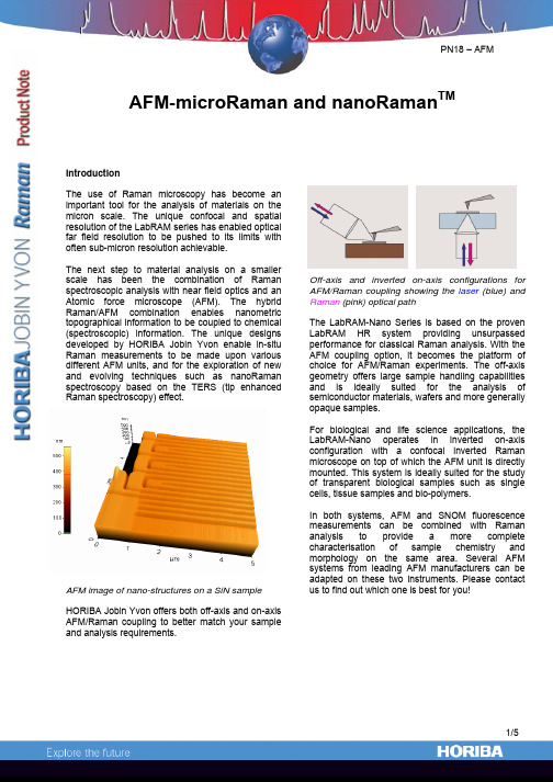
AFM-microRaman and nanoRaman TMIntroductionThe use of Raman microscopy has become animportant tool for the analysis of materials on themicron scale. The unique confocal and spatialresolution of the LabRAM series has enabled opticalfar field resolution to be pushed to its limits withoften sub-micron resolution achievable.The next step to material analysis on a smallerscale has been the combination of Ramanspectroscopic analysis with near field optics and anAtomic force microscope (AFM). The hybridRaman/AFM combination enables nanometrictopographical information to be coupled to chemical(spectroscopic) information. The unique designsdeveloped by HORIBA Jobin Yvon enable in-situRaman measurements to be made upon variousdifferent AFM units, and for the exploration of newand evolving techniques such as nanoRamanspectroscopy based on the TERS (tip enhancedRaman spectroscopy) effect.AFM image of nano-structures on a SiN sampleHORIBA Jobin Yvon offers both off-axis and on-axisAFM/Raman coupling to better match your sampleand analysis requirements.Off-axis and inverted on-axis configurations forAFM/Raman coupling showing the laser (blue) andRaman (pink) optical pathThe LabRAM-Nano Series is based on the provenLabRAM HR system providing unsurpassedperformance for classical Raman analysis. With theAFM coupling option, it becomes the platform ofchoice for AFM/Raman experiments. The off-axisgeometry offers large sample handling capabilitiesand is ideally suited for the analysis ofsemiconductor materials, wafers and more generallyopaque samples.For biological and life science applications, theLabRAM-Nano operates in inverted on-axisconfiguration with a confocal inverted Ramanmicroscope on top of which the AFM unit is directlymounted. This system is ideally suited for the studyof transparent biological samples such as singlecells, tissue samples and bio-polymers.In both systems, AFM and SNOM fluorescencemeasurements can be combined with Ramananalysis to provide a more completecharacterisation of sample chemistry andmorphology on the same area. Several AFMsystems from leading AFM manufacturers can beadapted on these two instruments. Please contactus to find out which one is best for you!AFM- microRaman dual analysisThe seamless integration of hardware and software of both systems onto the same platform enables fast and user-friendly operation of both systems at the same time. Furthermore, the AFM/Raman coupling does not compromise the individual capabilities of either system and the imaging modes of the AFM remain available (EFM, MFM, Tapping Mode, etc.)The operator has direct access to both the nanometric topography of a sample given by the AFM, and the chemical information from the micro-Raman measurement. An AFM image can berecorded as an initial survey map, in which regions of interest can be defined for further Raman analysis, using the same software.An example of such analysis is illustrated below by an AFM image of Carbon Nanotubes (CNTs) giving information on the CNTs’ length, diameters and aggregation state. A more detailed AFM image is then obtained in which Raman analysis can be performed.Carbon nanotubes AFM images with a gold-coated tip in contact mode. The diameter of the bundles of nanotubes is between 10 and 30 nm.NanoRaman for TERS experimentsSurface Enhance Raman Scattering (SERS) has long been used to enhance weak Raman signals by means of surface plasmon resonance using nanoparticle colloids or rough metallic substrates, allowing to detect chemical species at ppm levels.The TERS effect is based on the same principle, but uses a metal-coated AFM tip (instead of nanoparticles) as an antenna that enhances the Raman signal coming from the sample area which is in contact (near-field). Although not yet fully understood, the TERS effect has attracted a lot of interest, as it holds the promise of producing chemical images with nanometric resolution.The LabRAM-Nano offers an ideal platform,combining state-of-the-art AFMs with our Raman expertise to perform exploratory TERS experiments with confidence.Raman signal TERS enhancement on a Silicon sample with far field suppression thanks to adequate polarization configuration. Red : Far field + Near Field (tip in contact)– Blue : Far field only (tip withdrawn)Technical specificationsFlexure guided scanner is used to maintain zero background curvature below 2 nm out-of-planeFor non-TERS measurements, classical Raman measurements can be made on the same spot as AFM images by translating the sample with a high-accuracy positioning stage from the AFM setup to the Raman setup (and vice et versa). The AFM map can be used to define a region of interest for the Raman analysisusing a common software.LabRAM-Nano coupled with Veeco’s Dimension 3100 AFMThe on-axis coupling configuration enables both AFM-microRaman dual analysis and TERS measurementson transparent and biological samples. The AFM is directly coupled onto the inverted microscope and directlyinterfaced to the LabRAM HR microprobe. It can also be taken off the optical microscope to obtain AFMimages in a different location. Seamless software integration is realized to provide a common platform to bothsystems for both AFM and Raman analysis of the same area and TERS investigation.Bioscope II from VeecoLabRAM-Nano coupled with Park Systems(formerly PSIA) XE-120Off-axis coupling for AFM-microRaman and nanoRaman (TERS)For both dual AFM-microRaman dual analysis and TERS measurements, the off-axis coupling is ideally suited for opaque and large samples. For opaque samples, the inverted on-axis coupling is not possible as the sample will not transmit the laser beam. This can be solved by setting the microscope objective at some angle to avoid “shadowing” effects from the AFM cantilever. Here also, seamless software integration is realized to provide a common platform to both systems. The AFM can be controlled by the Raman software (LabSpec), and mapping areas can be defined on AFM images for further Raman analysis.France : HORIBA Jobin Yvon S.A.S., 231 rue de Lille, 59650 Villeneuve d’Ascq. Tel : +33 (0)3 20 59 18 00, Fax : +33 (0)3 20 59 18 08. Email : raman@jobinyvon.fr www.jobinyvon.frUSA : HORIBA Jobin Yvon Inc., 3880 Park Avenue, Edison, NJ 08820-3012. Tel : +1-732-494-8660, Fax : +1-732-549-2571. Email : raman@ Japan : HORIBA Ltd., JY Optical Sales Dept., 1-7-8 Higashi-kanda, Chiyoda-ku, Tokyo 101-0031. Tel: +81 (0)3 3861 8231, Fax: +81 (0)3 3861 8259. Email: raman@ LabRAM-Nano coupled with Park Systems (formerly PSIA) XE-100Combined polarized Raman and atomic force microscopy:In situ study of point defects and mechanical properties in individual ZnO nanobelts Marcel Lucas,1Zhong Lin Wang,2and Elisa Riedo1,a͒1School of Physics,Georgia Institute of Technology,Atlanta,Georgia30332-0430,USA2School of Materials Science and Engineering,Georgia Institute of Technology,Atlanta,Georgia30332-0245,USA͑Received8June2009;accepted23June2009;published online4August2009͒We present a method,polarized Raman͑PR͒spectroscopy combined with atomic force microscopy͑AFM͒,to characterize in situ and nondestructively the structure and the physical properties ofindividual nanostructures.PR-AFM applied to individual ZnO nanobelts reveals the interplaybetween growth direction,point defects,morphology,and mechanical properties of thesenanostructures.In particular,wefind that the presence of point defects can decrease the elasticmodulus of the nanobelts by one order of magnitude.More generally,PR-AFM can be extended todifferent types of nanostructures,which can be in as-fabricated devices.©2009American Instituteof Physics.͓DOI:10.1063/1.3177065͔Nanostructured materials,such as nanotubes,nanobelts ͑NBs͒,and thinfilms,have potential applications as elec-tronic components,catalysts,sensors,biomarkers,and en-ergy harvesters.1–5The growth direction of single-crystal nanostructures affects their mechanical,6–8optoelectronic,9 transport,4catalytic,5and tribological properties.10Recently, ZnO nanostructures have attracted a considerable interest for their unique piezoelectric,optoelectronic,andfield emission properties.1,2,11,12Numerous experimental and theoretical studies have been undertaken to understand the properties of ZnO nanowires and NBs,11,12but several questions remain open.For example,it is often assumed that oxygen vacancies are present in bulk ZnO,and that their presence reduces the mechanical performance of ZnO materials.13However,no direct observation has supported the idea that point defects affect the mechanical properties of individual nanostructures.Only a few combinations of experimental techniques en-able the investigation of the mechanical properties,morphol-ogy,crystallographic structure/orientation and presence of defects in the same individual nanostructure,and they are rarely implemented due to technical challenges.Transmis-sion electron microscopy͑TEM͒can determine the crystal-lographic structure and morphology of nanomaterials that are thin enough for electrons to transmit through,4,14–17but suf-fers from some limitations.For example,characterization of point defects is rather challenging.14–17Also,the in situ TEM characterization of the mechanical and electronic properties of nanostructures is very challenging or impossible.15–17 Alternatively,atomic force microscopy͑AFM͒is well suited for probing the morphology,mechanical,magnetic, and electronic properties of nanostructures from the micron scale down to the atomic scale.3,6,7,10In parallel, Raman spectroscopy is effective in the characterization of the structure,mechanical deformation,and thermal proper-ties of nanostructures,18,19as well as the identification of impurities.20Furthermore,polarized Raman͑PR͒spectros-copy was recently used to characterize the crystal structure and growth direction of individual single-crystal nanowires.21Here,an AFM is combined to a Raman microscope through an inverted optical microscope.The morphology and the mechanical properties of individual ZnO NBs are deter-mined by AFM,while polarized Raman spectroscopy is used to characterize in situ and nondestructively the growth direc-tion and randomly distributed defects in the same individual NBs.Wefind that the presence of point defects can decrease the elastic modulus of the NBs by almost one order of mag-nitude.The ZnO NBs were prepared by physical vapor deposi-tion͑PVD͒without catalysts14and deposited on a glass cover slip.For the PR studies,the cover slip was glued to the bottom of a Petri dish,in which a hole was drilled to allow the laser beam to go through it.The round Petri dish was then placed on a sample plate below the AFM scanner,where it can be rotated by an angle,or clamped͑see Fig.1͒.The morphology and mechanical properties of the ZnO NBs were characterized with an Agilent PicoPlus AFM.The AFM was placed on top of an Olympus IX71inverted optical micro-scope using a quickslide stage͑Agilent͒.A silicon AFM probe͑PointProbe NCHR from Nanoworld͒,with a normal cantilever spring constant of26N/m and a radius of about 60nm,was used to collect the AFM topography and modulated nanoindentation data.The elastic modulus of the NBs was measured using the modulated nanoindentation method22by applying normal displacement oscillations at the frequency of994.8Hz,at the amplitude of1.2Å,and by varying the normal load.PR spectra were recorded in the backscattering geometry using a laser spot small enough ͑diameter of1–2m͒to probe one single NB at a time.The incident polarization direction can be rotated continuouslywith a half-wave plate and the scattered light is analyzedalong one of two perpendicular directions by a polarizer atthe entrance of the spectrometer͑Fig.1͒.Series of PR spec-tra from the bulk ZnO crystals and the individual ZnO NBswere collected with varying sample orientation͑the NBs are parallel to the incident polarization at=0͒,in the co-͑parallel incident and scattered analyzed polarizations͒and cross-polarized͑perpendicular incident and scattered ana-lyzed polarizations͒configurations.For the ZnO NBs,addi-tional series of PR spectra were collected where the incidenta͒Electronic mail:elisa.riedo@.APPLIED PHYSICS LETTERS95,051904͑2009͒0003-6951/2009/95͑5͒/051904/3/$25.00©2009American Institute of Physics95,051904-1polarization is rotated and the ZnO NB axis remained paral-lel or perpendicular to the analyzed scattered polarization ͑see supplementary information 25͒.The exposure time for each Raman spectrum was 10s for the bulk crystals and 20min for NBs.After each rotation of the NBs,the laser spot is recentered on the same NB and at the same location along the NB.Prior to the PR characterization of ZnO NBs,PR data were collected on the c -plane and m -plane of bulk ZnO crystals ͓Fig.2͑a ͔͒.In ambient conditions,ZnO has a wurtzite structure ͑space group C 6v 4͒.Group theory predicts four Raman-active modes:one A 1,one E 1,and two E 2modes.11,20,23The polar A 1and E 1modes split into transverse ͑TO ͒and longitudinal optical branches.On the c -plane ͑0001͒-oriented sample,only the E 2modes,at 99͑not shown ͒and 438cm −1,are observed,and their intensity is independent of the sample orientation ͓Fig.2͑a ͔͒.On them -plane ͑101¯0͒-oriented sample,the E 2,E 1͑TO ͒,and A 1͑TO ͒modes are observed at 99,438,409,and 377cm −1,respectively ͓Fig.2͑a ͔͒,and their intensity depends on .Peaks at 203and 331cm −1in both crystals are assigned to multiple phonon scattering processes.The intensity,center,and width of the peaks at 438,409,and 377cm −1were obtained by fitting the experimental PR spectra with Lorent-zian lines ͑see supplementary information 25͒.The successful fits of the angular dependencies by using the group theory and crystal symmetry 23indicate that PR data can be used to characterize the growth direction of ZnO NBs.It is noted that the ZnO NBs studied here have dimensions over 300nm,so the determination of the growth direction is not ex-pected to be affected by any enhancement of the polarized Raman signal due to their high aspect ratio.24AFM images and PR data of three individual ZnO NBs are presented in Figs.2͑b ͒–2͑d ͒.These NBs,labeled NB1,NB2,and NB3,have different dimensions and properties assummarized in Table I .A comparison of the PR spectra in Figs.2͑a ͒–2͑d ͒reveals differences between bulk ZnO and individual NBs.First,the glass cover slip gives rise to a weak broadband centered around 350cm −1on the Raman spectra of the NBs ͓see bottom of Fig.2͑d ͔͒.Second,there are additional Raman bands around 224and 275cm −1for NB2and NB3.These bands are observed in doped or ion-implanted ZnO crystals.11,20Their appearance is explained by the disorder in the crystal lattice due to randomly distrib-uted point defects,such as oxygen vacancies or impurities.The defect peaks area increases in the order NB1ϽNB2ϽNB3.Since the laser spot diameter is larger than the width of all three NBs,but smaller than their length,L ,the NB volume probed by the laser beam is approximated by the product of the width,w ,with the thickness,t .ThevolumeFIG.1.͑Color online ͒Schematic of the experimental setup,showing the path of the laser beam.The ZnO NBs are deposited on a glass slide,which is placed inside a rotating Petridish.FIG.2.͑Color online ͒͑a ͒PR spectra from the c and m planes of a ZnO crystal,shown in blue and green,respectively.The wurtzite structure ͑Zn atoms are brown,O atoms red ͒is also shown,where a ء,b ء,and c ءare the reciprocal lattice vectors.͓͑b ͒–͑d ͔͒AFM images ͑3ϫ3m ͒of three NBs labeled NB1,NB2,and NB3and corresponding PR spectra.In ͑d ͒a PR spectrum of the glass substrate is shown at the bottom.All the PR spectra in ͑a ͒–͑d ͒are collected in the copolarized configuration for =0and 90°.The spectra are offset vertically for clarity.TABLE I.Summary of the PR-AFM results for NB1,NB2,and NB3.w ͑nm ͒t ͑nm ͒w /t L ͑m ͒͑°͒E ͑GPa ͒Defects NB11080875 1.24028Ϯ1562Ϯ5No NB21150710 1.64972Ϯ1538Ϯ5Yes NB315104553.35966Ϯ1517Ϯ5Yesprobed decreases in the order NB1͑wϫt=9.45ϫ103nm2͒ϾNB2͑8.17ϫ103nm2͒ϾNB3͑6.87ϫ103nm2͒.This indi-cates that the density of point defects is highest in NB3,and increases with the width to thickness ratio,w/t,in the order NB1ϽNB2ϽNB3.The PR intensity variations of the438cm−1peak as a function ofin the various polarization configurations were fitted by using group theory and crystal symmetry to deter-mine the anglebetween the NB long axis͑or growth di-rection͒and the c-axis͓͑0001͔axis͒of the constituting ZnO wurtzite structure21,23͑see supplementary information25͒.In-tensity variations of the377cm−1peak,when present,are used to confirm the obtained values of.The results are shown in Table I and indicate that growth directions other than the most commonly observed c-axis are possible,par-ticularly when point defects are present.Finally,the elastic properties of NB1,NB2,and NB3are characterized by AFM using the modulated nanoindentation method.6,7,22In a previous study,the elastic modulus of ZnO NBs was found to decrease with increasing w/t and this w/t dependence was attributed to the presence of planar defects in NBs with high w/t.6,7By using PR-AFM,we can study the role of randomly distributed defects,morphology,and growth direction on the elastic properties in the same indi-vidual ZnO NB.The measured elastic moduli,E,are62GPa for NB1,38GPa for NB2,and17GPa for NB3.These PR-AFM results confirm the w/t dependence of the elastic modulus in ZnO NBs,but more importantly they reveal that the elastic modulus of ZnO NBs can significantly decrease, down by almost one order of magnitude,with the presence of randomly distributed point defects.In summary,a new approach combining polarized Raman spectroscopy and AFM reveals the strong influence of point defects on the elastic properties of ZnO NBs and their morphology.Based on a scanning probe,PR-AFM pro-vides an in situ and nondestructive tool for the complete characterization of the crystal structure and the physical properties of individual nanostructures that can be in as-fabricated nanodevices.The authors acknowledge thefinancial support from the Department of Energy under Grant No.DE-FG02-06ER46293.1Y.Qin,X.Wang,and Z.L.Wang,Nature͑London͒451,809͑2008͒.2X.Wang,J.Song,J.Liu,and Z.L.Wang,Science316,102͑2007͒.3D.J.Müller and Y.F.Dufrêne,Nat.Nanotechnol.3,261͑2008͒.4H.Peng,C.Xie,D.T.Schoen,and Y.Cui,Nano Lett.8,1511͑2008͒. 5U.Diebold,Surf.Sci.Rep.48,53͑2003͒.6M.Lucas,W.J.Mai,R.Yang,Z.L.Wang,and E.Riedo,Nano Lett.7, 1314͑2007͒.7M.Lucas,W.J.Mai,R.Yang,Z.L.Wang,and E.Riedo,Philos.Mag.87, 2135͑2007͒.8M.D.Uchic,D.M.Dimiduk,J.N.Florando,and W.D.Nix,Science305, 986͑2004͒.9D.-S.Yang,o,and A.H.Zewail,Science321,1660͑2008͒.10M.Dienwiebel,G.S.Verhoeven,N.Pradeep,J.W.M.Frenken,J.A. Heimberg,and H.W.Zandbergen,Phys.Rev.Lett.92,126101͑2004͒. 11Ü.Özgür,Ya.I.Alivov,C.Liu,A.Teke,M.A.Reshchikov,S.Doğan,V. Avrutin,S.-J.Cho,and H.Morkoç,J.Appl.Phys.98,041301͑2005͒. 12Z.L.Wang,J.Phys.:Condens.Matter16,R829͑2004͒.13G.R.Li,T.Hu,G.L.Pan,T.Y.Yan,X.P.Gao,and H.Y.Zhu,J.Phys. Chem.C112,11859͑2008͒.14Z.W.Pan,Z.R.Dai,and Z.L.Wang,Science291,1947͑2001͒.15P.Poncharal,Z.L.Wang,D.Ugarte,and W.A.De Heer,Science283, 1513͑1999͒.16A.M.Minor,J.W.Morris,and E.A.Stach,Appl.Phys.Lett.79,1625͑2001͒.17B.Varghese,Y.Zhang,L.Dai,V.B.C.Tan,C.T.Lim,and C.-H.Sow, Nano Lett.8,3226͑2008͒.18M.Lucas and R.J.Young,Phys.Rev.B69,085405͑2004͒.19I.Calizo,A.A.Balandin,W.Bao,F.Miao,and u,Nano Lett.7, 2645͑2007͒.20H.Zhong,J.Wang,X.Chen,Z.Li,W.Xu,and W.Lu,J.Appl.Phys.99, 103905͑2006͒.21T.Livneh,J.Zhang,G.Cheng,and M.Moskovits,Phys.Rev.B74, 035320͑2006͒.22I.Palaci,S.Fedrigo,H.Brune,C.Klinke,M.Chen,and E.Riedo,Phys. Rev.Lett.94,175502͑2005͒.23C.A.Arguello,D.L.Rousseau,and S.P.S.Porto,Phys.Rev.181,1351͑1969͒.24H.M.Fan,X.F.Fan,Z.H.Ni,Z.X.Shen,Y.P.Feng,and B.S.Zou, J.Phys.Chem.C112,1865͑2008͒.25See EPAPS supplementary material at /10.1063/ 1.3177065for more information on the PR spectra.Growth direction and morphology of ZnO nanobelts revealed by combining in situ atomic forcemicroscopy and polarized Raman spectroscopyMarcel Lucas,1,*Zhong Lin Wang,2and Elisa Riedo1,†1School of Physics,Georgia Institute of Technology,Atlanta,Georgia30332-0430,USA 2School of Materials Science and Engineering,Georgia Institute of Technology,Atlanta,Georgia30332-0245,USA ͑Received26June2009;revised manuscript received28September2009;published14January2010͒Control over the morphology and structure of nanostructures is essential for their technological applications,since their physical properties depend significantly on their dimensions,crystallographic structure,and growthdirection.A combination of polarized Raman͑PR͒spectroscopy and atomic force microscopy͑AFM͒is usedto characterize the growth direction,the presence of point defects and the morphology of individual ZnOnanobelts.PR-AFM data reveal two growth modes during the synthesis of ZnO nanobelts by physical vapordeposition.In the thermodynamics-controlled growth mode,nanobelts grow along a direction close to͓0001͔,their morphology is growth-direction dependent,and they exhibit no point defects.In the kinetics-controlledgrowth mode,nanobelts grow along directions almost perpendicular to͓0001͔,and they exhibit point defects.DOI:10.1103/PhysRevB.81.045415PACS number͑s͒:61.46.Ϫw,61.72.Dd,78.30.Ly,81.10.ϪhI.INTRODUCTIONControl over the morphology and structure of nanostruc-tured materials is essential for the development of future de-vices,since their physical properties depend on their dimen-sions and crystallographic structure.1–15In particular,the growth direction of single-crystal nanostructures affects their piezoelectric,1,2transport,3catalytic,4mechanical,5–9 optoelectronic,10and tribological properties.11ZnO nano-structures with various morphologies͑wires,belts,helices, rings,tubes,…͒have been successfully synthesized in solu-tion and in the vapor phase,14–19but little is known about their growth mechanism,particularly in a process not involv-ing catalyst particles.17Understanding the growth mecha-nism and determining the decisive parameters directing the growth of nanostructures and tailoring their morphology is essential for the use of ZnO nanobelts as power generators or electromechanical systems.1,2,5,6From a theoretical stand-point,a shape-dependent thermodynamic model showed that the morphology of ZnO nanobelts grown in equilibrium con-ditions depends on their growth direction,but the role of defects was not considered.20Experimentally,it was shown that the growth direction of ZnO nanostructures can be di-rected by the synthesis conditions,such as the oxygen con-tent in the furnace.19A previous study combining scanning electron microscopy and x-ray diffraction suggested a growth-direction-dependent morphology.20An atomic force microscopy͑AFM͒combined with transmission electron mi-croscopy also suggested that the morphology of ZnO nano-belts is correlated with their growth direction and highlighted the potentially important role of planar defects.5 Growth modes out of thermodynamic equilibrium and the role of point defects5,17are particularly challenging to inves-tigate experimentally,21due to the lack of appropriate experi-mental techniques.Electron microscopy can determine the crystallographic structure and morphology of conductive nanomaterials,3,17,22–24but is not suitable for the character-ization of point defects,especially when their distribution is disordered.17,22–24Raman spectroscopy has been used for the characterization of the structure of carbon nanotubes,25,26the identification of impurities,27and the determination of the crystal structure28and growth direction of individual single-crystal nanowires.29Recently,polarized Raman͑PR͒spec-troscopy has been coupled to AFM to study in situ the inter-play between point defects and mechanical properties of ZnO nanobelts.30Here,PR-AFM is used to study the growth mechanism and the relationship between growth direction,point defects, and morphology of individual ZnO nanobelts.The morphol-ogy of an individual ZnO nanobelt is determined by AFM, while the growth direction and randomly distributed defects in the same individual nanobelt are characterized by polar-ized Raman spectroscopy.II.EXPERIMENTALThe ZnO nanobelts were prepared by physical vapor deposition͑PVD͒without catalysts following the method de-scribed in Ref.17.The ZnO nanobelts were deposited on a glass cover slip,which was glued to a Petri dish.The rotat-able Petri dish was then placed on a sample plate under an Agilent PicoPlus AFM equipped with a scanner of100ϫ100m2range.Topography images of the ZnO nanobelts were collected in the contact mode with CONTR probes͑NanoWorld AG,Neuchâtel,Switzerland͒of normal spring constant0.21N/m at a set point of2nN.The AFM was placed on top of an Olympus IX71inverted optical micro-scope that is coupled to a Horiba Jobin-Yvon LabRam HR800.PR spectra were recorded in the backscattering ge-ometry using a40ϫ͑0.6NA͒objective focusing a laser beam of wavelength785nm on the sample to a power den-sity of about105W/cm2and a spot size of about2m. The incident polarization direction can be rotated continu-ously with a half-wave plate.The scattered light was ana-lyzed along one of two perpendicular directions by a polar-izer at the entrance of the spectrometer.The intensity,center, and width of the Raman bands were obtained byfitting the spectra with Lorentzian lines.The polarization dependence of the quantum efficiency of the Raman spectrometer was tested by measuring the intensity variations of the377,409,PHYSICAL REVIEW B81,045415͑2010͒1098-0121/2010/81͑4͒/045415͑5͒©2010The American Physical Society045415-1and 438cm −1bands from two bulk ZnO crystals ͑c -plane and m -plane ZnO crystals,MTI Corporation ͒.The PR data from bulk crystals were successfully fitted using group theory and crystal symmetry 28without further calibration of the spectrometer or data correction.III.RESULTS AND DISCUSSIONAFM images and PR data of two individual ZnO nano-belts are presented in Fig.1.These nanobelts have different cross-sections,1320ϫ1080nm 2͑nanobelt labeled NB A͒FIG.1.͑Color online ͒PR-AFM results on individual ZnO nanobelts.͑a ͒AFM topography image,͑b ͒typical PR spectra for different sample orientations and polarization configurations,and ͑c ͒–͑f ͒polar plots of the angular dependence of the Raman intensities for the nanobelt NB A.͑g ͒AFM topography image,͑h ͒typical PR spectra,and ͑i ͒–͑l ͒polar plots of the angular dependence of the Raman intensities for the nanobelt NB B.The Raman spectra in ͑h ͒exhibit peaks centered at 224and 275cm −1͑triangles ͒that are characteristic of defects in the nanobelt NB B.The Raman spectra are offset vertically for clarity.In ͑c ͒,͑d ͒,͑i ͒,and ͑j ͒,the nanobelt axis is rotated in a fixed polarization configuration ͑solid squares:copolarized;open squares:cross polarized ͒and is parallel to the incident polarization for =0°.In ͑e ͒,͑f ͒,͑k ͒,and ͑l ͒,the incident polarization is rotated,while the analyzed polarization and the nanobelt axis are fixed.In ͑e ͒,͑f ͒,͑k ͒,and ͑l ͒,at the angle 0°,the nanobelt is perpendicular to the incident polarization and the incident and analyzed polarizations are parallel ͑solid squares ͒or perpendicular ͑open squares ͒.Typical Raman spectra of the glass cover slip in the copolarized and cross-polarized configurations are shown as a reference in ͑b ͒and ͑h ͒,respectively.LUCAS,WANG,AND RIEDO PHYSICAL REVIEW B 81,045415͑2010͒045415-2。
SpectrAA-110220880系列原子吸收光谱仪介绍
IntroductionThe SpectrAA-110/220/880 series atomic absorption spectrometers arecontrolled by Varian's innovative SpectrAA worksheet software. TheSpectrAA series includes the following instruments:SpectrAA 110, SpectrAA 220, SpectrAA 220FS, SpectrAA 220G,SpectrAA 220 Zeeman, SpectrAA 880, SpectrAA 880 ZeemanThis documentation is appropriate for all of the instruments listedabove.The software, based on a spreadsheet concept which mimics theanalyst's workbook, combines flame, furnace, Zeeman and vaporoperation in one integrated package.The SpectrAA software runs under Microsoft Windows95TM operatingsystem, and provides the following:Innovative, easy to use pages□Dedicated function keys for critical functionsO Extensive on-line Help including Multimedia demonstrations of howto set up your instrument□Button, control and field level context sensitive help (via the F1 keyor "What's This"button)□"Tool Tips" providing the allowed range for each field□Comprehensive error system.1.1Installation requirementsPrior to receiving your instrument you will have been provided with aSpectrAA Pre-installation manual, which describes the environmentalPublication date: 06/97and operating requirements of the SpectrAA system. You must prepareyour laboratory according to these instructions before the SpectrAAcan be installed. You should keep the Pre-installation manual, insertingit into this binder, for future reference. If you have misplaced your copy,you can obtain a replacement from your local Varian office.1.2SpectrAA documentationThis manual covers the installation of the SpectrAA software for110/220/880 series instruments. Instructions for installing, operatingand maintaining the instruments are included in the Multimedia on-linehelp (see section 3.3).Operating instructions for the Sample Introduction Pump System(SIPS) and other SpectrAA accessories are given in the manualsaccompanying the accessories, and some are also available on-linewith the SpectrAA software.To help you find the information you need, this operation manual isorganised into the following:Safety Practices and Hazards sectionChapter 1 IntroductionChapter 2 PC and printer setupChapter 3 Software overviewChapter 4 Getting startedConventionsThe following conventions have been used throughout this manual:O ltalics indicate menu items, menu options and field names (e.g.select Copy from the Edit menu).□Keyboard and mouse commands have been typed in bold(e.g.press the F2 key).口 Single quotes('') indicate a selection you can make from severalchoices, such as radio buttons and checkboxes.□Double quotes("") are used to signify the pushbuttons appearingthroughout the software (e.g. select "OK").□ALL CAPITALS indicates text you must type in from the keyboard 1-2Publication date: 06/97(e.g.type SETUP at the prompt).Publication date: 06/971.3SpecificationsYour SpectrAA instrument is designed for indoor use. It is suitable forthe following categories:□Installation category ll□Pollution degree 2□Safety Class 1 (EN 61010-1)1.3.1EnvironmentalCondition Altitude Tempt(℃)Humidity(%RH)non-condensingNon-operating 0-2133 m 5-4520-80(transport) (0-7000Non-operating andmeeting dielectricstrength testssea level 40 90-95Operating but not 0-2000 m 5-31≤80necessarily meetingperformance spec's(0-6562') 31-40 ≤{80-3.33(t-31)}Operating within 0-853 m 10-35 8-80performancespecifications(0-2800')853-2133 m(2800-7000')10-25 8-80For optimum analytical performance it is recommended that the ambient temperature of the laboratory be between 20-25℃and be held constant to within ±2 °℃throughout the entire working day. 1.3.2PowerVoltage 1-4 100+10%-5% VAC(non-Zeeman)120,220 or 240 VAC±10%(non-Zeeman)230+14%-6% VAC(non-Zeeman)230+6%-14% VAC (non-Zeeman)187-264 VAC(220 Zeeman)208,220,or 240 VAC±10%(880 Zeeman)Publication date: 06/97230+14%-6% VAC(880 Zeeman)230+6%-14% VAC (880 Zeeman) Frequency 50 or 60 Hz±1 HzNormal consumptionSpectrAA-110/220 series 170 VA(approx)SpectrAA-880 470 VA(approx)Rated firing consumption 3500VA**(approx)*Zeeman versions only. The Zeeman SpectrAA will draw surge currents in excess of the nominal rating of 15A (3500VA).Non-Zeeman Mains inlet coupler:SpectrAA- 110/220 series 3/2 A 120/250 VAC 50-60Hz IEC type SpectrAA-880 6A 250VAC IEC typeZeeman Mains inlet coupler:Hubbell 20A 250V Twist lock (NEMA L6-20P)ConnectionsNon-Zeeman Mains power cordAustralia USA Europe 10A 250VAC10A 125VAC6A 250VACNFC61.303VAComplies with AS3112Complies with NEMA 5- 15PComplies with CEE7 sheet vii orZeeman Mains power cordAustralia USA Europe15A 250VAC30A 250VACNEMA L6-30P15A 250VACClipsal 439D15MHubbel #2621 (complies withKaiser CEBEC 616 VDE (complies with DIN 49441R2)BearIEEE 488Accessory, 9-way female D-range typeAccessory, MCA, 6 way DIN type (SpectrAA-880 only)UltrAA-lamp connections: Burndy circular 6-way, optional (actual number depends on the model and option selected)Publication date: 06/97Warning-HIGH VOLTAGETo maintain safety, only the UltrAA-lamp power suppliesshould be used at these connections.1-6Publication date: 06/97FrontZeeman workhead CPC 14 way connection in the samplecompartment (Zeeman only).WarningTo prevent connector damage, switch OFF the instrumentbefore inserting the plug and ALWAYS turn the locknut fullyclockwise to the detent position. To maintain safety, only theZeeman workhead connector should be used at thisconnection.Front (lamp bay)Deuterium lamp:Molex 3-way connection, in lamp compartment (behind lamp panel in lamp compartment on SpectrAA- 110/220 series).Warning-HIGH VOLTAGETo maintain safety, only the Deuterium Lamp assembly shouldbe used at this connection.Hollow cathode lamps:4 lamp capacity on SpectrAA- 110/220 series8 lamp capacity on SpectrAA-880Warning-HIGH VOLTAGETo maintain safety, only hollow cathode lamps should be usedat these connections.FusesNon-ZeemanSpectrAA-110/220 seriesT2.5A H250V, IEC 127 sheet 5, 5x 20 mm (100- 120 &220-240 VAC) SpectrAA-880T5A H250V, IEC 127 sheet 5, 5×20 mm (220-240 VAC)T6.3A H250V, IEC 127 sheet 5,5×20 mm(100- 120 VAC)Publication date: 06/97Zeeman15 A long delayed action circuit breaker with a thermal cutout.SpectrAA-880T5A H250V,IEC 127 sheet 5,5x20 mm (208-240 VAC)Note For safety reasons, any other internal fuse or circuit breaker is notoperator accessible, and should be replaced only by Varian authorizedpersonnel.Fuse information on the rear of the instrument is the most up to date.1.3.3Gas suppliesRear of instrument"Adaptors are availableOther gas connectionsSample compartment: Push-on Air/N₂O connector for burnerPush-on C.H₂connector for burner1.3.4Weights and dimensions1-8 Publication date: 06/97Publication date: 06/97。
- 1、下载文档前请自行甄别文档内容的完整性,平台不提供额外的编辑、内容补充、找答案等附加服务。
- 2、"仅部分预览"的文档,不可在线预览部分如存在完整性等问题,可反馈申请退款(可完整预览的文档不适用该条件!)。
- 3、如文档侵犯您的权益,请联系客服反馈,我们会尽快为您处理(人工客服工作时间:9:00-18:30)。
BaySpec's Raman solutions begin with careful choice of optical, electrical and mechanical parts for optimal throughput and excellent long-term reliability Model Part Number Format Size Dimensions (mm3) Weight Excitation Source 532nm 785nm 1064nm Custom Laser FWHM Bandwidth Output power Spectrograph Grating Technology Range (532nm) (on request) Range (785nm) (on request) Range (1064nm) (on request) Spectral Resolution Stray Light Reduction Detector Type Number of pixels Pixel size Cooling temperature (Deep cooling option) Integration time Readout speed Digitalized output Dynamic range Quantum Efficiency (max.) Electronics Interface Input power Trigger mode Wireless Bluetooth Wireless Ethernet 802.11b/g Battery life Interchangable battery Software BaySpec "Spec 2020" Library support Xantus-0 HRAM-0B-xxxx Handheld 93 x 195 x 59 1 kg (2.2lbs.) Xantus-1 HRAM-1-xxxx Handheld 125 x 233 x 85 2.2kg (4.7lbs.) < < < First Guard HRAM-xxxx Handheld 140 x 218 x 85 1.8kg (4 lbs.) < < < Portability PRAM-xxxx Portable 240 x 260 x 135 4.5kg (9.9 lbs) < < < RamSpec-X BRAM-xxxx Benchtop 134 x 315 x 84 8.7 kg (19 lbs) < < < RamSpec-HR-X BRAM-HR-xxxx Benchtop 534 x 534 x 305 11.5 kg (25 lbs) < < < Gratings BaySpec's spectrometers utilize transmission Volume Phase Gratings (VPG®) high efficiency and compact size, covering any wavelength range from 100-3200cm-1
Micro 2020 , Windows CE
Spec 2020 , Windows XP/Vista SDK support for 3rd Party Libaries available
BaySpec, Inc. | 1101 McKay Drive, San Jose, California 95131 USA | Phone: +1(408)512-5928 | | © BaySpec, Inc. 1999-2011
100~220VDC 5V TTL edge or level < optional NA
100~220VDC 5V TTL edge or level < optional NA
BaySpec's vision is to design, manufacture and deliver VPG-based spectral engines enabling pervasive spectral sensing into biomedical, pharmaceuticals, food safety, industrial controls, fiber sensing, and telecom markets.
20ms - 300sec. 250kHz 16-bit 2000:1 typ. >90%
115~120VAC/+5VDC < 4 hrs < optional 4 hrs
USB2.0 / RS232 / Ethernet +15VDC 5V TTL edge or level < < optional optional 4 hrs 2 hrs <
400-1850 cm-1 100-1850 cm1
15-17 cm-1 0.05% Uncooled CCD 1024 14µm
200-3000 cm-1 100-1850 cm1 400-1850 cm-1 100-1850 cm1 300-1850 cm-1 100-1850 cm1 10-12 cm-1 0.05% TE cooled CCD 1024 14µm or 25µm -20°C 20ms - 300sec. 250kHz 16-bit 3000:1 typ. >90%0mW
contact BaySpec 0.2nm typ./ 0.3nm max. 0~500mW 0~500mW Transmission Volume Phase Grating (VPG ®) 200-3000 cm-1 200-3200 cm-1 100-1850 cm1 200-2000 cm-1 400-1850 cm-1 200-3200 cm-1 1 100-1850 cm 100-1850 cm1 300-1850 cm-1 200-3200 cm-1 100-1850 cm1 300-1850 cm-1 -1 10-12 cm 5-7 cm-1 0.05% 0.05% Uncooled and TE cooled CCD 1024 14µm or 25µm -20°C 20ms - 300sec. 250kHz 16-bit 3000:1 typ. >90% TE cooled CCD or InGaAs Array 512/1024/2048 14µm or 25µm -20°C 20ms - 300sec. 250kHz 16-bit 3000:1 typ. >80%
0~500mW
0~1500mW
Spectrograph Fast optics, typically f/2 or f/3 for high throughput utilizing BaySpec's VPGs and specialty optics designs for long-term repeatibility and reliability Detectors Uncooled, soft cooled (-20°C) and deepcooled (-55°C) detector array options allow for excellent signal/noise ratio over wide operating ranges Probe Flexible fiber coupling with ruggedized protecitve jacking delivers Rayleigh scattering rejection as high as 10 photons per billion. Available in a number of wavelength ranges: 532nm, 785nm, 1064nm. Others upon request.
200-3200 cm-1 custom 200-3200 cm-1 custom 200-3200 cm-1 custom 4-5 cm-1 0.05% TE cooled CCD or InGaAs Array 512/1024/2048 14µm or 25µm -20°C -55°C 20ms - 300sec. 250kHz 16-bit 6000:1 typ. >80%
200-3200 cm-1 custom 200-3200 cm-1 custom 200-3200 cm-1 custom 8-10 cm-1 0.05% TE cooled CCD or InGaAs Array 512/1024/2048 14µm or 25µm -20°C -55°C 20ms - 300sec. 250kHz 16-bit 6000:1 typ. >80%
