circulatory system循环系统
循环系统组织学

循环系统组织学循环系统【学习要点】1.血管壁的一般结构和动、静脉管壁的结构特点。
2.微循环的概念和构成。
3.毛细淋巴管的结构特点。
4.心壁的构造和心的传导系统。
循环系统(Circulatory syste)包括心血管系统和淋巴管系统两部分,是一套连续而封闭的管道系统。
心血管系统包括心脏、动脉、毛细血管和静脉。
心脏是推动血液在血管中不停流动的动力器官;动脉是将血液输送到各组织和器官的血管,根据其管径的大小,可将动脉分为大动脉、中动脉、小动脉和微动脉4种。
毛细血管是心血管系统中极为重要的组成部分,它连接在微动脉和微静脉之间,是实现物质交换的重要结构。
静脉是输送血液回流心脏的血管,起始于毛细血管,管径逐渐增粗,末端止于心房;根据静脉管径的大小,也可将其分为大静脉、中静脉、小静脉和微静脉4种。
淋巴管系统包括毛细淋巴管、淋巴管、淋巴干和淋巴导管。
毛细淋巴管起始于组织间隙,逐渐汇合成淋巴管,淋巴管汇合成9条淋巴干,淋巴干汇合成2条淋巴导管,即胸导管和右淋巴导管,淋巴导管最终注入静脉(表7-1)。
循环系统的主要功能是运输氧气、营养物质和代谢产物等,即把消化系统吸收的营养物质和呼吸系统交换的氧气运送到全身各器官、组织和细胞;同时也及时地将器官、组织和细胞产生的二氧化碳、尿素等代谢产物运送到泌尿、呼吸等系统排泄到体外;此外,心血管系统还能将内分泌系统分泌的激素运送至相应的器官,调节其生理功能。
而淋巴管系统则是心血管系统的重要补充部分,是脉管系统的辅助结构。
表7-1循环系统组成心血管系淋巴管系心毛细淋巴管血管动脉大动脉、中动脉、小动脉、微动淋巴管脉毛细血管淋巴干淋巴导管静脉微静脉、小静脉、中静脉、大静脉第一节血管一、血管管壁的一般微细结构血管管壁的微细结构,除毛细血管外,其管壁结构一般分为内膜、中膜和外膜三层(图7-1)。
虽然动脉和静脉都可分为大、中、小微4级,但4级血管在结构上并无明显的界限,而是逐渐移行的。
大动脉是指接近心的动脉,管径最粗,如主动脉和肺动脉等;管径在0.3mm~1mm的动脉属于小动脉;而接近毛细血管,管径在0.3mm 以下的动脉称微动脉;除大动脉外,凡管径在1mm以上的动脉均属中动脉,如肱动脉、桡动脉和尺动脉等。
循环系统讲课比赛-幻灯片
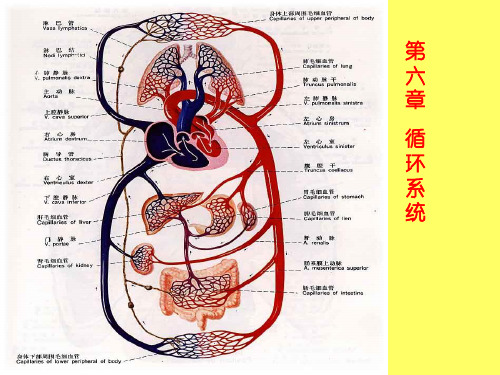
肺循环
一、心 heart
(一)心脏的位置和外形
1.位置: 心脏位于:胸腔的中纵隔内,第2-6肋软骨或第5-8胸椎之 间 。2/3偏在身体正中线的左侧。
胸腔的中纵隔内,
第2-6肋软骨或 第5-8胸椎之间 。 2/3偏在身体正中线的 左侧。
2.外形:
心脏呈前后稍扁的圆锥体,分心尖、心底、三条沟。
人体下1/2部 左上1/4的淋巴
右淋巴导管
胸导管
淋巴管 淋巴液来自组织液
毛细淋巴管
淋巴器官
淋巴器官包括淋巴结、扁桃体、脾和胸腺。
1、淋巴结lymph nodes扁圆形或椭圆形小体,成群 聚集,多沿血管分布,按所处动脉命名。
2、脾spleen是体内最大的淋巴器官,位于腹腔左季 肋部,第9-11肋之间,其长轴与第10肋一致,
一、静脉结构和配布特点 1.由小支汇合成大支,口径逐渐变粗。 2.静脉壁薄,腔内多有静脉瓣。 3.体循环静脉分深、浅两类,深静脉常与动脉伴行,浅静脉位
于浅筋膜内。 4.静脉之间有丰富的吻合支,并形成静脉丛。
5.脑部的静脉较为特殊,多为硬脑膜窦或板障静脉。
6.全身的静脉可分为肺循环和体循环静脉两部分。
(3)直肠静脉丛:门静脉→直肠上静脉→直肠静脉丛→直肠 下静脉、肛静脉→髂内静脉→髂总静脉→下腔静脉。
(4)腹后壁门静脉和腔静脉的小属支相互吻合,通过脊柱静 脉丛沟通上下腔静脉。
肝门静脉:为门静脉
系的静脉主干,门静 脉主要由肠系膜上静 脉和脾静脉汇合而成。
收集腹腔内除肝以外不 成对脏器:脾、胃、胆 囊、胰腺、小肠、大肠。
3、胸腺thymus 位于胸骨柄后方,分左右两叶。
二、淋巴系统的主要功能
(1)帮助组织液回流入静脉:回收蛋白质、运输营养物质
循环系统CirculatorySystemPPT课件
26
右半心的血流方向:
肺动脉 肺动脉口 上腔静脉口 下腔静脉口 右心房 右Biblioteka 室口 右心室 冠状窦口27
28
(三)左心房left atrium 位于右心房的左后方。左心耳 四个入口:两对肺静脉口。 一个出口:左房室口。
(四)左心室left ventricle 位于右心室的左后方。 一个入口:左房室口。口周围有二尖瓣(前瓣、后瓣)。 一个出口:主动脉口。口周围有主动脉瓣。主动脉窦(左、右、后窦)
25
(二)右心室right ventricle 位于右心房的左前下方。
一个入口:右房室口。口周围有三尖瓣环,其上附有三尖瓣tricuspid valve(借腱索连于乳头肌)。
三尖瓣复合体:三尖瓣环、三尖瓣、腱索、乳头肌共同构成,防止血液 逆流。
一个出口:肺动脉口。周围有肺动脉瓣,心室舒张时关闭,阻止血液逆 流入心室。
肺动脉
肺循环
主动脉
左心室 右心房
组织毛 细血管
上、下腔静脉 冠状窦
体循环
血液循环示意图 10
血管吻合及侧支循环
概念:血管吻合vascular anastomosis是指动脉与动脉、静脉与静脉 或动脉与静脉之间藉吻合支相互连接。
血管吻合的方式
动脉间吻合 静脉间吻合 动、静脉间吻合 侧支吻合
侧支循环collecteral circulation
11
12
13
淋巴系统 lymphatic system
组成:淋巴系统由淋巴管道、淋巴组织和淋巴器官组成。 淋巴管道:为静脉的辅助管道,流动着无色透明的淋巴液。 淋巴组织 淋巴器官
脉管学--循环系统

(2)心壁
心内膜 心肌层 心外膜
形成瓣膜和腱索
心房肌和心室肌不相连续,心室肌厚于心房 肌,左心室肌厚于右心室肌。心房肌分为两 层,心室肌分为外斜、中环和内纵三层。
1、心最小静脉 Smallest Cardiac Vein 2、心前静脉 Anterior Cardiac Vein
心脏各腔室 右心房
3、冠状窦 Coronary Sinus
心大静脉 心中静脉 心小静脉
冠状窦 右心房
心
大
心 前
静 脉
心 小 静
静
脉
脉
心 中 静 脉
冠 状 窦
7、心包 Pericardium
脉管学--循环系统
脉 管 学 Angiology
(循环系统 circulatory system)
一、组成
心血管系统
心Heart 动脉Artery 毛细血管Capillary 静脉Vein
淋巴系统
淋巴管道 淋巴器官 淋巴组织
循环系统的功能?
细胞
组织液
毛细血管
二、心血管系统 Cardiovascular System
一入口 右房室口:右房室瓣(三尖瓣)
RV
一出口 肺动脉口:肺动脉瓣
(3)左心房 LA
前部:左心耳 后部:固有心房(左心房窦)
固有心房
右
左心耳
肺 上
、
下
静
脉
口
左房室口
左肺静脉口(上、下):无瓣膜
四入口
【组胚】7.循环系统

内膜为血管壁的最内层; 内膜薄; 由内皮、内皮下层和内弹性 膜组成。
a. 内皮(vascular endothelium) 单层扁平细胞(ec); LM:表面光滑; EM:游离面有稀疏的胞质突起, 丰富的微丝和吞饮小泡,相邻 细胞间有紧密连接、缝隙连接 等细胞连接。
W-p小体[Weibel-Palade body]: 血管内皮细胞特有的膜性杆状 细胞器,其功能是合成和储存 凝血因子F Ⅷ 。
第 七 章
循 环 系 统
Chapter 7 Circulatory System
小隐静脉
外侧隐静脉
臂皮下静脉
头静脉
心 血 管 系 统
动 脉 心脏 毛细血管
静脉
组织液 淋 巴 管 系 统
淋巴导管
毛细淋巴管
淋巴管
循环系统(circulatory system)=心血管系统(cardiovascular system)+ 淋巴管系统(lymphatic vascular system)。
中动脉
B. 大动脉(large artery,aorta)
中膜内富含弹性膜和弹性纤维,故又称为弹性动脉(elastic artery)。 1.内膜(tunica intima) a.内皮下层较厚,有纵行平滑肌束; b.内弹性膜与中膜的弹性膜相连,内膜与中膜分界不清楚。
中动脉
2.中膜(tunica media) a. 最厚; b.主要由40-70层弹性膜构成,各层弹性膜由弹性纤维相连; c. 弹性膜之间有环行平滑肌,少量胶原纤维和基质; d. 弹性膜的层数随年龄和血压的增加而增加。
中动脉模式图 内皮 内皮下层 内弹性膜 平滑肌 外弹性膜 内膜
中膜 外膜 中动脉
I. 动脉的结构特点
英文医学课件:6 Circulatory system循环系统

• plasmalemmal vesicle
/60-70nm, constitute about 25-35% of total volume
/transendothelial channel
function: transport large molecules and storage of membrane (for enlarge, enlongated, pore-formed and microvilli)
circulatorycirculatorysystemsystemclosedtubularsystembloodcirculatorycardiovasculars心血管系统lymphvasculars淋巴管系统capillariescapillariesbasementmembrane
Chapter 9
ry
---Structural feature:
• endothelial cell: -have fenestrae or pore (6080nm in D, with 4-6 nm diaphragm)
• basal lamina: complete
of
endocardium:
endothelium + DCT
• prevent the back flow of blood
2) Conducting S
① components:
---sinoatriol node node): located epicardium of atrium
(SA in
1. Capillaries
1) LM: • endothelium: • basement membrane: • pericyte: /flattened with processes /function:
第十章循环系统(circulatorysystem)
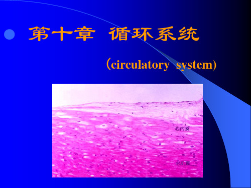
第十章 循环系统
(circulatory system)
一 心脏
heart
(一)心脏壁结构 内皮 endothelium 心内膜 内皮下层 subendothelial layer endocardium 心内膜下层 subendocardial layer 含蒲肯野纤维
心肌膜
myocardium
2
有孔毛细血管 fenestrated capillary 特点: * 一层有孔的内皮细胞 有窗孔,孔上有隔膜 * 基膜连续
3
血窦 sinusoid 特点: * 一层不连续的内皮细胞 有窗孔,有间隙 * 基膜不连续或缺如
四 静脉 vein
特点
:三层分界不清; 外膜厚,中、大V有纵行平滑肌 管腔内有静脉瓣,防血液逆流
重点
1
2
3
4
心脏壁的光镜结构 中动脉的结构及功能 大动脉.小动脉结构特点及功能 毛细血管光镜结构, 电镜下毛细血管类型及结构特点 名词:内弹性膜
medium-sized artery
medium-sized artery
(二)大动脉 large artery:弹性A
结构特点:1、内皮下层较厚
2、中膜主要为40-70层弹性膜 功 能:管壁富有弹性 保证血液连续而均匀流动
large artery
elastic lamina
(三)小动脉 small artery(肌性A) 特点: 直径 0.3mm--1mm 内弹性膜明显 中膜:数层平滑肌 无外弹性膜 (四)微动脉 arteriole 特点: 直径 < 0.3mm 无内弹性膜 中膜:1--2层平滑肌 功能:调节血流量 影响血压,外周阻力血管
循环系统Thecirculatorysystem

红血球:多数鱼类呈扁圆形。一般具细胞核,含血红蛋白,所以血液均为鲜红色。 白血球:分为粒细胞和无粒细胞两类。粒细胞分为:嗜中性白血球、嗜酸性白血球及嗜碱性白血球;无粒细胞:淋巴球和单核白血球。 血栓细胞:无色,小 于红血球。血栓细胞与血 液的凝固作用有关。鱼类 的血栓细胞与哺乳类的血 小板的不同点是它是一个 真细胞,有核和细胞质。
*
第一节 血 液 第二节 心 脏 第三节 动脉和静脉 第四节 淋巴和淋巴管 第五节 造血器官
第七章 循环系统 The circulatory system
202X
商务工作通用模板
功能:将氧气、营养物质以及激素运送到体内各器官和组织内,并把代谢废物排出体外。
特点:封闭型,单循环,心脏一心房一心室。
02
脾脏是鱼体最大最重要的淋巴髓质组织,此外还有不少淋巴髓质组织分布于鱼体不同部位:消化管粘膜下层、肝脏、生殖腺及中肾等。
软骨鱼类食道粘膜层下方的扁平的器官,能生成白血球,当脾脏移去后,它也能产生红血球。
01
头肾
02
一些硬骨鱼类的肾脏前部有前肾的残余组织,称为头肾,它已不起排泄作用,变成一种淋巴髓质组织,具有制造白血球、血栓细胞与毁灭陈旧红血
真骨鱼类:尾静脉→分成左右两支进入肾脏:左侧一支称肾门静脉,它在肾脏后部拆散成毛细血管,然后又汇集到左后主静脉;右侧的一支不形成肾门静脉,直接连到右后主静脉,这支静脉一般较粗大。
*
1
2
3
4
5
6
鱼类的血球可以在不同的器官内形成,在早期胚胎阶段,血管能形成血球,成体阶段,除了血管仍能制造血球外,已经形成更重要的造血中心:脾脏、淋巴髓质组织、赖迪氏器官、头肾等。
血量:仅为体重的1.5%--3%,而哺乳动物一般都在6%以上。 比重:平均为1.035,低于哺乳类(1.053)。 组成:由血浆(blood plasma)及血球(blood cell)组成。血球由红血球、白血球和血栓细胞(或称血小板)等组成。 血浆:略呈黄色,含有大量水分、盐分,蛋白质、氨基酸、脂肪、葡萄糖等营养物质及代谢产物。 血清:将血浆中的纤维蛋白元除去所残留的液体。
动物循环系统
一、循环系统概念:循环系统(Circulatory system)是生物体的细胞外液(包括血浆、淋巴和组织液)及其借以循环流动的管道组成的系统。
循环系统是进行血液循环的动力和管道系统,由心血管系统和淋巴系统组成。
从动物形成心脏以后循环系统分心脏和血管两大部分,叫做心血管系统。
淋巴系统包括淋巴管和淋巴器官,是血液循环的支流,协助静脉运回体液入循环系统,属循环系的辅助部分。
二、动物循环系统发展历程从环节动物门开始出现,环节动物有次生体腔的出现,相应的促进了循环系统的发生。
环节动物具有较完善的循环系统,结构复杂,由纵行血管和环行血管及其分支血管组成,各血管以微血管往相连,血液始终在血管内流动,不流入组织间的空隙中,构成了闭管式循环系统。
血液循环有一定方向,流速较恒定,提高了运输营养物质及携氧机能。
软体动物门的循环系统由心脏、血管、血窦及血液组成血液自心室经动脉,进入身体各部分,后汇入血窦,由静脉回到心耳,故软体动物为开管式循环。
节肢动物门循环系统开管式,包括心脏和动脉两部分。
鱼的循环系统包括液体和管道两部分,液体是指血液和淋巴液,管道为血管及淋巴管。
两栖类由单循环的血液循环方式发展为包括肺循环和体循环的双循环,循环系统包括血管系统和淋巴系统两部分。
鸟类的循环系统反映了较高的代谢水平,主要表现在:动静脉血液完全分开、完全的双循环,心脏容量大,心跳频率快、动脉压高、血液循环迅速。
三、循环系统分类1.开管式循环:大多数无脊椎动物的血液循环系统都是“开放式”的,例如蝗虫的循环系统、虾的循环系统。
2.闭管式循环系统:所有的脊椎动物和部分无脊椎动物的循环系统是“封闭式”的,如蚯蚓、人类的循环系统。
3.二者区别a.开管式循环:是指动物体内的血液不完全在心脏与血管内流动,而能流进细胞间隙的循环方式.如节肢动物体内,背有心脏和它发出的血管(动脉)。
心脏两侧有具活瓣的心门,动脉直接开口在体腔。
心脏收缩时,心门关闭,血液从动脉的开口进入体腔,浸润各组织和器官。
循环
心内膜下层:结缔组织,心室含蒲肯 野
心肌膜:厚的心肌 + 富于毛细血管的结缔组织 心房肌 细、短,心房颗粒,含心房利 钠尿多肽 心外膜:结缔组织+间皮(单扁),脂肪富,多血管神经
心内膜
束细胞
心肌膜
心外膜
心脏
心脏传导系统: 决定心肌的自律性收缩
房室束 窦房结 房室结
房室束分支
心脏传导系统
ቤተ መጻሕፍቲ ባይዱ
窦房结 房室结 房室束 左右房室束分支 (起博点) (传 导 束)
ground substance
Elastic membrane
pore
小动脉(small artery)
结构特点: φ 0.3~1mm 的肌性动脉 主要由内皮和几层平滑肌构成 较大的小动脉可见内弹性膜
功能: 增加血管阻力,调节血压,又称阻力血管
小动脉
大动脉
中动脉 小动脉 薄 明显
10-40层
内皮 内膜 内皮下层 薄层结缔组织 内弹性膜 弹性蛋白组成
中膜
平滑肌
外膜 外弹性膜 结缔组织
动脉
中动脉(medium-sized artery) 内皮 内膜 内皮下层 薄层结缔组织 内弹性膜 弹性蛋白组成,红色波纹状 中膜 10~40层环行平滑肌+胶原+弹性纤维 (产生、A硬化) 外膜 结缔组织,外弹性膜
功能:肌性动脉控制器官的血流量
大动脉(large artery)
结构特点:管壁三层中的中膜最厚,主 要由40-70层弹性窗膜组成。窗膜由弹性 蛋白组成,膜上有窗孔.
功能:弹性好(弹性动脉) ,保证血液在 血管内持续、均匀地流动。
中膜
Large arteries
Circulatory System 循环系统
Circulatory System 循环系统Circulatory System, or cardiovascular system, in humans, the combined function of the heart, blood, and blood vessels to transport oxygen and nutrients to organs and tissues throughout the body and carry away waste products. Among its vital functions, the circulatory system increases the flow of blood to meet increased energy demands during exercise and regulates body temperature. In addition, when foreign substances or organisms invade the body, the circulatory system swiftly conveys disease-fighting elements of the immune system, such as white blood cells and antibodies, to regions under attack. Also, in the case of injury or bleeding, the circulatory system sends clotting cells and proteins to the affected site, which quickly stop bleeding and promote healing.The human circulatory system is composed of the muscular heart and an intricate network of elastic blood vessels known as arteries, veins, and capillaries. These structures work together to circulate blood throughout the body, in the process delivering life-preserving oxygen and nutrients to tissue cells while also removing waste products. The heart, blood, and blood vessels are the three structural elements that make up the circulatory system. The heart is the engine of the circulatory system. It is divided into four chambers: the right atrium, the right ventricle, the left atrium, and the left ventricle. The walls of these chambers are made of a special muscle called myocardium, which contracts continuously and rhythmically to pump blood. The pumping action of the heart occurs in two stages for each heart beat: diastole, when the heart is at rest; and systole, when the heart contracts to pump deoxygenated blood toward the lungs and oxygenated blood to the body. During each heartbeat, typically about 60 to 90 ml (about 2 to 3 oz) of blood are pumped out of the heart. If the heart stops pumping, death usually occurs within four to five minutes.Blood consists of three types of cells: oxygen-bearing red blood cells, disease-fighting white blood cells, and blood-clotting platelets, all of which are carried through blood vessels in a liquid called plasma. Plasma is yellowish and consists of water, salts, proteins, vitamins, minerals, hormones, dissolved gases, and fats.Three types of blood vessels form a complex network of tubes throughout the body. Arteries carry blood away from the heart, and veins carry it toward the heart. Capillaries are the tiny links between the arteries and the veins where oxygen and nutrients diffuse to body tissues. The inner layer of blood vessels is lined with endothelial cells that create a smooth passage for the transit of blood. This inner layer is surrounded by connective tissue and smooth muscle that enable the blood vessel to expand or contract. Blood vessels expand duringexercise to meet the increased demand for blood and to cool the body. Blood vessels contract after an injury to reduce bleeding and also to conserve body heat.In an average healthy person, approximately 45 percent of the blood volume is cells, among them red cells (the majority), white cells, and platelets. A clear, yellowish fluid called plasma makes up the rest of blood. Plasma, 95 percent of which is water, also contains nutrients such as glucose, fats, proteins, and the amino acids needed for protein synthesis, vitamins, and minerals. The level of salt in plasma is about equal to that of sea water. The test tube on the right has been centrifuged to separate plasma and packed cells by density. Arteries have thicker walls than veins to withstand the pressure of blood being pumped from the heart. Blood in the veins is at a lower pressure, so veins have one-way valves to prevent blood from flowing backwards away from the heart. Capillaries, the smallest of blood vessels, are only visible by microscope—ten capillaries lying side by side are barely as thick as a human hair. If all the arteries, veins, and capillaries in the human body were placed end to end, the total length would equal more than 100,000 km (more than 60,000 mi)—they could stretch around the earth nearly two and a half times.The arteries, veins, and capillaries are divided into two systems of circulation: systemic and pulmonary. The systemic circulation carries oxygenated blood from the heart to all the tissues in the body except the lungs and returns deoxygenated blood carrying waste products, such as carbon dioxide, back to the heart. The pulmonary circulation carries this spent blood from the heart to the lungs. In the lungs, the blood releases its carbon dioxide and absorbs oxygen. The oxygenated blood then returns to the heart before transferring to the systemic circulation.Only in the past 400 years have scientists recognized that blood moves in a cycle through the heart and body. Before the 17th century, scientists believed that the liver creates new blood, and then the blood passes through the heart to gain warmth and finally is soaked up and consumed in the tissues. In 1628 English physician William Harvey first proposed that blood circulates continuously. Using modern methods of observation and experimentation, Harvey noted that veins have one-way valves that lead blood back to the heart from all parts of the body. He noted that the heart works as a pump, and he estimated correctly that the daily output of fresh blood is more than seven tons. He pointed out the absurdity of the old doctrine, which would require the liver to produce this much fresh blood daily. Harvey’s theory was soon proven correct and became the cornerstone of modern medical science.。
circulatorysystem循环系统英文资料实用PPT

The lymphatic system is a network of vessels that transport lymphatic fluid throughout the body, carrying away waste and helping to fight infections. It is also involved in immune function.
PVD can lead to problems with the circulation of blood to the arms, legs, and feet. Symptoms include pain or cramping in the legs, numbness, and skin changes.
removing carbon dioxide and other waste products.
Blood Vessels
01
Types
Blood vessels come in three types: arteries, veins, and capillaries.
02 03
Function
They transport blood throughout the body, delivering essential nutrients and gases to the cells and removing waste products.
Structure
Arteries and veins are thick-walled and elastic, while capillaries are tiny, thin-walled vessels that allow for the exchange of nutrients, gases, and waste products between the blood and the surrounding tissue.
第七节循环系统
第七节循环系统循环系统<Circulatory system)是封闭的管道系统,它包括心血管系统和淋巴管系统两部分。
心血管系统是一个完整的循环管道,它以心脏为中心通过血管与全身各器官、组织相连,血液在其中循环流动;淋巴管系统则是一个单向的回流管道,它以毛细淋巴管盲端起源于组织细胞间隙,吸收组织液形成淋巴液,淋巴液在淋巴管内向心流动,沿途经过若干淋巴结,并获得淋巴球和浆细胞,最后汇集成左、右淋巴导管开口于静脉。
循环系统的主要机能是①把机体从外界摄取的氧气和营养物质送到全身各部,供给组织进行新陈代谢之用,同时把全身各部组织的代谢产物,如CO2、尿素等,分别运送到肺、肾和皮肤等处排出体外,从而维持人体的新陈代谢和内环境的稳定;②它还将为数众多的与生命活动调节有关物质<如激素)运送到相应的器官,以调制各器官的活动;③淋巴系是组织液回收的第二条渠道,既是静脉系的辅助系统,又是抗体防御系统的一环。
b5E2RGbCAP一、心脏心脏heart位于胸腔的纵隔内,膈肌中心腱的上方,夹在两侧胸膜囊之间。
其所在位置相当于第2-6肋软骨或第5-8胸椎之间的范围。
整个心脏2/3偏在身体正中线的左侧。
p1EanqFDPw 心脏的外形略呈倒置的圆锥形<图2-44),大小约相当于本人的拳头。
心尖朝向左前下方,心底朝向右后上方。
心底部自右向左有上腔静脉、肺动脉和主动脉与之相连。
心脏表面有三个浅沟,可作为心脏分界的表面标志。
在心底附近有环形的冠状沟,分隔上方的心房和下方的心室。
心室的前、后面各有一条纵沟,分别叫做前室间沟和后室间沟,是左、右心室表面分界的标志。
左右心房各向前内方伸出三角形的心耳。
心脏是肌性的空腔器官。
与壁的构成以心脏层为主,其外表面覆以心外膜<即心包脏层),内面衬以心内膜,心内膜与血管内膜相续,心房、心室的心外膜、心内膜是互相延续的,但心房和心室的心肌层却不直接相连,它们分别起止于心房和心室交界处的纤维支架,形成各自独立的肌性壁,从而保证心房和心室各自进行独立的收缩舒张,以推动血液在心脏内的定向流动。
循环系统
第 6 章 循环系统循环系统(circulatory system)包括心血管系和淋巴系两部分(图 9-1),是一套密闭的 连续的管道系统,分布于全身。
心血管系由心脏、动脉、静脉和毛细血管所组成,血液在其 中循环流动。
淋巴系包括淋巴管道、淋巴器官和散在的淋巴组织。
淋巴液沿淋巴管道自周围 向心脏方向流动,最后注入静脉。
循环系统的功能,主要是不断地把营养物质和氧气输送到身体各器官组织和细胞,为 新陈代谢所用;同时,又将组织代谢产物(如二氧化碳、尿素等)运到肺、肾和皮肤等器官 排出体外,保证人体新陈代谢的正常进行。
此外,内分泌器官所产生的激素,也借血液循环 送至相应的器官、组织或细胞(称靶器官、靶组织或靶细胞),以调节其生理机能。
淋巴器 官还能产生淋巴细胞和抗体,参与机体的免疫机制。
因此,循环系统对维持机体内环境的相 对稳定起着重要的作用。
图 9-1 血液循环示意图第一节 心血管系统一、概述(一)心血管系统的组成心血管系统(cardiovascular system)包括心脏、动脉、毛细血管和静脉。
1.心脏(heart)位于胸腔内,处于两肺及胸骨与脊柱间的纵隔内。
主要由心肌组成, 是血液循环的动力器官。
心脏分为右心房、右心室,左心房和左心室四个腔,同侧心房与心 室之间有房室口相通,但左右心房、左右心室之间正常互不相通,分别有房间隔、室间隔分 隔。
心房有静脉的入口,心室有动脉的出口。
在房室口和动脉口处均有瓣膜。
生命过程中, 心脏不断地作收缩和舒张活动,舒张时收纳静脉血回心,收缩时则把血液从心脏射入动脉。
通过心脏这种节律性的活动以及由此而引起的瓣膜的规律性开启和关闭, 推动血液沿单一方 向循环流动。
2.动脉(artery)是心室发出的血管,运送血液离开心脏至肺和身体各部。
在行程中 不断分支,越分越细,管壁逐渐变薄,最后移行于毛细血管。
动脉内血液压力较高,流速较 快,因而动脉管壁较厚,富有弹性和收缩性等特点。
教学课件第四章昆虫的循环系统circulatorysystem

循环系统的主要功能: 运输养料、激素和代谢废物 维持正常生理所需的血压、渗透压和离子平衡 参与中间代谢 清除解离的组织碎片 修补伤口 对侵染物产生免疫反应 飞行时调节体温 协助昆虫的孵化、蜕皮、羽化
一、止血作用
止血作用分为4种类型 a.形成典型的凝血块(直、脉、长、毛翅目) 凝血细胞——破裂——凝血因子——可溶蛋白凝 固——凝血块 b.形成网状凝集物(鳞翅目、金龟甲、双翅目幼 虫) 凝血细胞——变形——线状伪足——网状——固 定、包囊血液中固体颗粒——凝血块
三、血液循环
昆虫的循环系统虽是开放式的,但血流仍有 一定的方向。
血细胞。 功能:贮存代谢,此外还参
与防卫作用 (四) 珠血细胞(spherulocyte)
含有较多大型膜泡的圆形或 卵形血细胞,由粒血细胞发育 而来。
功能:贮存和分泌作用
(五) 类绛色血细胞(oenocytoide) 胞质内含有针状、棒状的内含物,
另胞质中含酪氨酸酶、糖蛋白和中性 黏多糖
功能:参与物质代谢和分泌作用
昆虫的心脏是肌原性的,它不受神经的支配。昆 虫心脏的搏动周期可分为3个阶段:
收缩期(phase of systole) 舒张期(phase of diastole) 休止期(phase of diastasis)
二、影响心搏的因素
昆虫心博的速率因虫种、 性别、发育阶段、生理代 谢的强弱、环境条件和化 学毒物的影响而变化。
(三)血脂 昆虫血浆中非水溶性的脂类化合物,一般含量为
0.5%-2.5%,包括甘油一酯、甘油二酯、甘油三 酯、脂肪酸、甾醇、磷脂和其他烃类化合物,其中 以甘油二酯为主,通常结合成脂蛋白的形式运输。
脂类代谢受激素调控,其浓度与血糖呈负相关。
(四)氨基酸 血浆中具有高浓度的氨基酸( L-型) 氨基酸的功能 1. 为蛋白质合成提供原料 2. 调节血液的渗透压 3. 特殊功能 1)脯氨酸:能源物质(马铃薯叶甲、舌蝇 ) 2) L-谷氨酸:神经递质 3)酪氨酸:表皮鞣化剂的前体物质 4)谷氨酰胺:氮素代谢中传递-NH2
- 1、下载文档前请自行甄别文档内容的完整性,平台不提供额外的编辑、内容补充、找答案等附加服务。
- 2、"仅部分预览"的文档,不可在线预览部分如存在完整性等问题,可反馈申请退款(可完整预览的文档不适用该条件!)。
- 3、如文档侵犯您的权益,请联系客服反馈,我们会尽快为您处理(人工客服工作时间:9:00-18:30)。
1.General structure of artery
(1) Tunica intima endothelium subendothelial layer internal elastic membrane
Artery
(2) Tunica media
(3)Tunica adventitia
(1) Tunica intima
♠ Small artery:
0.3mm-1mm 1.tunic intima :
internal elastic
membrane 2.tunic media
smf :8~10layers
3.tunic adventia: have a same thickness with tunic media
Introduction
• Composition:
Cardiovascular system:
heart arteries capillaries veins
lymphatic vascular system
lymphatic capillaries lymphatic ducts lymphatic vessels
LCT with terminal branches of the conducting system
Purkinje fibers
Purkinje fibers vs cardiac muscle fiber Structure:
a) thicker b) fewer myofilaments c) 1-2 nuclei d) light-staining cytoplasm
Characteristics of each artery ♠ Medium-sized artery:named Artery 1.Easy to distinquish the 3 layers because of the prominent internal ,external elastic membrane 2. Subendothelial layer is thin almost absent. 3. Tunica media contains 10~40 layers of circularly arranged smf called muscular artery 4.The thickness of tunica media is similar to that of adventitia Function: to control the flow of blood into the organ
1.Continuous capi: • Characteristics ♠.tight junction between endothelial cells. ♠.intact basement membrane ♠.plasmalemmal vesicle pinocytotic transport materials
b. contracting cell actin, myosin, tropomyosin
Function of capi.:
supply a place for materials exchange
Distribution of capi:
二.Classification of capillary
• Function:
to carry blood and lymph to and from the tissues of the body.
Cardiovascular System Ⅰ.Heart
• hollow muscular organ
atria ventricles
(as a pump)
♠ Classification of vein 1.Venule : venule <200μm postcapillary venule :
10-50μm
clefts between endothelium
2. Small vein :
200μm ~1mm endothelium; smf
3.Medium–sized vein 1~10mm
• Thin tunica intima • thin tunica media, with some circular smf intermixed with reticular fiber and delicate network of elastic fiber • tunica adventitia: is thickest layer , well-developed
3. Tunic media contains 40~70 layers of elastic membrane called elastic artery. 4.The tunica media is thickest layer. Tunic adventitia contains vasa vasorum and nf Function: To maintain blood pressure and sustain blood flow
3.Sinusoidal capillary:
♠ Characters :
• large irregular lumen
Subendothelial layer a thin layer of delicate CT collagen fibers elastic fibers Internal elastic membrane: elastin fenestrae
Internal elastic membrane
2.Conducting system (1)composition: sinoatrial node atrioventricular node atrioventricular bundle left and right bundle branches (2) structure 1)pacemaker cells (P cells) 2)transitional cells 3)Purkinje fibers (3) function generate, conduct impulses
(3) Epicardium
LCT with adipose tissue and blood vessels
serous membrane ( mesothelium )
visceral layeCardiac valves
Composition : atrioventricular valves left: mitral valve right: tricuspid valve semilunar valves aortic valve pulmonary valve Structure: endothelium dense connective tissue core Function:preventing backflow of the blood
boundarie of the tunica intima and tunica media
(2) Tunica media smooth muscles elastic fibers collagenous fibers proteoglycan
myoendothelial junction
(3) Tunica adventitia L.C.T external elastic lamina vasa vasorum
(2) Myocardium
Characteristics of cardiac muscle fibers
1) thinner in atria, thicker in ventricles 2) arranged in a complex spiral way with three layers: inner --- longitudinal middle--- circular outer --- oblique 3) rich in blood vessels 4) specific atrial granules atrial natriuretic polypeptide (ANP) 5)cardiac skeleton: D.C.T.
collagenous adventitia
rge vein:
superior and inferior vena cava,
venous trunk • tunica media is thinner • have thickest adventitia with longitudinally placed bundles of smooth muscle fiber
Capillary
一.Structure
of capillary:
6-8μm
1.Endothelium:
flatten fusiform cell 1-3 plasmalemmal vesicle
2.Basement membrane:
pericyte:
a. undifferentiated cell
Endothelium: ruffled appearance (interior surface) plasmalemmal vesicle transendothelial channel
Weibel-Palade body coagulating factor Ⅷ related antigen FⅧRAg
The structure of the heart wall
Endocardium
Myocardiu m
Epicardium
♠ Endocardium: 1. Endothelium 2. Subendothelial layer: ct , smf ,no blood vessels 3. Subendocardiac layer:
