Fabrication and Characterization of VO2 Thin Films by Direct Current Facing Targets Magnetron Sp
DAPI染色液说明书

DAPI染⾊液说明书DAPI染⾊液产品简介:DAPI染⾊液(DAPI Staining Solution)是经过精⼼优化⼏乎适⽤于所有常见细胞和组织细胞核染⾊的染⾊液。
DAPI,即2-(4-Amidinophenyl)-6-indolecarbamidine dihydrochloride,也称DAPI dihydrochloride,分⼦式为C16H15N5 ·2HCl ,分⼦量为350.25 ,CAS Number 28718-90-3。
DAPI是⼀种可以穿透细胞膜的蓝⾊荧光染料。
和双链DNA结合后可以产⽣⽐DAPI⾃⾝强20多倍的荧光。
和EB(ethidium bromide)相⽐,对双链DNA的染⾊灵敏度要⾼很多倍。
DAPI染⾊常⽤于细胞凋亡检测,染⾊后⽤荧光显微镜观察或流式细胞仪检测。
DAPI也常⽤于普通的细胞核染⾊以及某些特定情况下的双链DNA染⾊。
DAPI的最⼤激发波长为340nm,最⼤发射波长为488nm;DAPI和双链DNA结合后,最⼤激发波长为364nm,最⼤发射波长为454nm。
本DAPI染⾊液可以直接⽤于固定细胞或组织的细胞核染⾊。
保存条件:-20℃避光保存,⼀年有效。
注意事项:本DAPI 染⾊液的浓度经过碧云天的优化,确保可以满⾜各种常规染⾊的需要。
如需使⽤特定浓度的DAPI,请选购碧云天的DAPI(C1002)。
荧光染料都存在淬灭的问题,建议染⾊后尽量当天完成检测。
为减缓荧光淬灭可以使⽤抗荧光淬灭封⽚液。
抗荧光淬灭封⽚液(P0126)可以向碧云天订购。
DAPI对⼈体有⼀定刺激性,请注意适当防护。
为了您的安全和健康,请穿实验服并戴⼀次性⼿套操作。
使⽤说明:1.对于细胞或组织样品,固定后,适当洗涤去除固定剂。
随后如果需要进⾏免疫荧光染⾊,则先进⾏免疫荧光染⾊,染⾊完毕后再按后续步骤进⾏DAPI染⾊。
如果不需要进⾏其它染⾊,则直接进⾏后续的DAPI染⾊。
2.对于贴壁细胞或组织切⽚,加⼊少量DAPI染⾊液,覆盖住样品即可。
学术会议 英文发言稿

学术会议英文发言稿Ladies and gentlemen, esteemed colleagues, it is my honor and privilege to address this esteemed gathering at the 2022 International Conference on Nanotechnology and Materials Science. This conference provides a platform for researchers, scientists, and scholars from around the world to share their knowledge and expertise in the field of nanotechnology and materials science. As we gather here today, we are poised to engage in meaningful discussions, collaboration, and the exchange of innovative ideas that will shape the future of these dynamic and fast-evolving fields.Nanotechnology, as we all know, is a rapidly growing and interdisciplinary field that holds tremendous promise for a wide range of applications, from medicine and healthcare to energy and environmental sustainability. The ability to manipulate and engineer materials at the nanoscale opens up a world of possibilities for creating new and improved technologies that can address some of the most pressing challenges facing our society today.In recent years, the field of nanotechnology has witnessed remarkable advancements, driven by significant breakthroughs in materials science, manufacturing techniques, and characterization methods. These developments have paved the way for the design and fabrication of novel nanomaterials with unique properties and functionalities, as well as the development of innovative nanodevices and systems that have the potential to revolutionize various industries and sectors.At the same time, however, the rapid pace of progress in nanotechnology presents its own set of challenges, including issues related to safety, ethical considerations, and environmental impact. As researchers and practitioners in this field, it is our collective responsibility to ensure that the development and deployment of nanotechnologies are carried out in a responsible and sustainable manner, taking into account the potential risks and implications associated with their use.In this regard, the theme of this year's conference, "Nanotechnology and Materials Science: Shaping the Future," is not only timely but also highly relevant. It underscores the crucial role that nanotechnology and materials science play in shaping the future of our world, and the need for us to approach these disciplines with foresight, wisdom, and a commitment to upholding the highest standards of excellence and ethics.Over the next few days, we will have the opportunity to delve into a wide range of topics and issues related to nanotechnology and materials science, including but not limited to:- The design and synthesis of advanced nanomaterials- The characterization and properties of nanomaterials- Nanoscale devices and systems- Nanomedicine and healthcare applications- Nanotechnology for energy and environmental sustainability- Ethical, legal, and societal implications of nanotechnologyIn addition to the technical presentations and panel discussions, I encourage all of you to take full advantage of the networkingopportunities provided by this conference. The interactions and collaborations that take place here have the potential to spark new ideas, foster new partnerships, and drive future research and innovation in nanotechnology and materials science.As we embark on this exciting journey together, I would like to challenge each and every one of you to think boldly, to push the boundaries of what is possible, and to envision a future where nanotechnology and materials science continue to make a positive and transformative impact on our world.In closing, I would like to express my sincere gratitude to the organizers of this conference for their hard work and dedication in bringing us all together. I would also like to thank the presenters, panelists, and attendees for their valuable contributions and insights.I am confident that the next few days will be filled with engaging discussions, stimulating exchanges, and new discoveries. I look forward to the discoveries that will emerge from this conference and the positive impact they will have on the future of nanotechnology and materials science.Thank you, and I wish you all a productive and inspiring conference.。
纳米纺织材料课题组

纳米纺织材料课题组[1] ZHOU H, NAEEM M A, LV P, et al. Effect Effect of pore distribution on the lithium storage properties of porous C/SnO2 nanofibers [J]. Journal of Alloys and Compounds, 2017, 711(414-23.[2] ZHANG J, YANG Q, CAI Y, et al. Fabrication and characterization of electrospun porous cellulose acetate nanofibrous mats incorporated with capric acid as form-stable phase change materials for storing/retrieving thermal energy [J]. International Journal of Green Energy, 2017, 14(12): 1011-9.[3] ZHANG J, HOU X, PANG Z, et al. Fabrication of hierarchical TiO2 nanofibers by microemulsion electrospinning for photocatalysis applications [J]. Ceramics International, 2017, 43(17): 15911-7.[4] ZHANG J, CAI Y, HOU X, et al. Fabrication of hierarchically porous TiO2 nanofibers by microemulsion electrospinning and their application as anode material for lithium-ion batteries [J]. Beilstein Journal of Nanotechnology, 2017, 8(1297-306.[5] ZHANG J, CAI Y, HOU X, et al. Fabrication and Characterization of Porous Cellulose Acetate Films by Breath Figure Incorporated with Capric Acid as Form-stable Phase Change Materials for Storing/Retrieving Thermal Energy [J]. Fibers and Polymers, 2017, 18(2): 253-63.[6] YUAN X, XU W, HUANG F, et al. Structural colors of fabric from Ag/TiO2 composite films prepared by magnetron sputtering deposition [J]. International Journal of Clothing Science and Technology, 2017, 29(3): 427-35.[7] SHAO D, GAO Y, CAO K, et al. Rapid surface functionalization of cotton fabrics by modified hydrothermalsynthesis of ZnO [J]. Journal of the Textile Institute, 2017, 108(8): 1391-7.[8] SHA S, JIANG G, CHAPMAN L P, et al. Fast Penetration Resolving for Weft Knitted Fabric Based on Collision Detection [J]. Journal of Engineered Fibers and Fabrics, 2017, 12(1): 50-8.[9] QIAO H, XIA Z, LIU Y, et al. Sonochemical synthesis and high lithium storage properties of ordered Co/CMK-3 nanocomposites [J]. Applied Surface Science, 2017, 400(492-7.[10] QIAO H, XIA Z, FEI Y, et al. Electrospinning combined with hydrothermal synthesis and lithium storage properties of ZnFe2O4-graphene composite nanofibers [J]. Ceramics International, 2017, 43(2): 2136-42.[11] PANG Z, NIE Q, YANG J, et al. Ammonia sensing properties of different polyaniline-based composite nanofibres [J]. Indian Journal of Fibre & Textile Research, 2017, 42(2): 138-44.[12] PANG Z, NIE Q, WEI A, et al. Effect of In2O3 nanofiber structure on the ammonia sensing performances of In2O3/PANI composite nanofibers [J]. Journal of Materials Science, 2017, 52(2): 686-95.[13] PANG Z, NIE Q, LV P, et al. Design of flexible PANI-coated CuO-TiO2-SiO2 heterostructure nanofibers with high ammonia sensing response values [J]. Nanotechnology, 2017, 28(22):[14] LV X, LI G, LI D, et al. A new method to prepare no-binder, integral electrodes-separator, asymmetric all-solid-state flexible supercapacitor derived from bacterial cellulose [J]. Journal of Physics and Chemistry of Solids, 2017, 110(202-10.[15] LV P, YAO Y, ZHOU H, et al. Synthesis of novel nitrogen-doped carbon dots for highly selective detection of iron ion [J]. Nanotechnology, 2017, 28(16):[16] LV P, YAO Y, LI D, et al. Self-assembly of nitrogen-dopedcarbon dots anchored on bacterial cellulose and their application in iron ion detection [J]. Carbohydrate Polymers, 2017, 172(93-101.[17] LUO L, QIAO H, XU W, et al. Tin nanoparticles embedded in ordered mesoporous carbon as high-performance anode for sodium-ion batteries [J]. Journal of Solid State Electrochemistry, 2017, 21(5): 1385-95.[18] LUO L, LI D, ZANG J, et al. Carbon-Coated Magnesium Ferrite Nanofibers for Lithium-Ion Battery Anodes with Enhanced Cycling Performance [J]. Energy Technology, 2017, 5(8): 1364-72.[19] LU H, WANG Q, LI G, et al. Electrospun water-stable zein/ethyl cellulose composite nanofiber and its drug release properties [J]. Materials Science & Engineering C-Materials for Biological Applications, 2017, 74(86-93.[20] LI G, NANDGAONKAR A G, WANG Q, et al. Laccase-immobilized bacterial cellulose/TiO2 functionalized composite membranes: Evaluation for photo- and bio-catalytic dye degradation [J]. Journal of Membrane Science, 2017, 525(89-98.[21] LI G, NANDGAONKAR A G, HABIBI Y, et al. An environmentally benign approach to achieving vectorial alignment and high microporosity in bacterial cellulose/chitosan scaffolds [J]. Rsc Advances, 2017, 7(23): 13678-88.[22] LI G, NANDGAONKAR A G, HABIBI Y, et al. An environmentally benign approach to achieving vectorial alignment and high microporosity in bacterial cellulose/chitosan scaffolds (vol 7, pg 13678, 2017) [J]. Rsc Advances, 2017, 7(27): 16737-.[23] HUANG X, MENG L, WEI Q, et al. Effect of substrate structures on the morphology and interfacial bonding properties of copper films sputtered on polyester fabrics [J]. InternationalJournal of Clothing Science and Technology, 2017, 29(1): 39-46.[24] CAI Y, SONG X, LIU M, et al. Flexible cellulose acetate nano-felts absorbed with capric-myristic-stearic acid ternary eutectic mixture as form-stable phase-change materials for thermal energy storage/retrieval [J]. Journal of Thermal Analysis and Calorimetry, 2017, 128(2): 661-73.[25] CAI Y, HOU X, WANG W, et al. Effects of SiO2 nanoparticles on structure and property of form-stable phase change materials made of cellulose acetate phase inversion membrane absorbed with capric-myristic-stearic acid ternary eutectic mixture [J]. Thermochimica Acta, 2017, 653(49-58.[26] ZHOU J, WANG Q, LU H, et al. Preparation and Characterization of Electrospun Polyvinyl Alcohol-styrylpyridinium/beta-cyclodextrin Composite Nanofibers: Release Behavior and Potential Use for Wound Dressing [J]. Fibers and Polymers, 2016, 17(11): 1835-41.[27] ZHOU H, LI Z, NIU X, et al. The enhanced gas-sensing and photocatalytic performance of hollow and hollow core-shell SnO2-based nanofibers induced by the Kirkendall effect [J]. Ceramics International, 2016, 42(1): 1817-26.[28] ZHOU H, LI Z, NIU X, et al. The enhanced gas-sensing and photocatalytic performance of hollow and hollow core shell SnO2-based nanofibers induced by the Kirkendall effect (vol 42, pg 1817, 2016) [J]. Ceramics International, 2016, 42(6): 7897-.[29] ZHANG J, SONG M, WANG X, et al. Preparation of a cellulose acetate/organic montmorillonite composite porous ultrafine fiber membrane for enzyme immobilizatione [J]. Journal of Applied Polymer Science, 2016, 133(33):[30] ZHANG J, SONG M, LI D, et al. Preparation of Self-clustering Highly Oriented Nanofibers by NeedlelessElectrospinning Methods [J]. Fibers and Polymers, 2016, 17(9): 1414-20.[31] YUAN X, XU W, HUANG F, et al. Polyester fabric coated with Ag/ZnO composite film by magnetron sputtering [J]. Applied Surface Science, 2016, 390(863-9.[32] YUAN X, WEI Q, CHEN D, et al. Electrical and optical properties of polyester fabric coated with Ag/TiO2 composite films by magnetron sputtering [J]. Textile Research Journal, 2016, 86(8): 887-94.[33] YU J, ZHOU T, PANG Z, et al. Flame retardancy and conductive properties of polyester fabrics coated with polyaniline [J]. Textile Research Journal, 2016, 86(11): 1171-9.[34] YANG J, LI D, PANG Z, et al. Laccase Biosensor Based on Ag-Doped TiO2 Nanoparticles on CuCNFs for the Determination of Hydroquinone [J]. Nano, 2016, 11(12):[35] YANG J, LI D, FU J, et al. TiO2-CuCNFs based laccase biosensor for enhanced electrocatalysis in hydroquinone detection [J]. Journal of Electroanalytical Chemistry, 2016, 766(16-23.[36] WANG X, WANG Q, HUANG F, et al. The Morphology of Taylor Cone Influenced by Different Coaxial Composite Nozzle Structures [J]. Fibers and Polymers, 2016, 17(4): 624-9.[37] QIU Y, QIU L, CUI J, et al. Bacterial cellulose and bacterial cellulose-vaccarin membranes for wound healing [J]. Materials Science & Engineering C-Materials for Biological Applications, 2016, 59(303-9.[38] QIAO H, FEI Y, CHEN K, et al. Electrospun synthesis and electrochemical property of zinc ferrite nanofibers [J]. Ionics, 2016, 22(6): 967-74.[39] PANG Z, YANG Z, CHEN Y, et al. A room temperatureammonia gas sensor based on cellulose/TiO2/PANI composite nanofibers [J]. Colloids and Surfaces a-Physicochemical and Engineering Aspects, 2016, 494(248-55.[40] NIE Q, PANG Z, LU H, et al. Ammonia gas sensors based on In2O3/PANI hetero-nanofibers operating at room temperature [J]. Beilstein Journal of Nanotechnology, 2016, 7(1312-21.[41] NARH C, LI G, WANG Q, et al. Sulfanilic acid inspired self-assembled fibrous materials [J]. Colloid and Polymer Science, 2016, 294(9): 1483-94.[42] LV P, XU W, LI D, et al. Metal-based bacterial cellulose of sandwich nanomaterials for anti-oxidation electromagnetic interference shielding [J]. Materials & Design, 2016, 112(374-82.[43] LV P, WEI A, WANG Y, et al. Copper nanoparticles-sputtered bacterial cellulose nanocomposites displaying enhanced electromagnetic shielding, thermal, conduction, and mechanical properties [J]. Cellulose, 2016, 23(5): 3117-27.[44] LV P, FENG Q, WANG Q, et al. Biosynthesis of Bacterial Cellulose/Carboxylic Multi-Walled Carbon Nanotubes for Enzymatic Biofuel Cell Application [J]. Materials, 2016, 9(3):[45] LV P, FENG Q, WANG Q, et al. Preparation of Bacterial Cellulose/Carbon Nanotube Nanocomposite for Biological Fuel Cell [J]. Fibers and Polymers, 2016, 17(11): 1858-65.[46] LUO L, XU W, XIA Z, et al. Electrospun ZnO-SnO2 composite nanofibers with enhanced electrochemical performance as lithium-ion anodes [J]. Ceramics International, 2016, 42(9): 10826-32.[47] LI W, LIU X, LIU C, et al. Preparation and Characterisation of High Count Yak Wool Yarns Spun by Complete Compacting Spinning and Fabrics Knitted from them [J]. Fibres & Textiles inEastern Europe, 2016, 24(1): 30-5.[48] LI G, WANG Q, LV P, et al. Bioremediation of Dyes Using Ultrafine Membrane Prepared from the Waste Culture of Ganoderma lucidum with in-situ Immobilization of Laccase [J]. Bioresources, 2016, 11(4): 9162-74.[49] LI G, SUN K, LI D, et al. Biosensor based on bacterial cellulose-Au nanoparticles electrode modified with laccase for hydroquinone detection [J]. Colloids and Surfaces a-Physicochemical and Engineering Aspects, 2016, 509(408-14.[50] LI G, NANDGAONKAR A G, LU K, et al. Laccase immobilized on PAN/O-MMT composite nanofibers support for substrate bioremediation: a de novo adsorption and biocatalytic synergy [J]. Rsc Advances, 2016, 6(47): 41420-7.[51] LI D, ZANG J, ZHANG J, et al. Sol-Gel Synthesis of Carbon Xerogel-ZnO Composite for Detection of Catechol [J]. Materials, 2016, 9(4):[52] LI D, AO K, WANG Q, et al. Preparation of Pd/Bacterial Cellulose Hybrid Nanofibers for Dopamine Detection [J]. Molecules, 2016, 21(5):[53] KE H, PANG Z, PENG B, et al. Thermal energy storage and retrieval properties of form-stable phase change nanofibrous mats based on ternary fatty acid eutectics/polyacrylonitrile composite by magnetron sputtering of silver [J]. Journal of Thermal Analysis and Calorimetry, 2016, 123(2): 1293-307.[54] KE H, GHULAM M U H, LI Y, et al. Ag-coated polyurethane fibers membranes absorbed with quinary fatty acid eutectics solid-liquid phase change materials for storage and retrieval of thermal energy [J]. Renewable Energy, 2016, 99(1-9.[55] KE H, FELDMAN E, GUZMAN P, et al. Electrospun polystyrene nanofibrous membranes for direct contactmembrane distillation [J]. Journal of Membrane Science, 2016, 515(86-97.[56] HUANG F, LIU W, LI P, et al. Electrochemical Properties of LLTO/Fluoropolymer-Shell Cellulose-Core Fibrous Membrane for Separator of High Performance Lithium-Ion Battery [J]. Materials, 2016, 9(2):[57] ZONG X, CAI Y, SUN G, et al. Fabrication and characterization of electrospun SiO2 nanofibers absorbed with fatty acid eutectics for thermal energy storage/retrieval [J]. Solar Energy Materials and Solar Cells, 2015, 132(183-90.[58] ZHENG H, ZHANG J, DU B, et al. Effect of treatment pressure on structures and properties of PMIA fiber in supercritical carbon dioxide fluid [J]. Journal of Applied Polymer Science, 2015, 132(14):[59] ZHENG H, ZHANG J, DU B, et al. An Investigation for the Performance of Meta-aramid Fiber Blends Treated in Supercritical Carbon Dioxide Fluid [J]. Fibers and Polymers, 2015, 16(5): 1134-41.[60] XU C, HINKS D, SUN C, et al. Establishment of an activated peroxide system for low-temperature cotton bleaching using N- 4-(triethylammoniomethyl)benzoyl butyrolactam chloride [J]. Carbohydrate Polymers, 2015, 119(71-7.[61] WANG Q, NANDGAONKAR A, LUCIA L, et al. Enzymatic bio-fuel cells based on bacterial cellulose (BC)/MWCNT/laccase (Lac) and bacterial cellulose/MWCNT/glucose oxidase (GOD) electrodes [J]. Abstracts of Papers of the American Chemical Society, 2015, 249([62] WANG H, XU Y, WEI Q. Preparation of bamboo-hat-shaped deposition of a poly(ethylene terephthalate) fiber web by melt-electrospinning [J]. Textile Research Journal, 2015, 85(17):1838-48.[63] SIGDEL S, ELBOHY H, GONG J, et al. Dye-Sensitized Solar Cells Based on Porous Hollow Tin Oxide Nanofibers [J]. Ieee Transactions on Electron Devices, 2015, 62(6): 2027-32.[64] QIAO H, LUO L, CHEN K, et al. Electrospun synthesis and lithium storage properties of magnesium ferrite nanofibers [J]. Electrochimica Acta, 2015, 160(43-9.[65] QIAO H, CHEN K, LUO L, et al. Sonochemical synthesis and high lithium storage properties of Sn/CMK-3 nanocomposites [J]. Electrochimica Acta, 2015, 165(149-54.[66] NANDGAONKAR A, WANG Q, KRAUSE W, et al. Photocatalytic and biocatalytic degradation of dye solution using laccase and titanium dioxide loaded on bacterial cellulose [J]. Abstracts of Papers of the American Chemical Society, 2015, 249([67] LUO L, QIAO H, CHEN K, et al. Fabrication of electrospun ZnMn2O4 nanofibers as anode material for lithium-ion batteries [J]. Electrochimica Acta, 2015, 177(283-9.[68] LUO L, FEI Y, CHEN K, et al. Facile synthesis of one-dimensional zinc vanadate nanofibers for high lithium storage anode material [J]. Journal of Alloys and Compounds, 2015, 649(1019-24.[69] LUO L, CUI R, LIU K, et al. Electrospun preparation and lithium storage properties of NiFe2O4 nanofibers [J]. Ionics, 2015, 21(3): 687-94.[70] LI W, SU X, ZHANG Y, et al. Evaluation of the Correlation between the Structure and Quality of Compact Blend Yarns [J]. Fibres & Textiles in Eastern Europe, 2015, 23(6): 55-67.[71] LI D, LV P, ZHU J, et al. NiCu Alloy Nanoparticle-Loaded Carbon Nanofibers for Phenolic Biosensor Applications [J]. Sensors, 2015, 15(11): 29419-33.[72] LI D, LI G, LV P, et al. Preparation of a graphene-loaded carbon nanofiber composite with enhanced graphitization and conductivity for biosensing applications [J]. Rsc Advances, 2015, 5(39): 30602-9.[73] HUANG F, XU Y, PENG B, et al. Coaxial Electrospun Cellulose-Core Fluoropolymer-Shell Fibrous Membrane from Recycled Cigarette Filter as Separator for High Performance Lithium-Ion Battery [J]. Acs Sustainable Chemistry & Engineering, 2015, 3(5): 932-40.[74] GONG J, QIAO H, SIGDEL S, et al. Characteristics of SnO2 nanofiber/TiO2 nanoparticle composite for dye-sensitized solar cells [J]. Aip Advances, 2015, 5(6):[75] GAO D, WANG L, WANG C, et al. Electrospinning of Porous Carbon Nanocomposites for Supercapacitor [J]. Fibers and Polymers, 2015, 16(2): 421-5.[76] FU J, PANG Z, YANG J, et al. Hydrothermal Growth of Ag-Doped ZnO Nanoparticles on Electrospun Cellulose Nanofibrous Mats for Catechol Detection [J]. Electroanalysis, 2015, 27(6): 1490-7.[77] FU J, PANG Z, YANG J, et al. Fabrication of polyaniline/carboxymethyl cellulose/cellulose nanofibrous mats and their biosensing application [J]. Applied Surface Science, 2015, 349(35-42.[78] FU J, LI D, LI G, et al. Carboxymethyl cellulose assisted immobilization of silver nanoparticles onto cellulose nanofibers for the detection of catechol [J]. Journal of Electroanalytical Chemistry, 2015, 738(92-9.[79] DU B, ZHENG L-J, WEI Q. Screening and identification of Providencia rettgeri for brown alga degradation and anion sodium alginate/poly (vinyl alcohol)/tourmaline fiber preparation[J]. Journal of the T extile Institute, 2015, 106(7): 787-91.[80] CUI J, QIU L, QIU Y, et al. Co-electrospun nanofibers of PVA-SbQ and Zein for wound healing [J]. Journal of Applied Polymer Science, 2015, 132(39):[81] CHEN X, LI D, LI G, et al. Facile fabrication of gold nanoparticle on zein ultrafine fibers and their application for catechol biosensor [J]. Applied Surface Science, 2015, 328(444-52.[82] CAI Y, SUN G, LIU M, et al. Fabrication and characterization of capric lauric palmitic acid/electrospun SiO2 nanofibers composite as form-stable phase change material for thermal energy storage/retrieval [J]. Solar Energy, 2015, 118(87-95.[83] CAI Y, LIU M, SONG X, et al. A form-stable phase change material made with a cellulose acetate nanofibrous mat from bicomponent electrospinning and incorporated capric-myristic-stearic acid ternary eutectic mixture for thermal energy storage/retrieval [J]. Rsc Advances, 2015, 5(102): 84245-51.[84] ZHANG P, WANG Q, ZHANG J, et al. Preparation of Amidoxime-modified Polyacrylonitrile Nanofibers Immobilized with Laccase for Dye Degradation [J]. Fibers and Polymers, 2014, 15(1): 30-4.[85] XIA X, WANG X, ZHOU H, et al. The effects of electrospinning parameters on coaxial Sn/C nanofibers: Morphology and lithium storage performance [J]. Electrochimica Acta, 2014, 121(345-51.[86] WANG Q, NANDGAONKAR A G, CUI J, et al. Atom efficient thermal and photocuring combined treatments for the synthesis of novel eco-friendly grid-like zein nanofibres [J]. Rsc Advances, 2014, 4(106): 61573-9.[87] WANG Q, LI G, ZHANG J, et al. PAN Nanofibers Reinforced with MMT/GO Hybrid Nanofillers [J]. Journal of Nanomaterials, 2014,[88] WANG Q, CUI J, LI G, et al. Laccase Immobilized on a PAN/Adsorbents Composite Nanofibrous Membrane for Catechol Treatment by a Biocatalysis/Adsorption Process [J]. Molecules, 2014, 19(3): 3376-88.[89] WANG Q, CUI J, LI G, et al. Laccase Immobilization by Chelated Metal Ion Coordination Chemistry [J]. Polymers, 2014, 6(9): 2357-70.[90] PANG Z, FU J, LV P, et al. Effect of CSA Concentration on the Ammonia Sensing Properties of CSA-Doped PA6/PANI Composite Nanofibers [J]. Sensors, 2014, 14(11): 21453-65.[91] PANG Z, FU J, LUO L, et al. Fabrication of PA6/TiO2/PANI composite nanofibers by electrospinning-electrospraying for ammonia sensor [J]. Colloids and Surfaces a-Physicochemical and Engineering Aspects, 2014, 461(113-8.[92] NANDGAONKAR A G, WANG Q, FU K, et al. A one-pot biosynthesis of reduced graphene oxide (RGO)/bacterial cellulose (BC) nanocomposites [J]. Green Chemistry, 2014, 16(6): 3195-201.[93] MENG L, WEI Q, LI Y, et al. Effects of plasma pre-treatment on surface properties of fabric sputtered with copper [J]. International Journal of Clothing Science and Technology, 2014, 26(1): 96-104.[94] LUO L, CUI R, QIAO H, et al. High lithium electroactivity of electrospun CuFe2O4 nanofibers as anode material for lithium-ion batteries [J]. Electrochimica Acta, 2014, 144(85-91.[95] LI X-J, WEI Q, WANG X. Preparation of magnetic polyimide/maghemite nanocomposite fibers by electrospinning[J]. High Performance Polymers, 2014, 26(7): 810-6.[96] LI X, WANG X, WANG Q, et al. Effects of Imidization Temperature on the Structure and Properties of Electrospun Polyimide Nanofibers [J]. Journal of Engineered Fibers and Fabrics, 2014, 9(4): 33-8.[97] LI D, YANG J, ZHOU J, et al. Direct electrochemistry of laccase and a hydroquinone biosensing application employing ZnO loaded carbon nanofibers [J]. Rsc Advances, 2014, 4(106): 61831-40.[98] LI D, PANG Z, CHEN X, et al. A catechol biosensor based on electrospun carbon nanofibers [J]. Beilstein Journal of Nanotechnology, 2014, 5(346-54.[99] LI D, LUO L, PANG Z, et al. Novel Phenolic Biosensor Based on a Magnetic Polydopamine-Laccase-Nickel Nanoparticle Loaded Carbon Nanofiber Composite [J]. Acs Applied Materials & Interfaces, 2014, 6(7): 5144-51.[100] LI D, LUO L, PANG Z, et al. Amperometric detection of hydrogen peroxide using a nanofibrous membrane sputtered with silver [J]. Rsc Advances, 2014, 4(8): 3857-63.[101] KE H, PANG Z, XU Y, et al. Graphene oxide improved thermal and mechanical properties of electrospun methyl stearate/polyacrylonitrile form-stable phase change composite nanofibers [J]. Journal of Thermal Analysis and Calorimetry, 2014, 117(1): 109-22.[102] KASAUDHAN R, ELBOHY H, SIGDEL S, et al. Incorporation of TiO2 Nanoparticles Into SnO2 Nanofibers for Higher Efficiency Dye-Sensitized Solar Cells [J]. Ieee Electron Device Letters, 2014, 35(5): 578-80.[103] HUANG X, MENG L, WEI Q, et al. Morphology and properties of nanoscale copper films deposited on polyestersubstrates [J]. International Journal of Clothing Science and Technology, 2014, 26(5): 367-76.[104] GAO D, WANG L, YU J, et al. Preparation and Characterization of Porous Carbon Based Nanocomposite for Supercapacitor [J]. Fibers and Polymers, 2014, 15(6): 1236-41.[105] FU J, QIAO H, LI D, et al. Laccase Biosensor Based on Electrospun Copper/Carbon Composite Nanofibers for Catechol Detection [J]. Sensors, 2014, 14(2): 3543-56.[106] FENG Q, ZHAO Y, WEI A, et al. Immobilization of Catalase on Electrospun PVA/PA6-Cu(II) Nanofibrous Membrane for the Development of Efficient and Reusable Enzyme Membrane Reactor [J]. Environmental Science & Technology, 2014, 48(17): 10390-7.[107] FENG Q, WEI Q, HOU D, et al. Preparation of Amidoxime Polyacrylonitrile Nanofibrous Membranes and Their Applications in Enzymatic Membrane Reactor [J]. Journal of Engineered Fibers and Fabrics, 2014, 9(2): 146-52.[108] DUAN F, ZHANG Q, WEI Q, et al. Control of Photocatalytic Property of Bismuth-Based Semiconductor Photocatalysts [J]. Progress in Chemistry, 2014, 26(1): 30-40.[109] CUI J, WANG Q, CHEN X, et al. A novel material of cross-linked styrylpyridinium salt intercalated montmorillonite for drug delivery [J]. Nanoscale Research Letters, 2014, 9([110] CAI Y, ZONG X, ZHANG J, et al. THE IMPROVEMENT OF THERMAL STABILITY AND CONDUCTIVITY VIA INCORPORATION OF CARBON NANOFIBERS INTO ELECTROSPUN ULTRAFINE COMPOSITE FIBERS OF LAURIC ACID/POLYAMIDE 6 PHASE CHANGE MATERIALS FOR THERMAL ENERGY STORAGE [J]. International Journal of Green Energy, 2014, 11(8): 861-75.[111] XIA X, LI S, WANG X, et al. Structures and properties ofSnO2 nanofibers derived from two different polymer intermediates [J]. Journal of Materials Science, 2013, 48(9): 3378-85.[112] WANG X, LI S, WANG H, et al. Progress in Research of Melt-electrospinning [J]. Polymer Bulletin, 2013, 7): 15-26.[113] WANG X, HE T, LI D, et al. Electromagnetic properties of hollow PAN/Fe3O4 composite nanofibres via coaxial electrospinning [J]. International Journal of Materials & Product Technology, 2013, 46(2-3): 95-105.[114] WANG Q, PENG L, LI G, et al. Activity of Laccase Immobilized on TiO2-Montmorillonite Complexes [J]. International Journal of Molecular Sciences, 2013, 14(6): 12520-32.[115] WANG Q, PENG L, DU Y, et al. Fabrication of hydrophilic nanoporous PMMA/O-MMT composite microfibrous membrane and its use in enzyme immobilization [J]. Journal of Porous Materials, 2013, 20(3): 457-64.[116] WANG Q, DU Y, FENG Q, et al. Nanostructures and Surface Nanomechanical Properties of Polyacrylonitrile/Graphene Oxide Composite Nanofibers by Electrospinning [J]. Journal of Applied Polymer Science, 2013, 128(2): 1152-7.[117] SHAO D, WEI Q, TAO L, et al. PREPARATION AND CHARACTERIZATION OF PET NONWOVEN COATED WITH ZnO-Ag BY ONE-POT HYDROTHERMAL TECHNIQUES [J]. Tekstil Ve Konfeksiyon, 2013, 23(4): 338-41.[118] QIAO H, YAO D, CAI Y, et al. One-pot synthesis and electrochemical property of MnO/C hybrid microspheres [J]. Ionics, 2013, 19(4): 595-600.[119] LIU H, CHEN D, WEI Q, et al. An investigation into thebust girth range of pressure comfort garment based on elastic sports vest [J]. Journal of the Textile Institute, 2013, 104(2): 223-30.[120] LI D, PANG Z, WANG Q, et al. Fabrication and Characterization of Polyamide6-room Temperature Ionic Liquid (PA6-RTIL) Composite Nanofibers by Electrospinning [J]. Fibers and Polymers, 2013, 14(10): 1614-9.[121] KUMAR D N T, WEI Q. Analysis of Quantum Dots for Nano-Bio applications as the Technological Platform of the Future [J]. Research Journal of Biotechnology, 2013, 8(5): 78-82.[122] KE H, LI D, ZHANG H, et al. Electrospun Form-stable Phase Change Composite Nanofibers Consisting of Capric Acid-based Binary Fatty Acid Eutectics and Polyethylene Terephthalate [J]. Fibers and Polymers, 2013, 14(1): 89-99.[123] KE H, LI D, WANG X, et al. Thermal and mechanical properties of nanofibers-based form-stable PCMs consisting of glycerol monostearate and polyethylene terephthalate [J]. Journal of Thermal Analysis and Calorimetry, 2013, 114(1): 101-11.[124] KE H, CAI Y, WEI Q, et al. Electrospun ultrafine composite fibers of binary fatty acid eutectics and polyethylene terephthalate as innovative form-stable phase change materials for storage and retrieval of thermal energy [J]. International Journal of Energy Research, 2013, 37(6): 657-64.[125] HUANG F, ZHANG H, WEI Q, et al. Preparation and characterization of PVDF nanofibrous membrane containing bimetals for synergistic dechlorination of trichloromethane [J]. Abstracts of Papers of the American Chemical Society, 2013, 246( [126] HUANG F, XU Y, LIAO S, et al. Preparation of Amidoxime Polyacrylonitrile Chelating Nanofibers and Their Application forAdsorption of Metal Ions [J]. Materials, 2013, 6(3): 969-80.[127] GAO D, WANG L, XIA X, et al. Preparation and Characterization of porous Carbon/Nickel Nanofibers for Supercapacitor [J]. Journal of Engineered Fibers and Fabrics, 2013, 8(4): 108-13.[128] FENG Q, WANG Q, TANG B, et al. Immobilization of catalases on amidoxime polyacrylonitrile nanofibrous membranes [J]. Polymer International, 2013, 62(2): 251-6.[129] CAI Y, ZONG X, ZHANG J, et al. Electrospun nanofibrous mats absorbed with fatty acid eutectics as an innovative type of form-stable phase change materials for storage and retrieval of thermal energy [J]. Solar Energy Materials and Solar Cells, 2013, 109(160-8.[130] CAI Y, ZONG X, BAN H, et al. Fabrication, Structural Morphology and Thermal Energy Storage/Retrieval of Ultrafine Phase Change Fibres Consisting of Polyethylene Glycol and Polyamide 6 by Electrospinning [J]. Polymers & Polymer Composites, 2013, 21(8): 525-32.[131] CAI Y, GAO C, ZHANG T, et al. Influences of expanded graphite on structural morphology and thermal performance of composite phase change materials consisting of fatty acid eutectics and electrospun PA6 nanofibrous mats [J]. Renewable Energy, 2013, 57(163-70.[1]张权,董建成,陈亚君,王清清,魏取福.水热反应温度对PMMA/TiO_2复合纳米纤维膜的形貌和性能的影响[J].材料科学与工程学报,2017,(05):785-789.[2]周建波,卢杭诣,张权,代雅轩,王清清,魏取福.醋纤基载药纳米纤维膜制备及药物缓释行为研究[J].化工新型材料,2017,45(10):223-225.[3]盛澄成,徐阳,魏取福,乔辉.Cu/Al_2O_3复合薄膜的制备及其抗氧化性能[J].材料科学与工程学报,2017,35(04):596-599+606.[4]张金宁,何慢,陈昀,曹建华,杨占平,宋明玉,魏取福.二醋酸纤维/OMMT复合增强纳米纤维膜及其过滤性能研究[J].化工新型材料,2017,45(08):84-86.[5]周建波,卢杭诣,张权,王清清,魏取福.CA/β-CD复合纳米纤维的制备与表征研究[J].化工新型材料,2017,45(07):244-246.[6]敖克龙,李大伟,吕鹏飞,王清清,魏取福.载钯细菌纤维素纳米纤维的制备及表征[J].化工新型材料,2017,45(07):214-216.[7]盛澄成,徐阳,魏取福,乔辉.双面结构电磁屏蔽材料的制备及抗氧化性能研究[J].化工新型材料,2017,45(07):57-59.[8]刘文婷,宁景霞,李沛赢,魏取福,黄锋林.PVDF-HFP/LLTO复合锂离子电池隔膜的电化学性能研究[J].化工新型材料,2017,45(07):50-53.[9]邱玉宇,蔡维维,邱丽颖,王清清,魏取福.负载王不留行黄酮苷纳米纤维作为创伤敷料的研究[J].生物医学工程学杂志,2017,34(03):394-400.[10]俞俭,李祥涛,高大伟,刘丽,魏取福,林洪芹.木棉/棉混纺机织物的服用性能[J].丝绸,2017,54(06):22-26.[11]盛澄成,徐阳,魏取福.层状复合电磁屏蔽材料的制备及性能研究[J].化工新型材料,2017,45(05):61-63.[12]张权,董建成,马梦琴,王清清,魏取福.柔性PMMA/TiO_2复合超细纤维的制备及表征[J].化工新型材料,2017,45(05):90-92.[13]张金宁,宋明玉,王小宇,陈昀,曹建华,杨占平,魏取福.多孔二醋酸超细纤维膜的固定化酶及染料降解性能[J].化工新型材料,2017,45(05):173-175.[14]高大伟,王春霞,林洪芹,魏取福,李伟伟,陆逸群,姜宇.二氧化钛纳米管的制备及其光催化性能[J].纺织学报,2017,38(04):22-26.[15]柯惠珍,李永贵,王建刚,袁小红,陈东生,魏取福.磁控溅射法提高定型相变材料的储热和放热速率[J].功能材料,2017,48(03):3163-3167.[16]张权,代雅轩,马梦琴,王清清,魏取福.光敏抗菌型静电纺丙烯酸甲酯/丙烯酸纳米纤维的制备及其性能表征[J].纺织学报,2017,38(03):18-22.。
现代传感技术
2010年春季学期研究生课程考试试题Q1 填空题(共10分,每空1分)a)现代信息技术的三大支柱是传感技术、通讯技术和计算机技术,它们分别构成信息系统的“( ①感觉器官)”、“神经”和“(大脑②)”。
b)往往一种量值在传感或检测技术上的突破,会带来对另外一种量值的突破。
例如,约瑟夫森效应器件的出现,不仅解决了对于10-13T超弱(磁场③)的检测,同时还解决了对微弱(电压④)量的检测。
c)汽车气囊安全系统的启动,应该依据汽车安装的(压力⑤)和(加速度⑥)等传感器输出的信号值。
d)传感技术的发展主要体现在以下几个方面,如集成化智能化、无线化(网络⑦)化、微机械微(电子⑧)化。
e)传感器结构设计采用反馈形式,可以使传感器的延迟时间常数(变小⑨);采用双敏感元件差动方式,不仅可以改善传感器的非线性问题,还可以抑制例如(温度⑩)等变量参数的干扰。
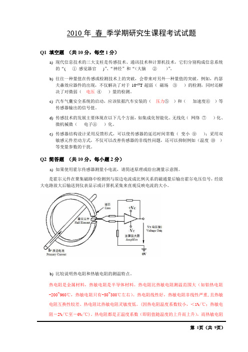
Q2 简答题(共10分,每小题2分)a)如果使用霍尔传感器测量小电流,请简述原理或给出测量示意图。
是霍尔元件在聚集磁路中检测到与原边电流成比例关系的磁通量后输出霍尔电压信号,经放大电路放大后输送到仪表显示或计算机采集来直观反映电流的大小。
b)比较说明热电阻和热敏电阻的测温特点。
热电阻是金属材料,热敏电阻是半导体材料。
热电阻比热敏电阻测温范围大(如铂热电阻-200~960℃,热敏电阻只有-50~300℃左右)。
热电阻线性好,热敏电阻非线性严重,且热敏电阻互换性较差。
热电阻比热敏电阻灵敏度低。
(因热电阻温度系数较小,<1%/℃;热敏电阻-2%/℃至-6%/℃)。
热电阻都是正温度系数(即阻值随温度的上升而上升),而热敏电阻分为负温度系数和正温度系数两种。
c) 无线传感器网络的核心技术问题有哪些?答:关键技术:拓扑控制、网络协议、网络安全、时间同步、定位技术、数据融合、数据管理、无线通信技术、嵌入式系统、应用层技术。
核心问题:能源、传感器、封装、部署、资源受限下的网络机制、大规模下的网络机制d) 功能型光纤传感器可以测量哪些物理量?(举3例即可)答:陀螺、声、磁、压力、温度、液面、e) 在传感器静态特性数据分析中,插值和回归的目的分别是什么?答:插值的目的在于减少或增大信息量。
核心原料:生物源胶原蛋白 RHC
生物源胶原蛋白(关键词:生物源、胶原蛋白、胶原) (备选名称:RHC、酵母胶原蛋白、发酵型胶原蛋白)1. 生物源胶原蛋白1.1 生物源胶原蛋白定义胶原蛋白被称为“骨中之骨,肤中之肤”,是人体最重要的结构蛋白质,约占蛋白总量30%,广泛存在于人体的皮肤、骨骼、肌肉、软骨等组织中,起支撑、修复、保护等重要作用,在肌肤细胞的生长、更新过程中起着重要作用。
20岁时真皮层中胶原蛋白含量达70%以上,随着年龄增长,胶原蛋白不断流失,25岁进入流失高峰,40岁时已不足年轻时一半。
科学合理的补充胶原蛋白能巩固肌底支撑,提供肌肤营养,起到保湿锁水,滋润皮肤,延缓衰老,消除皱纹等功效。
生物源胶原蛋白,又称RHC(Recombinant Human-source Collagen),是根据人胶原蛋白的结构特性,利用先进生物科学技术和国际领先发酵技术,通过微生物(如活性酵母)高密度发酵,绿色分离纯化工艺生产的一种高分子的生物源蛋白,与人胶原蛋白高度一致。
1.2 生物源胶原蛋白特点1.2.1 同质性生物源胶原蛋白是根据人胶原蛋白的特点进行结构设计,因此与人胶原蛋白结构高度一致,生物相容性好,亲和性和渗透性好,无排异反应,不易致敏。
1.2.2 无病毒隐患从动物组织提取的胶原蛋白存在携带病毒的隐患,直接限制了其应用领域。
生物源胶原蛋白是利用酵母高密度发酵获得,从根源上避免了病毒隐患,所使用的原料来源可控,成分简单,具有良好溯源性,不存在动物源杂质,弥补了传统胶原蛋白的缺陷,成为更安全的新型高科技胶原蛋白。
1.2.3 纯度高,分子量确切纯度可达99.5%以上,不含其它杂蛋白。
分子量与人胶原蛋白相近,犹如人体自身物质。
1.2.4 水溶性好通过生物发酵技术获得的胶原蛋白为水溶性蛋白,与各种性质的功能原料都能良好的复配,可加工性强,在下游应用和产品开发上更具优势。
1.2.5 无排异反应生物源胶原蛋白是利用生物科学技术,根据人胶原蛋白结构特性进行设计,摒弃了容易引起免疫排异反应的氨基酸残基,不存在动物胶原蛋白序列,其应用于人体时不会发生排异反应。
静电纺丝聚氨酯纳米纤维的应用研究进展

生物组织工程是修复或替换受损人体器官以重 建其功能的一项重要医学技术。生物组织工程涉及 的领域主要分为生物支架、细胞和生长因子3个部 分⑴],其中生物支架为细胞提供所需要的基体,通 过构建组织工程支架来替代原有的受损皮肤,将会 降低大面积皮肤修复的成本。静电纺丝纳米纤维与 天然细胞外基质结构类似,可以应用于生物组织工 程支架的构建。聚氨酯软硬段之间的微相分离结 构,利于细胞的附着和生长,因此静电纺丝聚氨酯纳 米纤维生物支架广泛应用于血管、心脏和皮肤等生 物组织工程中。Jaganathan等,12-将肉豆蔻油和聚氨 酯混合,利用静电纺丝制备生物组织工程支架。结 果发现,肉豆蔻油可有效降低聚氨酯的润湿性 ,改善 表面光滑度;此纳米复合材料的抗凝血性实验表明, 其抗血栓形成性比不加肉豆蔻油的静电纺丝聚氨酯 纤维更强。Puperi等⑴-通过静电纺丝得到聚氨酯 和聚乙二醇水凝胶组成的复合支架,该支架的多层 结构可实现细胞的3D培养。通过静电纺丝聚氨酯 网眼层的设计,调整支架可模拟自然主动脉瓣的拉 伸性、各向异性和可延展性,为进一步了解纤维化瓣 膜疾病提供模型。
[5 - HU X, LIF S, ZHOU G, et al. Electrospinning oi polymeac nanofibero for dag delivea applications[ J]. Jouaial oi Controlled Re lease, 2014,185:12—21.
高熵陶瓷研究进展

高熵陶瓷研究进展摘要高熵陶瓷是一种新兴的等摩尔多组分陶瓷材料,集抗氧化、耐烧蚀、耐腐蚀、超高硬度优秀性能于一体。
在空天技术,精密制造等高端领域有着广阔的应用前景。
当前高熵陶瓷制备工艺尚不成熟,本文基于近年相关实验,详细阐述了高熵硼化物相关研究成果,对当前高熵体系的相关体系与其特征进行了归纳和总结。
关键词高熵陶瓷,体系计算,制备方法0.引言2004年叶均蔚教授[1]提出了高熵的概念,认为高熵材料内部出现迟滞动力,晶格畸变和非原组元性能。
表现出良好的结构稳定性和优异的力学性能,并且展现了全新的电性能和催化性能等性质。
高熵陶瓷作为一种新兴等摩尔的多组分陶瓷材料,是一种抗氧化,抗烧蚀,耐腐蚀和超高硬度于一体的优秀材料,具有极大的发展潜力。
1.高熵效应在高混乱度无序系统中的特殊效应被称为高熵效应[1]。
高熵效应有四类:1.热力学中的高熵效应:在高熵系统作用下可以促进元素间的相容性使得多组元复合材料在制备后形成单一相。
2.结构的晶格畸变效应:高熵体系中的各组元的原子在晶格点阵中的随机分布,组元之间的结构差距较大,晶体内部的具有比传统复合材料更大的晶格畸变和缺陷。
3.动力学迟滞扩散效应:高熵材料内部的扩散和相变速度相对于传统材料较慢,内部反应滞后。
4.性能上的鸡尾酒效应[9],不同组元的性能的不同以及组元之间的相互作用会使得高熵材料产生更加复杂的性质,产生多组元协增效应从而实现性能的飞跃。
2.高熵氧化物最早提出高熵陶瓷概念并制备的陶瓷是Rost CM[2]等四制备的五元氧化物陶瓷。
他们以MgO、CoO、NiO、CuO、ZnO为原料,球磨混合后烧结制备,并从相转变的可逆性,体系熵与组元的关系和元素的化学环境来分析高熵陶瓷中的高熵效应,在此之后,相关学者将其扩展到到不同的氧化物体系,制备所得的材料具有优异的性能。
单相(Mg0.2Co0.2Ni0.2Cu0.2Zn0.2)1-x-yGyAxO (其中A= Li, Na或K)具有极高的介电常数和超离子电导率;快速燃烧降解法制备的(Mg0.2Co0.2Ni0.2Cu0.2Zn0.2)O陶瓷粉体在奈耳温度以下表现出长程反铁磁行为,并且在室温下显示出顺磁行为。
电泳沉积法制备高能量密度的非对称平面微型超级电容器

【电子技术/Electronic Technology】DOI: 10.19289/j.1004-227x.2021.03.001 电泳沉积法制备高能量密度的非对称平面微型超级电容器刘红彬1, *,赵方方2(1.中移(苏州)软件技术有限公司,江苏苏州215000;2.力神电池(苏州)有限公司,江苏苏州215000)摘要:首先采用光刻、蒸镀金的方法制备叉指电极,随后把合成的具有赝电容特性的二维MnO2和Ti3C2纳米片分别电泳沉积到叉指电极上,构建了非对称平面超级电容器。
其中MnO2为正极,Ti3C2为负极,滴涂凝胶为电解质,并利用透明的聚二甲基硅氧烷薄膜封装成器件。
通过能量色散X射线光谱(EDS)、傅里叶变换红外光谱(FT-IR)、扫描电子显微镜(SEM)、光学显微镜等手段证明了电泳沉积后材料的结构没有发生变化以及叉指电极的成功制备,也说明了电泳沉积后材料的形貌为薄膜结构。
最后通过二电极系统测试了器件的电化学性能,结果显示该器件具有高倍率性能和高能量密度,同时保持着高功率密度和优异的机械柔韧性,其容量在各种弯曲角度下基本没有衰减。
关键词:二氧化锰;碳化钛;电泳沉积;赝电容;二维材料;叉指电极;非对称平面微型超级电容器中图分类号:TQ174 文献标志码:A 文章编号:1004 – 227X (2021) 03 – 0171 – 06 Preparation of high-energy-density asymmetric planar microsupercapacitors by electrophoretic depositionLIU Hongbin 1, *, ZHAO Fangfang 2( 1. China Mobile (SuZhou) Software Technology Co., Ltd., Suzhou 215000, China;2. Lishen Battery (Suzhou) Joint-Stock Co., Ltd., Suzhou 215000, China)Abstract:The interdigitated electrodes were first prepared by photolithography and gold evaporation. Subsequently, the synthesized two-dimensional MnO2 and Ti3C2 nanosheets with pseudocapacitive properties were electrodeposited onto the interdigitated electrodes to fabricate asymmetric planar supercapacitors with gel electrolyte encapsulated with a transparent polydimethylsiloxane film, where MnO2 was the positive electrode and Ti3C2 was the negative electrode. The results of energy-dispersive X-ray spectroscopy, Fourier-transform infrared spectroscopy (FT-IR), scanning electron microscopy (SEM), and optical microscopy proved that the microstructures of MnO2 and Ti3C2 after electrophoretic deposition were unchanged and the interdigitated electrodes were prepared successfully, and also showed that the electrophoretically deposited materials were of a thin film structure. The electrochemical performance of the fabricated device was tested by a two-electrode system, which showed that it not only had high rate capability and high energy density, but also maintained high power density and good mechanical flexibility. There was basically no attenuation in capability for the device when being bended at various angles.Keywords:manganese dioxide; titanium carbide; electrophoretic deposition; pseudocapacitance; two-dimensional material; interdigitated electrode; asymmetric planar microsupercapacitor随着现代可穿戴电子设备的持续快速发展,对特征尺寸在微米范围内器件的制备开始成为人们比较感兴趣的问题[1-3]。
氧化亚铜的制备与性能
纳米Cu2O作为光催化剂的制备与性能摘要:光催化技术是一项新型的技术,与其他传统的技术相比具有降解完全、高效、价廉、稳定等优点,因而具有良好的应用前景。
氧化亚铜是一种重要的无机化工原料,因其独特的性质而在诸多领域有着广泛的应用,研究纳米氧化亚铜的制备及光催化性能有着深远意义。
关键词::纳米氧化亚铜,光催化,㈠纳米氧化亚铜的制备方法氧化亚铜具有能够便于对反应温度的操作和控制。
优点是不使用溶剂、并且还具有高选择性、高产率、节省能源、合成工艺简单, 制备方法有烧结法刚、电化学法、水热法和多元醇法等。
1烧结法刚烧结法又称为干法,该方法是将固体铜粉与氧化铜粉末预先混合,再送入锻烧炉内加热到1073一1173K密闭反应得到CuZO,其反应式为:CuO+Cu分CuZO 由于这种方法用铜粉作还原剂,与固体氧化铜进行固相反应制得,固相反应存在反应不均匀、不彻底等固有缺点,因而制得的CuZO粉末中往往含有铜和氧化铜杂质,难于去除。
该法制备得到的氧化亚铜粉末不仅纯度较低,而且粉末粒度取决于原料Cu粉和CuO粉的粗细,高温反应后得到的氧化亚铜容易板结、难于分散、劳动强度大、能耗高。
2电化学法电化学法也称电解法,该法具有流程短、成本低、操作简单、产量高、工作环境良好和产品质量高的优点,因而具有很好的工业化前景和比较成熟的生产工艺。
Yan沙6]等用电化学法制备纳米氧化亚铜时,两极分别采用含铜99.9%的铜板和铜片,电解液采用NaCI、NaOH和KZCrO7组成的混合液,在YB17ll型电化学装置中进行,并且比较了在不同的电流密度下所制样品的光催化性能。
采用紫铜板作阳极,铜片作阴极,在含有NaOH的NaCI碱性水溶液中电解金属铜。
从电极反应机理来看,氧化亚铜粉末是通过阳极铜溶解,并发生水解沉淀反应而生成的。
同时研究了电解液组成及其浓度、温度以及电流密度等因素对氧化亚铜产品质量的影响,从而得到了电化学法制备氧化亚铜的优化工艺条件。
sci中的长难句

sci中的长难句在科学论文中,长难句常常出现,主要是为了表达复杂的概念和关系。
以下是一些常见的长难句例子:1. "The development and implementation of a robust and scalable machine learning algorithm, combined with advanced data analytics techniques, have significantly improved the accuracy and efficiency of predicting and analyzing complex biological systems, thereby enabling researchers to gain deeper insights into the underlying mechanisms driving disease progression."“强大且可扩展的机器学习算法的开发和实施,结合先进的数据分析技术,显著提高了预测和分析复杂生物系统的准确性和效率,从而使研究人员能够更深入地了解驱动疾病进展的潜在机制。
”2. "The integration of nanomaterials with traditional construction materials, such as concrete and steel, not only enhances their mechanical properties, but also provides additional functionalities, such as self-healing, self-cleaning, and energy harvesting capabilities, contributing to the development of sustainable and smart infrastructure."“将纳米材料与混凝土和钢材等传统建筑材料相结合,不仅增强了它们的机械性能,还提供了额外的功能,如自我修复、自清洁和能量收集能力,有助于可持续和智能基础设施的发展。
20_-_accelerometers

ACCELEROMETERSBULK MICROMACHININGTwo fabrication processes are dominating, both rely on micromachining standard silicon wafers.Bulk Micromachining uses the full thickness of a wafer and is a subtractive process.Silicon is removed by wet or dry-etching techniques and forms a proof mass and a suspension system.Often these sensors consist of a sandwich of several wafers(either silicon or Pyrex) bonded together to provide electrical contacts and form an enclosure for the proof mass.Bulk-micromachining was used in earlier devices and most commercial accelerometers are fabricated in such a way.This technology is not very suitable for monolithic integration and often is used in conjunction with a separate integrated circuit in the same package or even with discrete electronics.SURFACE MICROMACHININGA technology which is suitable for integrating the electronics and the mechanical structures on the same chip is Surface Micromachining.This is an additive process in which thin films of typically poly-silicon and silicon-oxide are grown on a wafer. The oxide acts as a sacrificial layer and is removed by a release step by a wet-etchant such as HF. This results in free-standing beams and plates.The sensing elements are typically an order of magnitude smaller than bulk-micromachined devices.This technology is compatible with a standard CMOS process and has led to monolithically integrated devices.A commercial range of sensors is available from Analog Devices, the first device was the ADXL05 which has a ±5 g dynamic range.Another characteristics is the type of transduction from the mechanical to the electrical domain by means of measuring the deflection of the proof mass.BASICS –TRANSDUCTION TECHNIQUESPIEZORESISTIVE ACCELEROMETERSEarly research focused on usingpiezoresistors and anisotropicetching technologies developed forsilicon diaphragm pressure sensors.The first silicon micromachinedaccelerometer used bulkmicromachining with piezoresistorsand pyrex anodic bonding to senseacceleration.Roylance L.M., Angell J.B. “A batch fabricated silicon accelerometer”IEEE Trans. on Electron Devices 26, 1911-1917 (1979)The silicon mass was suspended by a single cantilever.Other bulk micromachined sensors contain a seismic mass suspended by one, two or four silicon beams.The beams carry piezoresistors, which in order to achieve a maximum output signal, are placed at the edges of the beams, where the maximum bending strain occurs.HITACHI PIEZORESISTIVE-TYPE TRIAXIALACCELERATION SENSOR•Ability to simultaneously detect acceleration in three axial directions (X, Y and Z)with a single chip•It is the world’s smallest,thinnest semiconductor type-3axial accelerating sensorIt is mainly due to these drawbacks that more modern devices use capacitive detection. Here, often differential capacitors are formed by using the proof mass as the common middle contact of a capacitive half-bridge. For small deflections the differential change in capacitance is proportional to the deflection of the proof mass and can be converted into a voltage by a charge amplifier and a synchronous demodulation technique.CAPACITIVE ACCELEROMETERS Capacitive Sensing is…Piezoresistive detection was used in the first sensors and still is used in many commercial devices (e.g. by SensoNor).The drawback is that the output level is not very high (a typical value is 100 mV for a 10V drive), the temperature coefficient is relatively large and intrinsically thermal noise is generated in the resistors.•Hitachi representatives have described thedevelopment of a closed-loop siliconaccelerometer (Tsuchitani et al. 1991)intended for automotive systems.•This device requires sophisticated double-sided etching to achieve the desiredsymmetrical structure that eliminates cross-axis sensitivity.•The 3.2 x 5 mm silicon chip is bonded between glass plates to form a differentialcapacitor, and mounted with hybrid electronics.HITACHI CAPACITIVE ACCELEROMETERCSEM (Centre Suisse d’Electronique et de Microtechnique) CAPACITIVE ACCELEROMETERCSEM (Centre Suisse d’Electronique et de Microtechnique) CAPACITIVE ACCELEROMETERSEM photograph of a vertical accelerometer;planar view (top) and a close up of the membranestructure (bottom) (MOTOROLA).MOTOROLA VERTICAL ACCELEROMETERMore than 10million units have been shipped !!!Motorola builds a two-dice accelerometer. The accelerometer sensing element, or g cell,is surface-micromachined in an adaptedsemiconductor process flow.The Motorola g cell is a z-axis device,meaning that the plane of accelerationsensitivity is perpendicular to the plane of thechip.Sensitivity is limited by the stiffness of the mechanical suspensionDynamic range is limited by:•nonlinearity of the suspension •nonlinearity of the transduction (piezoresistors or capacitors)•a stiffer (less compliant) suspension improves the dynamic range by limiting the displacement A trade-off is necessary between sensitivity and dynamic range foropen-loop accelerometersIn force-balance accelerometers the strain or displacement is sensed, amplified and fed back to counteract the displacement of the mass due to acceleration.During operation, the structure is virtually stationary and the linearity and effectiveThe suspension can be made very compliant without sacrificing dynamic range and the accelerometer can be made ultrasensitive.Accelerometers designed with a feedback actuator that can provide a restoring force to the proof mass can operate as a closed-loop sensor with a proof mass that moves negligibly in response to inertial forces.The electrical signal applied to the actuator is the sensor output.In capacitive sensing ⇒apply a signal to capacitor plates ⇒generate anelectrostatic restoring forceFor capacitive accelerometers,the same structure can often beused in both open-and closed-loop modes, with the latterrequiring additional signalprocessing circuitry to close theloop.FINAL REMARKSANALOG DEVICES ADXL-50ANALOG DEVICES ADXL-50 The ADXL50is a complete acceleration measurement system on asingle monolithic IC.It contains a polysilicon surface micromachined sensor(patterned on asacrificial oxide etched away with HF)and signal conditioning circuitry.The ADXL50 is capable of measuring both positive and negative ac-celeration to a maximum level of ±50 g.The structure of the sensor consists of 42unit cells and a commonbeam.The differential capacitor sensor consists of independent fixed platesand a movable“floating”central plate which deflects in response tochanges in relative motion.The two capacitors are series connected,forming a capacitive dividerwith a common movable central plate. A force balance techniquecounters any impeding deflection due to acceleration and servos thesensor back to its0 g position.RESONATING ACCELEROMETERSAn alternative approach to achieve high sensitivity andlarge dynamic range is to use a resonant microstructureas a strain sensing element.The frequency of a resonant structure can be madehighly sensitive to compressive or tensile strain and theresonance frequency can be measured with highprecision.RESONATING ACCELEROMETERSE062.swfPIEZOELECTRIC ACCELEROMETERS Piezoelectric accelerometers can be fabricated through surface micromachined using standard VLSI processing techniques.Pisano and DeVoe described the design, fabrication, and characterization of surface micromachined piezoelectric accelerometers based on ZnO filmsREMARK !!! Under constant strain, the piezoelectric generated charges slowly leak away, and the electric field disappears. Therefore, piezoelectric accelerometers cannot be used for low-frequency applications !!!•A second layer of 2.0 µm thick p-doped polysilicon is depositedon top of the PSG, and patterned by plasma etching to define themechanical accelerometer structure. This layer also acts as thelower electrode for the sensing film.•A thin layer of stoichiometric silicon nitride is next deposited byLPCVD. This film acts as a stress-compensation layer forbalancing the highly compressive residual stresses in the ZnO film.By varying the thickness of the Si 3N 4layer, the accelerometerstructure may be tuned to control bending effects resulting from thestress gradient through the device thickness.•A ZnO layer on the order of 0.5 µm is deposited by RF-magnetron sputtering.•Finally, 0.2 µm of sputtered Pt is deposited to form the upper electrode. •The Pt, Si3N4 and ZnO layers are patterned in a single ion milling etch step.•Finally, the devices are released by passivating the ZnO film with photoresist, immersing the wafer in 5:1 buffered HF to remove the sacrificial PSG layer, and removing the photoresist by O2 plasma ashing.Automotive ApplicationsMobile Phones ApplicationsORIENTATION DETECTIONTAP DETECTIONSHAKE DETECTIONEntertainment ApplicationsOne of the first applications of the 3-D sensor (accelerometer/gyroscope) was in laptops, where sensors guard against damage from a fall. In the split second of free fall that comes before the collision with the floor, the sensor tells a controller to park the read/write head safely away from the hard drive. Nintendo’s Wii is the currently hottest computer game. Its two wireless remote controls track any movement, encouraging players to engage opponents with physicality. Yet although detecting motion is critical to the success of the US $250 game, the job depends on $3 sensors the size of shirt buttons. The supplier of the sensors, STMicroelectronics, got into the business a decade ago in order to squeeze a few more dollars out of an obsolescent chip-making plant. ”We wanted something good for it that didn’t require deep submicron technology,” says Benedetto Vigna, the Italian physicist who developed the sensor.Entertainment Applications/parrot-ar-drone/usa/Textile ApplicationsE-TextilesTwo major motion pictures (Iron Man 2 and Alice in Wonderland) and two popular video game releases (Kill Zone 2 and Borderlands) recently used a MEMS motion suit to enable live actors to control virtual characters in real time. Actors in MEMS-sensor-studded suits performed the action scenes, but computer generated imagery (CGI) was what you saw. The common element here is a Lycra suit made by Xsens Technologies B.V. and embedded with 85 accelerometers and gyroscopes from Analog Devices Inc. (ADI)ADI assisted Xsens on the project and claims the MEMS motion suit is an example of a third phase in MEMS -after automobile airbags, and consumer devices like the Apple iPhone and Nintendo Wii.。
4H-SiC结型势垒肖特基二极管的制作与特性研究
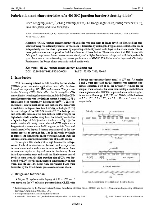
Vol.32,No.6Journal of SemiconductorsJune 2011Fabrication and characteristics of a 4H-SiC junction barrier Schottky diodeChen Fengping(陈丰平) ,Zhang Yuming(张玉明),L ¨uHongliang(吕红亮),Zhang Yimen(张义门),Guo Hui(郭辉),and Guo Xin(郭鑫)School of Microelectronics,Key Laboratory of Wide Band-Gap Semiconductor Materials and Devices,Xidian University,Xi’an 710071,ChinaAbstract:4H-SiC junction barrier Schottky (JBS)diodes with four kinds of design have been fabricated and char-acterized using two different processes in which one is fabricated by making the P-type ohmic contact of the anode independently,and the other is processed by depositing a Schottky metal multi-layer on the whole anode.The re-verse performances are compared to find the influences of these factors.The results show that JBS diodes with field guard rings have a lower reverse current density and a higher breakdown voltage,and with independent P-type ohmic contact manufacturing,the reverse performance of 4H-SiC JBS diodes can be improved effectively.Furthermore,the P-type ohmic contact is studied in this work.Key words:4H-SiC;junction barrier Schottky;field guard ring DOI:10.1088/1674-4926/32/6/064003PACC:7155D;7330;7340N1.IntroductionWith increasing interest in SiC Schottky barrier diodes(SBD)in power conversion applications,much effort has been focused on improving SiC SBD performance.The junction barrier Schottky (JBS)diode offers the Schottky-like ON-state with fast switching characteristics,and the PiN-like OFF-state characteristics with low leakage current.Several SiC JBS diodes have been reported by different groups Œ1 4 .The con-duction loss can be much lower than that of a PiN diode with a breakdown voltage of less than 3kV due to the high (2.7V)turn-on voltage of the SiC P–N junction Œ5 .The leakage cur-rent of the JBS diode is lower than that of a SBD,owing to the high electric field shielded away from the Schottky contact by a depletion layer of P–N junctions.As shown in Fig.1(a),the anode contains a Schottky contact above the SBD regions and a P-type ohmic contact above the P C regions,so it is fabricated simultaneously by deposit Schottky contact metal in the cus-tomary process,as shown in Fig.1(b).In this work,two kinds of processes to fabricate the anode were employed to study the influence on the electrical characteristics of the JBS diodes.To design a JBS diode with a high breakdown voltage,several kinds of termination can be used,such as a junction termination extension and a mesa termination.However,these terminations require etching and extra ion implanting.To re-duce the processing steps and avoid the disadvantages caused by these extra steps,the filed guarding ring (FGR)was fab-ricated with P C for the main junction simultaneously in this work.The 4H-SiC JBS diodes with and without FGRs were fabricated by the two different processes mentioned above.2.Design and fabricationA 10 m N epilayer with doping of 1.56 1015cm 3was grown on the N C substrate purchased from CREE,witha doping concentration of more than 1 1018cm 3.Samples 1and 2were processed on the substrate with different tech-niques separately.First of all,the circular P C regions for the samples were formed at the same time.Multiple implantations were implemented at 400ıC in argon ambience.Al ion implan-tation was with energy of 30,280,and 500eV ,while the doses of 8.6 1013,5.2 1014,and 7.8 1014cm 2were taken,respectively.Fig.1.Schematic cross section of the JBS diodes.*Project supported by the National Natural Science Foundation of China (No.61006060)and the 13115Innovation Engineering of Shannxi Province,China (No.2008ZDKG-30).Corresponding author.Email:fpchen@Received 1November 2010,revised manuscript received 22February 2011c2011Chinese Institute of ElectronicsParameters for all structures are shown in Fig.1(a),and all of the parameters’values were chosen to be the same for both samples.The P C junctions are characterized by the width of the P C implantation window (W /and the spacing in between (S/.FGRs are characterized by the width of a single ring (L/and the spacing between the two nearest rings (D/.According to Ref.[6],L was chosen to be the fixed value 5 m,and D D 2.5 m in the experiment.For convenient description in this paper,the JBS will be marked as JBS (S ,W /.In this work,the four structures with different designs are (a)JBS (2.5,4)with-out edge termination;(b)JBS (2.5,4)terminated by FGRs;(c)JBS (3,4)without edge termination;and (d)JBS (3,4)ter-minated by FGRs.FGRs with L D 5 m for each ring were implanted around the periphery of the forward conducting ac-tive area to reduce electric field crowding at the edge of the diode under reverse bias.All of the FGRs were formed simul-taneously with the P C junction regions with ion-implantation,thus the depth and concentration are the same as the P C junc-tion regions.The samples were annealed at 1650ıC for 45min in argon ambience.Then,the profile in 0.6 m depth was mea-sured.It is well known that a high temperature annealing (>1000ıC)is usually required for ohmic contact activation.The fact that the melting temperature of Al (660ıC)is much lower gives rise to contact morphological problems.It was reported that Al melted during the high temperature anneal Œ7 ,spilling over the surface of the devices,potentially damaged the periphery of the devices.To improve the contact morphology,according to Ref.[8],a multi-layer of Ti/Al/Ti/Al/Ti/Al/Ag was deposited on sample 1’s top of P C junction regions after P C regions done,and then an annealing at 1000ıC in a gas mixture of 97%N 2and 3%H 2for 2min was carried out to create a P-type ohmic contact.Both samples underwent tri-layer metallization of Ti/Ni/Ag to form a backside contact.Sample 1was annealed for 2min and sample 2was annealed for 5min at 1000ıC in a gas mixture of 97%N 2and 3%H 2.Finally,bi-layer metallization of Ti/Ag was used to form the front Schottky metal contact for both samples.Figure 2shows the scanning electron microscope (SEM)photographs of both samples.It was previously thought that sample 2had a much better periphery than sample 1since sample 2was not annealed to make P-type ohmic contact.As can be seen in Fig.2(a),the Al spilled a little over the edge of the anode area for sample 1.This can be improved if a thinner Al layer is chosen and the Ti/Al multi-layer superposition time is increased Œ8 .3.Results and discussionThe fabricated devices were electrically measured at room temperature using a Tektronix 370B programmable curve tracer and an Agilent B1500A semiconductor device analyzer.Figure 3shows a comparison of the reverse current den-sity versus reverse voltage between JBS (2.5,4)and JBS (3,4)from each sample.As we know,when the JBS is reverse biased,the depletion layers of the adjacent P–N junctions will spread wider,leading to a reduction in the width of the Schottky chan-nel.After the depletion layer is pinched off,a potential barrier for SBD is formed,then the depletion layer is extended toward the N C substrate with further increasing reversed voltage Œ9 .Fig.2.SEM photos of both samples.(a)Sample 1.(b)Sample 2.Fig.3.Reverse V –J characteristics of JBS diodes with different val-ues of S for samples 1and 2.However,as can be seen from this figure,the current densities from sample 1have much lower reverse current densities than those of sample 2.As mentioned above,the P-type contact for sample 2is not independently fabricated,which causes the bar-rier on the P C regions to be much higher then excepted,and the depletion layers between the two nearest P C regions will not pinch off effectively.Figure 3shows a comparison between two JBS diodes with different S .Apparently,the JBS diode with S D 2.5 m has a lower reverse current density than the one with S D 3 m.It can be seen from Fig.4that the reverse current den-Fig.4.Reverse V –J characteristics of JBS diodes with and without edge termination for samples 1and 2.sity of JBS diodes with FGRs are much lower than those with-out FGRs,since FGRs can effectively reduce the field crowd-ing in the edge of devices,it has a higher breakdown voltage.In this work,JBS diodes with a breakdown voltage of up to 400V when the current density is lower than 1A/cm 2is cre-ated with FGRs Œ10 .However,the FGR structure with different ring spacing and ring widths is difficult to optimize,and in-terface charges influence the breakdown voltage significantly,since we didn’t apply any passivation layer on the surface of the devices,so the reverse current in this work is a little higher correspondingly.The dominant mechanism of reverse current depends on the Schottky barrier height,temperature,applied voltage,sur-face status,and defects in the material.According to the tech-niques we employed during manufacture,the main reasons for a relatively high leakage current and low breakdown voltage are as follows:(1)Ti is used as the metal to form the Schottky ing the thermionic emission theory,the current through the SiC Schottky diode can be expressed byI D AAT 2exp  B kT ÃÄexp qVnkT1;(1)where A is the diode area,A is the Richardson’s constant, B is the Schottky barrier height,n is the ideality factor,and other constants have their usual meanings.The forward V –J characteristics of SBD and JBS diodes are shown in Fig.5.Calculated with Eq.(1),the barrier height formed in this work is 0.79eV ,which is smaller than that fab-ricated with Ni ( B D 1.26eV)Œ11 .(2)Multi-step ion implantation brought in damages in the space lattice.(3)The epilayer material deterioration in high temperature annealing,which was stated previously.To reduce the damage during a high-temperature activation anneal,AlN can be used to prevent silicon evaporation from the 4H-SiC surface Œ12 .4.SummaryThe process that fabricates a P-type ohmic contact inde-pendently has been employed to fabricate 4H-SiC JBS diodes.Fig.5.Forward V –J characteristics of SBD and JBS diodes.The influence of FGRs in the reverse characteristics of JBSdiodes has been studied.Results show that the JBS diodes with P-type ohmic contact fabricated independently have a better performance than those by the customary process and with P+regions doping concentration ( 1 1018cm 3)and window spacing (2.5 m),4H-SiC JBS diodes using FGRs termination have a reverse current density lower than 1 10 3A/cm 2be-low 100V .References[1]Jun W,Yu D,Bhattacharya S,et al.Characterization,modelingof 10-kV SiC JBS diodes and their application prospect in X-ray generators.IEEE ECCE,2009,20:1488[2]Yan G,Huang A Q,Agarwal A K,et al.Integration of 1200VSiC BJT with SiC diode.20th ISPSD,2008,18:233[3]Hull B A,Sumakeris J J,O’Loughlin M J,et al.Performance andstability of large-area 4H-SiC 10-kV junction barrier Schottky rectifiers.IEEE Trans Electron Devices,2008,55(8):1864[4]Feng Z,Mohammad M I,Biplob K D,et al.Effect of crystallo-graphic dislocations on the reverse performance of 4H-SiC p–n diodes.Mater Lett,2010,64(3):281[5]Lin Z,Chow T P,Jones K A,et al.Design,fabrication,and char-acterization of low forward drop,low leakage,1-kV 4H-SiC JBS rectifiers.IEEE Trans Electron Devices,2006,53(2):363[6]Cao L H.4H-SiC gate turn-off thyristor and merged P–i–N andSchottky barrier diode.PhD Thesis,New Jersey,Rutgers Univer-sity New Brunswick,2000[7]Luo Y ,Yan F,Tone K,et al.Searching for device processingcompatible ohmic contacts to implanted P-type 4H-SiC.Proceed-ings of the International Conference on Silicon Carbide and Re-lated Materials,1999,338:1013[8]Jennings M R,P ´e rez-Tom ´a s A,Davies M,et al.Analysis of Al/Ti,Al/Ni multiple and triple layer contacts to P-type 4H-SiC.Solid-State Electron,2007,51(5):797[9]Zhang Y M,Zhang Y M,Alexandrov P,et al.Fabrication of 4H-SiC merged PN-Schottky diodes.Chinese Journal of Semicon-ductors,2001,22(3):265[10]Chen F P,Zhang Y M,Zhang Y M,et al.Study of 4H-SiC junc-tion barrier Schottky diode using field guard ring termination.Chin Phys B,2010,19(9):097107[11]Zhang L,Zhang Y M,Zhang Y M,et al.Gamma-ray radiationeffect on Ni/4H-SiC SBD.Acta Phys Sin,2009,58(4):2737[12]Jones K A,Shah P B,Kirchner K W,et al.Annealing ion im-planted SiC with an AlN cap.Mater Sci Eng,1999,61:281。
智能材料结构中力与多物理场耦合理论及结构损伤断裂理论
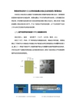
智能材料结构中力与多物理场耦合理论及结构损伤/断裂理论本研究方向的成员主要基于多物理场理论和超声波相关理论技术,利用新型的微纳米级的铁电功能材料,创新地提出了针对各类材料与结构(尤其是航空材料结构)中的毫米级的损伤进行的检测和监测相关理论与技术。
团队形成了较强的凝聚力和良好的学术风气,产生了较高水平的科研成果,以下为研究骨干在近几年以来已完成的代表性阶段成果:(1)超声相控阵损伤检测中PFC换能器的研究项目负责人:骆英研究骨干:王自平、赵国旗、韩伟、虞波研究了PZT、PMN-PT等系统压电陶瓷的组成、制备技术和性能,创新地提出了用有序生长制备技术制造压电纤维复合材料及智能驱动/传感器件的新方法。
建立了一种基于新型PFC相控阵超声驱动/传感器件的超声相控阵检测系统,并成功应用于金属结构和混凝土结构的损伤检测,研制了接近国际水平的相控阵超声检测系统的原型机。
PFC片状驱动/传感器用于金属结构检测的PFC超声相控阵换能器用于混凝土结构检测的超声相控阵换能器d 、a 、N 变化时的指向性分析阵元参数变化时的波场分析基于PFC 超声相控阵驱动/传感器件的相控阵检测结果 (2)基于声发射(AE)技术的结构损伤检测方法研究骨干:骆英、顾爱军、Adudrum Marfo 、刘红光、欧晓林AE 技术作为一项独特的无损检测方法在各工程领域发挥着巨大的作用,在土木工程领域也显示出巨大的潜力。
研究了钢筋与混凝土间粘结滑移的声发射特性,并开始应用于预应力混凝土结构、钢结构、玻璃幕墙等结构的无损检测中,研究将数字图像相干法(DIC)与AE监测技术相结合,进一步验证损伤监测的准确性,为工程结构声发射检测中利用AE特性进行损伤识别奠定了基础。
CFRP碳纤维加固混凝土开孔板损伤监测桥梁声发射检测声发射与DIC检测方法比较试验装置基于Gabor小波变换理论的声发射无损检测及信号处理技术a) b) c) d)利用DIC方法测得的水平应变场判断混凝土裂缝的形成和扩展a)、b)、c),应变场的演化,d)宏观裂缝(3)基于新型应变梯度传感器的结构损伤监测技术项目负责人:骆英研究骨干:徐晨光、李康、桑胜、王晶晶、李兴家在近10年跟踪前沿研究新型铁电功能材料及智能器件的基础上,揭示微米级挠曲电材料的力/电能量转换关系,基于微米级挠曲电结构对微损伤尖端附件的应变梯度极其敏感的特性,研制用于监测结构损伤的新型应变梯度传感器,实现在线监测损伤导致的应变梯度,进而达到超前监测结构中应力集中区域损伤的萌生。
二氧化硅的制备和表征含微胶囊丁基硬脂酸通过溶胶 - 凝胶法

Preparation and characterization of silica microcapsules containingbutyl-stearate via sol-gel methodMIAO Chun-yan(缪春燕)1, 2, YAO You-wei(姚有为)1, TANG Guo-yi(唐国翌)1,WENG Duan(翁端)21. Graduated School at Shenzhen, Tsinghua University, Shenzhen 518055, China;2. Department of Materials Science and Engineering, Tsinghua University, Beijing 100086, ChinaReceived 15 July 2007; accepted 10 September 2007Abstract: For thermal energy storage application in energy-saving building materials, silica microcapsules containing phase change material were prepared using sol-gel method in O/W emulsion system. In the system droplets in microns are formed by emulsifying an organic phase consisting of butyl-stearate as core material. The silica shell was formed via hydrolysis and condensation from tetraethyl silicate with acetate as catalyst. The SEM photographs show the particles possess spherical morphology and core-shell structure. The as-prepared silica microcapsules mainly consist of microsphere in the diameter of 3−7 µm and the median diameter of these microcapsules equals to 5.2 µm. The differential scanning calorimetry (DSC) curves indicate that the latent heat and the melting point of microcapsules are 86 J/g and 22.6 ℃, respectively. The results of DSC and TG further testify the microcapsules with core-shell structure.Key words: silica microcapsules; sol-gel; butyl-stearate; phase change materials1 IntroductionIncreasing energy cost and associated environmental problems have intensified efforts towards the energy storage and sustainable energy technologies. Over the past decade, the integration of phase change materials (PCMs) into building fabrics have been investigated as a potential technology for minimizing energy consumptions in buildings because PCMs allow large amounts of heat to be stored during their melting process and to be released during their solidifying process[1−6]. Butyl-stearate (BS) as a kind of PCMs with moderate energy-storing density, proper melting point and economic price has been studied in buildings.A laboratory scale energy-storing gypsum wallboard was produced by the direct incorporation of 21%−22%(mass fraction) commercial grades BS at the mixing stage of conventional gypsum board production. Compared with gypsum wallboard alone, the energy-storing capability of this PCM wallboard has a tenfold increase in capacity for the storage and release of heat[7]. ZHANG et al[8−10] produced the PCMs clay which contained BS as PCM and expanded perlite as matrix via penetrating method. In recent years, microencapsulation of PCM has been studied and applied in thermal energy fields due to its advantages, such as protection of the core materials, increasing the heat transfer area, and permitting the core material to withstand frequent changes in volume when the PCMs change their state from solid to liquid or vice versa. Many microencapsulation methods have been developed for paraffin[11−13], such as interfacial polymerization, polymerization in situ, and sol-gel, but micro- encapsulation of BS has not been reported. Microcapsules with silica as shell materials possess hydrophilic surface and anti-oxidization property compared with organic polymer microcapsules, and silica microcapsules with paraffin as PCM have been prepared via sol-gel method from O/W emulsion[14−15].In this study, spherical microcapsules with silica as shell materials and BS as core materials are successfully prepared from an O/W emulsion in the presence of polyvinyl alcohol (PV A) as stabilizer, and sorbitan monooleate (Span80) and polyoxyethylene(20) sorbitan monooleate (Tween80) as emulsifiers.Foundation item: Project(50572045) supported by the National Natural Science Foundation of China; project supported by Innovation Fund from the PetroChina Company LimitedCorresponding author: TANG Guo-yi; Tel: +86-755-26036752; E-mail: Tanggy@MIAO Chun-yan, et al/Trans. Nonferrous Met. Soc. China 17(2007) s10192 ExperimentalThe particle size and surface morphology of silica microcapsules were examined using a scanning electron microscope (S-4700). The particle size distribution was measured adapting particle size analyzer (Rise-2008). Thermogravimetry (TG) analysis was carried out on TA-2. Differential scanning calorimetry (DSC) curves were evaluated using DSC (Mettler Toledo, DSC823e) between the scales of 0−50 ℃ at a heating or cooling rate of 5 ℃/min and under nitrogen atmosphere.A typical microencapsulation procedure was carried out as follows: 1) 1.0 g of PV A was dissolved in 49.0 mL of distilled water; 2) an organic solution of 8 mL of BS and 1.5 g of mixed emulsifiers (45.0% Span 80 and 55.0% Tween 80) was prepared, then the organic solution was heated to 80 ℃; 3) maintaining the temperature of reaction system between 85 ℃ and 90 ℃, 10 g of PV A aqueous solution was added to the organic solution, and the mixture was emulsified mechanically at stirring rate of 300 r/min for 10 min, then the remains of PV A aqueous was added to the mixture and the mixture was emulsified at 600 r/min to form an O/W emulsion; 4) while stirring, 1.0 g of sodium chloride solution (2.5 mol/L) was added into the emulsion; 5) after stirring for 30 min, 8 mL of tetraethyl silicate (TEOS) and 0.2 g of acetate acid solution (10.0%) were slowly added into the emulsion system to start the hydrolysis and condensation of TEOS; 6) after the addition, the reaction mixture was cooled to 55.0 ℃for 3 h. The resultant microcapsules were centrifuged, washed with distilled water and dried at 55.0 ℃in oven for 24 h.3 Results and discussion3.1 Morphology of microcapsulesThe SEM photographs of silica microcapsules are shown in Fig.1. From Fig.1 (a), it is clear that the as-prepared silica microcapsules mainly consist of microsphere in diameter of 3−7 µm. The SEM photograph of the fractured microcapsules in Fig.1(b) illustrates the core/shell structure of microcapsules. According to the particle size distribution of silica microcapsules containing BS (shown in Fig.2), the median diameter of silica microcapsules is 5.2 µm and the diameter of 95% silica microcapsules is not more than 13 µm.3.2 TG analysisThe thermal gravimetry (TG) curves of BS and microcapsules are shown in Fig.3. The temperature of initial mass loss (5%) of BS is 170 ℃ and the mass loss ends at 235 ℃, while the initial mass loss of micro-Fig.1 SEM micrographs of silica microcapsules containing BSFig.2 Particle size distribution of silica microcapsules containing BSFig.3 TG curves of BS and microcapsules containing BSMIAO Chun-yan, et al/Trans. Nonferrous Met. Soc. China 17(2007) s1020capsules is different from that of the BS. In the temperature range of 100−170 ℃, the obvious mass loss (about 9%) of microcapsules appears, which is due to the water absorbed by silica gel, and the maximum mass loss of microcapsules is 78% at 250 ℃. This result, together with the SEM observation, verifies the formation of microcapsules with core-shell structure and the average content of core materials is about 69%(mass fraction).3.3 DSC curves of microcapsulesFig.4 shows the typical melting and solidifying curves of DSC for BS alone and silica microcapsules containing BS. As shown in Fig.4, BS and silica microcapsules exhibit similar thermal properties; the ∆H f of BS and silica microcapsules are determined to be 132 J/g and 86 J/g, and the melting points of BS and microcapsules are 22.6 and 23.0 ℃, respectively.Fig.4 DSC curves of BS and microcapsules containing BS The average content of BS in a silica microcapsule can be estimated by dividing the ∆H f of microcapsules by the ∆H f of BS alone, assuming that the energy of core materials does not change before and after microencapsulation. Accordingly, the average content of BS in microcapsules is 65%, which is basically in accordance with TG analysis.It remains to identify the micro-shell formation mechanism. In this study, nonionic surfactant is enriched at the oil-water interface and contributes to the stabilization of this emulsion; sodium chloride is added into the emulsion and the Na+ ions interact with the oxygen atom of the ethylene oxide group of the nonionic surfactant to form the complex Span80-Na+ and Tween80-Na+[16], which increases the volume of terminal hydrophilic group of the surfactants, brings the hydrophilic group of surfactants a spot of positive charge and contributes to the oil in water emulsion more stable. With acetic acid as a catalyst, on the one hand, microcapsules possess the lowest porosity among catalyst (HCl, H2SO4, HNO3, HF, NH3, HAc )[17], on the other hand, in acid condition, atomic group such as —OH, —OSi≡ can instabilize the positive charge around Si nucleus or increase the steric hindrance, which reduces the hydrolysis rate[18]. Under these conditions, the hydrolysis rate is smaller than the condensation rate. The oligomer of TEOS exists in the system primarily with the form of Si(OR)2(OH)2 or Si(OR)3OH that are both lipophilic and hydrophilic, and they are apt to gather around the oil droplets in emulsion. Thus, silica micro-shell formation is expected.4 Conclusions1) Silica microcapsules encapsulating BS as PCM are prepared via a combination of O/W emulsion technique with a sol-gel method.2) Micron size (3−7 µm) spherical silica capsules containing BS can be prepared from weak acidic solution by using nonionic surfactant as the emulsifiers, PV A as stabilizer and TEOS as silica resource.3) The microcapsule has a relatively higher energy-storing density of 86 J/g and proper melting point of 22.6 ℃.References[1] KHUDHAIR A M, FARID M M. A review on energy conservation inbuilding applications with thermal storage by latent heat using phasechange materials[J]. Energy Conversion and Management, 2004, 45:263−275.[2] DARKWA K. Evaluation of regenerative phase change drywalls:low-energy buildings application[J]. Int J Energy Res, 1999, 23:1205−1212.[3] AHMET K. Energy storage applications in greenhouse by means ofphase change materials (PCMs): a review[J]. Renewable Energy,1998, 13(1): 89−103.[4] HALAWA E, BRUNO F, SAMAN W. Numerical analysis of a PCMthermal storage system with varying wall temperature[J]. EnergyConversion and Management, 2005, 46(15/16): 2592−2604.[5] ZALBA B, MARIN J M, CABEZA L F, MEHLING H. Review onthe thermal energy storage with phase change: materials, heattransfer analysis and applications[J]. Appl Therm Eng, 2003, 23(3):251−283.[6] SCHOSSIG P, HENNING, GSCHWANDER S, HAUSSMANN T.Micro-encapsulated phase-change materials integrated into construction materials[J]. Solar Energy Material and Solar Cells,2005, 89: 297−306.[7] FELDMAN D, BANU D. Obtaining an energy storing buildingmaterial by direct incorporation of an organic phase change materialin gypsum wallboard[J]. Solar Energy Mater, 1991, 22: 231−242. [8] ZHANG Dong, ZHOU Jian-min, WU Ke-ru, LI Zong-jin. Granulatedphase change composite for energy storage[J]. Acta MaterialComposite Sinica, 2004, 21(5): 103−107. (in Chinese).[9] ZHANG Dong, ZHOU Jian-min, WU Ke-ru, LI Zong-jin. Study onfabrication method and energy-storing behavior of phase-changingenergy-storing concrete[J].Journal of Building Materials, 2003, 6(4):374−377.(in Chinese).[10] ZHOU Jian-min, ZHANG Dong, WU Ke-ru. Experiment study andanalysis on obtaining and energy storing composite material by directincorporating organic phase change materials into porous granule[J].Energy Conservation Technology, 2003, 21(6): 5−7.MIAO Chun-yan, et al/Trans. Nonferrous Met. Soc. China 17(2007) s1021[11] ZOU G L, LAN X Z, TAN Z C, SUN L X. Microencapsulation ofn-hexadecane as phase change material in polyurea[J]. ActaPhys-Chim Sin, 2004, 20: 90−93.[12] HAWLADER M N A, UDDIN M S, KHIN M M.Microencaopsulated PCM thermal-energy storage system[J]. AppliedEnergy, 2003, 74: 195−202.[13] ZHANG X X, FAN Y F, TAO X M, YICK K L. Fabrication andproperties of microcapsules and nanocapsules containing n-octadecane[J]. Materials Chemistry and Physics, 2004, 88:300−307.[14] WANG L Y, TSAI P S, YANG Y M. Preparation of silicamicrospheres encapsulating phase-change material by sol-gel methodin O/W emulsion[J]. Journal of Microencapsulation, 2006, 23(1):3−14. [15] MIAO C Y, LU G, YAO Y W, TANG G Y, WENG D. Preparation ofsilica microcapsules containing octadecane as temperature adjustingpowder[J]. Chemistry Letters, 2007, 36(4): 494−495.[16] MATSUBARA H, OHTA A, KAMEDA M, VILLENEUVET,IKEDA N, ARATONO M. Interaction between ionic and nonionicsurfactants in the adsorbed film and micelle: hydrochloric acid,sodium chloride, and tetraethylene glycol monooctyl ether[J].Langmuir, 1999, 15: 5496−5499.[17] POPE E J A, MACKENZIE J D. Sol-gel processing of silica .Ⅱ The role of the catalyst[J]. J Non-Cryst Solids 1986, 87: 185−198.[18] LIN J. The effect of catalysts on TEOS hydrolysis-condensationmechanism[J]. Journal of Inorgic Materials, 1997, 12(3): 363−369.(in Chinese)(Edited by YUAN Sai-qian)。
双相磷酸钙陶瓷化学组成对其材料性能的影响

双相磷酸钙陶瓷化学组成对其材料性能的影响尤琦;张赢心;李佳乐;刘敏;王梓霖;韩冰【摘要】Biphasic calcium phosphates, consisting of hydroxyapatite and beta-tricalcium phosphate, have been extensively applied as bone graft substitutes due to their similarity with the mineral portion of nature bone. They have been proved to have excellent biocompatibility, osteoinductivity and adjustable degradation, which are expected to be-come a good choice for bone graft substitutes. The paper is going to review influences of different chemical composition of biphasic calcium phosphates on the materials' compressive strength, degradation, biological compatibility, osteoinduc-tivity and the research progress of related mechanisms.%双相磷酸钙陶瓷是由一种由羟基磷灰石和β-磷酸三钙按照不同比例混合构成的生物活性陶瓷,其化学组成与骨组织的无机成分十分相近,目前大量研究表明该材料具有优良的生物相容性、骨诱导性、骨传导性及降解速率可调控等特点,因此有望成为理想的骨替代材料。
综述双相磷酸钙陶瓷化学组成对其抗压强度、降解性能、细胞生物学行为及骨诱导性的影响及相关机制的研究进展。
磷酸锂陶瓷靶材的制备 (1)
· 256 ·2015 年 6 月Journal of CeramicsV ol.36 No.3Jun. 2015第 36 卷 第 3 期2015 年 6 月DOI:10.13957/ki.tcxb.2015.03.008磷酸锂陶瓷靶材的制备皮陈炳 1,蔡雪贤 1,高庆庆 1,张忠健 2,肖 超 1,尚福亮 1,杨海涛1(1.深圳大学材料学院,深圳市特种功能材料重点实验室,深圳陶瓷制备先进技术工程实验室,广东 深圳 518060;2.株洲硬质合金集团有限公司,硬质合金国家重点实验室,湖南 株洲412000)摘 要:采用传统的常压固相烧结方法制备了Li 3PO 4陶瓷靶材,研究了烧结温度对Li 3PO 4陶瓷靶材的微观结构力学性能、致密度的影响。
通过XRD测定靶材相的结构,SEM观察靶材的断面形貌,万能实验机测量靶材的抗弯强度,维氏显微硬度仪测量靶材的维氏硬度,阿基米德排水法测量靶材密度等对Li 3PO 4靶材的性能进行了分析表征。
结果表明:其各项力学性能均达到了一个很高的水平,靶材的最佳烧结温度为800 ℃,抗弯强度为76.16 Mpa,维氏硬度为475.87HV0.3,相对密度为98.89%;而XRD数据则表明靶材物相单一且结晶性良好。
关键词:粉末冶金;材料加工;磷酸锂;陶瓷靶材;烧结;电解质薄膜中图分类号:TQ174.75 文献标志码:A 文章编号:1000-2278(2015)03-0256-04Preparation of Li 3PO 4 Ceramic TargetsPI Chenbing 1,CAI Xuexian 1, GAO Qingqing 1, ZHANG Zhongjian 2, XIAO Chao 1,SHANG Fuliang 1,YANG Haitao1(1. Shenzhen Engineering Laboratory of Advanced Technology for Ceramics, Key Laboratory of Functional Materials of Shenzhen,College of Materials Science and Engineering, Shenzhen University, Shenzhen 518060, Guangdong, China ; 2. State Key Lab ofCemented Carbide, Zhuzhou Cemented Carbide Group Co. Ltd., Zhuzhou 412000, Hunan, China)Abstract:Li 3PO 4 ceramic targets were prepared by traditional solid-state sintering method. The effects of the sintering temperature on themicrostructure, the mechanical properties and the density were discussed. The phase structure of the as-prepared targets was analyzed by X-ray diffractometer. The fracture morphology was observed by SEM. The bending strength was measured by the universal testing machine and the Vickers hardness was measured by the Vickers microhardness instrument. The density was measured using the Archimedes drainage method. The results show that the optimal sintering temperature is 800 ℃, resulting in a bending strength of 76.16 MPa, a Vickers hardness of 475.87HV0.3 and a relative density of 98.89%, which are excellent for a ceramic target material. XRD results indicated the sintered target having a good crystallinity.Key words:powder metallurgy; materials processing; Li 3PO 4; ceramic target; sintering; electrolyte film0 引 言 由Li 3PO 4靶材制备的LiPON固态电解质薄膜是全固态薄膜锂电池[1-4]的重要组成结构,LiPON与低电极电位的金属锂阳极和高电极电位的过渡金属氧化物阴极接触时都非常稳定,故采用LiPON电解质薄膜可以很好地克服锂枝晶的生长、钝化层的不断增厚和电池循环寿命低等问题[5],成为一种广泛应用在全固态锂电池中的电解质薄膜[6]。
低导通压降和低反向漏电流碳化硅肖特基二极管的研究
集成电路应用 第 38 卷 第 2 期(总第 329 期)2021 年 2 月 1Research and Design 研究与设计摘要:提出了一种具有低导通压降同时具有低漏电流的碳化硅650V肖特基二极管结构,基于半导体仿真软件Silvaco,验证了其正反向的器件性能,并与目前国际最新的商业化产品进行对比。
使用更低势垒高度的钼(Mo)金属作为肖特基势垒金属,显著降低器件的开启阈值电压,利用碳化硅等离子干法刻蚀技术形成深沟槽,在此基础上离子注入形成的深P+结,可以在反向阻断时较好的屏蔽肖特基金属表面的电场强度,从而降低器件的反向漏电流。
双外延的引入则大大降低了P+结之间的沟道电阻,提升器件整体导通性能。
关键词:碳化硅肖特基二极管,Silvaco,势垒高度,双层外延,沟槽。
中图分类号:TN402,TN311.7 文章编号:1674-2583(2021)02-0001-03DOI:10.19339/j.issn.1674-2583.2021.02.001中文引用格式:季益静.低导通压降和低反向漏电流碳化硅肖特基二极管的研究[J].集成电路应用, 2021, 38(02): 1-3.两个P+结之间区域形成肖特基接触,这些P+结会在正向导通大电流时开启,形成少数载流子注入,借助电导调制效应提升单极型器件的抗浪涌电流能力。
由于得到两侧P+结耗尽区的屏蔽保护,反向阻断时肖特基区域的电场强度也可以大大降低[3]。
目前商业化的碳化硅肖特基二极管各方面性能已非常出色,相比于硅基快恢复二极管,目前存在的主要问题是导通压降相对较高,主要表现为开启阈值电压较高(碳化硅二极管一般为1.0V,硅快恢复二极管仅为0.7V),这主要由于碳化硅材料与现有常用的势垒金属(一般为钛金属)形成的势垒高度较大。
目前国际上领先的碳化硅器件厂商已开发出基于更低势垒高度的钼(Mo)势垒金属的碳化硅肖特基二极管,可以将二极管的开启电压降低到0.6V,显著降低碳化硅二极管的正向导通损耗[4]。
聚乳酸多孔微球的制备及其表征

2018年第37卷第5期 CHEMICAL INDUSTRY AND ENGINEERING PROGRESS·1875·化 工 进展聚乳酸多孔微球的制备及其表征刘瑞来(武夷学院福建省生态产业绿色技术重点实验室,生态与资源工程学院,福建 武夷山 354300)摘要:以聚乳酸(PLLA )/四氢呋喃(THF )为淬火溶液,无其他添加剂条件下,通过低温淬火、萃取、洗涤和干燥得到直径为30.92μm±1.55μm 的PLLA 多孔微球,多孔微球由直径为0.34μm±0.06μm 向外辐射的纤维组成。
偏光显微镜表明多孔微球为球晶结构。
XRD 结果表明,多孔微球属于α晶型,晶粒尺寸大小为17.25nm 。
DSC 结果表明,PLLA 多孔微球的结晶度为36.05%。
与熔融挤出造粒得到PLLA 原料(结晶度小于10%)相比,低温淬火得到的多孔微球的结晶度大大提高。
N 2吸附-脱附结果分析表明,多孔微球的平均孔径和孔体积分别为42.92nm 和0.1135cm 3/g ,大部分为大孔和介孔结构,比表面积和孔隙率分别为14.18cm 2/g 和93.15%。
采用等温DSC 模拟低温淬火过程研究了PLLA 在THF 溶液中结晶动力学,利用Avrami 方程得到Avrami 指数n 平均值为2.29,说明PLLA 在THF 溶液中为异相成核和三维生长。
关键词:结晶;纳米材料;乳酸;多孔微球;低温淬火中图分类号:TQ317.3 文献标志码:A 文章编号:1000–6613(2018)05–1875–06 DOI :10.16085/j.issn.1000–6613.2017-1361Fabrication and characterization of poly(L-lactic acid) porousmicrospheresLIU Ruilai(College of Ecological and Resources Engineering ,Fujian Provincial Key laboratory of Eco-Industrial GreenTechnology ,Wuyi University ,Wuyishan 354300,Fujian ,China )Abstract :Poly(L-lactic acid) porous microspheres with diameter of 30.92μm±1.55μm were prepared from its tetrahydrofuran solution through four steps of low-temperature quenching ,extraction ,washing and drying while without the assistance of other additives. The microspheres were composed of radicalized nanofibers with diameter of 0.34μm±0.06μm. The polarized optical microscope observations show that the PLLA porous microspheres are spherulites ,while the XRD patterns show that they belong to α form with grain size of 17.25nm. DSC results show that the crystallinity of the microspheres obtained from low-temperature quenching are 36.05%,higher than the raw PLLA prepared by the melt-extrusion. N 2 adsorption-desorption results indicate that the average pore size and volume of the microspheres are 42.92nm and 0.1135cm 3/g ,respectively ,and most pores are macropore and mesopore. The specific surface area and porosity are 14.18m 2/g and 93.15%,respectively. DSC was used to study the isothermal crystallization kinetics of PLLA in THF solutions to mimic the low-temperature quenching process. The Avrami equation was used to analyze the data. Avrami exponent n was 2.29,indicating that the nucleation and crystal growth mechanism were heterogenous nucleation and three-dimensional ,respectively.(201710397014)及武夷学院引进人才项目(YJ201703,YJ201704)。
