细胞内吞的研究进展
磁性纳米颗粒细胞内吞评价方法研究及进展

545001;3江苏大学,江苏省镇江市 212013)
DOI:10.3969/j.issn.2095-4344.0766
ORCID: 0000-0003-1547-3460(王润生)
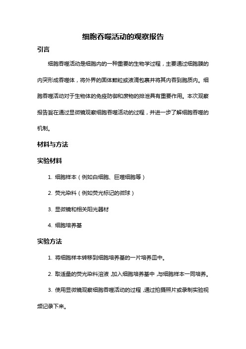
文酶体
吞噬 细菌
吞噬体 吞噬溶酶体 初级溶酶体
溶酶体酶
王润生,男,1985 年生, 安徽省六安市人,汉族, 解放军医学院在读博士, 主治医师,主要从事脊柱 外科、骨生物材料研究。
Evaluation methodology of cellular uptake of magnetic nanoparticles
Wang Run-sheng1, 2, Liu Yi-hao3, Mao Ke-ya1 (1Department of Orthopedics, Chinese PLA General Hospital, Beijing 100853, China; 2Department of Orthopedics, Third Affiliated Hospital of Guangxi University of Chinese Medicine, Liuzhou 545001, Guangxi Zhuang Autonomous Region, China; 3Jiangsu University, Zhenjiang 212013, Jiangsu Province, China)
Corresponding author: Mao Ke-ya, Chief physician, Department of Orthopedics, Chinese PLA General Hospital, Beijing 100853, China
中图分类号:R318 文献标识码:A 稿件接受:2018-01-05
细胞内吞作用与肿瘤治疗研究

细胞内吞作用与肿瘤治疗研究细胞内吞作用,简称内吞,是细胞由外向内摄取固体颗粒或液体的一种重要方式。
它以胞吞作用和胞吸作用为代表,对细胞的生长、分裂、代谢、免疫等过程产生着重要影响。
同时,它还在生物医学领域中得到广泛的研究和应用,特别是在肿瘤治疗方面。
1. 细胞内吞作用的原理与机制细胞内吞作用的实现主要依赖于细胞膜上的多种受体和适配分子。
这些分子对外界物质进行识别、结合,从而引导其进入细胞。
以胞吞作为例,一般包括以下几个步骤:首先,胞膜上的受体结合外界物质,形成一个复合物;接下来,复合物在细胞膜上聚集,并形成凹陷的小囊泡(内吞囊泡);最后,小囊泡将外界物质引入细胞内部,与细胞质融合,释放出物质。
2. 内吞的生物学意义和研究方法内吞在细胞生物学研究中占有重要地位。
首先,它对于细胞的生长和分裂至关重要。
细胞需要摄取和代谢外源性物质来维持正常生长和代谢状态,内吞作为一种稳定可靠的吞咽方式,为细胞供应了必需的营养物质。
其次,内吞与病原体侵入和免疫过程密切相关。
例如,受体介导的胞吞作用可以帮助身体从病原体中清除有害物质。
此外,内吞还是许多细胞信号通路、细胞器运输和细胞器间相互作用的基础和前提。
在内吞研究中,常用的方法包括形态学观察、细胞示踪、生化分析和分子遗传学等手段。
这些方法不仅对内吞现象的观察和分析有帮助,也为更深入理解内吞机制和其生物学功能提供了线索和证据。
3. 内吞与肿瘤治疗的关系及其应用现状近年来,内吞和肿瘤之间的关系成为了生物医学研究的热点。
内吞作为一种活性转运方式,可以促进药物和治疗物质进入肿瘤细胞内部,从而实现对肿瘤的治疗。
目前,内吞在肿瘤治疗中的应用主要包括以下几种形式:(1)靶向药物输送:利用纳米颗粒、肽或抗体等载体,将药物靶向输送至肿瘤细胞,有选择性地杀死肿瘤细胞,减轻药物对健康细胞的损害。
(2)免疫治疗:利用受体介导的内吞机制,将免疫细胞及其产生的细胞因子引导至肿瘤病变部位,激活免疫细胞,增强对肿瘤的攻击力度。
药物在肿瘤细胞中的内吞作用研究

药物在肿瘤细胞中的内吞作用研究药物在肿瘤细胞中的内吞作用是一项重要的研究领域,它涉及到肿瘤治疗的关键机制和策略。
本文旨在探讨这一领域的最新研究进展并分析其应用潜力。
一、背景概述肿瘤是细胞异常增殖导致的病理性生长,严重威胁人类健康。
传统的肿瘤治疗方法包括手术切除、放疗和化疗等,但不同程度上存在副作用和疗效不佳的问题。
因此,研究人员开始关注药物在肿瘤细胞中的内吞作用。
二、药物内吞作用的意义药物内吞作用是指细胞通过细胞膜上的受体介导将药物颗粒吸附到细胞内。
这种作用能够增加药物在肿瘤细胞内的浓度,提高药物的治疗效果,并减少对正常细胞的损伤。
因此,研究药物内吞作用对于开发新的肿瘤治疗策略具有重要的意义。
三、内吞作用的研究方法目前,科学家使用多种技术手段来研究药物内吞作用。
其中,最常用的方法是荧光探针技术和细胞内吞实验。
荧光探针技术通过标记药物颗粒上的荧光物质,实现对药物内吞过程的实时观察和定量分析。
而细胞内吞实验可以通过体外细胞培养体系来研究细胞对不同药物的内吞效率。
四、药物内吞作用的影响因素药物内吞作用的效率受多种因素的影响,包括细胞类型、药物化学性质以及环境条件等。
例如,一些肿瘤细胞具有特定的受体表面,可以增强对特定药物的内吞作用。
此外,药物的颗粒大小、形状和溶解度也会影响内吞效率。
五、内吞作用在肿瘤治疗中的应用药物内吞作用在肿瘤治疗中具有广阔的应用前景。
通过利用药物的内吞作用,可以实现靶向治疗,提高治疗效果,并减少药物的副作用。
此外,还可以通过纳米药物载体来增强药物与肿瘤细胞的结合,进一步提高内吞效率。
六、未来研究方向尽管药物内吞作用在肿瘤治疗中具有巨大潜力,但目前仍存在一些挑战和问题。
例如,如何提高药物的内吞效率,以及如何选择合适的药物内吞途径等。
因此,未来的研究方向应该集中在解决这些问题,并进一步完善药物内吞作用的理论基础和应用方法。
七、结论药物在肿瘤细胞中的内吞作用是一项具有重要意义和广阔应用前景的研究领域。
细胞吞噬活动的观察报告
细胞吞噬活动的观察报告引言细胞吞噬活动是细胞内的一种重要的生物学过程,主要通过细胞膜的内突形成吞噬体,将外界的固体颗粒或液滴包裹并将其内吞到胞质内。
细胞吞噬活动对于生物体的免疫防御和废物的排泄具有重要作用。
本次观察报告旨在通过显微镜观察细胞吞噬活动的过程,并进一步了解细胞吞噬的机制。
材料与方法实验材料1. 细胞样本(例如白细胞、巨噬细胞等)2. 荧光染料(例如荧光标记的微球)3. 显微镜和相关阳光器材4. 细胞培养基实验方法1. 将细胞样本转移到细胞培养基的一片培养皿中。
2. 取适量的荧光染料溶液,加入细胞培养基中,与细胞样本一同培养。
3. 使用显微镜观察细胞吞噬活动的过程,通过拍摄照片或录制实验视频记录下来。
实验结果观察过程中,我们发现细胞吞噬活动呈现出以下几个阶段:1. 识别阶段:细胞通过感知周围环境,活动自如地游动,并能迅速对潜在被吞噬物体进行识别。
我们观察到细胞在培养皿中自由移动,在接触到荧光标记的微球时,能够迅速转向并靠近微球。
2. 吞噬体形成阶段:当细胞接近微球时,细胞膜会突起形成一个或多个吞噬杯,将微球包裹其中,并与其他细胞膜结合形成一个完整的吞噬体。
我们观察到细胞膜突起形成吞噬杯的过程,最终形成一个完整的吞噬体。
3. 吞噬体的内吞和消化阶段:吞噬体会进一步收缩和内吞,将被包裹的微球引入细胞质内,并在胞质中进行消化分解。
我们观察到吞噬体逐渐收缩,微球被完全引入细胞质内,并通过显微镜观察到微球逐渐失去荧光信号,进入消化分解过程。
讨论与结论通过观察细胞吞噬活动的过程,我们可以看到细胞吞噬活动是一种高度有序的过程,其中涉及到多个细胞器和分子的协同作用。
细胞在识别阶段通过感知外界环境和目标物体的特征,迅速靠近并形成吞噬体;在吞噬体的内吞和消化阶段,细胞通过调控吞噬体的收缩和内吞过程,将目标物体引入细胞质内进行消化分解。
细胞内吞作用的研究进展

Chinese-German Journal of Clinical Oncology June 2009, Vol. 8, No. 6, P360–P365 DOI 10.1007/s10330-009-0023-9In the process of cell metabolism, various kinds of ma-terials including some ions, small molecules, macromo-lecular materials and some granular matters keep in and out of cells. The macromolecular substances and granular materials can not go through the membrane. They com-plete the transfer across the cell membrane in another way, that is, materials cross in and out of cell membrane in the form of vesicle which is fused with membrane and then sciss into the cell, which is currently recognized as the main pathway of intaking biological macromolecules-endocytosis. Endocytosis is broadly divided into two cat-egories, phagocytosis and pinocytosis; the latter is divided into four species according to their different mechanisms: clathrin dependent endocytosis, caveolin dependent en-docytosis, macropinocytosis, and clathrin and caveolin independent endocytosis.The recent study reported that the abnormal expres-sion of endocytosis may be involved in the mechanism of certain diseases, such as diabetes and neurological diseases and also closely related to the malignant transformation of cells. The role of endocytosis was paid increasing atten-tion. Further research in this area will help us understand these diseases, thereby found new treatments. Here we introduce about pathway of cell endocytosis, mechanisms of regulation and endocytotic proteins. Endocytosis pathwayPhagocytosisPhagocytosis refers to endocytosing large granular matters (> 250 nm), and it provides the host a direct way digesting exogenous substances, which is one of the most important immune protecting mechanisms [1]. In mamma-lian, phagocytosis can only be completed by specific cells, such as macrophages and neutrophils which are called phagocytic cells. Phagocytic cells recognize and combine with IgFc and complement packing pathogens through IgFc receptor and complement receptor respectively. When the cell surface receptors combined with the cor-responding ligands, downstream signal transduction was activated, which caused actin polymerization under plas-ma membrane of intaking site, actin contraction makes phagocytic cell membrane form pseudo-foot fusing into vesicle to pack pathogens. In cytoplasm, dynamin assem-bled into ring at the neck of vesicle, and hydrolysed the binding GTP, dynamin contraction forced vesicle to sciss from membrane at the neck and form phagosome which integrated with the lysosome, the acid hydrolysis enzyme of lysosomal digested pathogens. In addition to phagocy-tosing pathogens, macrophages of phagocytic cells canStudy progress of cell endocytosis*Li Chen1, Hui Li1, Ren Zhao2, jianwei Zhu31Department of Pathology, Nantong University Medical College, Nantong 226001, China2Department of General Surgery, Ruijin Hospital, Shanghai Jiaotong University, Shanghai 230002, China 3Department of General Surgery, Affiliated Hospital, Nantong University, Nantong 226001, ChinaReceived: 19 February 2009 / Revised: 13 March 2009 / Accepted: 10 April 2009Abstract Endocytosis is a process through which extracellular materials are transported into cell through membrane defor-mation. This process is not a simple step-by-step process in which a series of proteins function according to the chronological order, but rather a complex process comprising many members which are regulated precisely. The role of endocytosis is broadly divided into two categories, phagocytosis and pinocytosis, the latter is divided into four species in accordance with the size of endocytosis substances: clathrin dependent endocytosis, the diameter of clathrin-coated vesicle is 100–150 nm;caveolin dependent endocytosis, the diameter of caveolin protein-coated vesicle is 50–100 nm; macropinocytosis, the diam-eter of macropinocytosis is generally 0.5–2 μm, sometimes up to 5 μm; clathrin and caveolin independent endocytosis. Many proteins including endophilin A1, A2, A3, and endocytotic proteins B, B1a, and B1b as well as dynamin, actin and Rab protein families are involved in endocytosis and play an important role in different stages. The abnormal endocytosis may be involved in the development of certain diseases.Key words endocytosis; clathrin; caveolin; endophilin; signalling pathwayCorrespondence to: Jianwei Zhu. Email: usazhujianwei@* Supported by grants from the National Natural Sciences Foundationof China (No. 30771126 and 30772106).361 Chinese-German J Clin Oncol, June 2009, Vol. 8, No. 6swallow foreign bodies through ligand-receptor binding pattern. Identify and kill tumor cells, identify and remove degenerative plasma protein, lipids, and other macromol-ecules. Remove aging and damaged cells and cell debris. PinocytosisPinocytosis refers to the intake process that endocyto-ses extracellular liquid and the dissolving materials. Clathrin dependent endocytosisClathrin plays an important role in the regulation of composition of plasma membrane proteins, and research on clathrin can help us understand how cells interact with the surrounding environment, signal transduction of mitogenic, nutrition intake of the cell, establishment of extracellular environment cell identity including the interaction with the immune system, keep a balance in the stability of the environment of cell.(1) Clathrin and adaptor protein (AP)Clathrin is the skeleton protein outside the vesicle, clathrin-coated vesicle is 100–150 nm in diameter and exists in all eukaryotic cells and it mediates the way of transportation from the plasma membrane to intracellu-lar of proteins, lipids, nutrients, antibodies and growth factors and also the vector through which proteins and lipids transport from trans Golgi net work (TGN) to endosome. Clathrin is spider-like and polymerized by three chains at the top, which is known as triskelion [1]. Adapter lies inside the clathrin-coated vesicle, which mediates membrane binding, localization, sorting signals, identification and inositol phosphate. It not only serves to combine cargo and clathrin, but also connect with polyphosphatidylinositol headgroup. It is now found four kinds of adapters (AP1–4), all of which comprise a pair of 100–130 kD subunit. These subunits are able to identify 6-phosphate mannose receptor, transferrin, low-density lipoprotein and epidermal growth factor receptor, prote-ase and de-sialic acid receptor.(2) MechanismThe process from recruitment to disassemble is very short. It comprises as follows:1) Recruitment of adapter and clathrin. Recruitment of AP2 complex to high activity, saturated, easy enzymolysis site, activate formation of clathrin-coated vesicle in the plasma membrane under the effect of sorting signal and docking protein [2].2) Invagination, scission and budding of clathrin-coat-ed vesicle. The planar clathrin protein can be transformed into curved one without nucleic acid and cytoplasm in vitro; In vivo, clathrin-coated vesicle curves to some de-gree. Invagination may be due to structure changes or rearrangement of clathrin assembling in the crystal lat-tice or between clathrin. The budding of clathrin-coated vesicle is involved with GTPase dynamin, actin tubulizes or forms ring-shape in vitro, it is the trigger that vesicle dissociated from the membrane.3) Decapsulation of clathrin-coated vesicle. This is a wasting process that needs hsc 70, auxilin and ATP. The large chain of clathrin has two sites which interact with the adapter and also with hsc 70. The interaction between clathrin and hsc 70 destroyed the interaction between clathrin and adapter. Over-expression of hsc 70 mutant blocked the cycle of transferrin receptor so that the “as-sembly – dismantle” balance moved to the assembly di-rection. Hsc 70 dissociated clathrin from the vesicle in vitro, but could not dissociate adapter. Auxilin plays an important role in decapsulation. Auxilin can not only re-cruit hsc 70 to clathrin-coated vesicle, but also stimulate activity of hsc 70 ATP enzyme.Caveolin dependent endocytosisCaveolae/caveolins mediate endocytosis of many sub-stances and is the main form of clathrin-independent en-docytosis.(1) As early as 1950, Japanese scholar Yamada used transmission electron microscopy to observe some small caveolae which was about 50–100 nm in diameter for the first time. These vesicles appeared alone or string-like, in the form of invagination of the cytoplasmic membrane, it had typical lipid bilayer structure [2]. Caveolae is currently considered to be the signal transduction center, and many signal transduction receptors, protein kinase and binding proteins are highly enriched in caveolae region. In the regulation of caveolae endocytosis, activation of tyrosine kinase-dependent signal is an important step. After the treatment of phosphatase inhibitor, the endocytosis of cell by caveolae had increased notably.(2) Caveolin is the main surface marker of a caveolae, a kind of membrane integrin family modified in the inner surface of caveolae, 21–25 kD. It consists of N-terminal region, transmembrane region and C-terminal region, N-terminal and C-terminal cytoplasmic moves inward into the cytoplasm and its peptide chains like hairpin struc-ture. Caveolin plays a critical role in the assembly process of caveolae and cholesterol and is the signaling molecule of the scaffold proteins and negative regulatory proteins and belongs to a highly conserved integrity membrane protein family [3]. Rajjayabun [4] confirmed that caveolin-1 was the key factor of caveolae endocytosis. If caveolin gene was knockout, caveolae can not be formed. So far, four kinds of caveolin isomers have been found in mammals: caveolin-1α, 1β, 2 and 3, which are products of differ-ent genes, most cells expressed caveolin-1 and caveolin-2, the two form a stable heterologous oligomers complexes, particularly rich in terminal differentiated cells, such as fat cells, endothelial cells and fibroblasts, and caveolin-3 mainly exists in various muscle cells (such as myocardial cells, rhabdomyosarcoma cells and skeletal muscle cells) and is closely related to the synthesis of muscle cells [5]. Neither caveolin protein nor caveolae structure appeared362 www. springerlink. com/content/1613-9089in peripheral blood cells and nerve cells.(3) The biological feature of caveolin. Caveolae as a sig-naling molecule plays a role as a platform in signal trans-duction. Caveolin-1 is in the center of signaling pathways at all these platforms. Caveolin, as a signaling molecule’s scaffold protein and negative regulatory protein, inhibits kinase activities of signaling molecules in the normal sig-nal transduction pathways. Caveolin-2’s skeleton region has no inhibitory activities, and other signaling molecules inhibitory regions may exit in it. Caveolin-3 is involved in energy metabolisms in muscle cells. Caveolin-1 has strong affinity to cholesterol, thus the cholesterol levels of caveolae is far higher than other biofilm. The study showed that the absence of caveolae eventually resulted in formation of foam cells, suggesting that caveolae and caveolin-1 can maintain the cholesterol balance by re-moving the excessive lipoprotein cholesterol out of cells [6].The correlation between caveolin and tumors have become one of the hot spots in tumor biology, among which, the relationship between caveolin-1 and tumor occurrence and metastasis has been studied a lot. Gene coding for caveolin-1 (CAV) locates in the suspicious tu-mor suppressive site (D7S522; 7q31.1). This site depletes or fractures in a variety of tumors (such as liver cancer, ovarian cancer, breast cancer, uterine fibroids, gastric ad-enocarcinoma, etc. [7]). The normal NIH 3T3 cell which was introduced by antisense caveolin-1 was transplanted into nude mice to observe the formation of tumor, sug-gesting caveolin-1 has tumor suppressive function. In many tumors and activated oncogene transfected cells, the expression of mRNA and its protein of caveolin-1 de-creased or lost [8]. The experiments in vivo demonstrated mutation or deletion of caveolin-1 could lead to the ex-cessive proliferation of breast epithelial cells, and then increase the occurrence of breast cancers [9]. Besides, the expression of caveolin uprehulated in diabetes and hyper-cholesterolemia and plays a significant role in Alzheim-er’s disease [10], degenerative muscle disease [11], heart and lung diseases.MacropinocytosisLarge and irregular original endocytosis vesicles are formed by folding membrane on the verge of extending cell under stimulation by certain factors, and they are known as macropinosome whose size varies with diam-eter generally 0. 5–2 μm. Macropinocytosis plays a major role in macrophages and dendritic cells, also in many tu-mor cells [11].Macropinosome has no clathrin or caveolins coating; it is closely related to actin at the early stages of formation, which provides an effective way for non-selective endo-cytosis of extracellular nutrients and liquid phase macro-molecules. Macropinocytosis and phagocytosis, regulated by a variety of proteins, such as actin, Scar protein, Ap21 complexes and RabB. Different membrane wrinkle results in different rates of developing macropinocytosis.The formation of macropinosomes can be significantly inhibited by cytochalasin D and colchicine, suggesting that microtubules and microfilaments play an important role in this process. The mechanism may be the stimulus by a variety of factors that activate corresponding recep-tor tyrosine kinase and then self-phosphorylate quickly. Phosphorylated residues recruit PI3 kinase and activate it, the activated PI3 kinase activates Rac1 which may cause microfilament reconstruction through two ways. Firstly, the activated Rac1 activates PAK1 which regulates phos-phorylation of myosin light chain. The interaction be-tween myosin and actin is regulated by the phosphory-lated myosin light chain and the interaction promotes reconstruction of microfilaments, development of the cell membrane ruffles and formation of macropinosomes; Secondly, the activated Rac1 binds with the amino-ter-minal of its target protein IRSp53, SH3 region at the car-boxyl end of IRSp53 combines with WAVE to form three molecular complexes which could activate WAVE [12]. The latter further activates actin related protein Arp2/3 complexes which stimulate microfilament nucleation, promote microfilament reconstruction, development of cell membrane ruffles and formation of macropinosomes [13]. In-depth regulation mechanism needs further study. MicrodomainMicrodomain has similar lipid composition with caveolae but not function through caveolin. It functions through dynamin and Rho A-dependent mechanism, or through clathrin-independent, dynamin-2-independent, Cdc4 mediated pathway to endocytose glycosylated phos-phatidylinositol-hexanol anchored proteins [13]. The path-way plays a role in endocytosis of IL-2. EndophilinThe biological features of endophilinThe main function of endophilin is related to the en-docytosis of neurotransmitter. Endophilin has a common special structure, whose C-terminal has a unique SH3 do-main with the unique binding ability [14], which can in-teract with a number of special proteins such as dynamin, thus affecting the functions of other proteins. While N-terminal participates in membrane invagination of vesi-cles. They were named endophilin A1, A2, A3 in 2002. Huntingtin belongs to binding protein of endophilin A1, the complex composed of it and the huntingtin binding protein 40 (HAP40) inhibited early microtubule-depen-dent endosome movement, increased connection be-tween early endosome and actin [15]. Then a new family was discovered, which was named endophilin B.363 Chinese-German J Clin Oncol, June 2009, Vol. 8, No. 6Endophilin and its role in signal transduction Endophilin can interact with various proteins through which involved in different signal transduction pathway Endophilins which interact with cell membrane re-ceptors are mainly distributed in the cytoplasm and cell membrane, it can interact with cytoplasmic membrane receptor and conduct signals, such as beta 1-adrenergic receptor. Endophilin is also involved in phospholipid sig-nal transduction. Many endophilins contain the binding sites of inositol 4,5-diphosphate (PIP2). The degradation of PIP2 is necessary for the completion of the endocytosis. Mark PIP2 connected domain by immunohistochemical fluorescent protein and track endocytosis movement of living cells, and PIP2 was found present in the cell mem-brane surface in the form of many small plaques, these plaques entered gradually into the cell, which confirmed that the shift was the endocytosis. The disappearance of PIP2 matched the recruitment of phosphoinositide phosphatase enzyme, and we can propose that the latter can degraded the former. When PIP2 degradation was blocked, abnormal invagination can be observed, shear-ing machine of endocytosis gathered around the plaque on the membrane and failed to complete endocytosis. Therefore, PIP2 degradation is the necessary step of ves-icle scission [16].Recycling of the neurotransmitter Neurotransmitter recycle is essentially a process of cell endocytosis, clathrin interact with AP2 or AP180 (adapter protein) and membrane lipid to form clathrin-coated ves-icles. The process of its formation is just that endophilin A1 drove the rupture of membrane vesicles from the do-nor membrane at the neck of vesicles, LPAAT at N-termi-nal of endophilin A1 can catalyze lysophosphatidic acid (LPA) and arachidonic ene-CoA to form lysophosphatidic acid (PA), LPA is a three-dimensional structure like in-verted cone-type and PA is cone-type, the former struc-ture is beneficial for positive membrane deformation, the later is conducive to negative membrane deformation. It is the transformation of lysophosphatidic acid that drive rupture between vesicle and the donor vesicle membrane. After endophilin mutation, it will severely affect recycle mechanism of synaptic particle [17].Function besides endocytosisEndophilin A2 is actually involved in absorbing nu-trients and growth factors and endocytosis of pathogens and receptors, participating in a variety of membrane transport mechanism . While endophilin B1 locates in the membrane of the cell and its main function is relat-ed to cell membrane dynamics. Apoptosis receptor Fas/ CD95 can not correctly located in the membrane without endophilin, resulting in inhibition of the mechanism of apoptosis [18]. Endocytosis related proteinsDynaminDynamin is a 100 kD GTPase, and its GTPase region at N-terminal can bind and hydrolyse GTP, PH (pleck-strin homology) region can bind with membrane and me-diate polymerization between dynamins, PRD (praline arginine rich) region at C-terminal mediate the interac-tion between other proteins. In addition, dynamin has a small GED (GTPase effector) region which is necessary in hydrolysis of GTP [18], and also plays an important role in some clathrin independent endocytosis. Dynamin is necessary in pinching and formation of vesicles. After its binding with ligand amphiphysin-1, dynamin-1 plays a key role in dynamin dependent endocytosis under regu-lation of cycle-dependent kinase 5 (Cdk5). Dynamin-1-dependent endocytosis occurs quickly, usually only a few seconds, while dynamin-2 mediated endocytosis is slow, usually ten minutes.Dynamin uses mechanochemical activity – specifi-cally a twisting action – to pinch off endocytic vesicles [19]. Dynamin was, early on, localized to the collar around the neck of forming endocytic vesicles. This suggested that dynamin may use the energy of GTP hydrolysis to directly pinch a membranous neck. Indeed, dynamin could tubulate lipids and break apart the tubules in vitro, although later it seemed that the breaking apart was hap-pening as the samples dried on EM grids. Meanwhile, Schmid had come up with a “regulatory GTPase” mode: that dynamin was active not as it hydrolyzed GTP but in its GTPbound form, which recruited other proteins to do the pinching. The Yale group has more evidence for the earlier “pinchase” model. They used light microscopy rather than EM to follow tubulation and fission directed by dynamin in vitro. Longitudinal tension was needed with constriction to achieve fission. In mammalian, the dynamin collars are relatively short, so cortical actin is the most likely source of tension that would help the dynamin to wrench an endocytic vesicle free [20–23]. Dynamin has intrinsic activity of GTP and is also the cerebellum Dyrk1A kinase substrate, with its own ag-gregation feature of the endometrium can help new pro-cesses from the cell membrane vesicles isolated. Dynamin is involved in tubulation by PH epsin and ENTH (epsin NH2-terminal homology) [21].Wild-type dynamin mutant block the tube, which can be restricted to speculate dynamin of these membrane protein of the ability to control grid protein coated vesi-cle endocytosis and other media invagination degree. ActinActin is the main component of microfilament. Microfilament is involved in cell shape and polarity of the maintenance of endocytosis, intracellular transport,364 www. springerlink. com/content/1613-9089cell shrinkage and movement, cell division, and many other functions. Actin has two kinds: G-actin and F-ac-tin. The importance of F-actin in endocytosis was clearly illustrated by the effect of addition of the actin-mono-mersequestering drug latrunculin A. Within 5 minutes of its addition, actin patches were no longer visible and endocytosis was completely abrogated. This was the point at which the yeast WASP orthologue Las17p and the type I myosins (Myo3p and Myo5p) began to nucleate actin filaments through activation of the Arp2/3 complex [24]. This group of proteins has been called the “actin net-work growth machinery”. Some researchers combined epifluorescence with total internal reflection microscopy (TIRF) to follow the internalisation of individual coated pits in living cells expressing a fluorescent tagged form of clathrin light chain (DsRed clathrin). They showed that transient recruitment of actin coincides with the inward movement of vesicles, they also observed recruitment of the Arp2/3 complex to clathrin-coated pits. PH-sensitive probes have been developed that allow internalisation of cargo into individual clathrin-coated vesicles to be visual-ized, allowing us to follow the time course of invagination and identify the point of scission. The recruitment of actin during invagination is thought to provide the force that drives the invagination of the coated pit [25]. It is currently identified that the vesicle scission module contains the two yeast amphiphysin proteins Rvs161p and Rvs167p. These proteins contain BAR (Bin-Amphiphysin-Rvs) domains that bind to and tabulate, actin and associated proteins recruit amphiphysins and thus mark the site at which membrane tubulation should occur. Tubulation is then suggested to facilitate the scission process. Following scission, the vesicle is uncoated and moves away from the membrane until it fuses with an endosome. There are two possible roles for actin at this final stage: vesicles could move along actin cables; alternatively actin could be nu-cleated at the vesicle surface to facilitate their movement within the cell. Then the actin depolymerized from the endocytosis site. The most representative depolymeriz-ing factor is cofilin. Cofilin is necessary for endocytosis in mamamals, but its mechanism is still unknown [26]. In the process of invagination, Sac6p/microfilament binding protein/fimbrin is also required, invagination induced by actin failed without Sac6p [27].PCH (Pombe Cdc15 homology) / F-BARPCH (Pombe Cdc15 homology) / F-BAR is another family of proteins which plays an important role in en-docytosis. The BAR region of these proteins combine with phosphoinositide. FBP17 is a member of PCH fam-ily, containing PCH domain and extended FC domain. The EFC domains show weak homology to the Bin-am-phiphysin-Rvs (BAR) domain. The EFC domains bound strongly to phosphatidylserine and phosphatidylinositol 4,5-bisphosphate and deformed the plasma membrane and liposomes into narrow tubules. Most PCH proteins possess an SH3 domain that is known to bind to dynamin and that recruited and activated neural Wiskott-Aldrich syndrome protein (N-WASP) at the plasma membrane. FBP17 contributed to the formation of the protein com-plex, including N-WASP and dynamin-2, in the early stage of endocytosis. Furthermore, knockdown of endog-enous FBP17 impaired endocytosis, suggesting FBP17 is necessary for dynamin-dependent endocytosis [28].Rab proteinsRab protein which is a small GTP enzyme is an im-portant regulator of endocytosis. Rab5 is involved in tubulization and its role in endocytosis has been clear. Rab5 protein has three isomers, Rab5A, Rab5B and Rab5C. Rab5 protein exists primarily on the plasma mem-brane, clathrin-coated vesicle and early endosome, tak-ing charge of vesicle fusion and recycling. Rab5 protein functions with ongoing GTP/GDP cycle. As molecular switch of vesicle transport, the activated state of combin-ing with GTP is “open”, the unactivated state of combin-ing with GTP is called “close”. Rab5 regulates transport of endocytotic materials between membrane and early en-dosome, and help vesicle movement along microtubules. HAP40 is an effective regulatory factor of Rab5, and Rab5 increase connections of endosome with actin un-der existence of HAP40. RAB5A protein was related to endocytosis of prostacyclin [29]. A-synuclein is the root of Parkinson’s disease, Lewy body Dementia and Alzheim-er’s disease and other central nervous system diseases. Moreover, Young found the mutant can decrease endo-cytosis of A-synuclein according to RAB5A GTP mutant enzyme research while Lewy body-like decreased in cy-toplasm, the cytotoxicity also decreased. Over-expression of RAB5A protein in immune system deficiency, which can slower macrophage phagocytosis of pathogens and decrease the rate of degradation of pathogens and IFN-C-mediated phagocytosis weakened.ConclusionWe have found many molecules involved in endocy-tosis in the past few decades, and had a preliminary un-derstanding about its process and metabolism. We now know that the process of endocytosis is not a simple step-by-step process in which a series of proteins function ac-cording to the chronological order, but rather a complex process comprising many members which are regulated precisely. Although the mechanism of regulation of en-docytosis is still unknown, it can be predicted that as more and more new research techniques applied in this area, we will be able to understand the mechanism of cell endocytosis more comprehensively. Whether tumor cells365Chinese-German J Clin Oncol, June 2009, Vol. 8, No. 6endocytose more nutrients than normal cells and wheth-er tumor cells increase sizes through endocytosing more growth factors? Can we inhibit tumor growth through inhibiting cell endocytosis or can we cure tumor by in-ducing specific drug endocytosis of tumor cells? Whether the endocytosis of 6-phosphate mannose receptor (MPR) which plays a very important role in cell death signal transduction is restrained in tumor cells? The mechanism of cell endocytosis can be further elucidated by solving all of the above problems.ReferencesGiodini A, Rahner C, Cresswell P. Receptor-mediated phagocytosiselicits cross-presentation in nonprofessional antigen-presenting cells. Proc Natl Acad Sci USA, 2009, 106: 3324–3329.Kirchhausen T. Clathrin. Annu Rev Biochem, 2000, 69: 699–727.Kiss AL, Turi A, Müller N, et al . Caveolae and caveolin isoforms in ratperitoneal macrophages. Micron, 2002, 33: 75–93.Rajjayabun PH, Garg S, Durkan GC, et al . Caveolin-1 expressionis associated with high-grade bladder cancer. Urology, 2001, 58: 811–814.Frank PG, Woodman SE, Park DS, et al . Caveolin, caveolae andendothelial cell function. Arterioscler Thromb Vasc Biol, 2003, 23: 1161–1168.Krajewska WM, Masłowska I. Caveolins: structure and function in sig -nal transduction. Cell Mol Biol Lett, 2004, 9: 195–220.Pascariu M, Bendayan M, Ghitescu L. Correlated endothelial caveolinoverexpression and increased transcytosis in experimental diabetes. J Histochem Cytochem, 2004, 52: 65–76.Fra AM, Pasqualetto E, Mancini M, et al . Genomic organization andtranscriptional analysis of the human genes coding for caveolin-1 and caveolin-2. Gene, 2000, 243: 75–83.Cameron PL, Liu C, Smart DK, et al . Caveolin-1 expression is main-tained in rat and human astroglioma cell lines. Glia, 2002, 37: 275–290.Williams TM, Cheung MW, Park DS, et al. Loss of caveolin-1 gene ex-pression accelerates the development of dysplastic mammary lesions in tumor-prone transgenic mice. Mol Biol Cell, 2003, 14: 1027–1042.Gaudreault SB, Dea D, Poirier J. Increased caveolin-1 expression inAlzheimer′s disease brain. Neurobiol Aging, 2004, 25: 753–759.Müller JS, Piko H, Schoser BG, et al . Novel splice site mutation in thecaveolin-3 gene leading to autosomal recessive limb girdle muscular dystrophy. Neuromuscul Disord, 2006, 16: 432–436.Lim JP, Wang JT, Kerr MC, et al . A role for SNX5 in the regulation ofmacropinocytosis. BMC Cell Biol, 2008, 9: 58.1.2.3.4.5.6.7.8.9.10.11.12.13.Miki H, Yamaguchi H, Suetsugu S, et al . IRSp53 is an essential in -termediate between Rac and WAVE in the regulation of membrane ruffling. Nature, 2000, 408: 732–735.Sun P, Yamamoto H, Suetsugu S, et al . Small GTPase Rah/Rab34 isassociated with membrane ruffles and macropinosomes and promotes macropinosome formation. J Biol Chem, 2003, 278: 4063–4071.Daniels RL, Takashima Y, McKemy DD. Activity of the neuronal coldsensor TRPM8 is regulated by phospholipase C via the phospholipid phosphoinositol 4,5-bisphosphate. J Biol Chem, 2009, 284: 1570–1582.Gad H, Ringstad N, Löw P, et al . Fission and uncoating of synapticclathrin-coated vesicles are perturbed by disruption of interactions with the SH3 domain of endophilin. Neuron, 2000, 27: 301–312.Petrelli A, Gilestro GF, Lanzardo S, et al . The endophilin-CIN85-Cb1complex mediates ligand-dependent downregulation of c-Met. Na-ture, 2002, 416: 187–190.Williams R. PIP2 in endocytosis. J Cell Biol, 2007, 177: 185.Guichet A, Wucherpfennig T, Dudu V, et al . Essential role of endophilinA in synaptic vesicle budding at the Drosophila neuromuscular junc-tion. EMBO J, 2002, 21: 1661–1672.Takei K, McPherson PS, Schmid SL, et al . Tubular membrane invagi-nations coated by dynamin rings are induced by GTP-gamma S in nerve terminals. Nature, 1995, 374: 186–190.Zhang X, Wang F, Chen X, et al . Post-endocytic fates of delta-opi-oid receptor are regulated by GRK2-mediated receptor phosphory-lation and distinct beta-arrestin isoforms. J Neurochem, 2008, 106: 781–792.William A. Twisting endocytosis. J Cell Biol, 2006, 173: 456.Damke H, Binns DD, Ueda H, et al . Dynamin GTPase domain mu-tants block endocytic vesicle formation at morphologically distinct stages. Mol Biol Cell, 2001, 12: 2578–2589.Tsujita K, Suetsugu S, Sasaki N, et al . Coordination between the ac-tin cytoskeleton and membrane deformation by a novel membrane tubulation domain of PCH proteins is involved in endocytosis. J Cell Biol, 2006, 172: 269–279.Sun Y, Martin AC, Drubin DG. Endocytic internalization in buddingyeast requires coordinated actin nucleation and myosin motor activity. Dev Cell, 2006, 11: 33–46.Okreglak V, Drubin DG. Cofilin recruitment and function during actin-mediated endocytosis dictated by actin nucleotide state. J Cell Biol, 2007, 178: 1251–1264.Kaksonen M, Toret CP, Drubin DG. A modular design for the clathrin-and actin-mediated endocytosis machinery. Cell, 2005, 123: 305–320.O’Keeffe MB, Reid HM, Kinsella BT. Agonist-dependent internaliza-tion and trafficking of the human prostacyclin receptor: a direct role for Rab5a GTPase. Biochim Biophys Acta, 2008, 1783: 1914–1928.14.15.16.17.18.19.20.21.22.23.24.25.26.27.28.29.。
JEV入侵BHK-21细胞内吞途径的初步研究

Si c hu an A gr i c ul t u r al Uni v e r s i t y ,Ya’ an 6 25 01 4,Chi na;
2 . La b o r a t o r y o fZ o o n o s i s 。C o l l e g e o f V e t e r i n a r y Me d i c i n e,S i c h u a nAg r i c u l t u r a l Un i v e r s i t y,Ch e n g d u 6 1 1 1 3 0 ,C h i n a 3 . S i c h u a n S c i e n c e — o b s e r v a t i o n Ex p e r i me n t S t a t i o n o f V e t e r i n a r y Dr u gs a n d Ve t e r i n a r y
中 国 人 兽 共 患 病 学 报
7 6 6
Chi ne s e J o ur na l o f Zoo n os e s
2 01 3, 2 9 ( 8)
D O I : 1 0 . 3 9 6 9 / c j z j . i s s n . 1 0 0 2 —2 6 9 4 . 2 0 1 3 . 0 8 . 0 0 7
Bi o l o g i c a l Te c h n o l o g y,M i n i s t r y o f Agr i c u l t u r e,Y a’ a n 6 2 5 0 1 4,C hi n a )
细胞内吞作用的研究进展

细胞内吞作用的研究进展细胞内吞作用是指细胞将外界的物质通过包裹形成的小泡子,从而运输到细胞内部的过程。
这一过程在生物体中是非常重要的,因为它能够完成许多关键生理功能,例如细胞摄取营养物质、消化细菌和病毒、维持细胞内环境平衡等。
因此,细胞内吞作用的研究一直是生命科学领域的一个重要课题。
自20世纪初以来,人们已经开始研究细胞内吞作用,并提出了相关的理论和实验方法。
其中最为重要的是开发出了荧光显微镜技术,使得人们能够观察到细胞内吞作用的发生和细胞内小泡的运输情况。
通过这一技术,我们能够对细胞内吞作用的原理和机制有更加深入和准确的了解。
目前,已经有多种细胞内吞作用机制得到了研究和发现。
其中最为简单和明显的是非特异性内吞作用,即细胞通过伸展表面膜,包裹附着在上面的外来物质,从而形成小泡输运到细胞内部。
但是,针对不同类型和大小的外来物质,细胞内吞作用也存在特异性。
例如,一些大分子物质的吞噬需要通过细胞表面受体的介导。
在现今的研究中,细胞内吞作用已经成为多个研究领域的热点,例如癌症、神经退行性疾病、感染性疾病等。
对细胞内吞作用进行研究,不仅有助于理解各种疾病的发病机制,甚至可以为探求相关疾病的治疗方法提供新的思路和方向。
以癌症研究为例,近年来已经有多项研究表明,癌细胞在生长和转移过程中,不同于正常细胞的独特生命特性与细胞内吞作用耦合,即通过高度调节不同类型的内吞作用机制,促进细胞的生长、增殖和转移。
因此,通过研究癌细胞内吞作用的具体机制,不仅可以在理论上为癌症的发病机制提供更加深刻的认识,同时也有助于探索和开发新型的癌症治疗手段。
除了癌症研究,细胞内吞作用在感染性疾病中也具有重要的作用。
例如,病毒和细菌等病原体进入人体后,依托细胞内吞作用穿透到细胞内部,从而引起感染并且继续进行复制。
因此,人们可以通过对细胞内吞作用的研究来探索和开发能够对抗病原体的新型治疗手段。
总的来看,细胞内吞作用在生命科学领域的研究具有广泛的意义和重要性。
细胞膜融合与共内吞机制的研究进展

细胞膜融合与共内吞机制的研究进展细胞是生物体中最基本的单位,它们通过细胞膜与外界进行物质、信息交换。
细胞膜是由双层磷脂、膜蛋白、糖脂等组成的,通过一系列复杂的蛋白质机制,为细胞提供了一个与外界相互作用的平台。
细胞膜的重要性不言而喻,而细胞膜融合与共内吞等过程成为细胞生物学研究中的热点。
本文将对细胞膜融合和共内吞机制的研究进展进行概述。
一、细胞膜融合机制细胞膜融合是生命过程中一种最基本的现象,它不仅体现在细胞之间,也发生在细胞内部各个细胞器的融合与分裂过程中。
目前,关于细胞膜融合机制的研究主要分为三个方面,即融合启动、膜对齐和膜融合。
融合的启动涉及到蛋白质与膜蛋白之间的相互作用。
许多细胞膜上都存在膜融合素蛋白,它们能够识别和结合目标膜上的特定蛋白质或糖脂,从而启动膜融合。
一些研究发现,膜融合素蛋白能够在膜表面形成大片的聚集,从而形成许多近距离相互作用的细胞膜颗粒,这些颗粒能够在酵母菌和哺乳动物细胞质中促进膜融合事件的发生。
膜对齐是细胞膜融合过程中的第二个环节,其主要目的是将两个膜表面聚集在一起,形成紧密的相互作用。
最近的研究表明,细胞膜融合可以通过两种不同的方式进行。
第一种方式为完整膜双层的膜对齐,该方式是通过一系列复杂的蛋白质和膜蛋白相互作用完成的。
研究表明,很多蛋白质在细胞膜融合过程中发挥了重要的作用。
如 SNARE 蛋白是一类在细胞膜融合和新胞体形成过程中发挥重要作用的蛋白质。
它们通过特定的成对结合方式实现了膜对齐和膜融合。
近期研究还表明,细胞膜的一类新型脂质,即伪血蓝脂,也能够作用于 SNARE 蛋白,促进膜对齐和膜融合。
第二种方式是不完整膜双层的膜对齐,这类事件包括泡膜的扩张和吞噬等。
它与完整膜双层的膜对齐方式不同,不需要 SNARE 蛋白参与,而是利用了蛋白质和细胞膜磷脂的自组装能力。
有研究表明,草鱼毒液中的一个新型蛋白质 F-27 也能够参与细胞膜融合,进一步揭示了这一新型膜蛋白对细胞膜融合的作用。
细胞生物学中的细胞分泌和内吞机制研究

细胞生物学中的细胞分泌和内吞机制研究细胞生物学是研究细胞结构、功能和组织的科学领域。
在细胞生物学中,细胞分泌和内吞机制是两个重要的研究方向。
细胞分泌是指细胞将物质从细胞内排出到细胞外的过程;而细胞内吞则是指细胞将物质从细胞外摄入到细胞内的过程。
这两个过程在细胞的正常功能和生命周期中起着至关重要的作用,对细胞的存活、增殖和代谢等方面具有重要影响。
一、细胞分泌机制的研究细胞分泌是细胞的一种基本功能,通过分泌细胞可以释放细胞内合成的蛋白质、脂类、多糖等各种生物活性物质。
细胞分泌的机制可以分为两种:一种是通过囊泡运输方式,即囊泡分泌;另一种是通过膜蛋白介导的分泌方式,即透明分泌。
囊泡分泌是指细胞将合成的物质包装入囊泡中,然后囊泡通过与细胞膜融合的方式将物质释放到细胞外。
这种方式在动物细胞和植物细胞中都存在。
囊泡分泌过程中需要多种细胞器和分子机制的参与,如高尔基体、内质网、囊泡和膜转运蛋白等。
研究囊泡分泌的机制可以帮助我们更好地理解细胞如何调控物质的合成和释放过程。
透明分泌是指物质通过细胞膜的直接介导而被释放到细胞外。
细胞膜上的膜蛋白在这一过程中起到了关键作用,它们可以通过改变细胞膜的通透性来促进物质的释放。
研究透明分泌的机制可以帮助我们了解细胞如何与外界环境进行交流和信号传导。
二、细胞内吞机制的研究细胞内吞是指细胞通过吞噬细胞外的物质来获取营养、调节信号转导和清除废物等功能。
细胞内吞的过程包括三种主要类型:胞吞作用、胞饮作用和受体介导的内吞。
胞吞作用是指细胞通过细胞膜伸出伪足,将周围的固体颗粒或微生物包围并吞噬到胞内。
这一过程在吞噬细胞中经常发生,对于免疫系统的正常功能至关重要。
胞饮作用是指细胞通过细胞膜的突出,将周围的液体包围并吞噬到胞内。
这一过程在单细胞生物和某些多细胞生物中常见,可以帮助细胞保持内外环境的平衡。
受体介导的内吞是指细胞表面的受体与适配体结合后,通过膜蛋白的介导将受体和其结合的物质一起内吞到细胞内。
细胞生物学中胞吐和内吞作用机制研究

细胞生物学中胞吐和内吞作用机制研究胞吐和内吞作为细胞生物学中重要的生理过程,是细胞与外环境以及细胞与周围物质之间相互作用的重要形式。
在细胞生长、分化和代谢等方面都扮演着重要的角色。
本文将着重阐述胞吐和内吞作用机制的研究进展以及其在生理过程中所起作用的意义。
一、胞吐作用机制研究胞吐指的是细胞将细胞内物质通过胞膜外推出细胞外的过程。
该过程通过细胞表面及细胞外泡膜之间的相互作用来完成。
在该过程中,胞膜向外移动并将物质紧贴至细胞外泡膜表面,然后富含囊泡的胞膜由融合点处向外突出并完成胞吐过程。
关于胞吐作用的机制,随着现代生物技术的发展,人们对其研究越来越深入。
最近的研究表明,胞吐作用在缺氧性脑损伤、癌症、自身免疫疾病等生理和病理状态中都发挥着重要作用。
特别是在癌细胞中,细胞的过度胞吐作用导致肿瘤细胞生长不受控制,是一种导致肿瘤增殖、侵袭扩散的主要机制之一。
研究发现,细胞表面的蛋白质分子、钙离子、小分子脂质等对胞吐作用具有重要调控作用。
其中,蛋白质分子包括多种分子,如胞吐素、纤维粘附分子、促进胞吐蛋白等,它们可以参与调控细胞膜的完整性、胞吐囊泡的生成、以及识别胞吐物的种类等方面的功能。
此外,钙离子也是胞吐作用中不可缺少的调控分子。
研究表明,钙离子能够通过特定的信号转导机制,参与细胞膜透性的调控以及囊泡与膜的融合等过程。
小分子脂质也被认为是胞吐作用的重要调控分子之一。
磷脂由于其亲水性和疏水性,在胞吐作用中起到了关键的作用。
研究表明,膜所包含的越多的磷脂,胞吐作用的分析效率越高。
二、内吞作用机制研究内吞作用指的是细胞通过细胞膜内凹形成一定大小的囊泡,并将细胞外物质或细胞结构物质吞咽入囊泡中,然后带回到细胞内的一种生理过程。
细胞内物质如蛋白质、糖类等通过内吞作用进入细胞,参与各种生理过程并维持细胞正常生理功能。
与胞吐作用相同,内吞作用也参与细胞生长、分化、代谢和各种生理活动等方面。
在自噬、免疫防御、细胞末端回收以及由内吞泡产生的各种生理活动中都发挥着重要作用。
肿瘤细胞内吞及转移机制研究

肿瘤细胞内吞及转移机制研究肿瘤细胞内吞是指细胞将周围环境中的物质通过膜上凹陷和膜吞噬作用吞噬到细胞内部进行消化和吸收的过程。
肿瘤细胞内吞包括表面受体介导的内吞和无介导的内吞两种方式。
对于大多数的肿瘤细胞来说,内吞的过程十分重要,因为它涉及了不同因素之间的相互作用和各种信号的传递,而这些因素和信号往往对肿瘤细胞的转移和侵袭起到了至关重要的作用。
肿瘤细胞内吞的过程中,包括许多类型的细胞,如巨噬细胞、淋巴细胞、白细胞和肿瘤细胞所产生的分泌物在内,都扮演着重要的角色。
这些细胞或分泌物与肿瘤细胞表面的受体结合后,产生了一连串反应,进而形成了细胞内的作用机制。
在肿瘤细胞内吞的过程中,细胞膜上可能存在进行外侵和内吞的受体,通过对这些受体进行介导和激活,肿瘤细胞就可以进一步吞噬外部物质,这些物质有可能是与外界环境的变化有关或身体正在打击的外来病原菌。
肿瘤细胞内吞与转移是肿瘤元胞浸润和侵袭的主要原因之一,将肿瘤细胞的内吞和转移机制研究到位,将对肿瘤的预防和治疗产生积极影响。
目前对肿瘤细胞内吞的生物学机制已有深入的研究,其中最重要的是动态的内吞和转运蛋白作为肿瘤细胞内吞和转移的关键分子。
动态的内吞和转运蛋白动态的内吞和转运蛋白在肿瘤细胞内的紧密连接和结构自组装过程中起到了至关重要的作用。
在肿瘤细胞中,许多的内吞和转运蛋白可能会发生突变,从而失去或改变原有的功能。
这些蛋白的突变可能导致细胞信号传递通路的改变,这对肿瘤细胞的侵袭和转移起到了至关重要的作用。
同时,对于动态的内吞和转运蛋白的研究,还将有助于我们对肿瘤细胞内吞和转移的机制的理解。
动态的内吞和转运蛋白的发现为改善肿瘤的治疗提供了新的思路。
事实上,已经有多项研究表明,针对内吞和转运蛋白的免疫治疗可以有效地抑制肿瘤的转移和侵袭。
因此,在肿瘤治疗中,内吞和转运蛋白可能会成为一个重要的治疗目标。
研究动态的内吞和转运蛋白对于探索肿瘤内吞和转移的新机制和寻找新的治疗方法具有非常重要的意义。
细胞生物学中的细胞分泌和内吞检测技术

细胞生物学中的细胞分泌和内吞检测技术细胞分泌和内吞是细胞生物学中重要的过程,用于维持细胞功能和信号传导。
随着生命科学研究的发展,研究人员逐渐掌握了一系列细胞分泌和内吞检测技术,以更深入地了解这些过程以及它们在生理和病理条件下的重要性。
本文将介绍一些常用的细胞分泌和内吞检测技术,并分析它们的原理和应用。
一、细胞分泌检测技术细胞分泌是细胞释放物质到细胞外环境的过程,包括各种细胞因子、激素、酶和细胞外基质等,对于维持细胞和组织功能至关重要。
下面介绍两种常用的细胞分泌检测技术。
1. 酶联免疫吸附测定法(ELISA)ELISA是一种常用于检测细胞分泌蛋白的方法。
它基于酶联免疫吸附原理,通过特异性抗体与待测蛋白结合形成复合物,再使用酶标记的二抗或底物与该复合物反应产生可检测的信号。
ELISA技术具有高灵敏度、高特异性和高通量的优点,可以同时检测多种细胞因子。
2. 细胞素释放检测技术(CBA)CBA是一种常用于研究细胞因子分泌的方法。
它基于流式细胞术的原理,通过荧光标记的细胞素和待测细胞因子结合形成复合物,在流式细胞仪上进行检测。
CBA技术能够同时检测多种细胞因子的分泌,对于研究不同类型细胞对刺激物的反应具有重要意义。
二、细胞内吞检测技术细胞内吞是细胞通过吞噬囊泡将细胞外物质带入细胞内的过程,包括细胞吞噬、受体介导的内吞和胞吞作用等。
下面介绍两种常用的细胞内吞检测技术。
1. 荧光显微镜观察法荧光显微镜观察法是一种常用于观察细胞内吞过程的方法。
通过标记细胞吞噬囊泡或被吞噬物的荧光染料,在荧光显微镜下观察物质在细胞内的迁移和分布。
该技术对于研究细胞内吞的动力学和位置具有重要意义。
2. 流式细胞术检测流式细胞术是一种常用于检测细胞内吞的方法,通过荧光标记的细胞吞噬囊泡或被吞噬物,在流式细胞仪上检测细胞内荧光信号的强度。
流式细胞术具有高通量和高灵敏度的优点,可以同时检测大量样本。
总结:细胞分泌和内吞是细胞生物学中重要的过程之一,其异常可能导致多种疾病的发生。
细胞内吞作用与药物传递的研究

细胞内吞作用与药物传递的研究细胞内吞作用是细胞内摄取物质的一种方式。
它是指细胞通过将外界的物质包裹在细胞膜形成的小囊泡内,将其引入细胞内部。
细胞内吞作用在许多生物学过程中都发挥了重要的作用。
同时,这一过程也可以被应用于药物传递研究中。
细胞内吞作用的类型细胞内吞作用可以分为三种类型:吞噬作用、胞吐作用和受体介导的内吞作用。
其中,吞噬作用是指细胞对颗粒或大分子物质的摄取。
胞吐作用是指细胞将不需要的分子或细胞成分通过胞吐作用排出。
受体介导的内吞作用是指细胞表面的受体与物质结合并随后在内部形成小泡。
利用细胞内吞作用进行药物传递利用细胞内吞作用进行药物传递已被广泛应用于生物医学研究中。
这一技术可以利用细胞自身的吞噬作用,将药物包裹在脂质体中,通过此方法使药物从外部摄取到细胞内部。
目前,这一技术已经应用于肿瘤治疗中。
肿瘤细胞通常会以较高的速率吞噬物质,因此这种技术可以加速药物的摄取速度,从而提高药物的治疗效果。
研究发现,通过这种方式将化疗药物引入癌细胞内部可以减少药物的毒性,同时提高药物治疗效果。
然而,细胞内吞作用在生物医学研究中仍存在一些挑战。
首先,细胞膜的结构和功能非常复杂,因此需对其进行深入的了解。
其次,该技术对于不同细胞类型的适应能力也存在差异,因此需要更进一步的研究。
结论细胞内吞作用在细胞学、生物化学和生物医学等领域中都具有重要作用。
应用细胞内吞作用进行药物传递是一种前沿的研究领域,其具有巨大的潜力。
对于细胞内吞作用机制的深入研究将有助于开发更加有效的治疗方法,提高药物的治疗效果。
内吞作用资料

内吞作用的异常可能导致细胞信号传导障碍
• 吞噬囊泡形成障碍可能导致信号分子摄取不足 • 吞噬囊泡融合障碍可能导致信号分子降解障碍
04
内吞作用的病理生理意义
内吞作用异常与疾病的发生发展
内吞作用异常可能导致多种疾病的发生
内吞作用的调节因子及其作用
内吞作用受到多种调节因子的调控
• pH值:影响吞噬囊泡的酸化过程 • 离子浓度:影响吞噬囊泡的稳定性 • 蛋白质修饰:影响吞噬囊泡的功能
调节因子的异常Байду номын сангаас能导致内吞作用异常
• pH值异常可能导致吞噬囊泡酸化障碍 • 离子浓度异常可能导致吞噬囊泡稳定性障碍 • 蛋白质修饰异常可能导致吞噬囊泡功能障碍
CREATE TOGETHER
DOCS
DOCS SMART CREATE
内吞作用:细胞内部的消化吸收过程
01
内吞作用的定义与基本原理
内吞作用的概念及其重要性
内吞作用是细胞通过吞噬囊泡摄取外部物质的过程
• 细胞膜发生凹陷,形成吞噬囊泡 • 吞噬囊泡包裹外部物质,将其带入细胞内部 • 吞噬囊泡与溶酶体融合,降解内部物质
• 细胞通过内吞作用摄取药物分子 • 药物分子通过吞噬囊泡进入细胞内部
内吞作用在药物递送系统中的应用前景广阔
• 提高药物的靶向性:减少药物对正常细胞的损伤 • 提高药物的生物利用度:增加药物在体内的疗效
内吞作用在基因治疗中的应用
内吞作用是基因治疗的重要途径
• 细胞通过内吞作用摄取基因物质 • 基因物质通过吞噬囊泡进入细胞内部
03
内吞作用的生理功能
内吞作用在营养物质吸收中的作用
细胞分泌和内吞的过程和机制

细胞分泌和内吞的过程和机制细胞是生命体的基本单位,其内部的分泌和吞噬过程对于细胞正常功能和生物体的正常运作至关重要。
本文将详细介绍细胞的分泌过程和内吞过程,并探讨其机制。
一、细胞分泌的过程和机制细胞分泌是指细胞通过排泄物质的方式将物质从细胞内排出到细胞外的过程。
细胞分泌涉及到多种物质,如激素、酶、蛋白质等,这些物质对于调节生物体的生理功能和维持体内平衡至关重要。
在细胞分泌过程中,主要涉及到以下几个步骤:1. 合成:分泌物质在细胞内部通过合成过程产生。
合成的过程通常涉及到核糖体、内质网和高尔基体等细胞器的参与。
2. 包装:合成的物质在高尔基体中经过进一步的修饰和包装。
这个过程包括糖基化、磷酸化、脂质修饰等。
3. 运输:包装好的物质通过高尔基体和细胞质骨架的支持进入到细胞膜内。
4. 融合:物质与细胞膜融合,释放到细胞外。
细胞分泌的机制主要涉及到高尔基体、内质网、溶酶体和细胞膜等细胞器的协同作用。
这些细胞器通过运输物质的蛋白质、酶和信号传导分子等来实现物质的合成、包装、运输和释放。
二、细胞内吞的过程和机制细胞内吞是指细胞将外界固体或液体颗粒包围、吞噬到细胞内部形成囊泡的过程。
细胞内吞的主要目的是摄取营养物质、调节信号通路和清除细胞垃圾。
细胞内吞的过程中,主要包括以下几个步骤:1. 启动:细胞通过膜表面的受体或信号分子接收外界信号,启动内吞的过程。
2. 形成囊泡:细胞膜通过膜的弯曲形成小囊泡,将外界的物质包围进来。
3. 成熟囊泡:形成的囊泡通过内质网和高尔基体的参与,进一步转化为成熟的内吞体。
4. 融合:内吞体与溶酶体融合,形成消化泡。
5. 消化:内吞体内的物质被溶酶体内的酶降解,进行消化和吸收。
细胞内吞的机制主要涉及到细胞膜、受体蛋白、细胞骨架和溶酶体等细胞器的协同作用。
细胞膜上的受体蛋白可以识别并与特定的信号分子结合,触发细胞内吞的过程。
细胞骨架提供支持和动力,使细胞膜能够弯曲而形成囊泡,溶酶体则参与囊泡的成熟和物质的消化。
细胞膜的内吞和外泌过程

细胞膜的内吞和外泌过程细胞膜是细胞的外层保护屏障,它不仅控制着物质的进出,还参与了细胞的内吞和外泌过程。
内吞是细胞将外界物质通过膜囊形成以内吞方式包裹进细胞的过程,而外泌则是细胞将细胞内物质通过膜囊形成以外泌方式释放到细胞外的过程。
本文将深入探讨细胞膜的内吞和外泌过程。
一、细胞膜的内吞过程内吞是细胞膜向内凹陷,将物质包围进一个细胞器中的过程。
细胞膜内吞的方式有胞吞、胞内随形成和胞饮三种。
1. 胞吞:胞吞是细胞通过改变细胞膜的形状将大颗粒物质或其他细胞包裹进来的过程。
胞吞过程中,细胞膜发生凸起,并通过细胞骨架的调控将物质包囊引入细胞内部。
胞吞广泛参与了免疫、炎症反应和寄生等重要生理过程。
2. 胞内随形成:胞内随是一种细胞膜内吞的方式,它是通过细胞膜向内凹陷,形成一个小泡,将包裹着外界物质的细胞膜内侧翻转包裹进细胞内的过程。
胞内随形成后,这些小泡就成为细胞器或细胞内的囊泡,承载着特定功能并参与物质的运输和代谢。
3. 胞饮:胞饮是一种通过细胞膜向内凹陷将液态物质包裹进来的过程。
细胞膜在胞饮过程中形成了一个小的液泡,将周围的液体包围进来。
胞饮是细胞膜的重要功能之一,参与了细胞的营养摄取和废物排出。
二、细胞膜的外泌过程外泌是指细胞将细胞内物质通过细胞膜包裹形成囊泡,然后将这些囊泡释放到细胞外环境的过程。
细胞膜的外泌过程有胞吐、胞外随形成和释放细胞外囊泡三种方式。
1. 胞吐:胞吐是细胞通过改变细胞膜的形状将内部物质包囊向外排出的过程。
胞吐过程中,细胞膜发生凸起,并将包裹着细胞内物质的囊泡倒入细胞外部。
胞吐对于细胞外分泌蛋白、排泄废物以及细胞外囊泡的释放具有重要作用。
2. 胞外随形成:胞外随形成是指细胞膜向外凸起,将细胞内的物质包裹到细胞膜外部形成一个小囊泡,然后将其释放到细胞外的过程。
这种方式在细胞内外物质交换、信号传递和细胞间相互作用等过程中扮演着重要角色。
3. 释放细胞外囊泡:细胞膜释放细胞外囊泡的过程也是细胞外泌的一种方式。
细胞内吞实验操作步骤 (Transwell)

细胞内吞实验操作步骤 (Transwell)细胞内吞实验是一种常用的实验方法,用于研究细胞对外界物质的摄取过程。
Transwell是一种常用的实验仪器,用于模拟细胞内吞的过程。
以下是细胞内吞实验操作步骤的简要介绍:1. 准备细胞培养物- 从细胞库中选取需要进行实验的细胞系,并进行细胞培养。
- 在适当的培养基中培养细胞,使其达到合适的密度。
2. 准备Transwell装置- 取出Transwell装置,检查是否完好无损。
- 将适当的培养基添加到Transwell的上室和下室中。
3. 细胞接种- 将预先培养好的细胞通过传代方法收集。
- 计算细胞的浓度,根据实验需要进行稀释。
- 将适当浓度的细胞悬液加入Transwell的上室中。
- 将上室和下室之间的孔隙区域密封,避免交叉污染。
4. 培养细胞- 将装有细胞的Transwell装置放入孵化箱中。
- 设置适当的温度、湿度和气体组成以提供最佳的细胞生长条件。
- 将细胞培养在孵化箱中的适当时间,以确保细胞内吞的进行。
5. 固定和染色- 取出细胞培养装置,并将培养液倒掉。
- 用适当的缓冲液进行细胞固定。
- 根据需要选择适当的染色方法,如普鲁士蓝染色、荧光染色等。
6. 观察和分析- 使用显微镜观察固定并染色的细胞。
- 记录细胞内吞的现象和观察到的细胞形态等。
- 根据需要使用图像分析软件对细胞进行图像处理和数据分析。
注意事项:- 在操作过程中,注意细胞的无菌处理,避免细菌或其它污染物的引入。
- 每个步骤都应该严格按照实验室的操作规范进行操作。
- 根据实验需要,可以根据具体情况进行适当的调整和优化。
以上是细胞内吞实验操作步骤的简要介绍,希望能对您的实验有所帮助。
细胞内吞的研究进展

收稿日期:2013-11-05基金项目:国家自然科学基金项目(81041088) *通信作者:E-mail: hchen@ 细胞内吞的研究进展范真真,陈虹*,黄秉仁(中国医学科学院&北京协和医学院基础医学研究所医学分子生物学国家重点实验室,北京 100005)摘要:细胞外基质的各种分子经细胞膜进入真核细胞是一个复杂的过程。
细胞内吞是通过细胞质膜的变形运动将细胞外物质转运入细胞内的过程。
不同的细胞内吞途径需要不同的蛋白质分子参与,引起不同的信号转导通路。
目前认为细胞内吞和膜转运是细胞对其信号转导过程的一种精密的组织安排,细胞内吞在细胞信号转导,维持机体动态平衡方面起着重要作用。
细胞内吞途径通常可以分为网格蛋白依赖的内吞和非网格蛋白依赖的内吞,其中后者包括陷窝蛋白依赖和非陷窝蛋白依赖的内吞,以及巨胞饮介导的内吞。
本文将就这几种主要细胞内吞途径及与细胞信号转导通路关系的研究进展予以介绍。
关键词:细胞内吞;网格蛋白;陷窝蛋白;发动蛋白;巨胞饮Research progress of cell endocytosisFAN Zhenzhen, CHEN Hong*, HUANG Bingren(The National laboratory of Medical Molecular Biology, Department of Biochemistry & Molecular Biology,Institute of Basic Medical Sciences, Chinese Academy of Medical Sciences & School of Basic Medicine,Peking Union Medical College, Beijing 100005, China)Abstract: It is a complex process for the movement of multiple extracellular matrix molecules crossing the eukaryotic cell membrane. Endocytosis is such a process, through which extracellular materials can be transported across the membrane. Different endocytosis pathways require the participation of diverse protein factors, which can activate different signaling pathways. Currently, endocytosis and membrane trafficking are considered to be precise and highly organized in cellular signaling transduction system. In addition, endocytosis plays key roles in cellular signaling and homeostasis. In general, endocytosis can be divided into two types: the clathrin-dependent and clathrin-independent endocytosis pathways. The clathrin-independent endocytosis pathways could be subdivided into three categories: caveolin-dependent endocytosis, caveolin-independent endocytosis and macropinocytosis. In this review, we will outline the research advance in the relationship be-tween the main endocytosis pathways and cell signaling transduction.Key Words: cell endocytosis; clathrin; caveolin; dynamin; macropinocytosis细胞内吞作用(endocytosis)长期以来一直被认为是细胞将营养物质和与膜相关分子内置化的一个简单过程。
细胞胞吞作用的观察论文

细胞胞吞作用的观察XXX1XXX2 XXX3 XXX4(XXX大学 XXX学院 XXX专业)摘要:为了解细胞胞吞作用知识,观察胞吞作用的具体过程,细胞胞吞作用的观察实验一直是生物学相关专业学生开放性的一个综合性实验。
本文根据教学实际情况,针对植物细胞胞吞作用观察进行了研究,建立了用质壁分离差速离心法制备原生质体,运用FM4-64膜染料对原生质体进行标记并通过荧光显微镜对胞吞作用进行观察的方法进行了实验,在实验过程中这一方法取得了较好的实验结果。
关键词:胞吞作用;原生质体;FM4-64膜染料;脂质生物膜中图分类号:文献标识码:文章编号:Cell observation of swallowing functionJu hao-min1;chen chen2;hu zhi3;song xin-yi4(1 College of Biological Sciences and Biotechnology, Beijing Forestry University, 100083, P.R. China.)Abstract:In order to understand the relevant knowledge of endocytosis and to observe the specific process of it,Cell observation of swallowing function experiment has been a biology related majors open a comprehensive experiments.In this paper, based on teaching practice,In view of the plant cell effect observation are studied,Protoplast preparation from mass wall separation is established, using FM4-64 to tag and protoplast membrane dye cell swallowing function through a fluorescence microscope observation method.During the experiment this method achieved good experimental results.Key word:Cell swallowing function;protoplast;FM4-64 membrane dye;Lipid biofilm胞吞作用是是细胞摄取胞外物质的方式,也称入胞作用或内吞作用,质膜四陷将所摄取的液体或颗粒物质包裹,逐渐成泡,脂双层融合、箍断,形成细胞内的独立小泡。
- 1、下载文档前请自行甄别文档内容的完整性,平台不提供额外的编辑、内容补充、找答案等附加服务。
- 2、"仅部分预览"的文档,不可在线预览部分如存在完整性等问题,可反馈申请退款(可完整预览的文档不适用该条件!)。
- 3、如文档侵犯您的权益,请联系客服反馈,我们会尽快为您处理(人工客服工作时间:9:00-18:30)。
收稿日期:2013-11-05基金项目:国家自然科学基金项目(81041088) *通信作者:E-mail: hchen@ 细胞内吞的研究进展范真真,陈虹*,黄秉仁(中国医学科学院&北京协和医学院基础医学研究所医学分子生物学国家重点实验室,北京 100005)摘要:细胞外基质的各种分子经细胞膜进入真核细胞是一个复杂的过程。
细胞内吞是通过细胞质膜的变形运动将细胞外物质转运入细胞内的过程。
不同的细胞内吞途径需要不同的蛋白质分子参与,引起不同的信号转导通路。
目前认为细胞内吞和膜转运是细胞对其信号转导过程的一种精密的组织安排,细胞内吞在细胞信号转导,维持机体动态平衡方面起着重要作用。
细胞内吞途径通常可以分为网格蛋白依赖的内吞和非网格蛋白依赖的内吞,其中后者包括陷窝蛋白依赖和非陷窝蛋白依赖的内吞,以及巨胞饮介导的内吞。
本文将就这几种主要细胞内吞途径及与细胞信号转导通路关系的研究进展予以介绍。
关键词:细胞内吞;网格蛋白;陷窝蛋白;发动蛋白;巨胞饮Research progress of cell endocytosisFAN Zhenzhen, CHEN Hong*, HUANG Bingren(The National laboratory of Medical Molecular Biology, Department of Biochemistry & Molecular Biology,Institute of Basic Medical Sciences, Chinese Academy of Medical Sciences & School of Basic Medicine,Peking Union Medical College, Beijing 100005, China)Abstract: It is a complex process for the movement of multiple extracellular matrix molecules crossing the eukaryotic cell membrane. Endocytosis is such a process, through which extracellular materials can be transported across the membrane. Different endocytosis pathways require the participation of diverse protein factors, which can activate different signaling pathways. Currently, endocytosis and membrane trafficking are considered to be precise and highly organized in cellular signaling transduction system. In addition, endocytosis plays key roles in cellular signaling and homeostasis. In general, endocytosis can be divided into two types: the clathrin-dependent and clathrin-independent endocytosis pathways. The clathrin-independent endocytosis pathways could be subdivided into three categories: caveolin-dependent endocytosis, caveolin-independent endocytosis and macropinocytosis. In this review, we will outline the research advance in the relationship be-tween the main endocytosis pathways and cell signaling transduction.Key Words: cell endocytosis; clathrin; caveolin; dynamin; macropinocytosis细胞内吞作用(endocytosis)长期以来一直被认为是细胞将营养物质和与膜相关分子内置化的一个简单过程。
而就细胞信号转导而言,细胞膜受体介导的细胞内吞则被认为是通过内体到溶酶体,将膜受体及其配基-信号分子降解而使信号衰减或终止的过程。
但是这种观点在过去的十余年间发生了根本的变化。
1996年Vieira等[1]在Science 上首次报道了表皮生长因子受体(epidermal growth factor receptor, EGFR)由经典的网格蛋白(clathrin)依赖的内吞途径所产生的细胞信号转导级联反范真真, 等. 细胞内吞的研究进展· 493 ·应,指出细胞膜内吞转运活化的EGFR 不仅使EGFR 的信号衰减,而且在建立和调控特定的信号转导通路上发挥了关键作用。
这一开创性的研究给人们以新的视野,认识到了细胞内吞、膜转运与信号转导网络之间存在的错综复杂的关系[2-5]。
目前一般把细胞内吞分为两种类型,网格蛋白依赖的内吞和非网格蛋白依赖的内吞,而非网格蛋白依赖的内吞主要包括陷窝蛋白(caveolin)依赖的内吞、非陷窝蛋白依赖的内吞,以及介导大分子或病毒等进入细胞的巨胞饮(macropinocytosis)和吞噬(phagocytosis)等(图1)[6]。
网格蛋白依赖的内吞是目前研究得最为清楚的一种内吞途径,它存在于所有已知的真核细胞中,需要多种蛋白质的参与。
其它非网格蛋白依赖的内吞的研究也已取得了很大进展,但其具体的分子机制和运送物质的特异性尚不是很明确[7]。
细胞内吞途径从左至右依次为:吞噬(phagocytosis)、巨胞饮(macropinocytosis)、网格蛋白依赖的内吞(clathrin-dependent endocytosis)、陷窝蛋白依赖的内吞(caveolin-dependent endocytosis)、非网格蛋白及陷窝蛋白依赖的内吞(clathrin-and caveolin-independent pathways)。
图1 细胞内吞途径示意图[6]1 网格蛋白依赖的内吞网格蛋白是由3条重链蛋白(相对分子质量约为190 000)和3条与重链蛋白紧密伴随的轻链蛋白(相对分子质量约为25 000)组成的蛋白质复合体,形成3个曲臂,而许多网格蛋白的曲臂部分交织在一起,就形成一个具有5边形网孔的笼子,因此网格蛋白有时也被称为笼形蛋白[8]。
网格蛋白依赖的内吞过程大体可以分为四个步骤[7-9]:(1)网格蛋白包被小窝的成核化(nucleation of clathrin-coated pits):这是一个随机的过程,主要依赖于FCHo1/2(Fer/Cip4 homology domain-only proteins 1 and 2)、Eps l5(EGFR pathway sub-strate-15)和交叉蛋白-1(intersectin-1)组成的蛋白复合体参与。
在网格蛋白包被形成前,这个复合体汇集在包被小窝的核心位置,通过Eps l5上多个衔接蛋白-2(adaptor protein 2, AP-2)的结合位点,募集AP-2。
随后网格蛋白再被AP-2募集到细胞质膜表面。
参与网格蛋白招募过程的另一类结构和功能不同的重要蛋白是装配蛋白-180(assembly protein 180, AP180)。
AP180不能识别被转运分子,但是它具有很强的组装蛋白活性,可以辅助网格蛋白形成具有一定大小的均匀包被结构。
AP180是一种在神经组织特异性表达的蛋白。
研究表明,AP180的N-端同源结构域[AP180 N-terminal homology (ANTH) domain]可以和小突触小泡蛋白II(synapto-brevin II)的可溶性N-乙基马来酰亚胺敏感因子附着蛋白受体(soluble N-ethylmaleimide-sensitive factor attachment protein receptor, SNARE)相互作用,不《生命的化学》2014年34卷4期· 494 ·综述仅参与调控神经元突触囊泡的网格蛋白高效组装,而且对于突触囊泡蛋白质的转运和再循环也是至关重要的[10,11]。
(2)货物的捕获(cargo capture):这是一个对通过网格蛋白介导、内吞进入细胞的跨膜蛋白进行分选的过程,其利用货物上不同的分选信号,对该摄取的物质的内吞进行调节。
已经确定了两类主要的常用分选信号,一种是基于酪氨酸的模体[¢x(1-2)Yxx¢](x代表任何一种氨基酸,¢代表疏水性氨基酸);另一种是acidic cluster di-leucine ([DE]xxL[LIM])。
这两种信号直接结合到AP-2蛋白复合体不同的位置上[12]。
一般认为具有相同分选信号的货物会以相同的方式进入细胞。
AP-2识别分选信号,与货物结合。
货物和AP-2结合的复合物可以激活磷脂酰肌醇磷酸激酶I型γ异构体(PIPK Iγ),刺激激酶活性,从而提高磷脂酰肌醇二磷酸(phosphatidylinositol 4,5-bisphosphate, PIP2)的局部浓度,产生足够的与AP-2、eps和其他与网格蛋白依赖的内吞有关的蛋白结合的膜位点,来维持包被小窝的增长[13]。
(3)弯曲诱导和细胞膜内陷或包被的成长(cur-vature induction and membrane invagination or coat growth):尽管网格蛋白在细胞膜内陷中起着重要作用,但是只有网格蛋白和AP-2蛋白是不够的,质膜的内陷需要其他蛋白分子的辅助。
有报道显示ENTH结构域(epsin N-terminal homology do-main)、BAR、EFC/F-BAR[Extended Fer-CIP4 ho-mology (EFC)/FCH-BAR (F-BAR)]蛋白家族成员参与了膜弯曲的诱导和稳定[13]。
例如,Epsin与质膜的PIP2结合,插入到膜的两亲性螺旋结构中,使膜产生曲率变形。
又如:EFC/F-BAR结构域蛋白可以和已经弯曲的膜结合,以募集肌动蛋白聚合,并通过与富含脯氨酸结构域的N-WASP蛋白相互作用促进囊泡断裂。