MN-64_92831-11-3_DataSheet_MedChemExpress
Trigonox B(滴苷但羊水)产品数据表单说明书

Product Data SheetTrigonox BDi-tert-butyl peroxideTrigonox® B is a pure peroxide in liquid form.CAS number110-05-4EINECS/ELINCS No. 203-733-6TSCA statuslisted on inventory Molecular weight 146.2Active oxygen contentperoxide10.94%SpecificationsAppearance Clear liquidAssay≥ 99.0 %ApplicationsTrigonox® B (Di-tert-butyl peroxide) can be used for the market segments: polymer production, polymer crosslinking and acrylics production with their different applications/functions. For more information please check our website and/or contact us.Half-life dataThe reactivity of an organic peroxide is usually given by its half-life (t½) at various temperatures. For Trigonox® B in chlorobenzene half-life at other temperatures can be calculated by using the equations and constants mentioned below:0.1 hr at 164°C (327°F)1 hr at 141°C (286°F)10 hr at 121°C (250°F)Formula 1kd = A·e-Ea/RTFormula 2t½ = (ln2)/kdEa153.46 kJ/moleA 4.20E+15 s-1R8.3142 J/mole·KT(273.15+°C) KThermal stabilityOrganic peroxides are thermally unstable substances which may undergo self-accelerating decomposition. The lowest temperature at which self-accelerating decomposition may occur with a substance in the packaging as used for transport is the Self-Accelerating Decomposition Temperature (SADT). The SADT is determined on the basis of the Heat Accumulation Storage Test.SADT80°C (176°F)Method The Heat Accumulation Storage Test is a recognized test method for thedetermination of the SADT of organic peroxides (see Recommendations on theTransport of Dangerous Goods, Manual of Tests and Criteria - United Nations, NewYork and Geneva).StorageDue to the relatively unstable nature of organic peroxides, a loss of quality will occur over a period of time. To minimize the loss of quality, Nouryon recommends a maximum storage temperature (Ts max. ) for each organic peroxide product.Ts Max.40°C (104°F) andTs Min.-30°C (-22°F) to prevent crystallizationNote When stored according to these recommended storage conditions, Trigonox® Bwill remain within the Nouryon specifications for a period of at least 6 months afterdelivery.Packaging and transportIn North America Trigonox® B is packed in non-returnable, five gallon polyethylene containers of 30 lb net weight and steel drums of 100 or 340 lb net weight. In other regions the standard packaging is a 30-liter HDPE can (Nourytainer®) for 20 kg peroxide. Delivery in a 200 l steel drum for 150 kg peroxide is also possible in a number of countries. Both packaging and transport meet the international regulations. For the availability of other packed quantities consult your Nouryon representative. Trigonox® B is classified as Organic peroxide type E; liquid, Division 5. 2; UN 3107.Safety and handlingKeep containers tightly closed. Store and handle Trigonox® B in a dry well-ventilated place away from sources of heat or ignition and direct sunlight. Never weigh out in the storage room. Avoid contact with reducing agents (e. g. amines), acids, alkalis and heavy metal compounds (e. g. accelerators, driers and metal soaps). Please refer to the Safety Data Sheet (SDS) for detailed information on the safe storage, use and handling of Trigonox® B. This information should be thoroughly reviewed prior to acceptance of this product. The SDS is available at /sds-search.Major decomposition productsAcetone, Methane, tert-ButanolAll information concerning this product and/or suggestions for handling and use contained herein are offered in good faith and are believed to be reliable.Nouryon, however, makes no warranty as to accuracy and/or sufficiency of such information and/or suggestions, as to the product's merchantability or fitness for any particular purpose, or that any suggested use will not infringe any patent. Nouryon does not accept any liability whatsoever arising out of the use of or reliance on this information, or out of the use or the performance of the product. Nothing contained herein shall be construed as granting or extending any license under any patent. Customer must determine for himself, by preliminary tests or otherwise, the suitability of this product for his purposes.The information contained herein supersedes all previously issued information on the subject matter covered. The customer may forward, distribute, and/or photocopy this document only if unaltered and complete, including all of its headers and footers, and should refrain from any unauthorized use. Don’t copythis document to a website.Trigonox® and Nourytainer are registered trademarks of Nouryon Functional Chemicals B.V. or affiliates in one or more territories.Contact UsPolymer Specialties Americas************************Polymer Specialties Europe, Middle East, India and Africa*************************Polymer Specialties Asia Pacific************************2022-6-30© 2022Polymer crosslinking Trigonox B。
Omega Bio-tek Mag-Bind

1. Read the manufacturer’s instruction manual for the magnetic separation device, if provided.
3. Shake or vortex the Mag-Bind® Total Pure NGS to resuspend any particles that may have settled. Allow Mag-Bind® Total Pure NGS to come to room temperature before use.
2. Place the 96-well PCR plate on the bench and measure the volume of the PCR reaction. Determine the volume of Mag-Bind® Total Pure NGS that will be added to the reaction. If the reaction volume will exceed 200 µL transfer to a microtiter plate for processing. Note: PCR reactions >20 µL will need to be transferred to a processing plate.
magnetic bead volumes desired • Magnetic separation device (Recommend AlpAqua Cat# 001322). For elution volumes
微生物屏障试验 DIN 58953-6_2010 Test report
Interlaboratory T est …Microbial barrier testing of packa ging materials for medical devices which are tobe ster ili ze d“according to DIN 58953-6:2010Test re portJanuary 2013Author: Daniel ZahnISEGA Forschungs- und Untersuchungsgesellschaft mbHTest report Page 2 / 15Table of contentsSeite1.General information on the Interlaboratory Test (3)1.1 Organization (3)1.2 Occasion and Objective (3)1.3 Time Schedule (3)1.4 Participants (4)2.Sample material (4)2.1 Sample Description and Execution of the Test (4)2.1.1 Materials for the Analysis of the Germ Proofness under Humidityaccording to DIN 58953-6, section 3 (5)2.1.2 Materials for the Analysis of the Germ Proofness with Air Permeanceaccording to DIN 58953-6, section 4 (5)2.2 Sample Preparation and Despatch (5)2.3 Additional Sample and Re-examination (6)3.Results (6)3.1 Preliminary Remark (6)3.2 Note on the Record of Test Results (6)3.3 Comment on the Statistical Evaluation (6)3.4 Outlier tests (7)3.5 Record of Test Results (7)3.5.1 Record of Test Results Sample F1 (8)3.5.2 Record of Test Results Sample F2 (9)3.5.3 Record of Test Results Sample F3 (10)3.5.4 Record of Test Results Sample L1 (11)3.5.5 Record of Test Results Sample L2 (12)3.5.6 Record of Test Results Sample L3 (13)3.5.7 Record of Test Results Sample L4 (14)4.Overview and Summary (15)Test report Page 3 / 15 1. General Information on the Interlaboratory Test1.1 OrganizationOrganizer of the Interlaboratory Test:Sterile Barrier Association (SBA)Mr. David Harding (director.general@)Pennygate House, St WeonardsHerfordshire HR2 8PT / Great BritainRealization of the Interlaboratory Test:Verein zur Förderung der Forschung und Ausbildung fürFaserstoff- und Verpackungschemie e. V. (VFV)vfv@isega.dePostfach 10 11 0963707 Aschaffenburg / GermanyTechnical support:ISEGA Forschungs- u. Untersuchungsgesellschaft mbHDr. Julia Riedlinger / Mr. Daniel Zahn (info@isega.de)Zeppelinstraße 3 – 563741 Aschaffenburg / Germany1.2 Occasion and ObjectiveIn order to demonstrate compliance with the requirements of the ISO 11607-1:2006 …Packaging for terminally sterilized medical devices -- Part 1: Requirements for materials, sterile barrier systems and packaging systems“ validated test methods are to be preferably utilized.For the confirmation of the microbial barrier properties of porous materials demanded in the ISO 11607-1, the DIN 58953-6:2010 …Sterilization – Sterile supply – Part 6: Microbial barrier testing of packaging materials for medical devices which are to be sterilized“ represents a conclusive method which can be performed without the need for extensive equipment.However, since momentarily no validation data on DIN 58953-6 is at hand concerns emerged that the method may lose importance against validated methods in a revision of the ISO 11607-1 or may even not be considered at all.Within the framework of this interlaboratory test, data on the reproducibility of the results obtained by means of the analysis according to DIN 58953-6 shall be gathered.1.3 Time ScheduleSeptember 2010:The Sterile Barrier Association queried ISEGA Forschungs- und Unter-suchungsgesellschaft about the technical support for the interlaboratory test.For the realization, the Verein zur Förderung der Forschung und Ausbildungfür Faserstoff- und Verpackungschemie e. V. (VFV) was won over.November 2010: Preliminary announcement of the interlaboratory test / Seach for interested laboratoriesTest report Page 4 / 15 January toDecember 2011: Search for suitable sample material / Carrying out of numerous pre-trials on various materialsJanuary 2012:Renewed contact or search for additional interested laboratories, respectively February 2012: Sending out of registration forms / preparation of sample materialMarch 2012: Registration deadline / sample despatchMay / June 2012: Results come in / statistical evaluationJuly 2012: Despatch of samples for the re-examinationSeptember 2012: Results of the re-examination come in / statistical evaluationNovember 2012: Results are sent to the participantsDecember 2012/January 2013: Compilation of the test report1.4 ParticipantsFive different German laboratories participated in the interlaboratory test. In one laboratory, the analyses were performed by two testers working independently so that six valid results overall were received which can be taken into consideration in the evaluation.To ensure an anonymous evaluation of the results, each participant was assigned a laboratory number (laboratory 1 to laboratory 6) in random order, which was disclosed only to the laboratory in question. The complete laboratory number breakdown was known solely by the ISEGA staff supporting the proficiency test.2. Sample Material2.1 Sample Description and Execution of the TestUtmost care in the selection of suitable sample material was taken to include different materials used in the manufacture of packaging for terminally sterilized medical devices.With the help of numerous pre-trials the materials were chosen covering a wide range of results from mostly germ-proof samples to germ permeable materials.Test report Page 5 / 15 2.1.1 Materials for the Analysis of Germ Proofness under Humidity according to DIN 58953-6, section 3:The participants were advised to perform the analysis on the samples according to DIN 58953-6, section 3, and to protocol their findings on the provided result sheets.The only deviation from the norm was that in case of the growth of 1 -5 colony-forming units (in the following abbreviated as CFU) per sample, no re-examination 20 test pieces was performed.2.1.2 Materials for the Analysis of Germ Proofness with Air Permeance according to DIN 58953-6, section 4:The participants were advised to perform the analysis on the samples according to DIN 58953-6, section 4, and to protocol their findings on the provided result sheets.2.2 Sample Preparation and DespatchFor the analysis of the germ proofness under humidity, 10 test pieces in the size of 50 x 50 mm were cut out of each sample and heat-sealed into a sterilization pouch with the side to be tested up.Out of the 10 test pieces, 5 were intended for the testing and one each for the two controls according to DIN 58953-6, sections 3.6.2 and 3.6.3. The rest should remain as replacements (e.g. in case of the dropping of a test piece on the floor etc.).For the analysis of the germ proofness with air permeance, 15 circular test pieces with a diameter of 40 mm were punched out of each sample and heat-sealed into a sterilization pouch with the side to be tested up.Test report Page 6 / 15 Out of the 15 test pieces, 10 were intended for the testing and one each for the two controls according to DIN 58953-6, section 4.9. The rest should remain as replacements (e.g. in case of the dropping of a test piece on the floor etc.).The sterilization pouches with the test pieces were steam-sterilized in an autoclave for 15 minutes at 121 °C and stored in an climatic room at 23 °C and 50 % relative humidity until despatch.2.3 Additional Sample and Re-examinationFor the analysis of the germ proofness under humidity another test round was performed in July / August 2012. For this, an additional sample (sample L4) was sent to the laboratories and analysed (see 2.1.2). The results were considered in the evaluation.For validation or confirmation of non-plausible results, occasional samples for re-examination were sent out to the laboratories. The results of these re-examinations (July / August 2012) were not taken into consideration in the evaluation.3. Results3.1 Preliminary RemarkSince the analysis of germ proofness is designed to be a pass / fail – test, the statistical values and precision data were meant only to serve informative purposes.The evaluation of the materials according to DIN58953-6,sections 3.7and 4.7.6by the laboratories should be the most decisive criterion for the evaluation of reproducibility of the interlaboratory test results. Based on this, the classification of a sample as “sufficiently germ-proof” or “not sufficiently germ-proof” is carried out.3.2 Note on the Record of Test Results:The exact counting of individual CFUs is not possible with the required precision if the values turn out to be very high. Thus, an upper limit of 100 CFU per agar plate or per test pieces, respectively, was defined. Individual values above this limit and values which were stated with “> 100” by the laboratories, are listed as 100 CFU per agar plate or per test piece, respectively, in the evaluation.Test report Page 7 / 153.3 Comment on the Statistical EvaluationThe statistical evaluation was done based on the series of standards DIN ISO 5725-1ff.The arithmetic laboratory mean X i and the laboratory standard deviation s i were calculated from the individual measurement values obtained by the laboratories.The overall mean X of the laboratory means as well as the precision data of the method (reproducibility and repeatability) were determined for each sample3.4 Outlier testsThe Mandel's h-statistics test was utilised as outlier test for differences between the laboratory means of the participants.A laboratory was identified as a “statistical outlier” as soon as an exceedance of Mandel's h test statistic at the 1 % significance level was detected.The respective results of the laboratories identified as outliers were not considered in the statistical evaluation.3.5 Record of Test ResultsOn the following pages, the records of the test results for each interlaboratory test sample with the statistical evaluation and the evaluation according to DIN 58953-6 are compiled.Test report Page 8 / 153.5.1 Record of Test Results Sample F1Individual Measurement values:Statistical Evaluation:Comment:Laboratory 4, as an outlier, has not been taken into consideration in the statistical Evaluation.Outlier criterion: Mandel's h-statistics (1 % level of significance)Overall mean X:91.0CFU / agar plateRepeatability standard deviation s r:17.9CFU / agar plateReproducibility standard deviation s R:19.8CFU / agar plateRepeatability r:50.0CFU / agar plateRepeatability coefficient of variation:19.6%Reproducibility R:55.5CFU / agar plateReproducibility coefficient of variation:21.8%Evaluation according to DIN 58953-6, Section 3.7:Lab. 1 - 6:Number of CFU > 5, i.e. the material is classified as not sufficiently germ-proof.Conclusion:All of the participants, even the Laboratory 4 which was identified as an outlier, came to the same results and would classify the sample material as “not sufficiently germ-proof”Test report Page 9 / 153.5.2 Record of Test Results Sample F2Individual Measurement values:Statistical Evaluation:Comment:Laboratory 4, as an outlier, has not been taken into consideration in the statistical Evaluation.Outlier criterion: Mandel's h-statistics (1 % level of significance)Overall mean X:0CFU / agar plateRepeatability standard deviation s r:0CFU / agar plateReproducibility standard deviation s R:0CFU / agar plateRepeatability r:0CFU / agar plateRepeatability coefficient of variation:0%Reproducibility R:0CFU / agar plateReproducibility coefficient of variation:0%Evaluation according to DIN 58953-6, Section 3.7:Lab. 1 – 3:Number of CFU = 0, i.e. the material is classified as sufficiently germ-proofLab. 4:Number of CFU ≤ 5, i.e. a re-examination on 20 test pieces would have to be done Lab. 5 – 6:Number of CFU = 0, i.e. the material is classified as sufficiently germ-proofConclusion:All of the participants, except for the Laboratory 4 which was identified as an outlier, came to the same results and would classify the sample material as “sufficiently germ-proof”.Test report Page 10 / 153.5.3 Record of Test Results Sample F3Individual Measurement values:Statistical Evaluation:Overall mean X:30.1CFU / agar plateRepeatability standard deviation s r:17.2CFU / agar plateReproducibility standard deviation s R:30.9CFU / agar plateRepeatability r:48.2CFU / agar plateRepeatability coefficient of variation:57.1%Reproducibility R:86.5CFU / agar plateReproducibility coefficient of variation:103%Evaluation according to DIN 58953-6, Section 3.7:Lab. 1 - 4:Number of CFU > 5, i.e. the material is classified as not sufficiently germ-proof. Lab. 5:Number of CFU = 0, i.e. the material is classified as sufficiently germ-proof. Lab. 6:Number of CFU > 5, i.e. the material is classified as not sufficiently germ-proof.Conclusion:Five of the six participants came to the same result and would classify the sample as “not sufficiently germ-proof”. Only laboratory 5 would classify the sample material as “sufficiently germ-proof”.Test report Page 11 / 153.5.4 Record of Test Results Sample L1Individual Measurement values:Statistical Evaluation:Overall mean X:0.09CFU / test pieceRepeatability standard deviation s r:0.32CFU / test pieceReproducibility standard deviation s R:0.33CFU / test pieceRepeatability r:0.91CFU / test pieceRepeatability coefficient of variation:357%Reproducibility R:0.93CFU / test pieceReproducibility coefficient of variation:366%Evaluation according to DIN 58953-6, Section 4.7:Lab. 1 - 6:Number of CFU < 15, i.e. the material is classified as sufficiently germ-proof.Conclusion:All participants came to the same result and would classify the sample as “sufficiently germ-proof”.Test report Page 12 / 153.5.5 Record of Test Results Sample L2Individual Measurement values:Statistical Evaluation:Overall mean X:0.73CFU / test pieceRepeatability standard deviation s r: 1.10CFU / test pieceReproducibility standard deviation s R: 1.18CFU / test pieceRepeatability r: 3.07CFU / test pieceRepeatability coefficient of variation:151%Reproducibility R: 3.32CFU / test pieceReproducibility coefficient of variation:163%Evaluation according to DIN 58953-6, Section 4.7:Lab. 1:Number of CFU > 15, i.e. the material is classified as not sufficiently germ-proof. Lab. 2 - 6:Number of CFU < 15, i.e. the material is classified as sufficiently germ-proof.Conclusion:Five of the six participants came to the same result and would classify the sample as “sufficiently germ-proof”. Only laboratory 1 exceeds the limit value slightly by 1 CFU, so that the sample would be classified as “not sufficiently germ-proof”.Test report Page 13 / 153.5.6 Record of Test Results Sample L3Individual Measurement values:Statistical Evaluation:Overall mean X:0.36CFU / test pieceRepeatability standard deviation s r: 1.00CFU / test pieceReproducibility standard deviation s R: 1.06CFU / test pieceRepeatability r: 2.79CFU / test pieceRepeatability coefficient of variation:274%Reproducibility R: 2.98CFU / test pieceReproducibility coefficient of variation:293%Evaluation according to DIN 58953-6, Section 4.7:Lab. 1 - 6:Number of CFU < 15, i.e. the material is classified as sufficiently germ-proof.Conclusion:All participants came to the same result and would classify the sample as “sufficiently germ-proof”.Test report Page 14 / 153.5.7 Record of Test Results Sample L4Individual Measurement values:Statistical Evaluation:Overall mean X:35.1CFU / test pieceRepeatability standard deviation s r:18.8CFU / test pieceReproducibility standard deviation s R:42.6CFU / test pieceRepeatability r:52.7CFU / test pieceRepeatability coefficient of variation:53.7%Reproducibility R:119CFU / test pieceReproducibility coefficient of variation:122%Evaluation according to DIN 58953-6, Section 4.7:Lab. 1 - 3:Number of CFU > 15, i.e. the material is classified as not sufficiently germ-proof. Lab. 4:Number of CFU < 15, i.e. the material is classified as sufficiently germ-proof. Lab. 5 - 6:Number of CFU > 15, i.e. the material is classified as not sufficiently germ-proof.Conclusion:Five of the six participants came to the same result and would classify the sample as“not sufficiently germ-proof”.Test report Page 15 / 15 4. Overview and SummarySummary:In case of four of the overall seven tested materials, a 100 % consensus was reached regarding the evaluation as“sufficiently germ-proof”and“not sufficiently germ-proof”according to DIN 58 953-6.As for the other three tested materials, there were always 5 concurrent participants out of 6 (83 %). In each case, only one laboratory would have evaluated the sample differently.It is noteworthy that the materials about the evaluation of which a 100 % consensus was reached were the smooth sterilization papers. The differences with one deviating laboratory each occurred with the slightly less homogeneous materials, such as with the creped paper and the nonwoven materials.。
大动脉炎患者外周血单个核细胞RT-qPCR内参基因的选择

January 2021Vol.41 No.12021 年 1 月 第 41 卷 第 1 期基础医学与临床Basic & Clinical Medicine文章编号:1001-6325 ( 2021 ) 01-0087-06研究论文大动脉炎患者外周血单个核细胞RT-qPCR 内参基因的选择田苡箫,李菁*收稿日期:2019-11-18 修回日期:2020-04-30*通信作者(corresponding author ) :lijing6515@ (中国医学科学院北京协和医学院北京协和医院风湿免疫科风湿免疫病学教育部重点实验室国家皮肤与免疫疾病临床医学研究中心,北京100032)扌摘要:目的筛选适于在大动脉炎(TAK )患者和健康人群(HC )之间比较外周血单个核细胞(PBMC )中mRNA 表达水平的内参基因。
方法提取PBMC 中的总RNA,应用RT-qPCR ,分别采用geNorm 、NormFinder 、BestKeeper 3种软 件程序,分析 3-glucuronidase ,GAPDH ,ACTB ,SDHA ,HPRT1,RPL13A ,B2M , YWHAZ 和 PKG1 9 个基因的 mRNA 表达稳定性。
以T-bet 、GATA3和RORC 作为目的基因,比较不同稳定性的内参基因对mRNA 相对丰度的影响。
结果geNorm 筛选得到的基因组合为B 2M-SDHA , Nor^nFinder 和BestKeeper 筛选出最稳定的内参基因均为HPRT1 ; 3种方法均显示GAPDH 的稳定性较差。
结论自身免疫病患者在接受免疫抑制药物治疗时,原本稳定表达的基因可能会 上调或下调;在样本量较小时,稳定性更好的内参基因可能更有助于检测组间差异。
关键词:大动脉炎;实时定量聚合酶链式反应;RNA 稳定性;内参基因选择中图分类号:R593.2 文献标志码:AValidation of reference genes for the normalization of the RT-qPCRin peripheral blood mononuclear cells of patients with Takayasu arteritisTIAN Yi-xiao , LI Jing *(Department of Rheumatology and Immunology , Key Laboratory of Rheumatology and Clinical Immunology , Ministry of Education ,National Clinical Research Center for Dermatologic and Immunologic Diseases ( NCRC-DID ),Peking Union Medical College Hospital , CAMS & PUMC , Beijing 100032, China)Abstract : Objective To validate proper reference genes for quantitative real-time polymerase chain reaction ( RT-qPCR) used for comparing mRNA expression levels in Takayasu arteritis" (TAK) and healthy controls' ( HC ) pe ripheral blood mononuclear cells ( PBMC ). Methods Total RNA in PBMCs was extracted and used RT-qPCR to determine the profiles of 9 candidate genes , including 0-glucuronidase, GAPDH , ACTB , SDHA , HPRT1, RPL13A , B2M , YWHAZ and PKG1. Then compared their transcription stability by geNorm , NormFinder , and Best Keeper. Afterwards , with T-bet , GATA3 and RORC as the targeted genes , explored the influence of reference genes with different stability on mRNA relative abundance. Results The gene combination of B2M-SDHA was selected bygeNorm , and HPRT1 was the most stable one in analysis results of NormFinder and BestKeeper , while GAPDH was less stable. Conclusions Genes that have been expressed stably may be upregulated or downregulated whenpatients with autoimmune diseases received immunosuppressive drugs. When the sample size is small , the more sta ble internal reference may facilitate the identification of inter-groups difference.Key words : Takayasu arteritis ; real-time polymerase chain reaction ; RNA stability ; selection of reference gene88基础医学与临床Basic&Clinical Medicine2021.41(1)反转录实时荧光定量聚合酶链式反应(reverse quantitative real-time polymerase chain reaction,RT-qPCR)是目前分析基因表达水平的黄金标准,却经常表现出重复性欠佳的问题,选取合适的内参基因有助于改善这一情况[1]。
稳定性英文版
HPLC ASSAY with DETERMINATION OF META-FLUOXETINE HCl.ANALYTICAL METHOD VALIDATION10 and 20mg Fluoxetine Capsules HPLC DeterminationFLUOXETINE HClC17H18F3NO•HClM.W. = 345.79CAS — 59333-67-4STABILITY INDICATINGA S S A Y V A L I D A T I O NMethod is suitable for:ýIn-process controlþProduct ReleaseþStability indicating analysis (Suitability - US/EU Product) CAUTIONFLUOXETINE HYDROCHLORIDE IS A HAZARDOUS CHEMICAL AND SHOULD BE HANDLED ONLY UNDER CONDITIONS SUITABLE FOR HAZARDOUS WORK.IT IS HIGHLY PRESSURE SENSITIVE AND ADEQUATE PRECAUTIONS SHOULD BE TAKEN TO AVOID ANY MECHANICAL FORCE (SUCH AS GRINDING, CRUSHING, ETC.) ON THE POWDER.ED. N0: 04Effective Date:APPROVED::HPLC ASSAY with DETERMINATION OF META-FLUOXETINE HCl.ANALYTICAL METHOD VALIDATION10 and 20mg Fluoxetine Capsules HPLC DeterminationTABLE OF CONTENTS INTRODUCTION........................................................................................................................ PRECISION............................................................................................................................... System Repeatability ................................................................................................................ Method Repeatability................................................................................................................. Intermediate Precision .............................................................................................................. LINEARITY................................................................................................................................ RANGE...................................................................................................................................... ACCURACY............................................................................................................................... Accuracy of Standard Injections................................................................................................ Accuracy of the Drug Product.................................................................................................... VALIDATION OF FLUOXETINE HCl AT LOW CONCENTRATION........................................... Linearity at Low Concentrations................................................................................................. Accuracy of Fluoxetine HCl at Low Concentration..................................................................... System Repeatability................................................................................................................. Quantitation Limit....................................................................................................................... Detection Limit........................................................................................................................... VALIDATION FOR META-FLUOXETINE HCl (POSSIBLE IMPURITIES).................................. Meta-Fluoxetine HCl linearity at 0.05% - 1.0%........................................................................... Detection Limit for Fluoxetine HCl.............................................................................................. Quantitation Limit for Meta Fluoxetine HCl................................................................................ Accuracy for Meta-Fluoxetine HCl ............................................................................................ Method Repeatability for Meta-Fluoxetine HCl........................................................................... Intermediate Precision for Meta-Fluoxetine HCl......................................................................... SPECIFICITY - STABILITY INDICATING EVALUATION OF THE METHOD............................. FORCED DEGRADATION OF FINISHED PRODUCT AND STANDARD..................................1. Unstressed analysis...............................................................................................................2. Acid Hydrolysis stressed analysis..........................................................................................3. Base hydrolysis stressed analysis.........................................................................................4. Oxidation stressed analysis...................................................................................................5. Sunlight stressed analysis.....................................................................................................6. Heat of solution stressed analysis.........................................................................................7. Heat of powder stressed analysis.......................................................................................... System Suitability stressed analysis.......................................................................................... Placebo...................................................................................................................................... STABILITY OF STANDARD AND SAMPLE SOLUTIONS......................................................... Standard Solution...................................................................................................................... Sample Solutions....................................................................................................................... ROBUSTNESS.......................................................................................................................... Extraction................................................................................................................................... Factorial Design......................................................................................................................... CONCLUSION...........................................................................................................................ED. N0: 04Effective Date:APPROVED::HPLC ASSAY with DETERMINATION OF META-FLUOXETINE HCl.ANALYTICAL METHOD VALIDATION10 and 20mg Fluoxetine Capsules HPLC DeterminationBACKGROUNDTherapeutically, Fluoxetine hydrochloride is a classified as a selective serotonin-reuptake inhibitor. Effectively used for the treatment of various depressions. Fluoxetine hydrochloride has been shown to have comparable efficacy to tricyclic antidepressants but with fewer anticholinergic side effects. The patent expiry becomes effective in 2001 (US). INTRODUCTIONFluoxetine capsules were prepared in two dosage strengths: 10mg and 20mg dosage strengths with the same capsule weight. The formulas are essentially similar and geometrically equivalent with the same ingredients and proportions. Minor changes in non-active proportions account for the change in active ingredient amounts from the 10 and 20 mg strength.The following validation, for the method SI-IAG-206-02 , includes assay and determination of Meta-Fluoxetine by HPLC, is based on the analytical method validation SI-IAG-209-06. Currently the method is the in-house method performed for Stability Studies. The Validation was performed on the 20mg dosage samples, IAG-21-001 and IAG-21-002.In the forced degradation studies, the two placebo samples were also used. PRECISIONSYSTEM REPEATABILITYFive replicate injections of the standard solution at the concentration of 0.4242mg/mL as described in method SI-IAG-206-02 were made and the relative standard deviation (RSD) of the peak areas was calculated.SAMPLE PEAK AREA#15390#25406#35405#45405#55406Average5402.7SD 6.1% RSD0.1ED. N0: 04Effective Date:APPROVED::HPLC ASSAY with DETERMINATION OF META-FLUOXETINE HCl.ANALYTICAL METHOD VALIDATION10 and 20mg Fluoxetine Capsules HPLC DeterminationED. N0: 04Effective Date:APPROVED::PRECISION - Method RepeatabilityThe full HPLC method as described in SI-IAG-206-02 was carried-out on the finished product IAG-21-001 for the 20mg dosage form. The method repeated six times and the relative standard deviation (RSD) was calculated.SAMPLENumber%ASSAYof labeled amountI 96.9II 97.8III 98.2IV 97.4V 97.7VI 98.5(%) Average97.7SD 0.6(%) RSD0.6PRECISION - Intermediate PrecisionThe full method as described in SI-IAG-206-02 was carried-out on the finished product IAG-21-001 for the 20mg dosage form. The method was repeated six times by a second analyst on a different day using a different HPLC instrument. The average assay and the relative standard deviation (RSD) were calculated.SAMPLENumber% ASSAYof labeled amountI 98.3II 96.3III 94.6IV 96.3V 97.8VI 93.3Average (%)96.1SD 2.0RSD (%)2.1The difference between the average results of method repeatability and the intermediate precision is 1.7%.HPLC ASSAY with DETERMINATION OF META-FLUOXETINE HCl.ANALYTICAL METHOD VALIDATION10 and 20mg Fluoxetine Capsules HPLC DeterminationLINEARITYStandard solutions were prepared at 50% to 200% of the nominal concentration required by the assay procedure. Linear regression analysis demonstrated acceptability of the method for quantitative analysis over the concentration range required. Y-Intercept was found to be insignificant.RANGEDifferent concentrations of the sample (IAG-21-001) for the 20mg dosage form were prepared, covering between 50% - 200% of the nominal weight of the sample.Conc. (%)Conc. (mg/mL)Peak Area% Assayof labeled amount500.20116235096.7700.27935334099.21000.39734463296.61500.64480757797.52000.79448939497.9(%) Average97.6SD 1.0(%) RSD 1.0ED. N0: 04Effective Date:APPROVED::HPLC ASSAY with DETERMINATION OF META-FLUOXETINE HCl.ANALYTICAL METHOD VALIDATION10 and 20mg Fluoxetine Capsules HPLC DeterminationED. N0: 04Effective Date:APPROVED::RANGE (cont.)The results demonstrate linearity as well over the specified range.Correlation coefficient (RSQ)0.99981 Slope11808.3Y -Interceptresponse at 100%* 100 (%) 0.3%ACCURACYACCURACY OF STANDARD INJECTIONSFive (5) replicate injections of the working standard solution at concentration of 0.4242mg/mL, as described in method SI-IAG-206-02 were made.INJECTIONNO.PEAK AREA%ACCURACYI 539299.7II 540599.9III 540499.9IV 5406100.0V 5407100.0Average 5402.899.9%SD 6.10.1RSD, (%)0.10.1The percent deviation from the true value wasdetermined from the linear regression lineHPLC ASSAY with DETERMINATION OF META-FLUOXETINE HCl.ANALYTICAL METHOD VALIDATION10 and 20mg Fluoxetine Capsules HPLC DeterminationED. N0: 04Effective Date:APPROVED::ACCURACY OF THE DRUG PRODUCTAdmixtures of non-actives (placebo, batch IAG-21-001 ) with Fluoxetine HCl were prepared at the same proportion as in a capsule (70%-180% of the nominal concentration).Three preparations were made for each concentration and the recovery was calculated.Conc.(%)Placebo Wt.(mg)Fluoxetine HCl Wt.(mg)Peak Area%Accuracy Average (%)70%7079.477.843465102.27079.687.873427100.77079.618.013465100.0101.0100%10079.6211.25476397.910080.8011.42491799.610079.6011.42485498.398.6130%13079.7214.90640599.413080.3114.75632899.213081.3314.766402100.399.618079.9920.10863699.318079.3820.45879499.418080.0820.32874899.599.4Placebo, Batch Lot IAG-21-001HPLC ASSAY with DETERMINATION OF META-FLUOXETINE HCl.ANALYTICAL METHOD VALIDATION10 and 20mg Fluoxetine Capsules HPLC DeterminationED. N0: 04Effective Date:APPROVED::VALIDATION OF FLUOXETINE HClAT LOW CONCENTRATIONLINEARITY AT LOW CONCENTRATIONSStandard solution of Fluoxetine were prepared at approximately 0.02%-1.0% of the working concentration required by the method SI-IAG-206-02. Linear regression analysis demonstrated acceptability of the method for quantitative analysis over this range.ACCURACY OF FLUOXETINE HCl AT LOW CONCENTRATIONThe peak areas of the standard solution at the working concentration were measured and the percent deviation from the true value, as determined from the linear regression was calculated.SAMPLECONC.µg/100mLAREA FOUND%ACCURACYI 470.56258499.7II 470.56359098.1III 470.561585101.3IV 470.561940100.7V 470.56252599.8VI 470.56271599.5(%) AverageSlope = 132.7395299.9SD Y-Intercept = -65.872371.1(%) RSD1.1HPLC ASSAY with DETERMINATION OF META-FLUOXETINE HCl.ANALYTICAL METHOD VALIDATION10 and 20mg Fluoxetine Capsules HPLC DeterminationSystem RepeatabilitySix replicate injections of standard solution at 0.02% and 0.05% of working concentration as described in method SI-IAG-206-02 were made and the relative standard deviation was calculated.SAMPLE FLUOXETINE HCl AREA0.02%0.05%I10173623II11503731III10103475IV10623390V10393315VI10953235Average10623462RSD, (%) 5.0 5.4Quantitation Limit - QLThe quantitation limit ( QL) was established by determining the minimum level at which the analyte was quantified. The quantitation limit for Fluoxetine HCl is 0.02% of the working standard concentration with resulting RSD (for six injections) of 5.0%. Detection Limit - DLThe detection limit (DL) was established by determining the minimum level at which the analyte was reliably detected. The detection limit of Fluoxetine HCl is about 0.01% of the working standard concentration.ED. N0: 04Effective Date:APPROVED::HPLC ASSAY with DETERMINATION OF META-FLUOXETINE HCl.ANALYTICAL METHOD VALIDATION10 and 20mg Fluoxetine Capsules HPLC DeterminationED. N0: 04Effective Date:APPROVED::VALIDATION FOR META-FLUOXETINE HCl(EVALUATING POSSIBLE IMPURITIES)Meta-Fluoxetine HCl linearity at 0.05% - 1.0%Relative Response Factor (F)Relative response factor for Meta-Fluoxetine HCl was determined as slope of Fluoxetine HCl divided by the slope of Meta-Fluoxetine HCl from the linearity graphs (analysed at the same time).F =132.7395274.859534= 1.8Detection Limit (DL) for Fluoxetine HClThe detection limit (DL) was established by determining the minimum level at which the analyte was reliably detected.Detection limit for Meta Fluoxetine HCl is about 0.02%.Quantitation Limit (QL) for Meta-Fluoxetine HClThe QL is determined by the analysis of samples with known concentration of Meta-Fluoxetine HCl and by establishing the minimum level at which the Meta-Fluoxetine HCl can be quantified with acceptable accuracy and precision.Six individual preparations of standard and placebo spiked with Meta-Fluoxetine HCl solution to give solution with 0.05% of Meta Fluoxetine HCl, were injected into the HPLC and the recovery was calculated.HPLC ASSAY with DETERMINATION OF META-FLUOXETINE HCl.ANALYTICAL METHOD VALIDATION10 and 20mg Fluoxetine Capsules HPLC DeterminationED. N0: 04Effective Date:APPROVED::META-FLUOXETINE HCl[RECOVERY IN SPIKED SAMPLES].Approx.Conc.(%)Known Conc.(µg/100ml)Area in SpikedSampleFound Conc.(µg/100mL)Recovery (%)0.0521.783326125.735118.10.0521.783326825.821118.50.0521.783292021.55799.00.0521.783324125.490117.00.0521.783287220.96996.30.0521.783328526.030119.5(%) AVERAGE111.4SD The recovery result of 6 samples is between 80%-120%.10.7(%) RSDQL for Meta Fluoxetine HCl is 0.05%.9.6Accuracy for Meta Fluoxetine HClDetermination of Accuracy for Meta-Fluoxetine HCl impurity was assessed using triplicate samples (of the drug product) spiked with known quantities of Meta Fluoxetine HCl impurity at three concentrations levels (namely 80%, 100% and 120% of the specified limit - 0.05%).The results are within specifications:For 0.4% and 0.5% recovery of 85% -115%For 0.6% recovery of 90%-110%HPLC ASSAY with DETERMINATION OF META-FLUOXETINE HCl.ANALYTICAL METHOD VALIDATION10 and 20mg Fluoxetine Capsules HPLC DeterminationED. N0: 04Effective Date:APPROVED::META-FLUOXETINE HCl[RECOVERY IN SPIKED SAMPLES]Approx.Conc.(%)Known Conc.(µg/100mL)Area in spikedSample Found Conc.(µg/100mL)Recovery (%)[0.4%]0.4174.2614283182.66104.820.4174.2614606187.11107.370.4174.2614351183.59105.36[0.5%]0.5217.8317344224.85103.220.5217.8316713216.1599.230.5217.8317341224.81103.20[0.6%]0.6261.3918367238.9591.420.6261.3920606269.81103.220.6261.3920237264.73101.28RECOVERY DATA DETERMINED IN SPIKED SAMPLESHPLC ASSAY with DETERMINATION OF META-FLUOXETINE HCl.ANALYTICAL METHOD VALIDATION10 and 20mg Fluoxetine Capsules HPLC DeterminationED. N0: 04Effective Date:APPROVED::REPEATABILITYMethod Repeatability - Meta Fluoxetine HClThe full method (as described in SI-IAG-206-02) was carried out on the finished drug product representing lot number IAG-21-001-(1). The HPLC method repeated serially, six times and the relative standard deviation (RSD) was calculated.IAG-21-001 20mg CAPSULES - FLUOXETINESample% Meta Fluoxetine % Meta-Fluoxetine 1 in Spiked Solution10.0260.09520.0270.08630.0320.07740.0300.07450.0240.09060.0280.063AVERAGE (%)0.0280.081SD 0.0030.012RSD, (%)10.314.51NOTE :All results are less than QL (0.05%) therefore spiked samples with 0.05% Meta Fluoxetine HCl were injected.HPLC ASSAY with DETERMINATION OF META-FLUOXETINE HCl.ANALYTICAL METHOD VALIDATION10 and 20mg Fluoxetine Capsules HPLC DeterminationED. N0: 04Effective Date:APPROVED::Intermediate Precision - Meta-Fluoxetine HClThe full method as described in SI-IAG-206-02 was applied on the finished product IAG-21-001-(1) .It was repeated six times, with a different analyst on a different day using a different HPLC instrument.The difference between the average results obtained by the method repeatability and the intermediate precision was less than 30.0%, (11.4% for Meta-Fluoxetine HCl as is and 28.5% for spiked solution).IAG-21-001 20mg - CAPSULES FLUOXETINESample N o:Percentage Meta-fluoxetine% Meta-fluoxetine 1 in spiked solution10.0260.06920.0270.05730.0120.06140.0210.05850.0360.05560.0270.079(%) AVERAGE0.0250.063SD 0.0080.009(%) RSD31.514.51NOTE:All results obtained were well below the QL (0.05%) thus spiked samples slightly greater than 0.05% Meta-Fluoxetine HCl were injected. The RSD at the QL of the spiked solution was 14.5%HPLC ASSAY with DETERMINATION OF META-FLUOXETINE HCl.ANALYTICAL METHOD VALIDATION10 and 20mg Fluoxetine Capsules HPLC DeterminationSPECIFICITY - STABILITY INDICATING EVALUATIONDemonstration of the Stability Indicating parameters of the HPLC assay method [SI-IAG-206-02] for Fluoxetine 10 & 20mg capsules, a suitable photo-diode array detector was incorporated utilizing a commercial chromatography software managing system2, and applied to analyze a range of stressed samples of the finished drug product.GLOSSARY of PEAK PURITY RESULT NOTATION (as reported2):Purity Angle-is a measure of spectral non-homogeneity across a peak, i.e. the weighed average of all spectral contrast angles calculated by comparing all spectra in the integrated peak against the peak apex spectrum.Purity Threshold-is the sum of noise angle3 and solvent angle4. It is the limit of detection of shape differences between two spectra.Match Angle-is a comparison of the spectrum at the peak apex against a library spectrum.Match Threshold-is the sum of the match noise angle3 and match solvent angle4.3Noise Angle-is a measure of spectral non-homogeneity caused by system noise.4Solvent Angle-is a measure of spectral non-homogeneity caused by solvent composition.OVERVIEWT he assay of the main peak in each stressed solution is calculated according to the assay method SI-IAG-206-02, against the Standard Solution, injected on the same day.I f the Purity Angle is smaller than the Purity Threshold and the Match Angle is smaller than the Match Threshold, no significant differences between spectra can be detected. As a result no spectroscopic evidence for co-elution is evident and the peak is considered to be pure.T he stressed condition study indicated that the Fluoxetine peak is free from any appreciable degradation interference under the stressed conditions tested. Observed degradation products peaks were well separated from the main peak.1® PDA-996 Waters™ ; 2[Millennium 2010]ED. N0: 04Effective Date:APPROVED::HPLC ASSAY with DETERMINATION OF META-FLUOXETINE HCl.ANALYTICAL METHOD VALIDATION10 and 20mg Fluoxetine Capsules HPLC DeterminationFORCED DEGRADATION OF FINISHED PRODUCT & STANDARD 1.UNSTRESSED SAMPLE1.1.Sample IAG-21-001 (2) (20mg/capsule) was prepared as stated in SI-IAG-206-02 and injected into the HPLC system. The calculated assay is 98.5%.SAMPLE - UNSTRESSEDFluoxetine:Purity Angle:0.075Match Angle:0.407Purity Threshold:0.142Match Threshold:0.4251.2.Standard solution was prepared as stated in method SI-IAG-206-02 and injected into the HPLC system. The calculated assay is 100.0%.Fluoxetine:Purity Angle:0.078Match Angle:0.379Purity Threshold:0.146Match Threshold:0.4272.ACID HYDROLYSIS2.1.Sample solution of IAG-21-001 (2) (20mg/capsule) was prepared as in method SI-IAG-206-02 : An amount equivalent to 20mg Fluoxetine was weighed into a 50mL volumetric flask. 20mL Diluent was added and the solution sonicated for 10 minutes. 1mL of conc. HCl was added to this solution The solution was allowed to stand for 18 hours, then adjusted to about pH = 5.5 with NaOH 10N, made up to volume with Diluent and injected into the HPLC system after filtration.Fluoxetine peak intensity did NOT decrease. Assay result obtained - 98.8%.SAMPLE- ACID HYDROLYSISFluoxetine peak:Purity Angle:0.055Match Angle:0.143Purity Threshold:0.096Match Threshold:0.3712.2.Standard solution was prepared as in method SI-IAG-206-02 : about 22mg Fluoxetine HCl were weighed into a 50mL volumetric flask. 20mL Diluent were added. 2mL of conc. HCl were added to this solution. The solution was allowed to stand for 18 hours, then adjusted to about pH = 5.5 with NaOH 10N, made up to volume with Diluent and injected into the HPLC system.Fluoxetine peak intensity did NOT decrease. Assay result obtained - 97.2%.ED. N0: 04Effective Date:APPROVED::HPLC ASSAY with DETERMINATION OF META-FLUOXETINE HCl.ANALYTICAL METHOD VALIDATION10 and 20mg Fluoxetine Capsules HPLC DeterminationSTANDARD - ACID HYDROLYSISFluoxetine peak:Purity Angle:0.060Match Angle:0.060Purity Threshold:0.099Match Threshold:0.3713.BASE HYDROLYSIS3.1.Sample solution of IAG-21-001 (2) (20mg/capsule) was prepared as per method SI-IAG-206-02 : An amount equivalent to 20mg Fluoxetine was weight into a 50mL volumetric flask. 20mL Diluent was added and the solution sonicated for 10 minutes. 1mL of 5N NaOH was added to this solution. The solution was allowed to stand for 18 hours, then adjusted to about pH = 5.5 with 5N HCl, made up to volume with Diluent and injected into the HPLC system.Fluoxetine peak intensity did NOT decrease. Assay result obtained - 99.3%.SAMPLE - BASE HYDROLYSISFluoxetine peak:Purity Angle:0.063Match Angle:0.065Purity Threshold:0.099Match Threshold:0.3623.2.Standard stock solution was prepared as per method SI-IAG-206-02 : About 22mg Fluoxetine HCl was weighed into a 50mL volumetric flask. 20mL Diluent was added. 2mL of 5N NaOH was added to this solution. The solution was allowed to stand for 18 hours, then adjusted to about pH=5.5 with 5N HCl, made up to volume with Diluent and injected into the HPLC system.Fluoxetine peak intensity did NOT decrease - 99.5%.STANDARD - BASE HYDROLYSISFluoxetine peak:Purity Angle:0.081Match Angle:0.096Purity Threshold:0.103Match Threshold:0.3634.OXIDATION4.1.Sample solution of IAG-21-001 (2) (20mg/capsule) was prepared as per method SI-IAG-206-02. An equivalent to 20mg Fluoxetine was weighed into a 50mL volumetric flask. 20mL Diluent added and the solution sonicated for 10 minutes.1.0mL of 30% H2O2 was added to the solution and allowed to stand for 5 hours, then made up to volume with Diluent, filtered and injected into HPLC system.Fluoxetine peak intensity decreased to 95.2%.ED. N0: 04Effective Date:APPROVED::HPLC ASSAY with DETERMINATION OF META-FLUOXETINE HCl.ANALYTICAL METHOD VALIDATION10 and 20mg Fluoxetine Capsules HPLC DeterminationSAMPLE - OXIDATIONFluoxetine peak:Purity Angle:0.090Match Angle:0.400Purity Threshold:0.154Match Threshold:0.4294.2.Standard solution was prepared as in method SI-IAG-206-02 : about 22mg Fluoxetine HCl were weighed into a 50mL volumetric flask and 25mL Diluent were added. 2mL of 30% H2O2 were added to this solution which was standing for 5 hours, made up to volume with Diluent and injected into the HPLC system.Fluoxetine peak intensity decreased to 95.8%.STANDARD - OXIDATIONFluoxetine peak:Purity Angle:0.083Match Angle:0.416Purity Threshold:0.153Match Threshold:0.4295.SUNLIGHT5.1.Sample solution of IAG-21-001 (2) (20mg/capsule) was prepared as in method SI-IAG-206-02 . The solution was exposed to 500w/hr. cell sunlight for 1hour. The BST was set to 35°C and the ACT was 45°C. The vials were placed in a horizontal position (4mm vials, National + Septum were used). A Dark control solution was tested. A 2%w/v quinine solution was used as the reference absorbance solution.Fluoxetine peak decreased to 91.2% and the dark control solution showed assay of 97.0%. The difference in the absorbance in the quinine solution is 0.4227AU.Additional peak was observed at RRT of 1.5 (2.7%).The total percent of Fluoxetine peak with the degradation peak is about 93.9%.SAMPLE - SUNLIGHTFluoxetine peak:Purity Angle:0.093Match Angle:0.583Purity Threshold:0.148Match Threshold:0.825 ED. N0: 04Effective Date:APPROVED::HPLC ASSAY with DETERMINATION OF META-FLUOXETINE HCl.ANALYTICAL METHOD VALIDATION10 and 20mg Fluoxetine Capsules HPLC DeterminationSUNLIGHT (Cont.)5.2.Working standard solution was prepared as in method SI-IAG-206-02 . The solution was exposed to 500w/hr. cell sunlight for 1.5 hour. The BST was set to 35°C and the ACT was 42°C. The vials were placed in a horizontal position (4mm vials, National + Septum were used). A Dark control solution was tested. A 2%w/v quinine solution was used as the reference absorbance solution.Fluoxetine peak was decreased to 95.2% and the dark control solution showed assay of 99.5%.The difference in the absorbance in the quinine solution is 0.4227AU.Additional peak were observed at RRT of 1.5 (2.3).The total percent of Fluoxetine peak with the degradation peak is about 97.5%. STANDARD - SUNLIGHTFluoxetine peak:Purity Angle:0.067Match Angle:0.389Purity Threshold:0.134Match Threshold:0.8196.HEAT OF SOLUTION6.1.Sample solution of IAG-21-001-(2) (20 mg/capsule) was prepared as in method SI-IAG-206-02 . Equivalent to 20mg Fluoxetine was weighed into a 50mL volumetric flask. 20mL Diluent was added and the solution was sonicated for 10 minutes and made up to volume with Diluent. 4mL solution was transferred into a suitable crucible, heated at 105°C in an oven for 2 hours. The sample was cooled to ambient temperature, filtered and injected into the HPLC system.Fluoxetine peak was decreased to 93.3%.SAMPLE - HEAT OF SOLUTION [105o C]Fluoxetine peak:Purity Angle:0.062Match Angle:0.460Purity Threshold:0.131Match Threshold:0.8186.2.Standard Working Solution (WS) was prepared under method SI-IAG-206-02 . 4mL of the working solution was transferred into a suitable crucible, placed in an oven at 105°C for 2 hours, cooled to ambient temperature and injected into the HPLC system.Fluoxetine peak intensity did not decrease - 100.5%.ED. N0: 04Effective Date:APPROVED::。
脂滴尼罗红染色
2
1. 开启荧光显微镜 2. 小心抽掉 25cm2 细胞培养瓶里的培养液
3. 加入 3 毫升用户自备的 HANK 平衡盐缓冲溶液或 PBS 缓冲溶液到细胞培养瓶,覆盖培养瓶表面
4. 小心抽掉清洗液
5. 加入
毫升 GENMED 染色工作液
6. 在 37℃培养箱孵育 10 分钟,避免光照
7. 在荧光显微镜下观察荧光细胞:激发波长 543nm,散发波长 598nm――显示强烈桔红色荧光细胞的为
产品内容
GENMED 染色液(Reagent A) (0.5 毫克/毫升) 产品说明书
1 毫升 1份
保存方式
保存 GENMED 染色液(Reagent A)在-20℃冰箱里,避免光照,有效保证 6 月
用户自备
HANK 平衡盐缓冲溶液(GMS12028)或 PBS 缓冲溶液(GMS12033):用于清理细胞 胰蛋白酶乙二胺四乙酸混合液(GMS12024):用于细胞脱离 完全细胞培养液(GMS12052):用于细胞处理所需的培养基 15 毫升锥形离心管:用于细胞收集的存放 微型台式离心机:用于细胞沉淀收集 台式离心机:用于细胞沉淀收集 1.5 毫升离心管:用于细胞检测操作的容器 2 毫升离心管:用于染色工作液配制的容器 37℃培养箱:用于孵育反应物 比色皿:用于荧光定量分析 荧光显微镜:用于观察荧光细胞 荧光分光光度仪:用于定量检测荧光细胞和脂类产量
18.小心抽去上清液
19.加入 1 毫升用户自备的 HANK 平衡盐缓冲溶液或 PBS 缓冲溶液,充分混匀
20.(选择步骤)放进微型台式离心机离心 30 秒,速度为 16000g(或 13000RPM,例如 eppendorf 5415)
21.(选择步骤)小心抽去上清液
分子对接参考文献
Synthesis and SAR of 20,30-bis-O -substituted N 6,50-bis-ureidoadenosine derivatives:Implications for prodrug delivery and mechanism of actionJadd R.Shelton a ,Christopher E.Cutler a ,Megan S.Browning a ,Jan Balzarini b ,Matt A.Peterson a ,⇑a Department of Chemistry and Biochemistry,Brigham Young University,Provo,UT 84602-5700,United States bRega Institute for Medical Research,KU Leuven,B-3000Leuven,Belgiuma r t i c l e i n f o Article history:Received 5June 2012Revised 1August 2012Accepted 13August 2012Available online 21August 2012Keywords:Purine nucleosidesBio-active adenosine derivatives Antiproliferative nucleosides BMPR1b inhibitorsa b s t r a c tA series of 20,30-bis-O -silylated or -acylated derivatives of lead compound 3a (20,30-bis-O -tert -butyldi-methylsilyl-50-deoxy-50-(N -methylcarbamoyl)amino-N 6-(N -phenylcarbamoyl)adenosine)were prepared and evaluated for antiproliferative activity against a panel of murine and human cancer cell lines (L1210,FM3A,CEM,and HeLa).20,30-O -Silyl groups investigated included triethylsilyl (10a ),tert -butyldi-phenylsilyl (10b ),and triisopropylsilyl (10c ).20,30-O -Acyl groups investigated included acetyl (13a ),ben-zoyl (13b ),isobutyryl (13c ),butanoyl (13d ),pivaloyl (13e ),hexanoyl (13f ),octanoyl (13g ),decanoyl (13h ),and hexadecanoyl (13i ).IC 50values ranged from 3.0±0.3to >200l g/mL,with no improvement relative to lead compound 3a .Derivative 10a was approximately equipotent to 3a ,while compounds 13e –g were from three to fivefold less potent,and all other compounds were significantly much less active.A desilylated derivative (50-deoxy-50-(N -methylcarbamoyl)amino-N 6-(N -phenylcarbamoyl)adeno-sine;5)and several representative derivatives from each subgroup (10a –10c ,13a –13c )were screened for binding affinity for bone morphogenetic protein receptor 1b (BMPR1b).Only compound 5showed appre-ciable affinity (K d =11.7±0.5l M),consistent with the inference that 3a may act as a prodrug depot form of the biologically active derivative 5.Docking studies (Surflex Dock,Sybyl X 1.3)for compounds 3a and 5support this conclusion.Ó2012Elsevier Ltd.All rights reserved.As part of research directed toward the design,synthesis,and biological evaluation of potential inhibitors of HIV integrase,we discovered potent antiproliferative activities associated with a new class of N 6,50-bis-ureidoadenosine derivatives exemplified by compounds 1–3(Fig.1).1IC 50values for 1–3a (R =Ph)ranged from approximately 1–8l M against a majority of the human cancer cell lines in the NCI-60.IC 50values for 3b –i ranged from 3–182l g/mL against a panel of tumor cell lines consisting of murine leukemia (L1210),murine mammary carcinoma (FM3A),human T-lympho-cyte (CEM),and human cervix carcinoma (HeLa).Preliminary SAR studies revealed that for optimal cytostatic activities (low l M),the N 6-and 50-urea moieties are required,and substitution with at least one 20(30)tert -butyldimethylsilyl (TBS)group is also neces-sary.Interestingly,compounds 5and 6were essentially inactive against the NCI-60screen at 10l M concentrations.Similarly,50-carbamates 4a –i were significantly less active than the analogous 50-ureas (3a –i )against L1210,FM3A,CEM,and HeLa—in spite of the fact that 4a –i possess nearly identical substitutions as the 50-ureas.1aThe above observations support the conclusion that the 20,30-O-TBS groups are necessary,but not sufficient,for biological activity and have prompted us to investigate the role of the 20,30-O -substi-tution in this class of compounds.Herein we report the synthesis and antiproliferative activities for a series of variously substituted 20,30-O -derivatives of the most potent of these compounds (3a ),and draw preliminary conclusions from the mechanistic implica-tions of this SAR study.The synthesis begins with 50-azido-50-deoxyadenosine (7)and gives 20,30-bis-O -silylated or 20,30-bis-O -acylated products in good to excellent yields (Scheme 1).The synthesis is very straightforward and is amenable to scale-up.Silylation of 7with triethylsilylchlo-ride,tert -butyldiphenylsilylchloride,or triisopropylsilylchloride gave compounds 8a –c in 42–60%yield.Acylation of compounds 8a –c with phenylisocyanate gave N 6-phenylurea derivatives 9a –c (54–82%).A one-pot,two-step reaction sequence involving reduc-tion of the 50-azido group of compounds 9a –c followed by acylation with the relatively safe and innocuous methylisocyanate surrogate,N-methyl p -nitrophenylcarbamate,2gave 10a –c in 66–77%yield.20,30-Bis-O -acylated compounds 13a –c and 13d –i were obtained via two different pounds 13a –c were obtained in good yields via a five-step protocol analogous to the one employed in preparing 10a –c .However,the more lipophilic 20,30-bis-O -acylated compounds 13g –i were obtained in very low yields following this procedure.An alternative route involving one step from compound 5was investigated.This route was generally much more efficient,0960-894X/$-see front matter Ó2012Elsevier Ltd.All rights reserved./10.1016/j.bmcl.2012.08.050Corresponding author.E-mail address:matt_peterson@ (M.A.Peterson).and yields for13d–i ranged from46–63%(the highest yield for13e was26%,even with this more efficient method,presumably due to the steric bulk of the pivaloyl esters).As a point of comparison,only trace amounts of13i were obtained when thefive-step sequence—steps e,f,b,c,and d—was attempted.Finally,compounds14a–c were obtained in moderate to good yields(31–66%)by treating 11a–c with the aforementioned one-pot,two-step reduction/acyla-tion(steps c and d).The antiproliferative activities for compounds 3a,4a,10a–c,13a–i,and14a–c are shown in Table1.Interestingly, the IC50values for20,30-bis-O-triethylsilyl derivative10a were very similar to those for the20,30-bis-O-TBS derivative3a.In contrast, IC50values for20,30-bis-O-tert-butyldiphenylsilyl and/or20,30-bis-O-triisopropylsilyl derivatives(10b and10c,respectively),were significantly inferior to3a.Acyl derivatives13a–i were generally much less active than3a,especially the O-benzoyl,O-decanoyl, and O-hexadecanoyl derivatives(13b,13h,and13i,respectively). The O-pivaloyl,O-hexanoyl,and O-octanoyl derivatives(13e,13f, and13g,respectively)exhibited nearly equivalent antiproliferative activities,but IC50values for these compounds were from three to fivefold higher than those for pounds14a–c (each of which lacks the N6-phenylurea)showed generally lower antiproliferative activity than their corresponding N6-substituted analogues(13a–c).Recently,we demonstrated that compound5(Fig.1)binds to the ATP-binding site of bone morphogenetic protein receptor1b (BMPR1b)with low l M affinity(K d=11.7±0.5l M).1a When screened against a panel of441protein kinases,compound5 exhibited its greatest activity against BMPR1b,inhibiting binding of BMPR1b to an ATP-binding site ligand by approx.50%at 10l M pound3a,in contrast,did not bind to BMPR1b at concentrations as high as30l M.1a BMPR1b is a trans-membrane receptor with serine/threonine protein kinase activity. The ATP-binding domain lies within the cytoplasm and phosphor-ylates downstream targets(SMADs1,5,and8),which in turn regulate expression of inhibitor of differentiation gene1(Id1).3 Overexpression of Id1has been reported in a number of cancers, including lung,4breast,5colon,6ovarian,7pancreas,8prostate,9 and renal cancers.10Downregulation,inhibition,and/or inactiva-tion of Id1have been shown to induce apoptosis in several of these cancers.11Inhibition of BMPR1b by the desilylated analogue of3a, compound5,could constitute a plausible mechanism for the broad-spectrum antiproliferative activity exhibited by compound 3a.12In this context,compound3a would most likely serve as a prodrug form of the active species,desilylated derivative com-pound5.A commonly used strategy for enhancing membrane permeabil-ity of nucleosides has been to increase the lipophilicity by protect-ing hydroxyls as acetyl,benzoyl,or isobutyryl esters that are cleaved once the compound has crossed the cell membrane.13 TBS-protection has been shown to enhance the activities of a num-ber of antiproliferative compounds,and activities of several of these compounds have been positively correlated with the increased lipophilicity of the biologically active derivative.14 TBS-protected cytidine has been shown to facilitate transport of guanosine50-monophosphate through a model membrane(in con-junction with a lipophilic phosphonium ion co-carrier),15and sily-lated nucleosides have been shown to penetrate the blood–brain barrier where it is presumed they are desilylated to generate the active species.16The lipophilic20,30-bis-O-TBS groups could en-hance membrane permeability of compound3a and serve as a pro-drug depot form of the active derivative compound5.Docking studies performed using the Surflex docking program (Sybyl X1.3)are supportive of such an interpretation.17As illus-trated in Fig.2,the highest ranked pose for compound5is oriented within the ATP binding cleft of BMPR1b(pdb3mdy)with the50-urea undergoing hydrogen bonding interactions with the highly conserved catalytic triad18(Lys231,Glu244,Asp350;Fig.2). The N6-phenyl urea moiety in this pose is oriented toward the sol-vent accessible surface,which is consistent with the relative lack of sensitivity of the antiproliferative activity of3a–i to the substitu-tion pattern in the N6-urea moiety.1a In contrast,the top ranked pose for compound3a had nearly the opposite orientation to com-pound5,with the N6-phenyl urea moiety undergoing nonpolar binding interactions with the‘gatekeeper’residue(Leu277;blue residue;Fig.2)near the end of the catalytic cleft,in close proximity to the catalytic triad.In this pose,the very hydrophobic20,30-bis-O-TBS groups are exposed to the solvent accessible surface.If such a pose were biologically relevant,substitution at the N6-urea posi-tion would be expected to have a much greater effect on the bio-logical activity than the negligible effect that was observed experimentally.(The nature of the R group in3a–i had very little impact on their antiproliferative activities).1a Furthermore,the hydrophobic effect resulting from protrusion of the very nonpolar TBS groups into the aqueous environment would contribute to an unfavorable entropic term in the overall free energy of binding.Consistent with these modeling results is the aforementioned observation that compound5binds to BMPR1b with K d=11.7±0.5l M),while compound3a did not bind at concentrations as high as30l M(Fig.3A and3B,respectively).1a The negative impact of the 20,30-O-substitution on binding was also illustrated for several rep-resentative members of the presently discussed series of20,30-O-derivatives of3a,none of which showed appreciable binding to BMPR1b in a competitive inhibition of binding experiment19at 10l M concentrations(Fig.3C).The relative reactivity of silyl pro-tecting groups toward hydrolysis(TES>TBS TIPS>TBDPS)20is in harmony with these results,and is consistent with a mechanism involving cleavage of the silyl moiety before the nucleoside deriva-tive can interact with its primary biological receptor.21 In conclusion,we have developed efficient methods for the preparation of a variety of20,30-O-substituted derivatives ofour 6068J.R.Shelton et al./Bioorg.Med.Chem.Lett.22(2012)6067–6071recently discovered antiproliferative N 6,50-bis-ureidoadenosine compounds.Bis-O -protection of 50-azido-50-deoxyadenosine with either silyl or acyl protecting groups,followed by sequential acyl-ation of the N 6and 50-amino groups (with phenylisocyanate or N-methyl p -nitrophenylcarbamate,respectively)gave 20,30-O -substi-tuted derivatives of lead compound 3a (10a –c and 13a –c )in good to excellent yields.An alternative route from the more advanced intermediate compound 5gave 13d –i more efficiently than the route applied for 13a –c .Screening of compounds 10a –c ,13a –i ,and 14a –c against a panel of murine and human cancer cell lines did not reveal any improved activity relative to lead compound 3a .Several representative 20,30-O -substituted derivatives were shown to lack binding affinity for BMPR1b at concentrations near the K d for desilylated analogue 5.Taken together,these results sug-gest that the role of the TBS group in compound 3a may be to facil-itate membrane permeability.Cleavage of the TBS groups within the cytoplasm could give rise to the active derivative (5)which previously published screening data 1a suggest may target BMPR1b as its primary biomolecular target.BMPR1b is part of the BMP-sig-naling pathway that regulates expression of Id1.Overexpression of Id1has been reported in numerous cancers.4–10Inhibition of the BMP-signaling cascade by desilylated derivative 5may account for the broad-spectrum activity of compound 3a.Table 1Inhibitory effects of the test compounds on the proliferation of murine leukemia cells (L1210),murine mammary carcinoma cells (FM3A),human T-lymphocyte cells (CEM)and human cervix carcinoma cells (HeLa)CompoundIC 50a (l g/ml)L1210FM3A CEM HeLa 3a 3.8±0.3 5.9±1.18.3±2.9 3.2±0.24a 160±56>200>200P 20010a 3.8±0.1 3.0±0.3 4.2±0.2 3.7±0.410b >200>200P 200104±7110c >200>200142±81P 20013a 97±17150±39107±8>20013b 154±3061±2>200>20013c 29±444±428±073±1313d 20±218±12958±2513e 9.7±3.515±12017±113f 9.5±0.320±110±215±513g 11±032±112±416±913h >100140±16>100>10013i >100>200>100>10014a 112±31>200>200>20014b 16±136±319±840±714c87±1107±1388±3399±14a50%Inhibitory concentration or compound concentration required to inhibit tumor cell proliferation by 50%.J.R.Shelton et al./Bioorg.Med.Chem.Lett.22(2012)6067–60716069We are currently designing50-analogues that may more fully exploit interactions with the catalytic triad(Lys231,Glu244,Asp350)and gatekeeper residues(Leu277),which may lead to en-hanced binding,as indicated by the docking study,and thus,in-creased antiproliferative activity.AcknowledgmentsGenerous support from the BYU Cancer Research Center and BYU College of Physical and Mathematical Sciences and the KU Leuven(GOA10/14)to J.B.is gratefully acknowledged.10010010010053100100Figure2.Docking results for3a and5docked into the active site of BMPR1b(pdb3mdy).Yellow residues:catalytic triad(K231,E244,D350);blue residue:gate-keeper(L277);magenta tube:G-loop or activation loop(I210,G211,K212,G213,R214,Y215,G216);magenta ribbon:hinge region(I278,T279,D280,Y281,H282,E283,N284,G285,S286).18(A)Space-filling model of highest ranked pose ofcompound5.(B)Tube model of highest ranked pose of compound5(G-Loopomitted for clarity).(C)Space-filling model of highest ranked pose of compound3aChem.Lett.22(2012)6067–6071Supplementary dataSupplementary data(experimental procedures and NMR data for all new for compounds)associated with this article can be found,in the online version,at /10.1016/j.bmcl. 2012.08.050.References and notes1.(a)Shelton,J.R.;Cutler,C.E.;Oliveira,M.;Balzarini,J.;Peterson,M.A.Bioorg.Med.Chem.2012,20,1008;(b)Peterson,M.A.;Oliveira,M.;Christiansen,M.A.;Cutler,C.E.Bioorg.Med.Chem.Lett.2009,19,6775;(c)Peterson,M.A.;Oliveira, M.;Christiansen,M. A.Nucleosides Nucleotides Nucleic2009,28,394;(d) Peterson,M.A.;Ke,P.;Shi,H.;Jones,C.;McDougal,B.R.;Robinson,W.E.Nucleosides Nucleotides Nucleic2007,26,499.2.Peterson,M.A.;Shi,H.;Ke,P.Tetrahedron Lett.2006,47,3405.3.(a)Ruzinova,M.B.;Benezra,R.Trends Cell Biol.2003,13,410;(b)Ying,Q.L.;Nichols,J.;Chambers,I.;Smith,A.Cell2003,115,281;(c)Korchynskyi,O.;ten Dijke,P.J.Biol.Chem.2002,277,4883;(d)López-Rovira,T.;Chalaux, E.;Massagúe,J.;Rosa,J.L.;Ventura,F.J.Biol.Chem.2002,277,3176.4.Cheng,Y.J.;Tsai,J.W.;Hsieh,K.C.;Yang,Y.C.;Chen,Y.J.;Huang,M.S.;Yuan,S.S.Cancer Lett.2011,307,191.5.Schoppmann,S.F.;Schindl,M.;Bayer,G.;Aumayr,K.;Dienes,J.;Horvat,R.;Rudas,M.;Gnant,M.;Jakesz,R.;Birner,P.Int.J.Cancer2003,104,677.6.Zhao,Z.R.;Zhang,Z.Y.;Zhang,H.;Jiang,L.;Wang,M.W.;Sun,X.F.Oncol.Rep.2008,19,419.7.Schindl,M.;Schoppmann,S.F.;Ströbel,T.;Heinzl,H.;Leisser,C.;Horvat,R.;Birner,P.Clin.Cancer Res.2003,9,779.8.Lee,K.T.;Lee,Y.W.;Lee,J.K.;Choi,S.H.;Rhee,J.C.;Paik,S.S.;Kong,G.Br.J.Cancer2004,90,1198.9.Ling,M.T.;Lau,T.C.;Zhou,C.;Chua,C.W.;Kwok,W.K.;Wang,Q.;Wang,X.;Wong,Y.C.Carcinogenesis2005,26,1668.10.Li,X.;Zhang,Z.;Xin,D.;Chua,C.W.;Wong,Y.C.;Leung,S.C.L.;Na,Y.;Wang,X.Histopathology2007,50,484.11.(a)Wong,Y.-C.;Wang,X.;Ling,M.-T.Apoptosis2004,9,279;(b)Ling,M.-T.;Kwok,W.K.;Fung,M.K.;Wang,X.H.;Wong,Y.C.Carcinogenesis2006,27,205;(c)Ling,Y.X.;Tao,J.;Fang,S.F.;Hui,Z.;Fang,Q.R.Eur.J.Cancer Prev.2011,20,9;(d)Mern,D.S.;Hoppe-Seyler,K.;Hoppe-Seyler,F.;Hasskarl,J.;Burwinkel,B.Breast Cancer Res.2010,124,623;(e)Mern,D.S.;Hasskarl,J.;Burwinkel,B.Br.J.Cancer2010,103,1237.12.Shelton,J.R.;Burt,S.R.;Peterson,M.A.Bioorg.Med.Chem.Lett.2011,21,1484.13.(a)Li,F.;Maag,H.;Alfredson,T.J.Pharm.Sci.2008,97,1109;(b)Mackman,R.L.;Cihlar,T.Ann.Rep.Med.Chem.2004,305.14.(a)Pungitore,C.R.;León,L.G.;García,C.;Martín,V.S.;Tonn,C.E.;Padrón,J.M.Bioorg.Med.Chem.Lett.2007,17,1332;(b)Donadel,O.J.;Martín,T.;Martín,V.S.;Villarc,J.;Padrón,J.M.Bioorg.Med.Chem.Lett.2005,15,3536;(c)Szilágyi,A.;Fenyvesi,F.;Majercsik,O.;Pelyvás,I.F.;Bácskay,I.;Fehér,P.;Váradi,J.;Vecsernyés,M.;Herczegh,P.J.Med.Chem.2006,49,5626.15.Lee,S.B.;Choo,H.;Hong,J.–I.J.Chem.Res.1998,304.16.Montana,J.G.;Bains,W.Internatl.Patent App.PCT/GB2003/005056,2003;Internatl.Pub.WO2004/050666A1.17.Surflex has been validated as a robust molecular docking method.In terms ofdocking accuracy,it performs as well as other commonly used methods;and in terms of screening utility,its performance has been shown to be superior to other methods for which comparative data are available(a)Jain, A.N.J.Comput.Aided Mol.Des.2007,21,281;(b)Jain,A.N.J.Med.Chem.2003,46,499.18.BMPR1b is a member of the TGF b super family of protein kinases.BMPR1b(also known as Alk6)has68%sequence homology with Alk5(unpublished results).Assignments for the catalytic triad,gatekeeper,G-loop,and hinge region are consistent with published assignments for Alk5and for known sequences for protein kinases in general(a)Goldberg,F.W.;Ward,R.A.;Powell,S.J.;Debreczeni,J.É.;Norman,R.A.;Roberts,N.J.;Dishington,A.P.;Gingell,H.J.;Wickson,K.F.;Roberts,A.L.J.Med.Chem.2009,52,7901;(b) Ghose,A.K.;Herbertz,T.;Pippin,D.A.;Salvino,J.M.;Mallamo,J.P.J.Med.Chem.2008,51,5149.19.Fabian,M.A.;Biggs,W.H.I.I.I.;Treiber,D.K.;Atteridge,C.E.;Azimioara,M.D.;Benedetti,M.G.;Carter,T.A.;Ciceri,P.;Edeen,P.T.;Floyd,M.;Ford,J.M.;Galvin,M.;Gerlach,J.L.;Grotzfeld,R.M.;Herrgard,S.;Insko,D.E.;Insko,M.A.;Lai,A.G.;Lélias,J.-M.;Mehta,S.A.;Milanov,Z.V.;Velasco,A.M.;Wodicka,L.M.;Patel,H.K.;Zarrinkar,P.P.;Lockhart,D.J.Nature Biotech.2005,23,329.20.Nelson,T.D.;Crouch,R.D.Synthesis1996,1031.21.The possibility exists that BMPR1b may not be the primary biomolecular targetfor this class of compounds.However,from a panel of441protein kinases, compound5bound to BMPR1b with greatest affinity(see Ref.1a).Thus, amongst this class of receptors,BMPR1b certainly shows greatest potential.Optimization of binding to BMPR1b could lead to discovery of more potent derivatives and/or discovery of additional related inhibitors.J.R.Shelton et al./Bioorg.Med.Chem.Lett.22(2012)6067–60716071。
人甘油醛-3-磷酸脱氢酶(GAPDH)ELISA 试剂盒 使用说明书

人甘油醛-3-磷酸脱氢酶(GAPDH)ELISA试剂盒使用说明书产品编号:D711286包装规格:48 TESTS / 96 TESTS声明:使用前仔细阅读本说明书。
只能用于研究用途,不得用于医学诊断。
用途用于人血清、血浆或其他相关生物液体中甘油醛-3-磷酸脱氢酶的测定。
工作原理本试剂盒采用的是双抗夹心酶联免疫吸附检测技术(ELISA)。
测定样品中人甘油醛-3-磷酸脱氢酶水平。
向预先包被了抗人甘油醛-3-磷酸脱氢酶抗体的酶标孔中,加入标准品和样本,温育后,加入生物素标记的抗甘油醛-3-磷酸脱氢酶抗体。
再与HRP标记的链霉亲和素结合,形成免疫复合物,再经过温育和洗涤,去除未结合的酶,然后加入显色底物TMB,产生蓝色,并在酸的作用下转化成最终的黄色。
最后,在450 nm处测定反应孔样品吸光度(OD)值,样本中的人甘油醛-3-磷酸脱氢酶浓度与OD值成正比,通过绘制标准曲线计算出样本中人甘油醛-3-磷酸脱氢酶的浓度。
1 / 262 / 26原理图:试剂盒组成说明书1份1份封板膜5片5片预包被酶标板8孔X 6条8孔X 12条-20°C 标准品1瓶2瓶-20°C 标准品/样本稀释液SD120 mL X 1瓶20 mL X 1瓶2-8°C 浓缩生物素标记甘油醛-3-磷酸脱氢酶抗体(100X )60 μl120 μl-20°C生物素标记抗体稀释液SD214 mL X 1瓶14 mL X 1瓶2-8°C浓缩HRP 标记链霉亲和素(100X )60 μl120 μl-20°C (避光)HRP标记链霉亲和素稀释液SD314 mL X 1瓶14 mL X 1瓶2-8°C显色剂10 mL X 1瓶10 mL X 1瓶2-8°C (避光)终止液10 mL X 1瓶10 mL X 1瓶2-8°C浓缩洗涤液(25×)30 mL X 1瓶30 mL X 1瓶2-8°C需要而未提供的试剂和器材1.37°C恒温箱2.酶标仪(450 nm波长滤光片)3.精密移液器及一次性吸头4.去离子水或蒸馏水5.一次性试管6.洗板机或洗瓶,吸水纸注意事项1.试剂盒应在有效期内使用,请不要使用过期的试剂。
RSR AQP4 Ab Version2 Aquaporin4 (AQP4) Autoantibod

ElisaRSR TM AQP4 Ab Version 2 Aquaporin-4 (AQP4) AutoantibodyELISA Version 2 Kit –Instructions for useRSR LimitedParc Ty Glas, Llanishen, CardiffCF14 5DU United KingdomTel.: +44 29 2068 9299 Fax: +44 29 2075 7770 Email: Website: EC REP Advena Ltd. Tower Business Centre, 2nd Flr., Tower Street, Swatar, BKR 4013 Malta.INTENDED USEThe RSR AQP4 Autoantibody ELISA Version 2 kit is intended for use by professional persons only, for the quantitative determination of AQP4 autoantibodies (AQP4 Ab) in human serum. Neuromyelitis optica (NMO), also known as Devic’s syndrome, is an immune-mediated neurologic disease that involves the spinal cord and optic nerves. It can be considered to be a disorder distinct from multiple sclerosis (MS). A serum immunoglobulin G autoantibody (NMO-IgG) has been shown to be a specific marker for NMO and the water channel aquaporin 4 (AQP4) has been identified as the antigen for NMO IgG. Measurement of AQP4 Ab can be of considerable value in distinguishing NMO from MS when full clinical features may not be apparent and early intervention may prevent or delay disability. REFERENCESV. A. Lennon et al.A serum autoantibody marker of neuromyelitis optica: distinction from multiple sclerosis.Lancet 2004 364(9451): 2106 - 2112V. A. Lennon et al.IgG marker of optic-spinal multiple sclerosis bindsto the aquaporin-4 water channel.The Journal of Experimental Medicine 2005 202: 473 - 477B. G. Weinshenker et al.Neuromyelitis optica IgG predicts relapse after longitudinally extensive transverse myelitis.Annals of Neurology 2006 59: 566 - 569N. Isobe et al.Quantitative assays for anti-aquaporin-4 antibody with subclass analysis in neuromyelitis optica. Multiple Sclerosis Journal 2012 18: 1541 – 155S. Jarius et al.Testing for antibodies to human aquaporin-4 by ELISA: Sensitivity, specificity and direct comparison with immunohistochemistry.Journal of the Neurological Sciences 2012 320:32 - 37PATENTSThe following patents apply:European patent EP 1 700 120 B1, US patents US 7,101,679 B2, US 7,947,254 B2 and US 8,889,102 B2, Chinese patent ZL200480040851.3 and Japanese patent 4538464. ASSAY PRINCIPLEIn RSR’s AQP4 Ab ELISA Version 2 kit, AQP4 Ab in patient s’ sera, calibrators and controls are allowed to interact with AQP4 coated onto ELISA plate wells and liquid phase biotinylated AQP4 (AQP4-Biotin). After incubation at room temperature for 2 hours with shaking, the well contents are discarded. AQP4 Ab bound to the AQP4 coated on the well will also interact with AQP4-Biotin through the ability of AQP4 Ab in the samples to act divalently leaving AQP4-Biotin bound to the well via an AQP4 Ab bridge. The amount of AQP4-Biotin bound is then determined in a second incubation step involving addition of streptavidin peroxidase (SA-POD), which binds specifically to biotin. Excess, unbound streptavidin peroxidase is then washed away and addition of the peroxidase substrate, 3,3’,5,5’-tetramethlybenzidine (TMB), results in formation of a blue colour. This reaction is stopped by the addition of a stop solution, causing the well contents to turn yellow. The absorbance of the yellow reaction mixture at 450nm and 405nm is then read using an ELISA plate reader. A higher absorbance indicates the presence of AQP4 autoantibody in the test sample. Reading at 405nm allows quantitation of high absorbances. It is recommended that values below 10 u/mL should be measured at 450nm. If it is possible to read at only one wavelength 405nm may be used. The measuring interval is 3.0 –80 u/mL (arbitrary RSR units).STORAGE AND PREPARATION OF SERUM SAMPLESSera to be analysed should be assayed soon after separation or stored, preferably in aliquots, at or below –20o C. 100 μL is sufficient for one assay (duplicate 50 μL determinations). Repeated freeze thawing or increases in storage temperature should be avoided. Do not use lipaemic or haemolysed samples. Studies in which EDTA, citrate and heparin plasma samples were spiked with AQP4 Ab positive sera showed minor changes in signal compared with spiked serum from the same donor. In particular OD450values with spiked EDTA, citrate and heparin plasmas were 79% - 128% of spiked serum (15 samples with serum concentrations ranging from 2.6 u/mL – 30 u/mL) or 87% - 130% in terms of u/mL. When required, thaw test sera at room temperature and mix gently to ensure homogeneity. Centrifuge serum prior to assay (preferably for 5 min at 10-15,000 rpm in a microfuge) to remove particulate matter. Please do not omit this centrifugation step if sera are cloudy or contain particulates.SYMBOLSSymbol MeaningEC Declaration of Conformity IVD In Vitro Diagnostic DeviceREF Catalogue NumberLOT Lot NumberConsult InstructionsManufactured bySufficient forExpiry DateStoreNegative ControlPositive ControlMATERIALS REQUIRED AND NOT SUPPLIED Pipettes capable of dispensing 25 μL, 50 μL and 100 μL.Means of measuring various volumes to reconstitute or dilute reagents supplied.Pure water.ELISA Plate reader suitable for 96 well formats and capable of measuring at 450nm and 405nm.ELISA Plate shaker, capable of 500 shakes/min (not an orbital shaker).ELISA Plate cover.PREPARATION OF REAGENTS SUPPLIEDStore unopened kit and all kit components at 2-8o C.A AQP4 Coated Wells12 breakapart strips of 8 wells (96 in total) in a frame and sealed in foil bag. Allow foil bag to stand at room temperature (20-25o C) for 30 minutes before opening.Ensure wells are firmly fitted in the frame provided. After opening return any unused wells to the original foil bag and seal with adhesive tape. Then place foil bag in the self-seal plastic bag with desiccant provided and store at 2-8o C for up to4 months.B1-5 Calibrators1.5, 5, 20, 40, 80 u/mL (arbitrary RSR units)5 x 0.7 mLReady for useC1-2 Positive Controls I & II(see label for concentration range) 2 x 0.7 mLReady for useD Negative Control0.7 mLReady for useEAQP4–Biotin3 vialsLyophilisedImmediately before use, reconstitute withreconstitution buffer for AQP4-Biotin (F),1.5 mL per vial. When more than one vialis to be used, pool the contents of thevials and mix gently.FReconstitution Buffer for AQP4-Biotin10 mLReady for useGStreptavidin Peroxidase (SA-POD)0.8 mLConcentratedDilute 1 in 20 with diluent for diluting SA-POD (H). For example, 0.5 mL (G) + 9.5mL (H). Store for up to 16 weeks at 2-8o Cafter dilution.HDiluent for SA-POD15 mLReady for useIPeroxidase Substrate (TMB)15 mLReady for useJConcentrated Wash Solution120 mLConcentratedDilute 1 in 10 with pure water before use.Store at 2-8o C up to kit expiry date.KStop Solution14 mLReady for useASSAY PROCEDUREAllow all reagents to stand at room temperature (20-25o C) for at least 30 minutes prior to use. Do notreconstitute AQP4-Biotin until step 2 below. AnEppendorf type repeating pipette is recommended forsteps 2, 5, 8, and 9.1. Pipette 50 μL(in duplicate) of patientsera, calibrators (B1-5) and controls (C1-2 and D) into respective wells. Leave onewell empty for blank.2. Reconstitute AQP4-Biotin and pipette25μL into each well (except blank).3. Cover the frame and shake the wells for2 hours at room temperature on an ELISAplate shaker (500 shakes per min).4. Use an ELISA plate washer to aspirateand wash the wells three times withdiluted wash solution (J). If a platewasher is not available, discard the wellcontents by briskly inverting the frame ofwells over a suitable receptacle, washthree times manually and tap the invertedwells gently on a clean dry absorbentsurface to remove excess wash.RESULT ANALYSISA calibration curve can be established by plotting calibrator concentration on the x-axis (log scale) against the absorbance of the calibrators on the y-axis (linear scale). The AQP4 Ab concentrations in patient sera can then be read off the calibration curve [plotted at RSR as a spline log/lin curve (smoothing factor = 0)]. Other data reduction systems can be used. The negative control can be assigned a value of 0.15 u/mL to assist in computer processing of assay results. Samples with AQP4 Ab concentrations above 80 u/mL can be diluted (e.g.10 x and/or 100 x) in AQP4 Ab negative serum. Some sera will not dilute in a linear way. TYPICAL RESULTS (Example only; not forAbsorbance readings at 405nm can be converted to 450nm absorbances by multiplying by the appropriate factor (3.4 in the case of equipment used at RSR).This cut off has been validated at RSR. However each laboratory should establish its own normal and pathological reference ranges for AQP4 Ab levels. Also it is recommended that each laboratory include its own panel of control samples in the assay. CLINICAL EVALUATION(The information below is derived from 450nm data) Clinical SpecificitySera from 358 individual healthy blood donors were tested in the AQP4 Ab ELISA Version 2 kit. 356 (99%) sera were identified as being negative for AQP4 Ab.Clinical SensitivityOf 62 sera from patients with NMO or NMO spectrum disorder (NMOSD) 48 (77%) were positive for AQP4 Ab.Lower Detection LimitThe negative control was assayed 20 times and the mean and standard deviation calculated. The lower detection limit at 2 standard deviations was0.17 u/mL.Clinical AccuracyAnalysis of 205 sera from patients with autoimmune diseases other than neuromyelitis optica spectrum disorders (NMOSD) indicated no interference from autoantibodies to the TSH receptor (n=110), glutamic acid decarboxylase (n=26), 21-hydroxylase (n=12), the acetylcholine receptor (n=10), thyroid peroxidase (n=15), thyroglobulin (n=10), IA-2 (n=7) or from rheumatoid factor (n=15) in the RSR AQP4 Ab ELISA Version 2. InterferenceNo interference was observed when samples were spiked with the following materials; bilirubin at 20 mg/dL or intralipid up to 3000 mg/dL. Interference was seen from haemoglobin at 500 mg/dL. SAFETY CONSIDERATIONSStreptavidin Peroxidase (SA-POD)Signal word: WarningHazard statement(s)H317: May cause an allergic skin reaction Precautionary statement(s)P280: Wear protective gloves/protective clothing/ eye protection/face protectionP302 + P352: IF ON SKIN: Wash with plenty of soap and waterP333 + P313: If skin irritation or rash occurs: Get medical advice/attentionP362 + P364: Take off contaminated clothing and wash it before reusePeroxidase Substrate (TMB)Signal word: DangerHazard statement(s)H360: May damage fertility or the unborn child Precautionary statement(s)P280: Wear protective gloves/protective clothing/eye protection/face protectionP308 + P313: IF exposed or concerned: Get medical advice/attentionThis kit is intended for use by professional persons only. Follow the instructions carefully. Observe expiry dates stated on the labels and the specified stability for reconstituted reagents. Refer to Safety Data Sheet for more detailed safety information. Avoid all actions likely to lead to ingestion. Avoid contact with skin and clothing. Wear protective clothing. Material of human origin used in the preparation of the kit has been tested and found non-reactive for HIV1 and 2 and HCV antibodies and HBsAg but should, none-the-less, be handled as potentially infectious. Wash hands thoroughly if contamination has occurred and before leaving the laboratory. Sterilise all potentially contaminated waste, including test specimens before disposal. Material of animal origin used in the preparation of the kit has been obtained from animals certified as healthy but these materials should be handled as potentially infectious. Some components contain small quantities of sodium azide as preservative. With all kit components, avoid ingestion, inhalation, injection or contact with skin, eyes or clothing. Avoid formation of heavy metal azides in the drainage system by flushing any kit component away with copious amounts of water.ASSAY PLANAllow all reagents and samples to reach room temperature (20-25 o C) before usePipette: 50 μL Calibrators, controls and patient seraPipette: 25 μL AQP4-Biotin (reconstituted) into each well (except blank)Incubate: 2 Hours at room temperature on an ELISA plate shaker at 500 shakes/min Aspirate/Decant: PlateWash: Plate three times and tap dry on absorbent material1Pipette: 100 μL SA-POD (diluted 1:20) into each well (except blank)Incubate: 20 Minutes at room temperature on a ELISA plate shaker at 500 shakes/min Aspirate/Decant: PlateWash: Plate three times and tap dry on absorbent material1, 2Pipette: 100 μL TMB into each well (including blank)Incubate: 20 Minutes at room temperature in the dark without shakingPipette: 100 μL Stop solution into each well (including blank) and shake for 5 seconds Read absorbance at 450nm and 405nm within 10 minutes of adding stop solution31It is not necessary to tap the plates dry after washing when an automatic plate washer is used2Use pure water for the final wash when washing manually3If it is possible to read at only one wavelength, 405nm may be used。
重组贻贝粘蛋白的表征及功效评价

生物技术进展 2023 年 第 13 卷 第 4 期 596 ~ 603Current Biotechnology ISSN 2095‑2341研究论文Articles重组贻贝粘蛋白的表征及功效评价李敏 , 魏文培 , 乔莎 , 郝东 , 周浩 , 赵硕文 , 张立峰 , 侯增淼 *西安德诺海思医疗科技有限公司,西安 710000摘要:为了推进重组贻贝粘蛋白在医疗、化妆品领域的应用,对大肠杆菌规模化发酵及纯化生产获得的重组贻贝粘蛋白进行了表征及功效评价。
经Edman 降解法、基质辅助激光解吸电离飞行时间质谱、PITC 法、非还原型SDS -聚丙烯酰胺凝胶电泳法、凝胶法、改良的Arnow 法对重组贻贝粘蛋白进行氨基酸N 端测序、相对分子量分析、氨基酸组成分析、蛋白纯度分析、内毒素含量测定、多巴含量测定;通过细胞迁移、斑马鱼尾鳍修复效果对重组贻贝粘蛋白进行功效评价。
结果显示,获得的重组贻贝粘蛋白与理论的一级结构一致,蛋白纯度达95%以上,内毒素<10 EU ·mg -1,多巴含量大于5%;重组贻贝粘蛋白浓度为60 μg ·mL -1时能够显著促进细胞增殖的活性(P <0.01);斑马鱼尾鳍面积样品组与模型对照组相比极显著增加(P <0.001)。
研究结果表明,重组贻贝粘蛋白具有显著的促细胞迁移和修复愈合的功效,具备作为生物医学材料的潜质。
关键词:贻贝粘蛋白;基因重组;生物材料;表征;功效评价DOI :10.19586/j.20952341.2023.0021 中图分类号:S985.3+1 文献标志码:ACharacterization and Efficacy Evaluation of Recombinant Mussel Adhesive ProteinLI Min , WEI Wenpei , QIAO Sha , HAO Dong , ZHOU Hao , ZHAO Shuowen , ZHANG Lifeng ,HOU Zengmiao *Xi'an DeNovo Hith Medical Technology Co., Ltd , Xi'an 710000, ChinaAbstract :In order to promote the application of recombinant mussel adhesive protein in the medical and cosmetics field , the recombi⁃nant mussel adhesive protein obtained from scale fermentation and purification of Escherichia coli was characterized and its efficacy was evaluated. Amino acid N -terminal sequencing , relative molecular weight analysis , amino acid composition analysis , protein purityanalysis , endotoxin content , dihydroxyphenylalanine (DOPA ) content of recombinant mussel adhesive protein were determined by the following methods : Edman degradation , matrix -assisted laser desorption ionization time -of -flight mass spectrometry (MALDI -TOF -MS ), phenyl -isothiocyanate (PITC ), nonreductive SDS -polyacrylamide gel electrophoresis (SDS -PAGE ), gel method , modified Ar⁃now. The efficacy of recombinant mussel adhesive protein was evaluated by cell migration and repairing effect of zebrafish tail fin. Re⁃sults showed that the obtained recombinant mussel adhesive protein was confirmed to be consistent with the theoretical primary structure , protein purity of more than 95%, endotoxin <10 EU ·mg -1, DOPA content above 5%. When the recombinant mussel adhesive protein concentration was 60 μg ·mL -1, the effect of promoting cell proliferation was the most obvious , and it had very significant activity (P <0.01). The caudal fin area of zebrafish in sample group was significantly increased compared with model control group (P <0.001). The results indicated that recombinant mussel adhesive protein can promote cell migration and repair healing and has the potential to be used as biomedical materials.Key words :mussel adhesive protein ; gene recombination ; biological materials ; representation ; efficacy evaluation贻贝粘蛋白(mussel adhesive protein , MAP )也称作贻贝足丝蛋白(mussel foot protein ,Mfps ),收稿日期:2023⁃02⁃24; 接受日期:2023⁃03⁃31联系方式:李敏 E -mail:*******************;*通信作者 侯增淼 E -mail:***********************.cn李敏,等:重组贻贝粘蛋白的表征及功效评价是海洋贝类——紫贻贝(Mytilus galloprovincalis)、厚壳贻贝(Mytilus coruscus)、翡翠贻贝(Perna viri⁃dis)等分泌的一种特殊的蛋白质,贻贝中含有多种贻贝粘蛋白,包括贻贝粘蛋白(Mfp 1~6)、前胶原蛋白(precollagens)和基质蛋白(matrix proteins)等[1]。
依达拉奉右莰醇通过铁死亡-脂质过氧化通路对脑出血大鼠神经保护的作用机制

实验研究依达拉奉右莰醇通过铁死亡-脂质过氧化通路对脑出血大鼠神经保护的作用机制毛权西,李作孝△摘要:目的探讨依达拉奉右莰醇对脑出血大鼠的神经保护作用及血肿周围脑组织脂质过氧化的影响。
方法将128只SD大鼠随机分为假手术组、脑出血组、依达拉奉组和依达拉奉右莰醇组,每组32只。
除假手术组外,其余组大鼠构建急性脑出血模型,依达拉奉组、依达拉奉右莰醇组于造模后分别腹腔注射依达拉奉6mg/kg、依达拉奉右莰醇7.5mg/kg,每12h注射1次,假手术组和脑出血组腹腔注射等量生理盐水。
术后1d、3d、7d和14d按Garcia评分标准进行神经功能评分,HE染色观察血肿周围脑组织病理变化,化学荧光法检测血肿周围脑组织活性氧(ROS)含量,微量酶标法检测血肿周围脑组织还原型谷胱甘肽(GSH)含量,蛋白免疫印迹法检测血肿周围脑组织谷胱甘肽过氧化物酶4(GPX4)、长链脂酰辅酶A合成酶4(ACSL4)和磷脂胆碱酰基转移酶3(LPCAT3)表达。
结果与假手术组比较,脑出血组大鼠神经功能评分降低,血肿周围脑组织出现大量炎性细胞浸润及神经细胞变性,ROS含量、ACSL4和LPCAT3蛋白表达水平升高,GSH含量、GPX4蛋白表达水平降低(P<0.05);与脑出血组比较,依达拉奉组和依达拉奉右莰醇组大鼠神经功能评分升高,血肿周围脑组织病理损伤明显减轻,ROS含量、ACSL4和LPCAT3蛋白表达水平降低,GSH含量、GPX4蛋白表达水平增加(P<0.05);依达拉奉右莰醇组干预效果优于依达拉奉组(P<0.05);除假手术组外,其余各组均在术后3d时变化最明显,术后7d、14d逐渐恢复(P<0.05)。
结论依达拉奉右莰醇可能通过调节脑出血大鼠神经细胞铁死亡相关蛋白的表达,减少脑组织脂质过氧化,抑制神经细胞铁死亡,从而发挥脑保护作用。
关键词:依达拉奉右莰醇;依达拉奉;脑出血;铁死亡;脂质过氧化中图分类号:R743.34文献标志码:A DOI:10.11958/20221777Neuroprotective mechanism of edaravone dexborneol in rats with cerebral hemorrhage throughferroptosis-lipid peroxidation pathwayMAO Quanxi,LI Zuoxiao△Department of Neurology,the Affiliated Hospital of Southwest Medical University,Luzhou646000,China△Corresponding Author E-mail:Abstract:Objective To investigate the neuroprotective effect of edaravone dexborneol on cerebral hemorrhage in rats and the effect of lipid peroxidation on perihematomal brain tissue.Methods A total of128SD rats were randomly divided into the sham-operated group,the cerebral hemorrhage group,the edaravone group and the edaravone dexborneol group, with32rats in each group.The acute cerebral hemorrhage model was constructed in all groups except for the sham-operated group.The edaravone group and edaravone dexamphene group were injected intraperitoneally with6mg/kg of edaravone and edaravone dexamphene7.5mg/kg,one injection every12hours.The sham-operated group and the cerebral hemorrhage group were injected intraperitoneally with equal amounts of saline.The neurological function was scored according to Garcia score at1d,3d,7d,and14d after surgery.Brain tissue around hematoma was stained with HE staining.Chemo fluorescence assay was used to observe pathological changes and reactive oxygen species(ROS)content of brain tissue around hematoma.Micro enzyme labeling assay was used to detect glutathione(GSH)content of brain tissue around hematoma.The expression levels of glutathione peroxidase4(GPX4),long-chain lipid acyl-coenzyme A synthase4(ACSL4) and phospholipid choline acyltransferase3(LPCAT3)in brain tissue around hematoma were detected by protein immunoblotting.Results Compared with the sham-operated group,neurological function scores were decreased in the cerebral hemorrhage group.Massive inflammatory cell infiltration and neuronal degeneration in brain tissue around hematoma were found,and ROS content,ACSL4and LPCAT3protein expression level increased.GSH content and GPX4 protein expression level decreased in the cerebral hemorrhage group(P<0.05).Compared with the cerebral hemorrhage group,neurological function scores were increased,histopathological damage around the hematoma was significantly基金项目:泸州市人民政府-西南医科大学科技战略合作基金项目(2018LZXNYD-ZK17)作者单位:西南医科大学附属医院神经内科(邮编646000)作者简介:毛权西(1990),男,硕士在读,主要从事神经免疫方向研究。
ODM-201_DataSheet_MedChemExpress

Inhibitors, Agonists, Screening Libraries Data SheetBIOLOGICAL ACTIVITY:ODM–201 is a potent androgen receptor (AR ) antagonist with an IC 50 of 26 nM in AR–HEK293 cells.IC50 & Target: IC50: 26 nM (AR–HEK293 cells, AR)[1]In Vitro: In competitive AR binding assays, the inhibition constant (Ki) values of ODM–201 are 11 nM. ODM–201and ORM–15341suppresse androgen–induced cell proliferation more efficaciously than enzalutamide or ARN–509, IC50 values being 230 and 170nM for ODM–201 and ORM–15341 vs. 410 and 420 nM for enzalutamide and ARN–509. ODM–201 has no effect on the viability of AR–negative cell lines tested, DU–145 prostate cancer cells and H1581 lung cancer cells confirming that the antiproliferative properties of ODM–201 and ORM–15341 are specific to AR–dependent PC cells [1].In Vivo: ODM–201 showes a significant antitumor activity with both doses, 50 mg/kg twice daily being more efficacious compared to castrated, untreated mice (p < 0.001) or enzalutamide (p = 0.0245), which also showes inhibition of tumor growth (p < 0.05) vs.castrated, untreated mice. Further, there is no sign of treatment–related toxicities; the body weights of mice treated with ODM–201twice daily do not decrease significantly during the treatment [1].PROTOCOL (Extracted from published papers and Only for reference)Cell Assay:[1]To study the antiproliferative properties of ODM–201 and ORM–15341, the VCaP cell line originally derived from a bone metastasis of a CRPC patient is used. The VCaP cell line is characterized with endogenous AR gene amplification and ARoverexpression30, typical for CRPC. VCaP cells are cultured in RPMI–1640 medium and supplemented with 10% fetal bovine serum (FBS), 100 UI/mL penicillin, 100 μ g/mL streptomycin, and 4 mM VCaP [1].Animal Administration:[1]To elucidate the in vivo efficacy of ODM–201 in a CRPC mouse model, castrated male nude mice with subcutaneously injected VCaP cells are treated orally with ODM–201 (50 mg/kg) once (qd) or twice daily (bid), or with enzalutamide (20 mg/kg, qd) for 37 days. The dose for enzalutamide is selected based on previously published studies9 and our pharmacokinetic (PK) analyses which reveales that in mice the systemic exposure (AUC0–24) for this dose of enzalutamide is 2.5 times higher than that for ODM–201 (50 mg/kg, bid). Moreover, enzalutamide exhibited a long plasma half–life (18.3 hours) while the half–life of ODM–201 in mice is not optimal (1.6 hours) supporting once daily dosing for enzalutamide and higher dose and more frequent dosing for ODM–201[1].References:[1]. Moilanen AM, et al. Discovery of ODM–201, a new–generation androgen receptor inhibitor targeting resistance mechanisms toandrogen signaling–directed prostate cancer therapies. Sci Rep. 2015 Jul 3;5:12007. doi: 10.1038/srep12007.Product Name:ODM–201Cat. No.:HY-16985CAS No.:1297538-32-9Molecular Formula:C 19H 19ClN 6O 2Molecular Weight:398.85Target:Androgen Receptor Pathway:Others Solubility:DMSO: ≥ 44 mg/mLCaution: Product has not been fully validated for medical applications. For research use only.Tel: 609-228-6898 Fax: 609-228-5909 E-mail: tech@Address: 1 Deer Park Dr, Suite Q, Monmouth Junction, NJ 08852, USA。
Quant-iT
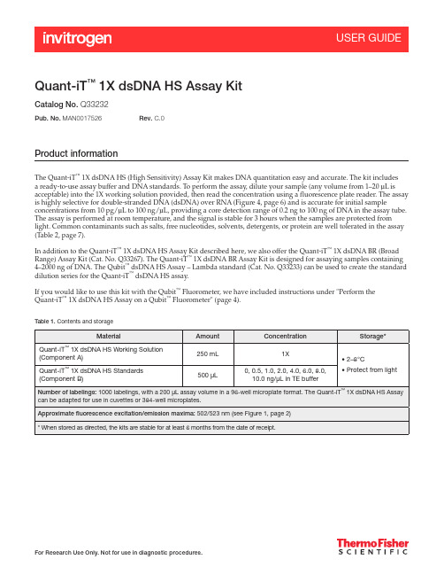
Quant-iT ™ 1X dsDNA HS Assay KitCatalog No. Q33232Product informationThe Quant-iT ™ 1X dsDNA HS (High Sensitivity) Assay Kit makes DNA quantitation easy and accurate. The kit includes a ready-to-use assay buffer and DNA standards. To perform the assay, dilute your sample (any volume from 1–20 μL is acceptable) into the 1X working solution provided, then read the concentration using a fluorescence plate reader. The assay is highly selective for double-stranded DNA (dsDNA) over RNA (Figure 4, page 6) and is accurate for initial sampleconcentrations from 10 pg/μL to 100 ng/μL, providing a core detection range of 0.2 ng to 100 ng of DNA in the assay tube. The assay is performed at room temperature, and the signal is stable for 3 hours when the samples are protected from light. Common contaminants such as salts, free nucleotides, solvents, detergents, or protein are well tolerated in the assay (Table 2, page 7).In addition to the Quant-iT ™ 1X dsDNA HS Assay Kit described here, we also offer the Quant-iT ™ 1X dsDNA BR (Broad Range) Assay Kit (Cat. No. Q33267). The Quant-iT ™ 1X dsDNA BR Assay Kit is designed for assaying samples containing 4–2000 ng of DNA. The Qubit ™ dsDNA HS Assay – Lambda standard (Cat. No. Q33233) can be used to create the standard dilution series for the Quant-iT ™ dsDNA HS assay.If you would like to use this kit with the Qubit ™ Fluorometer, we have included instructions under "Perform the Quant-iT ™ 1X dsDNA HS Assay on a Qubit ™ Fluorometer" (page 4).Table 1.Contents and storagePub. No. MAN0017526Rev. C.0Critical assay parametersAssay temperatureThe Quant-iT ™ 1X dsDNA HS Assay delivers optimal performance when all solutions are at room temperature (18–28˚C). Temperature fluctuations can influence the accuracy of the assay (Figure 5, page 6).To minimize temperature fluctuations, insert all assay tubes into the fluorescence microplate reader only for as much time as it takes for the instrument to measure the fluorescence. Do not hold the assay tubes in your hand before reading because this warms the solution and results in a different reading.Incubation timeTo allow the Quant-iT ™ 1X dsDNA HS Assay to reach optimal fluorescence, incubate the tubes for 2 minutes after mixing the sample or the standard with the working solution. After this incubation period, the fluorescence signal is stable for 3 hours at room temperature when samples are protected from light.Photostability of Quant-iT ™reagentsThe Quant-iT ™ reagents exhibit high photostability, showing <0.3% drop in fluorescence after 9 readings and <2.5% drop in fluorescence after 40 readings.Handling and disposalNo data are currently available that address the mutagenicity or toxicity of theQuant-iT ™ 1X dsDNA HS Reagent (the dye in Component A). This reagent is known to bind nucleic acids. Treat the Quant-iT ™ 1X dsDNA HS working solution with the same safety precautions as all other potential mutagens and dispose of the dye in accordance with local regulations.Figure 1. Excitation and emission maxima for the Quant-iT ™1X dsDNA HS reagent when bound to dsDNA.Perform the Quant-iT™ dsDNA HS Assay on a fluorescence microplate readerThis protocol describes the use of the Quant-iT™ 1X dsDNA HS Assay Kit with afluorescence microplate reader that is equipped with either a monochrometer orexcitation and emission filters appropriate for fluorescein or Alexa Fluor™ 488 dye(Figure 1, page 2). Some contaminating substances may interfere with the assay; formore information, see "Contaminants tolerated by the Quant-iT™ 1X dsDNA HS Assay"(page 7). For an overview of this procedure, see Figure 2.Figure 2. The Quant-iT™ dsDNA High-Sensitivity assay.Assay procedure IMPORTANT! For best results, ensure that all materials and reagents are at roomtemperature.1.1 Add 10 μL of each Quant-iT™ 1X dsDNA HS Standard to separate wells. Duplicates ortriplicates of the standards are recommended.1.2 Add 1–20 µL of each unknown DNA sample to separate wells. Duplicates or triplicatesof the unknown samples are recommended.1.3 Load 200 μL of the Quant-iT™ 1X dsDNA working solution into each microplate well.This can be done readily using a multichannel pipettor.If possible, mix your 96-well plate using a plate mixer or using the plate reader for1.4about 3–10 seconds. Following mixing, allow the plate to incubate at room temperaturefor 2 minutes..Measure the fluorescence using a microplate reader (excitation/emission maxima are1.5502/523 nm; see Figure 1, page 2). Standard fluorescein wavelengths (excitation/emission at ~480/530 nm) are appropriate for this dye. The fluorescence signal is stablefor 3 hours at room temperature when protected from light.Use a standard curve to determine the DNA amounts. For the dsDNA standards, plot1.6amount vs. fluorescence, and fit a straight line to the data points.Note: Many curve fitting programs will calculate the y-intercept. However, for bestresults, manually set the y-intercept as the RFU value obtained from the 0 ng/μLdsDNA standard.Data analysis considerations –standard curves and extendedranges The fluorescence of the Quant-iT™ 1X dsDNA HS reagent bound to dsDNA is extremelylinear from 0–100 ng. For best results at the low end of the standard curve, the lineshould be forced through the background point (or through zero, if backgroundhas been subtracted). When 10 μL volumes of the standards are used, the lowestDNA-containing standard represents 5 ng of DNA; nevertheless, highly accuratedeterminations of DNA down to 0.2 ng are attained using the standard curve asdescribed above.To assess the reliability of the assay in the low range, use smaller volumes of thestandards; for example, 2 μL volumes for a standard curve ranging from 0–20 ng.Alternatively, dilute the standards in buffer for an even tighter range. Duringdevelopment of the Quant-iT™ 1X dsDNA HS assay, we were able to detect 0.05 ng ofλ DNA under ideal experimental circumstances (using calibrated pipettors, octuplicatedeterminations, the best microplate readers, and Z-factor1 analysis). Your results mayvary.If desired, the utility of the Quant-iT™ 1X dsDNA HS assay can be extended beyond100 ng, up to 200 ng. For standards in this range, use 20 μL volumes of the providedstandards. Note that the standard curve may not be linear in the range 160–200 ng, andhigh levels of RNA may now interfere slightly with the results.Perform the Quant-iT™ dsDNA HS Assay on a Qubit™ FluorometerThe Quant-iT™ 1X dsDNA Assay Kit can be adapted for use with the Qubit™Fluorometer. The protocol below is abbreviated from the Qubit™ Fluorometer userguide, which is available at /qubit. Although a step-by-step protocoland critical assay parameters are given here, more detail is available in the Qubit™Fluorometer user guide and you are encouraged to familiarize yourself with thismanual before you begin your assay. See Figure 3 for an overview of the procedure.Figure 3. Overview for using the Quant-iT™ 1X dsDNA HS assay in the Qubit™ fluorometer.Assay procedure IMPORTANT! For best results, ensure that all materials and reagents are at roomtemperature.2.1 Set up the required number of 0.5-mL tubes for standards and samples. The Quant-iT™1X dsDNA HS Assay requires 2 standards.Note: Use only thin-wall, clear, 0.5-mL PCR tubes. Acceptable tubes include Qubit™assay tubes (Cat. No. Q32856).2.2 Label the tube lids.Note: Do not label the side of the tube as this could interfere with the sample read. Labelthe lid of each standard tube correctly. Calibration of the Qubit™ Fluorometer requiresthe standards to be inserted into the instrument in the right order.2.3 Add 10 µL of the 0 ng/μL and the 10 ng/μL Quant-iT™ 1X dsDNA HS Standard to theappropriate tube2.4 Add 1–20 µL of each user sample to the appropriate tube.2.5 Add the Quant-iT™ 1X dsDNA HS Working Solution to each tube such that the finalvolume is 200 µL.Note: The final volume in each tube must be 200 µL. Each standard tube requires 190 µLof Quant-iT™ working solution, and each sample tube requires anywhere from180–199 µL.2.6 Mix each sample vigorously by vortexing for 3–5 seconds.2.7 Allow all tubes to incubate at room temperature for 2 minutes, then proceed to read thestandards and samples. Follow the procedure appropriate for your instrument:• Qubit™ Flex Fluorometer• Qubit™ 4 Fluorometer• Qubit™ 3 FluorometerNote: If you are using the Qubit™ 3 Fluorometer, download the 1X dsDNA algorithmand assay button from /qubit, then install it onto your Qubit™Fluorometer.AppendixSelectivity of the Quant-iT™ 1XdsDNA HS AssayFigure 4. DNA selectivity and sensitivity of the Quant-iT™ 1X dsDNA HS Assay (Cat. No. Q33232). Triplicate10-μL samples of λ DNA, E. coli rRNA, or a 1:1 mixture of DNA and RNA were assayed with the Quant-iT™1X dsDNA HS Assay. Fluorescence was measured at 502/532 nm and plotted versus the concentration ofthe RNA or DNA sample alone, or versus the mass of the DNA component in the 1:1 mixture. The variation(CV) of replicate DNA determinations was ≤2%. The inset is an expanded view of the low range of the assayshowing the extreme sensitivity of the assay for DNA. Background fluorescence has not been subtracted.Effect of temperature on theQuant-iT™ 1X dsDNA HSAssayFigure 5. Plot of fluorescence vs. temperature for the Quant-iT™ 1X dsDNA HS Assay. The Quant-iT™assays are designed to be performed at room temperature, as temperature fluctuations can influence theaccuracy of the assay.Contaminants tolerated by the Quant-iT ™ 1X dsDNA HSAssayNote: While the contaminant tolerances of the Quant-iT ™ 1X dsDNA HS assay and theQuant-iT ™ dsDNA HS assay are largely similar, they are not identical.Reference1. J Biomol Screen 4, 67–73 (1999).Table 2. Effect of contaminants in the Quant-iT ™1X dsDNA HS Assay*/support | /askaquestion Limited Product WarrantyLife Technologies Corporation and/or its affiliate(s) warrant their products as set forth in the Life Technologies’ General Terms and Conditions of Sale found on Life Technologies’ website at /us/en/home/global/terms-and-conditions.html . If you have any questions, please contact Life Technologies at /support .Life Technologies Corporation | 29851 Willow Creek Road | Eugene, OR 97402 USAFor descriptions of symbols on product labels or prodoct documents, go to /symbols-definition .The information in this guide is subject to change without notice.DISCLAIMER: TO THE EXTENT ALLOWED BY LAW, LIFE TECHNOLOGIES AND/OR ITS AFFILIATE(S) WILL NOT BE LIABLE FOR SPECIAL, INCIDENTAL,INDIRECT, PUNITIVE, MULTIPLE OR CONSEQUENTIAL DAMAGES IN CONNECTION WITH OR ARISING FROM THIS DOCUMENT, INCLUDING YOUR USE OF IT.Important Licensing Information: These products may be covered by one or more Limited Use Label Licenses. By use of these products, you accept the terms and conditions of all applicable Limited Use Label Licenses.Revision history:Pub. No. MAN0017526©2021 Thermo Fisher Scientific Inc. All rights reserved. All trademarks are the property of Thermo Fisher Scientific and its subsidiaries unless otherwise specified .Ordering informationCat. No. Product name Unit size Q33232Quant-iT™ 1X dsDNA HS Assay Kit.................................................................... 1 kitRelated products Q33267 Quant-iT™ 1X dsDNA BR Assay Kit.................................................................... 1 kit Q33120 Quant-iT™ dsDNA Assay Kit, High Sensitivity............................................................ 1 kit Q33130 Quant-iT™ dsDNA Assay Kit, Broad Range.............................................................. 1 kit Q10213 Quant-iT™ RNA Assay Kit, Broad Range................................................................ 1 kit Q33140 Quant-iT™ RNA Assay Kit, 1000 assays ................................................................ 1 kit Q32882 Quant-iT™ microRNA Assay Kit, 1000 assays............................................................ 1 kit Q33210 Quant-iT™ Protein Assay Kit, 1000 assays .............................................................. 1 kit O11492 Quant-iT™ OliGreen™ ssDNA Assay Kit ................................................................ 1 kit Q33233 Qubit™ 1X dsDNA Assay- Lambda Standard ............................................................ 1 kit Q33238 Qubit™ 4 Fluorometer with WiFi....................................................................... 1 each Q33327 Qubit™ Flex Fluorometer ............................................................................ 1 each Q33252 Qubit™ Flex Assay Tube Strips .................................................................. 125 tube strips M33089 Microplates for fluorescence-based assays, 96-well (black-walled, clear bottom) ................................ 10 plates。
倍他米松磷酸钠注射液治疗早产有效性和安全性研究进展
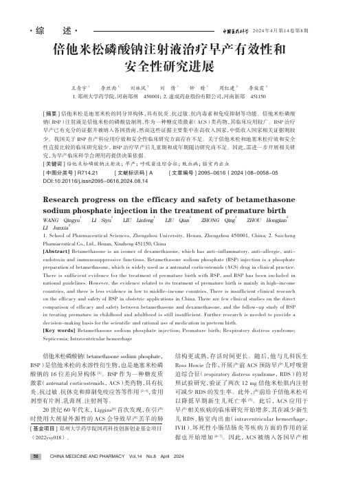
2024年4月第14卷第8期·综 述·[基金项目] 郑州大学药学院国药科技创新创业基金项目(2022yxy018)。
倍他米松磷酸钠注射液治疗早产有效性和安全性研究进展王青宇1 李丝雨1 刘林凤1 刘 倩1 钟 晴1 周红建2 李俊霞21.郑州大学药学院,河南郑州 450001;2.遂成药业股份有限公司,河南新郑 451150[摘要]倍他米松是地塞米松的同分异构体,具有抗炎、抗过敏、抗内毒素和免疫抑制等功能。
倍他米松磷酸钠(BSP)注射液是倍他米松的磷酸盐制剂,作为一种糖皮质激素(ACS)类药物,其临床应用较广。
BSP 治疗早产已有充分的证据并被纳入各国指南,然而这些证据主要集中在高收入国家,中低收入国家相关证据则较少。
我国关于BSP 在产科应用疗效和安全性临床研究方面存在不足。
关于倍他米松和地塞米松疗效和安全性直接比较的临床研究较少,BSP 治疗早产后儿童期和成年期随访研究尚不足。
因此,需进一步开展相关研究,为早产临床科学合理用药提供决策依据。
[关键词]倍他米松磷酸钠注射液;早产;呼吸窘迫综合征;败血病;脑室内出血[中图分类号] R714.21 [文献标识码] A [文章编号] 2095-0616(2024)08-0058-05DOI:10.20116/j.issn2095-0616.2024.08.14Research progress on the efficacy and safety of betamethasonesodium phosphate injection in the treatment of premature birthWANG Qingyu 1 LI Siyu 1 LIU Linfeng 1 LIU Qian 1 ZHONG Qing 1 ZHOU Hongjian2LI Junxia21. School of Pharmaceutical Sciences, Zhengzhou University, Henan, Zhengzhou 450001, China;2. Suicheng Pharmaceutical Co., Ltd., Henan, Xinzheng 451150, China[Abstract] Betamethasone is an isomer of dexamethasone, which has anti-inflammatory, anti-allergic, anti-endotoxin and immunosuppressive functions. Betamethasone sodium phosphate (BSP) injection is a phosphate preparation of betamethasone, which is widely used as a antenatal corticostemids (ACS) drug in clinical practice. There is sufficient evidence for the treatment of premature birth with BSP, and BSP has been included in national guidelines. However, the evidence related to its treatment of premature birth is mainly in high-income countries, and there is less evidence in low to middle-income countries. There is insufficient clinical research on the efficacy and safety of BSP in obstetric applications in China. There are few clinical studies on the direct comparison of efficacy and safety between betamethasone and dexamethasone, and the follow-up study of BSP in treating premature in childhood and adulthood is still insufficient. Further research is needed to provide a decision-making basis for the scientific and rational use of medication in preterm birth.[Key words] Betamethasone sodium phosphate injection; Premature birth; Respiratory distress syndrome; Septicemia; Intraventricular hemorrhage倍他米松磷酸钠(betamethasone sodium phosphate,BSP)是倍他米松的水溶性衍生物,也是地塞米松磷酸钠的16位差向异构体[1]。
NAD-苹果酸脱氢酶(NAD-MDH)活性检测试剂盒说明书

NAD-苹果酸脱氢酶(紫外分光光度法货号:BC1040规格:50T/48S产品组成:使用前请认真核对试剂体积与瓶内体积是否一致,有疑问请及时联系索莱宝工作人员。
试剂名称规格保存条件提取液液体50 mL×1瓶4℃保存试剂一液体40 mL×1瓶4℃保存试剂二粉剂×2支-20℃保存试剂三粉剂×2支-20℃保存溶液的配制:1、试剂二:临用前加入360 μL双蒸水,用不完的试剂仍-20℃保存;2、试剂三:临用前加入327 μL双蒸水,用不完的试剂仍-20℃保存。
产品说明:MDH(EC 1.1.1.37)广泛存在于动物、植物、微生物和培养细胞中,线粒体中MDH是TCA循环的关键酶之一,催化苹果酸形成草酰乙酸;相反,胞浆中MDH催化草酰乙酸形成苹果酸。
草酰乙酸是重要的中间产物,连接多条重要的代谢途径。
因此,MDH在细胞多种生理活动中扮演着重要的角色,包括线粒体的能量代谢、苹果酸-天冬氨酸穿梭系统、活性氧代谢和抗病性等。
根据不同的辅酶特异性,MDH分为NAD-依赖的MDH和NADP-依赖的MDH,细菌中通常只含有NAD-MDH,在真核细胞中,NAD-MDH分布于细胞质和线粒体中。
NAD-MDH催化NADH还原草酰乙酸生成苹果酸,导致340nm处光吸收下降。
注意:实验之前建议选择2-3个预期差异大的样本做预实验。
如果样本吸光值不在测量范围内建议稀释或者增加样本量进行检测。
需自备的仪器和用品:紫外分光光度计、台式离心机、水浴锅、可调式移液器、1mL石英比色皿、研钵/匀浆器、蒸馏水。
操作步骤:一、样本处理(可适当调整待测样本量,具体比例可以参考文献)细菌或培养细胞:收集细菌或细胞到离心管内,离心弃上清,按照每200万细菌或细胞加入400μL提取液,超声波破碎细菌或细胞(功率20%,超声3s,间隔10s,重复30次),8000g 4℃离心10min,取上清,置冰上待测。
组织:称取约0.05g组织,加入1mL提取液进行冰浴匀浆;8000g 4℃离心10min,取上清,置冰上待测。
Y-27632_DataSheet_MedChemExpress
Inhibitors, Agonists, Screening Libraries Data SheetBIOLOGICAL ACTIVITY:Y–27632 is an ATP–competitive inhibitor of ROCK–I and ROCK–II , with K i of 220 nM and 300 nM for ROCK–I and ROCK–II , respectively.IC50 & Target: Ki: 220/300 nM (ROCK–I/II)[1]In Vitro: Y–27632 inhibits the ROCK family of kinases 100 times more potently than other kinases including protein kinase C,cAMP–dependent kinase and myosin light chain kinase. Y–27632 prolongs the lag time and delays the appearance of BrdU–labeled cells in a concentration–dependent manner, delays of about 1 and 4 h are noticed in the Swiss 3T3 cells treated with 10 and 100 μM Y–27632, respectively [1]. Y–27632 promotes neuronal differentiation of adipose tissue–derived stem cells (ADSCs). Compared to 1.0and 2.5 μM Y–27632 induced groups, percentages of neuroal–like cells achieved a peak in the 5.0 μM Y–27632 induced group [2].In Vivo: Y–27632 (5 and 10 mg/kg) significantly prolongs the onset time of myoclonic jerks when compare with saline group.Y–27632 (5 and 10 mg/kg) significantly prolongs the onset time of clonic convulsions when compare with saline group [3].Treatment with Dimethylnitrosamine (DMN) causes a significant decrease in rat body and liver weight (DMN–S group) compared with control animals (S–S group). Oral Y27632 (30 mg/kg) essentially prevents this DMN–induced rat body and liver weight loss (DMN–Y group)[4].PROTOCOL (Extracted from published papers and Only for reference)Kinase Assay:[1]Recombinant ROCK–I, ROCK–II, PKN, or citron kinase is expressed in HeLa cells as Myc–tagged proteins by transfection using Lipofectamine, and is precipitated from the cell lysates by the use of 9E10 monoclonal anti–Myc antibodycoupled to G protein–Sepharose. Recovered immunocomplexes are incubated with various concentrations of [32P]ATP and 10 mg of histone type 2 as substrates in the absence or presence of various concentrations of either Y–27632 or Y–30141 at 30°C for 30 min in a total volume of 30 μL of the kinase buffer containing 50 mM HEPES–NaOH, pH 7.4, 10 mM MgCl 2, 5 mM MnCl 2, 0.02% Briji 35, and 2 mM dithiothreitol. PKCa is incubated with 5 μM [32P]ATP and 200 μg/mL histone type 2 as substrates in the absence or presence of various concentrations of either Y–27632 or Y–30141 at 30°C for 10 min in a kinase buffer containing 50 mM Tris–HCl,pH 7.5, 0.5 mM CaCl 2, 5 mM magnesium acetate, 25 μg/mL phosphatidyl serine, 50 ng/mL 12–O–tetradecanoylphorbol–13–acetate and 0.001% leupeptin in a total volume of 30 μL. Incubation is terminated by the addition of 10 μL of 43 Laemmli sample buffer.After boiling for 5 min, the mixture is subjected to SDS–polyacrylamide gel electrophoresis on a 16% gel. The gel is stained withCoomassie Brilliant Blue, and then dried. The bands corresponding to histone type 2 are excised, and the radioactivity is measured [1]. Cell Assay: Y–27632 is dissolved in water and stored [1].[1]HeLa cells are plated at a density of 3×10⁴ cells per 3.5–cm dish. The cells are cultured in DMEM containing 10% FBS in the presence of 10 mM Thymidine for 16 h. After the cells are washed with DMEM containing 10% FBS, they are cultured for an additional 8 h, and then 40 ng/mL of Nocodazole is added. After 11.5 h of theNocodazole treatment, various concentrations of Y–27632 (0–300 μM), Y–30141, or vehicle is added and the cells are incubated for another 30 min [1].Animal Administration: Y–27632 is dissolved in 0.9% NaCl (saline) (Mice)[3].Product Name:Y–27632Cat. No.:HY-10071CAS No.:146986-50-7Molecular Formula:C 14H 21N 3O Molecular Weight:247.34Target:ROCK; ROCK; ROCK Pathway:TGF–beta/Smad; Stem Cell/Wnt; Cell Cycle/DNA Damage Solubility:DMSO: ≥ 32 mg/mLY–27632 is dissolved in saline (final concentration 2%) (Rat)[4].[3][4]Mice[3]Male, inbred Swiss albino mice (2–3 months old) weighing 25–30 g are used. Mice are injected with a sub–convulsive dose of PTZ (35 mg/kg, i.p.) (on Mondays, Wednesdays and Fridays) of each week for a total of 11 injections. After each PTZ injection, mice are observed for 30 min and the occurrence of convulsive activity is recorded. After 30 min, the mice are then injected with either Fasudil (25 mg/kg, i.p.) or Y–27632 (5 mg/kg, i.p.) and returned to their home cages until the next injection. Control mice for Fasudil andY–27632 receives saline.Rat[4]Male Wistar Kind A rats (200–250 g) are used. DMN (1 g/mL) is diluted ten times with saline (final concentration 1%) and 10 mg/kg per day of DMN is injected intraperitoneally (i.p.) on the first 3 days of each week for 4 weeks. Y27632 is given orally once per day at a dose of 30 mg/kg for 4 weeks starting on the day of the first injection of DMN. The dose of 30 mg/kg corrects hypertension in several rat models without toxicity. Twenty rats are randomized into four experimental groups (n=5 in each group) as follows: (1) S–S (injection of saline i.p. and oral administration of saline); (2) S–Y (injection of saline i.p. and oral administration of Y27632); (3) DMN–S (DMN i.p. and oral administration of saline); (4) DMN–Y (DMN i.p. and oral administration of Y27632). The rats are weighed every week. They are sacrificed at the end of the fourth week and the liver is excised. In addition, a blood sample is taken immediately before the rats are sacrificed.References:[1]. Ishizaki T, et al. Pharmacological properties of Y–27632, a specific inhibitor of rho–associated kinases. Mol Pharmacol. 2000 May;57(5):976–83.[2]. Xue ZW, et al. Rho–associated coiled kinase inhibitor Y–27632 promotes neuronal–like differentiation of adult human adipose tissue–derived stem cells.Chin Med J (Engl). 2012 Sep;125(18):3332–5.[3]. Inan S, et al. Antiepileptic effects of two Rho–kinase inhibitors, Y–27632 and fasudil, in mice. Br J Pharmacol. 2008 Sep;155(1):44–51.[4]. Tada S, et al. A selective ROCK inhibitor, Y27632, prevents dimethylnitrosamine–induced hepatic fibrosis in rats. J Hepatol. 2001 Apr;34(4):529–36.Caution: Product has not been fully validated for medical applications. For research use only.Tel: 609-228-6898 Fax: 609-228-5909 E-mail: tech@Address: 1 Deer Park Dr, Suite Q, Monmouth Junction, NJ 08852, USA。
