美国药学科学家协会(AAPS) 2015年海报系列 - 多因子检测与Biomarker筛选
fda、tga、pmda、edqm一些基础知识

FDA、TGA、PMDA、EDQM一些基础知识CFDA系将食品安全办的职责、食品药品监管局的职责、质检总局的生产环节食品安全监督管理职责、工商总局的流通环节食品安全监督管理职责整合组建而成,负责药品、医疗器械、化妆品和消费环节食品安全的监督管理。
CFDA于2013年3月22日正式挂牌成立,食品药品监督管理局的官网也同步进行了更名,一律改成国家食品药品监督管理总局,英文简称由“SFDA”变成“CFDA”,就连原先的官方微博“中国药监”也改成了“中国食药监”美国食品和药物管理局(Foodand Drug Administration)简称FDA,FDA 是美国政府在健康与人类服务部(DHHS) 和公共卫生部 (PHS) 中设立的执行机构之一。
作为一家科学管理机构,FDA 的职责是确保美国本国生产或进口的食品、化妆品、药物、生物制剂、医疗设备和放射产品的安全。
它是最早以保护消费者为主要职能的联邦机构之一。
TGA[1] 是TherapeuticGoods Administration的简写,全称是治疗商品管理局,它是澳大利亚的治疗商品(包括药物、医疗器械、基因科技和血液制品)的监督机构。
依据1989年的治疗商品法案,TGA是递属于澳大利亚政府健康和老龄部下的一个部门。
TGA开展一系列的评审和监督管理工作,以确保在澳大利亚提供的治疗商品符合适用的标准,并保证澳大利亚社会的治疗水平在一个较短的时间内达到较高的水平。
欧洲药品质量理事会(European Directorate for Quality Medicines)简称EDQM,是欧洲理事会下属的药品管理系统的核心,旨在保证在欧洲生产和销售的药品具有同等优良的品质。
EDQM的职能是建立药品的质量标准以供欧洲药典委员会使用,制备标准品CRS,执行COS程序最终颁发COS证书等等。
欧洲药物管理局(European Medicines Agency)简称EMA,是欧洲官方药管机构之一,EMA主要负责欧盟市场药品的审查、批准上市,评估药品科学研究,监督药品在欧盟的安全性、有效性。
欧洲食品安全局第六次发现 益生菌与改善免疫系统之间没有任何因果关系
u eC crn 写道 。
场 条件制 约 了这项 工 作 的进 展 。希望 能 够 允许
A D排 污 系 统 公 司 总部 设 在 温 哥 华 , 们 T 他 过去 1 0年 当 中开发 了一套 固液 分离 系 统 , 将 可 废 水 回收 , 作动 物 的饮 水 。 用
该处 理系 统对土 地没 有任何 废 物排放 , 对水 体也 没有 任何废 物排 放 。 所有粪 污要 么制成 颗粒 , 么 回收成 为猪 只 要
的饮水 。 V nSy e 生 说 A D正 在 同 中 国 的 几 家 a lk 先 T
在希 腊 ,0 的 养 猪 生 产 者 陷 入 严 重 的 困 7%
造。
欧盟准 备禁用母猪 限位栏
欧盟最 近在 布鲁 塞尔 的会议 上 , 欧盟各 国的 代表很 不情 愿地 公 布 了 各 自为 禁 用 母 猪 限位 栏 做 好准 备 的情 况 , 项禁 令将 于 2 1 这 0 3年生 效 。 1 月早 些时候 在布 鲁 塞 尔 的欧 盟 猪 肉价 格 1 预 测小组 上 , 国国家养 猪协会 ( P 的 B re 英 N A) an y K y 求 各成员 国介 绍 自己 国家为 2 1 a要 0 3年部 分 禁用 限位栏 所做 准备 的情况 。 根据 B E 英 国养猪 委员 会 ) 报报 导 , P X( 周 他
我们 先从尿 液 中除去 氨 , 然后 再 降低总 溶解
固形 物 的含 量 , 后 用紫 外线 照射 , 时 回 收水 最 此 的总溶 解 固形物含 量可 降到 1rg k 。 / g 3 a
这 个指标 仅 相 当 于加 拿 大普 通 瓶 装 饮 用 水 的八分 之一甚 至更低 。 干粪 只需 干燥 , 而且 我们把 液粪 处理 过程 中 产 生的所 有 副产物都 加 到干粪 中一起 干燥 , 制 并
广告中的生物学资料

1红牛补充能量,里面含有大量糖分,糖类是主要的能源物质2纳爱斯牙膏,添加维生素C,防止牙龈出血,维生素C有促进伤口愈合的作用3汰渍加酶洗衣粉,酶有催化作用,如果加了蛋白酶,可以加速奶渍,血迹这一类污渍的分解4养生堂天然维E 维生素E是一种抗氧化剂,延缓衰老,有效减少皱纹的产生防止血液的凝固,减少斑纹组织的产生等等,功能较多5补铁、补血,效果好!补血口服液!——铁与血红蛋白有关,缺铁会得贫血症。
6健康的体魄,来源于‘碘碘’滴滴——碘与甲状腺素有关。
幼年缺碘得呆小症。
成年缺碘得大脖子病(地方性甲状腺肿)。
7万丈高楼平地起,层层都是‘钙’起来——钙与骨骼有关。
幼年缺钙得佝偻病,成年得骨质疏松症。
8.聪明伶俐,坚持补锌;有锌万事成;用心的妈妈会用‘锌’——缺锌生长发育停滞,味觉下降。
9哇哈哈AD钙奶,AD搭配更容易吸收!10.21金维他,保障生命为他——维生素对生命的重要性11大宝SOD蜜。
SOD(超氧化物歧化酶)是一种源于生命体的活性物质,能消除生物体在新陈代谢过程中产生的有害物质。
对人体不断地补充 SOD具有抗衰老的特殊效果。
12脑白金广告,脑白金富含褪黑激素,是人脑中松果体分泌的激素,有助于抗癌,提高免疫力。
13有关婴儿奶粉的广告,含有各种氨基酸、维生素等,是人体需要的必需氨基酸和维他命警惕产品广告中的伪科学有机食品并不完美注意虚假广告的识别:下列是几则广告语:①这种食品由纯天然谷物制成,不含任何糖类,糖尿病患者也可放心大量食用②这种饮料含有多种无机盐,能有效补充人体运动时消耗的能量③这种营养品含有人体所需的全部20种必需氨基酸④这种口服液含有丰富的钙、铁、锌、硒等微量元素请判断在上述广告语中,有多少条在科学性上有明显的错误?A.1条B.2条C.3条D.4条①这种食品由纯天然谷物制成,不含任何糖类,糖尿病患者也可放心大量食用淀粉也是糖类,谷物,糖尿病人是要限量摄入的②这种饮料含有多种无机盐,能有效补充人体运动时消耗的能量如果扔掉前后两句之间可疑的因果关系,也许还没关系,如果当做因果关系来读,就扯淡了,无机盐哪能提供能量?③这种营养品含有人体所需的全部20种必需氨基酸人体含有的20中氨基酸,但是所谓“必需氨基酸”是指8种人体自身不能合成的氨基酸④这种口服液含有丰富的钙、铁、锌、硒等微量元素钙是大量元素钙和铁不是微量元素卫生部通报确认珍奥核酸等6种保健品夸大宣传本报记者郑淑华报道: 昨天,卫生部向珍奥核酸胶囊、忘不了3A脑营养胶丸等6种曾经一度热销的保健食品亮起红灯,其原因是这些保健食品夸大宣传,误导了消费者。
plitidepsin 序列
plitidepsin 序列
Splitidepsin是一种天然产物,也被称为Aplidin或Plitidepsin。
它是由西班牙海洋微生物Aplidium albicans产生的一种环肽类化合物。
Splitidepsin的化学结构非常复杂,由148个氨基酸残基组成,其中包含多种非天然氨基酸。
Splitidepsin的序列是保密的商业机密,因此无法在公开的资源中获取到具体的序列信息。
只有相关研究人员和生产厂商才能获得该化合物的详细信息。
然而,根据已有的研究文献,Splitidepsin的结构和活性特点已经得到了一定程度的描述。
Splitidepsin被认为是一种强效的抗肿瘤药物,具有抗癌活性。
它通过多种机制抑制肿瘤细胞的生长和扩散,包括抑制蛋白质合成、诱导细胞凋亡、阻断细胞周期等。
尽管Splitidepsin在临床应用中显示出了一定的抗肿瘤活性,但目前仍然需要进一步的研究和临床试验来评估其安全性和疗效。
因此,具体的Splitidepsin序列信息仍然是受限的,并且可能受到专利保护。
总结起来,Splitidepsin是一种复杂的环肽类化合物,具有潜
在的抗肿瘤活性。
然而,具体的Splitidepsin序列信息是商业机密,只有相关研究人员和生产厂商才能获得。
药物化学SCI影响因子及分区
药物化学SCI影响因子及分区药物化学是研究药物分子的结构、合成、性质和作用机理的学科。
SCI影响因子是科学引文索引(Science Citation Index,简称SCI)中期刊的一个重要指标,它衡量着其中一期刊在特定年份中被引用的频率,并表示该期刊在学术界的影响力。
本文将介绍一些常见的药物化学领域SCI期刊的影响因子及分区情况。
药物化学领域的SCI期刊很多,不同期刊的影响因子及分区也存在一定的差异。
下面是一些常见的药物化学领域SCI期刊及其影响因子和分区情况:1. Journal of Medicinal Chemistry(药物化学杂志)影响因子:6.205(2024年数据)分区:Q12. European Journal of Medicinal Chemistry(欧洲药物化学杂志)影响因子:5.573(2024年数据)分区:Q13. Bioorganic & Medicinal Chemistry(生物有机与药物化学)影响因子:3.073(2024年数据)分区:Q24. ACS Medicinal Chemistry Letters(ACS医药化学快报)影响因子:4.609(2024年数据)分区:Q15. Pharmaceutical Research(药物研究)影响因子:4.729(2024年数据)分区:Q16. Medicinal Chemistry Research(药物化学研究)影响因子:1.787(2024年数据)分区:Q3总结起来,药物化学领域有许多有影响力的SCI期刊,其影响因子和分区情况会根据期刊的质量和影响力而有所差异。
以上所列期刊仅为常见的一部分,研究者可以根据具体需求选择适合自己研究方向的期刊。
UHPLC-MS

UHPLC-MS/MS法测定瑞戈非尼中两种痕量基因毒性杂质陈爽ꎬ刘柱ꎬ石云峰ꎬ朱价ꎬ罗英(浙江省食品药品检验研究院ꎬ浙江杭州310058)摘要:目的㊀建立超高效液相色谱-串联质谱(UHPLC-MS/MS)方法测定瑞戈非尼中两类基因毒性杂质[3-氟-4-氨基苯酚(SM2)㊁3-三氟甲基-4-氯异氰酸苯酯(SM3)]的含量ꎮ方法㊀采用色谱柱AgilentRRHDC18(2.1mmˑ100mmꎬ1.8μm)ꎬ流动相为0.05%甲酸水-甲醇ꎬ梯度洗脱ꎬ流速为0.3mL min-1ꎬ柱温为35ħꎻ采用Agilent1290-6470A液质联用仪和电喷雾离子源(AJSESI)源检测ꎬ正负离子模式采集数据ꎮ结果㊀本方法的专属性㊁线性与范围㊁定量限与检测限㊁准确度㊁精密度及稳定性均符合«中国药典»的验证指标ꎮ结论㊀本方法操作简便ꎬ结果可靠ꎬ用于两批瑞戈非尼中潜在毒性杂质(3-氟-4-氨基苯酚㊁3-三氟甲基-4-氯异氰酸苯酯)的测定ꎮ两批样品中均未检测到SM2ꎬ而基因毒性杂质SM3的含量分别为13.6ˑ10-6和41.4ˑ10-6ꎮ关键词:瑞戈非尼ꎻ基因毒性杂质ꎻ超高效液相色谱-串联质谱中图分类号:R927.1㊀文献标志码:A㊀文章编号:2095-5375(2023)07-0485-005doi:10.13506/j.cnki.jpr.2023.07.010DeterminationoftwotracegenotoxicimpuritiesinRegorafenibbyUHPLC-MS/MSCHENShuangꎬLIUZhuꎬSHIYunfengꎬZHUJiaꎬLUOYing(ZhejiangInstituteforFoodandDrugControlꎬHangzhou310058ꎬChina)Abstract:Objective㊀ToestablishanUHPLC-MS/MSanalyticalmethodforthedeterminationofgenotoxicimpuritiesinRegorafenib.Methods㊀ThemethodwasachievedonaAgilentRRHDC18column(2.1mmˑ100mmꎬ1.8μm)utilizingamobilephaseof0.05%Formicacidwater(A)-Methanol(B)withgradientelutionattheflowrateof0.3mL min-1.Thetemperatureofcolumnwassetat35ħ.TheAgilent1290-6470ALC-MSwasusedtodetect(AJSESIsourceꎬinMultipleReactionMonitoringmode).Results㊀ThespecificityꎬlinearityandrangeꎬLOQandLODꎬaccuracyꎬprecisionandstabilityofthemethodwereallinaccordancewiththevalidationofChinesePharmacopoeia.Conclusion㊀Theproposedmethodwassuccessfullyappliedtodeterminethetrace-levelPGIsintwobatchesofregorafenib.ThecontentsofSM2werenotobservedinthetwobatchesofsamplesꎬwhereasthecontentsofSM3weredeterminedtobe13.6ˑ10-6and41.4ˑ10-6ꎬrespectively.Keywords:RegorafenibꎻGenotoxicimpurityꎻUHPLC-MS/MS㊀㊀基因毒性杂质(或遗传毒性杂质ꎬgenotoxicim ̄purityꎬGTI)是少量即能引起细胞DNA损伤ꎬ诱导基因突变ꎬ并具有致癌倾向的化合物[1]ꎮ欧洲药品管理局(EMEA)㊁美国食品药品管理局(FDA)及人用药品注册技术要求国际协调会(ICH)先后颁布了基因毒性杂质控制的指导文件[2-4]ꎬ推荐以毒理学关注阈值(TTCꎬ1.5μg d-1)来控制用药风险ꎬ并发布基因毒性警示结构基团ꎮ瑞戈非尼(Regorafenib)是拜耳药业研发并于2017年3月在国内上市的一种新型口服多靶点的磷酸激酶抑制剂ꎬ本品以促进肿瘤血管生成和细胞增殖的多种蛋白激酶为靶标ꎬ达到治疗晚期及转移性结肠癌㊁进展期肝细胞癌㊁胃肠道恶性间质肿瘤㊁儿童神经母细胞瘤和胶质母细胞瘤及一些转移性实体肿瘤的目的ꎮ瑞戈非尼的合成路线中起始物料SM2㊁SM3结构中均含有苯胺类基因毒性警示结构基团ꎬ具有潜在基因毒性ꎬ详见图1ꎮ瑞戈非尼已收载于«中国药典»和«欧洲药典»ꎬ已经报道了一种LC-MS/MS法定量同时测定肝细胞癌患者血浆中瑞戈非尼中及其两种活性代谢物的含量的方法[17]ꎬ但目前也未见相关文献报道本品中两种苯胺类基因毒性杂质SM2㊁SM3的测定方法ꎮ㊀作者简介:陈爽ꎬ女ꎬ硕士ꎬ副主任药师ꎬ研究方向:药物质量研究㊁标准提高研究ꎬE-mail:credear@126.comA.3-氟-4-氨基苯酚(SM2)ꎻB.3-三氟甲基-4-氯异氰酸苯酯(SM3)图1㊀瑞戈非尼中苯胺类基因毒性杂质结构因此建立超高效液相色谱-串联质谱(UHPLC-MS/MS)方法对瑞戈非尼中少量的苯胺类基因毒性杂质的含量进行控制ꎮ本研究依照ICH推荐的条件ꎬ建立了方法并从方法的专属性㊁溶液稳定性㊁线性与范围㊁定量限与检测限㊁准确度㊁精密度方面进行了方法学研究ꎬ本法灵敏㊁可靠㊁简便ꎬ为瑞戈非尼工艺过程控制和质量保障提供参考依据ꎮ1㊀仪器与试剂Agilent1290-6470ALC-MS联用系统(美国安捷伦)ꎬ配备电喷雾离子源(AJSESI)ꎬ质谱软件为AgilentMassHunter工作站ꎻXS205DU百万分之一电子天平(瑞士梅特勒托利多公司)ꎮAgilentRRHDC18(2.1mmˑ100mmꎬ1.8μmꎬ填料:十八烷基硅烷键合硅胶ꎻAgilent公司)ꎮ3-氟-4-氨基苯酚对照品(批号:P1156811ꎬ纯度:98%)ꎬ3-三氟甲基-4-氯异氰酸苯酯对照品(杭州华东医药集团新药研究院有限公司ꎬ批号:191201ꎬ纯度:99.76%)ꎻ瑞戈非尼(批号:RGFN-B-190901㊁Z193-A1911001ꎬ由A公司提供)ꎻ甲醇(默克试剂有限公司)㊁异丙醇㊁甲酸(阿拉丁有限公司)均为色谱级ꎮ2㊀方法与结果2.1㊀色谱条件㊀液相色谱柱:AgilentRRHDC18(2.1mmˑ100mmꎬ1.8μm)ꎻ流速0.3mL min-1ꎻ0.05%甲酸水为流动相Aꎬ甲醇为流动相Bꎬ梯度洗脱(0~1.2minꎬ10%Bꎻ1.2~4.0minꎬ10%Bң60%Bꎻ4.0~8.0minꎬ60%Bꎻ8.0~13.0minꎬ95%Bꎻ13.0~13.10minꎬ95%Bң10%Bꎻ13.10~15.0minꎬ10%B)ꎻ柱温35ħꎻ进样体积5μLꎮ取0.6ng mL-1混标对照品溶液ꎬ在MRM模式下进样分析ꎬ结果如图2所示ꎬ各杂质之间分离度良好ꎮ2.2㊀质谱条件㊀采用电喷雾离子源(AJSESI)ꎬ正负离子检测模式MRM采集(SM2正离子模式㊁SM3㊀SM2[3-氟-4-氨基苯酚(3-Fluoro-4-aminophenol)]ꎻSM3[3-三氟甲基-4-氯异氰酸苯酯(3-trifluoromethyl-4-phenyl ̄chloroisocyanate)]图2㊀混合对照品溶液总离子流图负离子模式)ꎬ参数见表1ꎮ喷雾电压为3.5kVꎬ鞘气为250ħꎬ鞘气流速为11L min-1ꎬ雾化气为45psiꎬ干燥气温度为300ħꎬ干燥气流速为5L min-1ꎮ母离子及子离子:取0.6μg mL-1混标对照品溶液ꎬ在Scan模式下进样ꎬ进行MS分析ꎬ两种基因毒性杂质的质谱图如图4所示ꎬ其中3-氟-4-氨基苯酚的母离子为m/z128.05ꎬ3-三氟甲基-4-氯异氰酸苯酯的母离子为m/z280.00ꎮ各基因毒性杂质的MRM参数见表1ꎬ定量离子及定性离子的选择见表1ꎮ表1㊀MRM采集参数设置化合物类型母离子(m/z)子离子(m/z)碰撞能量/eV碎裂电压/V扫描时间/msSM2(ESI+)SM3(ESI-)定量离子128.0510817定性离子128.0581.125定量离子280219.913定性离子280199.92585112100㊀SM2[3-氟-4-氨基苯酚(3-fluoro-4-aminophenol)]ꎻSM3[3-三氟甲基-4-氯异氰酸苯酯(3-trifluoromethyl-4-phenyl ̄chloroisocyanate)]图3㊀两种基因毒性杂质的质谱图2.3㊀混标对照品溶液㊀精密称取3-氟-4-氨基苯酚对照品㊁3-三氟甲基-4-氯异氰酸苯酯对照品各约6mgꎬ加稀释剂异丙醇溶解并稀释成6ng mL-1的混标对照品储备液ꎮ精密量取适量ꎬ稀释10倍ꎬ作为0.6ng mL-1混标对照品溶液ꎮ2.4㊀供试品溶液㊀取本品约20mgꎬ精密称定ꎬ置20mL容量瓶ꎬ用稀释剂溶解并稀释到刻度ꎬ摇匀ꎻ再精密量取0.1mLꎬ置10mL容量瓶ꎮ异丙醇稀释至刻度ꎬ摇匀ꎬ作为供试品溶液(10μg mL-1)ꎮ2.5㊀100%加标供试品溶液㊀取本品约20mgꎬ精密称定ꎬ置20mL量瓶中ꎬ用异丙醇溶解稀释至刻度ꎬ摇匀ꎻ再精密量取0.1mLꎬ置10mL容量瓶ꎬ加入1mL对照品储备液后ꎬ再用异丙醇稀释至刻度ꎬ摇匀ꎬ作为100%加标供试品溶液ꎮ2.6㊀方法学考察㊀2.6.1㊀专属性试验㊀取稀释剂照上述条件进行试验ꎬ记录色谱图ꎬ结果显示稀释剂不干扰测定ꎬ表明该方法专属性良好ꎮ2.6.2㊀线性关系㊁检测限和定量限㊀精密量取混标对照品储备液适量ꎬ加稀释剂稀释得到浓度分别为0.15㊁0.3㊁0.45㊁0.6㊁0.9㊁1.2ng mL-1的混标对照品溶液ꎬ进样分析ꎬ绘制标准曲线ꎬ得线性方程(见表2)ꎮ逐步稀释ꎬ考察定量限及检测限(见图4)ꎮ表2㊀瑞戈非尼中两种基因毒性杂质的线性范围㊁定量检测限及精密度参数SM2SM3线性方程Y=4799.5X+27.1Y=893.3X+27.1范围/ng mL-10.15~1.170.15~1.19r0.99990.9991检测限/ng mL-10.060.06定量限/ng mL-10.150.15精密度RSD(%)1.321.97中间精密度RSD(%)2.532.24图4㊀定量限和检测限MRM质谱图2.6.3㊀稳定性试验㊀取 2.3 项下0.6ng mL-1的混标对照品溶液ꎬ 2.5 项下10μg mL-1的100%加标供试品溶液ꎬ连续进样16h进行分析ꎬ各峰面积偏离度的绝对值在8h内均小于17.22%ꎬ表明对照品溶液和100%加标供试品溶液在8h内稳定性良好ꎮ2.6.4㊀准确度和精密度试验㊀精密称取瑞戈非尼(批号:RGFN-B-190901㊁Z193-A1911001)约20mgꎬ一式12份ꎬ分别置20mL量瓶中ꎬ用异丙醇溶解稀释至刻度ꎬ摇匀ꎻ再精密量取0.1mLꎬ置10mL容量瓶ꎬ精密加入 2.3 项下浓度为6ng mL-1的混标对照品储备液溶液0.25㊁0.5㊁1.0㊁1.5mLꎬ分别溶解并稀释至刻度ꎬ摇匀ꎬ即得低(0.15ng mL-1ꎬ3份)㊁中(0.30ng mL-1ꎬ3份)㊁高(0.60ng mL-1ꎬ3份)㊁极高(0.90ng mL-1ꎬ3份)4个浓度的供试品加标溶液ꎬ取混标对照品溶液和各加标供试品溶液分别进样分析ꎬ外标法计算回收率结果见表3ꎬ各基因毒性杂质回收率均在90.64%~105.44%之间ꎬRSD均小于3.50%ꎬ表明各基因毒性杂质回收率和精密度良好ꎮ2.7㊀样品测定㊀取瑞戈非尼供试品2批(批号:RGFN-B-190901㊁Z193-A1911001)ꎬ每批精密称取2份约20mgꎬ置20mL量瓶中ꎬ加稀释剂溶解并稀释至刻度ꎬ摇匀ꎻ再精密量取0.1mLꎬ置10mL容量瓶ꎬ异丙醇稀释至刻度ꎬ摇匀ꎬ作为供试品溶液(10μg mL-1)ꎬ进样分析ꎬ结果2批次瑞戈非尼中SM2均未检出ꎬSM3均有检出(见表4)ꎮ表3㊀瑞戈非尼中各基因毒性杂质的回收率化合物加入量测得量回收率平均回收RSD(%)SM31.502.995.988.982.77101.332.78100.673.9790.644.1395.994.32102.347.0596.827.2299.677.42102.8410.25100.0010.0998.3310.2399.8999.153.41表4㊀瑞戈非尼中基因毒性杂质检测结果批号SM2含量(%)SM3含量(%)Z193-A19110010.00000.00136RGFN-B-1909010.00000.004143㊀讨论3.1㊀瑞戈非尼的合成工艺㊀如图1所示ꎬ以3-氟-4-氨基苯酚和N-甲基-4-氯-2-吡啶甲酰胺为原料㊁氢氧化钠为碱ꎬTHF为溶剂ꎬ回流反应得到中间体4-(4-氨基-3-氟苯氧基)-N-甲基吡啶-2-甲酰胺ꎬ该中间体再与4-氯-3-三氟甲基苯胺以羰基连接得到瑞戈非尼无水物(4-[4-[[[4-氯-3-(三氟甲基)苯基]氨基甲酰]氨基]-3-氟苯氧基]-N-甲基吡啶-2-甲酰胺)ꎬ加水而成ꎮ合成路线中涉及多个起始物料及中间体带有基因警示结构ꎬ因此ꎬ必须对瑞戈非尼合成路线中引入的带有基因警示结构的潜在基因毒性杂质进行控制ꎮ3.2㊀控制限度㊀参照瑞戈非尼片药品使用说明书ꎬ建议每日最低剂量为80mgꎬ每日最高剂量为160mgꎮ因为瑞戈非尼为抗肿瘤用药ꎬ可以不以ICHM7推荐的最严格的毒理学关注阈值(TTCꎬ1.5μg d-1)为控制限制ꎮ本研究基于治疗期ꎬ以相对严格的1~10年治疗期的TTC(10μg d-1)为控制限制计算ꎬ瑞戈非尼中基因毒性杂质限度应为62.5ˑ10-6ꎬ本研究以60ˑ10-6作为瑞戈非尼中基因毒性杂质的控制限度ꎮ本次研究用样品中未检出SM2ꎬ检出的SM3分别为13.6ˑ10-6㊁41.4ˑ10-6ꎬ均符合限度要求ꎬ但SM3检出量已超30%限度ꎬ值得关切ꎮ3.3㊀色谱条件的优化㊀色谱条件优化试验中ꎬ探索了甲醇㊁异丙醇和乙腈3种有机相(B相)ꎬ水相(A相)中添加不同比率的酸(0.1%甲酸水ꎬ0.05%甲酸水和0.1%乙酸水)对分离度和灵敏度的影响ꎮ结果显示ꎬ采用AgilentRRHDC18色谱柱ꎬ当流动相0.05%甲酸水溶液-甲醇梯度洗脱时ꎬ3-氟-4-氨基苯酚(SM2)㊁3-三氟甲基-4-氯异氰酸苯酯(SM3)响应较好ꎬ且两个化合物分离度良好ꎬ故流动相中选择0.05%甲酸水ꎮ本试验考察了流速0.2~0.4mL min-1范围内对苯胺类物质响应和峰形的影响ꎬ发现流速0.3mL min-1时ꎬSM2ꎬSM3离子化效率最高ꎬ出峰时间合适ꎬ且峰形较好ꎮ3.4㊀质谱解析㊀3-氟-4-氨基苯酚的质谱图中m/z128.05为[M+H]+离子ꎬ主要的碎片离子有m/z108和m/z81.1ꎬ其中m/z108为去氟基产物ꎬ反映了3-氟-4-氨基苯酚的结构特征ꎮ试验对3-氟-4-氨基苯酚2个离子对m/z128.05ң108和m/z128.05ң81.1的MRM参数见表1ꎬ其中ꎬ离子对m/z128.05ң108的信噪比较离子对m/z128.05ң81.1更高ꎬ因此选择m/z128.05ң108作为定量离子对ꎬm/z128.05ң81.1作为定性离子对ꎮ本法中SM3检测采用异丙醇衍生化ꎬ分析对象为SM3与异丙醇衍生后的产物ꎮ衍生产物精确分子量为281.04ꎬ其[M-H]-为280ꎬ反应过程见图5ꎮ图5㊀SM3衍生化反应(下转第525页)fattyacidbiogenesis:anewfamilyofanti-canceragents?[J].CurrPharmBiotechnolꎬ2006ꎬ7(6):483-493.[87]CHENXꎬHUANGKꎬHUSꎬetal.FASN-MediatedLipidMetabolismRegulatesGooseGranulosaCellsApoptosisandSteroidogenesis[J].FrontinPhysiolꎬ2020(11):600.[88]CHOYKꎬSONYꎬKIMSNꎬetal.MicroRNA-10a-5pregulatesmacrophagepolarizationandpromotestherapeuticadiposetissueremodeling[J].MolMetabꎬ2019(29):86-98.[89]XUEMꎬYANGMxingꎬZHANGWꎬetal.Characterizationꎬpharmacokineticsꎬandhypoglycemiceffectofberberineloadedsolidlipidnanoparticles[J].IntJNanomedicineꎬ2013(8):4677-4687.[90]XUEMꎬZHANGLꎬYANGMXꎬetal.Berberine-loadedsolidlipidnanoparticlesareconcentratedintheliverandamelioratehepatosteatosisindb/dbmice[J].IntJNano ̄medicineꎬ2015(10):5049-5057.[91]ZHAOJꎬZHAOQꎬLUJZꎬetal.NaturalNano-DrugDe ̄liverySysteminCoptidisRhizomaExtractwithModifiedBerberineHydrochloridePharmacokinetics[J].IntJNanomedicineꎬ2021(16):6297-6311.[92]DUONGTTꎬISOMÄKIAꎬPAAVERUꎬetal.Nanoformu ̄lationandEvaluationofOralBerberine-LoadedLiposomes[J].Moleculesꎬ2021ꎬ26(9):2591.[93]SAHIBZADAMUKꎬZAHOORMꎬSADIQAꎬetal.Bio ̄availabilityandhepatoprotectionenhancementofberberineanditsnanoparticlespreparedbyliquidantisol ̄ventmethod[J].SaudiJBiolSciꎬ2021ꎬ28(1):327-332.[94]CHOWYLꎬSOGAMEMꎬSATOF.13-Methylberberineꎬaberberineanaloguewithstrongeranti-adipogeniceffectsonmouse3T3-L1cells[J].ScientificReportsꎬ2016ꎬ6(1):38129.[95]REBELLOCJꎬGREENWAYFL.Obesitymedicationsindevelopment[J].ExpertOpinInvestigDrugsꎬ2020ꎬ29(1):63-71.(收稿日期:2022-12-31)(上接第488页)㊀㊀SM3衍生产物ꎬ在质谱离子源中ꎬ子离子为酯键断裂后重排的m/zꎬ碎裂途径见图6ꎮ图6㊀SM3子离子裂解途径本研究建立了超高效液相色谱-串联质谱法测定瑞戈非尼中的2种基因毒性杂质(3-氟-4-氨基苯酚(SM2)㊁3-三氟甲基-4-氯异氰酸苯酯(SM3)ꎬ并进行了方法学验证ꎬ可以有效控制瑞戈非尼的潜在基因毒性杂质ꎮ该方法专属性强ꎬ灵敏度高ꎬ满足基因毒性杂质测定要求ꎬ可以作为瑞戈非尼原料药中基因毒性杂质的质控方法ꎮ参考文献:[1]㊀EuropeanMedicinesAgency(EMA).GuidelineontheLimitsofGenotoxicImpurities(欧洲药品管理局关于基因毒性杂质限度指导原则)[EB/OL].[2015-03-02].http://www.Ema.Europa.eu/docs/en_GB/document_li ̄brary/Scientific_guideline/2009/09/WC500002903.pdf.[2]EuropeanMedicinesAgency(EMA).QuestionsandAnswersontheGuidelineontheLimitsofGenotoxicIm ̄purities[EB/OL].(2010-09-23)[2015-03-02].http//www.ema.europa.eu/docs/en_GB/document_library/Sci ̄entific_guideline/2009/09/WC500002907.pdf.[3]FoodandDrugAdministration(FDA).GenotoxicandCar ̄cinogenicImpuritiesInDrugSubstancesandProducts:RecommendedApproaches.[2015-08-26].[4]InternationalConferenceonHarmonizationofTechnicalRequirementsforRegistrationofPharmaceuticalsforHu ̄manUse(ICH).AssessmentandControlofDNAReactive(Mutagenic)ImpuritiesinPharmaceuticalstoLimitPo ̄tentialCarcinogenicRiskM7(R1).[2015-08-26].[5]RAMANNVVSSꎬPRASADAVSSꎬREDDYKR.Strategiesfortheidentificationꎬcontrolanddeterminationofgenotoxicimpuritiesindrugsubstances:apharmaceuticalindustryperspectives[J].JPharmBiomedAnalꎬ2011ꎬ55(4):662-667.[6]SZEKELYGꎬAMORESDESOUSAMCꎬGILMꎬetal.GenotoxicImpuritiesinPharmaceuticalManufacturing:SourcesꎬRegulationsꎬandMitigation[J].ChemicalReviewsꎬ2015ꎬ115(16):8182-8229.[7]甘勇军ꎬ王以武ꎬ张量ꎬ等.瑞戈非尼的合成工艺优化[J].中国医药工业杂志ꎬ2020ꎬ51(2):196-199.[8]曹琳ꎬ罗淑青ꎬ章燕ꎬ等.LC-QQQ-MS/MS分析苯磺酸氨氯地平中痕量苯磺酸酯类基因毒性杂质[J].中国现代应用药学ꎬ2020ꎬ37(11):1296-1300.[9]芦飞ꎬ邢珊珊ꎬ王咏.LC-MS测定醋酸甲羟孕酮中对甲苯磺酸甲酯及对甲苯磺酸乙酯的含量[J].中国现代应用药学ꎬ2018ꎬ35(5):657-659.(收稿日期:2023-02-24)。
药学期刊影响因子

药学期刊影响因子药学期刊影响因子(Impact Factor, IF)是衡量期刊学术影响力的一种指标,它通过统计其中一期刊的被引频次与该期刊所发表的论文数量之间的比值来计算。
影响因子越高,说明该期刊的论文在学术界的引用越多,影响力也越大。
药学期刊影响因子具有重要意义,因为它可以帮助研究者选择合适的期刊发表论文,并评估其中一领域的期刊的学术质量和影响力。
药学作为一门研究药物的学科,拥有大量的学术期刊。
其中,药学期刊影响因子较高的有一些国际知名的期刊,例如"Journal of the American Chemical Society"(美国化学学会杂志,IF≈14),"Nature Reviews Drug Discovery"(自然药物发现评论,IF≈62),"Pharmaceutical Research"(制药研究,IF≈3)等。
这些期刊代表了药学领域的研究前沿和最新进展。
药学期刊影响因子的高低可以反映期刊的学术质量和学术声誉。
高影响因子的期刊通常具有严格的审稿制度和高要求的论文质量,同时他们的读者群体也比较广泛,能更好地传播学术成果。
因此,发表在高影响因子期刊上的论文更容易受到同行的认可和引用。
影响因子虽然是衡量期刊影响力的一个重要指标,但也有一定的局限性。
首先,影响因子只是用于衡量一段时间内的期刊影响力,不能全面反映期刊的学术质量。
其次,影响因子受到领域偏好和引用习惯的影响,不同学科领域的期刊影响因子不能直接进行比较。
此外,由于少数论文存在高度引用现象,可能产生“寡头效应”,导致一些期刊的影响因子被过分夸大。
除了影响因子,还有一些其他指标可以衡量期刊的影响力,例如源刊分区(Journal Citation Reports, JCR)是Web of Science的一个子数据库,它提供了期刊的影响因子,以及关键性指标如引用频次、被引频次、引用半衰期等。
阿尔茨海默病的早期诊断生物标志物研究

阿尔茨海默病的早期诊断生物标志物研究阿尔茨海默病(Alzheimer's disease,AD)是一种常见的神经退行性疾病,主要影响老年人的认知功能,给患者、家庭和社会带来了沉重的负担。
早期诊断对于延缓疾病进展、提高患者生活质量以及开发有效的治疗方法至关重要。
近年来,研究人员一直在努力寻找可靠的生物标志物,以便能够在疾病的早期阶段进行准确诊断。
生物标志物是指可以客观测量和评估的生物学特征,能够反映正常生理过程、病理过程或对治疗干预的反应。
在阿尔茨海默病的研究中,生物标志物主要分为两大类:一类是基于脑脊液(CSF)的生物标志物,另一类是基于血液的生物标志物。
脑脊液中的生物标志物,如β淀粉样蛋白 42(Aβ42)、总tau 蛋白(ttau)和磷酸化 tau 蛋白(ptau),被认为是诊断阿尔茨海默病的重要指标。
Aβ42 水平的降低通常提示淀粉样蛋白斑块的形成,而 ttau 和ptau 水平的升高则反映了神经纤维缠结的存在和神经变性。
然而,脑脊液检测需要通过腰椎穿刺获取样本,这是一种侵入性的操作,可能会引起患者的不适和并发症,限制了其在临床实践中的广泛应用。
相比之下,血液生物标志物具有更易于获取、创伤小等优点,因此成为了近年来研究的热点。
其中,血浆Aβ42/Aβ40 比值、磷酸化 tau蛋白异构体(如 ptau181、ptau217、ptau231 等)以及神经丝轻链(NfL)等受到了广泛关注。
研究发现,AD 患者血浆中的Aβ42/Aβ40 比值降低,ptau181 水平升高。
这些生物标志物的变化与脑脊液中的相应指标以及大脑中的病理改变具有较好的相关性,为通过血液检测进行早期诊断提供了可能。
除了脑脊液和血液中的生物标志物,影像学检查也在阿尔茨海默病的早期诊断中发挥着重要作用。
结构磁共振成像(MRI)可以检测大脑的萎缩情况,特别是海马体和内侧颞叶的萎缩,这些区域与记忆和认知功能密切相关。
功能磁共振成像(fMRI)则能够反映大脑的功能活动,例如在 AD 早期阶段,默认模式网络的功能连接会出现异常。
多色免疫组织化学技术在肿瘤标志物分析中的研究进展

第52卷分析化学(FENXI HUAXUE)评述与进展第3期2024年3月Chinese Journal of Analytical Chemistry313~322DOI:10.19756/j.issn.0253-3820.221446多色免疫组织化学技术在肿瘤标志物分析中的研究进展杨马骏杨超杰贺月赵灿冀海伟王琦*秦玉岭*吴丽*(南通大学公共卫生学院,南通226019)摘要多色免疫组织化学(Multiplex immunohistochemistry,mIHC)技术是一种新型多靶点病理组织染色、成像及分析技术。
该技术通过在单张组织切片上检测多种生物标志物的表达水平及空间分布,实现对细胞的表型、组成、形态及细胞间相互作用机理的全面解析。
近年来,mIHC技术被成功应用于肿瘤免疫研究领域,为肿瘤的诊断和治疗开辟了新的途径。
本文从近年开发的mIHC检测技术出发,针对不同类型mIHC 技术的检测原理及其特点进行了详细阐述,重点讨论了新型mIHC检测技术在肿瘤标志物及肿瘤微环境检测等领域中的应用,并对mIHC在肿瘤免疫诊断和治疗领域的应用前景和发展趋势进行了展望。
关键词多色免疫组织化学;荧光多色标记;肿瘤诊断;循环荧光成像;评述2018年诺贝尔生理学和医学奖分别授予了美国得州大学免疫学家詹姆斯·艾利森(James P Allison)教授和日本京都大学本庶佑(Tasuku Honjo)教授,以表彰他们发现“负性免疫调节”治疗癌症的疗法。
这彻底颠覆了以往人类对抗肿瘤的策略,标志着肿瘤免疫治疗进入了一个新的历史阶段。
目前上市的肿瘤免疫治疗药物虽然效果很好,但是存在价格高、整体应答率较低以及部分患者的不良反应较大等缺点。
以免疫检查点PD-1/PD-L1单抗为例,其在肿瘤治疗中的临床整体应答率仅有20%~30%,无法实现有针对性的靶向用药,造成医疗资源浪费,限制了其在肿瘤治疗中的应用[1-3]。
肿瘤免疫微环境(Tumor immune microenvironment,TIME)生物标志物与免疫治疗反应息息相关。
新药研发必看数据库

新药研发必看数据库1 急性毒性数据库/data/acute/acute.html简介:本数据库为哥伦比亚环境研究中心(CERC)自1965年起对410种化学物质和66种水域动物所进行的4,901项急性毒性测试结果,并分析了各种不同因素(温度、水硬度、pH值等)对结果的影响。
检索者可通过“Searchable Database of Acute Toxicity Data”直接检索,也可在“ID Database”中先下载ID数据库(其中包括物质的化学分类名、化学名称、用途、毒性剂量单位、CA登录号)再编辑查寻。
2 化合物毒性相关数据库 Toxnet /Toxnet是美国国家医学图书馆(nlm)的化合物毒性相关数据库,包括药品毒理学、危险化学品和其它相关领域的信息,从Toxnet可对下列子数据库进行检索: HSDB (危险化合物数据库):内含4500种毒性(或可能具有毒性的)化学药品,以及其毒性、对环境的影响、化学安全性、废弃物处置等相关领域的信息。
TOXLINE? :包括药物和其它化学物质的生物化学、药理学、生理学、毒理学的文献数据库。
其中有300万条引文、几乎都有摘要和/或检索条、以及CA登录号。
ChemIDplus :对NLM数据库中的化学物质提供结构式和专业信息。
IRIS (综合风险信息系统):由美国环保署 (EPA)建立的在线数据库,内含500多种化学物的EPA致癌和非致癌性健康危险评估。
TRI (毒性化学药品的排放调查) :内含1995-1999年每年向外界排放的毒性化学药品估计量,其中包括这些化学物质的名称、性状描述,以及排向大气、水域或土地的毒性化学物质量。
CCRIS (化学致癌作用研究信息系统) :内含8,000多种化学物质短期或长期生物分析所得的评估数据及信息。
这些分析涉及到致癌物、诱变剂、辅致癌物质和肿瘤启动物质、致癌物的抑制剂和代谢物。
GENE-TOX:内含 3,000 多种化学物质的基因毒理学测试结果。
J.Pharm.Biomed.Anal.投稿指南

A Sponsored Journal of the American Association of PharmaceuticalScientistsGuide for AuthorsContributions which fulfil the Aims and Scope of the journal will be welcomed from anywhere in the world. The language of the journal is English. All manuscripts should be written in the past tense and impersonal style.Submission of an article implies that the work described has not been published previously (except in the form of an abstract or as part of a published lecture or academic thesis), that it is not under consideration for publication elsewhere, that itspublication is approved by all authors and tacitly or explicitly by the responsible authorities where the work was carried out, and that, if accepted, it will not be published elsewhere in the same form, in English or in any other language, without the written consent of the Publisher.Upon acceptance of an article, Authors will be asked to transfer copyright. For more information on copyright see . This transfer will ensure the widest possible dissemination of information. A letter will be sent to the corresponding Author confirming receipt of the manuscript. A form facilitating transfer of copyright will be provided.If excerpts from other copyrighted works are included, theAuthor(s) must obtain written permission from the copyright owners and credit the source(s) in the article. Elsevier has preprinted forms for use by Authors in these cases: contact Elsevier's Rights Department, Oxford, UK: phone (+44) 1865 843830, fax (+44) 1865 853333, e-mail permissions@ . Requests may also be completed online via the Elsevier homepage, /locate/permissions . This journal is an international medium for the publication of original research reports and authoritative reviews on pharmaceutical and biomedicalanalysis. It covers the interdisciplinary aspects of analysis in the pharmaceutical and biomedical sciences, including relevant developments in analytical methodology, instrumentation, computation and interpretation. Submissions on novel applications focussing on drug purity and stability studies, pharmacokinetics, therapeutic monitoring, metabolic profiling; drug-related aspects of analytical biochemistry and forensic toxicology; quality assurance in the pharmaceutical industry are welcome.Since classical UV-VIS methods (including derivative spectrophotometric and multi-wavelength measurements), solvent extraction, basic electroanalytical methods, titrimetry, etc. are well established, studies in such areas are accepted for publication in exceptional cases only, if a unique and substantial advantage over presently known systems is demonstrated. Studies reported should be supported by a demonstration of the application of the method to real samples. No papers dealing with the determination of drugs in biological samples based merely on spiked samples are acceptable. In determining the suitability of submitted articles for publication, particular scrutiny will be placed on the degree of novelty and significance of the research and the extent to which it adds to existing knowledge in pharmaceutical and biomedical analysis. In all submissions to the journal, authors must address the question of how their proposed methodology compares with previously reported methods. A substantial body of work cannot be fractionated into different shorter papers.The journal is directed towards the needs of academic, clinical, government and industrial analysis and presents a unique forum for the discussion of current developments at the interface between pharmaceutical, biochemical and clinical analysis.Submission: Authors should submit their manuscript online to one of the journal editors by using the Online Submission tool for the Journal of Pharmaceutical and Biomedical Analysis. To submit online authors should upload their article as a LaTeX, Microsoft® (MS) Word®, WordPerfect®, or PostScript via the journal's Author Gateway homepage at /jpba. The system generates an Adobe Acrobat PDF version of the article which is used for the reviewing process. Authors, Reviewers and Editors send and receive all correspondence by e-mail and no paper correspondence is necessary.Manuscripts must be double-spaced on one side only, with at least 2.5 cm (1 inch) margins all round. All pages should be numbered and the first page must contain the following: title; names of all authors with their addresses in full. Full instructions how to use the online submission tool are available at the online submission website of the journal /jpba.Peer Review: All manuscripts will be assessed by two independent Reviewers. Reviews will be assessed by one independent Reviewer. Authors will be informed of the Reviewers' comments and, where permission is given, of their identities.Proofs: Proofs must be returned to the Publisher within the time period specified, after which the Editors reserve the right to make any necessary corrections to a paper prior to publication. Only necessary amendments will be accepted at this stage and any changes not of a typographical nature may be charged to the Author.Reprints: Twenty-five reprints of each paper will be sent to the corresponding author free of charge. Additional copies may be ordered on the form accompanying the proofs.General Considerations:Please write your text in good english (American or British usage is accepted, but not a mixture of these).Language Polishing:Upon request, Elsevier will direct authors to an agent who can check and improve the English of their paper (before submission). Please contact authorsupport@for further information.A number of the commonly accepted abbreviations that may be used without further definition are described in the journal's list of abbreviations. Any other terms to be abbreviated should first be defined and then followed immediately by the abbreviation in parentheses. Articles should be written in the past tense and in the impersonal style(I, we, me, us, etc. are to be avoided, except in the Acknowledgements section).The following types of papers will be considered for publication:Reviews: Authors wishing to submit a review should send a short synopsis to one of the editors before starting detailed work on a manuscript. The structure and presentation of a review articlewill normally be at the author's discretion. Reviews may be relatively short, i.e. dealing with a limited subject, or longer and more general in nature.Full Length Research Papers:These papers should describe in detail original and important pieces of work in the fields covered by the Journal. Each paper should be set out as follows:Title:This should be as brief as possible consistent with clarity. Authors' names should be given with full addresses. The name of the corresponding author, to whom reprints will be addressed, should be marked with an asterisk and the corresponding author's telephone number; fax number and email address should be indicated.Keywords: The detailed subject index of the journal is compiled annually with the aid of keywords furnished by authors. These keywords (or key phrases) must be carefully selected to reflect the scope of the paper. General words (e.g. immunoassay, chromatography) should be avoided in favour of more specific terms (e.g. enzyme immunoassay, reversed-phase chromatography). Normally six keywords or key phrases will be sufficient.Abstract: This should be a concise self-contained summary of the principal results of the work described, together with any essential experimental details.Introduction:This should be a concise statement of the background to the work presented, including relevant earlier work, suitably referenced. The importance of the subject and reasons for the readers' presumed interest should be indicated.Experimental (or Materials and Methods): This section should contain reasonably detailed accounts of materials and experimental procedures, and/or references to previously published methods used. Sufficient information should be provided to permit repetition of the work by other workers. When describing mixed solvents for chromatography, extraction or other purposes, the following convention must be adopted: solvent A–solventB–solvent C (a:b:c, v/v/v) or (a:b:c, w/w/w) where a:b:c are the proportions (by volume or weight as appropriate) of the components A, B and C, respectively.The method of preparation of buffers should be clearly expressed, with the pH value and molarity stated in parentheses, e.g. sodiumacetate (pH 4.7; 0.1 M). For mixed solvent systems, it should be clearly stated whether the pH value quoted is the pH of the original aqueous component or the apparent pH (i.e. pH*) of the mixed solvent system. Typical examples of mobile phases employed in liquid chromatography might be:acetonitrile–sodium octylsulphate (10 mM)–sodium acetate (pH 4.7;0.1 M) (25:25:50, v/v/v), and acetonitrile–sodium octylsulphate (10 mM)–sodium acetate (0.1 M)(25:25:50, v/v/v )(pH* 4.7). Discussion of the optimisation procedure for the proposed method / assay should be given in detail.Results:The important results of the work should be clearly stated and illustrated where necessary by tables and figures. The latter should be kept to the minimum consistent with clarity. In particular figures showing linear analytical response curves are generally unnecessary, and will be deleted. The details of slope, intercept, standard error of slope, standard error of intercept, concentration range and number of standards are essential and they should be given in the text or tabulated. This section may also contain experimental detail such as that obtained when describing the development of new analytical procedures. It should include all relevant validation data, e.g. Specificity (Selectivity), Precision (repeatability, intermediate precision, reproducibility), Accuracy, Linearity, Range, Limit of detection, Limit of quantitation, Robustness, Ruggedness.Discussion: The results, and their wider implications, should be fully discussed. In some cases, this section may conveniently be combined with the Results section.Conclusions: Where appropriate, a section may be included, which concisely summarizes the principal conclusions of the work and highlights the wider implications. This section should not merely duplicate the abstract.Acknowledgments:Where necessary, these should be given at the end of the paper.References: Responsibility for the accuracy of bibliographic citations lies entirely with the authors.Citations in the text: Please ensure that every reference cited in the text is also present in the reference list (and vice versa). Any references cited in the abstract must be given in full. Unpublished results and personal communications should not be in the reference list, but may be mentioned in the text. The Author(s) should make clear that there is new valuable information in the submitted manuscript . Citation of a reference as 'in press' implies that the item has been accepted for publication.Citing and listing of web references. As a minimum, the full URL should be given. Any further information, if known (author names, dates, reference to a source publication, etc.), should also be given. Web references can be listed separately (e.g., after the reference list) under a different heading if desired, or can be included in the reference list.Text: Indicate references by number(s) in square brackets in line with the text. The actual authors can be referred to, but the reference number(s) must always be given.Example: "..... as demonstrated [3,6]. Barnaby and Jones [8] obtained a different result ...."List: Number the references (numbers in square brackets) in the list in the order in which they appear in the text.Examples:Reference to a journal publication:[1] J. van der Geer, J.A.J. Hanraads, R.A. Lupton, J. Sci. Commun. 163 (2000) 51-59.Reference to a book:[2] W. Strunk Jr., E.B. White, The Elements of Style, third ed., Macmillan, New York, 1979.Reference to a chapter in an edited book:[3] G.R. Mettam, L.B. Adams, in: B.S. Jones, R.Z. Smith (Eds.), Introduction to the Electronic Age, E-Publishing, Inc. New York, 1994, pp. 281-304.Journal names should be abbreviated according to CAS (Chemical Abstracts Service).Use of the Digital Object Identifier (DOI)The digital object identifier (DOI) may be used to cite and link to electronic documents. The DOI consists of a unique alpha-numeric character string which is assigned to a document by the publisher upon the initial electronic publication. The assigned DOI never changes. Therefore, it is an ideal medium for citing a document,particularly 'Articles in press' because they have not yet received their full bibliographic information. The correct format for citing a DOI is shown as follows (example taken from a document in the journal Physics Letters B):doi:10.1016/j.physletb.2003.10.071When you use the DOI to create URL hyperlinks to documents on the web, they are guaranteed never to change.Tables: Should each be typed on a separate page, numbered in sequence with the body of the text. Tables should be headed with a short, descriptive caption. They should be formatted with horizontal lines only: vertical ruled lines are not required. Any annotation to the headings or to the tabulated items must be numbered and added in sequence at the foot of the table.List of Figure Legends:A list of Figure Legends must be submitted on a separate sheet to accompany the figures. Each legend must give a concise description of the figure concerned, together with any essential experimental detail not described in the text. In particular, the key to any symbols or distinctive line formats used on the figure must be given.Illustrations:. Graphic files should be uploaded via the online submission page of this journal via /jpba.General points• You may be asked to supply high-quality printouts of your artwork, in case conversion of the electronic artwork is problematic. • Make sure you use uniform lettering and sizing of your original artwork.• Save text in illustrations as "graphics" or enclose the font. • Only use the following fonts in your illustrations: Arial, Courier, Helvetica, Times, Symbol.• Number the illustrations according to their sequence in the text.• Use a logical naming convention for your artwork files, and supply a separate listing of the files and the software used.• Provide all illustrations as separat e files.• Provide captions to illustrations separately.• Produce images near to the desired size of the printed version. • Mark the appropriate position of a figure in the article.A detailed guide on electronic artwork is available on our website:/artworkYou are urged to visit this site; some excerpts from the detailed information are given here.Formats:Regardless of the application used, when your electronic artwork is finalised, please "save as" or convert the images to one of the following formats (Note the resolution requirements for line drawings, halftones, and line/halftone combinations given below.):EPS: Vector drawings. Embed the font or save the text as "graphics". TIFF: Colour or greyscale photographs (halftones): always use a minimum of 300 dpi.TIFF: Bitmapped line drawings: use a minimum of 1000 dpi. TIFF: Combinations bitmapped line/half-tone (colour or greyscale): a minimum of 500 dpi is required.DOC, XLS or PPT: If your electronic artwork is created in any of these Microsoft Office applications please supply "as is".Please do not:• Supply embedded graphics in your wordprocessor (spreadsheet, presentation) document;• Supply files that are optimised for screen use (like GIF, BMP, PICT, WPG); the resolution is too low;• Supply files that are too low in resolution;• Submit graphics that are disproportionately large for the content.Non-electronic illustrationsNumber illustrations consecutively in the order in which they are referred to in the text. They should accompany the manuscript, but should not be included within the text. Clearly mark all illustrations on the back (or - in case of line drawings - on the lower front side) with the figure number and the Author's name and, in cases of ambiguity, the correct orientation. Mark the appropriate position of a figure in the article.Captions: Ensure that each illustration has a caption. Supply captions on a separate sheet, not attached to the figure. A caption should comprise a brief title (not on the figure itself) and a description of the illustration. Keep text in the illustrations themselves to a minimum but explain all symbols and abbreviationsused.Line drawings: The lettering and symbols, as well as other details, should have proportionate dimensions, so as not to become illegible or unclear after possible reduction; in general, the figures should be designed for a reduction factor of two to three. The degree of reduction will be determined by the Publisher. Illustrations will not be enlarged. Consider the page format of the journal when designing the illustrations. Do not use any type of shading on computer-generated illustrations.Photographs (halftones): Photographs should have good contrast and intensity. Sharp, glossy photographs are required to obtain good half tones. References to the illustrations should be included in appropriate places in the text by Arabic numerals and the approximate position of the illustration should be indicated in the margin of the manuscript. Each illustration should have a caption, all the captions being typed (with double spacing) together on a separate sheet.Colour illustrations:If, together with your accepted article, you submit usable colour figures then Elsevier will ensure, at no additional charge, that these figures will appear in colour on the web (e.g., ScienceDirect and other sites) regardless of whether or not these illustrations are reproduced in colour in the printed version. For colour reproduction in print, you will receive information regarding the costs from Elsevier after receipt of your accepted article. Because of technical complications which can arise by converting colour figures to 'grey scale' (for the printed version should you not opt for colour in print) please submit in addition usable black and white illustrations corresponding to all the colour illustrations.More information on the preparation of your illustrations and information for authors submitting their article online can be found at /locate/authorartwork.Preparation of supplementary data:Elsevier now accepts electronic supplementary material to support and enhance your scientific research. Supplementary files offer the Author additional possibilities to publish supporting applications, movies, animation sequences, high-resolution images, background datasets, sound clips and more. Supplementary files supplied will be published online alongside the electronic version of your article in Elsevier web products, including ScienceDirect(). In order to ensure that your submitted material is directly usable, please ensure that data is provided in one of our recommended file formats. Authors should submit the material in electronic format together with the article and supply a concise and descriptive caption for each file. For more detailed instructions please visit our Author Gateway at .Short Communications:These should describe complete and original pieces of research whose length and/or importance do not justify a full-length paper. The format is the same as that for afull-length Research Paper, except that the total number of figures and/or tables should not normally exceed six. The approximate length should be 10 pages of double-spaced type- script, including Tables and Figures, Keywords are essential. The Editors reserve the right to publish as a Short Communication a paper originally submitted as a full-length Research Paper.For further information please visit the Author Gateway from Elsevier (/journal/jpba) for the facility to track accepted articles and set up e-mail alerts to inform you when an article's status has changed. The Author Gateway also provides detailed artwork guidelines, copyright information, frequently asked questions。
biomolecules 2023影响因子
biomolecules 2023影响因子影响因子(Impact Factor),是最常用于评估学术期刊影响力的指标之一。
它是以期刊上一年发表的文章被引用次数(包括被同行评议的文章和通讯)除以该期刊在前两年发表的总文章数量得出的。
因此,影响因子被认为是衡量期刊的学术影响力和声誉的重要指标之一。
在生物化学和生物医学领域,一个重要的学术期刊是《Biomolecules》。
该期刊于2011年创办,是MDPI (Multidisciplinary Digital Publishing Institute)出版社旗下的开放获取期刊。
《Biomolecules》致力于发表高质量的原创研究论文、评论、通讯和综述文章,涵盖生物大分子的各个方面,包括蛋白质、核酸、糖类和脂质等。
自创刊以来,《Biomolecules》的影响因子逐年稳步增长。
2020年,该期刊的影响因子为4.082,位列生物化学和分子生物学领域的前列期刊之一。
值得一提的是,《Biomolecules》还被收录在一些重要的数据库中,如PubMed、PubMed Central和Scopus等。
那么,如何解释《Biomolecules》的影响因子呢?影响因子的高低反映了该期刊文章被引用的频率和影响力。
较高的影响因子意味着该期刊的文章被学术界广泛引用,进而证明了该期刊在该领域的影响力。
值得一提的是,《Biomolecules》的成功背后有以下几个重要因素:1. 质量导向:《Biomolecules》坚持严格的同行评议流程,确保发表的文章具有高科学和实践价值。
该期刊拥有国际知名科学家和专家组成的编委会,他们对提交的文章进行仔细审查,确保研究的可靠性和合理性。
2. 开放获取:作为开放获取期刊,文章发表后立即对所有读者免费开放。
这样做的好处是,可以迅速传播科学知识,从而拓宽研究成果的影响范围。
3. 多样性:《Biomolecules》涵盖了生物大分子的各个方面,从蛋白质结构与功能研究到基因组学和代谢研究等。
伪三元相图外文1
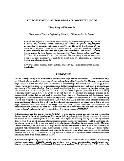
PSEUDO-TERNARY PHASE DIAGRAMS OF A DRUG DELIVERY SYSTEMZiheng Wang and Rajinder PalDepartment of Chemical Engineering, University of Waterloo, Ontario, CanadaAbstract: The purpose of this research was to develop the pseudo-ternary phase diagrams fora model drug delivery system consisting of vitamin E (model drug)+soybeanoil+surfactant+co-surfactant (anhydrous glycerol)+water. The model drug (vitamin E) wasloaded in the oil phase. The effects of different surfactants (pure and mixed) on the phasediagram, especially the microemulsion region, were investigated. The influence of drugloading level on the phase diagram was also determined. The surfactants studied were Tween20, Tween 80, Cremopher EL, and their mixtures. The size (area) of the microemulsionregion of the phase diagram was found to be dependent on the type of surfactant used and theloading level of drug (vitamin E)Keywords: Phase diagram, microemulison, drug delivery, self-microemusifying system,drug loading1.INTRODUCTIONOral-based drug delivery is the most common way to deliver drugs into the bloodstream.. The water-soluble drugs can diffuse freely and easily in gastrointestinal tract and they have a high bioavailability. However, more and more drugs being discovered nowadays with the advances in biotechnology and pharmaceutical technology are oil-soluble. The oil-soluble drugs pose serious problems in that they cannot diffuse freely and easily in gastrointestinal tract because of their poor solubility. One way to deliver oil-soluble drugs is to incorporate them into an inert lipid vehicle, such as an emulsion, oil [Burcham D.L. et al. 1997], surfactant dispersion [Serajuddin A.T.M. et al. 1988], or liposome [Schwendener R.A. et al. 1996]. At present, there are at least four drug products available in the pharmaceutical market that are delivered in emulsion form; they are: Sandimmune and Sandimmun Neoral (cyclosporin A), Norvir (ritonavir), and Fortovase (saquinavir). A significant improvement in the oral bioavailability of these drug compounds has been demonstrated [Gursoy R.N. et al. 2004]. Therefore, much attention is focused on using emulsions as vehicles to deliver oil-based drugs. Recently, microemulsions have been used to deliver oil-based drugs. Microemulsions offer several advantages over the usual (coarse) emulsions. Microemulsions are thermodynamically stable and the droplets of microemulsions are of very small size. The microemulsion delivery system is also referred to as the “self-microemulsifying drug delivery system (SMEDDS)”.Self-microemulsifying drug delivery system (SMEDDS) is a pre-mixture of drug, oil, surfactant and co-surfactant that can be used to deliver oil-based drugs. Upon gentle shaking and gastric juice dilution in stomach, it can form microemulisons spontaneously [Shah N.H. et al. 1994]. It is a highly suitable drug delivery system for hydrophobic drugs because it can be self-emulsified to microemulsion easily and steadily under mild condition in stomach. The pre-mixture can be stored for a very long period in capsules because of the high thermodynamic stability. However, a major disadvantage of SMEDDS is the large amount of surfactant needed to form a SMEDDS. Normally the amount of surfactant required to form a microemuslion is around 4 to 5 times of oil [Pouton C.W. 2000]. Another disadvantage related to large surfactant requirement is the potential toxic effects associated with the surfactant [Humberstone et al. 1997]. Therefore, it is highly desirable to reduce the usage of surfactants and at the same time, maintain the droplet size at a microemulsion level.A pseudo-ternary phase diagram of drug, oil, surfactant, co-surfactant, and water can be very helpful in formulating a suitable composition of SMEDDS. Usually there are three types of phases encountered in a pseudo-ternary phase diagram: microemulsion (ME), liquid crystal (LC) and coarse emulsion (EM). Microemulsion (ME) region is themain region of interest for formulation of SMEDDS. A large microemulsion region can offer more flexibility to find the optimal dosage composition. Microemulsions are identified with their clear and transparent appearance. Liquid crystal (LC) is a gel-like material that exhibits oil streaks under stirring condition. They also exhibit birefringence under crossed polarized microscope. Coarse emulsion (EM) is the traditional thermodynamic unstable emulsion; it appears as milky white during the preparation and storage. The particles size of coarse emulsion can range from sub-microns to microns [Li P. et al., 2005].In this work, the pseudo-ternary phase diagrams for a system consisting of oil, surfactant, co-surfactant, and water, with and without a model drug (vitamin E) are developed. The influence of different surfactants and drug on the size of the microemulsion region is examined.2.MATERIALS AND METHODS2.1MaterialsVitamin E (α-Tocopherol, HPLC grade), glycerol anhydrous (GC grade) and Cremophor EL were purchased from Fluka. Soybean oil, Tween 80, Tween 20 were purchased from Sigma. All chemicals were used as received.2.2Experimental ProcedureThe experimental work consisted of into two parts. In the first part, the influence of different surfactants (pure and mixed) on the pseudo-ternary phase diagram of oil + surfactant + co-surfactant + water system, without any drug (vitamin E) loading, was determined. Based on this part of experimental work, the best surfactant was selected. In the next part, the drug (vitamin E) was loaded in the oil phase and the influence of different drug loadings on the pseudo-ternary phase diagram was determined. The oil phase was loaded with 10%w/w, 20%w/w, 30%w/w, 40%w/w and 50%w/w of vitamin E.The ratio of surfactant to co-surfactant was fixed at 1:1 on the weight basis. The mixture of surfactant and co-surfactant is referred to as “surfactant phase” in the following discussion. Six types of surfactant phases (Tween 80+Glycerol, Tween 20+Glycerol, Cremophor EL+Glycerol, Tween 80+Tween 20+Glycerol, Tween 80+Cremophor EL+Glycerol, Tween 20+Cremophor EL+Glycerol) were prepared. The soybean oil was mixed with each of surfactant phases in the ratios (weight basis) of 1:9, 2:8, 3:7, 4:6, 5:5, 6:4, 7:3, 8:2 and 9:1 . A titration technique was employed for the preparation of the pseudo ternary phase diagrams. Deionized water was added in small increments (less than 5% w/w) to the mixture of soybean oil/surfactant phase at room temperature. After each water addition, the mixture was stirred in a beaker for 2-3 min using a stirring bar and a magnetic stirring plate. The titration process followed the tie lines (dash lines) shown in the pseudo-ternary phase diagram of Figure. 1. The phases were identified using visual inspection, microscopic inspection, and measurement of droplet size.. The droplet size was measured by DLS (Dynamic Light Scattering)Figure 1 - Tie lines of a pseudo-ternary phase diagram3.RESULTS AND DISCUSSION3.1 Influence on surfactant-type on the phase diagramFigures 2 to 7 show the pseudo ternary phase diagrams obtained for the system (soybean oil + surfactant + glycerol + water) using different surfactants. No drug (vitamin E) was present in the system..Figure 2 - Phase diagram for the system Figure 3 - Phase diagram for the system soybean oil+Tween80+glycerol+water soybean oil+Tween 20+glycerol+waterFigure 4 - Phase diagram for the system Figure 5 - Phase diagram for the system soybean oil+cremophor EL+glycerol+water soybean oil+Tween 80+Tween 20+glycerol+waterFigure 6 - Phase diagram for the system Figure 7 - Phase diagram for the system soybean oil+Tween 80+cremophor EL+glycerol+water soybean oil+Tween 20+cremophor EL+glycerol+water Upon comparing Figures 2 to 7, it is clear that Tween 20 (shown in Figure 3) is the worst surfactant in that it gives a negligibly small microemulsion (ME) region. The usage of mixed surfactants (see Figures 5 to 7) does not give any substantial enlargement of the ME region. Therefore, the combination of different surfactants is not a good choice for this system The combination of two surfactants can actually enhance the potential risk of drug administration.. The best surfactant for the present system appears to be Tween 80. While Cremophor EL gives a similar sized microemulsion (ME) region, it has a lower HLB value of 13.5 as compared with Tween 80 (HLB of 15). The larger the HLB value, easier it is for the surfactant to form oil-in-water emulsions.. In conclusion, the best surfactant (among the pure and mixed ones investigated in this work) for the system (soybean oil +surfactant+glycerol+water) appears to be Tween 80..3.2 Influence of drug loading on the phase diagramFigures 8 to 12 show the effect on drug (vitamin E) loading on the phase diagram. The phase diagrams are shown for five different loading levels vitamin E in soybean oil (10% w/w, 20% w/w, 30% w/w, 40% w/w and 50% w/w).Figure 8 - Phase diagram for the system soybean Figure 9 - Phase diagram for the system soybean oil (10% vitamin E loading)+Tween 80+glycerol + water oil (20% Vitamin E loading)+Tween 80+glycerol+waterFigure 10 - Phase diagram for the system soybean Figure 11 - Phase diagram for the system soybean oil (30% vitamin E loading)+Tween 80+glycerol+water oil (40% vitamin E loading)+Tween 80+glycerol+waterFigure 12 - Phase diagram for the system soybeanoil (50% vitamin E loading)+Tween 80+glycerol+waterAccording to Figures 8 to 12, the influence of drug loading on the phase diagram (particularly the microemulsion region) is quite significant. The microemulsion (ME) region of the phase diagram undergoes enlargement when the drug loading is increased from 0 to 30% in the oil phase. With further increase in drug loading, the microemulsion region tends to shrink. It should also be noted that for drug loading levels of 40% and 50%, the microemulsions were not very stable; the samples exhibited phase separation when left for a few days. Thus, the best loading level of vitamin E is 30% based on the oil phase. At this level of drug loading, the microemulsion region is large enough to allow some flexibility in choosing an optimal composition for the SMEDDS.Table 1 shows the effect of drug loading on the mean droplet size of microemulsions. In column 1 of the table, O5S45W50 represents the composition with 5% w/w of oil phase, 45% w/w of surfactant phase and 50% w/w of water phase. In any given row of the table, the effect of drug loading level on the mean droplet size (nm) is shown. The data with asterisk means that phase separation happened in this case within a few days of storage. From the information given in the table, it can be concluded that at a drug loading level of 30% the microemulsions are stable and have the smallest mean droplet size as compared with microemulsions at other drug levels.Table 1- Mean droplet size (nm) information4.CONCLUSIONSPseudo-ternary phase diagrams are developed for a model drug delivery system consisting of soybean oil (loaded with vitamin E as a model drug) + surfactant + glycerol (as co-surfactant) + water. The influence of different types of surfactants (pure and mixed) and different drug loading levels on the phase diagram are determined experimentally. Based on this in vitro study, the best SMEDDS is soybean oil (30% w/w vitamin E) +Tween 80+glycerol+water.ACKNOWLEDGMENTFinancial support for this research is provided by NSERC in the form of a discovery grant awarded to Professor R. PalREFERENCESBurcham D.L., Maurin M.B., Hausner E.A., Huang S.M.. (1997) Improved oral bioavailability of hypocholesterolemic DMP in dogs following dosing in oil and glycol soluteons. Biopharm Drug Dispos;18:737-42Pouton C.W.. (2000) Lipid formulations for oral administration of drugs: non-emulsifying, self-emulsifying and …self-microemulsifying‟ drug delivery systems. European Journal of Pharmaceutical Sciences 11 Auppl. 2 S93-S98Humberstone, A.J., Charman, W.N., (1997). Lipid-based vehicles for the oral delivery of poorly water soluble drugs.Adv. Drug Deliv. Rev. 25, 103–128Kang B.K., Lee J.S., Chon S.K., Jeong S.Y., Yuk S.H., Khang G., Lee H.B., Cho S.F.. (2004) Int J Pharm; 274:65. Li P., Ghosh A., Robert F., Krill S., Yatindra M. J., Abu T.M. et al.. (2005) Effect of combined use of nonionic surfactant on formation of oil-in-water microemulisons. International Journal of Pharmaceutics 288 27-34. Gursoy R. N., Benita S.. (2004) Self-emulsifying drug delivery systems (SEDDS) for improved oral delivery of lipophilic drugs. Biomed Pharmacother. Apr ;58 (3):173-82 15082340 (P,S,E,B)Schwendener R.A., Schott H. (1996) Lipophilic 1-beta-D-arabinofuranosyl cytosine derivatives in liposomal formations for oral and parenteral antileukemic therapy in the murine L1210 leukemia model. J Cancer Res Clin Oncol; 122:723-6Serajuddin A.T.M., Shee P.C., Mufson D., Bernstein D.F., Augustine M.A.. (1988) Effect of vehicle amphiphilicity on the dissolution and bioavailability of a poorly water-soluble drug from solid dispersion. J Pharm Sci;77:414-7.。
医学检验类杂志有哪些
医学检验类杂志有哪些医学检验类杂志是指专门刊登医学检验领域科研成果、临床技术应用、疾病诊断治疗相关信息的期刊。
以下是一些著名的医学检验类杂志。
1. Clinical ChemistryClinical Chemistry是一份由美国临床化学学会出版的月刊,该杂志报道了临床化学和实验室医学领域的研究成果和技术开发,涵盖了分子生物学、微生物学、免疫学等多个方面。
Clinical Chemistry以其高质量的学术论文和权威性而备受认可,被广泛阅读和引用。
2. The Journal of Molecular DiagnosticsThe Journal of Molecular Diagnostics是一份由美国分子病理学协会出版的月刊,涵盖了分子生物学和病理学领域的研究,旨在发表有关分子生物学、未发现临床疾病、致病菌分类和感染、免疫学等的研究。
该杂志以其高质量的论文和学术评审委员会而闻名。
3. Clinical Microbiology ReviewsClinical Microbiology Reviews是一份由美国微生物学学会出版的四次/年度的综述期刊,是临床微生物学领域中最有影响力的期刊之一。
该杂志旨在通过发表与诊断、治疗和预防感染相关的检验和研究来支持医学检验和临床研究,如病原体的鉴定、药敏性测试、病原体的生长、细胞 / 免疫学测量、基因组学和元组学等方面的研究。
4. American Journal of Clinical PathologyAmerican Journal of Clinical Pathology是一份月刊,曾被誉为世界上最主要的病理学期刊之一,涵盖了临床病理学、实验室医学、微生物学和分子生物学。
该杂志致力于发表新技术、新测试、病例研究和病理学研究的原始或综述论文。
本期刊拥有一个高度评估的审稿委员会,以保证其作品的学术性和质量。
5. Analytical ChemistryAnalytical Chemistry是由美国化学会出版的一份权威性和高度评价的月刊,发表了对医学检验领域有重大贡献的文章。
收藏外泌体鉴定外泌体示踪方法,你都知道吗?

收藏外泌体鉴定外泌体⽰踪⽅法,你都知道吗?(⼀)外泌体鉴定⽅法外泌体内容物 (芯⽚、测序、质谱、WB、QPCR、流式等⽅法)Gutierrez-Vazquez, C., et al.ImmunolRev, 2013. 251(1): p. 125-42.蛋⽩组分:细胞⾻架蛋⽩、信号转导相关蛋⽩、代谢酶类、抗原结合提呈相关蛋⽩典型的Exosomes标记蛋⽩有:四跨膜蛋⽩超家族,如CD9、CD63、CD81等;细胞质蛋⽩,如肌动蛋⽩(actin)、钙磷脂结合蛋⽩(annexins);参与⽣物功能的分⼦,如凋亡转接基因2互作蛋⽩X(ALIX)、肿瘤易感基因101蛋⽩(TSG101)、热休克蛋⽩(HSP70、HSP90);整合素等。
Exosomes还含有细胞类型特异的标记蛋⽩,这种蛋⽩分⼦由分泌Exosomes的细胞所决定;核酸组分:Exosomes内还能够包裹mRNA、miRNA、lncRNA、mtDNA、circRNA,并转移⾄其它细胞中发挥⽣物学作⽤,Exosomes所包裹的内容物也是细胞类型特异的。
1、蛋⽩质-WB鉴定Journalof Extracellular Vesicles 2015, 4: 27032、蛋⽩质- ELISA鉴定Journalof Histochemistry & Cytochemistry 2015, Vol. 63(3) 181-1893、蛋⽩质-流式细胞术鉴定doi:10.1038/nature223414、蛋⽩质-质谱鉴定Methods in Molecular Biology,vol. 15455、RNA-RNA提取Molecular Immunology 50(2012) 278–2866、RNA-测序鉴定PLoSOne. 2016; 11(1): e0146353.7、纳⽶颗粒跟踪分析:纳⽶颗粒跟踪分析(Nanoparticle TrackingAnalysis ,NTA)是对每个颗粒的布朗运动进⾏追踪和分析,结合Stockes-Einstein⽅程式计算出纳⽶颗粒的流体⼒学直径和浓度。
汤姆森数据库功能介绍
汤姆森三大数据库功能简介1Thomson Reuters Pharma®是一个整合了汤森路透提供的所有科学、医疗卫生和金融信息数据库的工作流工具。
Thomson Reuters Pharma允许您自由地浏览重要的市场情报,这些经过深加工的信息整合在由我们的行业专家团队所撰写的独一无二的摘要、总结、评论及分析报告中。
通过Thomson Reuters Pharma®,您能够:及时掌握最新的药物、试验、新闻报道及专利信息获取重大的公司新闻,并链接至全文报告和新闻发布稿查看会议报告在各种内容中交互链接汇集众多来源的数据定制检索,过滤内容∙Thomson Reuters Pharma提供∙40,162个药物专论(每月增加300个以上)∙7,600多个详尽的公司报告(共涵盖75,867 个机构)∙7,636个会议报告(每年增加约500个会议报告)∙11,000个医学期刊和100个有机化学期刊∙3,889,193个核心专利报告(覆盖87个专利授权机构)∙3,505,665种化合物(每月增加约5,000种)∙26,578个交易报告∙70,861个临床方案报告(每月增加800个以上新的临床方案)∙28,204个临床结果报告(每月增加200个以上新的临床结果报告)∙24,000个药物靶标为何选择Thomson Reuters Pharma®?∙权威性:Thomson Reuters Pharma®涵盖药物发现和开发流程全过程–最新药物、化合物、基因序列和靶标、临床试验、专利、期刊、会议、学术文章等∙∙相关性:直观的引导式检索可提供您所需要的情报个性化:个性化地定制您的检索条件,满足您不同的实际需求市场意识:有深度的竞争情报,包含涉及超过7,500家制药和生物科技公司的详细研发流程、财务及营销概况以用户为中心:易于使用的个人信息中心,与您的日常工作流需求整合在一起协作:直观的工作流和导出工具帮助您与团队进行数据共享,推动创新。
美国FDA 分析方法验证指南
美国FDA 分析方法验证指南(中文)U.S. Department of Health and Human ServicesFood and Drug AdministrationCenter for Drug Evaluation and Research (CDER)Center for Biologics Evaluation and Research (CBER) August 2000目录一、结论………………………………………………………..…………………二、背景……………………………………………………………..……….…..三、分析方法的类型…………………………………………………………….A. 法定分析方法……………………………………………………………B. 替代分析方法……………………………………………………………C. 稳定性指示分析…………………………………………………………四、标准品………………………………………………………………………..A.标准品的类型……………………………………………………………B.分析报告单………………………………………………………………C.标准品的界定……………………………………………………………五、IND 中的分析方法验证……………………………………………………..六、NDA、ANDA、BLA 和PLA 中分析方法的内容和格式…………………A.基本方法…………………………………………………………………B.取样………………………………………………………………………C.仪器和仪器参数………………………………………………………….D.试剂………………………………………………………………………E.系统适应性实验………………………………………………………….F.标准品的制备……………………………………………………………..G.操作过程…………………………………………………………………….H.操作程序……………………………………………………………………I.计算…………………………………………………………………………J.结果报告…………………………………………………………………….1.通则……………………………………………………………………2.杂质分析规程…………………………………………………………七、NDA,ANDA,BLA 和PLA 中的分析方法验证………………………….. A.非药典分析方法…………………………………………………………1. 验证项目……………………………………………………………2. 其它验证资料……………………………………………………….(1) 讨论可能会形成的异构体并讨论异构体的控制…………………..a. 耐用性…………………………………………………….b. 强降解实验………………………………………………c.仪器输出/原始资料………………………………………i. 有机杂质……………………………………………ii. 原料药……………………………………………….iii. 制剂………………………………………………….(2) 各类检测的推荐验证项目…………………………………………..a. 鉴别………………………………………………………....b. 杂质………………………………………………………..c. 含量………………………………………………………..d. 特定实验…………………………………………………….B.药典分析方法(21CFR 211.194(a)(2))…………………………………..八. 统计分析…………………………………………………………………….A.基本原则………………………………………………………………B:对比研究…………………………………………………………………C:统计………………………………………………………………………九、再验证………………………………………………………………………十、分析方法验证资料:内容和数据处理…………………………………….A.分析方法验证资料…………………………………………………….B:样品的选择和运输…………………………………………………….C:各方职责……………………………………………………………….1.申请人……………………………………………………………….2.化学评审官………………………………………………………….3.FDA 实验室………………………………………………………….4.检查官……………………………………………………………….十一、方法学……………………………………………………………………A.高效液相色谱(HPLC)………………………………………………….1.色谱柱……………………………………………………………….2.系统适应性研究…………………………………………………….3.操作参数…………………………………………………………….B.气相色谱(GC)………………………………………………………….1.色谱柱……………………………………………………………….2.操作参数……………………………………………………………..3.系统适应性实验……………………………………………………..C:分光光度法,光谱法和相关的物理方法………………………………D:毛细管电泳(CE)…………………………………………………………E:旋光度……………………………………………………………………F:和粒径分析相关的分析方法……………………………………………G:溶出度…………………………………………………………………..H:其它仪器分析方法………………………………………………………附录A……………………………………………………………………………….. 附录B……………………………………………………………………………….. 术语表……………………………………………………………………………….一、绪论本指南旨在为申请者提供建议,以帮助其提交分析方法,方法验证资料和样品用于支持原料药和制剂的认定,剂量,质量,纯度和效力方面的文件。
光动力治疗在气道肿瘤中的临床应用

2)病人注射光敏剂后需及时戴墨镜、入住暗房 3)注射光敏剂24~48h后做PDT 4)PDT术后三天内亲密注意观察病情 5)PDT术后第2天至4周及时去除肿瘤坏死物质
光动力治疗在气道肿瘤中的临床应用
第31页
相关避光问题
第一周:严格避光;
第二周:逐步接触光线;
第三、四面:夜间可活动,防止阳光直射。
致死性大咯血:肿块坏死脱落,瘘形成造成较 大血管破裂出血;(D67、D187、D567)
狭窄:PDT后局部纤维化疤痕形成狭窄; 急性黏膜水肿:PDT后48h内出现支气管及喉头
水肿引发呼吸道阻塞
光动力治疗在气道肿瘤中的临床应用
第42页
Harubumi Kato;Our Experience with Photodynamic Diagnosis and Photodynamic Therapy for lung cancer JNatl compr canc Netw;10
1968年,Gregorie报道226例肿瘤患者在静脉注射HPD后, 鳞癌和腺癌活检标本内75%~85%荧光阳性,而53份良性标 本中仅23%荧光阳性,为肿瘤PDT奠定了理论基础
1976年Kelly 和Snell应用HPD作为光敏剂治疗了1例复发 膀胱癌。
1982年,国际抗癌联盟(UICC)首次将PDT专题列入第十 三届代表大会议程,扩充了这项新技术影响。同年,美国 人创建了激光医学研究基金(LMRF),重点资助PDT临床 试验和医生培训。
肺癌可到达 长久缓解目标,AFB 对发觉多发病灶和确 定病变部位有主要补充作用
Harubumi Kato;Our Experience with Photodynamic Diagnosis and Photodynamic Therapy for lung cancer JNatl compr canc Netw;10
- 1、下载文档前请自行甄别文档内容的完整性,平台不提供额外的编辑、内容补充、找答案等附加服务。
- 2、"仅部分预览"的文档,不可在线预览部分如存在完整性等问题,可反馈申请退款(可完整预览的文档不适用该条件!)。
- 3、如文档侵犯您的权益,请联系客服反馈,我们会尽快为您处理(人工客服工作时间:9:00-18:30)。
美国药学科学家协会(AAPS) 2015年会海报系列
Fit-for-Purpose多因⼦子检测与⽣生物标志物筛选
Fit-for-Purpose Multiplex Panels and
Their Utility in Biomarker Screening
(1)Abstract
⽬目的:
以筛选Biomarker为⽬目的的探索性实验对于药物研发和临床诊断⾮非常重要,可能会涉及到100多种Biomarkers。
然⽽而,⼤大规模多因⼦子检测产⽣生的信号⼲干扰问题,会误导筛选结果。
在此研究中,选择部分多因⼦子检测衡量其筛选的效果。
⽅方法:
使⽤用MSD的MULTI-ARRAY专利技术,仅1mL样品即可检测122个指标。
这些⽅方法使⽤用了15个不同的多因⼦子检测组合,并遵循fit-for-purpose原则。
针对稀释倍数,稀释液成分,以及试剂的特异性均进⾏行了优化。
检测组合⾥里包含MSD的经过验证的V-plex Human Biomarker 40-plex,检测指标涉及炎症,免疫,⾎血管⽣生成,⾎血管创伤等;还包含10种不同的多因⼦子检测组合。
结果:
为便于进⾏行⽅方法学的优化及确保性能,多因⼦子组合使⽤用10-plex的形式,即每孔检测10个因⼦子。
检测⽅方法学展现出低于1%的⾮非特异性结合。
每个10因⼦子组合的的线性范围横跨3-4个数量级,以便于同时检测出正常样本和疾病样本。
疾病样本组选择了⾎血清,EDTA⾎血浆,脑脊液,尿液。
每个检测⽅方法在不同板⼦子之间有良好的重复性。
在定量检测范围以内的样本中,⼤大多数⽅方法学的板内差异均值(CV)低于10%。
结论:
使⽤用⽆无偏差的⽅方法学进⾏行Biomarker筛选,确保了快速鉴定出有临床重要意义的标志物。
不同多因⼦子组合的检测辅助了病⼈人群体的区分,并能⽤用于监控疾病活动。
这种多因⼦子检测技术对于使⽤用少量样本筛选⼤大量指标的项⽬目⼗〸十分理想。
(2)Methods
样本检测使⽤用MSD MULTI-ARRAY技术的Biomarker检测组合。
根据fit-for-purpose的⽅方法学,122个biomarker划分为15个不同的多因⼦子检测组合。
针对稀释倍数,稀释液成分,以及试剂的特异性均进⾏行了优化。
其中5个组合是MSD的经过验证的V-plex Human Biomarker 40-plex试剂盒,剩下的使⽤用了10个多因⼦子组合。
使⽤用少于1mL样品即可检测全部122个指标。
电化学发光技术:
1)最⼩小化⾮非特异性背景信号,
获得⾼高信号反应,从⽽而产⽣生
极⾼高的信号背景⽐比例。
2)激发机制(电)与信号(光)
分离,最⼩小化了基质干扰。
3)只有在电极表⾯面处的结合标
记被激发,从⽽而可以建⽴立
non-washed⽅方法。
4)标记稳定,⽆无放射性,并容
易偶联到⽣生物分⼦子。
5)620nm激发光消除了颜⾊色衰
减问题。
6)每个标记的多重激发循环提
⾼高光信号级别,并提升灵敏
度
7)⽯石墨电极板表⾯面的结合能⼒力
⽐比普通聚苯⼄乙烯⼤大10倍
8) 可以定制表⾯面的包被物体。
(3)Specificity
为确认检测抗体的特异性,混合的校准液与单个检测抗体⼀一起测试。
15个多因⼦子组合都进⾏行了这种测试。
我们发现绝⼤大多数的⾮非特异⼲干扰低于1%。
代表性数据如下:
(4)Sensitivity
LLOD(最低检测下限)是⼀一种计算出的浓度,其基于空⽩白的2.5标准偏差。
最少重复检测6次以⽤用于计算平均LLOD。
ULOD(最⾼高检测上限)是最⾼高的校准液浓度。
在以下表格中罗列了本研究中的检测极限,并标注了稀释⽐比例。
⼤大多数⽅方法针对所有样本基质采⽤用了相同的稀释倍数。
CRP,ICAM-1, SAA 和VCAM-1的⾎血清⾎血浆稀释了1000倍,脑脊液和尿液稀释了5倍。
(5)Sample Testing
20份⼈人⾎血清,20份EDTA⾎血浆,8份尿液,8份脑脊液,使⽤用15个多因⼦子组合检测。
⼤大多数测试中,样本都被检测出来。
IL-17B,IL-17D和IL-21在普通⼈人样本中并未检测出来。
检测浓度在前⼀一页表格中。
(6)Reproducibility
根据在检测范围内的样本计算出平均CV。
V-PLEX Human Biomarker 40-Plex的⽅方法,从COA中即可获得其定量检测上下限(LLOQ和ULOQ)。
其他⽅方法可以估计其定量极限。
LLOQ估为平均LLOD的5倍,ULOQ估为ULOD的80%。
在⾎血清,EDTA⾎血浆,脑脊液中,⾄至少90%⽅方法的平均CV低于10%。
在尿液中,70%的平均CV低于10%。
⼀一些较⾼高CV经常和低内源性浓度(见本⽂文第5节)。
以下⽅方法的平均CV ⼤大于20%。
> ⾎血清 - IL-2,IL-13,IL-17,PYY(total), Osteopontin.
> EDTA⾎血浆 - Eotaxin-3
> 脑脊液 - IL-2,IL-4,IL-13, RANTES
> 尿液 - IL-1a, VEGF-A, IL-16, C-Peptide, E-Selectin, NT-proBNP, cTnT, Myl3, CKMB, Myoglobin, Osteoprotegerin, MCP-2, MET。
这些标准曲线的平均CV也计算了。
98%的标准5浓度的平均信号CV低于10%。
CKMB ⽅方法的平均CV⼤大于20%。
这个⽅方法已经进⼀一步优化并延长了动态范围。
(未在此列出数据)
重复性(精确度)使⽤用基于基质的对照,并在⼀一天内⽤用6块板测试。
代表数据如下:
(7)Conclusion
MSD的Biomarker筛选试剂盒包含了性能稳定的定量⽅方法。
这些⽅方法线性范围⼴广,使科研⼈人员容易获得精确的定量数据。
对于⼤大部分⽅方法,多种不同的样本类型可以使⽤用⼀一个稀释浓度检测。
这些试剂盒使⽤用了简单的操作流程,可以在⼀一个板上检测10个biomarker。
这122个biomarker只使⽤用了不到1mL的样本。
这些试剂盒可以⽤用于定量不同基质样本(如⾎血清,⾎血浆,脑脊液,尿液)的biomaker。
页码:/
1111。
