Lepton Photon Symposium 2005 Summary and Outlook
第一次报道光遗传
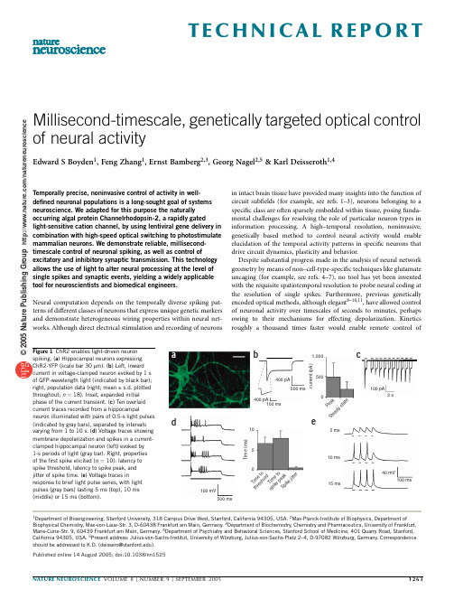
Millisecond-timescale,genetically targeted optical control of neural activityEdward S Boyden1,Feng Zhang1,Ernst Bamberg2,3,Georg Nagel2,5&Karl Deisseroth1,4Temporally precise,noninvasive control of activity in well-defined neuronal populations is a long-sought goal of systems neuroscience.We adapted for this purpose the naturally occurring algal protein Channelrhodopsin-2,a rapidly gatedlight-sensitive cation channel,by using lentiviral gene delivery in combination with high-speed optical switching to photostimulate mammalian neurons.We demonstrate reliable,millisecond-timescale control of neuronal spiking,as well as control of excitatory and inhibitory synaptic transmission.This technology allows the use of light to alter neural processing at the level of single spikes and synaptic events,yielding a widely applicable tool for neuroscientists and biomedical engineers.Neural computation depends on the temporally diverse spiking pat-terns of different classes of neurons that express unique genetic markers and demonstrate heterogeneous wiring properties within neural net-works.Although direct electrical stimulation and recording of neurons in intact brain tissue have provided many insights into the function of circuit subfields(for example,see refs.1–3),neurons belonging to a specific class are often sparsely embedded within tissue,posing funda-mental challenges for resolving the role of particular neuron types in information processing.A high–temporal resolution,noninvasive, genetically based method to control neural activity would enable elucidation of the temporal activity patterns in specific neurons that drive circuit dynamics,plasticity and behavior.Despite substantial progress made in the analysis of neural network geometry by means of non–cell-type-specific techniques like glutamate uncaging(for example,see refs.4–7),no tool has yet been invented with the requisite spatiotemporal resolution to probe neural coding at the resolution of single spikes.Furthermore,previous genetically encoded optical methods,although elegant8–10,11,have allowed control of neuronal activity over timescales of seconds to minutes,perhaps owing to their mechanisms for effecting depolarization.Kinetics roughly a thousand times faster would enable remote controlof100 mV500 ms105Time(ms)TimetothresholdTimetospikepeakSpikejitter15 ms10 ms5 ms400 pA400 pA100 ms500 ms100 ms40 mV100 pA2 sPeakSteadystate5001,000current(pA)d eb cFigure1ChR2enables light-driven neuronspiking.(a)Hippocampal neurons expressingChR2-YFP(scale bar30m m).(b)Left,inwardcurrent in voltage-clamped neuron evoked by1sof GFP-wavelength light(indicated by black bar);right,population data(right;mean±s.d.plottedthroughout;n¼18).Inset,expanded initialphase of the current transient.(c)Ten overlaidcurrent traces recorded from a hippocampalneuron illuminated with pairs of0.5-s light pulses(indicated by gray bars),separated by intervalsvarying from1to10s.(d)Voltage traces showingmembrane depolarization and spikes in a current-clamped hippocampal neuron(left)evoked by1-s periods of light(gray bar).Right,propertiesof thefirst spike elicited(n¼10):latency tospike threshold,latency to spike peak,andjitter of spike time.(e)Voltage traces inresponse to brief light pulse series,with lightpulses(gray bars)lasting5ms(top),10ms(middle)or15ms(bottom).Published online14August2005;doi:10.1038/nn15251Department of Bioengineering,Stanford University,318Campus Drive West,Stanford,California94305,USA.2Max-Planck-Institute of Biophysics,Department of Biophysical Chemistry,Max-von-Laue-Str.3,D-60438Frankfurt am Main,Germany.3Department of Biochemistry,Chemistry and Pharmaceutics,University of Frankfurt, Marie-Curie-Str.9,60439Frankfurt am Main,Germany.4Department of Psychiatry and Behavioral Sciences,Stanford School of Medicine,401Quarry Road,Stanford, California94305,USA.5Present address:Julius-von-Sachs-Institut,University of Wu¨rzburg,Julius-von-Sachs-Platz2–4,D-97082Wu¨rzburg,Germany.Correspondence should be addressed to K.D.(deissero@).T E C H N I C A L R E P O R T ©25NaturePublishingGrouphttp://www.nature.com/natureneuroscienceindividual spikes or synaptic events.We have therefore devised a new strategy using a single-component ion channel with submillisecond opening kinetics,to enable genetically targeted photostimulation with fine temporal resolution.Two rhodopsins in the unicellular green alga Chlamydomonas rein-hardtii were recently identified independently by three groups 12–15.One of them is a light-gated proton channel (Channelrhodopsin-1;ref.13),whereas the other,Channelrhododopsin-2(ChR2),is a light-gated cation channel 12.The N-terminal 315amino acids of ChR2are homologous to the seven-transmembrane structure of many microbial-type rhodopsins;they compose a channel with light-gated conductance (as proposed earlier 16).Inward currents in ChR2-expressing cells could be evoked within 50m s after a flash of blue light in the presence of all-trans retinal 12,suggesting the possibility that ultrafast neuronal stimulation might be possible with equipment commonly used for visualizing green fluorescent protein (GFP).ChR2therefore combines some of the best features of previous photostimulation methods,including the speed of a monolithic ion channel 9,and the efficacy of natural light-transduction machinery 11.We found that ChR2could be expressed stably and safely in mammalian neurons and could drive neuronal depolarization.When activated with a series of brief pulses of light,ChR2could reliably mediate defined trains of spikes or synaptic events with millisecond-timescale temporal resolution.This technology thus brings optical control to the temporal regime occupied by the fundamental building blocks of neural computation.RESULTSRapid kinetics of ChR2enables driving of single spikesT o obtain stable and reliable ChR2expression for coupling light to neuronal depolarization,we constructed lentiviruses containing a ChR2-yellow fluorescent protein (YFP)fusion protein for genetic modification of neurons.Infection of cultured rat CA3/CA1neurons led to membrane-localized expression of ChR2for weeks after infection (Fig.1a ).Illumination of ChR2-positive neurons with blue light (bandwidth 450–490nm via Chroma excitation filter HQ470/40Â;300-W xenon lamp)induced rapid depolarizing currents,whichreached a maximal rise rate of 160±111pA/ms within 2.3±1.1msafter light pulse onset (mean ±s.d.reported throughout paper,n ¼18;Fig.1b ,left).Mean whole-cell inward currents were large:496pA ±336pA at peak and 193pA ±177pA at steady-state (Fig.1b ,right).Light-evoked responses were never seen in cells expressing YFP alone (data not shown).Consistent with the known excitation spectrum of ChR2(ref.12),illumination of ChR2-expressing neurons with YFP-spectrum light in the bandwidth 490–510nm (300-W xenon lamp filtered with Chroma excitation filter HQ500/20Â)resulted in currents that were smaller (by 42%±20%)than those evoked with the GFP filters.Despite the inactivation of ChR2with sustained light exposure (Fig.1b and ref.12),we observed rapid recovery of peak ChR2photocurrents in neurons (Fig.1c ;recovery t ¼5.1±1.4s;recovery trajectory fit with Levenberg-Marquardt algorithm;n ¼9).This rapid recovery is consistent with the well-known stability of the Schiff base (the lysine in transmembrane helix seven,which binds retinal)in microbial-type rhodopsins,and the ability of retinal to re-isomerize to the all-trans ground state in a dark reaction,without the need for other enzymes.Light-evoked current amplitudes remained unchanged in patch-clamped neurons during 1h of pulsed light exposure (data not shown).Thus ChR2was able to sustainably mediate large-amplitude photocurrents with rapid activation kinetics.We next examined whether ChR2could drive spiking of neurons held in current-clamp mode,with the same steady illumination protocol we used for eliciting ChR2-induced currents (Fig.1d ,left).Early in an epoch of steady illumination,single neuronal spikes were rapidly and reliably elicited (8.0±1.9ms latency to spike peak,n ¼10;Fig.1d ,right),consistent with the fast rise times of ChR2currents described above.However,any subsequent spikes elicited during steady illumination were poorly timed (Fig.1d ,left).Thus,steady illumina-tion is not adequate for controlling the timing of ongoing spikes with ChR2,despite the reliability of the first spike.Earlier patch-clamp studies using somatic current injection showed that spike times were more reliable during periods of rapidly rising membrane potential than during periods of steady high-magnitude current injection 17.This is consistent with our finding that steady illumination evoked a single reliably timed spike,followed by irregular spiking.In searching for a strategy to elicit precisely timed series of spikes with ChR2,we noted that the single spike reliably elicited by steadyλ = 100λ = 200J i t t e r , n e u r o n -t o -n e u r o n(m s )# of neurons firing a given spikeλ = 100,λ =100λ = 200λ= 100λ = 200λ = 100λ = 200λ = 20λ = 10λ = 20λ = 10# o f s p i k e sThree different neurons:same pulse series1 s60 mVTimeto spike Jitter throughout traini iim sP e r c e n t a g e f i d e l i t y o f s p i k e t r a n s m i s s i o nJ i t t e r , t r i a l -t o -t r i a l (m s )R e p e a t a b i l i t y ,t r i a l -t o -t r i a lOne neuron: repeated pulse seriesafghbcdeFigure 2Realistic spike trains driven by series of light pulses.(a )Voltage traces showing spikes in a single current-clamped hippocampal neuron,in response to three deliveries of a Poisson train (with mean interval l ¼100ms)of light pulses (gray dashes).(b )Trial-to-trial repeatability of light-evoked spike trains,as measured by comparing the presence or absence of a spike in two repeated trials of a Poisson train (either l ¼100ms or l ¼200ms)delivered to the same neuron (n ¼7neurons).(c )Trial-to-trial jitter of spikes,across repeated light-evoked spike trains.(d )Percent fidelity of spiketransmission throughout entire 8-s light pulse series.(e )Latency of spikes throughout each light pulse series (i)and jitter of spike times throughout train (ii).(f )Voltage traces showing spikes in three different hippocampal neurons,in response to the same temporally patterned light stimulus (gray dashes)used in a .(g )Histogram showing how many of the seven neurons spiked in response to each light pulse in the Poisson train.(h )Neuron-to-neuron jitter of spikes evoked by light stimulation.T E C H N I C A L R E P O R T©2005 N a t u r e P u b l i s h i n g G r o u p h t t p ://w w w .n a t u r e .c o m /n a t u r e n e u r o s c i e n c eillumination had extremely low temporal jitter from trial to trial,as reflected by the small standard deviation of the spike times across trials (Fig.1d ,right;0.5±0.3ms,average of n ¼10neurons).This observation led us to devise a pulsed-light strategy that would take advantage of the low jitter of the single reliable spike evoked at light pulse onset.In order for this to work,the conductance and kinetics of ChR2would have to permit peak currents of sufficient amplitude to reach spike threshold,during a light pulse of duration shorter than the desired interspike interval.We found that multiple pulses of light with interspersed periods of darkness could elicit trains of multiple spikes (Fig.1e ;shown for a 25-Hz series of four pulses).Longer light pulses evoked single spikes with greater probability than short light pulses (Fig.1e ).In the experiments described here,we used light pulse durations of 5,10or 15ms.Thirteen high-expressing neurons fired reliable spikes,and five low-expressing neurons could reliably be depolarized to subthreshold levels.The ability to easily alter light pulse duration suggests that a straightforward method for eliciting spikes,even in multiple neurons having different ChR2current densities,would involve titrating the light pulse duration until single spikes were reliably obtained in all the neurons being illuminated.Modulation of light intensity would also allow for this kind of control.Precise spike trains elicited by series of light pulsesThe precise control described above raised the prospect of generating arbitrary spike patterns,even mimicking natural neural activity.T o test this possibility,we generated series of light pulses,the timings of which were selected according to a Poisson distribution,commonly used to model natural spiking.A single hippocampal neuron could fire repeatable spike trains in response to multiple deliveries of the same Poisson-distributed series of light pulses (Fig.2a ;response to a repeated 59-pulse-long series of light pulses,with each pulse of 10-ms duration,and with mean interpulse interval of l ¼100ms).These optically driven spike trains were very consistent across repeated deliveries of the same series of light pulses:on average,495%of the light pulses in a series elicited spikes during one trial if and only if they elicited spikes on a second trial,for both the l ¼100ms series (Fig.2a )and a second series (mean interval l ¼200ms)comprising 46spikes (Fig.2b ;n ¼7neurons).We increased light pulse duration until reliable spiking was obtained:we used trains of 10-ms light pulses for four of the seven neurons and trains of 15-ms light pulses for the other three (for the analyses of Fig.2,all data were pooled).The trial-to-trial jitter for each individual spike was very small across repeated deliveries of the same Poisson series of light pulses (on average,2.3±1.4ms and 1.0±0.5ms for l ¼100ms and l ¼200ms,respectively;Fig.2c ).Throughout a series of pulses,the efficacy of eliciting spikes throughout the train was maintained (76%and 85%percent of light pulses successfully evoked spikes,respectively;Fig.2d ),with small latencies (Fig.2e ).As another indication of how precisely spikes can be elicited throughout an entire series of pulses,we measured the standard deviation of the latencies of each spike across all the spikes in the train.This ‘throughout-train’spike jitter was quite small (3.9±1.4ms and 3.3±1.2ms;Fig.2e ),despite presumptive channel inactivation during the delivery of a series of pulses.Hence,pulsed optical activation of ChR2elicits precise,repeatable spike trains in a single neuron,over time.Even in different neurons,the same precise spike train could be elicited by a particular series of light pulses (shown for three hippocampal neurons in Fig.2f ).Although the large hetero-geneity of different neurons—for example,in their membrane capaci-tance (68.8±22.6pF)and resistance (178.8±94.8M O )—might be expected to introduce significant variability in their electrical responses to photostimulation,the strong nonlinearity of light-spike coupling dominated this variability.Indeed,different neurons responded in similar ways to a given light pulse series,with 80–90%of the light pulses in a series eliciting spikes in at least half the neurons examined (Fig.2g ).T o quantitatively compare the reliability of spike elicitation in different neurons,for each pulse,we calculated the standard deviation of spike latencies (jitter)across all the neurons.Remarkably,this across-neuron jitter (3.4±1.0ms and 3.4±1.2ms for the pulses in the l ¼100ms and l ¼200ms trains,respectively;Fig.2h )was similar to the within-neuron jitter measured throughout the light pulse series (Fig.2e ).Thus,heterogeneous populations of neurons can be controlled in concert,with practically the same precision observed for the control of single neurons over time.Having established the ability of ChR2to drive sustained naturalistic trains of spikes,we next probed the frequency response of light-spike coupling.ChR2enabled driving of spike trains from 5to 30Hz (Fig.3a ;here tested with series of twenty 10-ms light pulses).It was easier to evoke more spikes at lower frequencies than at higher frequencies (Fig.3b ;n ¼13neurons).Light pulses delivered at 5or 10Hz could elicit long spike trains (Fig.3b ),with spike probability dropping off at higher frequencies of stimulation.For these experiments,the light pulses used were 5ms (n ¼1),10ms (n ¼9)or 15ms (n ¼3)in duration (data from all 13cells were pooled for the population analyses of Fig.3).As expected from the observation that light pulses generally elicited single spikes (Figs.1d and 2),almost no extraneous spikes occurred during the delivery of trains of light pulses (Fig.3c ).Even at higher frequencies,the throughout-train temporal jitter of spike timing remained very low (typically o 5ms;Fig.3d )and the latency to spike remained constant across frequencies (B 10ms throughout;Fig.3e ).Thus ChR2can mediate spiking across a physiologically relevant range of firing frequencies.5 Hz10 Hz20 Hz30 HzStimulus frequency(Hz)Stimulus frequency(Hz)Stimulus frequency(Hz)Stimulus frequency(Hz)# o f e x c e s s s p i k e sT i m e t o s p i k e (m s )# o f s u c c e s s f u l s p i k e s (o u t o f 20 p o s s i b l e )J i t t e r t h r o u g h o u t t r a i n (m s )aedFigure 3Frequency dependence of coupling between light input and spike output.(a )Voltage traces showing spikes in a current-clamped hippocampal neuron evoked by 5-,10-,20-or 30-Hz trains of light pulses (gray dashes).(b )Population data showing the number of spikes (out of 20possible)evoked in current-clamped hippocampal neurons.(c )Number of extraneous spikes evoked by the trains of light pulses,for the experiment described in b .(d )Jitter of spike times throughout the train of light pulses for theexperiment described in b .(e )Latency to spike peak throughout the light pulse train for the experiment described in b .T E C H N I C A L R E P O R T©2005 N a t u r e P u b l i s h i n g G r o u p h t t p ://w w w .n a t u r e .c o m /n a t u r e n e u r o s c i e n c eRemote activation of subthreshold and synaptic responsesFor many cellular and systems neuroscience processes (and for nonspiking neurons in species like Caenorhabditis elegans )subthres-hold depolarizations convey physiologically significant information.For example,subthreshold depolarizations are potent for acti-vating synapse-to-nucleus signaling 18,and the relative timing of subthreshold and suprathreshold depolarizations can determine thedirection of synaptic plasticity 19.But compared with driving spiking,it is in principle more difficult to drive reliable and precisely sized subthreshold depolarizations.The sharp threshold for action potential production facilitates reliable ChR2-induced spiking,and the all-or-none dynamics of spiking produces virtually identical waveforms from spike to spike (as seen throughout Figs.1–3),even in the presence of significant neuron-to-neuron variability in electrical properties.In contrast,subthreshold depolarizations,which operate in a more linear regime of membrane voltage,will lack these intrinsic normalizing mechanisms.Nevertheless,subthreshold depolarizations evoked by repeated light pulses were reliably evoked over a range of frequencies (Fig.4a ),with spaced repeated depolarizations resulting in a coefficientof variation of 0.06±0.03(Fig.4b ;n ¼5).Thus,ChR2can be used to drive subthreshold depolarizations of reliable amplitude.Finally,synaptic transmission was also easily controlled with ChR2.Indeed,both excitatory (Fig.4c )and inhibitory (Fig.4d )synapticevents could be evoked in ChR2-negative neurons receiving synaptic input from ChR2-expressing neurons.Expression of ChR2has minimal side effectsWe conducted extensive controls to test whether simply expressing ChR2would alter the electrical properties or survival of neurons.Lentiviral expression of ChR2for at least 1week did not alter neuronal membrane resistance (212±115M O for ChR2+cells versus239.3±113M O for ChR2Àcells;Fig.5a ;P 40.45;n ¼18each)or resting potential(À60.6±9.0mV for ChR2+cells versus À59.4±6.0mV for ChR2Àcells;Fig.5b ;P 40.60),when measured in the absence of light.This suggests that in neurons,ChR2has little basalelectrical activity or passive current-shunting ability.It also suggests that expression of ChR2did not compromise cell health,as indicated by electrical measurement of mem-brane integrity.As an independent measure ofcell health,we stained live neurons with themembrane-impermeant DNA-binding dye propidium iodide.ChR2expression did not affect the percentage of neurons that took up propidium iodide (1/56ChR2+neurons ver-sus 1/49ChR2Àneurons;P 40.9by w 2test).Neither did we see pyknotic nuclei,indicative of apoptotic degeneration,in cells expressingChR2(data not shown).We also checked foralterations in the dynamic electrical propertiesof neurons.In darkness,there was no differ-ence in the voltage change resulting from 100pA of injected current,in either the hyperpolarizing (À22.6±8.9mV for ChR2+neurons versus À24.5±8.7mV for ChR2Àneurons;P 40.50)or depolarizing (+18.9±4.4mV for ChR2+versus 18.7±5.2mV for ChR2Àneurons;P 40.90)directions.Nor was there any difference in the number ofspikes evoked by a 0.5-s current injection of +300pA (6.6±4.8for ChR2+neurons versus 5.8±3.5for ChR2–neurons;Fig.5c ;P 40.55).Thus,ChR2does not significantly jeopardize cell health or basal electrical properties of the expressing neuron.We also measured the electrical properties described above,24h afterexposing ChR2+neurons to a typical light pulse protocol (1s of 20-Hz15-ms light flashes,once per minute,for 10min).Neurons expressing ChR2and exposed to light had basal electrical properties similar to non-flashed ChR2+neurons:cells had normal membrane resistance (178±81M O ;Fig.5a ;P 40.35;n ¼12and resting potential (À59.7±7.0mV;Fig.5b ;P 40.75).Exposure to light also did not predispose neurons to cell death,as measured by live-cell propidium iodide uptake (2/75ChR2+neurons versus 3/70ChR2-neurons;P 40.55by w 2test).Finally,neurons expressing ChR2and exposed to light also had normal spike counts elicited from somatic current injection (6.1±3.9;Fig.5c ;P 40.75).Thus,membrane integrity,cell health and electrical proper-ties were normal in neurons expressing ChR2and exposed to light.500 ms 40 mV1 s 500 pA+Gabazine Inhibitory synaptic transmission5 HzSub-threshold responsesExcitatory synaptic transmission 100 pA1 s +NBQXab Figure 4Simulated and natural synaptic transmission evoked via ChR2.(a )Voltage traces showing trainsof subthreshold depolarizations in a current-clamped hippocampal neuron in response to trains of light pulses (gray dashes).(b )Repeated light pulses induced reliable depolarizations.(c )Excitatory synaptic transmission driven by light pulses.The selective glutamatergic transmission blocker NBQX abolishedthese synaptic responses (right).(d )Inhibitory synaptic transmission driven by light pulses.Theselective GABAergic transmission blocker gabazine abolished these synaptic responses (right).M e m b r a n e r e s i s t a n c e (M O h m )Ch R2C h R 2f l a s h e dCh R 2Ch R2Ch R 2f l a s h e dCh R2–Ch R2+Ch R 2+f l a s h e d Ch R2R e s t i n g p o t e n t i a l (m V )a b Figure 5Basal and dynamic electrical properties of neurons expressingChR2.(a )Membrane resistance of neurons expressing ChR2(black;n ¼18),not expressing ChR2(white;n ¼18)or expressing ChR2and measured 24h after exposure to a typical light-pulse protocol (gray;n ¼12).(b )Membrane resting potential of the same neurons described in a .(c )Number of spikes evoked by a 300-pA depolarization of the same neurons described in a .T E C H N I C A L R E P O R T©2005 N a t u r e P u b l i s h i n g G r o u p h t t p ://w w w .n a t u r e .c o m /n a t u r e n e u r o s c i e n c eDISCUSSIONCombining the best aspects of earlier approaches that use light to drive neural circuitry,the technology described here demonstrates voltage control significantly faster than previous genetically encoded photo-stimulation methods 8–11.Notably,the ChR2method does not rely on synthetic chemical substrates or genetic orthogonality of the transgene and the host organism.Although the ChR2molecule does require the cofactor all-trans retinal for light transduction 12,no all-trans retinal was added either to the culture medium or recording solution for any of the experiments described here.Background levels of retinal may be sufficient in many cases;moreover,the commonly used culture medium supplement B-27used here (see Methods)includes retinyl acetate,and additional supplementation with all-trans retinal or its precursors may assist in the application of ChR2to the study of neural circuits in various tissue environments.We used pulsed light delivery to take full advantage of the fast kinetics and high conductance of ChR2.This strategy was made possible with fast optical switches,but other increasingly common equipment,such as pulsed lasers,would also suffice.Unlike electrical stimulation,glutamate uncaging 20–22and high-powered laser excitation methods 23,ChR2can be genetically targeted to allow probing of specific neuron subclasses within a heterogeneous neural circuit,avoiding fibers of passage and the simultaneous stimulation of multiple cell types.Because ChR2is encoded by a single open reading frame of only 315amino acids,it is feasible to express ChR2in specific subpopulations of neurons in the nervous system through genetic methods including lentiviral vectors (as we have done)and in transgenic mice,thus permitting the study of the function of individual types of neurons in intact neural circuits and even in vivo .Cell-specific promoters will allow targeting of ChR2to various well-defined neuronal subtypes,which will permit future exploration of their causal function in driving downstream neural activity (measured using electrophysiological and optical techniques)and animal behavior.ChR2also could be used to resolve functional connectivity of particular neurons or neuron classes in intact circuits in response to naturalistic spike trains (Fig.2)or rhythmic activity (Fig.3),for example,by using acute slice prepara-tions after intracranial viral injections.Recent papers have explored the topics of static and dynamic microcircuit connectivity using calcium imaging of spontaneous activity 24,multi-neuron patch-clamping 25,26and glutamate uncaging 6.These studies have reported surprisingly refined and precise connec-tions between neurons.However,finer-scale dissection of micro-circuits,at the level of molecularly defined neuron classes (such as cannabinoid receptor–expressing cortical neurons,parvalbumin-positive interneurons or cholinergic modulatory neurons)would be greatly facilitated through use of a genetically targeted,temporally precise tool like ChR2.This holds true also for recent microstimulation experiments that have demonstrated profound influences of a cluster of neurons in controlling attention,decision making or action 2,3,27,28.Understanding precisely which cell types contribute to these functions could provide great insight into how they are computed at the circuit level.Because the light power required for ChR2activation (8–12mW/mm 2)is fairly low,it is possible that ChR2will be an effective tool for in vivo studies of circuit maps and behavior,even in mammals.Finally,the efficacious and safe transduction of light with a single natural biological component also could serve biotechnological needs,in high-throughput studies of activity-dependent signal transduction and gene expression programs,for example,in guiding stem cell differentiation 29and screening for drugs that modulate neuronal responses to depolar-ization.Thus,the technology described here may fulfill the long-sought goal of a method for noninvasive,genetically targeted,temporallyprecise control of neuronal activity,with potential applications ranging from neuroscience to biomedical engineering.METHODSPlasmid constructs.The ChR2-YFP gene was constructed by in-frame fusing EYFP (Clontech)to the C terminus of the first 315amino acid residues of ChR2(GenBank accession number AF461397)via a Not I site.The lentiviral vector pLECYT was generated by PCR amplification of ChR2-YFP with pri-mers 5¢-GGCAGCGCTGCCACCATGGATTATGGAGGCGCCCTGAGT-3¢and 5¢-GGCACTAGTCTATTACTTGTACAGCTCGTC-3¢and ligation into pLET (gift from E.Wexler and T.Palmer,Stanford University)via the Afe I and Spe I restriction sites.The plasmid was amplified and then purified using MaxiPrep kits (Qiagen).Viral production.VSVg pseudotyped lentiviruses were produced by triple transfection of 293FT cells (Invitrogen)with pLECYT,pMD.G and pCMV D R8.7(gifts from E.Wexler and T.Palmer)using Lipofectamine 2000.The lentiviral production protocol is the same as previously described 30except for the use of Lipofectamine 2000instead of calcium phosphate precipitation.After harvest,viruses were concentrated by centrifuging in a SW28rotor (Beckman Coulter)at 20,000rpm for 2h at 41C.The concentrated viral titer was determined by FACS to be between 5Â108and 1Â109infectious units (IU)per ml.Hippocampal cell culture.Hippocampi of postnatal day 0(P0)Sprague-Dawley rats (Charles River)were removed and treated with papain (20U/ml)for 45min at 371C.The digestion was stopped with 10ml of MEM/Earle salts without L -glutamine along with 20mM glucose,Serum Extender (1:1000),and 10%heat-inactivated fetal bovine serum containing 25mg of bovine serum albumin (BSA)and 25mg of trypsin inhibitor.The tissue was triturated in a small volume of this solution with a fire-polished Pasteur pipette,and B 100,000cells in 1ml plated per coverslip in 24-well plates.Glass coverslips (prewashed overnight in HCl followed by several 100%ethanol washes and flame sterilization)were coated overnight at 371C with 1:50Matrigel (Collaborative Biomedical).Cells were plated in culture medium:Neurobasal containing 2ÂB-27(Life T echnologies)and 2mM Glutamax-I (Life T echnol-ogies).The culture medium supplement B-27contains retinyl acetate,but no B-27was present during recording and no all-trans retinal was added to the culture medium or recording medium for any of the experiments described.One-half of the medium was replaced with culture medium the next day,giving a final serum concentration of 1.75%.Viral infection.Hippocampal cultures were infected on day 7in vitro (DIV 7)using fivefold serial dilutions of lentivirus (B 1Â106IU/ml).Viral dilutions were added to hippocampal cultures seeded on coverslips in 24-well plates and then incubated at 371C for 7d before experimentation.Confocal imaging.Images were acquired on a Leica TCS-SP2LSM confocal microscope using a 63Âwater-immersion lens.Cells expressing ChR2-YFP were imaged live using YFP microscope settings,in Tyrode solution containing (in mM)NaCl 125,KCl 2,CaCl 23,MgCl 21,glucose 30and HEPES 25(pH 7.3with NaOH).Propidium iodide (Molecular Probes)staining was carried out on live cells by adding 5m g/ml propidium iodide to the culture medium for 5min at 371C,washing twice with Tyrode solution and then immediately counting the number of ChR2+and ChR2Àcells that took up propidium iodide.Coverslips were then fixed for 5min in PBS +4%paraformaldehyde,permeabilized for 2min with 0.1%Triton X-100and then immersed for 5min in PBS containing 5m g/ml propidium iodide for detection of pyknotic nuclei.At least eight fields of view were examined per coverslip.Electrophysiology and optical methods.Cultured hippocampal neurons were recorded at approximately DIV 14(7d post-infection).Neurons were recorded by means of whole-cell patch clamp,using Axon Multiclamp 700B (Axon Instruments)amplifiers on an Olympus IX71inverted scope equipped with a 20Âobjective lens.Borosilicate glass (Warner)pipette resistances were B 4M O ,range 3–8M O .Access resistance was 10–30M O and was monitored throughout the recording.Intracellular solution consisted of (in mM)97T E C H N I C A L R E P O R T©2005 N a t u r e P u b l i s h i n g G r o u p h t t p ://w w w .n a t u r e .c o m /n a t u r e n e u r o s c i e n c e。
多聚磷酸盐在原核和真核生物中的研究进展

・综 述・多聚磷酸盐在原核和真核生物中的研究进展3魏 峥 聂琰晖 刘乐庭 卢 洁 于常海△(北京大学神经科学研究所/基础医学院神经生物学系,教育部神经科学重点实验室,卫生部神经科学重点实验室,北京100191)摘要 多聚磷酸盐(polyphos phate,poly P )广泛存在于自然界无机环境和生命有机体。
细菌学研究显示,poly P 可增强细胞抵抗外界恶劣环境的能力,促进严瑾反应(strigent res ponse )和孢子形成,促进生物被膜的形成,提高捕食能力,增强细菌毒力等。
对真核生物的研究发现,poly P 可以促进正常成骨细胞和成纤维细胞分化成熟,参与胞内钙的贮存与释放,刺激一些肿瘤细胞增殖等。
本文从现有的信息入手,推测poly P 在神经系统中的功能。
关键词 多聚磷酸盐;中枢神经系统;多聚磷酸盐激酶;外切聚磷酸酶中图分类号 Q42Progress i n Functi ona l Polyphospha te i n Prokaryoti c and Eukaryoti c L i v i n g O rgan is m s W E I Zheng ,N I E Yan 2Hui ,LAU Lok 2Ting ,LU J ie ,Y U A lbert Cheung 2Hoi (N euroscience R esearch Institu 2te,Peking U niversity,D epart m ent of N eu robiology,School of B asic M edical Sciences,Peking U niversity,Key L ab for N euroscience,theM inistry of Education,and Key L ab for N euroscience,theM in istry of Public Health,Beijing 100191,China )Abstract Polyphos phate (poly P )has been widely identified in both inorganic envir onment and living organis m s .Research sho ws that poly P in bacteria enhances their resistance t o severe envir on ment,trig 2gers their p r otective res ponses,increases bi ofil m f or mati on and involves in p redati on and bacterial viru 2lence .I n eukaryotes,poly P has been found t o enhance the p r oliferati on of fibr oblast and many tumor cell lines,induce the calcificati on of osteoblast and be involved in calcium i on release .Based on the existing infor mati on,we atte mp t t o discuss the possible functi ons of poly P in the nervous syste m.Key words polyphos phate;central nervous syste m;polyphos phate kinase;exopolyphos phatase 多聚磷酸盐(polyphos phate,poly P )作为一种广泛存在的线性多聚体,由几个至数百个磷酸盐残基(Pi )通过与ATP 磷酸酐键相同的高能磷酸键相互聚合形成(图1)。
抑郁症与糖尿病共病的运动干预机制--瘦素抵抗假说

抑郁症与糖尿病共病的运动干预机制--瘦素抵抗假说刘微娜;季浏【摘要】Diabetic patients with depression phenomenon is attracting more and more attention , long‐term use of tricyclic antidepressants and SSRIs will increase the risk of diabetes .Exercise promotes recovery of depressive patients and patients with diabetes ,but the mechanisms under‐lying the exercise intervention still remain unknown .For diabetes patients with co‐morbid de‐pression ,focus should be on downstream of leptin signaling and leptin resistance .JAK2/STAT3 pathway is involved in leptin signaling via leptin receptor (LepRb) phosphorylation , and inflammatory cytokines (ICK) induces IKKβ/NFκB activation .JAK2/STAT3 and IKKβ/NFκB pathway are inhibited by the suppressor of cytokine signaling 3 (SOCS3 ) in leptin re‐sistance .Therefore ,the current study puts forward that LepRb/ICK‐SOCS3 pathway mediates leptin resistance in exercise intervention for comorbidity of depression and diabetes .The pro‐spective findings may contribute to our understanding of the comorbidity between depression and diabetes ,and suggest a new target or guide for pharmacological research and development against metabolic diseases .%糖尿病患者罹患抑郁症现象正吸引越来越多的关注,常用抗抑郁药又会增加糖尿病风险。
2005Anti-inflammatory
The marine environment is an exceptional reservoir ofbioactive natural products having potential biomedical appli-cations, many of which differ from those of terrestrial organ-isms in both chemical structures and peculiarities of biologi-cal actions.1)Several marine natural products have become the source of potentially novel chemical leads for the ongo-ing global search of therapeutic agents for the treatment of multiple disease categories. Majority of marine natural prod-ucts and their derivatives are produced by invertebrates such as sponges, soft corals, tunicates, mollusks or bryozoans and currently in advanced preclinical evaluation.Many compounds were reported from marine sources,having anti-inflammatory activity. Amomg them manoalide,a sesterterpene isolated from the sponge Luffariella vari-abilis 2)found to have a selective anti-inflammatory profile.3)The pseudopterosins 4,5)are a family of natural products pro-duced by sea whips in Florida and the Bahamas, also show anti-inflammatory activity. These compounds are extremely potent chemicals in reducing swellings and skin irritations and accelerating wound healing.3)A sesterterpene, palaulol,from the sponge Fascaplysinopsis sp.,6)a sesquiterpene furan from the coelenterate, Sinularia sp.7)and the ceramide iso-lated from the tunicate Cystodytes cf.dellechiajei 8)are some other anti-inflammatory agents.Since non-steroidal compounds and sphingosine 8)deriva-tives were reported to have significant anti-inflammatory ac-tivity, and some of them have even entered into the clinical trial, the new sphingosine derivative (1) and the cembrenoid diterpene (2) obtained from soft corals of Sinularia crassa T IXIER -D URIV AULT and Lobophytum species respectively were evaluated for their anti-inflammatory activity. It was already reported that this diterpene has cytotoxic 9)and anti-H IV 10)activities. But so far no anti-inflammatory activity was re-ported. This is the first report of sphingosine derivative hav-ing anti-inflammatory activity from soft corals.MATERIALS AND METHODSCollection of Soft Corals The soft corals Sinularia crassa T IXIER -D URIV AULT and Lobophytum sp., were collected in March 1993, on the coasts of the Andaman and Nicobar Islands [Kalipur (Diglipur) and Natkal Islands 93°02ЈE,13°20ЈN] by hand picking at 1m depth in the inter-tidal rocky region. The animals were collected in plastic net bags and brought to the shore, washed with fresh water, cut into thin slices and preserved in ethanol in plastic carboy. The large specimens with clear morphological features were sep-arately preserved in ethanol in small plastic container for identification purpose. The organisms were brought to the laboratory for processing.Identification The soft corals were identified as Sinu-laria crassa T IXIER -D URIV AULT and Lobophytum sp., throughJuly 2005Notes Biol. Pharm. Bull. 28(7) 1311—1313 (2005)1311∗To whom correspondence should be addressed.e-mail: radhika_parvataneni_in@ © 2005 Pharmaceutical Society of JapanAnti-inflammatory Activity of a New Sphingosine Derivative andCembrenoid Diterpene (Lobohedleolide) Isolated from Marine Soft Corals of Sinularia crassa T IXIER -D URIV AULT and Lobophytum species of the Andaman and Nicobar IslandsParvataneni R ADHIKA ,*,a Pragada Rajeswara R AO ,b Jorige A RCHANA ,b and Nalamolu Koteswara R AO caMedic inal Chemistry, b Pharmac ology Divisions, University College of Pharmac eutic al Sc ienc es, Andhra University;Visakhapatnam-530 003, Andhra Pradesh, India: and c Pharmacology Division, GITAM Dental College; Visakhapatnam-530 045, Andhra Pradesh, India.Received February 7, 2005; accepted April 15, 2005The aim of this study is to determine the anti-inflammatory activity of a new sphingosine derivative (1) and cembrenoid diterpene (lobohedleolide) (2) isolated from the soft corals of Sinularia crassa and Lobophytum species respectively, collected on the coasts of Andaman and Nicobar Islands. The anti-inflammatory activity was evaluated using carrageenin-induced rat hind paw edema model for acute inflammation and cotton pellet granu-loma model for chronic inflammation. Indomethacin was used as a standard drug in this study. Both the sphin-gosine derivative (1) and the cembrenoid diterpene (2) produced the maximum effect at a dose of 10mg/kg and this is comparable to that of indomethacin (2mg/kg, p Ͻ0.001). The observed anti-inflammatory activity is almost identical in both the types of experimental inflammation.Key words Sinularia crassa ; Lobophytum sp.; sphingosine derivative; cembrenoid diterpene; carrageenin edema; cotton pelletgranulomathe courtesy of Dr. V. Jayasree, National Institute of Oceanography, Goa, India. The specimens of both the soft corals were deposited in National Institute of Oceanography Museum, Goa and the voucher specimens of these soft corals were also preserved in the Department of Organic Chemistry, Andhra University, Visakhapatnam, Andhra Pradesh, India with code numbers MF-V A/45, MF-V A/41 respectively. Extraction, Isolation and Purification The extraction of the fragmented organisms Sinularia crassa (900g) and Lobophytum sp. (2000g) was carried out at room tempera-ture using ethanol by percolation every four days. The process was repeated until no substantial amount was extracted. The solvent was stripped off by distillation under reduced pressure and the dark colored residue was extracted with ethyl acetate several times. The ethyl acetate solubleportion was passed over anhydrous MgSO4and concentratedin vacuo.The crude extracts of Sinularia crassa (50g) and Lobophy-tum sp. (30g) were separately subjected to column chro-matography (silica gel, Acme, 100—200 mesh, 500g) using eluants of increasing solvent polarities of hexane (bp 35—80°C), hexane–ethyl acetate (0—100%) and ethyl acetate–methanol (0—100%) to get several fractions. Fractions (800 ml) were collected and monitored with silica gel G (mesh size 200, Acme) TLC. The visualization of the spots was car-ried out by spraying with 10% methanol–sulphuric acid reagent and heating at 105—110°C. Similar fractions were combined by observing homogeneity over silica gel G TLC. Further purification of these fractions were carried out by rechromatography onto small silica gel columns (finer than 200, Acme), eluted with solvent polarities of hexane, hexane–ethyl acetate (0—100%) and then ethyl acetate–methanol (0—100%) to yield several fractions. The fractions obtained upon elution with hexane–ethyl acetate 4:1 were separately subjected to reversed-phase HPLC, using a gradi-ent of acetonitrile–water (0—100%) and methanol–water (0—100%). The eluates when crystallized from 10% methanol in chloroform afforded sphingosine derivative (1) (40mg)11)as colourless buttons and cembrenoid diterpene (2) (200mg)12)as colourless prisms.These two compounds, sphingosine derivative (1) and cembrenoid diterpene (2) were homogeneous and found to be pure when examined on silica gel G TLC plates as well as silver nitrate impregnated silica gel plates. They were charac-terized by mp, [a]D , UV, IR, 1H-, 13C-NMR and Mass spec-tral data.11,12)Anti-inflammatory Effect Animals Sprague-Dawley rats of either sex (180—200g) procured from National Insti-tute of Nutrition, H yderabad, India were used in the study. The animals were housed under standard environmental con-ditions (temperature of 22Ϯ1°C with an alternating 12-h light–dark cycle and relative humidity of 60Ϯ5%), fed with standard diet and water ad libitum.Carrageenin-Induced Rat Paw Edema Rats were di-vided into 7 groups of 6 animals each. The control group I was injected saline (1ml/kg) into the sub-plantar region of the right hind paw. Anti-inflammatory activity was evaluated by injecting carrageenin (Sigma, 100m g/rat) subcutaneously into the sub-plantar region of the right hind paw and measur-ing the induced paw edema according to the method of Win-ter et al.13)One hour prior to carrageenin injection, groups III and IV were treated with sphingosine derivative (1) at the dose level of 5mg/kg and 10mg/kg respectively. In a similar way groups V and VI treated with cembrenoid diterpene (2) at the dose level of 5mg/kg and 10mg/kg respectively. Saline (1ml/kg) given to group II was used as carrageenin treated control and the standard drug indomethacin (2mg/kg) ad-ministered to group VII rats. All the doses were administered orally.The thickness of right paw was measured before car-rageenin injection and after carrageenin injection at time intervals 1, 3, 5 and 7h using the Zeitlin’s constant loaded lever method.14)The percentage increase of paw edema thickness is calculated using the method of Duwiejua et al.15) Cotton Pellet Implantation Rats were divided into four groups of six animals each. Group I used as control received 0.5ml of 1% sodium carboxy methyl cellulose suspension orally. Group II, III and IV received sphingosine derivative (10mg/kg), cembrenoid diterpene (10mg/kg) and In-domethacin (4mg/kg) in 1% sodium carboxy methyl cellu-lose respectively. After 1h rats were anaesthetised with anaesthetic ether, laid on a dissection board on its back and forelegs stretched with threads. A small incision was made at groin region16)on either side and sterilized pellets of surgical cotton weighing 9.0Ϯ1.0mg were implanted and incision was closed, according to the method of Winter and Portar.17) The drug treatment was continued for a period of 6d and on the seventh day pellets were dissected out under light ether anaesthesia. They were kept separately in small glass vials, dried for 2h at a temperature of 150°C or more till it gave a constant weight and weighed after cooling. The inhibition in granulation tissue formation was calculated by comparing with vehicle treated group.Statistical Analysis The results are expressed as meanϮS.E.M. Dunnet’s t-test was used to verify the statistical sig-nificance at pϽ0.05 between the treated and control groups.RESULTSSignificant percent increase in paw edema (pϽ0.001) is observed in carrageenin treated control group when com-pared with saline treated control group rats. When compared to carrageenin treated control group there is significant de-crease (pϽ0.001) in paw edema of sphingosine derivative (1), cembrenoid diterpene (2) and indomethacin treated groups of rats from third hour onwards (Fig. 1). Both sphin-gosine derivative (1) and cembrenoid diterpene (2) at the dose levels tested (5mg and 10mg/kg) possessed significant anti-inflammatory activity and is very much comparable to that of standard drug indomethacin (2mg/kg).The anti-inflammatory activity of these compounds was also tested using cotton pellet induced granuloma model. Similar results were observed for compounds (1) and (2) at the same dose level (10mg/kg) in this study also (Table 1). DISCUSSIONThe methods employed to screen these compounds for anti-inflammatory activity may involve transudative, exuda-tive or proliferative phases of the inflammatory reactions.18) Subcutaneous injection of carrageenin into the rat paw pro-duces plasma extravasation19)and the inflammation produced1312V ol. 28, No. 7July 200513131.Effect of Sphingosine Derivative (1) 5mg/kg, 10mg/kg, Cem-brenoid Diterpene (2) 5mg/kg, 10mg/kg and Indomethacin (2mg/kg) onCarrageenin Induced Paw Edema in Ratsis characterized by increased tissue water and plasma proteinexudation with neutrophil extravasation and metabolism ofarachidonic acid by both cyclooxygenase and lipoxygenaseenzyme pathways.20)There are biphasic effects in carrageenin-induced edema. The first phase begins immediately afterinjection and diminishes in 1h and the second phasebegins after 1h.21,22)It is suggested that the early hyperemiaof carrageenin-induced edema results from the release of his-tamine and serotonin.23)On the other hand, the delayed phaseof carrageenin-induced edema results mainly from the poten-tiating effect of prostaglandins on mediator release, espe-cially of bradykinin.24)Cotton pellet implantation is the mostsuitable method for studying the efficacy of drugs againstproliferative phase of inflammation.The present study revealed that the compounds sphingo-sine derivative (1) and cembrenoid diterpene (2) are effectiveagainst both carrageenin induced edema and cotton pellet。
胡杨_Populuseuphratica_异形叶叶绿素荧光动力学_刘晓晴
刘晓晴,常宗强,马亚丽,等.胡杨(Populus euphratica)异形叶叶绿素荧光动力学[J].中国沙漠,2014,34(3):704-711.[LiuXiaoqing,Chang Zongqiang,Ma Yali,et al.Characteristics of the fast chlorophyll fluorescence induction kinetics of heteromor-phic leaves in Populus euphratica[J].Journal of Desert Research,2014,34(3):704-711.].doi:10.7522/j.issn.1000-694X.2014.00007.胡杨(Populus euphratica)异形叶叶绿素荧光动力学 收稿日期:2013-12-12;改回日期:2014-01-26 基金项目:国家自然科学基金项目(31370396,91025024);国家科技支撑计划项目(2012BAC08B05)资助 作者简介:刘晓晴(1988—),女,山东青岛人,硕士研究生,主要从事干旱区生态水文研究。
Email;liuxiaoqing0532@126.com 通讯作者:常宗强(Email:changzq@lzb.ac.cn)刘晓晴1,常宗强1,马亚丽1,吴雨霞2(1.中国科学院寒区旱区环境与工程研究所阿拉善荒漠生态水文试验研究站,甘肃兰州730000;2.兰州大学生命科学学院/草地农业生态系统国家重点实验室,甘肃兰州730000)摘要:荒漠地区的胡杨(Populus euphratica)叶片为多态形,呈现出由披针形叶到锯齿卵圆形叶的规律性变化。
为了研究胡杨异形叶的电子传递和光能吸收、分配和耗散对额济纳荒漠地区的适应特征,分别连续测定了生长季内胡杨两种典型叶片(披针形叶和锯齿卵圆形叶)的叶绿素荧光诱导动力学曲线。
2006年苏州大学论文被《SCI(E)》,《EI》,《ISTP》收录情况表

序号 作者 Li, Shan-Liang; Zhu, Dao-Li 论文名称 Principal-agent analysis of supply chain incentive contract with asymmetric information 李善良 第一(通讯)作者 所在院系 政治与公共管 理学院 刊名 Jisuanji Jicheng Zhizao Xitong/Computer Integrated Manufacturing Systems, CIMS ICEMI 2005: CONFERENCE 2 Xu, DC; Zhou, XJ; Wen, WX; Wang, CF An injector beam automatic shutdown control system based on MCU PROCEEDINGS OF THE SEVENTH 徐大诚 应用技术学院 INTERNATIONAL CONFERENCE ON ELECTRONIC MEASUREMENT & INSTRUMENTS, VOL 2 Zhang, XN; Tang, LH; 3 Gonga, JH; Xu, XY; Zhang, Q Alternative albendazole polybutylcyanoacrylate nanoparticles preparation, pharmaceutical properties and tissue distribution in rats Involvement of nitric oxide in oxidative 4 Wang, JW; Zheng, LP; Wu, JY; Tan, RX burst, phenylalanine ammonia-lyase activation and Taxol production induced by low-energy ultrasound in Taxus yunnanensis cell suspension cultures Chen, Gang; Wu, 5 Jian-Hong; Chen, Xin-Rong; Liu, Quan Measurement of profile parameters of holographic photoresist grating mask made on top of chrome stack in spectroscopic way 吴建宏 信息光学工程 研究所 Zhongguo Jiguang/Chinese Journal of Lasers 王剑文 药学院 NITRIC OXIDE-BIOLOGY AND CHEMISTRY SCI\SCIE 张学农 药学院 LETTERS IN DRUG DESIGN & DISCOVERY SCIE ISTP EI 检索系统
X-Ray Photoelectron Spectroscopy 麻茂生
X射线光电子能谱及其特性 电子能级及其表示 光电效应 俄歇效应 表面与表面灵敏性
1.1、X射线光电子能谱及其特性
l X射线光电子能谱(X-Ray
Photoelectron Spectroscopy),简称XPS,别称ESCA
l X射线光电子能谱学是最近四十年来发展起
来的一门独立完整的综合性学科。它与多种 学科相互交叉,融合了物理学,化学,材料 学,真空电子学,以及计算机技术等多学科 领域。
表2-1:量子数、光谱学符号和X射线符号间的关系
量子数 n 1 2 l 0 0 1 j 1/2 1/2 1/2 3/2 3 0 1 1/2 1/2 3/2 2 3/2 5/2 4 0 1 1/2 1/2 3/2 2 3/2 5/2 3 5/2 7/2 5 0 1/2 电子能级 X射线符号 K L1 L2 L3 M1 M2 M3 M4 M5 N1 N2 N3 N4 N5 N6 N7 O1 光谱学符号 1s1/2 2s1/2 2p1/2 2p3/2 3s1/2 3p1/2 3p3/2 3d3/2 3d5/2 4s1/2 4p1/2 4p3/2 4d3/2 4d5/2 4f5/2 4f7/2 5s1/2
l
f i EK = hν − E ( n − 1, k ) − E (n) tot tot
或
EK = hν − EB
此即爱因斯坦光电发射定律。 f i 其中结合能定义为: EB = Etot (n − 1, k ) − Etot ( n)
1.3.3 光电离截面
l 电离过程中产生的光电子强度与整个过程发生的几率有关,后者常称为电离截面σ。 一个原子亚壳层的总截面σnl与电子的主量子数n和角量子数l有关。当n一定时,随l值
前列腺不典型小腺泡增生
ASAP与前列腺微小癌 (m inimal volume p rostatic adeno2 carcinoma,癌占活检组织总量的 5%以下 )之间的鉴别标准 中 ,腺泡数目和病灶大小是最主要的一条 , ASAP腺泡的数目 是癌腺泡数目的 2 /3 (11、17) , ASAP病灶比癌性病灶小一半 (014 mm、018 mm ) 。核增大 、明显的核仁 、核分裂象 、腔内蓝 色黏液及并存 P IN等形态特征在前列腺微小癌中更明显 ,但 核深染及中 ~重度萎缩在 ASAP 比癌中更为常见 (分别为 44%、9%和 59%、35% ) 。 100%前列腺微小癌呈浸润性生 长 ,但浸润性的生长方式也存在于 75%的 ASAP病例中 。嗜 酸性颗粒性分泌物与类晶体在两者无明显差异 [12 ] 。
前列腺癌占男性恶性肿瘤的第 2位 ,在发达国家 ,前列 腺癌占全部恶性肿瘤的 19% ,在发展中国家为 513% [1 ] 。前 列腺穿刺活检是发现和确诊前列腺癌的重要手段 ,但穿刺标 本中经常会遇到少量不典型腺泡 ,疑似癌却又不能确定为 癌 ,这便是前列腺不典型小腺泡增生 ( atyp ical small acinar p roliferation, ASAP) 。现将 ASAP形态特征 、诊断标准 、发病 率 、临床意义以及对发现前列腺癌的预测价值等作一综述 。
1 A SA P的病理特征及应用现状
ASAP也称不典型腺体 ( atyp ia / atyp ical glands) [2 ] ,是由 Bostw ick等 [3 ]于 1993年首次提出的一个描述不典型腺样前 列腺增生的诊断术语 。4 年后这一诊断的临床意义得到首 次阐述 [4 ] 。
ASAP为不典型腺泡病变 ,表现为排列紧密的灶性增生 的小腺泡集落 。这些小腺泡被覆一层几近透明的分泌细胞 上皮 ,而基底细胞呈断片状或消失 (可经 34βE12 免疫组化 证实 ) 。组织特点为 : ①有限数量的腺体 ; ② 极少腺体出现 细胞不典型性 ,包括核增大 、核仁增大 ; ③ 组织异型 :缺乏核 异型的小腺泡杂乱无章地排列 ; ④ 腔内可见蓝色黏液 、结晶 体或粉红色蛋白样分泌物 [5 ] 。这些腺泡的结构形态和 /或细 胞形态类似于分化较好的前列腺癌 ,但数量太少 ,只是怀疑 为癌但不能明确诊断 。不足以诊断为癌而做出 ASAP这一 诊断主要见于两种情况 [6 ] : ①质的方面 ,缺乏足够的前列腺 癌细胞和组织结构特点 。例如一个病灶可能包括 12 个腺 泡 ,腺泡缺乏基底细胞层 ,呈浸润性生长 ,但细胞形态和组织 结构上尚未达到癌的诊断标准 (如缺少明显的核仁和明显 的核增大 ) ; ②量的方面 ,包含的腺泡数量太少 ,腺泡的细胞 和组织结构方面已经达到癌的诊断标准 ,但病灶的大小是其 主要限制 (如 1~3个腺泡 ) 。
2005年科学幻想摄影奖作品

2005 Visions of Science Photographic Awards科學幻想攝影獎(2005 Visions of Science Photographic Awards)是由諾華制藥公司(Novartis)和倫敦每日電訊報(London Daily Telegraph)共同發起的。
去年度由科學家、攝影師和新聞記者組成的評選小組分別選出了2005年度科學幻想攝影獎各個類別中的優勝作品,其中主要五大獎項為:Action, Close-up, People, Concepts and Art 。
Action:Shrimp cleaning the teeth of a lizard fish生物的的共生,攝影師吉姆‧格林非爾德(Jim Greenfield)是在荷蘭安地列斯群島(Netherlands Antilles)的古拉索島(Curacao)海岸下拍攝到這張照片的。
一隻狗母魚張開了寬寬的大嘴,讓一隻小蝦為它清理牙齒。
Close-up:Salt and Pepper照片的作者大衛‧麥卡錫(David McCarthy)是倫敦藥劑學校的一位研究人員。
他使用一台電子顯微鏡拍下了一個胡椒子和一粒海鹽的特寫照片,讓人們清楚地看到了這兩種生活中常見物體的真實構造。
People:Clinical skills tutor一位臨床技能師,抱著一把修護用手臂。
Concepts:The Origin of Life英國劍橋大學的講師約翰‧布拉肯伯裡(John Brackenbury)把一系列高速照片組合在一起,向人們闡述有生源說(panspermia)的概念。
這張照片顯示出,雞蛋擊打水面時,蛋殼發生破碎,從而將裡面的物質釋放了出來。
Art:Paracetamol Crystals這張照片的作者斯派克‧沃克(Spike Walker)使用一台顯微照相機捕捉到了一種解熱鎮痛藥物扑熱息痛(Paracetamol Crystals)溶液中的晶體成長畫面。
四羟基二苯乙烯苷对慢性脑缺血致痴呆模型大鼠的保护作用_刘丽
基金项目:国家重点基础研究发展计划/9730项目(2003CB517104);北京市自然科学基金重大项目(7050001);北京市自然科学基金项目(7032013);北京市科委资助首都二四八重大创新工程项目(H010********);首都医学发展基金项目(03III21)作者简介:刘丽,女,硕士,助理研究员 *通讯作者:李林,博士,教授,博士生导师 Tel:(010)63132779 Fax:(010)63042809 E -mail:li nli97@hotmail 1com 四羟基二苯乙烯苷对慢性脑缺血致痴呆模型大鼠的保护作用刘丽1,2,赵玲1,李雅莉1,张丽1,叶翠飞1,李林1*(11首都医科大学宣武医院药物研究室,教育部神经变性学重点实验室,北京100053;21中国药品生物制品检定所,国家药物安全评价监测中心,北京100176)摘要:目的 检测慢性脑缺血模型大鼠学习记忆能力的改变、海马神经元的损伤和脑内过氧化水平的改变,并观察何首乌成分四羟基二苯乙烯苷(2,3,5,4c -tetrahydroxystilbene -2-O -B -D -glucoside,TSG )在上述各方面是否具有显著的改善和保护作用。
方法 雄性SD 大鼠双侧颈总动脉永久性结扎制备慢性脑缺血致痴呆模型。
采用Morris 水迷宫和避暗实验检测大鼠的学习记忆能力,用尼氏染色方法检测海马神经元细胞,采用生化方法检测皮层及海马脂质过氧化物丙二醛(MDA)含量和谷胱甘肽过氧化物酶(GSH -PX)活性。
结果 慢性脑缺血模型组大鼠空间学习记忆能力和被动回避学习记忆能力较假手术对照组明显降低,海马神经元细胞减少,尼氏体显著减少,皮层和海马MDA 含量较假手术对照组明显升高,GSH -PX 活性显著降低。
TSG 灌胃给药可显著改善模型动物的空间和被动回避学习记忆能力,减轻脑缺血后海马神经元的损伤,增强皮层和海马GS H -PX 活性,降低脑组织MDA 含量。
LTP好总结

LTP的分子机制1。
CaMKII(1)CaMKII这个蛋白是个很特殊的蛋白,在脑内含量非常高,大约占总蛋白量的1-2%。
在突触部位的含量很高,并且是PSD(postsynaptic density)主要蛋白。
但这个蛋白最特殊之处是其具有自身调节能力,仿佛自己本身就是一个具有学习记忆的功能。
让我们看看这个蛋白的“光辉形象”。
从外部看这个蛋白的形状象两片重叠在一起的雪花,12个亚基(subunit)形成一个双层的六角形,12个亚基排列的非常规则。
除了这个美丽动人的外表之外,这个蛋白作为与学习记忆,与LTP密切相关的重要蛋白之一,有着她自己独特的分子生物学特性---自身具有记忆功能。
CaMKII有四种亚型(isoform),分别为α,β,γ,δ,其中以α,β在脑内的含量最高。
这里我只说α亚型,也就是αCaMKII的故事。
说到分子结构,首先要知道的就是这个分子有几个结构域(domain)。
αCaMKII的每一个亚基有四个结构域,一个催化活性结构域,自身抑制结构域,自身磷酸化结构域,一段Self-association domain (包含一段自由可变区)。
催化活性域由底物结合位点,具有催化活性,这一结构域具有催化磷酸转移酶反应的作用;Self-association domain是多个亚基互相结合不可缺少的,我想不需要过多的讨论。
但这个蛋白的自身抑制结构域和自身磷酸化结构域非常特殊,咳,咳,注意了,你可以忽略刚说的这一段让人感觉很枯燥的话,但不要忽略下边一段。
刚说了,这个蛋白分子有一个催化活性结构域,但由于有自身抑制结构域的存在,在正常的情况下催化活性结构域并没有催化活性。
自身抑制结构域的作用就像是一把锁,在自身抑制结构域内有一段蛋白结构和CaMKII的催化底物结构相似,这个假催化底物区占据催化域中的底物结合位点(S-site), 使酶无法和底物结合,抑制其磷酸激酶活性。
开这把锁的钥匙就是“钙离子/钙调蛋白复合物”,当细胞受到高频刺激,NMDA受体开放,钙离子内流,细胞内钙离子浓度增高,就有了开锁的钥匙了。
金的等离子共振等综述
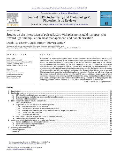
a b s t r a c t
This review describes the fundamental aspects of laser–gold nanoparticle (Au NP) interaction that leads to nanoscale energy deposition to the surroundings through light amplification and heat generation. Besides the importance of the primary process in physics and chemistry, application of the light–NP interaction has attracted significant interest from various areas ranging from analytical chemistry to material chemistry and biomedicine. Here we consider both mechanistic and application aspects. Our attention is focused on pulsed-laser-induced fast processes that revealed the heating–cooling dynamics of electrons, lattice (particle), and particle’s environment. On the application side, we focus on material fabrication and processing that beat diffraction-limited resolution. Together, we will shed a light on the essence of research activities carried out in the past 10 years. In addition to an abundance of latest information obtained from currently available literature, this review includes figures obtained by our own calculations to provide readers with a better understanding of the basics of the optical properties and energy and heat-transfer processes of Au NPs, which are not familiar to photochemists. © 2012 Elsevier B.V. All rights reserved.
脑成像技术
7
PET 是通过回旋加速器产生放射性核素, 合 成能发射正电子的放射性示踪物, 然后把它 注入人体内, 通过与计算机相连的射线照像 机进行检测,从而了解标记物的分布
17
问题
fMRI不是直接检测神经活动,而是对其次 级反映的检测,那么所检测到的信号变化 是否精确反映了神经活动变化的位置?
为了提高成像定位的精确性,目前采用两 种方法:使用高磁场强度;使用造影剂。
18
信号的变化是否精确反映了神经活动变化 的大小?
目前,所有的FMRI实验均假设BOLD信号变 化的幅值越大.相应的神经活动的变化就越 大;
微弱脉冲电流
42
刺激参数
视 刺觉 激 参 数听
觉
体 感
对比度 亮度 视角
频率 声压级 上升/下降时间
0.1~0.5ms, 1~5Hz,50uV
刺激呈现时间
刺激呈现概率 刺激间隔 SOA、ISI
三大要素
43
刺激呈现——E-Prime简介
E-Prime是实现心理实验计算机化的一个 可视化编程语言平台,是一个涵盖实验生成 到毫秒精度的数据收集分析的应用软件套装。
3
脑成像技术
以ERP、fMRI等为代表的脑成像技术的发明和发展,
已成为现代高科技的一个竞争热点,汇集了信息科学、 物理科学以及其它工程科学的众多高科技成果
脑成像技术可以直接“观察”大脑的活动。采用脑成
像技术研究脑的认知结构和功能,已成为脑科学与认 知科学发展的一大趋势
脑成像技术还有待完善。脑认知成像技术可以为我们
EEG主要来自突触后电位变化--胞体和 树突的电位变化。
PMMA和PS掺杂螺吡喃光致变色膜的制备及其变色性能

PMMA和PS掺杂螺吡喃光致变色膜的制备及其变色性能蒋淑恋;黄立漳;张小清;胡世荣【摘要】The photochromic behaviour of 6-nitro-l ',3',3' -trimethyl indolinebenzopyran photochromic com-pounds were studied, which synthesed by 5 -nitrosalieylaldehyde and 1 , 3 , 3-trimethyl-2-methyleneindo-line. The spiropyran photochromic thin films were made by dispersion method, in the way that 6-nitrb-1 ',3',3'-trimethyl indoline was dispersed in the PMMA and PS in THF solution. At the same time,the pho-tochromic effect and the stability of the film has also been researched.%由5-硝基水杨醛和吲哚啉合成了6-硝基-1′,3′,3′-三甲基吲哚啉苯并吡喃光致变色化合物,研究其在溶液中的光致变色行为.将6-硝基-1′,3′,3′-三甲基吲哚啉苯并吡喃分散在PMMA和PS的四氢呋喃溶液中,制成螺吡喃光致变色薄膜,并研究其成膜和变色效果以及膜的稳定性.【期刊名称】《应用化工》【年(卷),期】2012(041)011【总页数】4页(P1912-1914,1918)【关键词】合成;吲哚啉苯并吡喃;光致变色薄膜【作者】蒋淑恋;黄立漳;张小清;胡世荣【作者单位】漳州市产品质量检验所,福建漳州363000;漳州市产品质量检验所,福建漳州363000;漳州师范学院化学与环境科学系,福建漳州363000;漳州师范学院化学与环境科学系,福建漳州363000【正文语种】中文【中图分类】TQ577螺吡喃化合物绝大多数只能在溶液中变色,而在固体状态下,几乎不表现出光致变色效应,这在很大程度上限制了其应用。
马尾松针叶聚戊烯醇对阿尔茨海默病模型小鼠学习记忆的影响
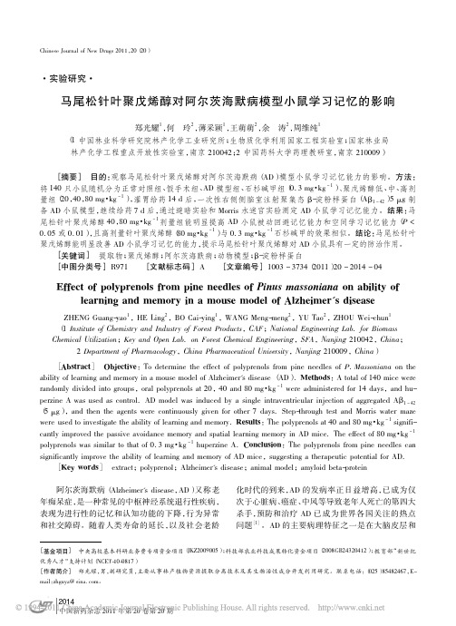
2014中国新药杂志2011年第20卷第20期[基金项目]中央高校基本科研业务费专项资金项目(JKZ2009005);科技部农业科技成果转化资金项目(2008GB24320412);教育部“新世纪优秀人才”支持计划(NCET-10-0817)[作者简介]郑光耀,男,副研究员,主要从事林产植物资源提取分离技术及其生物活性成分开发利用研究。
联系电话:(025)85482467,E-mail :zhguya@sina.com 。
·实验研究·马尾松针叶聚戊烯醇对阿尔茨海默病模型小鼠学习记忆的影响郑光耀1,何玲2,薄采颖1,王萌萌2,余涛2,周维纯1(1中国林业科学研究院林产化学工业研究所;生物质化学利用国家工程实验室;国家林业局林产化学工程重点开放性实验室,南京210042;2中国药科大学药理教研室,南京210009)[摘要]目的:观察马尾松针叶聚戊烯醇对阿尔茨海默病(AD )模型小鼠学习记忆能力的影响。
方法:将140只小鼠随机分为正常对照组、假手术组、AD 模型组、石杉碱甲组(0.3mg ·kg -1)、聚戊烯醇低、中、高剂量组(20,40,80mg ·kg -1),灌胃给药14d 后,一次性右侧侧脑室注射聚集态β-淀粉样蛋白(A β1 42)5μg 制备AD 小鼠模型,继续给药7d 后,通过避暗实验和Morris 水迷宫实验测定AD 小鼠学习记忆能力。
结果:马尾松针叶聚戊烯醇40,80mg ·kg -1剂量组能明显提高AD 小鼠被动回避记忆能力和空间学习记忆能力(P <0.05或0.01),且高剂量针叶聚戊烯醇(80mg ·kg -1)与0.3mg ·kg -1石杉碱甲的效果相似。
结论:马尾松针叶聚戊烯醇能明显改善AD 小鼠学习记忆的能力,提示马尾松针叶聚戊烯醇对AD 小鼠具有一定的防治作用。
[关键词]提取物;聚戊烯醇;阿尔茨海默病;动物模型;β-淀粉样蛋白[中图分类号]R971[文献标志码]A [文章编号]1003-3734(2011)20-2014-04Effect of polyprenols from pine needles of Pinus massoniana on ability oflearning and memory in a mouse model of Alzheimer's diseaseZHENG Guang-yao 1,HE Ling 2,BO Cai-ying 1,WANG Meng-meng 2,YU Tao 2,ZHOU Wei-chun 1(1Institute of Chemistry and Industry of Forest Products ,CAF ;National Engineering Lab.for Biomass Chemical Utilization ;Key and Open Lab.on Forest Chemical Engineering ,SFA ,Nanjing 210042,China ;2Department of Pharmacology ,China Pharmaceutical University ,Nanjing 210009,China )[Abstract ]Objective :To determine the effect of polyprenols from pine needles of P.Massoniana on the ability of learning and memory in a mouse model of Alzheimer's disease (AD ).Methods :A total of 140mice wererandomly divided into groups ,oral polyprenols at 20,40and 80mg ·kg -1were administered for 14days ,and hu-perzine A was used as control.AD model was induced by a single intraventricular injection of aggregated A β1 42(5μg ),and then the agents were continuously given for other 7days.Step-through test and Morris water mazewere used to investigate the ability of learning and memory.Results :The polyprenols at 40and 80mg ·kg -1signifi-cantly improved the passive avoidance memory and spatial learning memory in AD mice.The effect of 80mg ·kg -1polyprenols was similar to that of 0.3mg ·kg -1huperzine A.Conclusion :The polyprenols from pine needles can significantly improve the ability of learning and memory of AD mice ,suggesting a therapeutic potential for AD.[Key words ]extract ;polyprenol ;Alzheimer's disease ;animal model ;amyloid beta-protein 阿尔茨海默病(Alzheimer's disease ,AD )又称老年痴呆症,是一种常见的中枢神经系统退行性疾病,表现为进行性的记忆和认知功能的下降,行为异常和社交障碍。
NTP非肿瘤性病变图谱

景病变发病 率之时, 差异可能 成为病理 学家的挑 战。
许多非 肿瘤性疾病与 环境暴露有关, 出现于人类疾病的 病变也可在化学处理的 动物中观察到。例如,当通过吸入 暴露于名为二乙酰(一种人造黄油调 味剂的成分)的化学物时,C57BL/6 小鼠发生类似于称之为闭塞性细支气 管炎的人类支气管病变。已经知道这 种不可逆的阻塞性肺疾病发生在职业 暴露人造奶油调味剂的工人中。该图 谱通过提高病理学家分类与解释以上 及其它非肿瘤性病变的水平,从观念 上提高我们对人类环境相关疾病的理 解。
化学暴露
NTP 非肿瘤性病变图谱
毒性病理学的新工具
一种新的在线图谱与诊断指导有望显著提高研究人员评估动物研究中非癌性组织病理学发现的能力。© Shutterstock; Joseph Tart
12
《环境与健康展望》2015年2月刊·VOLUME 123 / NUMBER 1C
© Emilio Ereza/Alamy
《环境与健康展望》2015年2月刊·VOLUME 123 / NUMBER 1C
13
化学暴露
越来越受重视的非肿瘤终点
尤其是《Fisher大鼠病理学:参考文 兽医病理学家越来越依赖于SD大鼠,
“肿瘤”源于希
献 与 图 谱 》 (P a t h o l o g y 因为Fisher 344大鼠的单核细胞白血病
子中,同一非肿瘤的术语‘嗜碱性病 灶’可应用于迥然不同的情形。”他 指出,在尝试比较染毒组动物的嗜碱 性病灶的剂量相关发病率与对照组背
工作越来越多,意味着分析非肿瘤性 病变需要更多的时间。”
据NTP细胞与分子病理室主任 Robert Sill说,非肿瘤性病变往往发
该图谱最终将扩大到涵盖所有 56 个不同组织类型 的 13 个解剖系统。“我们认为它将会作为活文件 而 不 断 得 到 更 新,” 该 图 谱 主 编,NTP 病 理 学 家 Mark Cesta 说。
如何写医学科普有趣

1960年,一位科学家将一小罐白色粉末交给初来美国 的日籍科学家下村修,并告诉他这是从一些能发光的水母 中取得的“精华干粉”。下村修立刻被这神秘的粉末吸引, 随着第二年初夏的暖风来到了华盛顿盛产发光水母的星期 五港(Friday Harbor)。
下村修所在的实验室正好位于港湾。每天,柔软的小 水母都乘着早晚的潮汐成群结对地漂过他实验室的两侧。 其中一些会被下村修用小网兜截住,带回实验室。这些水 母只有掌心大小,就像一把圆圆张开的小伞,在昏暗的水 中发出幽幽绿光。发光的器官是一百多个小颗粒,就像点 缀在小伞边缘的微型“灯泡”。下村修将小伞边缘剪下来, 用棉布攥出水母“精华”。只是离开了原来的环境,它们 似乎不太乐意发光……
资料仅供参考,不当之处,请联系改正。
试着写一写
十一过后宋大妈来到武汉老同学家玩,当老同学得知她的病情后带着宋大妈 来到古田三路的武汉市普爱医院就诊,接诊神经内科医生在仔细询问后了解到: 宋大妈第一次出现症状时吃的黄瓜是她自己去地里摘的,而在这之前两天,她曾 亲眼看到农民给这片地喷农药,而后她一直担心黄瓜上的农药没洗干净会中毒。
缓解医患矛盾
深圳一儿童因腹胀去市儿童医院。医院称孩子疑为先天性 巨结肠,建议进行造瘘活检手术,手术及后续费用超过十 万。随后,带儿子到广州市儿童医院就诊,称接诊医生开 了八毛钱的药,“孩子就治好了,能吃能拉”。 当天,深圳当地媒体以《婴儿被诊断要做10万元手术 最 终吃8毛钱药痊愈》为题做出报道,被全国各大媒体及电 视广泛转载转播,掀起轩然大波。
下村修是首位从水母中分离绿色荧光蛋白的科学家,他 发现这种蛋白在紫外线光中呈现亮色。马丁-查尔菲展示了绿 色荧光蛋白作为各种生物现象的亮光基因标签的价值。钱永 健对我们理解绿色荧光蛋白如何发光作出了贡献,他还将颜 色标签扩展至除绿色之外的颜色,以便可以用各种颜色标识 不同的蛋白和细胞。
- 1、下载文档前请自行甄别文档内容的完整性,平台不提供额外的编辑、内容补充、找答案等附加服务。
- 2、"仅部分预览"的文档,不可在线预览部分如存在完整性等问题,可反馈申请退款(可完整预览的文档不适用该条件!)。
- 3、如文档侵犯您的权益,请联系客服反馈,我们会尽快为您处理(人工客服工作时间:9:00-18:30)。
a r X i v :h e p -p h /0510276v 2 2 N o v 2005LEPTON PHOTON SYMPOSIUM 2005:SUMMARY AND OUTLOOK ∗FRANCIS HALZENPhysics Department,University of Wisconsin,Madison,WI 53706,USAEmail:halzen@Lepton Photon 2005told the saga of the Standard Model which is still exhilarating because it leaves all questions of consequence unanswered.University of Wisconsin -MadisonMADPH-05-1444hep-ph/0510276Over the last decade the biennial gather-ing discussing leptons and photons has broad-ened its horizons to reflect the excursions par-ticle physics techniques have made into as-tronomy and cosmology.It was in the grand-est of particle physics traditions however that five days of talks in the historic aula of one of Europe’s oldest universities,the home of Linnaeus,Manne and Kai Siegbahn and Dag Hammarskjold,erected the impressive edifice that is called the Standard Model.Experi-mental ingenuity has not been able to pierce the Model’s armor and I cannot help think-ing of the prophetic words of Leon Lederman at the Rochester meeting held in Madison twenty five years ago:“the experimentalists do not have enough money and the theorists are overconfident”.Where experimentalists are concerned,nobody could have anticipated that today we would be studying the pro-ton structure to one thousandth its size and would have established the Standard Model as a gauge theory with a precision of one in a thousand,pushing any interference of pos-sible new physics to energy scales beyond 10TeV.The theorists can modestly claim that they have taken revenge for Leon’s remark.Because all the big questions remain unan-swered,there is no feeling though that we are now dotting the i’s and crossing the t’s of a mature theory.Worse,the theory has its own demise built into its radiative corrections.The most evident of unanswered ques-tions is why the weak interactions are weak.√√1−m 2W /m 2Z(1+∆r ).(1)Fermi could certainly not have anticipated that we now have a renormalizable gauge the-ory that allows us to calculate the radiative corrections ∆r to his formula.Besides reg-ular higher order diagrams,loops associated with the top quark and the Higgs boson con-tribute;they have been observed 2,3,4.I once heard one of my favorite physicists refer to the Higgs as the “ugly”particle,but this is nowadays politically incorrect.Indeed,scalar particles are unnatural.If one calcu-lates the radiative corrections to the mass m appearing in the Higgs potential,the same gauge theory that withstood the onslaught of precision experiments at LEP/SLC and the Tevatron yields a result that grows quadrat-ically:δm2=32m2H†H+1ΛL5+1m2 = δv2Figure1.The Tevatron roadmap to the Higgs by increased luminosity and improved detector performance.lepton identification and jet mass resolution. The performance of the detectors has been nothing short of spectacular as illustrated by the identification of the top quark in6-jet events3.Baryogenesis is another one of the grand issues left unresolved by the Standard Model. We know that at some early time in the evo-lution of the Universe quarks and antiquarks annihilated into light,except for just one quark in1010that failed tofind a partner and became us.We are here because baryogene-sis managed to accommodate the three Za-kharov conditions;one of them dictates CP-violation.Evidence for the indirect violation of CP-invariance wasfirst revealed in1964 in the mixing of neutral kaons.Direct CP-violation,not mixing-assisted,was not dis-covered until1999.Today,precision data on neutral kaons have been accumulated over 40years;the measurements can,without ex-ception,be accommodated by the Standard Model with three families8.History has re-peated itself for B mesons,but in three years only,thanks to the magnificent performance of the B-meson factories Belle and BaBar9.Direct CP-violation has been established in the decay B d→Kπwith a significance in excess of5sigma.Unfortunately,this re-sult,as well as a wealth of data contributed by CLEO,BES and Dafne,fails to reveal evidence for new physics10.Whenever the experimental precision increases,the higher precision measurements invariably collapse onto the Standard Model values;see Fig.2. Given the rapid progress and the better the-oretical understanding of the Standard Model expectations relative to the K system11,the hope is that at this point the glass is half full and that improved data will pierce the Standard Model’s resistant armor12.Where theory is concerned,it is noteworthy that lat-tice techniques have reached the maturity to perform computer experiments that are con-firmed by experiment.The rise and fall of theories,or at least of their popularity,can be easily assessed by consulting the citation index.The number of citations to Wolfenstein’s seminal paper on neutrino oscillations in the presence of mat-ter has,after a steady increase from1978–2000,dropped by almost a factor of two since.3Figure2.Geometry of the CKM triangle converges on the standard model by measuring sides,angles,or both.Progress in neutrino physics has been led by a string of fundamental experimental measure-ments summarized by the simple vacuum re-lations between the neutrino states produced by the weak interactions in e,mu and taufla-vors and propagating as mixed statesν1,ν2 andν3:ν1=−cosθνe+sinθ νµ−ντ2 ,ν2=sinθνe+cosθ νµ−ντ2 ,ν3= νµ+ντ2 .(5)Hereθis the solar mixing angle.Discovery of neutrino oscillations in solar and atmospheric beams has been confirmed by supporting ev-idence from reactor and accelerator beams13.As usual,next-generation experimentsare a lot more challenging and the boom times of neutrino physics are probably over as reflected by Wolfenstein’s citations.Also, high-precision data from the pioneering ex-periments trickle in at a slower pace,al-though new evidence for the oscillatory be-havior in L/E of the muon-neutrinos in the atmospheric neutrino beam has become very convincing.The new results included first data from the reborn SuperKamiokande experiment14.The future of neutrino physics is undoubtedly bright.Construction of the KATRIN spectrometer measuring neutrino mass to0.2eV by studying the kinematics of tritium decay is in progress and a wealth of ideas on double beta decay and long-baseline experiments is approaching reality15. These experiments will have to answer the great“known-unknowns”of neutrino physics: 4their absolute mass and hierarchy,the precise value of the second and third small mixing angle and its associated CP-violating phase and whether neutrinos are really Majorana particles.In Eq.(5)we assumed that the mixing of mu and tau neutrinos is maxi-mal,with no admixture of electron neutrinos. Observing otherwise will most likely require next-generation experiments.Among these,discovery of neutrino-less double beta decay would be especially rewarding16.Its observation would confirm the theoretical bias that neutrinos are their own antiparticles,yield critical information on the absolute mass scale and,possibly,re-solve the hierarchy problem.In the meantime we will keep wondering whether small neu-trino masses are ourfirst glimpse at grand unified theories via the see-saw mechanism, or represent a new Yukawa scale tantalizingly connected to lepton conservation and,possi-bly,the cosmological constant.The cosmological constant represents a thorny issue for the Standard Model17.New physics is also required to control the Stan-dard Model calculation of the vacuum en-ergy,also known as the cosmological con-stant,which diverges asΛ12sponsible for renewed interest in decipheringbaryogenesis—a tally of the rapidly increas-ing number of citations to the1986paper by Fukugita and Yanagida underscores the point.The problem is more clearly framed than ever before.The imprint on the sur-face of last scattering of the acoustic waves driven into the hot gas of nuclei and pho-tons reveals a relative abundance of baryons to photons of6.5+0.4−0.3×10⁻¹⁰(WMAP obser-vation).Gamov realized that a Universe born as hot plasma must consist mostly of hydro-gen and helium,with small amounts of deu-terium and lithium added.The detailed bal-ance depends on basic nuclear physics as well as the same relative abundance of baryons to photons;the state of the art result of this ex-ercise yields4.7+1.0−0.8×10⁻¹⁰.The agreement of the two observations is stunning,not just because of the precision,but because of the concordance of two results derived by totally unrelated ways to probe the early Universe.Physics at the high energy frontier is the physics of partons.For instance,at the LHC gluons produce the Higgs boson and the high-est energy neutrinos interact with sea-quarks in the detector.We master this physics with unforeseen precision because of a decade of steadily improving HERA measurements of the nucleon structure19.These now include experiments using targets of polarized pro-tons and neutrons.HERA is our nucleon microscope,tunable by the wavelength and thefluctuation time of the virtual photon ex-changed in the electron proton collision.The wavelength of the virtual photons probing the nucleon is reduced with increased momentum transfer Q.The proton has now been probed to distances of one thousandth of its size of1fm.In the interaction thefluctuations of the virtual photons survive over distances ct∼1/x,where x is the relative momentum of the parton.HERA now studies the pro-duction of chains of gluons as long as10fm, an order of magnitude larger than and prob-ably totally insensitive to the proton target.These are novel QCD structures,the under-standing of which has been challenging20.We should not forget however that theorists ana-lyze HERA data with calculations performed to next-to-next to leading order in the strong coupling.In fact,beyond this precision one has to include the photon as a parton inside the proton21.These electromagnetic struc-ture functions violate isospin and differen-tiate a u-quark in a proton from a d-quark in a neutron because of the different electric charge of the quark.Interestingly,their in-clusion in the structure functions modifies the extraction of the Weinberg angle from NuTeV data,bridging roughly half of its discrepancy with the particle data book value.Added to already anticipated intrinsic isospin viola-tions associated with sea-quarks,the NuTeV anomaly may be on its way out.Recalling Lederman,whatever the actual funding,the experimenters managed to de-liver most highlights of this conference.And where history has proven that theorists had the right to be confident in1980,they have not faded into the background,and provided some highlights of their own.Developing QCD calculations to the level that the pho-ton structure of the proton becomes a factor is a tour de force and,there were others at this meeting.Progress in higher order QCD computations of hard processes is mind bog-gling—progress useful,sometimes essential, for the interpretation of LHC experiments22. Discussions of strings,supersymmetry and additional dimensions were very much fo-cused on the capability of experiments to con-firm or debunk these concepts23.Theory and experiment joined forces in the ongoing attempts to read the information supplied by the data on heavy ion collisions from Brookhaven.Rather than the antici-pated quark gluon plasma,the data suggests the formation of a strongly interactingfluid with very low viscosity for its entropy24.Sim-ilarfluids of cold6Li atoms have been cre-ated in atomic traps.Interestingly,theorists 6are exploiting the Maldacena connection be-tween four dimensional gauge theory and ten dimensional string theory to model such a thermodynamic system25.The model is that of a10-D rotating black hole with Hawking-Beckenstein entropy.It accommodates the low viscosities observed.This should put us on notice that very high energy collisions of nuclei may be more interesting than antici-pated from QCD-inspired logarithmic extrap-olations of accelerator data.This is relevant to the analysis of cosmic ray experiments. Enter particle astrophysics.Conventional astronomy spans60oc-taves in photon frequency,from104cm radio-waves to10−14cm photons of GeV energy. This is an amazing expansion of the power of our eyes that scan the sky over less than a single octave just above10−5cm wavelength. Recently detection and data handling tech-niques of particle physics26are reborn in in-strumentation to probe the Universe at new wavelengths,smaller than10−14cm,or pho-ton energies larger than10GeV.Besides gamma rays,gravitational waves and neutri-nos as well as very high-energy protons that are only weakly deflected by the magnetic field of our galaxy,have become astronom-ical messengers from the Universe27.As ex-emplified time and again,the development of novel ways of looking into space invariably results in the discovery of unanticipated phe-nomena.For particle physicists the sexiest astrophysics problem is undoubtedly how Na-ture manages to impart an energy of more than108TeV to a single elementary particle.Although cosmic rays were discovered al-most a century ago,we do not know how and where they are accelerated28.This may be the oldest mystery in astronomy and solving it is challenging as can be seen by the follow-ing argument.It is sensible to assume that,in order to accelerate a proton to energy E in a magneticfield B,the size R of the accelerator must encompass the gyroradius of the parti-cle:R>R gyro=E/B,i.e.the acceleratingmagneticfield must contain the particle or-bit.This condition yields a maximum energy E<ΓBR by dimensional analysis and noth-ing more.The factorΓhas been included to allow for the possibility that we may not be at rest in the frame of the cosmic accelerator resulting in the observation of boosted par-ticle energies.Opportunity for particle ac-celeration to the highest energies is limited to dense regions where exceptional gravita-tional forces create relativistic particleflows: the dense cores of exploding stars,inflows on supermassive black holes at the centers of ac-tive galaxies,annihilating black holes or neu-tron stars?All speculations involve collapsed objects and we can therefore replace R by the Schwartzschild radius R∼GM/c2to obtain E<ΓBM.The above speculations are reinforced by the fact that the sources listed happen to also be the sources of the highest energy gamma rays observed.At this point a real-ity check is in order.Note that the above dimensional analysis applies to the Fermi-lab accelerator:kGaussfields over several kilometers(covered with a repetition rate of 105revolutions per second)yield1TeV.The argument holds because,with optimized de-sign and perfect alignment of magnets,the accelerator reaches efficiencies close to the dimensional limit.It is highly questionable that Nature can achieve this feat.Theorists can imagine acceleration in shocks with effi-ciency of perhaps1–10%.Given the microgauss magneticfield of our galaxy,no structures seem large or mas-sive enough to reach the energies of the high-est energy cosmic rays.Dimensional analysis therefore limits their sources to extragalac-tic objects.A common speculation is that they may be relatively nearby active galactic nuclei powered by a billion solar mass black holes.With kilo-Gaussfields we reach100 EeV,or1020eV.The jets(blazars)emit-ted by the central black hole could reach similar energies in accelerating substructures 7boosted in our direction by aΓ-factor of 10,possibly higher.The neutron star or black hole remnant of a collapsing super-massive star could support magneticfields of1012Gauss,possibly larger.Shocks with Γ>102emanating from the collapsed black hole could be the origin of gamma ray bursts and,possibly,the source of the highest en-ergy cosmic rays.The astrophysics problem is so daunt-ing that many believe that cosmic rays are not the beam of cosmic accelerators but the decay products of remnants from the early Universe,for instance topological defects as-sociated with a grand unified GUT phase transition near1024eV.A topological de-fect will suffer a chain decay into GUT par-ticles X,Y,that subsequently decay to famil-iar weak bosons,leptons and quark-or gluon jets.Cosmic rays are the fragmentation prod-ucts of these jets.HERA again revealed to us the composition of these jets that count rel-atively few protons,i.e.cosmic rays,among their fragmentation products and this is in-creasingly becoming a problem when one con-fronts this idea with data.We conclude that,where the highest en-ergy cosmic rays are concerned,both the ac-celerator mechanism and the particle physics are enigmatic.There is a realistic hope that the oldest problem in astronomy will be re-solved soon by ambitious experimentation: air shower arrays of104km2area(Auger),ar-rays of air Cerenkov detectors(H.E.S.S.and Veritas,as well as the Magic17m mirror tele-scope)and kilometer-scale neutrino observa-tories(IceCube and NEMO).Some of these instruments have other missions;all are likely to have a major impact on cosmic ray physics. While no breakthroughs were reported,pre-liminary data forecast rapid progress and im-minent results in all three areas27.The Auger air shower array is con-fronting the low statistics problem at the highest energies by instrumenting a huge col-lection area covering3000square kilometerson an elevated plane in Western Argentina. The instrumentation consists of1600water Cherenkov detectors spaced by1.5km.For calibration,showers occurring at night,about 10percent of them,are also viewed by four fluorescence detectors.The detector will ob-serve several thousand events per year above 10EeV and tens above100EeV,with the ex-act numbers depending on the detailed shape of the observed spectrum.The end of the cosmic ray spectrum is a matter of specula-tion given the somewhat conflicting results from existing experiments,most notably the HiResfluorescence detector and the AGASA scintillator array;see Fig.4.FluxxE3/124(eV2m-2s-1sr-1)10110Log10 E (eV) Figure4.Extragalactic cosmic ray spectrum before Auger and HiRes stereo measurements.Above a threshold of50EeV the cos-mic rays interact with cosmic microwave pho-tons and lose energy to pions before reaching our detectors.This is the Greissen-Zatsepin-Kuzmin cutoffthat limits the sources to our supercluster of galaxies.The feature in the spectrum is claimed at the5sigma level in the latest HiRes data.It is totally absent in the AGASA data,a fact that would require some radical departure from established par-ticle physics or astrophysics.At this meet-ing Auger presented thefirst results from8the partially deployed array28.The expo-sure is similar to that of thefinal AGASA data.The data confirms the existence of su-per EeV events.There is no evidence,ei-ther in the latest HiRes or the Auger data, however,for anisotropies in the arrival di-rections of the cosmic rays claimed mostly on the basis of the AGASA data.Impor-tantly,Auger observes a discrepancy between the energy measurements of showers obtained from thefluorescence and particle array tech-niques.The discrepancy suggests that very high energy air showers do not develop as fast as modeled by the particle physics simu-lations used to analyze previous experiments, i.e.the data necessitate deeper penetration of the primary,less inelasticity and more energy in fewer leading particles than anticipated. Auger data definitely indicate that the exper-iment is likely to qualitatively improve exist-ing observations of the highest energy cosmic rays in the near future.Cosmic accelerators are also cosmic beam dumps producing secondary photon and neu-trino beams29.Particles accelerated near black holes pass through intense radiation fields or dense clouds of gas leading to pro-duction of secondary photons and neutri-nos that accompany the primary cosmic-ray beam.The target material,whether a gas or photons,is likely to be sufficiently tenuous so that the primary beam and the photon beam are only partially attenuated;see Fig.5.Although gamma ray and neutrino tele-scopes have multiple interdisciplinary sci-ence missions,in the case of neutrinos the real challenge has been to develop a re-liable,expandable and affordable detector technology30.The South Pole AMANDA neutrino telescope,now in itsfifth year of operation,has improved its sensitivity by more than an order of magnitude since re-porting itsfirst results in2000.It has now reached a sensitivity close to the neutrino flux anticipated to accompany the highest energy cosmic rays,dubbed theWaxman-Figure 5.Cosmic ray accelerators are also cosmic beamdumps producingfluxes of neutrinos and TeV photons accompanying the cosmic rays.Bahcall bound.Expansion into the Ice-Cube kilometer-scale neutrino observatory, required to be sensitive to the best esti-mates of potential cosmic neutrinofluxes, is in panion experiments in the deep Mediterranean are moving from the R&D into the construction phase with the goal to eventually build an IceCube size de-tector.With the sun and SN87neutrino observations as proofs of concepts,next-generation neutrino experiments will also scrutinize their data for new particle physics, from the signatures of dark matter to the ev-idence for additional dimensions of space.It is however the H.E.S.S.array of four air Cherenkov gamma ray telescopes de-ployed under the southern sky of Namibia that delivered the highlights in the particle astrophysics corner27.For thefirst time an instrument is capable of imaging astronomi-cal sources in TeV gamma rays.Its images of young galactic supernova remnants showsfil-ament structures of high magneticfields that are capable of accelerating protons to the en-ergies,and with the energy balance,required to explain the galactic cosmic rays;Fig.6.Al-though the smoking gun for cosmic ray accel-9Figure6.TeV gamma ray image of a young supernova remnant.eration is still missing,the evidence is tanta-lizingly close.The big event of the next biennium is the commissioning of the LHC.With dark mat-ter and energy18,31,astronomers have raised physics problems that seem as daunting as the problem of the lifetime of the sun over one century ago.Evolution and geology re-quired a sun that was older than several tens of millions of years.Chemistry established its lifetime at3000years.Neither chemistry nor astronomy solved the puzzle,Rutherford did. May history repeat itself with the solution re-vealed by the accelerators in our future,LHC and a linear collider32!Rendez vous in2007in Daegu,Korea. AcknowledgmentsI am grateful for the superb hospitality of Professor Tord Ekelof,my Uppsala IceCube colleagues and other organizers of the meet-ing.This talk is not only inspired by the excellent talks at this meeting,but also by a series of stimulating and provocative talks atmeetings I attended prior to this conference, by Alan Martin,K.Kinoshita and C.W.Chi-ang at Pheno05in Madison and by Pilar Hernandez,Hitoshi Murayama,Carlos Pena-Garay and G.Raffelt at a neutrino workshop in Madrid.I thank Vernon Barger,Hooman Davoudiasl,Concha Gonzalez-Garcia,Tao Han,and Patrick Huber for reading the manuscript.This research was supported in part by the National Science Foundation under Grant No.OPP-0236449,in part by the U.S.Department of Energy under Grant No.DE-FG02-95ER40896,and in part by the University of Wisconsin Research Committee with funds granted by the Wisconsin Alumni Research Foundation.References1.E.Fermi,Z.Phys.88,161(1934);NuovoCim.11,1(1934).2.S.Dawson,these proceedings.3.A.Juste,these proceedings.4.D.Diaconnu,these proceedings.5.For a nice update,see J.A.Casas, 10J.R.Espinosa and I.Hidalgo,arXiv:hep-ph/0410298,JHEP0411,057(2004).6.F.Gianotti and L.Rolandi,these pro-ceedings.mmel,these proceedings.8.E.Blucher,these proceedings.9.K.Abe and F.Forti,these proceedings.10.M.Artuso,R.Jesic,X.Shen andV.Burkert,these proceedings.11.U.Nierste and I.Stewart,these proceed-ings.12.L.Silvestrini,these proceedings.13.A.Poon, E.Zimmerman and S.Go-swami,these proceedings.14.Y.Suzuki,these proceedings.15.C.Weinheimer,these proceedings.16.O.Cremonesi,these proceedings.17.E.Witten,Proceedings of the2003Lepton-Photon Conference,Fermilab,Chicago(2003)18.S.Hannestad,these proceedings.19.J.Butterworth and J.-M.Le Goff,theseproceedings.20.G.Ingelman,these proceedings.21.M.Gluck,P.Jimenez-Delgado andE.Reya,arXiv:hep-ph/0503103,Phys.Rev.Lett.95,022002(2005);A.D.Mar-tin,R.G.Roberts,W.J.Stirling andR.S.Thorne,arXiv:hep-ph/0411040,Eur.Phys.J.C39,155(2005).22.G.Salam,these proceedings.23.I.Antoniadis,these proceedings.24.J.Stachel,these proceedings.25.I.Klebanov,these proceedings.26.M.Danilov,these proceedings.27.R.Ong,these proceedings.28.S.Westerhoff,these proceedings.29.E.Waxman,these proceedings.30.P.Hulth,these proceedings.31.L.Baudis,these proceedings.32.K.Moenig,F.Zimmerman and J.Dor-fan,these proceedings.11。