Merck-NASA-Knowledge-2020-FINAL-MS(NASA 知识管理资料)
植根中国,服务中国,以创新的产品赢得市场

植根中国,服务中国,以创新的产品赢得市场作者:来源:《食品安全导刊》2015年第12期科学服务领域的世界领导者赛默飞世尔科技(以下简称“赛默飞”)于10月27~30日携百余款产品亮相“第十六届北京分析测试学术报告会暨展览会”(BCEIA 2015),并隆重推出应用于食品、生命科学、环境监测、健康医疗等行业的十多款新品,全方位展示赛默飞在分析测试领域为客户提供的领先技术和高效解决方案。
赛默飞今年以“聚·创”为主题,旨在与客户协同合作,实现创新加速度。
在这个理念的指导下,赛默飞积极践行创新科技及“植根中国,服务中国”的本地化战略,最大化满足中国客户的多样化需求,实现合作多赢的目标和愿景。
在展会现场,赛默飞中国区总裁江志成先生、赛默飞软件服务全球首席技术官兼副总裁Mark Field先生及赛默飞中国区生命科学质谱市场经理陈伟接受了记者的采访。
记者:今年赛默飞在中国的业务发展情况是怎样的?和去年相比,哪些领域表现良好?中国业务销售额在全球整体业务中占的比重大概为多少?江志成:中国是赛默飞的第二大市场,仅次于美国,年销售额约12亿美元,从业绩来看,赛默飞在中国的表现十分抢眼。
长期以来,赛默飞在中国市场一直持续两位数的增长,今年第三季度更取得15%的增长率。
我们在食品安全、环境还有医疗方面的增长尤为强劲。
赛默飞进入中国发展已有30多年,员工人数约3700名。
对赛默飞来说,中国不仅是赛默飞的产品销售市场,也是产品生产地。
目前赛默飞在中国有8家工厂,它们分别位于北京、上海、苏州、广州等地,生产的产品不仅在中国销售也出口到其他国家。
未来,我们将植根于中国,服务于中国。
今年5月,中国政府出台了“中国制造2025”战略,而我们也于2013年在上海建立了中国创新中心,希望将创新的技术带到中国,和中国的合作伙伴一起携手,使世界更健康、更清洁、更安全。
同时,我们今年也在中国市场上推出了Thermo Fisher Cloud云端服务平台,服务于中国客户,通过这个平台可以使中国的客户拥有更多的数据,这对他们工作将有很大的帮助。
Waters Protein-Pak Hi Res Q Column 分离 Low Range ss

Size and Purity Assessment of Single-Guide RNAs by Anion-Exchange Chromatography (AEX)Hua Yang,Stephan M. Koza,Ying Qing YuWaters CorporationAbstractSingle-guide RNA (sgRNA) is a critical element in the CRISPR/Cas9 Technology for gene editing, the size of which usually ranges from 100 to 150 bases. In this application note, we show that the size of several sgRNAs could be estimated by comparison to a Low Range ssRNA Ladder (50–500 bases) using an optimized anion-exchange method developed on a Waters Protein-Pak Hi Res Q Column. In addition, the purity of the sgRNA samples can be assessed using the same anion exchange method, providing an informative and non-complex method for sgRNA product consistency.BenefitsWaters Protein-Pak Hi Res Q Column separation of a Low Range ssRNA Ladder with the size ranging from ■50 to 500 basesWaters Protein-Pak Hi Res Q Column separation of ssRNAs and their impurities■Size and purity estimation of ssRNAs having a size range of 100–150 mer under the same gradient conditions ■using the AEX method on Waters Protein-Pak Hi Res Q ColumnIntroductionThe discovery of clustered regularly interspaced short palindromic repeats (CRISPR)/CRISPR associated (Cas) bacterial immunity systems and the rapid adaptation of RNA guided CRISPR/CRISPR Associated Protein 9 (Cas9) Technology to mammalian cells have had a significant impact in the field of gene editing.1–3 The Cas9 protein, a non-specific endonuclease, is directed to a specific DNA site by a guide RNA (gRNA), where it makes a double-strand break of the DNA of interest. The gRNA consists of two parts: CRISPR RNA (crRNA) and trans-activating crRNA (tracrRNA). The crRNA is usually a 17–20 nucleotide sequence complementary to the target DNA, and the tracrRNA serves as a binding scaffold for the Cas9 nuclease. While crRNAs and tracrRNAs exist as two separate RNA molecules in nature, the single-guide RNA (sgRNA), which combines both the crRNA sequence and the tracrRNA sequence into a single RNA molecule, has become a commonly used format. The length of a sgRNA is in the range of 100–150 nucleotides. It is critical to characterize the sgRNA, as it is the core of the CRISPR/Cas9 technology.Anion-exchange chromatography (AEX) separates molecules based on their differences in negative surface charges. This analytical technique can be robust, reproducible, and quantitative. It is also easy to automate, requires small amounts of sample, and allows for the isolation of fractions for further analysis. AEX has been utilized in multiple areas related to gene therapy, including adeno-associated virus empty and full capsid separation, plasmid isoform separation, and dsDNA fragment separation.4–6 Since the sgRNAs are negatively charged due to the phosphate groups on the backbone, we investigated AEX for size and purity assessment of sgRNAs.In this application note, we show that using a Waters Protein-Pak Hi Res Q strong Anion-Exchange Column on an ACQUITY UPLC H-Class Bio System, a single-stranded RNA (ssRNA) ladder ranging from 50 to 500 bases can be separated and used for estimating the size of ssRNAs in the approximate range of 100–150 bases, including the sgRNAs for CRISPR/Cas9 System. Moreover, the purity of these ssRNAs can be estimated with the same gradient conditions.ExperimentalSample DescriptionHPRT (purified and crude) is a pre-designed CRISPR/Cas9 sgRNA (Hs.Cas9.HPRT1.1AA, 100 mer). GUAC is acustomized ssRNA (150 mer), which contains repeats of GUAC sequence. HPRT sgRNA and GUAC ssRNA were purchased from Integrated DNA Technologies (IDT). Rosa26 and Scrambled #2 are both pre-designedCRISPR/Cas9 sgRNAs purchased from Synthego (100 mer). Low Range ssRNA Ladder was purchased from New England Biolabs (N0364S).Method ConditionsLC ConditionsLC system:ACQUITY UPLC H-Class BioDetection:ACQUITY UPLC TUV Detector with 5 mm titaniumflow cellWavelength:260 nmVials:Polypropylene 12 x 32 mm Screw Neck Vial, withCap and Pre-slit PTFE/Silicone Septum, 300 µLVolume, 100/pk (P/N 186002639)Column(s):Protein-Pak Hi Res Q Column, 5 µm, 4.6 x 100 mm(P/N 186004931)Column temp.:60 °CSample temp.:10 °CInjection volume:1–10 µLFlow rate:0.4 mL/minMobile phase A:100 mM Tris-HClMobile phase B:100 mM Tris baseMobile phase C: 3 M Tetramethylammonium chloride (TMAC)Mobile phase D:WaterBuffer conc. to deliver:20 mMGradient Table (an AutoBlend Plus Method, Henderson-Hasselbalch derived).In the above gradient table, the buffer is 20 mM Tris pH 9.0. The initial salt concentration is set to 0 mM to ensure all the analytes are strongly bound onto the column. After 5 mins, the salt concentration is increased to 1400 mM where most of the impurities will elute, based on prior investigation. After 4 mins equilibration, the separation gradient starts. The salt concentration increases linearly from 1400 m to 2100 mM in 20 mins for the Low Range ssRNA Ladder separation, as well as individual ssRNAs. Then it is ramped up to 2400 mM to strip off any remaining bound molecules. Finally, an equilibration step to the initial condition takes place, preparing for the next injection.An equivalent gradient table for a generic quaternary LC system is shown above.Data ManagementChromatography software:Empower 3 (FR 4)Results and DiscussionSize AssessmentVarious mobile phase conditions were tested using a Low Range ssRNA Ladder for size assessment of the ssRNAs, including pH (7.4 and 9.0), column temperature (30 °C and 60 °C) and salt (NaCl and TMAC).The results from the optimal conditions are shown in Figure 1B. Using a pH 9.0 Tris buffer with 60 °C column temperature and a TMAC salt gradient, the Low Range ssRNA Ladder (50–500 bases) along with four pre-made sgRNAs (100 mer), and one customized ssRNA (150 mer) were separated on a Waters Protein-Pak Hi Res Q Column. The separation for the Low Range ssRNA Ladder on this strong anion exchange column was very similar to that on an agarose gel, as shown in Figure 1A. A calibration curve was constructed based on the retention time and the logarithm of the number of bases of each ssRNA in the ladder (Figure 1C, blue dots). Thelinear fit from the Low Range ssRNA Ladder indicates a strong correlation between the logarithm of the size andthe retention time (R2=0.993). Using this plot, the size of the ssRNAs was calculated from their individual retention time. The percent error is calculated using the formula {(calculated size – theoretical size)/theoretical size}. The percent error was less than 6% for all the RNAs tested (Figure 1d), as evidenced by the orange data points residing on or very closely to the trendline of the calibration curve. Notice that small percent error was obtained from four pre-made sgRNAs from two different manufacturers and a customized ssRNA with an artificial sequence. Although ssRNAs with shorter than 100 bases and larger than 150 bases were not tested, it is possible that this method can be used for the ssRNAs size assessment in the range of 50–500 bases.Figure 1A.Agarose gel separation of Low Range ssRNA Ladder (Reprinted from (2021) with permission from New England Biolabs); 1B. Anion-exchange separation of Low Range ssRNA Ladder and ssRNAs on a Waters Protein-Pak Hi Res Q Column; 1C. A plot of log(size) vs. retention time of Low Range ssRNA Ladder (blue dots) and individual ssRNAs (orange dots); 1D. Size estimation of individual ssRNAs based on retention time and calibration curve. Small percent error was obtained for all ssRNAs.It is noteworthy that a mobile phase condition with pH 7.4 Tris buffer, 60 °C column temperature and a TMAC salt gradient also resulted in good size estimation with percent error <5% for all pre-made sgRNAs (100 mer) and ~12% for the artificially made GUAC ssRNA (150 mer). Overall, 60 °C column temperature resulted in one singlepeak for each ssRNA which is needed to determine the retention time of the peak for size assessment. 30 °C column temperature resulted in more than one major peaks, which are presumably the isomers of the ssRNAs. Multiple peaks were also observed when using NaCl as the salt, regardless of the pH and column temperature.Purity AssessmentPurified and crude HPRT sgRNA was separated on the Protein-Pak Hi Res Q Column (Figure 2) using the same gradient conditions for size assessment. The relative purities of the crude and purified samples were measured as 37.4% and 88.0%, respectively, based on the peak areas indicated. The majority of the impurities eluted prior to 50 bases although lower abundance impurities appear to be present up to the size of the HPRT sgRNA.Figure 2. Crude and purified HPRT sgRNA for CRISPR/Cas 9 System were separated on a Waters Protein-Pak Hi Res Q Column using the same conditions as in Figure 1B (see Experimental for details).ConclusionAnion-exchange chromatography is robust, reproducible, easy to automate, yields quantitative information, andrequires a small amount of sample. We demonstrate here that the components of a Low Range ssRNA Ladder, ranging from 50 to 500 bases, can be separated on a Waters Protein-Pak Hi Res Q Column with a linear correlation between the log of base-number and observed retention time when TMAC is used as an elution salt. The size of ssRNAs ranging from 100 to 150 bases can be estimated by comparing the retention time of the ssRNAs with that of the Low Range ssRNA Ladder. In addition, the purity of a sgRNAs may also be observed from the same chromatographic separation. This method can potentially be applied to the analysis of sgRNAs which are the key element for CRISPR/Cas9 gene editing technology.ReferencesDunbar C E, High K A, J. Joung K, Kohn D B, Ozawa K, Sadelain M. Gene Therapy Comes of Age. Science 1.2018; 359: 175.2.Rath D, Amlinger L, Rath A, Lundgren M. The CRISPR-Cas Immune System: Biology, Mechanisms and Applications. Biochimie 2015; 117: 119–128.3.Patrick D. Hsu P D, Eric S. Lander E S, and Zhang F. Development and Applications of CRISPR-Cas9 for Genome Engineering. Cell 2014; 157: 1262–1278.Yang H, Koza S and Chen W. Anion-Exchange Chromatography for Determining Empty and Full Capsid4.5.Yang H, Koza S and Chen W. Plasmid Isoform Separation and Quantification by Anion-Exchange6.Yang H, Koza S and Chen W. Separation and Size Assessment of dsDNA Fragments by Anion-ExchangeFeatured Products■■720007428, November 2021© 2021 Waters Corporation. All Rights Reserved.。
CellASIC ONIX M04S-03微流体板用户指南说明书

User GuideCellASIC ® ONIX M04S-03 Microfluidic PlateFor research use only. Not for use in diagnostic procedures.IntroductionThe CellASIC ® ONIX M04S-03 Microfluidic Plate is a 4-chamber cell culture plate designed for use with the CellASIC ® ONIX2 Microfluidic System and ONIX2 Manifolds for enabling perfusion-based, long-term, live-cell analysis with solution switching. This bio-inspired plate provides a controlled and dynamic microenvironment for culture of cells in standard planar (2D) and 3-dimensional formats. The easy-to-use format and superior technology redefine the standard for microfluidics-based experimentation.Applications• Time-lapse analysis of adherent cells• Long-term continuous perfusion experiments (3 days typical)• Solution exchange experiments (induction, inhibition, drug dosing, etc.)• Automated immunostaining and “on-demand” fixation of live cells within the culture chamber • Comparison of up to four different cell types or exposure conditions (media components) in parallel • Temperature and gas atmospheric control (temperature shift, anoxic conditions, etc.)Plate DescriptionA BC D1234578Solution inlets Cell inletGravityflow inletFlow outletsFigure 1. Plate configurationThe M04S microfluidic plate has four independent culture units (A–D), each with a gravity flow inlet (1), four solution inlets (2–5), a cell inlet (6) and two shared outlets (7 and 8). Flow channels are resistance matched for uniformity. Each row of wells (A–D) addresses the corresponding culture chamber . The plate is shipped preprimed with a PBS (phosphate-buffered saline) solution, which can be replaced with a buffer of choiceprior to experiment. The plate is for single use only.Flow inletsFlow outletsAir channelCulture chamber(A–D)Figure 2. Chamber viewing windowAll four culture chambers are located under a single viewing window to minimize travel distance for high-magnification phase objectives.OutletsPerfusion barrier123451Inlets78Figure 3. Culture chamberA perfusion barrier surrounds the chamber to separate it from the flow channels. The inlet/outlet functions and minimum/maximum volumes for each culture unit are listed below.FunctionMinimum Volume (µL)Maximum Volume (µL)Inlet 1Inlet for gravity-driven perfusion 10350Inlet 2Inlet for solution switching 50350Inlet 3Inlet for solution switching 50350Inlet 4Inlet for solution switching 50350Inlet 5Inlet for solution switching 50350Inlet 6Cell inlet for loading cells into culture chamber10350Outlets 7 and 8Accept flow-through from culture chamber50900** Outlets 7 and 8 combinedThe life science business of Merck operatesManifold DescriptionThe CellASIC ® ONIX2 heated (CAX2-MXT20) or basic(CAX2-MBC20) manifolds connect the microfluidic plate to the CellASIC ®ONIX2 Microfluidic System.Well group 8Gas environmentVacuum 12345678Gas inletWell groups Well groupsFigure 4. Lines to CellASIC ® ONIX2 Microfluidic SystemFlow control is achieved using air pressure above the liquid in each well. Multiple wells on a plate are grouped together and addressed by a single pneumatic line via the manifold. Each set of wells is called a “well group.” A vacuum line is used to seal the plate to the manifold, and a gas line enables atmospheric control.Flow PropertiesThe flow properties of wells 2–5 are shown in Figure 5 and those of wells 1 and 6 are shown in Figure 6. Each figure shows the flow rate out of the well as a function of pressure. If more than one channel is pressurized, multiply the well flow rate by thenumber of pressurized channels to derive the overall flow rate.020406080100120140160180051015202530354045505560Pressure (kPa)F l o w R a t e (µL /h r )Figure 5. Flow rate for wells 2–5020040060080010001200140016001800012345678Pressure (kPa)F l o w R a t e (µL /h r )Figure 6. Flow rate for wells 1 and 6Plate StorageStore at room temperature. Do not store in direct sunlight.LimitationsThe plate is incompatible with acetic acid and organic solvents such as acetone, ethanol, and methanol. Plates should be tested for compatibility with other acids or organic solvents prior to use.Plate OperationIf fewer than four chambers are going to be used, aspirate wells 1, 6, 7, and 8, but not the bottom holes, of the unused chamber row(s). This will prevent liquid from overflowing during the experiment. After the experiment, add PBS back into these wells to prevent drying out. Unused chambers may be used at a later time.If temperature control is needed, use the CellASIC ® ONIX2 Manifold XT (CAX2-MXT20). Refer to the CellASIC ® ONIX2 Microfluidic System User Guide for setup instructions.Precoating with ECM (Optional)NOTE: For some cell types, pretreating the chambers with medium or ECM (extracellular matrix) coating solutions may be necessary. This step is recommended when using capillary loading to ensure even distribution of cells in the cell chamber .1. Prepare the ECM coating solution or medium according todesired procedure.2. Aspirate the PBS solution from the upper part of wells 1 and8, leaving PBS in the bottom holes.3. Aspirate the PBS solution from well 6, including the bottomhole with the PTFE (polytetrafluoroethylene) ring around it (Figure 7).PTFE ringFigure 7. Well 6 cell inlet4. Aspirate the PBS solution from well 7, including the bottomhole.5. Pipette 10 μL of ECM coating solution (or medium) into thebottom hole of well 6. This initiates capillary flow from well 6 to the culture chamber . The empty well 7 causes capillary action to pull the ECM coating solution or medium into the chamber .6. Incubate according to desired coating protocol.7. If wash step(s) are required, aspirate ECM coating solution(or medium) from well 6 (including bottom hole) and add 10 μL wash solution. Aspirate wells 7 and 8 (including bottom holes) to induce capillary flow.Cell LoadingCapillary MethodNOTE: When choosing capillary action for loading, ECM coating or priming is recommended to ensure even distribution of cells in the cell chamber .1. Prepare a cell suspension of 1–5 × 10⁶ cells/mL.2. Aspirate solution from cell inlet well 6 including the bottomhole with the PTFE ring around it. (Figure 7).3. Aspirate well 7, including the bottom hole.4. Pipette 10 μL of cell suspension into the bottom hole ofwell 6. This initiates capillary action to pull the cells into the chamber.5. Place the plate on a microscope to monitor loadingprogression in real time.6. Allow the cells to flow for up to 30 minutes. If more cellsare desired, repeat steps 4 and 5.7. Proceed to Cell Culture section.Pressure-Driven Method Using the CellASIC® ONIX2 Microfluidic System1. Prepare a cell suspension of 1–5 × 10⁶ cells/mL.2. Aspirate solution from cell inlet well 6 including the bottomhole with the PTFE ring around it at the bottom of the well (Figure 7).3. Aspirate PBS from upper part of wells 7 and 8 but leave PBSin bottom holes.NOTE: If loading cells using the pressure-driven method after ECM coating, or priming with the capillary method, make sure to add medium or PBS to the bottom hole of well 7 (10 µLor enough volume to fill the bottom hole) before adding cellsuspension to the bottom hole of well 6. This prevents capillary action from pulling unwanted cells into the cell chamber.4. Pipette 10 μL of cell suspension into the bottom hole of well 6.5. Seal the microfluidic plate to the ONIX2 manifold accordingto the CellASIC® ONIX2 Microfluidic System User Guide.6. Open the CellASIC® ONIX2 Software, select one of the NewExperiment options, and find the M04S plate on the dropdown list. On the Manual Mode tab (Figure 8), click on the Run cell loading sequence button. The recommendedpressure and flow time for well group 6 are 1.7 kPa(0.25 psi) and 6 seconds but you may need to optimize theseconditions depending on your cell type and/or concentration.7. Check loading density on a microscope. If more cells aredesired, repeat steps 4–7.8. Proceed to Cell Culture section.Cell CulturePreculture in incubator (optional)1. Cells can be perfused in the cell chamber using gravity-driven flow. Some cell types may require a preculture step in the incubator for several hours or this culture methodcan be used when environmental control is not available. 2. Aspirate PBS solution from the top of well 1. Do not aspiratethe bottom hole. Pipette 350 μL of growth medium into well1 and 50 μL into well 7 to initiate gravity-driven perfusion.3. Place plate in incubator. Replace the medium in well 1 andempty wells 7 and 8 every 2–3 days for long-term cell culture. Cell culture with CellASIC® ONIX2 Microfluidic System1. Aspirate PBS solution from wells that will be used forperfusion (wells 2–5). Add 350 μL medium to these wells.NOTES: If not all solution inlet wells are being used, leave the unused wells (2–5) filled with PBS, but remove thePBS from inlet well 1. This prevents the fluid in well 1 from interfering with flow in the other inlet wells.For any experiment, even if not all culture units (A, B, C,or D) on a microfluidic plate are to be used, remember tomonitor wells 2–5 for solution level. If wells become empty, dehydration and bubble formation can occur within thecell chamber, preventing use of these units in subsequentexperiments. Also, wells 7 and 8 must be emptiedperiodically to prevent solution overflow into the manifoldtubing and microfluidic system. The combined volume ofwells 7 and 8 is approximately 900 µL.2. Seal the microfluidic plate to the ONIX2 manifold accordingto the CellASIC® ONIX2 Microfluidic System User Guide.3. Open the CellASIC® ONIX2 Software, select one of the NewExperiment options, and find the M04S plate on the drop down list. Click on the Protocol Editor tab (Figure 9) and enter the desired steps and conditions. For wells 2–5, therecommended pressure of 3.4–6.9 kPa (0.5–1 psi) provides adequate nourishment with minimal stress. Once theexperiment protocol is ready, it can be executed using the Run tab. For information on creating a protocol, refer to the CellASIC® ONIX2 Microfluidic System User Guide.4. To monitor cell growth, place the sealed plate/manifoldassembly on an inverted microscope.5. During extended perfusion experiments, empty wells 7 and 8periodically to avoid outlet overflow into the manifold tubingand perfusion system. On the Run tab in the CellASIC® ONIX2 Software, click the Pause button. Press the Seal button on the instrument or in the Tools drop down menu, click on Unseal Plate. Remove the manifold from the plate, and aspirate wells7 and 8. Reseal the manifold to the plate, then on the Runtab, click Resume to restart the perfusion protocol.NOTE: For cell types that do not require preculture in an incubator, it is possible to load cells and perfuse in a single uninterrupted protocol, rather than in two separate protocols. Set up the loading cell step for well group 6, followed by the perfusion steps for well groups 2–5 when creating the protocol. Refer to basic protocol example in Figure 9. Load 10 µL of cell suspension into well 6, and 350 µL of medium into the wells that will be used for perfusion (2–5).Solution Switching1. Fill the four sets of solution inlet wells (2–5) with up to350 μL of solution.NOTE: If not all solution inlet wells are being used, leavethe unused wells (2-5) filled with buffer, but remove thePBS from inlet well 1. This prevents the fluid in well 1 from interfering with flow in the other inlet wells.2. We do not recommend using wells 1 and 6 for solutionswitching experiments when using the CellASIC® ONIX2Microfluidic System. Fully aspirate the liquid from these wells.NOTE: If the experiment requires more than four inlets,there is an option to use well 1. If well 1 is used inexperiments and contains a solution volume, the solutioninlet wells (2–5) need to flow at 48.3 kPa (7 psi).3. Seal the microfluidic plate to the manifold according to theCellASIC® ONIX2 Microfluidic System User Guide.4. Open the CellASIC® ONIX2 Software, select the M04S plateon the drop down list, and click on the Protocol Editortab (Figure 9) to create and initiate custom protocols.To manually control flow, use the Manual Mode tab toselect the desired wells, pressure, and temperature (ifusing heated manifold). For information on automatedprotocols or manual perfusion, refer to the CellASIC® ONIX2 Microfluidic System User Guide.NOTE: For experiments requiring rapid solution exchange, the following technique can be applied: Flow at high pressure for the initial transition (34.5 kPa [5 psi] for 1 minute), then reduce flow to standard pressure (3.4–6.9 kPa [0.5–1 psi]) for long-term exposure.For symmetric flow switching between two solutions, useinlets 2 and 5 for the first solution and 3 and 4 for the second solution.Software OperationThe figures below show two modes for running experiments using the CellASIC ® ONIX2 software. Refer to the CellASIC ®ONIX2 Microfluidic System User Guide for details on software features.Manual pressure activation (click colored bar to= 0.0 kPaGas flow control(On/Off, source, and flow)Culturechamber layout (animated when pressure is on)Preset protocols populated with defaults according to plate type Temperature control (On/Off, set point)Status bar (shows current system conditions and operations)Figure 8. Manual Mode allows interactive operation of the ONIX2 System. Operating parameters can be set manually and this mode also provides the option to run short automated plate setup sequences that are prepopulated with plate-specific defaults. These setup sequences can be edited if desired.In the culturing protocol example outlined below, cells were loaded into the culture chamber from well 6 by applying pressure(1.7 kPa [0.25 psi] for 6 seconds) to well group 6. Next, cells were perfused with standard growth medium for one day (24 hours) from well 2, one day from well 3, and one day from well 4. Temperature was controlled with the CAX2-MXT20 manifold, using a setpoint of 37 °C.Perfusion: Sets pressure and well group(s) states fora specified durationopen well groups while maintaining temperature and gas environments Protocol steps are listed in this paneAdditional comments Figure 9. Protocol Editor mode allows the creation and editing of an experimental protocol. A protocol is comprised of a sequence of environmental control and/or perfusion steps. Steps can be added and altered as desired. When the protocol is ready, it can be executed using the Run tab.SpecificationsCulture Plate Dimensions Length × widthHeight without lid 127.3 mm (5.0 in.) × 85.2 mm (3.4 in.)14.3 mm (0.6 in.)Culture Chamber Dimensions DiameterHeightCulture chamber sample volumeGlass bottom thickness (#1.5 slide)2.8 mm (0.1 in.) 120 µm0.9 µL170 µmPlate materials of construction Polycarbonate, silicone, acrylic, glass Product Ordering InformationThis section lists catalogue numbers for the CellASIC® ONIX products. You can purchase these products and find themost up-to-date software, plate maps, and user guides at /cellasic.Microfluidic PlatesCellASIC® ONIX Plate for Bacteria Cells(4-chamber, trap heights of 0.7, 0.9, 1.1,1.3,2.3, and 4.5 µm)5B04A-03-5PKCellASIC® ONIX Gradient Plate forMammalian Cells (4-chamber)5M04G-02-5PK CellASIC® ONIX Open-top Plate forMammalian Cells (4-chamber)5M04L-03-5PK CellASIC® ONIX Switching Plate forMammalian Cells (4-chamber)5M04S-03-5PK CellASIC® ONIX Pad Trap Plate(4-chamber, trap heights 12.0 µm)5M04T-01-5PK CellASIC® ONIX Plate for Haploid YeastCells (4-chamber, trap heights of 3.5, 4.0,and 4.5 µm)5Y04C-02-5PKCellASIC® ONIX Plate for Diploid YeastCells (4-chamber, trap heights of 5.0, 6.0,and 7.0 µm)5Y04E-01-5PKCellASIC® ONIX Pad Trap Plate(4-chamber, trap height of 4.0 µm)5Y04T-04-5PK CellASIC® ONIX2 Microfluidic System and ManifoldsCellASIC® ONIX2 Microfluidic System1CAX2-S0000 CellASIC® ONIX2 Manifold XT(temperature controlled)1CAX2-MXT20 CellASIC® ONIX2 Manifold Basic(no temperature control)1CAX2-MBC20Replacement Parts/AccessoriesCellASIC® ONIX2 Filter Multiconnector(includes filters)1CAX2-AMC00 CellASIC® ONIX2 Software USB Drive1CAX2-SSW01 CellASIC® ONIX2 Gasket1CAX2-AGK20 CellASIC® ONIX2 Self Check Plate1CAX2-ASP20 CellASIC® ONIX2 Cleaning Plate1CAX2-ACP20 CellASIC® ONIX2 Replacement Filter Pack(9 × 4 mm and 1 × 13 mm Millex® 0.45 µmPTFE filters)1CAX2-AFP00CellASIC® ONIX2 Accessory Fittings(quick-connect gas fitting, 2/pk)1CAX2-ABF00 CellASIC® ONIX2 Temperature CalibrationPlate1CAX2-ACT20 CellASIC® ONIX2 Premixed Gas Regulator(for use with 103 L or 112 L gas cylinderswith a C10 connection)1CAX2-ABR00 CellASIC® ONIX2 Microfluidic ServicesCellASIC® ONIX2 Essential Service Plan1CAX2-ESVC CellASIC® ONIX2 Total Service Plan1CAX2-TSVC CellASIC® ONIX2 Installation1CAX2-INST NoticeWe provide information and advice to our customers on application technologies and regulatory matters to the best of our knowledge and ability, but without obligation or liability. Existing laws and regulations are to be observed in all casesby our customers. This also applies in respect to any rights of third parties. Our information and advice do not relieve our customers of their own responsibility for checking the suitability of our products for the envisaged purpose.The information in this document is subject to change without notice and should not be construed as a commitment by the manufacturing or selling entity, or an affiliate. We assume no responsibility for any errors that may appear in this document. Contact InformationFor the location of the office nearest you, go to/offices.Technical AssistanceVisit the tech service page on our web site at/techservice.Standard WarrantyThe applicable warranty for the products listed in this publication may be found at /terms.。
生物实验室仪器国际知名品牌简介

生物实验室仪器国际品牌简介1、赛默飞世尔科技(热电)Thermo Fisher Scientific :2006年11月9日,热电公司与飞世尔科学国际公司合并完成,Thermo Fisher Scientific公司成立。
科学仪器行业最大的供应商由此诞生:合并后,公司年销售额将达到约90亿美元(是第二名销售额的三倍),全球员工约30000名。
Thermo Fisher Scientific公司将拥有两个旗舰品牌——Thermo Scientific和Fisher Scientific:Thermo Scientific 代表可提供综合实验室工作流程解决方案的广泛高端分析仪器、化学品和耗材、实验室设备、软件与服务。
Thermo Scientific 是倍受信赖品牌 Thermo Electron 的新名称,保留原Thermo 旗下的所有优质仪器并融入原Fisher旗下的某些高科技品牌,以提供完整的从实验室到生产乃至产品包装的整个过程一体化仪器解决方案。
具有世界一流的竞争力,比同类竞争对手涵盖的领域更广泛、全面。
Fisher Scientific 代表公司全球分销和服务品牌,包括用于保健、科学研究、安全和教育的实验室设备、化学品、耗材和服务的完整产品系列。
Fisher Scientific将这些解决方案带给全世界的常规研究客户,此外,通过Fisher HealthCare、Fisher Safety 和 Fisher Science Education,Fisher公司为客户提供他们所需要的专业设备、耗材和服务,以及满足他们需要这些产品的方式。
中国区总裁Lew Rosenblum(罗瑞德)介绍,公司目前在中国已有四处生产基地:上海浦东区两个生产基地面积分别为8300平方米和2000平方米,可生产分析仪器、过程仪表和实验室仪器设备等;位于上海南汇区的工厂面积达9000平方米,主要生产显微镜载玻片产品;位于北京的面积达1000平方米的生产基地,则主要生产生化类产品,如Hyclone培养基等。
如新华茂生物光子扫描仪Q&A

如新华茂生物光子扫描仪Q&AQ1:为什么如新华茂生物光子扫描仪是独一无二的?如新华茂拥有的生物光子扫描仪科技是第一个使用生物光子科技的应用机器,以非侵入性的技术来测量皮肤里的类胡萝卜素浓度。
华茂生技已取得本科技在全球的食品补充品市场的独家使用权,并且拥有两张美国政府专利(分别为测量技术及非侵入性测量类胡萝卜素技术专利)。
换句话说,其它公司在往后的15年里都不得使用本科技。
我们相信以生化光子测量将会成为未来的预估工具,而华茂生技的伙伴拥有可优先使用这无与伦比的科技.如新华茂生物光子扫描仪能证明如沛及其它华茂生技产品改善我们整体的健康。
Q2.如新华茂生物光子扫描仪是用来帮助销售更多的补充品吗?如新华茂生物光子扫描仪是用来帮助人们记录及追踪自己的营养健康程度(身体防御能力),确定是否有摄取到足够及合适的营养素,如蔬菜水果及营养补充品。
华茂生技鼓励参予扫描者要摄取均衡的饮食。
大部分的人觉得每天要摄取5-10份建议的蔬菜水果量是很困难的,但是当无法从饮食中摄取足够的营养素时,为保持最佳的身体防御功能则需要服用营养补充品。
选择要食用营养补充品,我们将推荐我们的如沛家族产品,同时配合自动循环订货方式来确保每天都能补充到足够的营养素维持最佳的健康,并且同时可轻松巩固您每月业绩及下线推广。
如沛配方中含有40多种维生素及矿物质,可帮助您促进健康的身体防御功能。
Q3.上次所测量的身体防御指数与这次的不同,为什么会不一样呢?身体防御指数会稍微有点差距是正常的,其误差有以下几个原因:第一,因为仪器测量指数的准确度是+/-5%中,造成重复测量里的小差异。
第二,皮肤上的类胡萝卜素分布会因手掌上测量的位置、油脂或疤痕而有所差异。
第三,饮食习惯改变,或营养补充品的使用,曝晒及抽烟都会造成皮肤上的胡萝卜素有长期性的变化,使得测量指数有所不同。
Q4.如新华茂生物光子扫描仪是根据什么来决定身体防御指数标准的数据范围?身体防御指数标准范围是依照1,375人所参予的扫描研究。
MODIS数据介绍、下载及处理
MODIS产品介绍及下载流程1.数据获取1)MODIS 发射背景及综述为了加强对地球大气、海洋和陆地的综合观测研究,美国国家宇航局(NASA)于1991年发起了一个综合性项目,称为地球科学事业(ESE),其主要目的是通过卫星及其它工具对地球进行更深入的研究。
ESE包括三个主要部分:一是地球观测卫星系列(EOS);二是先进的数据系统(EOSDIS);三是进行资料分析研究的科学队伍。
重点观测研究领域包括水与能量循环、海洋、大气化学、陆地表层系统、水和生态系统过程、冰川和极地冰盖以及固体地球。
EOS将在近地轨道提供至少18年系统连续的卫星观测数据用于定量研究地球系统的变化。
Terra作为EOS观测计划中的第一颗卫星,在美国(国家宇航局)、日本(国际贸易与工业厅)、加拿大(空间局、多伦多大学)的共同合作下于1999年12月18日成功发射,Terra的字源是拉丁语“地球、土地”,由于Terra卫星每天上午从北向南通过赤道,因此又被称为地球观测第一颗上午星(EOS-AM1)。
NASA的EOS第二颗星命名为Aqua,是美国、巴西和日本共同合作研制的,其拉丁语意为“水”,于2002年5月4日发射成功,为了与Terra卫星在数据采集时间上相互配合,Aqua卫星每天下午从南向北通过赤道,因此被称为地球观测第一颗下午星(EOS-PM1)。
两颗星均为太阳同步极轨卫星。
此外,美国对地观测系统计划还将陆续发射用于不同观测内容的卫星系列,如以观测大气化学成分为主的AULA卫星(EOS-CHEM)、以观测冰雪、云层和地面高程为主的ICESAT卫星、以观测太阳辐射及其对气候影响为主的SORCE卫星和以观测陆地为主的LANDSAT-7卫星(1999年已发射成功)等。
中分辨率成像光谱仪(MODerate-resolution Imaging Spectroradiometer) -MODIS是Terra和Aqua卫星上搭载的主要传感器之一,两颗星相互配合每1-2天可重复观测整个地球表面,得到36个波段的观测数据,这些数据将有助于我们深入理解全球陆地、海洋和低层大气内的动态变化过程,因此,MODIS在发展有效的、全球性的用于预测全球变化的地球系统相互作用模型中起着重要的作用,其精确的预测将有助于决策者制定与环境保护相关的重大决策。
英国新一代特定蛋白分析系统
英国新一代特定蛋白分析系统MININEPH迷你派红色激光散射蛋白比浊仪是英国邦地塞公司推出的新一代全定量物定蛋白分析系统,其经济、快速、简便、全定量、独特的性能在世界上属于领先地位。
产品通过了国际认可的FDA、EC批准、ISO9001质量体系认证。
可检测:超敏CRP,C-反应蛋白,抗"O",中国总代理北京美创公司IgE,类风湿,IgA,IgG,IgM,C3,C4等28个项目•原理:采用红色激光,散射比浊原理测定单个样本中的特种蛋白含量•光源:红色激光,灵敏度高•测定范围广:能检测血清、尿液、脑脊液和唾液中的多种特定蛋白•检测速度快:15秒-3分钟自动检出打印结果,可随时检测,立即出结果•无需做定标曲线:简便的刷卡定标系统,同一批号的试剂只需刷卡一次即可•仪器自动做空白:不用另加比色杯做对照、空白•配备电子加样器,稀释精确,方便,节省时间•内设温度平衡系统,试剂从冰箱取出后,可立即使用,不需预热•内设自动搅拌系统,主动混匀•设自动计时系统,检测时自动计时•红色激光光源,敏感度高•程序开方式,用户可自设参数,自备抗血清开展新项目•试验结果:显示在液晶屏上,并可通过选配的外接打印机打印出检测项目ZK086.R APO-B 载脂蛋白B 25T 以上每个试剂质控高值,或低值0.5ml特种蛋白分析仪原理特种蛋白分析仪是近年来国内常购的临床检验设备。
其实特定蛋白的分析无论从思路还是技术上都不能算是新东东。
由于观念和临床运用的滞后,使其在国内成了新生事物。
使用单一专门化的仪器在国外比较普遍,对于一些非专门化的仪器也常用作单一的测定,如色谱仪,众所周知只要更换分离柱,可以测定多种的成分,但在国外实验室里常用有数十台的色谱仪以避免和减少柱的更换。
这样做的好处很明显:首先,能提高效率。
由于国外人工成本很高,提高效率成了降低成本的关键,从经济角度是合算的,而国内人工成本低,因而缺少这种意识。
其次,能提高检测质量。
美天旎公司磁珠分选产品
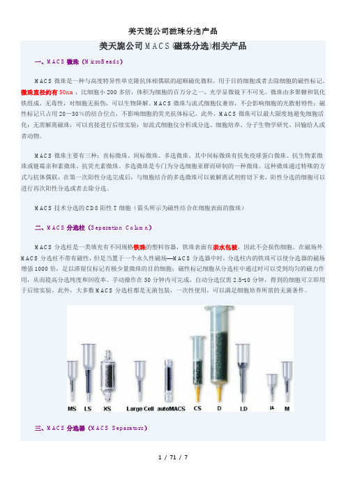
美天旎公司MACS(磁珠分选)相关产品一、MACS微珠(MicroBeads)MACS微珠是一种与高度特异性单克隆抗体相偶联的超顺磁化微粒,用于目的细胞或者去除细胞的磁性标记。
微珠直径约有50nm,比细胞小200多倍,体积为细胞的百万分之一,光学显微镜下不可见。
微珠由多聚糖和氧化铁组成,无毒性,对细胞无损伤,可以生物降解。
MACS微珠与流式细胞仪兼容,不会影响细胞的光散射特性;磁性标记只占用20-30%的结合位点,不影响细胞的荧光抗体标记。
此外,MACS微珠可以最大限度地避免细胞活化;无需解离磁珠,可以直接进行后续实验:如流式细胞仪分析或分选、细胞培养、分子生物学研究、回输给人或者动物。
MACS微珠主要有三种:直标微珠、间标微珠、多选微珠。
其中间标微珠有抗免疫球蛋白微珠、抗生物素微珠或链霉亲和素微珠、抗荧光素微珠。
多选微珠是专门为分选细胞亚群而研制的一种微珠。
这种微珠通过特殊的方式与抗体偶联,在第一次阳性分选完成后,与细胞结合的多选微珠可以被解离试剂剪切下来,阳性分选的细胞可以进行再次阳性分选或者去除分选。
MACS技术分选的CD8阳性T细胞(箭头所示为磁性结合在细胞表面的微珠)二、MACS分选柱(Separation Column)MACS分选柱是一类填充有不同规格铁珠的塑料容器,铁珠表面有亲水包被,因此不会损伤细胞。
在磁场外MACS分选柱不带有磁性,但是当置于一个永久性磁场—MACS分选器中时,分选柱内的铁珠可以使分选器的磁场增强1000倍,足以滞留仅标记有极少量微珠的目的细胞;磁性标记细胞从分选柱中通过时可以受到均匀的磁力作用,从而提高分选纯度和回收率。
手动操作在30分钟内可完成,自动分选仅需2.5-10分钟,得到的细胞可立即用于后续实验。
此外,大多数MACS分选柱都是无菌包装,一次性使用,可以满足细胞培养所需的无菌条件。
三、MACS分选器(MACS Separators)MACS分选器由永久性磁铁和支架构成。
Seagate NAS 4-bay 网络存储解决方案:商品说明书

Data SheetA complete network storage solution and private cloud for power users andbusinesses with up to 25 employeesSeagate® NAS 4-bay is a full-featured network storage solution offering the easiest setupand the industry’s most intuitive interface. The NAS 4-bay features hardware, software andSeagate drives designed to work together seamlessly, which reduces your setup time to justa few minutes and delivers reliable performance for years to come. In addition, Seagate runshours of extensive tests on every NAS 4-bay before it leaves our manufacturing facility, tofurther ensure the system is running at its best.The NAS 4-bay runs Seagate NAS OS 4 software, completely designed from the groundup to deliver ease of use, performance, reliability and a flexible platform for running add-onapps, enabling you to do more with your NAS. With the App Manager in NAS OS 4, you canchoose to install apps that enhance the functionality of the NAS, such as our surveillanceapp that turns the NAS into a recording solution for IP cameras or our anti-virus app that canscan files and identify potential threats before it’s too late.Drives are the core of any high-capacity storage solution, and Seagate delivers a specializeddrive specifically for NAS systems. Seagate NAS HDD drives, included with the NAS4-bay, are engineered to run 24×7 to deliver the performance needed by various businessapplications, yet run cooler than other drives on the market.1 Furthermore, NAS HDD drivesare rated for higher reliability than a standard desktop drive, offering more protection for yourbusiness’s critical files.1 Except diskless modelAMERICAS Seagate Technology LLC 10200 South De Anza Boulevard, Cupertino, California 95014, United States, 408-658-1000 ASIA/PACIFIC Seagate Singapore International Headquarters Pte. Ltd. 7000 Ang Mo Kio Avenue 5, Singapore 569877, 65-6485-3888 EUROPE, MIDDLE EAST AND AFRICASeagate Technology SAS 16–18, rue du Dôme, 92100 Boulogne-Billancourt, France, 33 1-4186 10 00© 2014 Seagate Technology LLC. All rights reserved. Printed in USA. Seagate, Seagate Technology and the Wave logo are registered trademarks of Seagate Technology LLC in the United States and/or other countries. Sdrive and SimplyRAID are either trademarks or registered trademarks of Seagate Technology LLC or one of its affiliated companies in the United States and/or other countries. All other trademarks or registered trademarks are the property of their respective owners. When referring to drive capacity, one gigabyte, or GB, equals one billion bytes and one terabyte, or TB, equals one trillion bytes. Your computer’s operating system may use a different standard of measurement and report a lower capacity. In addition, some of the listed capacity is used for formatting and other functions, and thus will not be available for data storage. The export or re-export of hardware or software containing encryption may be regulated by the U.S. Department of Commerce, Bureau of Industry and Security (for more information, visit ), and controlled for import and use outside of the U.S. Complying with all applicable copyright laws is the responsibility of the user. Seagate reserves the right to change, without notice, product offerings orspecifications. DS1817.1-1405 AMER1 Except diskless model2 One terabyte, or TB, equals one thousand billion bytes when referring to drive capacity.The storage volume on the NAS 4-bay is preconfigured, which can save you hours of setup time compared to traditional RAIDconfigurations.1 If you choose to purchase our diskless model or want to change the default configuration, we offer SimplyRAID ™ technology to speed up and simplify that process too. SimplyRAID technology, built into the NAS OS software, will help you choose the RAIDconfiguration that is best for your business by balancing performance, capacity and redundancy. Another benefit from our SimplyRAID technology is the ability to begin storing files and using the NASinstantly after choosing your preferred configuration. No more waiting hours or days for the RAID volume to build!One of the best things about owning a NAS is the ability to collaborate, share and access files from anywhere when on-the-go. Seagateenhances that experience with Seagate Sdrive ™ software for PC, Mac, Android, iPhone and iPad. The Sdrive software for PC and Mac gives you the most intuitive access to the files stored on your NAS from anywhere you have an Internet connection. Sdrive software makes the NAS appear as another drive connected to your computer, so you can drag-and-drop, view thumbnails and open files with your favorite programs. The Sdrive app for mobile devices keeps you connected to your files with enhanced security. All files are encrypted before they leave the NAS and only the Sdrive app on your mobile device, combined with your user credentials, can decrypt and access the files.。
Orbitrap的过去、现在和未来

Orbitrap的过去、现在和未来2013-10-08 08:35Orbitrap的过去、现在和未来关键词:thermo质谱质量分析器orbitrap赛默飞——访Orbitrap发明人Alexander Makarov 【导语】Orbitrap无疑是近20年来质谱技术上最重要的发明,不仅使赛默飞在这个质量分析器上花样翻新、喜获丰收,也给无数的质谱用户带来超乎寻常的极致体验。
在重庆举办的第八届中国蛋白质组学大会上,我们有幸采访到Orbitrap的发明人、赛默飞色谱质谱部生命科学质谱研发总监Alexander Makarov,以及赛默飞色谱质谱部负责全球销售的副总裁Herb Kenny,色谱质谱部生命科学质谱的总经理John F. Sos,他们为我们分享了Orbitrap诞生的很多故事,并展望了Orbitrap的未来,希望对所有的质谱爱好者有帮助,激励更多的科学家们为我们带来更多的发明……Orbitrap的发明人Alexander MakarovOrbitrap是如何诞生的迈入质谱门亚力山大马可洛夫(Alexander Makarov)1966年出生在俄罗斯Irkutsk 的Siberian镇。
回顾自己的经历,Makarov说:“我小时候就喜欢发明,并不是喜欢发现大自然界的奥秘,而是喜欢发明世界上还没有的东西。
小时候我就发明了一些东西,比如不同的机械,不过可没什么专利,现在,这些机械已经被真正的发明并应用。
虽然小时候的发明并不是很‘成功’,但这是我开始发明的起步。
”大学期间Makarov就显示了惊人的发明才能。
他1983-1989年在莫斯科工程物理学院(MEPhI)读大学,88-89年在分子物理系做研究助手,就发明设计了超对数场(hyper-logarithmic field)的多电极飞行时间质量分析器用于火花离子源(spark ion source),并提供了原理的证明,那时候Makarov只有23岁,同时他直接获得了科学硕士的学位奖励。
艾博思生物转录组测序样品要求
艾博思⽣物转录组测序样品要求1. 艾博思⽣物质量控制体系质量标准严格:QA系统为您的实验全程护航,实验严格按照153个SOP⽂件进⾏。
实验记录完备:⼯⼚化的流⽔线作业项⽬流程,保证所有实验记录可供回溯。
多重实验质控:实验室QC、数据分析QC、项⽬QC—3重QC,减少低质量数据。
(1)样品运输和储存的质量控制(2)实验过程的质量控制(3)结果数据运输和储存的质量控制2.艾博思⽣物RNA-Seq服务样品要求A 样品要求(RNA)1) 样品纯度:OD 260/280值应在1.9~2.2 之间,RNA 28S:18S≥1.5推荐 RIN≥8;DNA 应该去除⼲净。
2) 样品浓度:最低浓度不低于100ng/µl。
3) 样品总量:每个样品总量不少于15µg。
4) 样品溶剂:要溶解在H20或TE (pH 8.0)中。
5) 样品运输: RNA⽤冻存管保存,并⽤⼲冰或液氮运输。
B 样本准备应该遵循的基本原则1) 代表性原则:取样的代表性关系到实验结果是否具有科学意义,因此您应该根据实验⽬的慎重选择您的取样⽅案。
病变组织样本中应不夹带正常组织,正常组织样本中不能含有病变组织。
有条件时,应做到实验组与对照组的样本在取材时间、部位、处理条件等⽅⾯尽可能保持⼀致,否则可能会影响实验结果的可信度。
2) 准确性原则:代表性样本的各种特征数据必须被准确记录,并按要求(低温、迅速)采集、制备、贮存、运输,最终正确地按实验设计进⾏实验和数据处理。
3) 迅速性原则:样本质量是实验中影响实验结果的最关键因素,因此⽤于实验的样本,在采集、制备、贮存、运输过程中应尽可能地做到迅速,最⼤限度的缩短从样本采集到实验的时间。
4) 低温原则:所取样本离体后,应尽快置于液氮、⼲冰或-80℃冰箱中,并保证在实验前始终处于-70℃以下,以避免RNA的降解。
C 样本准备⽅法:详见《艾博思⽣物样品采集操作指南》D 注意事项1) 我们不接收未经TRIzol试剂处理的细胞样本。
icp-ms讲解解析

多元素快速分析 (>75)
动态线性范围宽
检测限低
在大气压下进样,便于与其它进样技术联用(HPLCICP-MS)
可进行同位素分析、单元素和多元素分析,以及有机物
中金属元素的形态分析
缺点:
运行费用高
需要有好的操作经验
样品介质的影响较大( TDS < 0.2%)
ICP高温引起化学反应的多样化,经常使分子离子的强
正离子被拉出并按其质荷比分离;
检测器将离子转化为电子脉冲,然后由积分测量线路计数;
电子脉冲的大小与样品中分析离子的浓度有关,通过与已知的
标准或参比物质比较,实现未知样品的痕量元素定量分析。
标准的ICP-MS仪器分
为三个基本部分:
(1)电感耦合等离
子体(样品引入系统;
离子源)
(2)接口(采样锥;
或固体小颗粒的形式进入中心通道气流中。
样品导入的三大类型:
溶液气溶胶进样系统
(气动雾化或超声雾化法)
气体进样系统
(氢化物发生、电热气化、激光烧蚀以及气
相色谱等)
固体粉末进样系统
(粉末或固体直接插入或吹入等离子体)
ICP-MS离子源
ICP特别适合作质谱的离子源,由于其具有以下特点:
光子挡板或90度转弯,就可以将其与非带电粒子
(光子和中性粒子)分离。
四极杆质量分析器
四极杆的工作是基于在四根电极之间的空间产生一个随时间
变化的特殊电场,只有给定荷质比(m/z)的离子才能获得稳定
的路径而通过极棒,从其另一端出射,其它离子将被过分偏转,
MilliporeSigma Viresolve Pro Shield 和 Shield H 过滤膜

The life science business of Merck operates as MilliporeSigma in the U.S. and Canada.Data SheetViresolve ® Pro Shield and Shield HSelection GuideSize based membrane filtration provides criticalvirus removal assurance in most established and new biopharmaceutical molecule production processes. Even for highly purified process intermediates, the presence of protein aggregates, denatured proteins and other impurities limit hydraulic performance of virus filters, increasing the required filter area and costs.Pretreatment of feed streams can remove plugging species, resulting in more favorable economics and robust processing. Our Viresolve ® Pro Shield or Shield H are effective at removing these species from many monoclonal antibody (mAb) processes. They provide an adsorptive removal that improves the capacity of the Device by an average of two-fold, while allowing the Device to deliver high parvovirus removal and flux. Results of the Viresolve ® ProDevice capacity improvement are shown in Figure 1.We were issued two patents that cover the use of adsorptive depth filters and charged or surface modified microfiltration (MF) membranes to remove aggregates from protein solutions in order to enhance performance of virus filters.Shield H + DeviceShield + Device Device onlyV i r e s o l v e P r o V o r 4 h r s . (k g /m )Conductivity (mS/cm)3 6.5101620144545452013Figure 1.The Viresolve ® Pro Shield or Shield H improves the Viresolve ® Pro Device performance across a broad range of pH and/or conductivity.Merck KGaAFrankfurter Strasse 250 64293 Darmstadt Germany© 2017 Merck KGaA, Darmstadt, Germany and/or its affiliates. All Rights Reserved. Merck, the vibrant M, Millipore and Viresolve are trademarks of Merck KGaA, Darmstadt, Germany or its affiliates. All other trademarks are the property of their respective owners. Detailed information on trademarks is available via publicly accessible resources. /life-science To Place an Order or Receive Technical AssistancePlease visit/contactPS For additional information, please visit TB1140EN00 Ver. 2.02017 - 0329611/2017or+=Expansion of Viresolve ® Pro SolutionConductivity (mS/cm)876p H54Figure 2.The contour plot shows the optimal performance for Shield or Shield H depending on the pH and conductivity of the mAb.Viresolve ® Pro Shield • Membrane based • Cation exchange adsorptive chemistry • Caustic stable •Low extractablesViresolve ® Pro Shield H • Membrane based • Mixed modeadsorptive chemistry • Caustic stable •Low extractablesUS Patent 7,118,675 B2 covers the use of adsorptive depth filters or charged or surface modified microfiltration membranes in front of a viral filter to enhance the virus filter performance by removing aggregates from protein solutions.US Patent 7,465,397 B2 involves the process of removing aggregates and viruses using a depth filter upstream of a viral filter in either a constant pressure or pump system. Adsorptive depth filters and modified microfiltration filters that allow for increased capacity and robustness of our virus filters are part of the Viresolve offering. As part of our continued goal to provide our customers with the right tools and technologies to perform effective and robust virus filtration, we provide customers with a free license to use the process.。
微波消解-电感耦合等离子体-质谱法测定柏子养心丸中总汞的含量

·324·药品评价 Drug Evaluation 2021,18(06)基金项目:江西省食品药品监督管理局科研项目(2017YX01)作者简介:任琦,硕士,主管药师。
研究方向:中药质量控制与评价研究。
E-mail :jiangxizhongyi2008@通信作者:邓维,硕士,编辑。
研究方向:药品监管。
E-mail :***************微波消解-电感耦合等离子体-质谱法测定柏子养心丸中总汞的含量任琦1,许妍1,刘桂凤1,徐兰2,邓维11.江西省药品检验检测研究院,国家药品监督管理局中成药质量评价重点实验室,江西省药品与医疗器械质量工程技术研究中心,江西 南昌 330029;2.解放军联勤保障部队第九○八医院,江西 南昌 330002[摘要]目的:建立微波消解-电感耦合等离子体-质谱法测定柏子养心丸中总汞的含量。
方法:将样品经微波消解处理后,采用ICP-MS 法以209Bi 为内标元素测定柏子养心丸中总汞的含量。
结果:总汞质量浓度在0~5.0 ng/mL 范围内与测量值线性关系良好,平均加样回收率为104.65%,RSD 为1.82%(n =6)。
结论:该方法简便、快速、准确,适用于柏子养心丸中总汞的测定。
[关键词]柏子养心丸;微波消解;电感耦合等离子体-质谱法;总汞;含量DOI: 10.19939/ki.1672-2809.2021.06.02Determination of Total Mercury in Baizi Yangxin Pills by Microwave Digestion-ICP-MSREN Qi 1, XU Yan 1, LIU Guifeng 1, XU Lan 2, DENG Wei 11. Jiangxi Institute for Drug Control , NMPA Key Laboratory of Quality Evaluation of Traditional Chinese Patent Medicine, Jiangxi Province Engineering Research Center of Drug and Medical Device Quality, Nanchang Jiangxi 330029, China;2.The 908th Hospital of PLA Joint Logistic Support Force, Nanchang Jiangxi 330002, China.[Abstract] Objective: To establish a method for the determination of total mercury in Baizi Yangxin pills by microwave digestion-ICP-MS. Methods: After microwave digestion, the content of total mercury in Baizi Yangxin pills was determined by ICP-MS with 209Bi as internal standard element. Results: The linear range of total mercury was 0~5.0 ng/mL. The average recovery was 104.65%, RSD was 1.82% (n =6). Conclusion: The method is simple, rapid, accurate and suitable for the determination of total mercury in Baizi Yangxin pills.[Key Words] Baizi Yangxin pills; Microwave digestion; ICP-MS; Total mercury; Content·药物研究·柏子养心丸处方来源于《古今医统》卷七叶引《集验方》。
美国国家食品安全与技术中心选择ThermoScientific质谱检测三聚氰胺

薯 0
■ 5 ■ 一 ● 4
_ __ _一 _■ 行 业 动 态
GL 》 CBALF O0D I NDUSTR Y
背景
关于宠物食 品、动物 饲料 、小麦麸和其它合蛋白质的 日常食物 中存在三聚氰胺 的消息 ,引起 了人们对 动 物和 人的食物链 中的化 学品的关注 。三聚氰 胺是生产塑料 、阻燃剂和其它产 品常用的 工业化 学品 ,禁止用 于 食品和动物饲料 中。美 国从 中国进 口的宠物 食品中 ,发现 了污染的植物蛋 白,这也导致了2 0 年3 l日起 07 月 5 全国性的召 回宠物食品事件 。此外 ,一部分 污染的宠物食品被用于生产农场的动物饲料和鱼饲料 。美 国食 品 药品监督管理曷和美国农业部芨现一些 吃了污染饲料的动物被加工威了人类的食品 。 由于美 国国家食品安全与技术 中心要对食 品加 工过程对 特定的威份的影响进行评估 ,伊利诺斯工学院生 物 、化学和物理学院的教 ̄P t aei ee V r l博士知 道美国国家食品安全与技术 中心需要掌握 当三聚氰 胺和三聚 r s 氰酸一一种与三聚氰胺相 关的化合物一被加 工进食 品中后 ,到底会发生什么 。这J  ̄ 识对监测食品 中存在三 'H - 聚氰胺和三聚氰酸时应该测定的 降解产物是非 常必要 的。
仪器的选择
在进行三聚氰胺的研究之前 ,美国国家食品安全与技术 中心 的实验 室并没有安装L— S CM 系统 。V rl博 aei s
士以前在 其它的实验室有使用T em c ni 的仪器的经验 ,他对T em c ni 公司的客户支持服务印 h r oS i t c e f i h r oS i t c e f i 象非常深刻 。这两个因素导致了美国国家食品安全与技术 中心决定 选择 T em c ni 的L — 仪器。 h r o i t c C MS S e f i “ 公司给我们提供了关于色谱柱 、色谱条件 、质谱仪设置等方 面的有用 的建 议 ,以获得最佳灵敏度。” V rl博士说 “ aei s 这是一种巨大的帮助 。T Q Q a t 系统在三聚 氰胺的分析上 得到了应得的认可。我们可 S u nu m 以很容易 的在 含有 l p  ̄聚氰胺 的鲶鱼 的分析 中 ,获得超过3 0 的信 噪比。” V rl博 士是这样评 论的。 O b p 80 ae s i 测定鲶鱼 中的 '聚氰胺残 留的现有方 法 ( 二 实验 室信 息通 报N 3 6 是 由位于丹 佛的食 品药品监督管理 049 ) 局动物药物研究中心建立的 。该方法使用T Q Q a t 质谱仪 ,配备 了T em c ni 的S re o丁型L S u nu m h r oSi ti uv y r e f c M C M 型液相泵和 自动进样器 。方法 表明T em c ni 的仪器能 够检测 出鲶 鱼提取物中lp b s h r o i ti Se f c O p 水平的三聚氰 胺 ,超过了食品药品监督 管理局规定的要求 。
默克表达载体简介

病毒导入 AdEasy™腺病毒系统 感染分裂和非分裂期细胞效
系统
率 100%,在大肠杆菌中同
源重组,克隆片段大,高水
平表达外源蛋白,适合用于
病毒导入
疫苗开发
系统
AAV 系统
感染分裂和非分裂期细胞,
不需要辅毒,生物安全性高。
免疫原性低,是长期基因治
疗研究的首选工具
Viraport® 逆转录病毒 可以整合到宿主染色体上,
克隆 siRNA 序列用于基 因沉默研究
PDF 文件使用 "pdfFactory" 试用版本创建
抑制 免费热线:800-820-8872,Email:elaine.liang@
由德国 MERCK 授权 生物通 设计制作
PDF 文件使用 "pdfFactory" 试用版本创建
(转录调 系统
产物对于信号传导通路的增 系统
准确鉴定 DNA 结合蛋
控)
强子的作用
白,用于分析转录调控
因子研究等
PathDetect® 反式报告 检测、评估多种信号传导通 pMLuc
系统
路的增强子上游元件的功能 MlightyLight™Rluc
通过检测荧光素酶活性 非常灵敏地评估启动子 /增强子,LIC 克隆
T7 启动子控制的带钙结合 pETcoco™ 蛋白标签表达,温和纯化
大肠杆菌、哺乳动物细胞表 pBiEx™
达,Lambda 表达载体,克
隆片段达 10kb
pTriEx™
pDual® pDual® 1GC
大肠杆菌、哺乳动物细胞表 Radiance™系列 达天然或带标签融合蛋白, CBP 纯化 大肠杆菌、哺乳动物细胞表 Duet 系列原核表达 达,融合 c-myc 和 His 标签,
EZ-Fit 过滤器单元用户指南说明书
User GuideEZ-Fit® Filtration UnitThe life science business of Merck KGaA,Darmstadt, Germany operatesas MilliporeSigma in theUS and Canada.CoverFunnelMembraneSupport pad (blue unit only)BasePlug (blue unit bulk packaging and selected versions only)IntroductionThe EZ-Fit ® Filtration Unit is a disposable filtration device for bioburden testing of liquid samples like water, process samples, or final products. It is designed for optimizing and securing the laboratory workflow to provide time-saving and reliable microbiologicalresults. After the filtration of the sample, the membrane can be transferred and cultured on an agar plate. Alternately, on the blue unit only, liquid media can be added after the filtration, and the device converts into a Petri dish.This disposable device is designed to fit onto the EZ-Fit ® Manifold, which features specific EZ-Fit ® Filtration heads. It can also fit onto a standard #8 stopper.The EZ-Fit ® filtration device consists of:The information in this document is subject to change without notice and should not be construed as a commitment by Merck KGaA, Darmstadt, Germany. Merck KGaA, Darmstadt, Germany assumes no responsibility for any errors that may appear in this document. This manual is believed to be complete and accurate at the time of publication. In no event shall Merck KGaA, Darmstadt, Germany be liable for incidental or consequential damages in connection with, or arising from, the use of this manual.Materials of constructionFunnelStyrene butadiene copolymer MembraneMixed cellulose esters, PVDF Support pad (blue unit versions only)CelluloseBaseAcrilonitrile butadiene styrene Plug (blue unit in bulk packaging & selected versions only)Low-density polyethylene DimensionsHeight100 mL: 66.5 mm (2.6 in.)250 mL: 108.5 mm (4.3 in.)Largest diameter75.8 mm (3.0 in.)Filtration surface 12.56 cm2Sterilization method Electron beamStorage conditions Store in a cool and dry place Maximum temperature of the sample45 °CFiltration1. R emove the device from the packaging by grasping the funnel.CAUTION: G rasping the cover could result inremoving the cover from the funnel.NOTE: T he devices can be stacked.2. R emove the plug from the bottom of the base (blue unit bulk packaging & selected versions only).NOTE: W hen culturing using ampoule liquidmedia, do not discard the plug as it will be used during incubation step.6. T urn the knob to the upright positionto apply vacuum to perform filtration.5. Place the membrane into a solid medium.Ampoule Liquid Media(blue units bulk packaging versions only) 1. T urn the knob to the horizontal positionto stop the vacuum.2. R emove the cover and pour the liquidmedium evenly onto the membrane.3. Q uickly perform the followingoperations:1 Turn the knob to the vertical position. 2S lightly pinch the funnel and remove it.3P lace the cover onto the base.4R eturn the knob to the horizontal position to stop the vacuum.5. R eplace the plug and twist it ontothe support.NOTE: T he devices can be stacked in the incubator.Discarding the productRefer to the materials of construction of the device for proper disposal/recycling of each component.NOTE: The funnels can be stacked to reduce waste volume.Ordering Information®®1EZ-Fit ® Manifold, 3-place for EZ-Fit ® Filtration Units EZFITEFUN3EZ-Fit ® Manifold, 6-place for EZ-Fit ® Filtration Units EZFITEFUN6EZ-Fit ® filtration head for EZ-Fit ® Filtration Units EZFITMVHE1EZ-Fit ® filtration head for EZ-Fit ® Filtration Units 3EZFITMVHE3Visit our website: /EZ© 2020 Merck KGaA, Darmstadt, Germany and/or its affiliates. All Rights Reserved. The vibrant M, Millipore, Sigma-Aldrich,Lit. No. VM_PG5162EN Ver. 4.0。
赛默飞推出革新性三重四极杆LC-MS 灵敏度、稳定性、适用性更上台阶
赛默飞推出革新性三重四极杆LC-MS 灵敏度、稳定性、适用性更上台阶佚名【期刊名称】《理化检验:化学分册》【年(卷),期】2013()8【摘要】全球科学服务领域的领导者赛默飞世尔科技今天推出新一代三重四极杆液相色谱质谱(LC-MS)平台,该平台专为定量实验设计,具有极佳的灵敏度,更高的运行通量,更优的精度和易用性。
平台包括新型ThermoScientificQuantiva三重四极杆质谱仪和新型ThermoScientificEndura三重四极杆质谱仪,在2013年6月美国质谱年会(ASMS)上首度亮相。
“应用领域的不断发展持续推动了对仪器极限性能的提升,我们已创建了完整的新仪器平台,能够为客户提供最佳灵敏度和运行时间。
同样重要的还有令人满意的用户体验、简便实现方法创建和便捷的数据分析和回顾。
我们相信该仪器是目前市场中更高效、更实用的三重四极杆。
”赛默飞世尔科技色谱质谱研发副总裁lainMylchreestz如是说。
【总页数】1页(PI0002-I0002)【关键词】四极杆;灵敏度;稳定性;适用性;仪器平台;MS;LC;革新【正文语种】中文【中图分类】TP332【相关文献】1.赛默飞世尔科技新品——Nicolet iS50红外光谱仪、TSQ 8000三重四极杆GC-MS/MS系统、TRACE 1300系列气相色谱、iCAP QICP-MS、Exactive Plus LC-MS [J],2.赛默飞推出针对食品安全检测的《TSQ三重四极杆质谱简明应用手册》 [J], 邵帅;3.赛默飞推出革新性三重四极杆LC-MS,灵敏度、稳定性、适用性更上台阶 [J],4.赛默飞世尔科技推出革新性的三重四极杆LC-MS,灵敏度、稳健性、适用性更上台阶 [J],5.赛默飞发布三重四极杆液质联用仪定量测定物质的解决方案 [J],因版权原因,仅展示原文概要,查看原文内容请购买。
- 1、下载文档前请自行甄别文档内容的完整性,平台不提供额外的编辑、内容补充、找答案等附加服务。
- 2、"仅部分预览"的文档,不可在线预览部分如存在完整性等问题,可反馈申请退款(可完整预览的文档不适用该条件!)。
- 3、如文档侵犯您的权益,请联系客服反馈,我们会尽快为您处理(人工客服工作时间:9:00-18:30)。
Explicit Knowledge
>
“The Work”
Core business processes & practices powered by knowledge Tacit Knowledge
e.g. QbD, investigations
Core K workflow processes
Mt. Everest
2009: Base Camp KM Maturity <1
This looks so straight forward when viewed this way…
5
Learnings & Happenings…
Emphasis on Connectivity & creating a Boundaryless Organization
Continual Improvement
Challenges in Process & People
6
Connectivity & Boundaryless Organization
Boundaryless = Rapid flow of our science, knowledge, processes, and people to meet critical business needs, grow and develop our people and deliver value to our patients and customers.
Customers
8
MMD’s Improvement Journey…
…powered by KNOWLEDGE & CONNECTIVITY
Standardized, repeatable, robust channels for knowledge flow …
Commercial Plant B
Rapidly changing business environment demands this for our survival…and it’s simply good business
7
MMD’s Improvement Journey
Last several years: Focus “within 4 walls of a plant” to reduce variability and improve performance…
• Learn
– Must be a role model and know what you are asking for
• Commit
– Time (culture) & Effort & Visible Upward Advocacy – Resources: KM SMEs, functional experts, stewards, …
MMD: HOW WE MANUFACTURE SUCCESS
15
“Making Problems Visible and solving them once, collaboratively, at root cause…”
Provide Leadership …words from our sponsor
Product Flow Product Flow
Commercial Plant B
Commercial Plant A
Commercial Plant C
Commercialized Product
Product Flow
Late Stage Development & Commercialization
• Create the right environment
– “Top down and bottom up”
• Set expectations
– Managing knowledge is “part of your job” – Cannot change culture without clear expectations, accountability and consequences
Link to & Leverage Organizational Norms
• KM as the backbone to enable the speed and effectiveness of knowledge transfer
Ask who else needs to be involved and create 360º vision… People and teamwork are at the heart of Merck Production System
Commercial Plant A
Commercial Plant C
Product Knowledge
Late Stage Development & Commercialization
Commercial Plant D
Customers
9
…enabling connectivity, rapid problem solving, sharing of issues and best practices, higher engagement, and more
• Remain Resolved!
16
Thanks for your attention!
17
With Special Thanks to…
• • • • • • • Dr. Michael Thien Dr. Jean Wyvratt Anando Chowdhury David Vossen GSTC Leadership Team MMD KM CoE Team APQC and many fellow KM practitioners!
(behaviors which capture, seek, share & leverage knowledge)
4
Merck’s Journey in KM Capability
2015: Today 2015: Well into KM Maturity 3+
our journey
(KM Maturity >3)
Structured Methodology (CEM)
Intent
Create an Energized Network
People
Delivery
Adapted from Conner Partners
© Rob Cross
Copyright 2012 - Kaleel Jamison Consulting Group
Tap into Culture
Behaviors are Critical
Ensuring people have the right: • Direction • Competence • Opportunity • Motivation
14 *R. D. Guenard, M.J. Lipa, S. Bruno and J. Katz, "Enabling a New Way of Working through Inclusion and Social Media: A Case Study", OD Practitioner, 45(4), 2013
We are going to prevent the problem before it happens again!
“We could have that problem!” “Fixed it, here’s how!” “Sorry, does not apply to us.”
Continual Improvement: AAR
2.1.5.x: SLA between Nodes
Added
And the power of KNOWLEDGE MAPPING to understand how
knowledge flows
13
People: Key Change Execution Concepts
blending social technologies for success*
Connectivity Application: Packaging Defects
Monitor for Packaging‐ related Defects Investigate and Capture defects Share via standard work Assess impact Act
MMD Lessons Learned System ‐ Generalized Life of a Lesson
Natural Flow of Work
Work Processes/Procedures Peer Assist Future Focus Business Practices People Institutional Knowledge AAR Selection Current Focus
•2
Guiding Principles
credit: APQC
credit: APQC
3
Merck Knowledge Ecosystem
Lessons Learned
“Knowledge knows no organizational boundaries… it is boundaryless”
Capture & Share
Lesson (s) 1n
11AAR of Wor来自EventAfter Action Review