脑颜面血管瘤病的MRI表现(附5例报告)

脑颜面血管瘤病的MRI表现(附5例报告)
李显忠
【期刊名称】《中外健康文摘》
【年(卷),期】2013(000)002
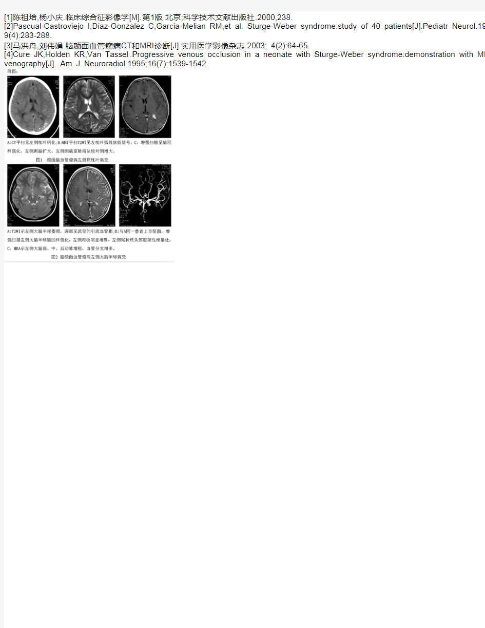
【摘要】目的探讨磁共振成像(MRI)对脑颜面血管瘤病的诊断价值.方法收集5例经临床和手术证实的脑颜面血管瘤病,对其磁共振资料、临床表现和发病机理进行回顾性的总结和分析.结果5例患者均表现为不同程度脑萎缩,3例可见脑表面弧线状低信号影,2例合并脑梗塞,2例可见同侧侧脑室扩大,3例脉络膜增大,2例颅骨增厚.3例增强后呈广泛脑回样强化;1例磁共振血管造影(MRA)显示病变侧动脉血管增粗、扭曲.结论结合典型的临床症状和体征,MRI可以对脑颜面血管瘤病作出比较明确的诊断.%Objective To explore the diagnostic value of MRI in encephalofacial angiomatosis. Methods The MRI findings were retrospectively studied in 5 cases of encephalofacial angiomatosis proved by histological examinations,its MRI data, clinical performance and pathogenesis were summaried and analysed. Results There was varying degrees of brain atrophy in all the patients,arcshaped low signal were observed in three patients, two cases of cerebral infarction, ipsilateral lateral ventricles enlarged in two cases, choroidal increased in three cases,skull thickened in two cases.Contrast enhanced examine was performanced to three patients, syrus-lide enhancement was observed in the three cases. dilated and proliferated artery was finded in one case of MRA. Conclusion Combine with typical clinical symptoms and