人iPS细胞DYR0100说明书
IPS细胞

子区域。
返回
为报告基因 原理是Fbx25对于多能性的维持和胚胎
的发育来说是不可缺少的基因
五、IPS细胞的鉴定 表面标志分子鉴定 IPS细胞能够表达多能干细胞特异的表 面标志分子,如:ssea-1(阶段特异性胚胎 抗原1),ssea-3等。这些物质在已分化体细 胞表面不表达,从而鉴定。
表观遗传状态鉴定 IPS细胞与ES细胞具有相似的DNA甲基化模
构建IPS细胞示意图
IPS细胞形成的分子机制
四、IPS细胞的筛选方法
a.形态学筛选 不对供体细胞做任何额外遗传修
饰的情况下,仅依靠形态学筛选的标准对细胞进行
筛选也能够分离出IPS细胞。
b.药物筛选 利用基因重组技术建立了具有药物
抗性基因的体细胞,并用特定药物进行筛选。
c.选用对多能性更为关键的基因(如Fbx25)作
六、IPS细胞的应用前景
用于器官移植 建立疾病模型,了解发病机理
治疗某些遗传病
如:患有镰刀型贫血症
的小鼠皮肤成纤维细胞建立了IPSc,并将其移植
到小鼠体内,小鼠的症状得以缓解。
七、面临的问题
a.解析诱导体细胞重编程为 IPS 细胞的分 子机制 b.研究 IPS 细胞生物学特性和行为 (如自我 复制、增殖和分化等)调控的机制及 IPS 细 胞体外定向诱导分化机制 c.提高IPS细胞制备效率 d.充分评价IPS 细胞临床应用的安全性
织等是亟待阐明的问题.
返回
胚胎干细胞(embryonic stem cells,ES cells)
是来源于哺乳动物囊胚内细胞团(inner cell mass,
ICM)的多潜能干细胞,具有无限自我更新和多向
分化能力.ES 细胞在疾病模型建立与机理研究、 细胞治疗、药物发现与评价等方面极具应用价 值. 返回
氯化钴诱导hiPSC-CMs体外缺氧模型的建立

浙江理工大学学报,2021,45(1):84-93Journal of Zhejiang Sci-Tech UniversityDOI:10.3969力.issn.l673-3851(n),2021.01.011氯化钻诱导hiPSC-CMs体外缺氧模型的建立朱梦怡-柯敏霞I,王皓齐念民益3,吴月红(1.浙江理工大学生命科学与医药学院,杭州310018;2.杭州标模生物科技有限公司,杭州310018;3.上海丽坤生物科技股份有限公司,上海201499)摘要:为进一步了解缺氧性心血管疾病的发病机制.通过使用CHIR99021和IWP2抑制剂的组合诱导和单层诱导分化方法获得人诱导性多能干细胞来源心肌细胞(Human induced pluripotent stem cells-derived cardiomyocytes,hiPSC-CMs).并在hiPSC-CMs的培养系统中加入氯化祐进行处理,通过CCK-8检测,Hoechst荧光染色分析、台盼蓝染色分析、qRT-PCR检测和Western blot检测确定最佳处理浓度和时间。
CCK-8检测结果显示,低浓度CoCl2(100,300M mol/L)处理显著提高hiPSC-CMs细胞活力(/><0.01),高浓度CoCl2(900,1200M mol/L)处理显著抑制hiPSC-CMs细胞活力(p<0.001).并且作用趋势呈剂量和时间依赖性,600M mol/L CoCl2处理对于hiPSC-CMs的细胞活力影响不明显(/><0.05);Hoechst荧光染色结果显示,CoCl2处理24h和48h后,低浓度CoCl2处理对细胞无影响,Hoechst染色阳性细胞数量少(p>0.05),高浓度CoCl2处理剂量依赖性增加阳性细胞数量(^<0.0001),且48h处理组的结果与24h处理组无显著差异;台盼蓝染色结果表明,CoCl2处理可剂量依赖性促进细胞凋亡;CoCl2处理后,促凋亡相关基因Bax和蛋白Bax表达呈剂量依赖性上调(/><0.001),抗凋亡相关基因B4-2和蛋白Bcl-2表达呈剂量依赖性下调(p<0.OODsCoCb处理可剂量依赖性促进hiPSC-CMs细胞的凋亡。
人胰岛素原(PI)酶联免疫分析试剂盒 说明书
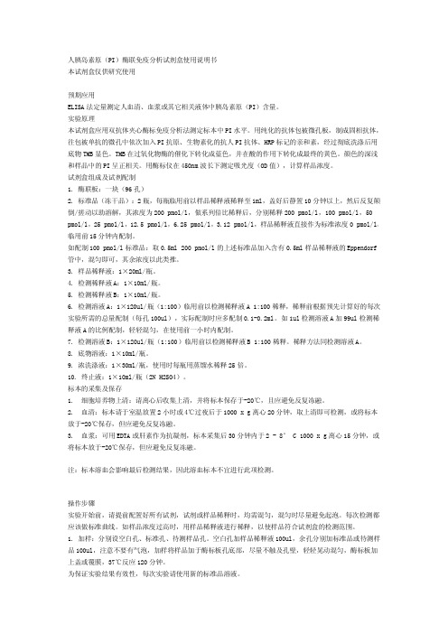
人胰岛素原(PI)酶联免疫分析试剂盒使用说明书本试剂盒仅供研究使用预期应用ELISA法定量测定人血清、血浆或其它相关液体中胰岛素原(PI)含量。
实验原理本试剂盒应用双抗体夹心酶标免疫分析法测定标本中PI水平。
用纯化的抗体包被微孔板,制成固相抗体,往包被单抗的微孔中依次加入PI抗原、生物素化的抗人PI抗体、HRP标记的亲和素,经过彻底洗涤后用底物TMB显色。
TMB在过氧化物酶的催化下转化成蓝色,并在酸的作用下转化成最终的黄色。
颜色的深浅和样品中的PI呈正相关。
用酶标仪在450nm波长下测定吸光度(OD值),计算样品浓度。
试剂盒组成及试剂配制1.酶联板:一块(96孔)2.标准品(冻干品):2瓶,每瓶临用前以样品稀释液稀释至1ml,盖好后静置10分钟以上,然后反复颠倒/搓动以助溶解,其浓度为200pmol/l,做系列倍比稀释后,分别稀释200pmol/l,100pmol/l,50 pmol/l,25pmol/l,12.5pmol/l,6.25pmol/l,3.12pmol/l,样品稀释液直接作为标准浓度0pmol/l,临用前15分钟内配制。
如配制100pmol/l标准品:取0.5ml200pmol/l的上述标准品加入含有0.5ml样品稀释液的Eppendorf 管中,混匀即可,其余浓度以此类推。
3.样品稀释液:1×20ml/瓶。
4.检测稀释液A:1×10ml/瓶。
5.检测稀释液B:1×10ml/瓶。
6.检测溶液A:1×120ul/瓶(1:100)临用前以检测稀释液A1:100稀释,稀释前根据预先计算好的每次实验所需的总量配制(每孔100ul),实际配制时应多配制0.1-0.2ml。
如1ul检测溶液A加99ul检测稀释液A的比例配制,轻轻混匀,在使用前一小时内配制。
7.检测溶液B:1×120ul/瓶(1:100)临用前以检测稀释液B1:100稀释。
稀释方法同检测溶液A。
IPS技术手册

人类诱导性多能干细胞(iPS细胞)技术指导手册目录:1.前言 (1)2.人类胚胎成纤维细胞培养 (2)3.重编程载体构建 (3)4.病毒包装 (4)5.人类iPS细胞的诱导 (6)6.iPS细胞鉴定 (8)6.1碱性磷酸酶活性检测 (9)6.2干细胞表面marker的免疫染色检测 (9)6.3干性因子的去甲基化程度分析 (11)6.4干细胞内源基因的表达分析 (13)6.5端粒酶活性检测 (14)6.6核型检测 (15)6.7拟胚体形成 (15)6.8畸胎瘤形成实验 (15)7.干细胞技术培训及服务一览表 (15)8.附录 (16)1.前言iPS细胞最初从成纤维细胞重编程而来,因为它们准备和操作相对简单。
其他细胞类型,包括来自外胚层、中胚层和内胚层的细胞也可以用于产生iPS细胞(Eminili et al2008)。
2006年Yamanaka和Takahashi利用逆转录病毒系统在成鼠的成纤维细胞导入四个转录因子(Oct3/4,Sox2,c-Myc,和Klf4,Yamanaka因子),将其重编程为iPS细胞,它具有跟小鼠ES十分相似的特性,尤其重要的是,iPS细胞也能产生后代。
2007年,iPS技术在人类体细胞中得以应用,人类iPS 细胞的产生对退行性疾病的治疗产生巨大的影响(Takahashi et al;Yu et al,2007)。
由于iPS细胞具有和ES类似的功能,却绕开了胚胎干细胞研究一直面临的伦理和法律等诸多瓶颈,因此在医疗领域的应用前景非常广阔,成为干细胞研究的热点,《自然》和《科学》杂志分别将其评为2007年第一和第二大科学进展。
随后,iPS细胞的研究日新月异。
.近几年来,iPS研究方面取得的一系列的重大成果让我们欣喜不已,然而,这才刚刚开始,iPS细胞能真正用于临床惠及大众还面临着很多技术上的问题。
一方面iPS产生的方法有待改进;另一方面iPS细胞的定向分化手段需继续探索。
斯丹赛干细胞生物技术公司拥有成熟稳定的胚胎干细胞/iPS细胞技术平台,将竭力为全国各类进行干细胞/iPS研究的科研机构、高校、相关医疗机构和制药企业等提供高品质产品和优质的服务。
人NK细胞(NK)ELISA试剂盒使用说明书
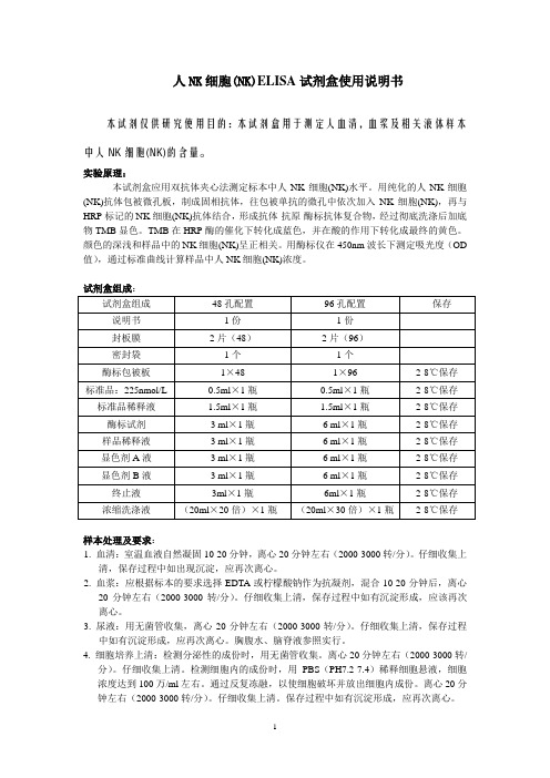
人NK细胞(NK)ELISA试剂盒使用说明书本试剂仅供研究使用目的:本试剂盒用于测定人血清,血浆及相关液体样本中人NK细胞(NK)的含量。
实验原理:本试剂盒应用双抗体夹心法测定标本中人NK细胞(NK)水平。
用纯化的人NK细胞(NK)抗体包被微孔板,制成固相抗体,往包被单抗的微孔中依次加入NK细胞(NK),再与HRP标记的NK细胞(NK)抗体结合,形成抗体-抗原-酶标抗体复合物,经过彻底洗涤后加底物TMB显色。
TMB在HRP酶的催化下转化成蓝色,并在酸的作用下转化成最终的黄色。
颜色的深浅和样品中的NK细胞(NK)呈正相关。
用酶标仪在450nm波长下测定吸光度(OD 值),通过标准曲线计算样品中人NK细胞(NK)浓度。
样本处理及要求:1. 血清:室温血液自然凝固10-20分钟,离心20分钟左右(2000-3000转/分)。
仔细收集上清,保存过程中如出现沉淀,应再次离心。
2. 血浆:应根据标本的要求选择EDTA或柠檬酸钠作为抗凝剂,混合10-20分钟后,离心20分钟左右(2000-3000转/分)。
仔细收集上清,保存过程中如有沉淀形成,应该再次离心。
3. 尿液:用无菌管收集,离心20分钟左右(2000-3000转/分)。
仔细收集上清,保存过程中如有沉淀形成,应再次离心。
胸腹水、脑脊液参照实行。
4. 细胞培养上清:检测分泌性的成份时,用无菌管收集。
离心20分钟左右(2000-3000转/分)。
仔细收集上清。
检测细胞内的成份时,用PBS(PH7.2-7.4)稀释细胞悬液,细胞浓度达到100万/ml左右。
通过反复冻融,以使细胞破坏并放出细胞内成份。
离心20分钟左右(2000-3000转/分)。
仔细收集上清。
保存过程中如有沉淀形成,应再次离心。
5. 组织标本:切割标本后,称取重量。
加入一定量的PBS,PH7.4。
用液氮迅速冷冻保存备用。
标本融化后仍然保持2-8℃的温度。
加入一定量的PBS(PH7.4),用手工或匀浆器将标本匀浆充分。
冻干精制人白细胞干扰素使用说明书

冻干精制人白细胞干扰素使用说明书本品系用特定诱生剂,诱导健康人白细胞,经提取后制成的冻干干扰素注射剂,为白色或淡黄色疏松体,加水溶解后为无色或淡黄色澄清液体。
用于某些病毒性疾病和肿瘤的辅助治疗,对某些免疫缺陷性疾病也有一定疗效。
主要用于治疗慢性乙型肝炎、丙型肝炎、流行性出血热、尖锐湿疣、毛细胞白血病、小儿婴幼儿病毒性肺炎、子宫颈炎、疱疹性角膜炎、带状疱疹、流行性腮腺炎等疾病。
如与化疗、放疗配合治疗肿瘤,可改善患者血象和全身症状。
用法
本品用1~2ml灭菌注射用水溶解。
可肌内注射或病变局部使用。
肌内注射,每日1~2次,每次一支,连续5~10天为一疗程,每疗程间隔2~3天,或遵医嘱。
注意事项
1.对鸡蛋过敏者忌用。
2.注射本品后,一般无反应,个别患者注射后有低热反应或不同程度不适,停药后即可消失。
3.如安瓿有裂纹或溶解后有摇不散的颗粒,不可使用。
ips操作指南第五版-慢病毒版本
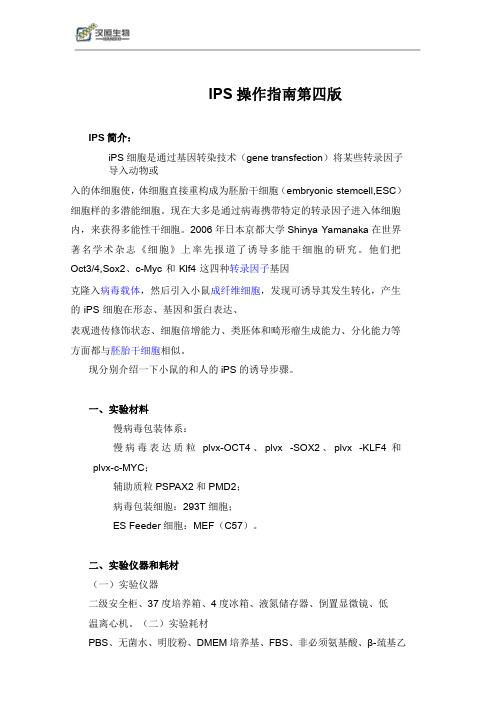
IPS操作指南第四版IPS简介:iPS细胞是通过基因转染技术(gene transfection)将某些转录因子导入动物或入的体细胞使,体细胞直接重构成为胚胎干细胞(embryonic stemcell,ESC)细胞样的多潜能细胞。
现在大多是通过病毒携带特定的转录因子进入体细胞内,来获得多能性干细胞。
2006年日本京都大学Shinya Yamanaka在世界著名学术杂志《细胞》上率先报道了诱导多能干细胞的研究。
他们把Oct3/4,Sox2、c-Myc和Klf4这四种转录因子基因克隆入病毒载体,然后引入小鼠成纤维细胞,发现可诱导其发生转化,产生的iPS细胞在形态、基因和蛋白表达、表观遗传修饰状态、细胞倍增能力、类胚体和畸形瘤生成能力、分化能力等方面都与胚胎干细胞相似。
现分别介绍一下小鼠的和人的iPS的诱导步骤。
一、实验材料慢病毒包装体系:慢病毒表达质粒plvx-OCT4、plvx-SOX2、plvx-KLF4和plvx-c-MYC;辅助质粒PSPAX2和PMD2;病毒包装细胞:293T细胞;ES Feeder细胞:MEF(C57)。
二、实验仪器和耗材(一)实验仪器二级安全柜、37度培养箱、4度冰箱、液氮储存器、倒置显微镜、低温离心机。
(二)实验耗材PBS、无菌水、明胶粉、DMEM培养基、FBS、非必须氨基酸、β-巯基乙醇、胰酶、血清替代物(SR)、mTESR1、T75培养瓶、无菌水、15ml离心管、50ml离心管,巴氏管等。
三、ips诱导方法(一)Feeder细胞1、Feeder细胞的分离(MEF)取妊娠13.5-14.5d的孕鼠,在无菌情况下取出胎鼠,去除鼠头,尾,四肢及内脏,然后用PBS进行冲洗。
将胎体剪碎成小于1mm2的组织块,1000rpm,3min,去掉上层PBS,加入0.25%胰酶-0.04%EDTA2~3ml浸泡组织块,常温消化2~3min。
轻微吹打,待较多细胞溢出后,加入等量的10%FBS DMEM终止消化,通过100目滤网过滤后,将获得的液体离心,1000rpm,5min。
Human TB IFN-γ Precoated ELISPOT Kit strips 说明书

Human TB IFN-γPrecoated ELISPOT Kit(strips)Cat#:2110025产品描述:深圳市达科为生物工程有限公司生产的达优®系列ELISPOT预包被试剂盒采用原装进口高亲和力高效价抗体对,经预包被PVDF板、低温干燥、真空密封包装等工艺流程制备。
成品PVDF板上预包被的抗体分布均匀、效价稳定,2-8ºC可存放12个月。
达优®系列预包被ELISPOT试剂盒使实验检测时间从3天缩短为2天,大幅度减少无菌操作的实验步骤,减轻实验者的劳动强度和减少污染的几率。
使得实验者能够轻松、高效地完成复杂的ELISPOT检测实验。
试剂盒提供的试剂、规格:名称规格×数量Biotinylated Antibody100μL×1支Streptavidin-HRP100μL×1支Dilution Buffer R(1×)10mL×2瓶结核杆菌特异混合多肽A320μL×1瓶结核杆菌特异混合多肽B320μL×1瓶阳性刺激物50TWashing Buffer(50×)15mL×1瓶AEC Dilution10mL×1瓶AEC SolutionⅠ(20×)500μL×1支AEC SolutionⅡ(20×)500μL×1支AEC SolutionⅢ(200×)50μL×1支预包被PVDF可拆板1块实验者自备的试剂与仪器:1.RPMI-1640基础培养基(需要添加双抗)2.无血清培养基(完全培养基,即用型),推荐使用达优@ELISPOT无血清培养基。
3.超净工作台4.CO2细胞培养箱5.微量移液器及配套Tip头6.8通道微量移液器7.0.5mL,1.5mL EP管8.酶联斑点分析仪检测操作:第一天:接种细胞,加入刺激物,培养(严格注意无菌操作)试剂配制1.阳性刺激物:现配现用。
人淋巴细胞无血清培养基(ELISPOT 专用)说明书

第1页共2页人淋巴细胞无血清培养基(ELISPOT 专用)说明书【产品名称】通用名称:人淋巴细胞无血清培养基(ELISPOT 专用)【规格】100mL/瓶;500mL/瓶【预期用途】仅用于在ELISPOT 检测技术中对细胞的培养,不具备对细胞的选择、诱导、分化功能。
培养后的细胞用于体外诊断。
【检验原理】ELISPOT 技术是根据ELISA 技术的基本原理,建立的体外检测特异性抗体分泌细胞和细胞因子分泌细胞的固相酶联免疫斑点技术。
由于其灵敏度高、操作简便经济,因此备受青睐。
传统的ELISPOT 检测技术中,细胞培养这一步使用的多为含有FBS 或者FCS 的培养基,由于血清中含有的很多未知成分能够抑制或者刺激细胞,从而干扰实验,并且由于血清的不确定性,不同批次对于实验的影响也不同,造成了实验的重复性降低。
本产品可有效避免血清质量变动及血清组分对实验研究的影响,提高细胞培养和实验结果的重复性。
【主要组成成分】无机盐、氨基酸、维生素、糖类、蛋白等。
【储存条件及有效期】1、2℃~8℃密封避光保存,避免污染。
2、在储存条件下有效期为一年。
【样本要求】1、细胞要求:新鲜分离的人淋巴细胞。
2、细胞悬液要求吹打混匀,成单细胞悬液。
【检验方法】1、取ELISPOT 板,每孔加入200μL 本产品,室温静置5-10分钟后将其扣出。
2、取新鲜分离的人淋巴细胞,使用本产品调整细胞浓度。
3、将调整好浓度的细胞悬液加入各实验孔,100μL/well 。
4、正对照孔加入阳性刺激剂工作液;负对照孔(含背景负对照孔)加入本产品;实验孔:加入实验者自己的刺激物。
5、所有样品和刺激物加完后,盖好板盖。
放入37℃,5%CO 2培养箱培养16-24小时。
6、显色:按照标准ELISPOT 操作流程进行细胞裂解、洗板、检抗孵育、酶孵育、显色,记录板底斑点数并分析。
【检验结果的解释】结果显示阳性孔斑点分布均匀、频率较高、斑点较大、圆润、线性好、背景干净;阴性对照孔无斑点或有极少斑点,背景干净,无杂色。
人外周血淋巴细胞分离液说明书
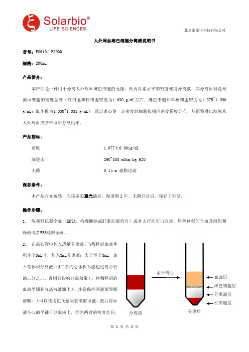
分离后水平离心分离前血浆层淋巴细胞层分离液层红细胞层人外周血淋巴细胞分离液说明书货号:P8610/P8900规格:200mL产品简介:本产品是一种用于分离人外周血淋巴细胞的无菌、低内毒素水平的密度梯度分离液。
其分离原理是根据血细胞的密度差异(红细胞和粒细胞密度为1.090g/mL左右;淋巴细胞和单核细胞密度为1.075~1.090g/mL;血小板为1.030~1.035g/mL),通过离心使一定密度的细胞按相应密度梯度分布,从而将淋巴细胞从人外周血或脐带血中分离出来。
产品指标:密度1.077±0.001g/mL 渗透压290~350mOsm/kg H2O 无菌0.1μm 滤膜过滤保存条件:本产品对光敏感,应该室温避光储存,保质期2年。
无菌开封后,保存于室温。
操作步骤:1.取新鲜抗凝全血(EDTA、枸橼酸钠或肝素抗凝均可)或者去纤维蛋白血液,用等体积的全血及组织稀释液或者PBS稀释全血。
2.在离心管中加入适量分离液(当稀释后血液体积小于3mL时,加入3mL分离液;大于等于3mL,加入等体积分离液。
但二者的总体积不能超过离心管的三分之二,否则会影响分离效果),将稀释后的血液平铺到分离液液面上方,注意保持两液面界面清晰。
(可以使用巴氏德吸管吸取血液,然后将血液小心的平铺于分离液上,因为两者的密度差异,将形成明显的分层界面。
如果样品较多加样时间较长,在离心之前出现红细胞成团下沉属正常现象。
)3.室温,水平转子500~1000g,离心20~30min(血液的体积越大所需的离心力越大,离心时间越长,最佳的分离条件需摸索,离心转速最大不超过1200g)。
4.离心后将出现明显的分层:最上层是稀释的血浆层,中间是透明的分离液层,血浆与分离液之间的白膜层即为淋巴细胞层,离心管底部是红细胞与粒细胞。
5.小心的吸取白膜层细胞到15mL洁净的离心管中,10mL PBS或细胞洗涤液洗涤白膜层细胞。
250g,离心10min。
人类胚胎干细胞基础培训教材

人类胚胎干细胞基础技术高级培训班2010-12目录1. 绪论 (4)2. 饲养层细胞的制备 (5)2.1 从胎鼠获得小鼠胚胎成纤维细胞(MEF) (5)2.2 MEF的传代 (6)2.3 MEF的冻存 (6)2.4 MEF的失活 (7)3. 饲养层细胞的准备 (8)4. ES/iPS细胞的复苏 (10)5. ES/iPS细胞的传代 (11)6. ES/iPS细胞的冻存 (13)7. 人类胚胎干细胞的无滋养层培养 (14)7.1 人类胚胎干细胞条件培养基(CM)的收集 (14)7.2 无饲养层人胚胎干细胞系的培养及传代 (14)8. 拟胚体的形成 (15)9. 胚胎干细胞全能性的鉴定 (16)9.1碱性磷酸酶活性检测 (16)9.2干细胞表面marker的免疫染色检测 (17)9.3干性因子的去甲基化程度分析 (18)9.4干细胞内源基因的表达分析 (21)9.5端粒酶活性检测 (22)9.6核型检测 (23)9.7拟胚体形成 (23)9.8畸胎瘤形成实验 (23)10. 干细胞技术培训及服务一览表 (24)11. 附录 (25)1.绪论本操作手册将为您详细介绍人胚胎干细胞/iPS细胞培养的基础知识、技术细节及相关的产品信息。
目的是为了让您更好地培养您购买的细胞,请按照此手册进行培养操作直至获得可靠的冻存细胞。
上海斯丹赛生物技术有限公司专业提供各类胚胎干细胞/iPS细胞及成套产品服务,为您的干细胞研究提供一站式解决方案。
2.饲养层细胞的制备材料与仪器♦成纤维细胞完全培养基,货号EFM-01♦胰酶,货号M-005-1♦成纤维细胞冻存液(2x),货号EFFM-01♦基质胶(Matrigel matrix),BD Pharmingen,货号356235♦CF-1小鼠胚胎成纤维细胞(MEF),货号MEF-CF1-01♦T25透气培养瓶,NUNC,货号156367♦T75透气培养瓶,NUNC,货号156499♦ 2 ml移液管,NUNC,货号159617♦5ml移液管,NUNC,货号159625♦10 ml移液管,NUNC,货号159633♦15ml离心管,货号366036♦50ml离心管,货号373660♦冻存管,NUNC,货号377267♦Thermo-Fisher Finnpipette F3移液器和灭过菌的枪头♦生物安全柜,货号♦CO2培养箱,Thermo,HERA cell 150i♦电子计数器,millipore,货号PHCC00000♦电动移液器,Thermo Scientific Finnpipette C1,货号4580800♦低速离心机,Thermo Scientific CL10♦液氮罐,Thermofisher,货号CN509002.1从胎鼠获得小鼠胚胎成纤维细胞(MEF)1. 拉断颈椎处死怀孕12-13天的CF-1小鼠,浸入70%的酒精中消毒。
人脂肪间充质干细胞系说明书
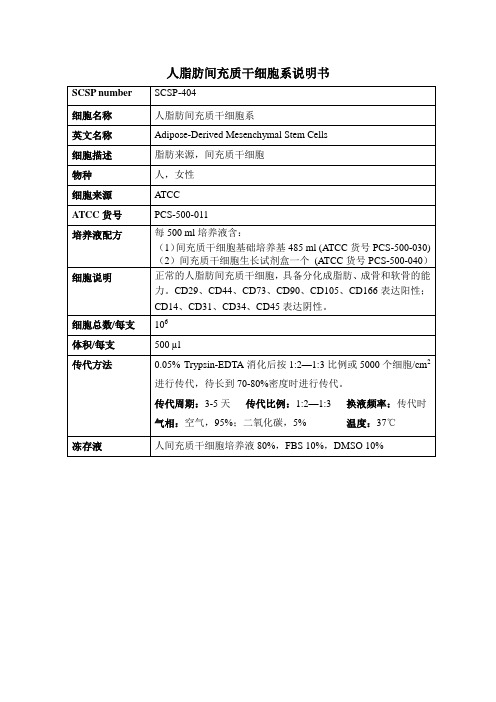
气相:空气,95%;二氧化碳,5%温度:37℃
冻存液
人间充质干细胞培养液80%,FBS10%,DMSO 10%
细胞说明
正常的人脂肪间充质干细胞,具备分化成脂肪、成骨和软骨的能力。CD29、CD44、CD73、CD90、CD105、CD166表达阳性;CD14、CD31、CD34、CD45表达阴性。
细胞总数/每支
106
体积/每支
500µl
传代方法
0.05%Trypsin-EDTA消化后按1:2—1:3比例或5000个细胞/cm2进行传代,待CSP number
SCSP-404
细胞名称
人脂肪间充质干细胞系
英文名称
Adipose-Derived Mesenchymal Stem Cells
细胞描述
脂肪来源,间充质干细胞
物种
人,女性
细胞来源
ATCC
ATCC货号
PCS-500-011
培养液配方
(1)间充质干细胞基础培养基485 ml (ATCC货号PCS-500-030)(2)间充质干细胞生长试剂盒一个(ATCC货号PCS-500-040)
Vi-Cell细胞计数仪使用说明

参考手册
PN 383674 Rev. A
操作人员在使用仪器前应认真阅读本产品手册,具体问题请咨询美国贝克曼库尔特有限公司的培训人员。 技术支持服务电话:1-800-523-3713
11800 SW 147th Ave. 弗罗里达州迈阿密 33116-9015
美国贝克曼库尔特有限公司建议,用户在使用 Vi-CELLTM XR 活力分析仪时,应遵循所有的国家卫生安全 标准,如使用隔离防护装置。即在对此仪器(或任意其他的实验室自动分析仪)进行操作或维护时,应佩 戴护目镜、手套并穿上适当的实验服,以及其他相应的防护装置。
第 2 章 VI-CELL XR 介绍.............................................................................................................. 6
1. 系统概况................................................................................................................................. 6 2. 细胞活力及细胞参数测量........................................................................................................ 6 3. 如何测量细胞活力? ............................................................................................................... 7 3.1 台盼蓝染色排除法 ................................................................................................................... 7 3.2 图像分析方案 .......................................................................................................................... 7
Immobilon-P 传输膜用户指南说明书
User Guide Immobilon®-P Transfer MembraneFor High Sensitivity ImmunodetectionNoticeWe provide information and advice to our customers on application technologies and regulatory matters to the best of our knowledge and ability, but without obligation or liability. Existing laws and regulations are to be observed in all cases by our customers. This also applies in respect to any rights of third parties. Our information and advice do not relieve our customers of their own responsibility for checking the suitability of our products for the envisaged purpose.The initial M, Millipore, Immobilon, SNAP i.d., Calbiochem, and Milli-Q are trademarks of Merck KGaA, Darmstadt, Germany or its affiliates. All other trademarks are the property of their respective owners. Detailed information on trademarks is available via publicly accessible resources.© 2018 Merck KGaA, Darmstadt, Germany and/or its affiliates. All rights reserved.PR04715, Rev. 05/18ContentsIntroduction (1)Guidelines for Working with Immobilon®-P membrane (2)Materials Recommended for Western Blotting (2)Protein Transfer (3)Immunodetection (4)Common Protocols Used in Western Blotting and Immunodetection (4)Membrane Wetting (4)Semi-dry Transfer (4)Protein Visualization (Optional) (5)Immunodetection (5)Standard Immunodetection (5)SNAP i.d.® 2.0 Immunodetection Using Vacuum Filtration (6)Guidelines for Choosing an Immobilon® PVDF Membrane (6)Ordering Information (7)Related products for general Western blotting applications (8)Description (8)Catalogue Number (8)Technical Assistance (9)Standard Warranty (9)IntroductionImmobilon®-P transfer membrane is a polyvinylidene fluoride (PVDF) microporous membrane used for transfer of proteins from a variety of gel matrices. When compared to a nitrocellulose membrane, it has improved handling characteristics and staining capabilities, increased solvent resistance, and a higher signal-to-noise ratio for enhanced sensitivities.This hydrophobic membrane has a nominal pore size of 0.45 micron (µm) and is capable of binding a wide range of molecular weight proteins. While Immobilon®-P membrane is capable of binding proteins smaller than 20,000 in molecular weight, Immobilon®-P SQ 0.2 micron (µm) membrane could be considered for proteins smaller than 20,000, because it has more surface area and higher binding capacity for small proteins.Immobilon®-P membrane has excellent protein retention, high physical strength, and broad chemical compatibility, making it ideal for a variety of staining applications and reprobing in immunodetection methods. This user guide describes some of the most common techniques for Western blotting. For more detailed information please refer to TP001EN, the “Protein Blotting Handbook” (available at).Table 1. Immobilon-P® Membrane Properties and ApplicationsComposition PVDFPore size0.45 µmPhobicity HydrophobicProtein binding capacity Insulin: 160 µg/cm2Bovine serum albumin (BSA): 215 µg/cm2Goat IgG: 294 µg/cm2Applications Binding assaysDot/slot blottingGlycoprotein visualizationLipopolysaccharide analysisMass spectrometryAmino acid analysisN- terminal protein sequencingDetection methods*Chemiluminescent (Immobilon® HRP substrates)Chromogenic (TMB, Insoluble)RadioactiveProtein visualization methods Transillumina-tionReversible stains Ponceau-SCopper phthalocyanine tetrasulfonic acid, tetrasodium salt (CPTS)Toluidine blueSypro® blot stainsIrreversible stains Coomassie™ Brilliant Blue dye Amido blackIndia inkColloidal gold* For fluorescence detection methods, low-autofluorescent Immobilon®-FL membrane is rec-ommended.Guidelines for Working with Immobilon®-P membrane •Always wear gloves when handling the membrane, in order to avoid fingerprints.•Use blunt forceps to prevent membrane damage.•Keep the patapar (blue paper) with the membrane during cutting or handling, but discard when wetting the membrane.•Handle with care to avoid scratches on the membrane surface. Do not fold the membrane.•Hydrophobic Immobilon®-P membrane must be wet in an alcohol solution (> 50% v/v methanol, ethanol, or isopropanol) before use. Once the membrane is wet, it changes from opaque to semi-transparent.•After protein transfer, wash the blot with Milli-Q® water to eliminate any gel residues.•Blots can be air dried and stored at 4° C for several months (for later use) or they can be used immediately.Materials Recommended for Western Blotting•Immobilon®-P membrane cut to the dimensions of the gel•Alcohol (> 50% methanol, ethanol, or isopropanol) for wetting dry membrane•Milli-Q® water•Transfer buffer: 25 mM Tris-base, 192 mM glycine, pH 8.3, 10% alcohol for tank transferor 48 mM Tris, 39 mM glycine, pH 9.2, 10% alcohol for semi-dry transfer)•Sheets of filter paper, cut to the dimensions of the gel and soaked in transfer buffer for at least 30 seconds•Blocking buffer: Immobilon® Block-CH buffer (cat. no. WBAVDCH01) or 0.5–5% (w/v) blocking agent (bovine serum albumin, casein, nonfat dry milk) in wash buffer•Wash buffer: Phosphate-buffered saline (PBS) or Tris-buffered saline (TBS) containing0.5-0.1% Tween®-20 surfactant (PBST or TBST)PBS: 10 mM sodium phosphate, pH 7.2, 0.9% NaClTBS: 10 mM Tris, pH 7.4, 0.9% NaCl•Primary antibody (specific for the protein of interest), diluted in blocking buffer or wash buffer•Secondary antibody (specific for the primary antibody), labeled with a detection enzyme (e.g., horseradish peroxidase [HRP] or alkaline phosphatase [AP]), diluted in blocking buffer or wash bufferProtein TransferProteins can be transferred to Immobilon®-P membrane by two common electro-transfer meth-ods: tank and semi-dry transfer. Table 2 describes the general conditions and major differences for the two methods.Table 2. Transfer Methodstions; it stabilizes the gel dimensions and strips complexed sodium dodecyl sulfate (SDS) from protein molecules, improving protein binding to the membrane. However, for large proteins, or proteins that exhibit solubility problems, it is recommended that the alcohol concentration be decreased and that a small amount of SDS be added to the transfer buffer. This improves protein elution from the gel while maintaining protein solubility during the transfer process.ImmunodetectionImmunodetection is an antibody-based method that allows the detection, identification, and quantitation of a protein or antigen in the blotting membrane. The typical protocol follows these six general steps:1. Block unoccupied membrane sites to prevent nonspecific binding of antibodies.2. Incubate the membrane with a primary antibody that binds to the protein of interest.3. Wash to remove any unbound primary antibody.4. Incubate the membrane with a conjugated secondary antibody, which binds to the firstantibody.5. Wash to remove any unbound secondary antibody.6. Incubate the membrane with a substrate that reacts with the conjugated secondary anti-body to reveal the location of the protein.Standard immunodetection takes at least 4 hours and is widely used, but the SNAP i.d.® 2.0 Protein Detection System (/snapwb) can perform the same process with significant time savings (Table 3).Table 3. Comparison of Standard vs. SNAP i.d.® 2.0 ImmunodetectionStandard Immunodetec-tion SNAP i.d.® 2.0 Immunode-tection1. Block membrane 1 hour10 seconds2. Incubate with primary anti-body1 hour10 minutes3. Wash membrane 3 times, 5 minutes each 4 times, ~10 seconds each4. Incubate with secondary anti-body1 hour10 minutes5. Wash membrane 3 times, 5 minutes each 4 times, ~10 seconds each Total time 4 hours22 minutes Common Protocols Used in Western Blottingand ImmunodetectionMembrane Wetting1. Wet the dry membrane in alcohol (> 50% methanol, ethanol, or isopropanol) for 10–20seconds, or until it changes from a opaque white to uniform, translucent gray.2. Immerse the membrane in Milli-Q® water for 1–2 minutes to displace the alcohol.3. Equilibrate the membrane in transfer buffer for 2–3 minutes or until ready to use.CAUTION: Once the membrane has been wet out, do not allow it to dry out. It can be kept in buffer until protein transfer. If the membrane dries out (turns opaque white)even partially, it must be wet out again (steps 1–3).Semi-dry Transfer1. Resolve the protein mixture on a 1D or 2D polyacrylamide gel.2. Immerse the gel in the transfer buffer and allow it to equilibrate for 10–15 minutes.3. Assemble the transfer stack according to manufacturer’s instructions for the transferSemi-dry Transfer, continue dapparatus used.CAUTION: To ensure an even transfer, remove air bubbles by carefully rolling a clean pipette or blot roller over the surface of each layer in the stack. Do not applyexcessive pressure, as this may damage the gel and membrane.4. Transfer proteins according to transfer apparatus manufacturer’s instructions.5. Remove the blot from the transfer system and rinse the membrane briefly in Milli-Q® waterto remove gel debris. The blot may be air dried for storage, or it can be use immediately for the immunodetection step.NOTE: Drying the blot before immunodetection may enhance the binding of some proteins and reduce background noise.Protein Visualization (Optional)To visualize the protein transfer efficiency, Immobilon®-P membrane may be stained with any reversible blot stain compatible with immunodetection (e.g., Ponceau-S, toluidine blue, CPTS, Sypro® blot stains) or viewed by transillumination using a light box. For a list of reversibleand nonreversible compatible stains and protocols refer to TP001EN, the “Protein Blotting Handbook” at .ImmunodetectionThe following is a general protocol for immunodetection with Immobilon®-P membrane. Some of the critical factors for obtaining a “perfect” Western blot such as protein concentration, blocking solution, and antibody concentration may require optimization.Standard Immunodetection1. If blot has been dried, rewet it in alcohol (> 50% methanol, ethanol, or isopropanol) for 15seconds or until it changes from opaque white to translucent gray.2. Rinse the blot in Milli-Q® water for 1 minute.3. Place the blot in blocking buffer and incubate for 1 hour with gentle agitation. Dilute theprimary antibody in wash or blocking buffer.4. Place the blot in diluted primary antibody solution and incubate for 1 hour with gentle agi-tation.5. Wash the blot with wash buffer (tris- or phosphate-buffered saline solution, supplementedwith Tween®-20 surfactant (TBST or PBST)) 3–5 times for 5 minutes each. Preparesecondary antibody in wash or blocking buffer.6. Place the blot in diluted enzyme-labeled secondary antibody solution and incubate for 1hour with gentle agitation.7. Wash the blot with wash buffer 3–5 times for 5 minutes each.8. Place the blot into a clean container and add the appropriate detection reagent (HRP, AP,or chromogenic).9. Incubate 1–5 minutes, according to the detection reagent manufacturer’s instructions.10. For HRP or AP chemiluminescent reagents, expose blot to x-ray film or acquire the imageusing a digital imaging system. For chromogenic detection, add the reagent and wait until signal appears.SNAP i.d.® 2.0 Immunodetection Using Vacuum Filtration(refer to the SNAP i.d.® 2.0 Protein Detection System User Guide for full protocol details)1. If blot has been dried, rewet it in > 50% alcohol (methanol, ethanol, or isopropanol) for15 seconds, or until it changes from opaque white to translucent gray. Prepare all the re-quired solutions and antibodies ahead of time.NOTE: Antibodies should be 3 to 5 times more concentrated than in standard immunode-tection, but in volumes of 2.5 to 10 mL depending of the blot size/blot holder.2. Wet the SNAP i.d.® 2.0 blot holder in Milli-Q® water and assemble the blot with the proteinside down.3. Using the blot roller, remove all air bubbles and excess water, and insert the blot holderinside the SNAP i.d.® 2.0 frame.4. Block by adding 15–30 mL of blocking solution and immediately turn the vacuum on.5. Depending of the blot holder size used, add 2.5 to 10 mL of diluted primary antibody andincubate for 10 minutes.6. Turn vacuum on to flush the antibody, then with the vacuum still on, wash 4 times with15–30 mL of wash buffer.7. Turn vacuum off, add 2.5–10 mL (depending of the blot holder size used) of dilutedsecondary antibody, and incubate for 10 minutes.8. Turn vacuum on to flush the antibody, then wash 4 times with 15–30 mL of wash buffer.9. Remove the blot from the blot holder and continue with the detection method of choice(chemiluminescence or chromogenic).Guidelines for Choosing an Immobilon® PVDF Membrane The following table provides general guidelines for choosing the appropriate membrane for a specific post-Western blot application. Due to variations in protein properties such as charge density, conformation, and hydrophobicity, not all proteins behave the same way on a given membrane surface. Experiments with a variety of Immobilon® membranes may be necessary to optimize results for your specific application.Application after Western blotting Membrane of choice for most proteins General immunodetection Immobilon®-P or Immobilon®-EAmino acid analysis Immobilon®-PImmunodetection of low molecular weight orImmobilon®-P SQlow-abundance proteinsSequencing of low molecular weight orImmobilon®-P SQlow-abundance proteinsFluorescence immunodetection andImmobilon®-FLchemifluorescence methodsOrdering InformationSee the Technical Assistance section for contact information. You can purchase these products on-line at /westernblot.Immobilon®-P Membrane (0.45 µm pore size) for general Western blotting applications Size Qty/Pk Catalogue Number8.5 × 1000 cm roll 1IPVH85R26.5 × 375 cm roll1IPVH0001026.5 × 187.5 cm roll1IPVH0000526 × 26 cm sheet10IPVH304F020 × 20 cm sheet10IPVH2020015 × 15 cm sheet10IPVH1515010 × 10 cm sheet10IPVH101009 × 12 cm sheet10IPVH091208.5 × 13.5 cm sheet10IPVH081308 × 10 cm sheet10IPVH081007 × 8.4 cm sheet50IPVH07850Immobilon®-P SQ Membrane (0.2 µm pore size) for blotting applicationsof proteins with molecular weights less than 20,000Size Qty/Pk Catalogue Number8.5 × 1000 cm roll1ISEQ85R26.5 × 375 cm roll1ISEQ0001026.5 × 187.5 cm roll1ISEQ0000526 x 26 cm sheet10ISEQ2626020 x 20 cm sheet10ISEQ2020015 x 15 cm sheet10ISEQ1515010 × 10 cm sheet10ISEQ101009 × 12 cm sheet10ISEQ091208.5 × 13.5 cm sheet10ISEQ081308 × 10 cm sheet10ISEQ081007 × 8.4 cm sheet50ISEQ07850Immobilon®-FL Membrane (0.45 µm pore size) for fluorescence detection applications Size Qty/Pk Catalogue Number8.5 × 1000 cm roll1IPFL85R26.5 × 375 cm roll1IPFL0001026.5 × 187.5 cm roll1IPFL0000520 × 20 cm sheet10IPFL2020010 × 10 cm sheet10IPFL101007 × 8.4 cm sheet10IPFL07810Related products for general Western blotting applicationsDescription Catalogue NumberImmobilon® NOW Dispenser for 8.5 x 1000 cm rolls IMDISPImmobilon® Block Noise Cancelling Reagents for chemiluminescence detection,WBAVDCH01 500 mLImmobilon® blotting filter paper, 7 × 8.4 cm sheet, 100/pk IBFP0785CImmobilon® blotting filter paper, 8.5 × 13.5 cm sheet, 100/pk IBFP0813CImmobilon® ECL Ultra Western HRP substrate, 100 mL WBULS0100Immobilon® Signal Enhancer for immunodetection, 500 mL WBSH0500Immobilon® Western HRP substrate, 100 mL WBKLS0100Immunoblot Blocking Reagent, 20 g20-200Immobilon® Forte Western HRP substrate, 100 mL WBLUF0100Immobilon® Crescendo Western HRP substrate, 100 mL WBLUR0100Immobilon® Classico Western HRP substrate, 100 mL WBLUC0100Phosphate-buffered saline with 3% nonfat milk, pH 7.4, dry powder P2194Phosphate-buffered saline with Tween® 20 surfactant, pH 7.4, tablet08057Ponceau S solution, 0.1% (w/v) in 5% acetic acid, 1 L P7170Re-Blot™ Plus Strong Antibody Stripping solution, 10X, 50 mL2504TMB substrate, insoluble (Calbiochem®), 100 mL613548Tris-buffered saline with Tween® 20 surfactant, pH 7.6, tablet91414Tris-glycine buffer 10X Concentrate, 1 L T4904-1LThe initial M, Millipore, and Immobilon are trademarks of Merck KGaA, Darmstadt, Germany or its affiliates. All other trademarks are the property of their respective owners. Detailed information on trademarks is available via publicly accessible resources..© 2018 Merck KGaA, Darmstadt, Germany and/or its affiliates. All rights reserved.All rights reserved.PR04715, Rev. 05/18Technical AssistanceVisit the tech service page on our web site at /techservice. Standard WarrantyThe applicable warranty for the products listed in this publication may be found at /terms (“Conditions of Sale”).。
mTeSR1 培养体系使用说明书
人iPS细胞DYP0530说明书目录编号:SCSP-1302细胞名称:DYP0530细胞描述:将人皮肤细胞诱导成iPS细胞,重编程转录因子OCT4、SOX2、KLF4、MYC 来源性别:男性疾病:帕金森症年龄:63岁细胞来源:2013年引进生物安全等级:BSL-2培养液:见【培养方案】冻存日期/冻存代数:详见冻存管/培养瓶标识冻存管建议复苏培养体系:1个T25培养瓶参考传代周期:4-5天参考传代比例:1:4参考换液频率:每天冻存液:见【培养方案】细胞状态:克隆状,贴壁生长支原体检测结果:阴性STR鉴定结果:D5S818:11,12;D13S317:11,11;D7S820:10,12;D16S539:10,11;vWA:17,18;THO1:7,7;Amelogenin:X,Y;TPOX:8,11;CSF1PO:10,12.中国科学院典型培养物保藏委员会细胞库/干细胞库【培养方案】:使用STEM CELL公司mTeSR1配套培养体系进行培养1.试剂和材料mTeSR TM1培养基试剂盒(产品号#85850)包括:成分规格存放条件mTeSR TM1基础培养基(#85851)400mL2-8℃mTeSR TM15X添加物(#85852)100mL-20℃所需的其他试剂和材料产品品牌/货号Cryostor CS10STEM CELL/07930温和分离液STEM CELL/07174DPBS(不含钙镁离子)GIBCO/14190DMEM/F12GIBCO/11330Y27632STEM CELL/72302BD Matrigel hESC-qualified Matrix CORNING/354277(BD该系列产品已被CORNING收购)细胞刮如,CORNING/3010另需细胞培养板/瓶/皿,15ml离心管和移液管等细胞培养耗材。
2.试剂的制备2.1mTeSR1的制备2.1.1在室温(15-25°C)下或冰箱内(2-8°C)过夜解冻mTeSR™15X添加物(成分编号#05852)。
科普:什么是人工诱导性多能干细胞(简称“iPS细胞”)
科普:什么是人工诱导性多能干细胞(简称“iPS细胞”)什么是iPS细胞?iPS细胞是2006年诞生的新的多功能干细胞,被期待为实现再生医疗发挥重要作用。
但是,iPS细胞究竟是怎么产生的呢?iPS细胞的什么是划时代的呢?并且,什么时候,被预测怎样对医疗有用呢?在本节中,对这些疑问进行简单易懂的说明。
iPS细胞是什么样的细胞?在人的皮肤等体细胞中,导入极少数的因子,通过培养,会变成与各种组织和脏器细胞分化的能力几乎无限增殖的多能性干细胞。
这种细胞被称为“人工多功能干细胞”。
英语中写作“induced pluripotent stem cell”,所以取头文字称为“iPS细胞”。
命名世界上第一个成功制作IPS细胞的京都大学山中伸弥教授。
体细胞变成多功能性干细胞,用专业用语称为“再编程序”。
山中教授的研究小组发现的少数因素引起重新编程序的技术,再现性高,而且比较容易,可以说是干细胞研究的突破。
PS细胞被认为是如何活用的?iPS细胞可以用于再生医疗、查明疾病的原因,开发新药物等。
所谓再生医疗,是以恢复因疾病、受伤等而失去的机能为目的的治疗法。
利用IPS细胞的多分化能力制造出各种各样的细胞,例如糖尿病的话,有调整血糖值能力的细胞,如果神经受到像被切断一样的外伤的话,为了连接丢失的网络而移植神经细胞等情况可以考虑。
可以期待应用于从iPS细胞分化诱导细胞的细胞移植治疗。
另一方面,从难治性疾病患者的体细胞中制作IPS细胞,将其分化成神经、心肌、肝脏、胰脏等患部的细胞,研究其患部的状态和机能会如何变化,并期待研究查明疾病原因的研究。
例如,脑内神经细胞发生变化而引起的疾病,很难从外部进入,而且,从不断变化的细胞中,很难推测出正常状态是怎样的。
通过使用iPS细胞,这样的研究有飞跃性地进行的可能性。
另外,如果利用那个细胞的话,可以进行对人体无法进行的药物的有效性和副作用进行评价的检查和毒性测试,预计新药物的开发会有很大的进展。
ips细胞的有关实验

工
作
量
1.至少阅读15篇以上的相关科技文献
2.设计文字至少在15000字以上
工
作
计
划
12.28——1.1 查阅资料
1.2——1.4 整理文献
1.5 ——1.6 提出设计方案
1.7 ——1.11 撰写说明书
1.12——1.13 检查内容,准备答辩
1.13——1.14 答辩
参
考
资
料
[1]刘辉,黎江等.细胞提取物介导的体细胞重编程[J].细胞生物学杂志,2008,30(5): 553-557.
年月日
设计撰写成绩
(70%)
答辩成绩
(3பைடு நூலகம்%)
合计
教师签字:
年月日
2010-2011秋季学期
生物工程专业课程设计
结题论文
小鼠胚细胞液诱导小鼠成纤维细胞转化IPS的研究
学院(系):环境与化学工程学院
年级 专业:08级 生 物 化 工
学 号:080110050051
学生 姓名:周纪
指导 教师:赵红卫
教师 职称:讲 师
1.3 转录因子的优化
四种转录因子又到体细胞转变为多能干细胞的机制还不是很清楚,并且先前的IPS细胞诱导技术带有致癌基因c-Myc而备受关注。2008年1月Yamanaka小组和Jaenisch小组利用Oct4、Sox2和Klf4 3种基因将鼠和人成纤维细胞重编程为更安全的iPS细胞,2008年4月Jaenisch小组运用Oct4、Sox2、c-Myc、Klf4和C-EBPα将终末分化的鼠成熟B细胞重编程为iPS细胞。2008年6月1 Sch ler小组利用Oct4和Klf4 2种基因将鼠神经干细胞重编程为iPS细胞。这使转录因子越来越少而且一些容易引起细胞发生癌变的基因也被一些其他的基因所代替,这是细胞基因突变和癌变的概率大大降低。这使IPS细胞的安全性大大提高。
