Muscarinic Antagonists:Preclinical Evaluation, BI的,把几个在研药物比较了一番√
雅思真题阅读词汇 剑桥雅思6 test 3 passage 3 抗衰老药物.doc
雅思真题阅读词汇剑桥雅思6 test 3 passage 3 抗衰老药物今天我们雅思真题阅读词汇的文章来研究下剑桥雅思6 test 3 passage 3。
这篇雅思阅读的主题为抗衰老药物。
文章一开始先介绍了一种发现,即限制热量摄入可以延缓衰老。
之后描述了相关药物开发的过程,以及对猴子进行实验的情况。
最后说明了相关药物的缺陷和前景,并解释了其作用机理。
按照惯例,我们总结下其中出现的雅思阅读常考词汇。
第一自然段vigor n. 活力。
“在政府的实验室里,以及别的地方,科学家们正在寻找一种能够延长生命和年轻活力的药物”。
vulnerability n. 脆弱。
“随着年龄的增大,分子和细胞所积累的损害是我们对于身体衰弱十分脆弱”。
longevity n. 寿命。
“一种低卡路里但营养均衡的食物在各种各样的动物上都起到了很好的效果,提升寿命并延长良好的身体状况”。
第二自然段intake n. 摄入。
“不幸的是,为了好处最大化,人们可能不得不减少他们热量摄入的百分之30”。
mortal n. 凡人,人类。
“很少有普通人能够连续几年坚持这样一种严苛的养生疗法”。
mimic v. 模仿。
“但是如果有人能够发明一种药物,它可以模仿更少进食的心里效果,而不同强迫人们真的吃的更少”。
postpone v. 延迟。
“这种卡路里限制药物能够使人们更长时间的保持健康,延迟老年相关疾病的发生吗?”。
第三自然段incidence n. 发生频率。
“他们发现低卡路里饮食的老鼠平均比自由喂食的文章来自雅思老鼠活的更长,并且沾染老年常见现象的发生频率也更低”。
lifespan n. 寿命。
“这意味着最大寿命,而不仅仅是普通寿命,有所上升”。
第四自然段replicate v. 复制,重复。
“这一老鼠身上的发现已经重复了需多次,并且扩展到各种各样的动物身上”。
creature n. 生物。
“直到最近,研究被局限于寿命较短的生物。
它们跟人类的基因相差甚远”。
法国埃维昂文化
evian-les-bains,中文翻译“埃维昂”,1893年诞生于法国,总部设立在浪漫的巴黎,专家们集医学、生化学、分子生物学、细胞学、免疫学、蛋白质组学、美容、整形于一体,研制出埃维昂系列产品在法国巴黎精确严密的科学标准下,接受过多项资格细菌检验证明安全可靠,现已遍布全球30多个国家。
埃维昂科学研究院,特别针对新生细胞形成所需的必要能源做研究,努力朝细胞再生探讨方向迈进,研究成果证实:细胞有加速新陈代谢的潜力,进而研制出含丰富生物细胞再生活性元素和活细胞换肤术,它是真正的生物活性元素的“大熔炉”,含丰富的氨基酸、EGF、六胜肽、端粒酶、醣蛋白、多肽复合物、胶原4D保湿因子、寡肽分子、海洋堇、金属硫蛋白、牛樟芝、石莼提取物、纳诺HA透明质酸等等....法国皇帝拿破仑三世和皇后对埃维昂的泉水情有独钟,1864年,拿破仑正式赐名埃维昂。
埃维昂镇背后雄伟的阿尔卑斯山是依云水的源头,来自高山融雪和山地雨水在阿尔卑斯山脉腹地经过长达15年的天然过滤和冰川砂层的矿化形成了依云水。
也正是这种泉水,使泡温泉的法国姑娘纤尘不染、艳光照人,也使法国的温泉赢得“温泉城市中女王”的美誉。
埃维昂是“水城”之意,因为这里的温泉源于阿尔卑斯山,泉水清明洁净,含钙、镁、锌、锡多种矿物质与微量元素,因此对治疗皮肤疾病、泌尿消化、等方面疾病有较好的疗效。
埃维昂温泉水经过层层矿盐的过滤,在地下孕育了几百年以后,在地下100米处以固定的流速(400000I/J)温度(27°C)不断涌出。
埃维昂泉水纯净无菌,其独特之处还在于它是世界上唯一一款渗透性温泉水,它的渗透压力等于人体细胞的渗透压,符合皮肤生理的PH值。
具备渗透性和矿物质丰富两大特点,在全球众多温泉中最受皮肤科专家的推崇,是专门用于治疗皮肤问题的温泉圣水。
2007暮色中,河湾里落满云霞 scientific and technological advances
暮色中,河湾里落满云霞,与天际的颜色混合一起,分不清哪是流云哪是水湾。
也就在这一幅绚烂的图画旁边,在河湾之畔,一群羊正在低头觅食。
它们几乎没有一个顾得上抬起头来,看一眼这美丽的黄昏。
也许它们要抓紧时间,在即将回家的最后一刻再次咀嚼。
这是黄河滩上的一幕。
牧羊人不见了,他不知在何处歇息。
只有这些美生灵自由自在地享受着这个黄昏。
这儿水草肥美,让它们长得肥滚滚的,像些胖娃娃。
如果走近了,会发现它们那可爱的神情,洁白的牙齿,那丰富而单纯的表情。
如果稍稍长久一点端详这张张面庞,还会生出无限的怜悯。
参考译文:Besidethispicturewithprofusionsofcolors,agroupofsheeparelowingtheir heads, eating by the river bank. Hardly none of them would spare some time toraise their eyes to have a glance at the beautiful dusk. They are, perhaps, takinguseofeveryminutetoenjoytheirlastchewbeforebeingdrivenhome.Thisapictureof the Yellow River bank, in which the shepherd disappears, and no one knows wherehe is resting himself. Only the sheep, however, as free creatures, are joyfullyappreciating the dusk. The exuberant water plants have nutrited the sheep, makingthem grow as fat as balls. When approaching near, you would find their lily-whiteeth and a variety of innocent facial impressions.Globalizationismakingtheworldsmaller,fasterandricher.Still,9/11,avianflu,and Iran remind us that a smaller, fatster world is not necessarily a safer world.Our world is bursting with knowledge - but desperately in need of wisdom. Now, whensound bites are getting shorter, when instant messages crowd out essays, and whenindividual lives grow more frenzied, college graduates capable of deep reflectionare what our world needs.For all these reasons I believed - and I believe even more strongly today - in theunique and irreplaceable mission of universities.科技进步正在使我们能够探索宇宙的边陲、物质最基本的成分及生命的奇迹.与此同时,今天,人类所做的及没能做到的事情,不仅危害到这个星球上的生命,也危害到该星球的寿命。
2019年托福听力科学美国人60秒:药品外表改变
2019年托福听力科学美国人60秒:药品外表改变科学美国人60秒中英文翻译:药品外表改变Don't judge a book by its cover.And don't judge a pill by its color…or shape.When prescriptions for post-heart-attack care gotrefilled and the same drugs suddenly came in different shapes or colors, patients were significantly more likely to stop taking their meds.That's the finding from a study involving more than11,000 patients that appears in the Annals of Internal Medicine.药品.jpgEven when different looking pills contained the exact same active ingredient, a change in color boosted the odds that patients would stop taking their heart medication by 34 percent.A change in shape jumped the chance that patients would cease their meds by 66 percent.There's no legal requirement to make generic and brand-name drugs appear alike.But the study's authors say that the Food and Drug Administration should consider making consistent appearance the standard, as such uniformity would likely result in more patients adhering to their medication regimens.For now, we're all stuck reading pharmaceutical labels.科学美国人60秒中文翻译:不要从封面来判断一本书的好坏,当然也不能凭借颜色和形状来判断一片药。
当代研究生英语 第七单元 B课文翻译
价格的利润生物公司正在吞噬可改变动物DNA序列的所有专利。
这是对阻碍医学研究发展的一种冲击。
木匠认为他们的贸易工具是理所当然的。
他们买木材和锤子后,他们可以使用木材和锤子去制作任何他们所选择的东西。
多年之后来自木材厂和工具储藏室的人并没有任何进展,也没有索要利润份额。
对于那些打造明日药物的科学家们来说,这种独立性是一种罕见的奢侈品。
发展或是发现这些生物技术贸易中的工具和稀有材料的公司,对那些其他也用这些工具和材料的人进行了严格的监控。
这些工具包括关键基因的DNA序列,人类、动物植物和一些病毒的基因的部分片段,例如,HIV,克隆细胞,酶,删除基因和用于快速扫描DNA样品的DNA 芯片。
为了将他们这些关键的资源得到手,医学研究人员进场不得不签署协议,这些协议可以制约他们如何使用这些资源或是保证发现这些的公司可以得到最终结果中的部分利益。
许多学者称这抑制了了解和治愈疾病的进程。
这些建议使Harold得到了警示,Harold是华盛顿附近的美国国家卫生研究院的院长,在同年早期,他建立了一个工作小组去调查此事。
由于他的提早的调查,下个月出就能发布初步的报告。
来自安阿伯密歇根大学的法律教授,该工作组的主席Rebecea Eisenberg说,她们的工作组已经听到了好多研究者的抱怨,在它们中有一份由美国联合大学技术管理组提交的重量级的卷宗。
为了帮助收集证据,NIH建立了一个网站,在这个网站上研究者们可以匿名举报一些案件,这些案件他们相信他们的工作已经被这些限制性许可证严重阻碍了。
迫使研究人员在出版之前需要将他们的手稿展示给公司的这一保密条款和协议是投诉中最常见的原因之一。
另一个问题是一些公司坚持保有自动许可证的权利,该许可证是有关利用他们物质所生产的任何未来将被发现的产品,并且这些赋予他们对任何利用他们的工具所赚取的利润的支配权利的条款也有保有的权利。
Eisenberg说:“如果你不得不签署了许多这样的条款的话,那真的是一个大麻烦”。
美从光秃库盘尼树皮中分离出新的可抗癌细胞和病原细菌化合物
7 6
浙 江 林
业 科
技
2 卷 6
【 5 】张泽华.日本青森县 苹果省力化低 成本 栽培技术的发展 【. J 落叶果 树 ,20 ,3 6 1 04 6( ):5 —5 0 1
L b r a iga d E oo a n g me t d l a o vn n c lgc l S i Ma a e n Mo es a d E au t nf r l ie F ut c a d n v la i l d r i0r h r o o Hi s
1 . , ,4 乙酰..- .[ 3 . O2 三 a 鼠李糖吡喃糖. 1+ ) BD 吡喃型葡萄糖】 L (— 2 . . . 十六烷 , 并将这种新的长链脂肪醇糖苷 称之为库盘尼糖苷。还利用 2 维核磁共振分析了这种化合物的化学结构。 试验证明 , 库盘尼糖苷具有离体抗人肝细胞癌 H p 2 eC 、乳腺腺癌 MD . .3 、 AMB2 1 乳腺导管癌 H 58 、乳 5 T 7
Zh -i n1 LU e .h ng , HE io y ng , XI Ja . u W iz o X a .o 2 E in qi3 LAIJ n s e 3 W AN G il u h ng
 ̄
,
,
( .uh F rsy ueu f eag Yn e 2 60C i ; .i u Si c rm t n et o Ze agLsu 330, h ; 1Yne oet B ra  ̄ j n。 uh 3 30, hn 2Ls ic ne o oi ne f  ̄j n, i i 200 C i r o i a h e P oC r i h a n
腺腺癌 MC .、原发性乳腺癌 5 3 和前列腺癌 P 细胞毒性。还证明,库盘尼糖苷具有抗蜡样芽孢杆菌、金 F7 67 C3 黄色葡萄球菌和大肠杆菌的抗病原细菌毒性 。
本草纲目青蒿素的记载
本草纲目青蒿素的记载英文回答:Artemisinin, also known as qinghaosu, is a chemical compound found in the plant Artemisia annua, also known as sweet wormwood. It is a sesquiterpene lactone with the chemical formula C15H22O5. Artemisinin is a colorless, crystalline solid with a melting point of 156-158 °C and a boiling point of 290-295 °C. It is soluble in water, ethanol, and other organic solvents.Artemisinin is a powerful antimalarial drug that has been used for centuries in traditional Chinese medicine. It is effective against both Plasmodium falciparum and Plasmodium vivax, the two most common species of malaria parasites. Artemisinin is also effective against drug-resistant strains of malaria parasites.The mechanism of action of artemisinin is not fully understood, but it is believed to involve the generation offree radicals that damage the parasite's cell membrane. Artemisinin is also thought to inhibit the parasite'sability to take up nutrients.Artemisinin is a valuable antimalarial drug, but it is not without its side effects. The most common side effectsof artemisinin include nausea, vomiting, and abdominal pain. Artemisinin can also cause liver damage in rare cases.Artemisinin is used in combination with other antimalarial drugs to prevent and treat malaria. The most common combination therapy is artemisinin-based combination therapy (ACT), which includes artemisinin and one or more other antimalarial drugs. ACT is the most effective treatment for malaria, and it is recommended by the World Health Organization (WHO).Artemisinin is also used to treat other conditions,such as cancer and inflammatory diseases. However, there is not enough scientific evidence to support the use of artemisinin for these conditions.中文回答:青蒿素,又名黄花蒿素,是一种发现于菊科植物青蒿中的化学化合物。
微生物学英汉双语对照
微生物学专业词汇英汉双语对照(生物科学专业用)Microorganism 微生物 Microbiology微生物学 Leeuwenhock列文虎克 Pasteur 巴斯德 Whittaker魏塔克 Koch 科赫Jenner琴纳 Fleming弗莱明 Florey佛罗理 Chain钱恩 Beijerinck贝哲林克 Stanley斯坦莱Lister 李斯特 coccus 球菌 rod杆菌 spirillum螺旋菌 vibrio弧菌Tetanus破伤风 Anthracnose炭疽病peptidoglycan肽聚糖protoplast原生质体spheroplast球形体bacterial L-form细菌L形 nucleoid拟核 flagellum鞭毛 capsule荚膜colony菌落 lawn菌苔 schizogenesis裂殖 algocyan藻蓝素 heterocyst异形胞Bacteria细菌Actinomycetes放线菌Rickettsia立克次氏体Chlamydozoan衣原体 mycoplasma支原体 cyanobacteria蓝细菌 yeast酵母菌mold霉菌 virus病毒 virion 病毒粒子budding芽殖 aflatoxin黄曲霉毒素septum隔膜 arthrospore节孢子conidium分生孢子 sporangiospore孢囊孢子plasmogamy质配 karyogamy核配 meiosis减数分裂 oospore卵孢子 zygospore 接合孢子 progametangium原配子囊 homothallism同宗接合 heterothallism 异宗接合 ascospore子囊孢子 ascocarp子囊果 ascus子囊 basidiospore担孢子Guarnieri’s body顾氏小体Negri’s body内基氏小体 X-body X体capsomere衣壳粒 capsid衣壳 multiplication增殖 phage噬菌体 plague噬菌斑rabies狂犬病burst size烈解量Water activity水的活度photoautotroph光能自养生物photoheterotroph光能异养生物chemoautotroph化能自养生物 chemoheterotroph化能异养生物 simple diffusion单纯扩散 facilitated diffusion促进扩散 active transport 主动运输 group translocation基团移位 medium培养基 solid medium固体培养基 liquid medium液体培养基 semisolid medium半固体培养基agar琼脂 potato dextrose agar medium PDA培养基 beef extract peptone medium肉膏蛋白胨培养基 Gause's No 1 synthetic medium高氏1号合成培养基 Czapek' s medium察氏培养基 malt extract medium麦芽汁培养基Ashby medium阿须贝培养基 eosin-methylene blue agar medium伊红美兰琼脂培养基 carbon source碳源 nitrogen source氮源 growth factor生长因子 mineral element 矿质元素 metabolism新陈代谢 amylase 淀粉酶 cellulase纤维素酶 ammonification氨化作用 proteinase蛋白酶extracellular enzyme胞外酶 intracellular enzyme胞内酶 fermentation 发酵pasteur effect巴斯德效应denitrification反硝化作用desulfurization反硫酸化作用 methanogen产甲烷细菌 nitrobacteria硝化细菌 nitrification硝化作用 nitrogen fixation固氮作用 nitrogenase 固氮酶 Mo-Fe-protein钼铁蛋白 Fe-protein铁蛋白 alcohol fermentation 酒精发酵 fermention of lactic acid乳酸发酵 acetic acid fermentation 醋酸发酵growth生长 growth curve生长曲线 lag phase延迟期 log phase 对数期 stationary phase稳定期 decline phase死亡期 synchronous cultivation同步培养 continuous cultivation连续培养 continuous fermentation连续发酵 antisepsis 防腐 disinfection消毒 sterilization 灭菌 boiling煮沸 incineration焚烧 baking烘烤 autoclaving高压灭菌autoclave高压锅 tyndallization间歇灭菌 pasteurization巴斯德消毒photoreactivation光复活作用 antimetabolites抗代谢物 antibiotic抗生素penicillin青霉素nystatin制霉菌素streptomycin链霉素sulfonamide磺胺inoculate接种ultraviolet紫外线dye 染料formaldehyde甲醛 Griffith格里菲斯 transformation转化 Hershey 侯喜Chase蔡斯 Fraenkel-Conrat法郎克-康勒特 Luria鲁里亚 replica plating 影印培养 Lederberg莱德伯格 mutagen诱变剂 donor供体 recipient受体 transduction转导 conjugation接合 plasmid质粒 gene engineering 基因工程 mycorrhiza菌根 ectomycorrhiza外生菌根 symbiosis共生lichen地衣 rumen瘤胃 parasitism寄生 antagonium拮抗 predation捕食 competition竞争 activated sludge活性污泥 coli-index大肠菌群指数 colititre大肠菌群值 infection传染 immunity免疫 toxin毒素exotoxin外毒素 endotoxin内毒素 toxoid类毒素 antitoxin抗毒素complement补体antigen抗原antibody抗体interferon干扰素immunoglobulin免疫球蛋白 natural active immunity自然自动免疫 natural passive immunity自然被动免疫 artificial active immunity人工自动免疫artificial passive immunity人工被动免疫 vaccine 疫苗 Strain 菌株。
青蒿素英语介绍作文
青蒿素英语介绍作文The Miracle of Artemisinin.Artemisinin, derived from the Chinese herb Artemisia annua, has revolutionized the global fight against malaria. This remarkable compound has become a lifesaver for millions, particularly in regions where malaria is endemic. Its discovery and subsequent development into a clinically effective drug is a testament to the richness oftraditional medicine and the power of scientific inquiry.Malaria, caused by parasites transmitted through thebite of infected mosquitoes, has been a scourge of humanity for centuries. It is estimated that malaria has caused over a billion cases of illness and millions of deaths worldwide. The disease is particularly devastating in sub-Saharan Africa, where it often affects children and pregnant women, leading to high mortality rates.The search for an effective treatment for malaria hasbeen ongoing for decades. In the 1970s, when the global health community was facing a crisis due to the emergence of drug-resistant malaria parasites, Chinese scientists embarked on a mission to find new anti-malarial agents from traditional Chinese medicine.One such scientist was Tu Youyou, who led a team at the China National Academy of Medical Sciences. She focused her attention on Artemisia annua, a plant that had been used in traditional Chinese medicine for centuries to treat fever and other ailments. Through meticulous research and experimentation, Tu Youyou and her team isolatedartemisinin from the plant and found that it had remarkable anti-malarial activity.The development of artemisinin into a clinically useful drug was a challenging task. Artemisinin was initially extracted from the plant in a low yield and was unstable. However, through meticulous research and collaboration with pharmaceutical companies, Tu Youyou and her team developed a way to make artemisinin more stable and suitable for clinical use.The results of these efforts were transformative. Artemisinin-based combination therapies (ACTs) have proven to be highly effective against multidrug-resistant malaria parasites. These therapies are now the recommended first-line treatment for malaria in many parts of the world, particularly in Africa where the disease is most prevalent.The impact of artemisinin on global health has been immense. It has significantly reduced malaria mortality rates in many countries, saving millions of lives. Children and pregnant women, who are particularly vulnerable to malaria, have benefited greatly from the availability of ACTs.The discovery of artemisinin is not only a scientific breakthrough but also a powerful reminder of the value of traditional medicine. It demonstrates that knowledge and practices from ancient cultures can contributesignificantly to modern medicine. The collaboration between Chinese scientists and pharmaceutical companies in the development of artemisinin further illustrates theimportance of international cooperation in addressing global health challenges.In conclusion, artemisinin is a remarkable compound that has revolutionized the fight against malaria. Its discovery and development into a clinically effective drug is a testament to the power of traditional medicine and scientific inquiry. The impact of artemisinin on global health has been immense, saving millions of lives and providing hope for a malaria-free future.。
商务英语词汇-医学寄生虫学英语词汇翻译_外贸商务英语四级六级
商务英语词汇 - 医学寄生虫学英语词汇翻译_外贸商务英语四级六级考研雅思英语翻译写作作文听力单词在线字典,learn english dictionary,spkcn首页英语听力词汇学习英语童话英语演讲 ESL资源 Delphi 站内搜索联系我们设为首页您的位置:商务英语词汇 > 医药卫生 > 医学寄生虫学英语词汇翻译医学寄生虫学英语词汇翻译2006-09-27 spkcn 点击: 14Medical Parasitology 医学寄生虫学Aedes 伊蚊alternation of generations 世代交替amastigote 无鞭毛体AmoebiasisAncylostoma duodenale 十二指肠钩口线虫Anopheles 按蚊ascariasis 蛔虫病ascaris lumbricoides 似蚓蛔线虫arthropod 节肢动物bradysporozoite 迟发型子孢子bradyzoite 缓殖子Brugia malayi 马来布鲁线虫capsule 荚膜,被膜,囊胞carrier 携带者,载体,载流子,带虫者cercaria 尾蚴cercarial dermatitis 尾蚴性皮炎daughter cyst 子囊ectopic parasitism 异位寄生egg 卵elephantiasis 象皮肿enterobiasis 蛲虫病Enterobius vermiculariserythrocytic stage 红细胞内期facultative parasite 兼性寄生虫fasciolopsiasisfasciolopsis buskifertile egg 受精卵filaria 丝虫filariasis 丝虫病filariform larvae 丝状蚴final host 终宿主flea 蚤fly 蝇gametocyte 配子体Giardia lamblia 蓝氏贾第鞭毛虫Giardiasis 贾第虫病gravid proglottid 孕节helminth 蠕虫helminthiasis 蠕虫病hemimetabola 不全变态hexacanth 六钩蚴hookworm disease 钩虫病host 宿主human parasitology 人体寄生虫学hydatid cyst 棘球蚴囊hydatid diseaseimmature proglottid 幼节immune evasion 免疫逃避infective stage 感染阶段infertile cyst 不育囊larva 幼虫larva migrans 幼虫移行症Leishmania donovani 杜氏利什曼原虫Leishmaniasis 利什曼病life cycle 生活史louse 虱macrogametocyte 大配子体malaria 疟疾malaria pigment 疟色素mature proglottid 成节medical arthropodology 医学节肢动物学merozoite 裂殖子metacercaria 囊蚴microfilaria 微丝蚴microgametocyte 雄配子体,小配子体miracidium 毛蚴mosquito 蚊myiasis 蛆病Necator americanus 美洲板口线虫Nematode 线虫nocturnal periodicity 夜现周期性nymph 若虫obligatory parasite 专性寄生虫onchosphere 六钩蚴oocyst 卵囊ovum 卵,卵细胞Pagumogonimus skrjabini 斯氏狸殖吸虫paragonimiasis 肺吸虫病parasite 寄生虫parasitic zoonosis 人兽共患寄生虫parasitism 寄生paratenic host (transport host) 转续宿主plerocercoid (sparganum) 裂头蚴Pneumocystis carinii 卡氏肺孢子虫premunition 带虫免疫procercoid 原尾蚴promastigote 前鞭毛体protoscolex 原头蚴protozoon (protozoa) 原生动物pseudocyst 假包囊pupa 蛹recrudescence 再燃redia 雷蚴relapse 复发reservoir host 保虫宿主sandfly 白蛉sarcoptes mites 疥螨Sarcoptes scabiei 人疥螨scabies 疥疮Schistosoma haematobium 埃及血吸虫Schistosoma japomicum 日本血吸虫Schistosoma mansoni 曼氏血吸虫Schistosomiasis 血吸虫病schistosomule (schistosomula) 童虫schizont 裂殖体Schuffners dots 薛氏小点scolex 头节soft ticks 软蜱somatic antigen 虫体抗原sparganosis 裂头蚴病Spirometra mansoni 曼氏迭宫绦虫sporocyst 胞蚴sporozoite 子孢子sterilizing immunity 消除性免疫surface antigen 表面抗原tachysporozoite 速发型子孢子tachyzoite 速殖子taeniasis 带绦虫病tapeworm 绦虫Copyright © 2005-2010 商务英语词汇, Inc. All Rights Reserved.版权声明:未经本站许可,任何人不得复制本站内容。
听故事记GRE单词:青霉素的发现
听故事记GRE单词:青霉素的发现一次大战期间,因为第一次动用了artillery(炮的总称, 炮兵的总称)和armada,每次assail/assault之后总会有很多士兵受伤。
即使使用astringent能够使他们止血,但很快伤口就会出现arcane(神秘的, 不可思议的)的symptom,不久就死去了。
没有人知道是为什么,更不用说找到合适的antidote(解毒剂, 矫正方法)了。
这件appalling的事情在士兵中引起了很大的apprehension(理解, 忧惧,拘捕),人们不得不满怀aspiration的向医生appeal for协助。
一天医生带来了一种新的药粉,有了它以后,奇迹出现了,垂死的病人recovered了。
这种神奇的药粉是一种antibiotic,叫penicillin,是一位叫弗莱明的医生发现的,他又是怎样发现这种arcane的药物的呢?即使巴甫洛夫早就apprised(通知,告知)大家伤口腐败的原因是bacteria在作怪,但当时,所有antique/antiquated的药物都无法有效的杀死bacteria。
弗莱明培养了一些bacteria来研究。
一天,他发现了个antic的apparition(离奇出现的东西):有些培养皿生了mildew(霉, 霉菌, (植物的)霉病),再一看,mildew旁边的bacteria都死掉了。
弗莱明有着aquiline一样锐利的观察力,他立刻asserted 这种mildew里一定有一种。
因为以往的archive中从未报道过,弗莱明就给它起了个artless的appellation叫penicillin。
最初,因为arrogant(傲慢)而arbitrary(专横)的医学界arbiter/arbitrator总以assay(化验)不通过为由,认为这种药不appropriate/apposite/apropos用于人体,所以即使ardent的弗莱明一再assure绝对无害,仍然没人敢尝试它。
英语翻译科学家发现近视基因或研制早期预防药物

科学家发现近视基因或研制早期预防药物Genetic code linked to short sight foundDrugs to be given to children at genetic risk of myopia may now be a real possibility.()Get Flash PlayerScientists have discovered strands of genetic code linked to short sight, the most common eye disorder in the world.The findings shed light on what goes awry to make distant objects look blurred, and raises the prospect of developing drugs to prevent the condition.Understanding the biological glitches behind short-sightedness could help researchers develop eye drops or tablets that could be given to children to stop their vision from failing as they get older.Short-sightedness, or myopia, usually starts to manifest early on in life. The extent to which genes are to blame varies, but for those with the worst vision, around 80% of the condition is caused by genetic factors. Two separate studies, published in Nature Genetics journal, found variations in DNA that were more common in people with short sight. Chris Hammond, at King's College, London, found one section of DNA on chromosome 15 was more common in people with myopia. Caroline Klaver, at Erasmus Medical Centre in Rotterdam, found another strand, also on chromosome 15, linked to short sight.The variations in DNA amount to misspellings in the genetic code. These alter the activity of three genes that control the growth of the eyeball and ensure light entering the eye is converted into electrical pulses 脉冲 in the retina. 视网膜The discovery helps scientists piece together how a healthy eye becomes short-sighted and points the way to medicines to prevent it in children. "My hope is that we can identify a pathway that we can block with eyedrops or tablets that will stop the eye growing too much and without interfering with normal brain development or other processes in the body," Hammond told the Guardian.(Read by Renee Haines. Renee Haines is a journalist at the China Daily Web site.)科学家最近发现了一组与近视有关的遗传密码。
60-second science 听力原文
M ummy Says P rincess H ad C oronary DiseaseP rinc ess Ahmos e-Meryet-A mon enjoyed a privileged lif es tyle in what is now Luxor about 3,500years ago. But she may not have been a happy princess towards the end. T wo of her three main coronary arteries were c alc ified, a marker of atherosc lerosis. Someone in her s ituation today would have bypass s urgery.3500年前,雅赫摩斯-梅尔耶特-艾蒙公主在现在叫卢克索的地方享受着优越的生活。
但是在生命最后阶段她可能就不那么幸福了。
她的三条主冠状动脉中的两条都出现硬化,这是动脉粥样硬化的标志。
今天如果有人得了同样的病会做个心脏搭桥手术。
Sc ientists disc overed her c ondition with a full-body CT sc an of her mummified remains. T he princ ess has thus become the oldest known c ase of c oronary artery disease. Researc hers pres ented their finding at the I nternational Conference of N on-I nvasive Cardiovasc ular I maging in Ams terdam.科学家是在对她的木乃伊进行了全身CT扫描后发现这一情况的。
这位公主成为已知最古老的冠状动脉疾病的患者。
关于艾草的英语介绍作文
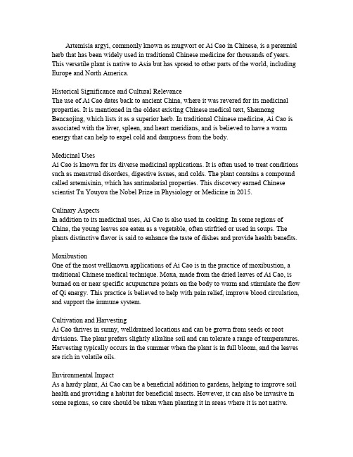
Artemisia argyi,commonly known as mugwort or Ai Cao in Chinese,is a perennial herb that has been widely used in traditional Chinese medicine for thousands of years. This versatile plant is native to Asia but has spread to other parts of the world,including Europe and North America.Historical Significance and Cultural RelevanceThe use of Ai Cao dates back to ancient China,where it was revered for its medicinal properties.It is mentioned in the oldest existing Chinese medical text,Shennong Bencaojing,which lists it as a superior herb.In traditional Chinese medicine,Ai Cao is associated with the liver,spleen,and heart meridians,and is believed to have a warm energy that can help to expel cold and dampness from the body.Medicinal UsesAi Cao is known for its diverse medicinal applications.It is often used to treat conditions such as menstrual disorders,digestive issues,and colds.The plant contains a compound called artemisinin,which has antimalarial properties.This discovery earned Chinese scientist Tu Youyou the Nobel Prize in Physiology or Medicine in2015.Culinary AspectsIn addition to its medicinal uses,Ai Cao is also used in cooking.In some regions of China,the young leaves are eaten as a vegetable,often stirfried or used in soups.The plants distinctive flavor is said to enhance the taste of dishes and provide health benefits. MoxibustionOne of the most wellknown applications of Ai Cao is in the practice of moxibustion,a traditional Chinese medical technique.Moxa,made from the dried leaves of Ai Cao,is burned on or near specific acupuncture points on the body to warm and stimulate the flow of Qi energy.This practice is believed to help with pain relief,improve blood circulation, and support the immune system.Cultivation and HarvestingAi Cao thrives in sunny,welldrained locations and can be grown from seeds or root divisions.The plant prefers slightly alkaline soil and can tolerate a range of temperatures. Harvesting typically occurs in the summer when the plant is in full bloom,and the leaves are rich in volatile oils.Environmental ImpactAs a hardy plant,Ai Cao can be a beneficial addition to gardens,helping to improve soil health and providing a habitat for beneficial insects.However,it can also be invasive in some regions,so care should be taken when planting it in areas where it is not native.Modern ResearchIn recent years,there has been a resurgence of interest in Ai Cao due to its potential health benefits.Modern research is exploring its use in treating a variety of conditions, including cancer,as well as its potential role in improving mental health and wellbeing. ConclusionAi Cao,with its rich history and multifaceted uses,continues to be an important plant in both traditional and modern medicine.Its versatility as a medicinal herb,culinary ingredient,and component in traditional therapies makes it a valuable resource for health and wellbeing.As research continues,we can expect to learn even more about the potential benefits of this ancient plant.。
酿酒酵母 细胞内膜锚定蛋白英语
酿酒酵母细胞内膜锚定蛋白英语Membrane-Anchored Proteins in the Endomembrane System of Saccharomyces cerevisiae.The endomembrane system of Saccharomyces cerevisiae, a budding yeast, is a complex network of organelles that includes the nuclear envelope, endoplasmic reticulum (ER), Golgi apparatus, vacuole, and several types of vesicles. These organelles are responsible for a variety of cellular functions, including protein and lipid synthesis, modification, and trafficking; ion homeostasis; and autophagy.Membrane-anchored proteins are essential for the proper function of the endomembrane system. These proteins are anchored to the membrane by a variety of mechanisms, including transmembrane domains, lipid anchors, and glycosylphosphatidylinositol (GPI) anchors. Transmembrane domains are hydrophobic regions of the protein that span the lipid bilayer, while lipid anchors are hydrophobicmolecules that are covalently attached to the protein. GPI anchors are complex oligosaccharides that are attached to the protein via a phosphodiester bond.Membrane-anchored proteins perform a variety of functions in the endomembrane system. These functions include:Protein trafficking: Membrane-anchored proteins are involved in the trafficking of proteins between the ER, Golgi apparatus, and vacuole. These proteins include coat proteins, which form complexes that bud off vesicles from the ER and Golgi apparatus, and SNAREs, which mediate the fusion of vesicles with target membranes.Lipid metabolism: Membrane-anchored proteins are involved in the synthesis, modification, and trafficking of lipids. These proteins include enzymes that synthesize lipids, proteins that transport lipids between organelles, and proteins that regulate lipid metabolism.Ion homeostasis: Membrane-anchored proteins areinvolved in the transport of ions across the membranes of the endomembrane system. These proteins include ion channels, which allow ions to flow down their electrochemical gradients, and ion pumps, which use energy to transport ions against their electrochemical gradients.Autophagy: Membrane-anchored proteins are involved in autophagy, a process by which the cell degrades its own components. These proteins include autophagy receptors, which recognize damaged proteins and organelles, and autophagy proteins, which are involved in the formation and maturation of autophagosomes.The membrane-anchored proteins of the endomembrane system are a diverse group of proteins that play a critical role in the proper function of the cell. These proteins are essential for protein trafficking, lipid metabolism, ion homeostasis, and autophagy.Here are some specific examples of membrane-anchored proteins in the endomembrane system of Saccharomyces cerevisiae:Sec61: A transmembrane protein that forms the core of the translocon, a complex that mediates the translocation of proteins into the ER.COPII coat proteins: Proteins that form complexes that bud off vesicles from the ER.SNAREs: Proteins that mediate the fusion of vesicles with target membranes.Lipid flippases: Proteins that transport lipids between the two leaflets of the lipid bilayer.Ion channels: Proteins that allow ions to flow down their electrochemical gradients.Ion pumps: Proteins that use energy to transport ions against their electrochemical gradients.Autophagy receptors: Proteins that recognize damaged proteins and organelles.Autophagy proteins: Proteins that are involved in the formation and maturation of autophagosomes.These are just a few examples of the many membrane-anchored proteins that are found in the endomembrane system of Saccharomyces cerevisiae. These proteins are essential for the proper function of the cell and play a criticalrole in a variety of cellular processes.。
高角羚英文描写
高角羚英文描写The Sable Antelope: A Majestic Creature of the African SavannaAmidst the vast expanse of the African savanna, a magnificent creature roams the land with an unparalleled grace and power. The sable antelope, a species indigenous to the continent, stands as a testament to the wonders of the natural world. With its striking black and white coat, impressive horns, and regal bearing, the sable antelope captivates all who witness its presence.As the sun rises over the rolling grasslands, the sable antelope emerges from the shadows, its ebony-hued body glistening in the morning light. The males, with their impressive curved horns that can reach up to 5 feet in length, lead the herd, their heads held high as they survey the surrounding territory. The females, with their sleek and slender frames, follow closely, their delicate features complemented by the stark contrast of their white faces and bellies.The sable antelope is a true master of its environment, possessing a remarkable agility and speed that allows it to navigate the ruggedterrain with ease. Its long, powerful legs propel it forward with a graceful stride, enabling it to outrun even the most persistent predators. When threatened, the sable antelope will often stand its ground, its horns lowered in a defensive posture, ready to charge and defend its herd.One of the most striking features of the sable antelope is its intricate coat pattern. The males, in their prime, display a deep, glossy black hue that covers the majority of their body, with stark white markings on their face, belly, and legs. The females, on the other hand, exhibit a more subtle coloration, with a reddish-brown coat that blends seamlessly with the surrounding vegetation.As the herd moves across the savanna, their synchronized movements create a mesmerizing sight. The sable antelope is a highly social animal, and the members of the herd work together to ensure the safety and well-being of the group. They communicate through a range of vocalizations, from the deep, grunting calls used to maintain contact within the herd, to the high-pitched snorts that serve as alarm signals.The sable antelope's habitat is primarily found in the savannas and woodlands of southern and eastern Africa, where it thrives in areas with a mix of open grasslands and dense vegetation. These areas provide the perfect balance of grazing opportunities and cover frompredators, allowing the sable antelope to thrive in its natural environment.Unfortunately, the sable antelope, like many other African wildlife species, faces a number of threats to its survival. Habitat loss due to human encroachment, poaching for its valuable horns, and competition with livestock are just a few of the challenges this magnificent creature must overcome. Conservation efforts, both in Africa and around the world, are crucial in ensuring the continued existence of the sable antelope and the preservation of the delicate ecosystem it calls home.Despite these challenges, the sable antelope remains a symbol of the enduring strength and resilience of the African continent. Its presence in the savanna serves as a reminder of the incredible diversity and beauty of the natural world, and its majestic presence captivates all who are fortunate enough to witness it.In conclusion, the sable antelope is a truly remarkable creature, a living embodiment of the power and grace of the African wild. Its striking appearance, impressive stature, and remarkable adaptability make it a true wonder of the natural world, and a testament to the enduring beauty and resilience of the African continent.。
- 1、下载文档前请自行甄别文档内容的完整性,平台不提供额外的编辑、内容补充、找答案等附加服务。
- 2、"仅部分预览"的文档,不可在线预览部分如存在完整性等问题,可反馈申请退款(可完整预览的文档不适用该条件!)。
- 3、如文档侵犯您的权益,请联系客服反馈,我们会尽快为您处理(人工客服工作时间:9:00-18:30)。
Preclinical Evaluation of Long-Acting Muscarinic Antagonists:Comparison of Tiotropium and Investigational DrugsPaola Casarosa,Thierry Bouyssou,Sabine Germeyer,Andreas Schnapp,Florian Gantner,and Michael PieperDepartment of Pulmonary Diseases Research,Boehringer Ingelheim Pharma GmbH &Co.KG,Biberach an der Riss,GermanyReceived February 16,2009;accepted May 26,2009ABSTRACTChronic obstructive pulmonary disease (COPD)is character-ized by progressive airflow limitation caused by persistent in-flammatory processes in the airways.An increased cholinergic tone mediates different pathophysiological features of COPD,such as bronchoconstriction and mucus hypersecretion,mostly through activation of the human muscarinic M 3receptor (hM 3)subtype.Tiotropium bromide (Spiriva)is a well estab-lished muscarinic antagonist in the pharmacological manage-ment of COPD with a once-daily posology.The rationale behind the sustained bronchodilation obtained with tiotropium con-sists in its slow dissociation from hM 3receptors.In this study,we performed a comprehensive preclinical comparison of tiotropium with other long-acting muscarinic antagonists (LAMAs)currently in clinical development,namely aclidinium bromide and glycopyrrolate.The different muscarinic antago-nists were characterized for their 1)affinity toward the different human muscarinic receptor subtypes expressed in Chinesehamster ovary cells and kinetics of receptor dissociation,2)potency in inhibiting the agonist-induced activation of musca-rinic receptors through measurement of second messengers,and 3)efficacy and duration of bronchoprotection,as tested in a model of acetylcholine-induced bronchoconstriction in anes-thetized dogs over a period of 24h.All of the tested LAMAs showed high affinity and potency toward the hM 3receptor (tiotropium,p A 2ϭ10.4;aclidinium,p A 2ϭ9.6;and glycopyr-rolate,p A 2ϭ9.7).However,dissociation half-lives of the LA-MAs from the hM 3receptor differed significantly (tiotropium,t 1⁄2ϭ27h;aclidinium,t 1⁄2ϭ10.7h;and glycopyrrolate,t 1⁄2ϭ6.1h).In line with their kinetic properties at the hM 3,the tested LAMAs provided different levels of bronchoprotection in the in vivo setting 24h after administration (tiotropium ϭ35%,acli-dinium ϭ21%,and glycopyrrolate ϭ0%at 24h)when applied at equieffective doses.The term COPD describes a clinical condition character-ized by chronic airway obstruction caused by an abnormal inflammatory response of the respiratory system.The main causal factor in the pathogenesis of COPD is sustained inha-lation of cigarette smoke (Rabe et al.,2007).Occasionally,other factors are involved,including a genetically induced deficiency of the enzyme ␣1-antitrypsin or exposure to nox-ious agents at work and in the environment.COPD is char-acterized by a predominantly neutrophilic inflammation of the airway walls,and airflow limitation is mainly a result of hyperplasia of mucosal glands,hypertrophy,and,in partic-ular,constriction of the bronchial smooth muscle in the small airways (Barnes,2000).COPD is associated with a signifi-cant level of morbidity and mortality (Rabe et al.,2007).The Global Burden of Disease Study has projected that COPD,which ranked sixth as the cause of death in 1990,will become the third leading cause of death worldwide by 2020(Murray and Lopez,1997).National and international guidelines recommend that pa-tients with COPD should receive bronchodilators as first-line maintenance treatment,because they increase expiratory flow by decreasing airway smooth muscle tone,thus leading to reduced lung hyperinflation (Rabe et al.,2007).The most frequently prescribed bronchodilators for COPD treatment are muscarinic antagonists and 2-agonists.In COPD,bron-choconstriction and mucus secretion are increased,and the airways become hyper-responsive to contractile agents.These changes are caused mostly by increased parasympa-thetic nerve activity (Barnes,2004).Acetylcholine (ACh),released from parasympathetic nerve endings,activatesArticle,publication date,and citation information can be found at .doi:10.1124/jpet.109.152470.ABBREVIATIONS:COPD,chronic obstructive pulmonary disease;[3H]NMS,[N -methyl -3H]scopolamine methyl chloride;hM,human muscarinic receptor;ACh,acetylcholine;LAMA,long-acting muscarinic antagonist;CHO,Chinese hamster ovary;PTX,pertussis toxin;ipratropium bromide,(1R ,3r ,5S ,8r )-8-isopropyl-3-[(Ϯ)-tropoyloxy]tropanium bromide monohydrate;tiotropium bromide,6,7-epoxy-3-hydroxy-8-methyl-1␣H ,5␣H -tropanium bromide di-2-thienylglycolate;LAS34273,aclidinium bromide,(3R )-3-{[hydroxydi(thiophen-2-yl)acetyl]oxy}-1-(3-phenoxypropyl)-15-azabicyclo[2.2.2]octan-1-ylium bromide;NVA-237,glycopyrrolate bromide,3-(␣-cyclopentylmandeloyloxy)-1,1-dimethylpyrrolidinium bromide.0022-3565/09/3302-660–668$20.00T HE J OURNAL OF P HARMACOLOGY AND E XPERIMENTAL T HERAPEUTICSVol.330,No.2Copyright ©2009by The American Society for Pharmacology and Experimental Therapeutics 152470/3498433JPET 330:660–668,2009Printed in U.S.A.660by guest on April 22, 2011 Downloaded frompostjunctional muscarinic M 3receptors present on airway smooth muscle and submucosal glands to induce bronchocon-striction and mucus secretion,respectively.Thus,anticholin-ergic bronchodilators have a particular value in the treat-ment of COPD because they block the effects of an increased vagal cholinergic tone.In clinical trials,muscarinic antago-nists have been shown to produce an improvement in lung function,exercise endurance,and health-related quality of life and significant reductions of exacerbations,and—in sum-mary—to improve the clinical course of COPD (Vincken et al.,2002;Tashkin et al.,2008).Importantly,muscarinic an-tagonists show a favorable side-effect profile.Thanks to the quaternary structure of modern anticholinergics,which pre-vents a substantial absorption from mucosal surfaces and penetration of the blood-brain barrier,and the inhalative administration route,which further limits systemic expo-sure,these drugs are virtually free of clinically relevant side effects,besides occasional reports of dry mouth (Gross,2006).Tiotropium (Spiriva),introduced recently for the treatment of COPD,is the first example of a long-acting muscarinic antagonist (LAMA):following a single dose of tiotropium,clinically relevant improvement in lung function lasts for more than 24h,allowing once-daily dosing (Vincken et al.,2002).The mechanism behind its long duration of action relates to the slow rate of dissociation from its target,the human M 3muscarinic receptor (Disse et al.,1999).Long duration of action (preferably 24h)is an important feature of drugs intended to treat chronic diseases,enabling both pro-longed efficacy (Tashkin,2005)and a simple,once-daily dos-age regime that improves patient compliance (Tamura and Ohta,2007).Other drugs are currently being evaluated in clinical trials for their ability to function as LAMAs with a potential for once-daily administration,i.e.,aclidinium [also known as LAS34273from Almirall Prodesfarma (Barcelona,Spain),currently in phase IIb trials]and glycopyrrolate [NVA-237,from Novartis (Basel,Switzerland),in phase III trials].Currently,however,limited preclinical information is published concerning the pharmacological properties of these investigational drugs.Thus,in the present study,we have directly compared the pharmacology of the different LAMAs,namely tiotropium,aclidinium,and glycopyrrolate,in in vitro and in vivo models.To understand the antagonists’behavior at the molecular level,interaction with the different muscarinic receptor subtypes was analyzed in binding and functional assays.Given the rationale behind tiotropium’s long duration of action,particular attention was given to the kinetics of dissociation from the muscarinic receptors.Effi-cacy and duration of bronchoprotection were tested in a phar-macological model of acetylcholine-induced bronchoconstric-tion in anesthetized dogs over a period of 24h.Materials and MethodsChemicals and Reagents.[N -methyl -3H]Scopolamine methyl chloride ([3H]NMS,specific activity,82Ci/mmol)was obtained from PerkinElmer Life and Analytical Sciences (Waltham,MA).MgCl 2,carbachol,muscarine,atropine sulfate,pirenzepine,N -methyl sco-polamine bromide,EDTA,3-isobutyl-1-methylxanthine,NaCl,and HEPES were obtained from Sigma-Aldrich (St.Louis,MO).Ipratro-pium bromide,tiotropium bromide,aclidinium bromide,and glyco-pyrrolate bromide were synthesized in the chemical laboratories of Boehringer Ingelheim (Biberach an der Riss,Germany).The tritia-tion of tiotropium was performed by RC Tritec AG (Teufen,Switzer-land).[3H]Tiotropium was purified by high-performance liquid chro-matography on a XBridge C-8column (Waters GmbH,Eschborn,Germany)resulting in a radiochemical purity of Ն98%,and a specific activity of 65Ci/mmol.All cell culture reagents were purchased from Invitrogen (Carlsbad,CA).Cell Culture Techniques.Chinese hamster ovary (CHO)cells transfected with the cDNAs encoding the human M 1to M 5musca-rinic acetylcholine receptors were purchased from PerkinElmer Life and Analytical Sciences.CHO cells were grown in Ham’s F-12me-dium supplemented with 10%fetal calf serum in the presence of the selection agent G418(400g/ml).Cells were maintained at 37°C in humidified air containing 5%CO 2.Equilibrium Binding Experiments.Membrane isolation and purification from CHO cells stably expressing the human M 1to M 5receptors was performed as described previously (Casarosa et al.,2005).In brief,cells were suspended in buffer A (15mM Tris-HCl,pH 7.5,2mM MgCl 2,0.3mM EDTA,1mM EGTA),homogenized,and spun down for 30min at 48,000g .The pellet was resuspended in buffer B (7.5mM Tris-HCl,pH 7.5,12.5mM MgCl 2,0.3mM EDTA,1mM EGTA,250mM sucrose),aliquoted,and stored at Ϫ80°C until use.Protein content was measured with the BCA kit (Thermo Fischer Scientific,Rockford,IL).In all radioligand experiments,the binding buffer consisted of 10mM HEPES,1mM MgCl 2,pH 7.4.After the indicated incubation period,bound and free [3H]NMS were separated by rapid vacuum filtration using a Brandel Harvester (Gaithersburg,MD)on GF/B filters presoaked in 0.5%polyethylenimine and rapidly washed three times with ice-cold binding buffer.Filter disks were added to 3ml of scintillation fluid (Ultima Gold from PerkinElmer Life and Analyti-cal Sciences)in pony-vials,and radioactivity was quantified by use of liquid scintillation spectrometry on a Tri-Carb 2900TR Liquid Scin-tillation Analyzer (PerkinElmer Life and Analytical Sciences).In all experiments,total binding never reached 10%of that added,limiting complications associated with depletion of the free-radioligand con-centration.Saturation binding experiments were performed by incubating membranes (usually 5–10g/sample,adjusted according to the B max of the individual cell line),with a range of concentrations of [3H]NMS (4pM to 8nM)in a total volume of 4ml,to avoid significant ligand depletion at the lower concentrations.Samples were incubated at room temperature for 2h under gentle agitation before filtration.Nonspecific binding was determined for each radioligand concentra-tion by coincubating in parallel a set of membranes with an excess of unlabeled atropine (10M).To obtain affinity estimates of unlabeled antagonists,heterolo-gous competition experiments against [3H]NMS were performed at equilibrium.Membranes were incubated in the presence of [3H]NMS (final concentration,approximately 0.1nM)and different concentra-tions of unlabeled antagonist at room temperature with gentle agi-tation for 18to 20h before petition displacement binding data were fitted to the equation described by Hill (1909),and IC 50values obtained from the inhibition curves were converted to K i values by use of the method of Cheng and Prusoff (1973).Kinetic Studies.To determine dissociation kinetic parameters according to the classical method,membranes expressing the differ-ent muscarinic receptor subtypes were first allowed to equilibrate with 0.45nM [3H]NMS or 90pM [3H]tiotropium at room tempera-ture for at least 2h (300l/sample).Subsequently,samples were added to test tubes already containing 3ml of binding buffer with atropine 10Ϫ5M (time 0)to start dissociation.At the different time points,samples were filtered by use of a Brandel harvester,as described above.To monitor the potential membrane degradation occurring at later time points,some samples were added at time 0to tubes containing 3ml of binding buffer with the same concentration of radioligand present in the preincubation step (i.e.,0.45nM [3H]NMS or 90pM [3H]tiotropium),so that dissociation was not started and decrease in radioligand binding would reflect receptor breakdown.Dissociation data were fitted to a single-phase exponen-Preclinical Pharmacology of Long-Acting Anticholinergics661by guest on April 22, 2011 Downloaded fromtial decay function,and the K off rate obtained was transformed into a t 1⁄2value (dissociation half-life)using the following equation:t 1⁄2ϭln 2/K off .The dissociation kinetic parameters of unlabeled ligands were assessed with the use of the method of Motulsky and Mahan (1984).With this approach,the kinetics of an unlabeled ligand are deter-mined via competition with a radioligand ([3H]NMS),whose kinetic parameters have already been determined for a given receptor.This method involves the simultaneous addition of both radioligand and unlabeled competitor to the membranes,so that at time 0all recep-tors are unoccupied.Membranes were added at time 0to tubes containing 0.3nM [3H]NMS in the presence or absence of three different concentrations of unlabeled competitor (approximately 30-,100-,and 300-fold K i ).At the different time points (up to 8h),the degree of [3H]NMS bound to receptor was assessed by filtration harvesting,as described above.Dissociation rates for unlabeled an-tagonists were calculated with Prism (GraphPad Software Inc.,San Diego,CA)using the equation for kinetics of competitive binding by fitting (least-squares)the data from the competition kinetic experi-ments to a two-component exponential curve,with all parameters fixed apart from k 3and k 4.The kinetics of [3H]NMS,determined independently (average of three independent experiments)and used in these equations,are as follows:for hM 1receptor:k on ϭ3.22ϫ108M Ϫ1min Ϫ1and K off ϭ0.019min Ϫ1;for hM 2receptor:k on ϭ6.96ϫ108M Ϫ1min Ϫ1and K off ϭ0.035min Ϫ1;for hM 3receptor:k on ϭ5.96ϫ108M Ϫ1min Ϫ1and K off ϭ0.013min Ϫ1.cAMP Assay.To determine the functional antagonistic potency of the different muscarinic antagonists at the hM 2receptor,changes in intracellular cAMP levels were determined with CHO-hM 2cells in suspension (5000cells/well)with use of Lance technology (PerkinElmer Life and Analytical Sciences)and the 384-well plate format (Optiplate;PerkinElmer Life and Analytical Sciences),ac-cording to the manufacturer’s protocol.In brief,cells were stimulated with a range of agonists’concentrations (either carbachol or musca-rine,from 10Ϫ3to 10Ϫ12M)in Hanks’buffered saline solution sup-plemented with 5mM HEPES,0.1%bovine serum albumin,500mM 3-isobutyl-1-methylxanthine,and 1M forskolin in the absence or presence of at least six different concentrations of antagonists (usu-ally from 10Ϫ5to 10Ϫ10M)for 30min at room temperature.A 15-min preincubation with antagonists was allowed to proceed before the addition of the agonist.Cells were lysed with use of Lance reagents,and after an additional 2h,plates were read on an Envision plate reader (PerkinElmer Life and Analytical Sciences).The concentra-tion of cAMP in the samples was calculated from a standard curve.The Gaddum equation was used to fit the data,and shifts in the dose ratio were plotted according to Schild.Inositol Phosphate Accumulation Assay.Changes in intracel-lular levels of inositol phosphates were monitored in CHO-hM 1and CHO-hM 3cells by use of the IPone kit (Cisbio Bioassays,Bagnols-sur-Ce `ze Cedex,France)according to the manufacturer’s instruc-tions.In brief,cells were plated 1day in advance in 384-well white plates (Greiner Bio-One GmbH,Frickenhausen,Germany),with a density of 20,000cells per well.On the day of the assay,cells were stimulated with a range of agonists’concentrations (either carbachol or muscarine,from 10Ϫ3to 10Ϫ12M)in a buffer consisting of 10mM HEPES,1mM CaCl 2,0.5mM MgCl 2,4.2mM KCl,146mM NaCl,5.5mM glucose,and 50mM LiCl (final pH 7.4)in the absence or presence of at least six different concentrations of antagonists (usu-ally from 10Ϫ5to 10Ϫ10M).A 15-min preincubation with antagonists was allowed to proceed before addition of the agonist.After 1-h stimulation at 37°C,cells were lysed using reagents provided by the kit,and after an additional hour,plates were read on an Envision plate reader.The concentration of inositol phosphates in the samples was calculated from a standard curve.The Gaddum equation was used to fit the data,and shifts in the dose ratio were plotted accord-ing to Schild.In Vivo Experiments.Bronchoprotective effects of the musca-rinic antagonists were investigated in a model of acetylcholine(ACh)-induced bronchoconstriction in anesthetized,ventilated bea-gle dogs.All animal experiments were conducted in accordance with the German Animal Welfare Law and received ethics committee approval.Male dogs weighing between 10and 12kg were randomly assigned to four groups,each consisting of four animals.The dogs were anesthetized with a bolus injection of propofol (10mg/kg i.v.)followed by an infusion of 30mg/kg i.v.propofol per hour (B.Braun Melsungen,Melsungen Germany)into the cephalic vein.The dogs were intubated and ventilated with volume-controlled pressure with a mixture of room air and oxygen (3:1)with a Siemens respirator at a rate of 15strokes per minute.Optimal ventilation was ensured by regular measurement of acid-base status and oxygen saturation in the blood.Muscarinic antagonists were dissolved in water/ethanol (40:60,v/v)and administered by inhalation by use of the Respimat Soft Mist Inhaler (Boehringer Ingelheim Pharma GmbH,Ingelheim,Germany)connected to the tracheal tube of the ventilated dogs in a volume of 30l.Increases in airway resistance were induced by repeated intravenous injections of 10g/kg acetylcholine (Sigma-Aldrich).Experiments were performed as dose range-finding studies for 3h before studies for 24h.For the 3-h study setup,intravenous acetylcholine challenges were injected at:Ϫ45,Ϫ30,Ϫ15,5,10,30,60,90,120,150,and 180min after inhalation of the test compound.For the 24-h study setup,ACh was administered 30and 15min before and 5,10,and 30min and 6,12,and 24h after inhalation of the test compounds.Bronchoprotection was expressed as percentage of inhibition of the ACh-induced increase in airway resistance before the administration of the compound (predrug values).Data Analysis.All experiments were analyzed by either linear or nonlinear regression analysis with the equations mentioned under the different assay methodology using Prism version 5.1(GraphPad Software,San Diego,CA).Data are expressed as mean ϮS.E.M.ResultsDetermination of Ligand Affinity for the Human Muscarinic M 1to M 5Receptors.To analyze the pharma-cological behavior of the different muscarinic antagonists in vitro,CHO cell lines selectively and stably expressing either of the human muscarinic M 1,M 2,M 3,M 4,and M 5receptors were used to ensure that measurements were made at a single receptor subtype.For each cell line,the B max values and affinities toward [3H]NMS,as determined in saturation experiments,are reported in Table 1.The antagonists’affinities for the different receptor subtypes were determined in heterologous competitive binding experi-ments against [3H]NMS.Because the time needed to reach equilibrium can change significantly,depending on the kinetic parameters of the unlabeled compound (Motulsky and Mahan,1984),initial experiments were performed to decide how long incubation should proceed.This aspect is of particular impor-tance for compounds with longer kinetics of receptor interac-TABLE 1Pharmacological characterization of the different CHO cell lines stably expressing human muscarinic receptor subtypesDissociation constants for ͓3H ͔NMS (expressed in picomolars)and levels of receptor expression (expressed in picomoles per milligram of protein content of the membrane preparations)were determined in saturation binding assays,as described under Materials and Methods .The average of at least two independent experiments per-formed in triplicate is shown.Receptor SubtypeK d B max pMpmol/mghM 160Ϯ1 1.91Ϯ0.09hM 266Ϯ49.10Ϯ0.10hM 378Ϯ7 2.97Ϯ0.03hM 454Ϯ14 2.39Ϯ0.03hM 5201Ϯ168.99Ϯ0.20662Casarosa et al.by guest on April 22, 2011 Downloaded fromtion,as shown for tiotropium (Disse et al.,1993),where too short incubation would result in underestimation of receptor affinity.As a model system,membranes expressing the hM 3receptor were used,and the different muscarinic antagonists,i.e.,tiotropium,ipratropium (with fast dissociation from the hM 3receptor,see next paragraph),aclidinium and glycopyrro-late,were allowed to equilibrate in the presence of [3H]NMS for different times (Fig.1).Ipratropium reached equilibrium after 2h,as indicated by the absence of any further change in its IC 50over time.In contrast,tiotropium needed at least 20h of incu-bation time to reach pseudo-equilibrium,as indicated by the shift in pIC 50values at shorter time points (pIC 50values after 2,5,and 10h were statistically different from that obtained at 20h,P Ͻ0.05).An intermediate behavior was observed for aclidinium and glycopyrrolate,which needed at least 5h to reach pseudo-equilibrium in this experimental setting.Because the time needed to reach equilibrium is related to the receptor dissociation kinetics for each cold test compound (Motulsky and Mahan,1984),these data suggest that,among the substances tested,tiotropium has the longest half-life at the hM 3receptor.Based on these results,an assay incubation time of approxi-mately 20h was used in all subsequent experiments,because this provided a satisfactory compromise between attaining equilibrium while avoiding membrane degradation.Displacement curves were analyzed according to a model of orthosteric competition between [3H]NMS and the unlabeled competitors,so that IC 50values were transformed into K i val-ues according to the Cheng-Prusoff equation.To prove that this is the correct model for interpreting the data,the shifts in IC 50values in relation to the increase in amount of radioligand used in a given experiment were monitored for different ligands:the IC 50values shifted linearly in correlation with the increase of radioactive probe ([3H]NMS),as assumed in the model for com-petitive interaction (Cheng and Prusoff,1973)(data not shown).With this method,the different muscarinic antagonists were tested,and the p K i values obtained under these conditions are summarized in Table 2.Kinetics of Dissociation from Human Muscarinic Re-ceptors.Another important parameter in describing the in-teraction taking place between ligand and receptor is the dissociation rate constant,K off ,which refers to the rate at which the drug receptor complex dissociates.It has previ-ously been demonstrated for tiotropium that its slow disso-ciation from the target receptor hM 3is key to its long dura-tion,which allows for once-daily administration of tiotropium (Disse et al.,1999).The dissociation rates of compounds are traditionally assessed directly by monitoring the specific binding of a radiolabeled form of the ligand of interest over time.As an alternative,Motulsky and Mahan (1984)intro-duced a method by which a kinetically characterized radioli-gand is added simultaneously with the unlabeled ligand to the receptor preparation of interest.Because the pattern of radioligand binding over time depends on both the concen-tration and kinetic parameters of the competitor,the disso-ciation rate of the unlabeled compound can be derived exper-imentally in this setting.This methodology has been used previously with success to assess the dissociation of ligands from the human M 3receptor (Dowling and Charlton,2006).The dissociation kinetic parameters for tiotropium were determined with the classical method (i.e.,monitoring disso-ciation of [3H]tiotropium from hM 3receptors in time;Fig.2A)and with the “Motulsky and Mahan”method (i.e.,via com-petition kinetic experiments between unlabeled tiotropium and [3H]NMS,as explained under Materials and Methods ;Fig.2B).The dissociation half-life values of tiotropium ob-tained with the two different methods were in close agree-ment (K off 0.028Ϯ0.003and 0.026Ϯ0.005h Ϫ1derived with the classical and alternative method,respectively);therefore,the kinetic values of the other compounds were determined with the competition kinetic experiments,which do not re-quire the preparation of a specific radioligand for each com-pound to be tested.The K off and dissociation half-life values of the different muscarinic antagonists at the hM 1,hM 2,and hM 3receptors are reported in Table 3.The half-life ratioofFig.1.Shift of pIC 50values obtained in heterologous binding assays for different muscarinic antagonists over time.CHO-hM 3cell membranes were incubated with [3H]NMS and a range of competitor concentrations (ipratropium,E ;tiotropium,F ;aclidinium,‚;glycopyrrolate,Ⅺ)at room temperature for different times (i.e.,2,5,10,20,and 24h)before har-vesting through filtration.IC 50values were obtained by fitting a sigmoi-dal function.Data are shown as means ϮS.E.M.for three experiments,each performed in triplicate.TABLE 2Binding affinities of different muscarinic antagonists against the five human muscarinic receptor subtypesThe equilibrium dissociation constants of the different muscarinic antagonists were determined in heterologous competition experiments against ͓3H ͔NMS.The p K i values shown are the average of at least three independent experiments performed in triplicate,and the means ϮS.E.associated with the measurements were 0.1or less.hM 1hM 2hM 3hM 4hM 5Atropine 9.779.479.689.979.50NMS10.3110.2510.3210.429.59Ipratropium 9.409.539.589.659.07Pirenzepine 8.63Ͻ7.07.037.917.32Tiotropium 10.8010.6911.0211.029.96Aclidinium 10.7810.6810.7410.8410.26Glycopyrrolate 10.099.6710.0410.269.74Preclinical Pharmacology of Long-Acting Anticholinergics663by guest on April 22, 2011 Downloaded fromeach antagonist for dissociation from the hM 3and hM 2re-ceptors is included in Table 3as well.Quantification of Functional Competitive Antago-nism:Analysis of the G q Pathway.The M 1to M 5receptors can be subdivided into two major functional classes according to their G-protein-coupling preference.The “odd-numbered”M 1,M 3,and M 5receptors selectively couple to G-proteins of the G q /G 11-family,whereas the “even-numbered”M 2and M 4receptors preferentially activate G i /G o -type G-proteins (Wess et al.,2007).Given that the hM 4and hM 5receptors are expressed almost exclusively in the central nervous system and are basically absent in the lungs where predominantly hM 3and hM 2are expressed (Lee et al.,2001),the functional analysis of muscarinic antagonists was restricted to the hM 1to hM 3receptor subtypes.The assays of choice to monitor receptor activation con-sisted in measurement of second messengers (i.e.,inositol phosphates for G q -coupled receptors andcAMP for the G i -coupled receptors,respectively).Two different agonists be-longing to different chemical classes,i.e.,carbachol and mus-carine,were tested.Both behaved as full agonists (␣ϭ1)on the hM 1and hM 3receptors,with muscarine being slightly more potent (p D 2values of muscarine and carbachol are:6.7Ϯ0.1and 6.0Ϯ0.1at the hM 1receptor;7.3Ϯ0.1and 6.7Ϯ0.1at the hM 3receptor,respectively;n Ն11).To measure the antagonism of the different anticholin-ergics,the shift in the agonist response curves induced by the presence of different antagonist concentrations were plotted according to Schild (e.g.,in Fig.3),and p A 2values were determined.All muscarinic antagonists tested showed a com-petitive and surmountable antagonistic behavior (exempli-fied for ipratropium in Fig.3A).Most of the tested anticho-linergic drugs (i.e.,ipratropium,atropine,NMS,and pirenzepine)induced a parallel shift of the agonist concen-tration-response curve that could be perfectly fitted with the Schild equation,with a slope not significantly different from 1(e.g.,ipratropium,Fig.3,A and B).A different profile was observed for tiotropium,aclidinium,and glycopyrrolate,where fitting the dose ratios over a large range of antagonist concentrations (10Ϫ10-10Ϫ5M)resulted in a regression with a slope different from 1.Slopes are 1.29Ϯ0.05,1.27Ϯ0.07,and 1.27Ϯ0.05for tiotropium,aclidinium,and glycopyrrolate,respectively (see dashed lines in Fig.3,B–D).A slope that is significantly greater than 1could indicate a lack of antagonist equilibrium,as can be expected when compounds with slow receptor dissociation kinetics are tested.Because in conditions of nonequilibrium the speed of ligand association to the receptor is proportional to its concentration,the fractional antagonist receptor occu-pancy will be lower (compared with that at equilibrium)for the lower concentrations (Kenakin,1984),and this causes the slope of the Schild regression to be more than 1.To solve this problem,these antagonists were tested at higher con-centrations (i.e.,from 10Ϫ8to 10Ϫ5M)with half-logarithmic concentration steps.With this adjustment,Schild regression turned out to be linear with unit slope (Fig.3,B–D),allowingFig.2.Determination of the K off parameter of tiotropium for the hM 3receptor by two independent methods.A,according to the traditional method,CHO-hM 3cell membranes were preincubated with [3H]tiotro-pium,and then at time 0dissociation was started (F )by adding an excess of atropine,and the amount of receptor-bound [3H]tiotropium was mon-itored by filtering samples at the indicated time points.Data were best-fitted using a one-phase exponential decay.In some samples (E ),disso-ciation was not started,and total binding was monitored at the different time points to rule out membrane degradation.The average of three independent experiments,each performed in triplicate,is shown.B,to determine the K off value according to the method described by Motulski and Mahan (1984),competition kinetics curves were performed with CHO-hM 3membranes in the presence of [3H]NMS (approximately 0.45nM)and tiotropium (F ,0;f ,0.3nM;E ,1nM;Ⅺ,3nM).Samples were incubated at room temperature for the indicated time points before fil-tration occurred.Data were fitted with Prism equations as described under Materials and Methods to calculate K off values.Because the total binding somewhat varied from experiment to experiment,data are pre-sented as mean ϮS.D.from one representative of three independent experiments performed in triplicate.DR,drug receptor.D.P.M.,desinte-grations per minute.TABLE 3Koff values (h Ϫ1)and dissociation half-lives (h)for the different muscarinic antagonists against the human M 1,M 2,and M 3receptor subtypesThe dissociation constants were determined according to the method described by Motulski and Mahan (1984),by analyzing competition kinetics curves in the presence of ͓3H ͔NMS and different concentrations of unlabeled antagonist,as shown in Fig.2B for tiotropium.Data shown are the mean K off values ϮS.E.M.of three independent experiments,each performed in triplicate,and the corresponding dissociation half-life (in hours).hM 1hM 2hM 3t 1/2Ratio M 3/M 2K off t 1/2K off t 1/2K off t 1/2h Ϫ1hh Ϫ1hh Ϫ1hIpratropium 6.75Ϯ0.150.117.7Ϯ0.30.03 3.09Ϯ0.090.227.3Tiotropium 0.066Ϯ0.0110.50.26Ϯ0.05 2.60.026Ϯ0.0052710.4Aclidinium 0.11Ϯ0.007 6.40.39Ϯ0.03 1.80.071Ϯ0.0110.7 5.9Glycopyrrolate 0.35Ϯ0.072 1.84Ϯ0.10.370.11Ϯ0.02 6.116.5664Casarosa et al.by guest on April 22, 2011 Downloaded from。