A thin-gap cell for selective oxidation of 4-methylanisole to 4-methoxy-ben
新型自噬抑制剂杨橄榄叶素elai...
目录第一部分Elaiophylin可激活内质网应激相关性凋亡,发挥抗TP53突变型多发性骨髓瘤细胞的生物学效应 (1)1. 中文摘要 (2)2. Abstract (4)3. 前言 (6)4. 材料与方法 (8)4.1 细胞来源及细胞分选培养方法 (8)4.2 实验动物来源及饲养条件 (9)4.3 实验试剂来源及配制 (9)4.4 实验方法 (10)5. 结果 (22)5.1 Elaiophylin抑制多发性骨髓瘤细胞的体外存活能力 (22)5.2 Elaiophylin发挥了促进细胞凋亡和抑制细胞增殖的作用 (23)5.3 Elaiophylin具有阻断多发性骨髓瘤细胞自噬流的效应 (24)5.4 Elaiophylin诱导致死性内质网应激 (27)5.5内质网应激相关性凋亡部分参与了elaiophylin所致的细胞死亡 (29)5.6利用斑马鱼异种移植肿瘤模型快速评估Elaiophylin体内药效和安全性 (31)5.7利用NOD/SCID小鼠皮下瘤模型评估Elaiophylin在体抗瘤效应 (34)5.8 Elaiophylin有效治疗剂量的体内安全性评估 (35)6. 讨论 (37)7. 参考文献 (39)第二部分综述 (46)内质网应激:一把决定肿瘤命运的双刃剑 (46)参考文献 (54)附录 (64)在攻读学位期间发表的论文目录 (64)中英文缩略词表 (65)主要仪器和器材 (66)致谢 (67)1第一部分Elaiophylin激活内质网应激相关性凋亡途径发挥抗TP53突变型多发性骨髓瘤细胞的生物学效应中文摘要[目的]:本研究旨在评估该药物对多发性骨髓瘤细胞的生物学效应,尤其是临床预后差的TP53突变型,并探索可能涉及的分子作用机制。
[方法]:应用Cell Counting Kit-8 (CCK8)和克隆形成实验,分析药物对细胞株生存能力和克隆形成能力的影响。
通过流式技术检测Edu阳性和AnnexinV/PI双阴细胞比例,评估药物对细胞株增殖和凋亡的作用。
plitidepsin 序列
plitidepsin 序列
Splitidepsin是一种天然产物,也被称为Aplidin或Plitidepsin。
它是由西班牙海洋微生物Aplidium albicans产生的一种环肽类化合物。
Splitidepsin的化学结构非常复杂,由148个氨基酸残基组成,其中包含多种非天然氨基酸。
Splitidepsin的序列是保密的商业机密,因此无法在公开的资源中获取到具体的序列信息。
只有相关研究人员和生产厂商才能获得该化合物的详细信息。
然而,根据已有的研究文献,Splitidepsin的结构和活性特点已经得到了一定程度的描述。
Splitidepsin被认为是一种强效的抗肿瘤药物,具有抗癌活性。
它通过多种机制抑制肿瘤细胞的生长和扩散,包括抑制蛋白质合成、诱导细胞凋亡、阻断细胞周期等。
尽管Splitidepsin在临床应用中显示出了一定的抗肿瘤活性,但目前仍然需要进一步的研究和临床试验来评估其安全性和疗效。
因此,具体的Splitidepsin序列信息仍然是受限的,并且可能受到专利保护。
总结起来,Splitidepsin是一种复杂的环肽类化合物,具有潜
在的抗肿瘤活性。
然而,具体的Splitidepsin序列信息是商业机密,只有相关研究人员和生产厂商才能获得。
厚壳贻贝抗菌肽mytichitin-CB的固相化学合成、复性及功能
收稿日期:2017-10-17基金项目:国家自然科学基金(31671009)作者简介:宫延斌(1996-),男,吉林通化人,研究方向:海洋生物活性蛋白结构与功能.E-maili:756433280@通信作者:廖智,博士,教授.E-mail:liaozhi@文章编号押2096-4730穴2018雪01-0008-06厚壳贻贝抗菌肽mytichitin-CB 的固相化学合成、复性及功能宫延斌,秦传利,石戈,廖智(浙江海洋大学海洋科学与技术学院,海洋生物蛋白质工程实验室,浙江舟山316022)摘要:抗菌肽是贻贝免疫系统的重要组成部分,也是开发新型生物抗生素的先导分子。
厚壳贻贝抗菌肽mytichitin-CB 是厚壳贻贝几丁质酶的C 端天然裂解产物,具有几丁质结合结构域及抑菌活性。
Mytichitin-CB 分子在厚壳贻贝血清中含量极低,妨碍了后续研究。
为深入研究mytichitin-CB 的结构与功能,采用固相化学合成手段,成功合成了mytichitin-CB 肽段,采用反相高效液相色谱和质谱对合成后的mytichitin-CB 进行了纯度和精确分子量验证。
通过氧化复性策略对合成后myti -chitin-CB 进行了成功复性,复性后的mytichitin-CB 开展了抑菌活性测试,热稳定性和几丁质结合活性分析。
结果表明合成mytichitin-CB 对革兰氏阳性菌、革兰氏阴性菌以及真菌具有明显的抑制活性;经60℃温度处理后,其抑菌活性未见明显下降;合成mytichitin-CB 与几丁质具有可逆结合作用。
上述研究表明,合成mytichitin-CB 具有与天然mytichitin-CB 相似的结构与功能,可用于开展后续抗菌机制分析,同时,其较强的抑菌活性和较好的热稳定性为后续基于mytichitin-CB 的生物抗生素研发奠定了基础。
关键词:mytichitin-CB ;固相化学合成;抑菌活性;几丁质结合结构域中图分类号:Q789文献标识码:ASolid-Phase Synthesis,Oxidation and Function of Mytichitin-CB from Mytilus coruscusGONG Yan-bin,QIN Chuan-li,SHI Ge,et al (School of Ocean Science and Technology of Zhejiang Ocean University,Laboratory of Marine Biology Protin Engineering,Zhoushan 316022,China)Abstract:The antimicrobial peptides are important immune molecule of Mytilus.The researches on an -tibacterial peptides have significant value for providing leading molecule with novel bio-antibiotic.Mytichitin-CB is a novel antimicrobial peptide isolated from Mytilus coruscus serum and a chitin-binding domain was de -tected in its sequence.However,the abundance of mytichitin-CB is very low in Mytilus serum and thus inter -feres with the mollecullar mechenism research.For further understanding the relationship between structure and function of this peptide,mytichitin-CB was synthesized successfully using solid-phase chemical method第1期宫延斌等:厚壳贻贝抗菌肽mytichitin-CB的固相化学合成、复性及功能9 withFmoc-protected amino acids.Reversed-phase high performance liquid chromatograph y(RP-HPLC)and mass spectromitor were used for purity and mollecular mass identification.After oxidative refolding of linear peptide,the synthesized mytichitin-CB was tested for antimicrobial activity,thermo stability,and chitin-binding activity.The results showed that synthesized mytichitin-CB has strong inhibition to Gram-positive,Gram-neg-ativebacteria and fungus.Furthermore,synthesized mytichitin-CB also showed thermo stability even at60°C. The results of chitin-binding tests presentedthat mytichitin-CB has ability of reversible combination with chitin powder.These resultsindicatedthe similar structure and function between synthesized and natural mytichitin-CB and provided foundation for developing novel bio-antibiotic derived from mytichitin-CB.Key words:mytichitin-CB;solid-phase peptide synthesis;antimicrobial activity;chitin-binding domain抗菌肽因其广谱的抗菌活性和微生物对其难以产生抗性等优势而成为当前生物活性物质研究的重要内容。
生物医用材料
Biomimetic Cell Culture Proteins as Extracellular Matrices for Stem Cell Differentiation
Akon Higuchi,*,†,‡,§ Qing-Dong Ling,§,∥ Shih-Tien Hsu,⊥ and Akihiro Umezawa‡
© 2012 American Chemical Society
4507 4509 4509 4509 4511 4511 4512 4513 4514 4514 4514 4514 4515 4516 4516 4516 4516 4517 4517 4518 4520 4521
4507
4523 4523 4523 4524 4525 4527 4528 4528 4530 4531 4531
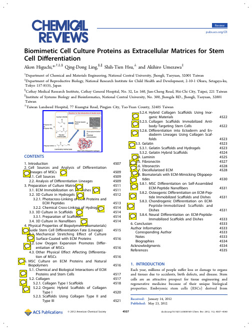
CONTENTS
1. Introduction 2. Cell Sources and Analysis of Differentiation Lineages of MSCs 2.1. Cell Sources 2.2. Analysis of Differentiation Lineages 3. Preparation of Culture Matrix 3.1. ECM Immobilization on 2D Dishes 3.2. 3D Culture in Hydrogels 3.2.1. Photocross-Linking of ECM Proteins and ECM Peptides 3.2.2. Chemical Cross-Linking of Hydrogels 3.3. 3D Culture in Scaffolds 3.3.1. Preparation of Scaffolds 3.4. 3D Culture in Nanofibers 4. Physical Properties of Biopolymers (Biomaterials) Guide Stem Cell Differentiation Fate (Lineage) 4.1. Mechanical Stretching Effect of Culture Surface-Coated with ECM Proteins 4.2. Low Oxygen Expansion Promotes Differentiation of MSCs 4.3. Other Physical Effect Affecting Differentiation of MSCs 5. MSC Culture on ECM Proteins and Natural Biopolymers 5.1. Chemical and Biological Interactions of ECM Proteins and Stem Cells 5.2. Collagen 5.2.1. Collagen Type I Scaffolds 5.2.2. Organic Hybrid Scaffolds of Collagen Type I 5.2.3. Scaffolds Using Collagen Type II and Type III
冬凌草甲素逆转硫代乙酰胺对破骨和成骨细胞分化的机制研究

骨质疏松症是21世纪的主要流行病之一,全球约有2亿人受到骨质疏松症的影响[1],其严重危害着人们的健康。
成骨细胞和破骨细胞活性失衡会导致骨骼脆性增加,骨折的发生率上升[2]。
近年来研究发现骨质流失与炎症疾病相关,如磨损颗粒可以诱导小鼠破骨细胞分化并增加骨溶解面积,造成炎性骨溶解[3]。
脂多糖(LPS )能够诱导过度炎症反应并增强破骨细胞活性引起骨丢失[4]。
硫代乙酰胺(TAA)常用于建立肝纤维化的动物模型。
TAA 不仅能够诱导巨噬细胞活化,也可以刺激促炎细胞因子和转录因子,促进核因子κB (NF-κB )p65和白细胞介素1β(IL-1β)的释放[5],引起氧化应激和炎症反应[6]。
先前的研究发现,TAA 与致炎因子LPS 一样能够引起炎性骨损伤[7],增强破骨细胞的活性,促进骨吸收[8]。
然而,TAA 对成骨分化的影响尚不清楚。
近年来,一些中药单体因其不良反应少和显著的抗炎作用[9-11],值得在抑制炎性骨损伤的预防和治疗中加以研究。
冬凌草甲素(ORI )是一种从冬凌草属植物中提取的四环二萜类化合物,在抗炎[12,13]和抗肿瘤[14,15]中有显Oridonin suppresses the effect of thioacetamide for promoting osteoclast differentiation of RAW264.7cells and inhibiting osteoblast differentiation of bone mesenchymal stem cellsJIN Xiaoli,XU Jia,CHEN Xuanwei,CHEN Jin,HUANG Hui,ZHANG Ting,REN Jun,XU JianSchool of Medical Technology and Information Engineering,Zhejiang Chinese Medical University,Hangzhou 310053,China摘要:目的探究冬凌草甲素(ORI )和硫代乙酰胺(TAA )对破骨分化和成骨分化的作用及其机制。
材料专业常用术语英语单词表
Unit 1property (材料的)性质heat treatme nt 热处理metal 金属glass 玻璃plastics 塑料fiber 纤维electro nic devices 电子器件component 组元,组分semic onducting materials 半导体材料materials scie nee and engin eeri ng 材料科学与工程materials scie nee 材料科学materials engin eeri ng 材料工程materials scie ntist 材料科学家materials engin eer 材料工程师syn thesize 合成syn thesissyn theticsubatomic structure 亚原子结构electro n 电子atom 原子nu clei 原子核nu cleusmolecule 分子microscopic 微观的microscope 显微镜n aked eye 裸眼macroscopic 宏观的specime n 试样deformati on 变形polished 抛光的reflect 反射magn itude 量级solid materials 固体材料mecha nical properties 力学性质load 载荷force 力elastic modulus 弹性模量stre ngth 强度electrical properties 电学性质electrical con ductivity 导电性dielectric con sta nt 介电常数electric field 电场thermal behavior 热学行为heat capacity 热容thermal con ductivity 热传导(导热性)magn etic properties 磁学性质magn etic field 磁场optical properties 光学性质electromag netic radiati on 电磁辐射light radiation 光辐射in dex of refract ion 折射率reflectivity 反射率deteriorative characteristics 劣化特性process ing 力口工performa nee 性能lin ear 线性的in tegrated circuit chip 集成电路芯片stre ngth 强度ductility 延展性deterioration 恶化,劣化mecha nical stren gth 机械强度elevated temperature 高温corrosive 腐蚀性的fabrication 制造Un it 2 chemical makeup 化学组成atomic structure 原子结构adva need materials 先进材料high-tech no logy 高技术smart materials 智能材料nanoengin eered materials 纟纳米工程材料metallic materials 金属材料nonl ocalized electr ons 游离电子con ductor 导体electricity 电heat 热tran spare nt 透明的visible light 可见光polished 抛光的surface 表面lustrous 有光泽的alumi num 铝silic on 硅alumi na 氧化铝silica 二氧化硅oxide 氧化物carbide 碳化物nitride 氮化物dioxide 二氧化物clay mi nerals 黏土矿物porcela in 瓷器ceme nt 水泥mecha nical力学行为behaviorceramic materials 陶瓷材料stiffness 劲度stre ngth 强度hard 坚硬brittle 脆的fracture 破裂in sulative 绝缘的resista nt 耐 .. 的resista nee 耐力,阻力,电阻molecular structures 分子结构chai n-like 链状backb one 骨架carb on atoms 碳原子low den sities 彳氐密度mecha nical characteristics 力学特性inert 隋性synthetic (人工)合成的fiberglass 玻璃纤维polymeric 聚合物的epoxy 环氧树脂polyester 聚酯纤维carbon fiber-rei nforced polymer composite 碳纤维增强聚合物复合材料glass fiber-rei nforced materials 玻璃纤维增强材料high-stre ngth, low-de nsity structural materials 高强度低密度结构材料solar cell 太阳能电池hydrogen fuel cell 氢燃料电池catalyst 催化剂nonren ewable resource 不可再生资源Un it 3 periodic table (元素)周期表atomic structure 原子结构magn etic 磁学的optical 光学的microstructure 微观结构macrostructure 宏观结构positively charged n ucleus 带正电的原子核atomic nu mber 原子序数proto n 质子atomic weight 原子量n eutro n 中子n egatively charged electr ons 带负电的电子shell壳层magn esium 镁chemical bonds 化学键partially-filled electro n shells 未满电子壳层bond 成键metallic bond 金属键nonm etal atoms 非金属原子covale nt bond 共价键ionic bond 离子键Un it 4physical properties 物理性质chemical properties 化学性质flammability 易燃性corrosi on 腐蚀oxidatio n 氧化oxidati on resista nee 抗氧化性vapor (vapour) 蒸汽,蒸气,汽melt 熔化solidify 凝固vaporize 汽化,蒸发condense 凝聚sublime 升华state 态plasma 等离子体phase tran sformatio n temperatures den sity 密度specific gravity 比重thermal con ductivity 热导lin ear coefficie nt of thermal expa nsion electrical con ductivity and resistivity 相变温度线性热膨胀系数电导和电阻corrosi on resista nee 抗腐蚀性magn etic permeability 磁导率phase tran sformatio ns 相变phase tran siti ons 相变crystal forms 晶型melt ing point 熔点boili ng point 沸腾点vapor pressure 蒸气压atm 大气压glass tran siti on temperature 玻璃化转变温度mass 质量volume 体积per un it of volume 每单位体积the accelerati on of gravity 重力加速度temperature depe ndent 随温度而变的,与温度有关的grams/cubic cen timeter 克每立方厘米kilograms/cubic meter 千克每立方米grams/milliliter 克每毫升grams/liter 克每升pounds per cubic inch 磅每立方央寸pounds per cubic foot 磅每立方央尺corrosi on resista nee 抗腐蚀性alcohol 酒精benzene 苯magn etize 磁化magn etic induction 磁感应强度magn etic field inten sity 磁场强度con sta nt 常数vacuum 真空magn etic flux den sity 磁通密度diamag netic 反磁性的factor 因数paramag netic 顺磁性的ferromag netic 铁磁性的non-ferrous metals 非铁金属,有色金属brass 黄铜ferrous 含铁的ferrous metals 含铁金属,黑色金属relative permeability 相对磁导率transformer 变压器,变换器eddy curre nt probe 涡流探针Un it 5hard ness 硬度impact resista neefracture tough nessstructural materialsani sotropic 各向异性orie ntati on 取向texture 织构 fiber rein forceme nt Ion gitudi nal 纵向tran sverse directi onshort tran sverse direction短横向 a function of temperature温度的函数,温度条件room temperature 室温elo ngatio n 伸长率tension 张力,拉力 compressi on 压缩ben di ng 弯曲shear 剪切torsio n 扭转static load ing 静负荷dyn amic loadi ng动态载荷 cyclic loading 循环载荷,周期载荷耐冲击性 断裂韧度,断裂韧性 结构材料 纤维增强 横向cross-sect ional area 横截面stress 应力stress distributi on 应力分布strain 应变engin eeri ng stra in 工程应变perpe ndicular 垂直no rmal axis 垂直车由elastic deformati on 弹性形变plastic deformati on 塑性形变quality con trol 质量控制non destructive tests 无损检测tensile property 抗张性能,拉伸性能Unit 6lattice 晶格positive ions 正离子a cloud of delocalized electr ons 离域电子云ionization 电离,离子化metalloid 准金属,类金属nonm etal 非金属cross-sect ional area 横截面diago nal line 对角线polonium 钋semi-metal 半金属lower left 左卜万upper right 右上方con ducti on band 导带vale nee band 价带electro nic structure 电子结构syn thetic materials (人工)合成材料oxygen 氧oxide 氧化物rust 生锈potassium 钾alkali metals 碱金属alkali ne earth metals 碱土金属volatile 活泼的tran siti on metals 过渡金属oxidize 氧化barrier layer 阻挡层basic 碱性的acidic 酸性的electrochemical series 电化序electrochemical cell 电化电池cleave 解理,劈开elemental 兀素的,单质的metallic form 金属形态tightly-packed crystal lattice 密排晶格,密堆积晶格atomic radius 原子半径nu clear charge 核电荷nu mber of bonding orbitals 成键轨道数overlap of orbital en ergies 轨道能重叠crystal form 晶型pla nes of atoms 原子面a gas of n early free electr ons 近自由电子气free electro n model 自由电子模型an electr on gas 电子气band structure 能带结构binding energy 键能positive pote ntial 正势periodic pote ntial 周期性势能band gap 能隙Brillouin zone 布里渊区n early-free electr on model 近自由电子模型solid solution 固溶体pure metals 纯金属duralumin 硬铝,杜拉铝Unit 9purification 提纯,净化raw materials 原材料discrete 离散的,分散的iodi ne 碘Ion g-cha in 长链alkane 烷烃,链烃oxide 氧化物nitride 氮化物carbide 碳化物diam ond 金刚石graphite 石墨inorganic 无机的mixed ion ic-covale nt bonding 离子一共价混合键con stitue nt atoms 组成原子con ducti on mecha nism 传导机制phonon 声子phot on 光子sapphire 蓝宝石visible light 可见光computer-assisted process con trol 计算机辅助过程控制solid-oxide fuel cell 固体氧化物燃料电池spark plug in sulator 火花塞绝缘材料capacitor 电容electrode 电极electrolyte 电解质electr on microscope 电子显微镜surface an alytical methods 表面分析方法Unit 12macromolecule 高分子repeati ng structural un its 重复结构单元covale nt bond 共价键polymer chemistry 高分子化学polymer physics 高分子物理polymer scie nee 高分子科学molecular structure 分子结构molecular weights 分子量long cha ins 长链cha in-like structure 链状结构mono mer 单体plastics 塑料rubbers 橡胶thermoplastic 热塑性thermoset 热固性vulca ni zed rubbers 硫化橡胶thermoplastic elastomer 热塑弹性体n atural rubbers 天然橡胶syn thetic rubbers 合成橡胶thermoplastic 热塑性thermoset 热固性resi n 树脂polyethyle ne 聚乙烯polypropyle ne 聚丙烯polystyre ne 聚苯乙烯polyvi nyl-chloride 聚氯乙烯polyvi nyl 聚乙烯的chloride 氯化物polyester 聚酉旨polyuretha ne 聚氨酉旨polycarbo nate 聚碳酸酯nylon 尼龙acrylics 丙烯酸树脂aery Ion itrile-butadie ne-styre ne ABS 树月脂polymerization 聚合(作用)conden sati on polymerizati on 缩聚additi on polymerizatio n 力口聚homopolymer 均聚物copolymer 共聚物chemical modificati on 化学改性termino logy 术语nomen clature 命名法chemist 化学家the Noble Prize in Chemistry 诺贝尔化学奖catalyst 催化剂atomic force microscope原子力显微镜(AFM)Unit 15composite 复合材料multiphase 多相bulk phase 体相matrix 基体matrix material 基质材料rei nforceme nt 增强体reinforcing phase 增强相rei nforci ng material 力口强材料metal-matrix composite 金属基复合材料ceramic-matrix composite 陶瓷基复合材料resi n-matrix composite 树脂基复合材料stre ngthe ning mecha nism 增强机理dispers ion stren gthe ned composite 弥散强化复合材料particle rei nforced composites 颗粒增强复合材料fiber-re in forced composites 纤维增强复合材料Unit 18nano tech no logy 纟纳米技术nano structured materials 纟纳米结构材料nano meter 纟纳米nano scale 纟纳米尺度nan oparticle 纟纳米颗粒nano tube 纟纳米管nanowire 纟纳米线matrix material 基质材料nanorod 纟纳米棒nanoonion 纟纳米葱nan obulb 纳米泡fullere ne 富勒烯size parameters 尺寸参数size effect 尺寸效应critical le ngth 临界长度mesoscopic 介观的qua ntum mecha nics 量子力学qua ntum effects 量子效应surface area per un it mass 单位质量的表面积surface physics and chemistry 表面物理化学substrate 衬底,基底graphe ne 石墨烯chemical an alysis 化学分析chemical compositi on 化学成分an alytical tech niq ues 分析技术sca nning tunn eli ng microscope 扫描隧道显微镜spatial resoluti on 空间分辨率de Brogile wavele ngth 德布罗意波长mean free path of electrons (电子)平均自由程qua ntum dot 量子点band gap 带隙连续态密度discrete en ergy level 离散能级con ti nu ous den sity of statesabsorpti on 吸收in frared 红夕卜ultraviolet 紫外visible 可见qua ntum confin eme nt (effect) 量子限域效应qua ntum well 量子势阱optoelectro nic device 光电子器件en ergy spectrum 能谱electr on mean free path 电子平均自由程spin relaxati on len gth 自旋弛豫长度Unit 21biomaterial 生物材料impla nt materials 植入材料biocompatibility 生物相容性in vivo 在活体内in vitro 在活体外organ tran spla nt 器管移植calcium phosphate 磷酸钙hydroxyapatite 羟基磷灰石research and development 研发R&DPreparati on & Characterizati onprocess ing tech niq ues 力口工技术cast ing 铸造rolling 轧制,压延weld ing 焊接ion impla ntati on 离子注入thin-film depositi on 薄膜沉积crystal growth 晶体生长sin teri ng 烧结glassblowi ng 玻璃吹制an alytical tech niq ues 分析技术characterizati on tech niq ues 表征技术electr on microscopy 电子显微术X-ray diffractio n X 射线衍射calorimetry 量热法Rutherford backscatteri ng 卢瑟福背散射n eutro n diffractio n 中子衍射nu clear microscopy 核子微探针。
重组大肠杆菌生产核苷磷酸转移酶的发酵条件优化
重组大肠杆菌生产核苷磷酸转移酶的发酵条件优化沈爱萍。
何宝龙。
孙丽慧。
郑裕国(浙江工业大学生物工程研究所,浙江杭州310014)摘要:核苷磷酸转移酶能够特异地将无机焦磷酸(PPi)的磷酸根转移到核苷5’一位的羟基上,该过程并不需要A T P的参与,且具有选择性高和反应条件温和等特点。
以实验室前期构建的重组大肠杆菌E.∞f f B L21(D E3)/pE T28b—A P/P T为茵株,进行了5L发酵罐中的培养条件优化,确定了以乳糖作为诱导剂时,最佳诱导时机oD鲫为7左右,诱导剂乳糖的浓度为10g/L;确定了最适通气量为1.5L/m i n。
在该条件下,核苷磷酸转移酶酶活达221.4U/L,O D600为20.4。
关键词:重组大肠杆茵;核苷磷酸转移酶;发酵;条件优化O pt i m i zat i on of f e r m e nt a t i on condi t i ons f or t he pr oduct i on ofphosphot r ansf e r as e by E sc her i chi a col iS H E N A i—pi ng,H E B ao—l ong,SU N L i—hui,Z H EN G Y u—guo(I nst i t ut e of B i oengi neer i ng,Zhej i ang U ni vers i t y of t e chnol ogy,H angzhou310014,C hi na)A bst r ac t:P hos phot r ans f e r as e,poss ess ed hi gh r egi o s peci f i ci t y of t he nucl eosi de phosphor yl at i ng act i vi t y t o t he C-57posi t on,i s of hi gh pot ent i al f or t he pr oduct i on of i nos i ne-5'-m onophosphat e due t o m i l d r eact i on condi t i ons.I n t hi s pa pe r,r ecom bi nant E c ol iB L21(D E3)/pE T28b-A P/PT w hi ch has e xpr e ss ed ac i d phos phat a se s w as use d,and t he ef fect s of cul t ur e condi t i ons o n ce l l gr ow t h and enzy m e act i vi t y w e r e i nves t i gat ed i n5L f e r m e nt er.T he opt i m al condi t i ons of cul t ur e w e r e as f ol l o w i ng:i ndu ct i on t i m e w as at O D c劬o=7,t he l act os e concent r a t i on w as of10g/L,vent i l at i on vol um e w as of1.5L/m i n.U nder t he cul t ur e condi t i ons,t he enzym e act i vi t y and O D600c oul d r ea ch221.4U几and20.4.r es pect i vel y.K ey w or ds:r ec om bi na nt E.col i;phosphot r ansf er ase;f er m ent at i on;opt i m i zi ng condi t i ons以57一肌苷酸(5'-I M P)为代表的呈味核苷酸,是新一代食品增鲜剂,常与5’一鸟苷酸(5’一G M P)按1:1混合而成。
如何获得蛋白质的开发阅读框
如何获得蛋白质的开发阅读框
蛋白质的开放阅读框(ORF)是指在DNA或RNA序列中,能够被
转录和翻译成蛋白质的部分。
要获得蛋白质的开放阅读框,首先需
要对DNA或RNA序列进行分析。
以下是一些常见的方法:
1. 手动分析,可以通过手动检查DNA或RNA序列,寻找起始密
码子(通常是ATG)和终止密码子(TAA,TAG或TGA)来识别ORF。
然而,这种方法对于长序列或大量数据来说非常耗时且容易出错。
2. 使用生物信息学工具,生物信息学工具如NCBI的ORF Finder、EMBOSS和ExPASy的Translate工具等可以帮助自动识别ORF。
这些工具可以搜索输入的核酸序列,找到可能的ORF,并提供
相关的翻译蛋白质序列。
3. 基因组学数据库,许多基因组学数据库(如GenBank、Ensembl等)提供了已经预测和注释好的ORF信息,可以直接查询
这些数据库来获取感兴趣的蛋白质的ORF信息。
4. 实验方法,除了计算方法外,还可以通过实验方法来验证ORF的存在,例如利用原核生物或真核生物的细胞系来表达候选ORF,
然后通过蛋白质质谱等技术来鉴定和验证蛋白质的存在。
总的来说,获得蛋白质的开放阅读框可以通过手动分析、生物信息学工具、基因组学数据库和实验方法来实现。
选择合适的方法取决于具体的研究目的和实验条件。
不同酶消化法提取猪原代肝细胞的效果比较
532024.4·试验研究0 引言猪圆环病毒(PCV )是Circoviridae 科Circovirus 属的一种无囊膜的单链环状DNA 病毒。
在已知的4个血清型中,PCV2为猪易感的致病性病毒[1]。
PCV2感染会诱导宿主免疫抑制引起猪圆环病毒病(PCVD ),包括断奶仔猪多系统衰竭综合征、新生仔猪先天性脑震颤、皮炎与肾病综合征、猪呼吸道病综合征、母猪繁殖障碍等,给全世界养猪业带来较大的经济损失,是世界各国的兽医与养猪业者公认的造成重大影响的猪传染病[2]。
PCV2的感染在猪生长发育的不同阶段有不同的组织嗜性。
但无论是胎儿阶段还是出生后,肝细胞都是PCV2感染和复制的靶细胞。
因此,PCV2也被视为一种能够诱导猪肝炎的病毒[3]。
且PCV2诱导的肝细胞凋亡在PCV2引发的相关病变和疾病的发病机制中具有关键性作用[4]。
因此,方便、快捷地获取大量有活性的猪肝细胞对于研究PCVD 的致病机制具有重大意义。
目前获取肝细胞常用的方法主要包括机械分离细胞法、非酶分离细胞法、离体酶消化法和酶灌流法等[5]。
因此,本试验采用简便、经济、无需特殊设备、仅需部分肝组织的离体酶消化法,比较不同酶消化分离猪原代肝细胞的效果,为一般实验室提取分离大量有活性的猪肝细胞提供参考。
1 材料与方法1.1 材料1.1.1 主要试剂新鲜猪肝组织,Hank's 平衡盐溶液(HBSS ),磷酸盐缓冲液(无菌PBS ),4%多聚甲醛(PFA ),收稿日期:2024-01-27基金项目:国家自然科学基金项目:复杂器官与组织在脾脏内的功能性再生(32230056)作者简介:周徐倩(1999-),女,汉族,浙江温州人,硕士在读,研究方向:组织工程与再生医学。
*通信作者简介:董磊(1978-),男,汉族,安徽阜阳人,博士,教授,研究方向:组织工程与再生医学、生物材料。
周徐倩,董磊.不同酶消化法提取猪原代肝细胞的效果比较[J].现代畜牧科技,2024,107(4):53-55. doi :10.19369/ki.2095-9737.2024.04.014. ZHOU Xuqian ,DONG Lei .Comparison of the Effect of Different Enzyme Digestion Methods on Extraction of Porcine Primary Hepatocytes[J].Modern Animal Husbandry Science & Technology ,2024,107(4):53-55.不同酶消化法提取猪原代肝细胞的效果比较周徐倩,董磊*(南京大学,江苏 南京 210023)摘要:猪肝细胞是猪圆环病毒的靶细胞,简单快速地提取猪原代肝细胞对于研究猪圆环病毒病的致病机制具有重要意义。
Cell Viability Analysis说明书

Cell V iability A nalysis u sing C alcein A M, P ropidium I odide a nd H oechstOvernightVariable 30 m in.10 m in5 m in.30 m inExperimental S etupMaterials:•A549 C ells•A549 g rowth m edia: E MEM + 10% F BS + 1% N EAA + 1% G lutaMax •Calcein A M, 20 x 50 µg (Life T echnologies, C at# C 3100MP)•Propidium I odide, 1 m g/mL s olution i n w ater (Life T echnologies, C at# P 3566)•Hoechst 33342, 10 m g⁄mL s olution i n w ater (Life T echnologies, C at# H 3570)•DMSO•Greiner 96-‐well p late C at# 655090Protocol:1.Reconstitute C alcein A M b y a dding 50.2 μl o f D MSO i n o ne o f t he 50 µg v ial o f l yophilized C alcein A M.This w ill r esults i n a 1 m M C alcein A M s olution.2.Seed 8000-‐10000 c ells/well i n 96-‐well p late i n a f inal v olume o f 100 μl/well.3.Incubate c ells o vernight a t 5% C O2, 37 ° C.Seed cellsTreat w ith d rugAdd L ive, D ead and T otal D yesImage o n C eligoAnalyze c ell imagesProduce g raphs and d ataApplication Cell v iability a nalysis u sing C alcein A M, P ropidium I odide a nd H oechst o n A 549 c ells. DescriptionThis p rotocol d escribes t he a nalysis o f c ell v iability o n A 549 c ells u sing C alcein A M a s a measure o f e nzymatic a ctivity i n l ive c ells, P ropidium I odide a s a m easure o f d ead c ells with c ompromised m embrane i ntegrity a nd H oechst a s a m arker f or a ll n ucleated c ells. The a pplication r eports t he c ounts a nd p ercentages o f l ive, d ead a nd t otal n umbers o f cells.Celigo A pplication Cell V iability (Live + D ead + T otal).Plate T ype Greiner c at# 655090 b lack w all c lear b ottom 96-‐well P late.Major StepsDay 1 Day 24.In t he e xample p rotocol p rovided h ere, w e u se h ydrogen p eroxide t o i nduce c ell d eath. B elow i s t heplate m ap f or h ydrogen p eroxide c oncentration. C oncentration s hould b e a djusted f or a ny o ther d rug.Skip t o p aragraph 6 i f u sing a d ifferent d rug o r p late m ap.5.The a ppropriate w ell c oncentrations w ere o btained b y p reparing a 2x c oncentration o f t he d ilutioncurve a s 100 μl o f d rug w ill b e a dded t o 100 μl o f m edia i n t he w ells. D ilution s teps w ere o btained a sfollow:a.Prepare a 1M s olution o f h ydrogen p eroxide b y a dding 500 μl o f 3% h ydrogen p eroxide t o4.4 m L o f w ater.b.Prepare a 100 m M s olution o f h ydrogen p eroxide b y a dding 400 μl f rom t he p revious s tep t o3.6 m L o f c ulture m edia.ing a s eries o f 12 t ubes, p repare a s erial d ilution b y d iluting h ydrogen p eroxide i n m ediaaccording t o t he f ollowing t able.6.Drug c ompound t reatmenta.Add 100 μl o f c ompound a t 2x d esired f inal c oncentration.b.After d esired t reatment t ime (4 h ours f or h ydrogen p eroxide), w ash w ells w ith m edia t wice a ndleave 100 μl v olume i n w ells.7.Prepare C alcein A M, P ropidium I odide a nd H oechst m ixed d ye s olution.a.12 m L o f m ixed d yes s olution i s r equired f or s taining a ll t he w ells o f a 96-‐well p late. A djustaccordingly i f n ot a ll w ells a re s tained.e t he f ollowing d ilution t able t o p repare t he m ixed d ye s olutionDyeStockconcentrationRecommendedconcentrationRecommendeddilution f actorVolume t o a ddto 12 m LmediaConcentrationrange f or o thercell t ypesCalcein A M 1 m M 1 μM 1000x 12 μl 0.1 – 10 μMPropidiumIodide 1 m g/mL 2 μg/mL 500x 24 μl 0.1 – 5 μg/mL Hoechst 33342 10 m g/mL 5 μg/mL 2000x 6 μl 1 – 10 μg/mLc.Remove m edia f rom a ll w ells a nd a dd 100 μl o f m ixed d yes s olutiond.Incubate c ells f or 30 m in a t 5% C O2, 37 °C.e.Wash w ells w ith m edia t wice a nd l eave 100 μl v olume i n w ells.f.As c ells a re l ive i n t he w ells, t he p late s hould b e i maged i mmediately o n t he C eligo.Celigo S etupStart T ab:1.Create a n ew s can a nd n ame f ile a ppropriately.2.If e xperimental s ettings h ad b een p reviously o ptimized t hen s elect t hese t o b e u sed.Scan T ab:1.Select C eligo A pplication: C ell V iability (live + D ead + T otal).2.Setup L ive C hannela.Select G reen i llumination f or C alcein A M s tain.b.Exposure t ime s hould b e a round 10,000 μs (Gain 0).3.Setup D ead C hannela.Select R ed i llumination f or P ropidium I odide s tain.b.Exposure t ime s hould b e a round 40,000 μs (Gain 0).4.Setup T otal C hannela.Select B lue i llumination f or H oechst s tain.b.Exposure t ime s hould b e a round 100,000 μs (Gain 0).5. Register H ardware a utofocus u sing t he B lue / H oechst c hannel.6. Switch t o G reen / C alcein A M c hannel, “Find f ocus” a nd “Set O ffset”.7. Switch t o R ed / P ropidium I odide c hannel, “Find f ocus” a nd “Set O ffset”.8.Select w ells t o a cquire a nd “Start S can”.Analyze T ab:The C eligo C ell V iability a pplication s egments a ll t hree f luorescent c hannels. F luorescent o bjects w ill b e identified i n e ach c hannel. G reen a nd R ed o bjects (for l ive a nd d ead c ells) w ill b e c ounted o nly i f t hey a re super-‐imposed w ith a b lue f luorescent o bject (for n uclear s tain). T herefore, s egmented g reen a nd r ed objects (such a s f luorescent d ebris) n ot a ssociated w ith a b lue n ucleus w ill b e r ejected f rom t he a nalysis.1. Select “Well M ask” a nd “Automatic” t o e xclude t he o uter p art o f t he w ell.2. Select “Fluorescence” a lgorithm f or a ll 3 c hannels.3. Adjust t he “Intensity T hreshold” t o e nsure p roper i dentification o f t he n ucleus. D efault v alue o f “4”with “High P recision” w orks w ell f or m ost f luorescent s tains. V alues o f “2” t o “3” c an b e u sed f or dimmer s amples.4. Keep t he “Cell D iameter” b etween “8” a nd “10”.5. Select “Separate T ouching O bjects” i n t he T otal c hannel t o e nsure p roper s eparation o f n uclei i nclose p roximity.Live Dead TotalLive Dead Total6. Expected S egmentation:a. Use t he “Image D isplay” a nd “Graphic O verlay” c ontrols t o v isualize s egmentation.b. Typical i mages a nd f luorescent o bjects i dentification s hould l ook a s f ollow:I m a g e sS e g m e n t a t i o nLive Dead Total7. Select w ells t o a nalyze a nd “Start A nalyze”.Gate T ab:The C ell v iability a pplication d oes n ot u se g ating. I nstead, q uantification o f l ive a nd d ead c ells i s p re-‐defined in t he a pplication a nd a vailable d irectly i n t he R esult T ab.Result T ab:1. Typical R esults f or t he p ercentage o f l ive c ells.Hydrogen P eroxide (12 m M)Control – N o T reatmentHydrogen P eroxide C onc. R esponseLive + D ead + T otal Segmentation O verlaysLive c ell w ith g reen o verlay s uper-‐imposing a b lue n ucleus o verlay Dead c ell w ith r ed o verlay s uper-‐imposing a b lue n ucleus o verlay Green o verlay d ismissed f rom a nalysis because n ot s uper-‐imposing a b lue nucleus o verlayRed o verlay d ismissed f rom a nalysis because n ot s uper-‐imposing a b lue nucleus o verlayData A nalysisThe e xample p rovided i n t his p rotocol u ses A 549 c ells t reated w ith a c oncentration r esponse c urve o f h ydrogen peroxide, a n i nducer o f c ell d eath. A s h ydrogen p eroxide c oncentration i ncreases, w e e xpect t o a d ecrease o f the p ercentage o f l ive c ells a nd a n i ncrease p ercentage o f d ead c ells.Data E xport1. Export t he d ata b y s electing “Export W ell-‐Level D ata”2. Save t he “.csv” f ile t o y our d esktop.3. Open t he f ile w ith M icrosoft E xcel.4. The d ata i s o rganized a ccording t o t he p late m ap f or e ach o f l ive a nd d ead c ells c lasses.Graphing1. Generate a “Scatter p lot” u sing M icrosoft E xcel u sing t he d rug c oncentration f or t he X -‐axis a nd 2series f or t he l ive a nd d ead c ells. I n t his e xample, t he a verage o f 4 d ata p oints w ere p lotted. 2. You s hould e xpect t he f ollowing r esults:% L ive 123456789101112A 86.92%90.05%90.93%94.20%92.86%91.73%0.10%0.02%0.04%0.07%0.03%0.00%B 95.30%97.64%95.07%95.69%96.80%96.68%0.38%0.27%0.29%0.13%0.23%0.04%C 95.61%96.69%95.89%96.54%97.51%97.42%0.32%0.60%0.48%0.27%0.28%0.12%D 96.62%97.89%96.10%98.13%97.79%97.59%0.23%0.19%0.32%0.18%0.12%0.07%E 0.02%0.95% 2.21% 4.31%8.40%80.73%97.26%97.94%97.27%92.94%93.37%95.46%F 0.04%0.57% 1.75% 3.77%8.54%82.48%97.52%97.70%96.77%90.97%94.21%95.11%G 0.08%0.59% 1.58%7.89%72.01%96.78%97.85%97.52%84.33%79.70%86.01%94.15%H 0.01%0.17%0.90% 1.93%15.78%54.26%97.22%97.73%87.77%86.77%83.21%95.14%% D ead 123456789101112A 1.05% 1.05% 1.19% 1.47% 1.57% 1.44%81.08%80.68%79.42%79.08%80.68%78.34%B 1.11% 1.18% 1.33% 1.33% 1.10% 1.18%74.68%76.62%77.77%74.93%77.09%78.48%C 1.01%0.89%0.84% 1.20% 1.36% 1.50%71.45%72.90%70.81%72.09%75.67%79.03%D 0.97%0.92% 1.14% 1.06% 1.19%0.93%67.78%72.30%72.66%71.54%75.25%80.36%E 80.55%81.84%87.48%88.49%86.34%17.13% 1.61% 2.19% 1.83% 1.49% 1.17% 1.13%F 78.30%81.28%86.58%89.36%86.27%15.85% 1.95% 1.86% 1.16%0.94% 1.03%0.79%G 84.44%88.56%91.97%86.97%22.62% 1.39% 1.43% 1.64% 1.03%0.76%0.94%0.88%H82.50%86.32%87.84%92.38%78.13%39.31%2.90%2.61%1.57%0.77%1.10%1.10%0%20% 40% 60% 80% 100% 110 P e r c e n t a g e o f T o t a lHydrogen P eroxide (mM)Live a nd D ead p ercentages o f A 549 c ells t reatedwith H ydrogen P eroxide% L ive % D eadVariations o f t he C eligo C ell V iability a pplicationThree o ther v ariants o f t he C ell V iability a pplication e xist i n t he C eligo s oftware. A ll t hree o f t hem a re2 c hannels a pplications r epresenting a s ubset o f t he3 c hannel p rotocol d escribe h ere. I n t his v ariants, t hesettings f or t he L ive, D ead a nd T otal c hannels r emain t he i dentical t o t he 3 c hannels a pplication.Additionally, t he T otal c hannel c an b e c hanged f rom u sing a b lue n uclear d ye t o b rightfield w here t he c ells can a lso b e c ounted. T he f ollowing t able p rovides d etails f or e ach a pplication.Cell V iabilityLive c hannel Dead C hannel Total C hannel Application o utputs ApplicationLive + D ead + T otal Green Red Blue o r B rightfield % L ive, % D ead, T otal c ounts Live + T otal Green Not A vailable Blue o r B rightfield % L ive, T otal c ountsDead + T otal Not A vailable Red Blue o r B rightfield % D ead, T otal c ountsLive + D ead Green Red Not A vailable % L ive, % D ead, T otal c ountsNote: A dditional o utputs a re r eported b y t he C ell V iability a pplication. R efer t o C ell V iability u ser g uide f or a c omplete l istof o utputs.Reference•Chen H Y, Y ang Y M, S tevens B M, N oble M. E MBO M ol. M ed (2013). I nhibition o f r edox/Fyn/c-‐Cbl pathway f unction b y C dc42 c ontrols t umour i nitiation c apacity a nd t amoxifen s ensitivity i n b asal-‐like breast c ancer c ells.。
北京大学成功研发染色质免疫沉淀测序技术

北京大学成功研发染色质免疫沉淀测序技术
佚名
【期刊名称】《生物学教学》
【年(卷),期】2015(40)2
【摘要】据2014年9月10日《科学网》援引报道,北京大学生物动态光学成像中心黄岩谊课题组与汤富酬课题组合作,发展了一种基于微流控芯片的微量细胞样品处理与核酸俘获方法,并成功实现了针对1000个细胞的染色质免疫沉淀测序(Ch IP-Seq)。
研究结果在线发表在《细胞研究》上。
应用Ch IP-Seq方法,通过有针对性地俘获和富集特定蛋白,得到与这些蛋白结合的DNA片段,
【总页数】1页(P74-74)
【关键词】染色质免疫沉淀;测序技术;北京大学;研发;微流控芯片;DNA片段;光学成像;样品处理
【正文语种】中文
【中图分类】Q786
【相关文献】
1.染色质免疫沉淀ChIP技术检测传代培养细胞Grb1O组蛋白乙酰化水平 [J], 郑芳芳;姚建凤;黄荣富
2.染色质免疫沉淀技术(ChIP)在基因转录调控中的作用 [J], 史双林
3.染色质免疫沉淀技术分析红藻氨酸处理后的大鼠大脑皮质神经元中p53与p21基因启动子的结合 [J], 贾玉红;马天舒;姜妙娜;赵海东
4.染色质免疫沉淀技术分析HePG2细胞TCF7L2蛋白与IDE基因转录启动子的结
合 [J], 许丹;任伟;郑晓雅;毕健琨
5.染色质免疫沉淀技术及其应用 [J], 王远锏
因版权原因,仅展示原文概要,查看原文内容请购买。
单细胞富集 ucell的原理

单细胞富集 ucell的原理单细胞富集(Single-cell enrichment)是一种用于分离和富集单个细胞的技术。
单细胞富集的原理是通过对细胞进行特定的分选,使得目标细胞能够被有效地分离和富集出来,以进行后续的单细胞分析。
在单细胞研究中,细胞数量有时非常有限,特别是针对罕见细胞类型或少数细胞的研究。
因此,需要一种高效、准确、可靠的方法来获取这些细胞并进行进一步的分析。
单细胞富集技术应运而生,为单细胞研究提供了重要的工具。
单细胞富集的原理可以分为两个主要步骤:细胞分选和细胞富集。
细胞分选是指将目标细胞从混合细胞组织中分离出来。
常用的细胞分选方法包括流式细胞术(flow cytometry)、磁力珠分选(magnetic bead sorting)和微流控技术(microfluidics)。
这些方法通过对细胞的某些特定性质进行标记或分离,如细胞表面标记物(surface markers)、细胞大小或细胞形态等,来实现对目标细胞的分选。
其中,流式细胞术是最常用的方法之一,通过利用细胞表面标记物的差异,使用荧光染料进行标记,并通过流式细胞仪的高速流动性将目标细胞从混合细胞中迅速分离出来。
细胞富集是指将分选得到的目标细胞集中到一个小体积的区域中。
常用的细胞富集方法包括单细胞吸管(single-cell pipetting)、微流控芯片(microfluidic chip)和电泳等。
这些方法可以有效地将目标细胞从大量的非目标细胞中分离出来,从而提高目标细胞的浓度和纯度。
其中,微流控芯片是一种常用的细胞富集方法,通过微流控技术的精确控制,可以将目标细胞定位到特定的位置,并实现单细胞的富集。
单细胞富集技术在许多领域都有广泛的应用。
在生物医学研究中,单细胞富集可以用于研究罕见细胞类型,如干细胞和肿瘤干细胞,以及特定细胞亚群的功能和表型。
在临床诊断中,单细胞富集可以用于早期癌症检测、预测疾病进展和治疗反应等。
多糖的铜藻抗氧化应激的巨噬细胞的保护作用
International Journal of Biological Macromolecules 68(2014)98–106Contents lists available at ScienceDirectInternational Journal of BiologicalMacromoleculesj o u r n a l h o m e p a g e :w w w.e l s e v i e r.c o m /l o c a t e /i j b i o m acProtective effect of polysaccharides from Sargassum horneri against oxidative stress in RAW264.7cellsZheng-Shun Wen ∗,Li-Jia Liu,Xiao-Kun OuYang,You-Le Qu,Yin Chen,Guo-Fang DingZhejiang Provincial Key Engineering Technology Research Center of Marine Biomedical Products,Food and Pharmacy College,Zhejiang Ocean University,Zhoushan 316000Zhejiang,Chinaa r t i c l ei n f oArticle history:Received 9March 2014Received in revised form 26March 2014Accepted 16April 2014Available online 24April 2014Keywords:Sargassum horneri polysaccharides RAW264.7cellsAntioxidant activitya b s t r a c tThis study was designed to investigate chemical composition and the protective effects of polysaccharides isolated from Sargassum horneri against hydrogen peroxide (H 2O 2)-induced oxidative injury in RAW264.7cells.Results showed that isolated polysaccharides (SHS c )and the major fractions (SHS 1,SHS 0.5)con-tained sulfate ester,and SHS 1was high fucose-containing sulfated polysaccharide.After preincubation with three isolated polysaccharides,RAW264.7cells viability were significantly restored and decreased in cellular LDH release (P <0.05).SHS 1and SHS 0.5decreased intracellular ROS level,intracellular NO and malonic dialdehyde (MDA)level (P <0.05),restoring activities of superoxide dismutase (SOD)and glutathione peroxidase (GSH-Px)(P <0.05),decreasing inducible nitric oxide synthase (iNOS)(P <0.05).Moreover,preincubation of SHS 1with RAW264.7cells resulted in the increase of the gene expression level of endogenous antioxidant enzymes such as MnSOD and GSH-Px (P <0.05).These results clearly showed that SHS c and its fractions could attenuate H 2O 2-induced stress injury in RAW264.7cells,and a similar efficiency in protecting RAW264.7cells against H 2O 2-induced oxidative injury between SHS 1and Vitamin C.Taken together,our findings suggested that SHS 1can effectively protect RAW264.7cells against oxidative stress by H 2O 2,which might be used as a potential natural antioxidant in the functional food and pharmaceutical industries.©2014Elsevier B.V.All rights reserved.1.IntroductionOxidative stress has become focus of interest in most biomedical disciplines and many types of clinical research.Increasing evidence from research on several diseases show that oxidative stress is associated with the pathogenesis of diabetes,obesity,cancer,aging,inflammation,neurodegenerative disorders,hypertension,apoptosis,cardiovascular diseases,and heart failure [1].Oxidative stress causes a complex dysregulation of cell metabolism and cell–cell homeostasis,producing large amounts of reactive oxygen species (ROS).ROS is a collective term describing the chemical species that are formed upon incomplete reduction of oxygen.It includes superoxide anion (O 2−),hydrogen peroxide (H 2O 2),and hydroxyl radical (HO)that attack biomolecules such as proteins,lipid and DNA [2].Moreover,inducible nitric oxide synthase (iNOS)is induced in diseases associated with inflammation and oxidative stress [3,4],generating large amounts of NO that have cytostatic∗Corresponding author.Tel.:+865802554781;fax:+865802554781.E-mail addresses:wenzhengshun@ (Z.-S.Wen),77280116@ (Y.-L.Qu).effects on parasitic target cells by inhibiting iron-containing enzymes and causing DNA fragmentation.Antioxidants may have a positive effect on human health since they can protect human body against deterioration by ROS.Intracellular ROS is influ-enced by a number of endogenous and exogenous processes,and controlled by several radical scavenging enzymes.Cells possess cellular defense system including nonenzymatic and enzymatic substance such as superoxide dismutase (SOD)and glutathione peroxidase (GSH-Px)to scavenge radicals and protect oxidative damage to major cellular biomolecules [5–7].When the cellular defense mechanisms are unable to cope with excessive generation of ROS,this oxidative stress has been shown to be involved in var-ious pathogenic processes,therefore,treatment with antioxidant might theoretically retard spreading of cell damage and improve intracellular antioxidant enzymatic and nonenzymatic system.Many species of seaweed (marine macroalgae)are used as food and they have also found use in traditional medicine because of their perceived health benefits.S.horneri ,a Sargassaceae brown alga,is one of the main components in the subtidal seaweed flora along the Chinese warmer water coast.S.horneri has been used as fundamental source of human food and drug in tradi-tional Chinese medicine for centuries.In the previous studies,the/10.1016/j.ijbiomac.2014.04.0370141-8130/©2014Elsevier B.V.All rights reserved.Z.-S.Wen et al./International Journal of Biological Macromolecules68(2014)98–10699sulfated polysaccharide and other water-soluble natural extracts from S.Horneri exerted potent antiviral[8]and antioxidant activity evaluated by examining the radical scavenging activity on1,1-diphenyl-2-picrylhydrazyl(DPPH),hydroxyl and alkyl radicals[9]. One particularly interesting feature of the seaweed is its richness in polysaccharides.So far,little study of the antioxidant activ-ity of the sulfated polysaccharides isolated from S.horneri has been performed in cellular system which is closer to a living sys-tem.In addition,the structure characteristics are unclear,and to carry out this research contributes to an understanding of its structure–activity relationship.The aim of the present study is to investigate the partial chemical characteristic of the sulfated polysaccharides isolated from marine brown algae S.horneri,and to evaluate its protective effects against hydrogen peroxide(H2O2)-induced oxidative damage in murine macrophages RAW264.7cells.2.Materials and methods2.1.Chemicals and reagentsDulbecco’s modified Eagle’s medium(DMEM),peni-cillin/streptomycin,and the other materials required for culturing of cells were purchased from Gibco BRL,Life Technologies (Grand Island,NY,USA).H2O2,dimethylsulfoxide(DMSO),3-(4,5-dimethylthiazol-2-yl)2,5-diphenyltetrazolium bromide(MTT), Vitamin C,bovine serum albumin(BSA)and were obtained from Sigma(St.Louis,MO,USA).The reagent kits for measurement of levels of lactate dehydrogenase(LDH),malonic dialdehyde(MDA), NO,SOD,GSH-Px and iNOS were purchased from Nanjing Institute of Jiancheng Bioengineering(Nanjing,China).Trizol,Power SYBR®Master Mix(2×)and1st-Strand cDNA Synthesis Kit were from Invitrogen,USA;diethylpyrocarbonate(DEPC)and ribonuclease inhibitor were from Biobasic,Canada.All other chemicals were of analytical grade or of the highest grade available commercially.2.2.Isolation and purification of the polysaccharide from Sargassum horneriSargassum horneri(Turner)C.Agardh(abbreviated as S.horneri) was collected at Zhoushan islands in Zhejiang province,China in May2012.Dried algae were dipped into20volumes of distilled water and kept at room temperature for2h.The algae were then homogenized and the solution was refluxed at60◦C for3h.After cooling,the supernatant was separated from the algae residues by centrifugation.The supernatants were concentrated under reduced pressure,dialyzed in cellulose membrane tubing(molecular weight cut off8000)against distilled water for24h.The retained fraction was recovered,concentrated under reduced pressure,and precip-itated by the addition of fourfold volume of95%(v/v)ethanol and washed twice with absolute ethanol,followed by drying at40◦C to obtain a polysaccharide.During extraction of polysaccharides, proteins were removed by the method of Sevag[10].The polysac-charide was fractionated by a Q Sepharose Fast Flow column with distilled water,0.5,1.0and1.5mol/L NaCl.Total sugar content of the eluate was determined by the phenol–sulfuric acid method[11].position analysisSulfate ester content was estimated according to the method reported by Therho and Hartiala[12].The composition of monosac-charide was determined by gas chromatography after converting them into acetylated aldononitrile derivatives.Briefly,10mg of polysaccharide was hydrolyzed in a sealed glass tube with2mol/L trifluoroacetic acid(TFA)at105◦C for10h.The hydrolysate was evaporated to dryness.TFA was removed under reduced pressure by repeated co-evaporations with methanol.The hydrolysates were then converted into alditol acetates according to conventional pro-cedures.After adding10mg of hydroxyl ammonium and3mg of inositol(as internal reference),the mixture was dissolved in0.5mL of pyridine and incubated at90◦C for30min.The mixture was cooled to room temperature.Acetic anhydride(0.5mL)was then added to the mixture and the solution was incubated at90◦C for another30min.Sugar identification was done by comparison with reference sugars(mannuronate,mannose,fucose,glucuronide acid,guluronic acid,glucose).Calculation of the molar ratio of the monosaccharide was carried out on the basis of the peak area of the monosaccharide.Gas chromatography was performed on an HP5890II instrument(Agilent,Waldbronn,Germany).2.4.Cell culture and treatmentMouse macrophages RAW264.7cell line was obtained from Shanghai Institute of Cell Biology(Shanghai,China)and main-tained in DMEM supplemented with heat-inactivated10%fetal bovine serum,100U/ml penicillin,100U/ml streptomycin in a humidified atmosphere of5%CO2at37◦C.When the cells reached sub-confluence,they were pretreated for24h with culture medium containing different concentrations of SHS c(isolated polysaccha-ride)and SHS1(the fraction of SHSc eluted with1mol/L NaCl), SHS0.5(the fraction of SHSc eluted with0.5mol/L NaCl)(50,100, 200,400and800g/ml)or Vitamin C(200M)that were tested in the experiments(n=6).Next,the culture supernatant was col-lected and cells were washed three times with phosphate-buffered saline(PBS,pH7.2).Subsequently,the cells were exposed to H2O2 (500M)diluted in culture medium for12h in a humidified atmo-sphere of5%CO2at37◦C until further assay.2.5.Cell viability measurementRAW264.7cells were seeded at density of1×106cells/ml in 6-well plates and the cell viability was measured using the MTT assay[13].Briefly,at the indicated time after the treatment as men-tioned above,the culture supernatant was removed.The cells were washed with PBS and incubated with MTT(5mg/ml)in culture medium at37◦C for another4h.After MTT removal,the colored formazan was dissolved in150l of DMSO.The absorption val-ues were measured at490nm using a SpectraMax M5Microplate Reader(MDS,USA).The viability of RAW264.7cells in each well was presented as percentage of control cells.2.6.Preparation of cell lysatesThe cells were seed at a density of1×106cells/ml in6-well plates and were allowed to attach for24h before treatment.Upon completion of the incubation studies,the culture supernatant was collected for analysis of LDH and NO release.The cells were scraped from the plates into ice-cold1%Triton X-100lysis buffer and pro-tein concentration was determined by the bicinchoninic acid(BCA) method,using BSA as a reference standard.Aliquots were stored at −80◦C until detection for MDA,SOD,GSH-Px and iNOS.2.7.Measurement of LDH and nitric oxide(NO)releaseLDH,an indicator of cell injury,was detected after the exposure to H2O2with an assay kit according to the manufacturer’s protocol. The absorbance was read at450nm and the activity of enzyme was expressed as units per liter.The concentration of nitriles(NO2−) and nitrates(NO3−),stable end products of nitric oxide(NO),were determined by the reagent kits from Nanjing Institute of Jiancheng Bioengineering(Nanjing,China).NO production was determined100Z.-S.Wen et al./International Journal of Biological Macromolecules68(2014)98–106Table1Real-time PCR primers and conditions.Gene Genbank accession Primer sequence Product size(bp)Annealing(◦C)GSH-Px NM0081605 ACAGTCCACCGTGTATGCCTTC3 238635 CTCTTCATTCTTGCCATTCTCCTG3MnSOD X049725 CTGTGGGAGTCCAAGGTTCA3 79635 GAGCAGGCAGCAATCTGTAAG318S NR0032785 CGGACACGGACAGGATTGACA3 94635 CCAGACAAATCGCTCCACCAACTA3by measuring the optical density at550nm and expressed as units per liter.2.8.Assay for intracellular contents of SOD,GSH-Px,iNOS andMDAThe activities of SOD and GSH-Px as well as the concentra-tion of MDA were all determined by using commercially available kits.All procedures completely complied with the manufacture’s instructions.The activities of enzymes were expressed as units per milligram protein.The assay of SOD activity was based on its abil-ity to inhibit the oxidation of hydroxylamine by O2−produced from the xanthine–xanthineoxiase system.One unit of SOD activity was defined as the amount that reduced the absorbance at450nm by 50%.GSH-Px activity was assayed by quantifying the rate of oxida-tion of the reduced glutathione to the oxidized glutathione by H2O2 catalyzed by GSH-Px.One unit of GSH-Px was defined as the amount that reduced the level of GSH at412nm by1M in1min/mg pro-tein.Activity of iNOS in the supernatant was quantified using the iNOS activity assay kit(Nanjing Jiancheng Bioengineering Institute, China)according to the manufacturer’s instructions.Values of iNOS level were expressed as activity units per milligram protein.MDA was measured at a wavelength of532nm by reacting with thio-barbituric acid(TBA)to form a stable chromophoric production. Values of MDA level were expressed as nanomoles per milligram protein.2.9.Measurement of the mRNA expression levels of MnSOD and GSH-Px by real-time PCRCells were lysed in1mL of Trizol reagent(Invitrogen,USA)and the total RNA was isolated according to the manufacture’s proto-col.The concentration of total RNA was quantified by determining the optical density at260nm.The total RNA was used and reverse transcription(RT)was performed using1st-Strand cDNA Synthe-sis Kit(Invitrogen,USA).Briefly,nuclease-free water was added giving afinal volume of5l after mixing2g of RNA with0.5g oligo(dT)18primer in a DEPC-treated tube.This mixture was incu-bated at65◦C for5min and chilled on ice for2min.Then,a solution containing3l of RT buffer Mix,0.65l of RT Enzyme Mix and 1.35ul Primer Mix,giving afinal volume of10l,and the tubes were incubated for10min at30◦C.The tubes were incubated for 30min at42◦C.Finally,the reaction was stopped by heating at 70◦C for15min.The samples were stored at−20◦C until further use.The primers used to amplify cDNA fragments were shown in Table1.Amplification was carried out in total volume of25l con-taining1l(5M)of each target and18S specific primers,1l of cDNA template,12.5l of Power SYBR®Master Mix(2×)(Invitro-gen,USA),and10.5l of DEPC-treated water.Reaction conditions were the standard conditions for the iQTM5PCR(Bio-Rad,Hercules, CA,USA)(10s denaturation at95◦C,25s annealing at63◦C)with40 PCR cycles.Ct values were obtained automatically using software (Bio-Rad,USA).The comparative Ct method(2− Ct method)[14] was used to analyze the expression levels of GSH-Px and MnSOD.2.10.Measurement of intracellular reactive oxygen species(ROS) levelsIntracellular ROS levels were measured using thefluorescent probe,DCFH-DA,which,after crossing the plasma membrane,is hydrolyzed to DCFH and oxidized to thefluorescent product,DCF. RAW264.7cells(1×10⁵/well in6-well plates)were cultured for 1day in cell culture containing SHS1,SHS0.5and SHS c(100,200 and400g/ml).Then the cells were washed three times with PBS (phosphate buffered solution),and incubated for12h at37◦C in cell culture containing H2O2(500M).Next treated RAW264.7 cells(5×10⁶/well in96-well plates)were incubated cell medium containing5M DCFH-DA for30min,followed by washed three times using fresh PBS and centrifuged(138g,5min).Fluorescence intensity was measured using a SpectraMax M5Microplate Reader (Molecular Devices,MDS Analytical Technologies,Sunnyvale,CA, USA)with excitation at485nm and emission at530nm.2.11.Statistical analysisData were expressed as mean±standard error(S.E.)and exam-ined for their statistical significance of difference with ANOVA and a Tukey post hoc test by using SPSS16.0.P-Values of less than0.05 were considered statistically significant.3.Results3.1.Chemical composition of polysaccharides isolated from S. horneriThe polysaccharide was extracted from S.Horneri,and purified by ion-exchange chromatography on Q Sepharose Fast Flow column.The fractions eluted with the0.5and1.0mol/L NaCl on a Q Sepharose Fast Flow column contained abundant total sugar. The major fractions were then pooled,dialyzed and lyophilized to obtain dry polysaccharide powder.The chemical composi-tion of the polysaccharide was shown in Table2.Two major polysaccharide fractions(SHS1and SHS0.5)contained sulfate ester. Monosaccharide composition analysis showed that SHS1consisted of mannose,glucuronide acid,glucose and high content of fucose (55.9%),was high fucose-containing sulfated polysaccharide.No mannuronate and guluronic acid were detected in SHS1.Moreover, Table2Chemical composition of the polysaccharides isolated from S.horneri.Component SHS1SHS0.5SHS cSulfate ester content(%) 4.48 2.95 4.95 Monosaccharide content(mol%)Mannuronate nd a67.314.4 Guluronic acid nd a24.78.3 Mannose11.7 2.018.9 Glucuronide acid11.7 3.016.7 Glucose20.7 3.034.1 Fucose55.9nd a7.6a Below detection limit(0.001);SHS1,the fraction eluted with1mol/L NaCl; SHS0.5,the fraction eluted with0.5mol/L NaCl;SHS c,isolated polysaccharide.Z.-S.Wen et al./International Journal of Biological Macromolecules68(2014)98–106101Fig.1.SHS1,SHS0.5and SHS c effects treated with different concentrations on cell viabilities of RAW264.7macrophages.Each cell population(1×10⁴cells/ml)was treated with SHS1,SHS0.5and SHS c at the indicated concentrations of50,100,200, 400,800g/ml for24h,respectively.Values are means±SE(n=6).Bars not sharing a common letter are significantly different(P<0.05)from each other.SHS0.5contained high level of mannuronate(67.3%),fucose was not detected in SHS0.5.3.2.Effects of SHS1,SHS0.5and SHS c on the cell viability ofRAW264.7cellsSHS1,SHS0.5and SHS c were selected for bio-assay using mouse macrophage RAW264.7cells.To evaluate possible cytotoxicities of SHS1,SHS0.5and SHS c treatments on RAW264.7macrophages, SHS1,SHS0.5and SHS c at the indicated concentrations of50,100, 200,400and800g/ml were cultured with cells for24h,respec-tively.The results showed that RAW264.7macrophages viability was not significantly(P>0.05)influenced by SHS1treatments at the indicated concentrations of50,100,200,400and800g/ml.How-ever,RAW264.7macrophages viability was significantly(P<0.05) inhibited by SHS0.5(50g/ml)(Fig.1).To avoid the cytotoxicities of test samples,SHS1,SHS0.5and SHS c at the indicated concentra-tions of100,200,400g/ml were selected to conduct antioxidant activity assays.3.3.Time-dependent and concentration-dependent viabilitylosses in RAW264.7cells induced by H2O2The time-dependent and concentration-dependent studies of viability losses were investigated in RAW264.7cells induced by H2O2.After treatment with increasing concentrations of H2O2for 12h,cell viability was then determined by MTT method.As shown in Fig.2,gradual losses of cell viability were found with increase of concentrations of H2O2.The degree of cell injury was maximum at 700M,the cell viability was close to27.5±0.7%as compared with the vehicle-treated control group.The results of time-response study in which cells were exposed to500M of H2O2up to24h (Fig.3),the concentration of H2O2started to increase oxidative injury in RAW264.7cells4h after treatment with H2O2.The magni-tude of cell injury at12h after H2O2exposure and the cell viability was about41.5±3.6%(P<0.05).Based on these results,RAW264.7 cells were treated with500M of H2O2for12h or vehicle as control in the extensivestudies.Fig.2.Viability losses in RAW264.7cells induced by various concentration of H2O2. Values are presented as means±SE.Mean with different asterisks are statistically different(P<0.05).3.4.Effects of SHS1,SHS0.5and SHS c on the viability ofH2O2-induced RAW264.7cellsEffects of SHS1,SHS0.5and SHS c on the viability of H2O2-induced RAW264.7cells were investigated by MTT analysis.The results in Fig.4A showed that the viability of RAW264.7cells was decreased markedly(P<0.05)after exposure to500M of H2O2for12h. However,preincubation of RAW264.7cells with different con-centrations of SHS1,SHS0.5and SHS c(200–400g/ml)for24h significantly increased the viability of H2O2-induced RAW264.7 cells in a dose-dependent manner(P<0.05).In addition,differ-ence was not found in cell viability between cells treated with SHS1(200g/ml and400g/ml)alone and vehicle-treated control. Apparently,SHS c and its purified components(SHS1and SHS0.5) were efficacious for the protection of RAW264.7cells against H2O2-induced injury.To further investigate the protective effects of SHS c and its purified components(SHS1and SHS0.5),extracellular lactate dehy-drogenase(LDH),another important indicator of cell toxicity,was evaluated.As shown in Fig.4B,LDH release in RAW264.7cellsFig.3.Viability losses in RAW264.7cells induced by H2O2for different time.Values are presented as means±SE.Mean with different asterisks are statistically different (P<0.05).102Z.-S.Wen et al./International Journal of Biological Macromolecules68(2014)98–106Fig.4.Protective effects of SHS1,SHS0.5and SHS c on viability losses in RAW264.7 cells induced by H2O2(500M).Values are presented as means±SE.Bars with different letters are statistically different(P<0.05).was minimal in the vehicle-treated control group and a dramatic increase was observed after12h exposed to500M of H2O2. Contrary to this,pretreatment with SHS1and SHS c for24h at concentrations above100g/ml remarkably attenuated the H2O2-induced increase in LDH release as well as SHS0.5(400g/ml).3.5.Effects of SHS1,SHS0.5and SHS c on intracellular ROS levels of RAW264.7cellsEffects of SHS1,SHS0.5and SHS c on intracellular ROS levels of RAW264.7cells were shown in Fig.5.Treatment with500M of H2O2significantly increased intracellular ROS levels.Pretreat-ment with SHS1and SHS0.5blocked H2O2-induced stimulation of increased intracellular ROS generation(P<0.05).However,pre-treatment with SHS c did not reduce intracellular ROS levels induced by500M of H2O2.Fig.5.Effects of SHS1,SHS0.5and SHS c on intracellular ROS levels of RAW264.7 cells.Values are presented as means±SE.Bars with different letters are statistically different(P<0.05).3.6.Measurement of SOD,GSH-Px and iNOS activities as well as MnSOD and GSH-Px mRNA expression levelsTreatment of RAW264.7cells with500M of H2O2for12h caused the decrease in the activities of SOD and GSH-Px compared with H2O2-untreated control,respectively(P<0.05).However, preincubation with SHS1(200–400g/ml)significantly attenuated the changes of SOD activity(Fig.6A).At400g/ml of SHS0.5and SHS c,the H2O2-induced depression in SOD activity was restored respectively(P<0.05).However,treatment with other concentra-tion of SHS1,SHS0.5and SHS c did not affect(P>0.05)the activities of SOD(Fig.6A)in RAW264.7cells treated with500M of H2O2 for12h.SHS1increased significantly the activity of GSH-Px at all concentrations(Fig.6B)(P<0.05)and decreased remarkably the activity of iNOS at400g/ml(P<0.05)as compared to H2O2alone control group(Fig.7),while SHS0.5and SHS c did not increase the activity of GSH-Px(Fig.6B).In parallel,as shown in Fig.8.treatment of RAW264.7cells with 500M of H2O2for12h decreased the mRNA expression of MnSOD and GSH-Px(P<0.05).After RAW264.7cells with preincubated with SHS1(100–400g/ml)or Vitamin C(200M)for24h,the mRNA expression of MnSOD was augmented in consistent with intracel-lular SOD level.Moreover,mRNA expression level of GSH-Px was up-regulated by SHS1(100,200g/ml)(P<0.05).3.7.Detection of MDA content in cell culture medium as well as nitric oxide(NO)releaseRAW264.7cells treated with500M of H2O2for12h induced a remarkable increase in the intracellular MDA level(P<0.05) compared with others,while preincubation of cells with SHS1 (100–400g/ml),SHS0.5(400g/ml)or SHS c(200,400g/ml) significantly decreased the intracellular MDA level(P<0.05) respectively(Fig.9A).In addition,the intracellular NO release in RAW264.7cells was also assayed according to protocol of commercial kits.As shown in Fig.9B,NO level was increased significantly at the vehicle-treated control group after treatment with500M of H2O2for12h. And preincubation with SHS1,SHS0.5,SHS c(100–400g/ml)before H2O2exposure significantly decreased the NO level as compared to H2O2treatment alone(P<0.05).Z.-S.Wen et al./International Journal of Biological Macromolecules68(2014)98–106103Fig.6.Effects of SHS1,SHS0.5,SHS c and antioxidant Vitamin C(Vc)on the activities of SOD(A),GSH-Px(B)in RAW264.7cells.Values are presented as means±SE.Bars with different letters are statistically different(P<0.05).4.DiscussionOxidative stress has been implicated in the pathogenesis of many disease states such as aging,atherosclerosis,carcinogene-sis,ischemia-reperfusing tissue injury,and acute and chronic inflammatory disorders[15,16].It can be defined as the imbal-ance between cellular oxidant species production and anti-oxidant capability.Toxic ROS,including the superoxide and hydrogen per-oxide(H2O2),generated from normal cellular respiration by aerobic metabolism or exogenous oxidants,can cause cellular damage by oxidizing nucleic acids,proteins and membrane lipids[16]. In biological systems,balance between oxidant formation and endogenous antioxidant defense mechanisms exists to protect cel-lular biomolecules against oxidation by ROS.ROS are generated excessively in oxidative stress and cause cell or tissue injury lead-ing to cell death if this balance is disturbed[17].Furthermore,ROS have direct or indirect relationships with oxidative process of cel-lular components and play an important role in variousdiseases Fig.7.Effects of SHS1,SHS0.5,SHS c and antioxidant Vitamin C(Vc)on the activity of iNOS in RAW264.7cells.Values are presented as means±SE.Bars with different letters are statistically different(P<0.05).such as cancer,arthritis,inflammation,Alzheimer’s,hypertension, diabetes,and aging[18–20].Macrophages participate in host defense and are main targets for action of pro-oxidants.H2O2is commonly used on macrophage cells for investigation of apoptosis or oxidative stress-mediated cell injury[21,22].We confirmed that500M H2O2and12h incubation is sufficient to test oxidant-induced cell injury.The present study was designed to investigate whether treatment of SHS c and its fractions(SHS1and SHS0.5)prior to acute oxidative stress(H2O2)in macrophage cell can afford to cytoprotection and, if so,the protection effect of SHS c,SHS1and SHS0.5are due to its ability to enhance cell antioxidant activities.Thus we deter-mined monosaccharide composition,cell viability,intracellular (ROS)levels,the mRNA expression of MnSOD and GSH-Px,enzyme and non-enzyme antioxidant activity.Current cell culture exper-iments demonstrated cell viability was protected significantly by the presence of SHS c,SHS1and SHS0.5(200,400g/ml).Lactate dehydrogenase(LDH)is a crucial biomarker for assessing cell via-bility of proliferation[23].Using LDH as a marker for cell viability and membrane integrity,in keeping with viability(MTT assay),the present study revealed that pretreatment of RAW264.7cells with SHS c,SHS1or SHS0.5(100–400g/ml)markedly decreased cellular LDH release as compared to H2O2-induction alone.Based on cellu-lar LDH release and viability,ourfindings suggested RAW264.7cells pretreated with SHS c,SHS1and SHS0.5could effectively attenuate H2O2-induced cell injury.ROS,include superoxide,singlet O2,H2O2,and the highly reac-tive hydroxyl radical,are well-established mediators of oxidative damage and cell demise[24].Its production has been implicated in important pathophysiological events,such as atherosclerosis, aging,and cancer.ROS are often overproduced locally in dis-eased cells and tissues,and they individually and synchronously contribute to many of the abnormalities associated with local pathogenesis[25].ROS generated by activated macrophages play an important role in the initiation of inflammation.Homeostatic control of ROS is one of the key determinants in maintaining cell growth pathways,which can incorporate proliferation,apo-ptosis and senescence.In the present study,pretreatment with SHS1and SHS0.5significantly inhibited intracellular ROS genera-tion induced by H2O2stimulation.In the previous study,sulfated。
keil rules for miscleavage by trypsin的具体内容 -回复

keil rules for miscleavage by trypsin的具体内容-回复关于肌酶肌裂解产物的规则,我假设你是指蛋白酶胰蛋白酶(Trypsin)对蛋白质的降解作用。
下面是一篇1500-2000字的文章,逐步回答你的问题。
第一步:介绍Trypsin的背景和特性(200字)Trypsin是一种消化酶,主要由胰腺细胞分泌,用于在胃肠道中分解蛋白质。
在生物体内,它是负责蛋白质的降解和水解的关键酶之一。
Trypsin 属于血清蛋白酶家族,其具体的功能是通过剪切蛋白质的肽键,将长链蛋白质切割成短链肽。
这使得Trypsin成为生物学研究中重要的工具,尤其是在蛋白质组学和蛋白质鉴定方面。
第二步:介绍Peptide Bond与肌酶裂解的关系(200字)蛋白质的主要结构是由氨基酸残基通过肽键连接而成的肽链。
肽键是两个相邻氨基酸残基之间的共价键。
Trypsin的降解作用主要是通过特定的肽链切割酶活性位点,这些位点通常是精氨酸(Arg)和赖氨酸(Lys)残基之后。
当Trypsin进入蛋白质分子中,它会与肽链上的这些位点的碱性氨基酸产生特定的相互作用,从而酶催化下发生肽键切割。
第三步:阐述Trypsin对蛋白质的裂解规则(600字)Trypsin降解的规则广泛应用于蛋白质序列分析和蛋白质质谱分析等领域。
根据Trypsin的作用规则,首先,肽链上必须有精氨酸或赖氨酸氨基酸残基;其次,肽链上该氨基酸的后续氨基酸不能是组氨酸(His)、精氨酸(Arg)和赖氨酸(Lys),否则会阻止Trypsin的切割作用。
在Trypsin的作用下,精氨酸和赖氨酸之后的肽链将被切割成两个片段。
这种切割过程是选择性的,对于具有相同酶切位点的多个蛋白质,Trypsin 可以产生相似的肽段。
因此,Trypsin使用起来相对简便,可以很容易地获得重复性好的结果。
第四步:肌酶裂解产物在蛋白质组学和质谱分析中的应用(500字)肌酶裂解产物在蛋白质组学和质谱分析中具有重要的应用价值。
细胞抗氧化英语
细胞抗氧化英语《Cellular Antioxidants: The Key to Fighting Oxidative Stress》Oxidative stress is a process that occurs in the body when there is an imbalance between free radicals and antioxidants. Free radicals, which are highly reactive molecules, can cause damage to cells and tissues, leading to various health issues such as inflammation, aging, and chronic diseases. Antioxidants play a crucial role in combating oxidative stress by neutralizing free radicals and protecting the body from their harmful effects.Within the body, cells are constantly exposed to oxidative stress due to various factors such as pollution, UV radiation, and poor diet. As a result, cells rely on antioxidants to protect themselves and maintain proper functioning. Cellular antioxidants, which include enzymes like superoxide dismutase and catalase, as well as nutrients like vitamins C and E, are essential in preventing cellular damage caused by free radicals.One of the key functions of cellular antioxidants is to scavenge free radicals and prevent them from causing oxidative damage. When cells are exposed to oxidative stress, antioxidants work to neutralize free radicals and reduce their harmful effects. This not only protects the cells from damage but also helps to maintain proper cellular function and overall health.In addition to their scavenging properties, cellular antioxidants also play a role in repairing oxidative damage. When cells are damaged by free radicals, antioxidants can help to repair the damage and restore the cell to its normal state. This process is crucial for maintaining the integrity of cellular structures and preventing further damage.Furthermore, cellular antioxidants are involved in regulating the body's response to oxidative stress. This includes activating various cellular defense mechanisms and signaling pathways that help to mitigate the effects of oxidative stress. By doing so, antioxidants help to protect cells from oxidative damage and maintain their overall integrity.In conclusion, cellular antioxidants play a crucial role in fighting oxidative stress and protecting the body from the harmful effects of free radicals. By scavenging free radicals, repairing oxidative damage, and regulating cellular responses to oxidative stress, antioxidants are essential for maintaining cellular function and overall health. Therefore, it is important to consume a diet rich in antioxidants and to engage in healthy lifestyle practices that can help to support the body's natural antioxidant defenses.。
孵育时间在二氧化钛选择性富集磷酸化肽中的影响
收稿日期: 2 0 1 1 1 0 2 0 ㊀㊀修回日期: 2 0 1 2 0 1 0 6 3 1 0 0 0 3 7 3 ) 、广 东 省 自 然 科 学 基 金 国家自然科学基金( ( 1 0 4 5 1 0 6 3 2 0 1 0 0 5 2 4 7 ) 、 暨南大学创新与培育项目( 2 1 6 1 1 2 0 1 ) 资助 项目 电子信箱: t s u n x s @j n u . e d u . c n 通讯作者,
6 0
1 . 2 ㊀试剂及仪器
中国生物工程杂志 C h i n aB i o t e c h n o l o g y
V o l . 3 2N o . 32 0 1 2
b u f f e r I ( 3 0 0 m m o l / LN H O H/ 5 0 %A C N ) 洗脱一次, 再用 4 2 0 0 l e l u t i o nb u f f e r I I ( 5 0 0 m m o l / LN H O H/ 6 0 %A C N ) μ 4 洗脱 2次, 每次 2 0 m i n 。所有操作都在常温下进行。最 后洗脱液真空冷冻干燥, 复融于 0 . 1 %F A/ H O供 L T Q 2 质谱鉴定。 1 . 6 ㊀质谱鉴定 ㊀㊀将富集之后的磷酸化肽段溶解之后,经过自动进 r a p 柱上面脱盐,之后 C 1 8反相柱( M i c h r o m , 样器到 T B i o r e s o u r c e s ,A u b u r n ,C A ) 分离后直接至质谱 L T Q O r b i t r a pX L检测。0 % 3 5 %C A N( 含0 . 1 % 的甲酸) 5 0 n l / m i n的 流 速 洗 脱。其 余 质 谱 参 数 主 要 参 考 以2
【高中生物】Nature:饿死肿瘤的新方法
【高中生物】Nature:饿死肿瘤的新方法RPF的9,12和15位点分别对应核糖体的E,P和A位点肿瘤的生长和代谢可能会局限于某些氨基酸用于蛋白合成。
最近已经被证明,某些类型的癌细胞依赖于甘氨酸、谷氨酰胺,亮氨酸,丝氨酸代谢和增殖。
此外,运用左旋门冬酰胺酶诱导天冬酰胺缺乏,已经用于治疗急性淋巴细胞白血病。
然而,本研究之前还不能在每个肿瘤中检测哪些氨基酸缺乏可以导致其生长受阻。
在2月25日的Nature中,研究者用核糖体分析来检测限制性氨基酸。
他们开发了diricore,一种核糖体检测不同密码子的步骤,用来评估特定氨基酸用于蛋白合成的可用性。
Diricore是基于核糖体分析检测(ribosomeprofiling)而开发的,核糖体分析是一种基于深度测序的技术可以定量地分析核苷酸翻译的方法。
使用核酸酶消化mRNA时,在翻译过程中发挥作用的核糖体结合并保护了大约30bp的mRNA片段(RPF)。
将细胞中这些被保护的mRNA片段构建成DNA文库,再使用测序仪对文库中所有的片段进行深度测序,最终得到了有关细胞中蛋白质翻译情况的图谱。
而在此基础上如图,diricore运用RPF进行亚克隆测序和5’末端密度两种互补的方法进行分析。
他们首先运用代谢抑制和营养缺乏分析验证了diricore的功能与抑制。
值得注意的是使用左旋门冬酰胺酶在天冬酰胺密码子诱导特殊diricore信号会引起高水平天冬酰胺合成酶(ASNS)。
然后研究者将diricore运用到肾癌上,从信号上发现脯氨酸是限制性的氨基酸。
对于天冬酰胺来说,观察的结果与高水平的PYCR1,脯氨酸产生的一个关键酶相关。
这提示了一个允许肿瘤扩增的代偿性的机制。
PYCR1是由脯氨酸前体不足引起的,当脯氨酸缺乏时抑制该酶可以抑制肾癌细胞的增殖。
高水平的PYCR1经常在浸润性的乳腺癌中被观察到。
在这个肿瘤的体内模型系统中,研究人员也发现了脯氨酸是限制性氨基酸。
在限制生长的条件下,需要PYRC1来维持肿瘤生长。
- 1、下载文档前请自行甄别文档内容的完整性,平台不提供额外的编辑、内容补充、找答案等附加服务。
- 2、"仅部分预览"的文档,不可在线预览部分如存在完整性等问题,可反馈申请退款(可完整预览的文档不适用该条件!)。
- 3、如文档侵犯您的权益,请联系客服反馈,我们会尽快为您处理(人工客服工作时间:9:00-18:30)。
improvement of both production rates and selectivity. This study deals with design, construction and validation of a new microreactor for organic electrosynthesis. The model reaction is the anodic oxidation of 4-methylanisole (4methoxy-toluene) to 4-methoxy-benzaldehyde-dimethylacetal. The reaction is important in industrial organic electrochemistry, due to the wide spread of the resulting aldehyde as a precursor and fragrance for fine chemicals production, including pharmaceuticals, dye-stuffs, plating additives, pesticides and flavour ingredients [1–4]. Because of poor space–time yields and considerable problems with work up and recycling of the electrolyte, indirect electrochemical processes have not been established industrially to date [5]. Hence, the direct oxidation of 4-methylanisole appears most promising for commercial production of benzaldehydes, and the reaction is performed by BASF with a production rate of 3,500 tons per year [2]. In methanol solution, the overall reaction of the electrochemical formation of anisaldehyde is described in Fig. 1 [6, 7]. The reaction mechanism consists of two distinctive steps: the electrochemical formation of the diacetal and the chemical hydrolysis to the respective aldehyde. A mechanism analogous to Fig. 2 has been proposed for the direct oxidation of toluenes in an alcohol solution [8–10]. The oxidation begins with an anodic electron transfer resulting in formation of a radical cation (step 1). This radical cation is stabilised in the methanol solvent by the formation of a benzylradical (step 2). Due to the lower oxidation potential of the benzylradical, it is transformed rapidly to a benzylcation (step 3). The benzylcation reacts with the methanol solvent (step 4), leading to the formation of the intermediate ether. The ether is then oxidised to the dimethylacetal by repeated electron transfers, stabilisation and addition of the methanol solvent (step 5). In the
J Appl Electrochem (2008) 38:339–347 DOI 10.1007/s10800-007-9444-8
ORIGINAL PAPER
A thin-gap cell for selective oxidation of 4-methylanisole to 4-methoxy-benzaldehyde-dimethylacetal
COH
4-methoxy-benzaldehyde or anisaldehyde (p-AH)
Fig. 2 Reaction sequence of anodic toluene oxidation in methanol
OCH3
OCH3
OCH3
OCH3 +CH3OH
OCH3 +CH3OH -2H+ -2eCH2(OCH3) 4 5
1 Introduction Electroorganic synthesis is a domain in which microstructured devices are especially promising, with potential
A. Attour Á S. Rode Á M. Matlosz Á F. Lapicque (&) ´ nie Chimique, CNRS-ENSIC, Laboratoire des Sciences du Ge BP 20451, 54001 Nancy, France e-mail: picque@ensic.inpl-nancy.fr A. Ziogas ¨ r Mikrotechnik Mainz (IMM) GmbH, Carl-Zeiss Institut fu Strasse 18-20, 55129 Mainz, Germany
OCH3
-eCH3 1Biblioteka -H+ CH3 ]+. 2 CH2 ].
-e
-
-H+ CH2 ]+
CH(OCH3)2
3
presence of water and in acidic media, the dimethylacetal undergoes hydrolysis which leads to the formation of 4methoxybenzaldehyde (anisaldehyde) of commercial interest. In methanol, the reaction proceeds in a very rapid two-electron oxidation of the reagent to the benzylether followed by a subsequent rapid two-electron oxidation of the intermediate ether to the diacetal [8, 9]. The counter electrode (cathode) reaction is the reduction of methanol leading to hydrogen evolution (Fig. 1). In parallel with the main reaction, side-reactions may also occur. Among these reactions, the following should be mentioned: (1) side oxidation of diacetal into 4-methoxytrimethoxytoluene through further exchange of two electrons [8], (2) radical dimerisation and polymerisation reactions due to high concentration of protons and radicals, produced by step 2 especially at high current densities and reactant concentrations [9, 11], and (3) side addition of methanol to the benzene ring [9]. The oxidation reaction on carbon materials has been shown to be effective in methanol solution [2, 12, 13]. Kinetic constants of the successive formations of the ether, diacetal and ester—involving two electrons—have been determined on glassy carbon and on graphite surfaces [14]. Preparative oxidation of anisole has been carried out in conventional electrochemical cells [13, 14], or with the assistance ultrasonic generators [10], or in thin-gap cells such as the capillary-gap cell. The capillary-gap cell, with radial flow of electrolyte solution in the sub-millimetre gap formed by two neighbouring disk electrodes, has been patented by BASF for electroorganic synthesis [2, 15]. The cell is operated in a continuous mode and placed in a recycle loop. A process diagram including the separation
