GTS-21_dihydrochloride_LCMS_20261_MedChemExpress
GTS 21 dihydrochloride_烟碱型乙酰胆碱受体(nAChRs)激动剂_156223-05-1_Apexbio
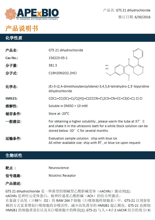
化学性质
产品名: Cas No.: 分子量: 分子式:
GTS 21 dihydrochloride 156223-05-1 381.3 C19H20N2O2.2HCl
产品名: GTS 21 dihydrochloride 修订日期: 6/30/2016
化学名: SMILESdihydrochloride 是一种新型的烟碱型乙酰胆碱受体(nAChRs)激动剂[1]。 nAChRs 是神经元受体蛋白,被神经递质乙酰胆碱(ACh)的结合所激活。 在暴露于高氧(≥99% O2)的 RAW 264.7 细胞(巨噬细胞样细胞系)中,GTS-21 以剂量依 赖的方式显著增加巨噬细胞的吞噬活性,减少高氧诱导的 HMGB1 超乙酰化。GTS-21 也抑制 HMGB1 的细胞质易位以及从巨噬细胞中的释放[1]。GTS-21 与人α4β2 nAChR 结合的效力(Ki
Evaluation sample solution : ship with blue ice All other available size: ship with RT , or blue ice upon request
靶点 :
Neuroscience
信号通路:
Nicotinic Receptor
参考文献: [1]. Sitapara RA, Antoine DJ, Sharma L, et al. The α7 nicotinic acetylcholine receptor agonist GTS-21 improves bacterial clearance in mice by restoring hyperoxia-compromised macrophage function. Mol Med, 2014, 20: 238-247. [2]. Briggs CA, Anderson DJ, Brioni JD, et al. Functional characterization of the novel neuronal nicotinic acetylcholine receptor ligand GTS-21 in vitro and in vivo. Pharmacol Biochem Behav, 1997, 57(1-2): 231-241.
盐酸倍他司汀及其关键中间体的合成综述

盐酸倍他司汀及其关键中间体的合成综述盐酸倍他司汀(Escitalopram hydrobromide)是一种常用的抗抑郁药物,广泛用于治疗抑郁症和焦虑症。
它是一种选择性去甲肾上腺素再摄取抑制剂(SSRI),能有效增加大脑中去甲肾上腺素的水平,从而改善情绪和心理状态。
盐酸倍他司汀的合成工艺涉及多个中间体的合成步骤,下面将从中间体入手,对盐酸倍他司汀及其关键中间体的合成进行综述。
一、关键中间体的合成1. 3-氯-3-(二氯甲基)丙烯(3-Chloro-3-(dichloromethyl)propene)3-氯-3-(二氯甲基)丙烯是合成盐酸倍他司汀的关键中间体之一,它通常通过氯乙腈和三氯乙酸的反应制备。
首先将氯乙腈和三氯乙酸在二氯甲烷中反应,再加入三甲胺,使用活性碳吸附,最后蒸馏得到3-氯-3-(二氯甲基)丙烯。
2. 3-(二氯甲基)-7-氟-1,3-二氢-5-(4-甲基-1-哌啶基)-2H-1,4-苯并二氮在-3-酮(3-(Dichloromethyl)-7-fluoro-1,3-dihydro-5-(4-methyl-1-piperazinyl)-2H-1,4-be nzodiazepin-2-one)以上述3-氯-3-(二氯甲基)丙烯为起始原料,通过串联反应得到3-(二氯甲基)-7-氟-1,3-二氢-5-(4-甲基-1-哌啶基)-2H-1,4-苯并二氮在-3-酮。
将3-氯-3-(二氯甲基)丙烯和对氨基苯甲酮在三甲苯中加热反应,生成中间体,再用氢醌处理,发生串联反应,得到目标产物。
3. 盐酸倍他司汀通过将3-(二氯甲基)-7-氟-1,3-二氢-5-(4-甲基-1-哌啶基)-2H-1,4-苯并二氮在-3-酮与盐酸的反应,得到盐酸倍他司汀的合成。
二、盐酸倍他司汀的合成综述盐酸倍他司汀的合成工艺包括多个中间体的合成步骤,其合成路线如下:以上合成路线中间体的合成步骤相对繁琐,需要多步反应和纯化过程,针对每个中间体的合成过程进行优化是十分重要的。
特异性二氧化碳胺胶囊21S 产品数据表说明书
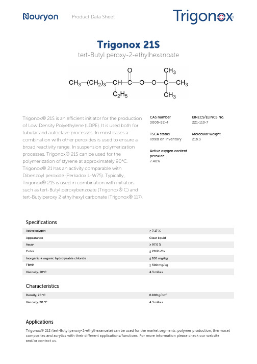
Product Data SheetTrigonox 21Stert-Butyl peroxy-2-ethylhexanoateTrigonox® 21S is an efficient initiator for the production of Low Density Polyethylene (LDPE). It is used both for tubular and autoclave processes. In most cases a combination with other peroxides is used to ensure a broad reactivity range. In suspension polymerization processes, Trigonox® 21S can be used for the polymerization of styrene at approximately 90°C. Trigonox® 21 has an activity comparable with Dibenzoyl peroxide (Perkadox L-W75). Typically, Trigonox® 21S is used in combination with initiators such as tert-Butyl peroxybenzoate (Trigonox® C) and tert-Butylperoxy 2 ethylhexyl carbonate (Trigonox® 117).CAS number3006-82-4EINECS/ELINCS No.221-110-7TSCA statuslisted on inventoryMolecular weight216.3Active oxygen contentperoxide7.40%SpecificationsActive oxygen≥ 7.17 %Appearance Clear liquidAssay≥ 97.0 %Color≤ 20 Pt-CoInorganic + organic hydrolysable chloride≤ 100 mg/kgTBHP≤ 500 mg/kgViscosity, 20°C 4.3 mPa.sCharacteristicsDensity, 20 °C0.900 g/cm³Viscosity, 20 °C 4.3 mPa.sApplicationsTrigonox® 21S (tert-Butyl peroxy-2-ethylhexanoate) can be used for the market segments: polymer production, thermoset composites and acrylics with their different applications/functions. For more information please check our websiteand/or contact us.Half-life dataThe reactivity of an organic peroxide is usually given by its half-life (t1/2) at various temperatures. For Trigonox® 21S in chlorobenzene half-life at other temperatures can be calculated by using the equations and constants mentioned below:0.1 hr at 113°C1 hr at 91°C10 hr at 72°CFormula 1kd = A·e-Ea/RTFormula 2t½ = (ln2)/kdEa124.90 kJ/moleA 1.54E+14 s-1R8.3142 J/mole·KT(273.15+°C) KThermal stabilityOrganic peroxides are thermally unstable substances, which may undergo self-accelerating decomposition. The lowest temperature at which self-accelerating decomposition of a substance in the original packaging may occur is the Self-Accelerating Decomposition Temperature (SADT). The SADT is determined on the basis of the Heat Accumulation Storage Test.SADT35°CEmergency temperature (Tₑ)25°CControl temperature (Tc)20°CMethod The Heat Accumulation Storage Test is a recognized test method for thedetermination of the SADT of organic peroxides (see Recommendations on theTransport of Dangerous Goods, Manual of Tests and Criteria - United Nations, NewYork and Geneva).StorageDue to the relatively unstable nature of organic peroxides a loss of quality can be detected over a period of time. To minimize the loss of quality, Nouryon recommends a maximum storage temperature (Ts max. ) for each organic peroxide product.Ts Max.10°C andTs Min.-30°C to prevent crystallizationNote When stored according to these recommended storage conditions, Trigonox®21S will remain within the Nouryon specifications for a period of at least 3 monthsafter delivery.Packaging and transportIn North America Trigonox® 21S is packed in non-returnable, vented, five gallon polyethylene containers of 35 lb net weight. In other regions the standard packaging is a 30-liter HDPE can (Nourytainer®), vented, for 25 kg peroxide content. Both packaging and transport meet the international regulations. For the availability of other packed quantities consult your Nouryon representative. Trigonox® 21S is classified as Organic peroxide type C; liquid, temperature controlled, Division 5. 2; UN 3113; PG II.Safety and handlingKeep containers tightly closed. Store and handle Trigonox® 21S in a dry well-ventilated place away from sources of heat or ignition and direct sunlight. Never weigh out in the storage room. Avoid contact with reducing agents (e. g. amines), acids, alkalis and heavy metal compounds (e. g. accelerators, driers and metal soaps). Please refer to the Safety Data Sheet (SDS) for further information on the safe storage, use and handling of Trigonox® 21S. This information should be thoroughly reviewed prior to acceptance of this product. The SDS is available at /sds-search.Major decomposition productsCarbon dioxide, tert-Butanol, Heptane, 3-tert-ButoxyheptaneAll information concerning this product and/or suggestions for handling and use contained herein are offered in good faith and are believed to be reliable.Nouryon, however, makes no warranty as to accuracy and/or sufficiency of such information and/or suggestions, as to the product's merchantability or fitness for any particular purpose, or that any suggested use will not infringe any patent. Nouryon does not accept any liability whatsoever arising out of the use of or reliance on this information, or out of the use or the performance of the product. Nothing contained herein shall be construed as granting or extending any license under any patent. Customer must determine for himself, by preliminary tests or otherwise, the suitability of this product for his purposes.The information contained herein supersedes all previously issued information on the subject matter covered. The customer may forward, distribute, and/or photocopy this document only if unaltered and complete, including all of its headers and footers, and should refrain from any unauthorized use. Don’t copythis document to a website.Trigonox® and Nourytainer are registered trademarks of Nouryon Functional Chemicals B.V. or affiliates in one or more territories.Contact UsPolymer Specialties Americas************************Polymer Specialties Europe, Middle East, India and Africa*************************Polymer Specialties Asia Pacific************************2022-6-30© 2022Polymer production Trigonox 21S。
加速溶剂萃取-超高效液相色谱-串联质谱法测定大枣中3种五环三萜酸

食品与药品Food and Drug2021年第23卷第1期17加速溶剂萃取-超高效液相色谱-串联质谱法测定大枣中3种五环三祜酸张萍,何婷,王颖,胡克特,顾丁,陈荣祥**(遵义医科大学基础医学院,贵州遵义563000)摘要:目的建立加速溶剂萃取-超高效液相色谱-串联质谱法(ASE-UPLC-MS/MS)同时测定大枣中桦木酸、齐墩果酸和熊果酸的方法。
方法样品釆用ASE提取,优化提取条件,并与超声辅助提取法进行比较。
优化 后的提取条件为:以80%甲醇为提取溶剂,提取温度100-C,静态萃取时间15min,萃取1次。
提取液釆用Waters ACQUITY BEH C18色谱柱分离,以乙ffi-15mmol/L乙酸钱(pH9.3)为流动相,梯度洗脱,经UPLC-MSZMS仪,釆用电喷雾电离源,负离子模式下多反应监测模式检测。
结果桦木酸、齐墩果酸、熊果酸在0.5~10 mg/L范围内,浓度与峰面积线性关系良好,相关系数大于0.9900。
不同浓度3种化合物的加标回收率93.6%〜101.7%,相对标准偏差在1.18%~6.83%之间。
结论此法简单快速、准确稳定、重复性好,可用于大枣中桦木酸、齐墩果酸、熊果酸的含量测定。
关键词:加速溶剂萃取;超高效液相色谱;串联质谱法;大枣中图分类号:R284.1文献标识码:A文章编号:1672-979X(2021)01-0017-06DOI:10.3969/j.issn.l672-979X.2021.01.004Simultaneous Determination of Three Pentacyclic Triterpenic Acids in Jujubae Fructus byAccelerated Solvent Extraction-UPLC-MS/MSZHANG Ping,HE Ting,WANG Ying,HU Ke-te,GU Ding,CHEN Rong-xiang(School of B asic Medical Sciences,Zunyi Medical University,Zu^yi563000,China)Abstract:Objective To establish a method for the simultaneous determination of betulinic acid,oleanolic acid and ursolic acid in Jujubae fructus by ultra-high performance liquid chromatography-tandem mass spectrometry(UPLC-MS/MS)with accelerated solvent extraction(ASE).Methods The extract parameters of A SE were optimized and the efficiency was compared with the ultrasound-assisted extraction method.The optimum extraction conditions were as follows:80%methanol was selected as extraction solvent,oven temperature was100°C,the static extraction time was 15min and one extraction cycle was adopted.A waters ACQUITY BEH C18(2.1mmx]00mn,1.7“m)column was used as the stationary phase,acetonitrile and ammonium acetate solution(15mmol/L,pH9.3)was used as the mobile phase.Mass detection was conducted by electrospray ionization in negative ion multiple reaction monitoring mode. Results The calibration curves were linear over a concentration range of0.5-10mg/L for betulinic acid,oleanolic acid and ursolic acid.The correlation coefficients were greater than0.9900.The recoveries of different spiked levels were between93.6%and101.7%,with RSDs between1.18%and6.83%.Conclusion The method is simple,rapid,收稿日期:2020-09-04基金项目:国家自然科学基金(31660131,81760652);贵州省联合基金(黔科合J字LKZ[2013]17号);遵义医学院博士启动基金(F-568)作者简介:张萍,硕士研究生,研究方向:药用植物开发与利用E-mail:******************通讯作者:陈荣祥,教授,博士,研究方向:药用植物开发与利用E-mail:*************************18食品与药品Food and Drug2021年第23卷第1期accurate,stable and reproducible.It can be used for the determination of betulinic acid,oleanolic acid and ursolic acid in Jujubae f ructus.Key Words:accelerated solvent extraction;ultra-high performance liquid chromatography;tandem mass spectrometry; Jujubae f ructus.大枣为鼠李科植物枣树(Ziziphus jujuba Mill.)的干燥成熟果实,不仅作为食物,还是传统中医药中的常用药材。
高效液相色谱-串联质谱法检测泮托拉唑钠原料药中的水合肼
·药物研发·高效液相色谱-串联质谱法检测泮托拉唑钠原料药中的水合肼赵会明 张振洋 樊华军[英格尔检测技术服务(上海)有限公司 上海 201100]摘要建立了泮托拉唑钠原料药中的基因毒性杂质水合肼的高效液相色谱-串联质谱(LC-MSMS)检测方法。
采用反相色谱,以水-乙腈(含0.1%甲酸)为流动相,梯度洗脱,流速0.5 mL/min,以ESI正离子多反应监测(MRM)模式进行质谱检测。
结果显示,水合肼的检测限和定量限可达到0.23、0.47 ng/mL,其在0.47~9.37 ng/mL浓度范围内线性关系良好(r=0.999 9),准确度试验中低、中、高浓度回收率均在81.6%~90.9%之间。
在3批次泮托拉唑钠原料药中均未检出水合肼。
关键词高效液相色谱-串联质谱法基因毒性杂质泮托拉唑钠水合肼痕量检测中图分类号:R917; O657 文献标志码:A 文章编号:1006-1533(2022)11-0072-04引用本文 赵会明, 张振洋, 樊华军. 高效液相色谱-串联质谱法检测泮托拉唑钠原料药中的水合肼[J]. 上海医药, 2022, 43(11): 72-75.Determination of hydrazine hydrate in pantoprazole sodium by high performance liquid chromatography-tandem mass spectrometryZHAO Huiming, ZHANG Zhenyang, FAN Huajun[ICAS Testing Technology Service (Shanghai) CO., LTD., Shanghai 201100, China]ABSTRACT To establish a high-performance liquid chromatography-tandem mass spectrometry (LC-MSMS) method for the determination of hydrazine hydrate in active pharmaceutical ingredient (API) pantoprazole sodium. HPLC was carried out by reverse chromatography using water-acetonitrile containing 0.1% formic acid as flow phase and gradient elution at a flow rate of 0.5 mL/min. Mass spectrometry was performed with multi-reaction monitoring (MRM) in positive ESI mode. The detection and quantitative limits of hydrazine hydrate reached 0.23, 0.47 ng/mL and hydrazine hydrate showed good linear relationship in the range of 0.47-9.37 ng/mL (r=0.999 9). The recoveries of samples at low, medium and high-level concentrations reached81.6% to 90.9% in the accuracy experiment. No hydrazine hydrate was detected in 3 batches of pantoprazole sodium.KEY WORDS HPLC-tandem mass spectrometry; genotoxic impurities; pantoprazole sodium; hydrazine hydrate; trace determination上消化道出血是近年的临床疾病中常见且多发的一种疾病,其临床表现为呕血、黑便等,如得不到及时有效治疗,可能引发失血性休克。
Incucyte
Product Information Presentation, Storage and StabilityThe Incucyte® Fabfluor-pH Antibody Labeling Reagents for antibody internalization are supplied as lyophilized solids in sufficient quantity to label 50 μg of test antibody, when used at the suggested molar ratio (1:3 of test antibody to labeling Fab). The lyophilized solid can be stored at 2-8° C for one year. Once re-hydrated, any unused reagent should be aliquoted and stored at -80° C for up to one year. Avoid repeated freeze-thaw cycles.Incucyte® Fabfluor-pH Antibody Labeling ReagentsFor Antibody Internalization AssaysAntibody Labeling Reagent Rehydrated: -80° C *Excitation and Emission maxima were determined at a pH of 4.5.Fabfluor_quick_guideBackgroundIncucyte ® Fabfluor-pH Antibody Labeling Reagents are designed for quick, easy labeling of Fc-containing test antibodies with a Fab fragment-conjugated pH-sensitive fluorophore. The pH-sensitive dye based system exploits the acidic environment of the lysosomes to quantify in-ternalization of the labeled antibody. As Fabfluor labeled antibodies reside in the neutral extracellular solution (pH 7.4), they interact with cell surface specific antigens and are internalized. Once in the lysosomes, they enter an acidic environment (pH 4.5–5.5) and a substantial in-crease in fluorescence is observed. In the absence of ex-pression of the specific antigen, no internalization occurs and the fluorescence intensity of the labeled antibodies remains low. With the Incucyte ® integrated analysis soft-ware, background fluorescence is minimized. These reagents have been validated for use with a number of different antibodies in a range of cell types. The Incucyte ® Live-Cell Analysis System enables real-time, kinetic eval -uation of antibody internalization.Recommended UseWe recommend that the Incucyte ® Fabfluor-pH Antibody Labeling Reagents are prepared at a stock concentration of 0.5 mg/mL by the addition of 100 μL of sterile water and triturated (centrifuge if solution not clear). The reagent may then be diluted directly into the labeling mixture with test antibody. Do NOT sonicate the solution.Additional InformationThe Fab antibody was purified from antisera by a combination of papain digestion and immunoaffinity chromatography using antigens coupled to agarose beads. Fc fragments and whole IgG molecules have been removed.Human Red (Cat. No. 4722) or Human Orange (Cat. No. 4812)—Based on immunoelectrophoresis and/ or ELISA, the antibody reacts with the Fc portion of human IgG heavy chain but not the Fab portion of human IgG. No antibody was detected against human IgM, IgA or against non-immunoglobulin serum proteins. The anti-body may cross-react with other immunoglobulins from other species.Mouse IgG1 (Cat. No. 4723), IgG2a (Cat. No. 4750) or IgG2b (Cat. No. 4751)—Based on antigen-binding assay and/or ELISA, the antibody reacts with the Fc portion of mouse IgG, IgG2a or IgG2b, respectively, but not the Fab portion of mouse immunoglobulins. No antibody was detected against mouse IgM or against non–immunoglobulin serum proteins. The antibody may cross-react with other mouse IgG subclasses or with immunoglobulins from other species.Rat (Cat. No. 4737)—Based on immunoelectrophoresis and/or ELISA, the antibody reacts with the Fc portion of rat IgG heavy chain but not the Fab portion of rat IgG. No antibody was detected against rat IgM, IgA or against non-immunoglobulin serum proteins. The antibody may cross-react with other immunoglobulins from other species.A.B.C.D.R e d O b j e c t A r e a (x 105 μm 2 p e r w e l l )Time (hours)A U C x 106 (0–12 h )log [α–CD71] (g/mL)Example DataFigure 1: Concentration-dependent increase in antibody internalization of Incucyte ® Fabfluor labeled-α-CD71 in HT1080 cells. α-CD71 and mouse IgG1 isotype control were labeled with Incucyte ® Mouse IgG1 Fabfluor-pH Red Antibody Labeling Reagent. HT1080 cells were treated with either Fabfluor-α-CD71 or Fabfluor-IgG1 (4 μg/mL); HD phase and red fluorescence images were captured every 30 minutes over 12 hours using a 10X magnification. (A) Images of cells treated with Fabfluor-α-CD71 display red fluorescence in the cytoplasm (images shown at 6 h). (B) Cells treated with labeled isotype control display no cellular fluorescence. (C) Time-course of Fabfluor-α-CD71 internalization with increasing concentrations of Fabfluor-α-CD71 (progressively darker symbols). Internalization has been quantified as the red object area for each time-point. (D) Concentration response curve to Fabfluor-α-CD71. Area under the curve (AUC) values have been determined from the time-course shown in panel C (0-12 hours) and are presented as the mean ± SEM, n=3 wells.CD71-FabfluorIgG-FabfluorProtocols and ProceduresMaterialsIncucyte® Fabfluor-pH Antibody Labeling ReagentTest antibody of interest containing human, mouse, or rat IgG Fc region (at known concentration)Target cells of interestTarget cell growth mediaSterile distilled water96-well flat bottom microplate (e.g. Corning Cat. No. 3595) for imaging96-well round black round bottom ULA plate (e.g. Corning Cat. No. 45913799) or amber microtube (e.g. Cole Parmer Cat. No. MCT-150-X, autoclaved) for conjugation step0.01% Poly-L-Ornithine (PLO) solution (e.g. Sigma Cat. No. P4957), optional for non-adherent cells Recommended control antibodiesIt is strongly recommended that a positive and negative control is run alongside test antibodies and cell lines. For example, CD71, which is a mouse anti-human antibody, is recommended as a positive control for the mouse Fab.Anti-CD71, clone MEM-189, IgG1 e.g. Sigma Cat. No. SAB4700520-100UGAnti-CD71, clone CYG4, IgG2a e.g. BioLegend Cat. No. 334102Isotype controls, depending on isotype being studied—Mouse IgG1, e.g. BioLegend Cat. No. 400124, Mouse IgG2a e.g. BioLegend Cat. No. 401501Preparation of Incucyte® Antibody Internalization Assay 1. Seed target cells of interest1.1 Harvest cells of interest and determine cell concentra-tion (e.g. trypan blue + hemocytometer).1.2 Prepare cell seeding stock in target cell growth mediawith a cell density to achieve 40–50% confluence be-fore the addition of labeled antibodies. The suggested starting range is 5,000–30,000 cells/well, although the seeding density will need to be optimized for each cell type.Note: For non-adherent cell types, a well coating may be required to maintain even cell distribution in the well. For a 96-well flat bottom plate, we recommend coating with 50 μL of either 0.01% Poly-L-Or-nithine (PLO) solution or 5 μg/mL fibronectin diluted in 0.1% BSA.Coat plates for 1 hour at ambient temperature, remove solution from wells and then allow the plates to dry for 30-60 minutes prior to cell addition.1.3 Using a multi-channel pipette, seed cells (50 µL perwell) into a 96-well flat bottom microplate. Lightly tapplate side to ensure even liquid distribution in well. Toensure uniform distribution of cells in each well, allowthe covered plate sit on a level surface undisturbed at room temperature in the tissue culture hood for 30minutes. After cells are settled, place the plate insidethe Incucyte® Live-Cell Analysis System to monitor cell confluence.Note: Depending on cell type, plates can be used in assay once cells have adhered to plastic and achieved normal cell morphology e.g.2-3 hours for HT1080 or 1-2 hours for non-adherent cell types. Some cell types may require overnight incubation.2. Label Test Antibody2.1 Rehydrate the Incucyte® Fabfluor-pH Antibody Label-ing Reagent with 100 µL sterile water to result in a final concentration of 0.5 mg/mL. Triturate to mix (centrifuge if solution is not clear).Note: The reagent is light sensitive and should be protected fromlight. Rehydrated reagent can be aliquoted into amber or foilwrapped tubes and stored at -80° C for up to 1 year (avoid freezing and thawing).2.2 Mix test antibody with rehydrated Incucyte® Fabfluor–pH Antibody Labeling Reagent and target cell growth media in a black round bottom microplate or ambertube to protect from light (50 µL/well).a. Add test antibody and Incucyte® Fabfluor–pH Anti-body Labeling Reagent at 2X the final concentration.We suggest optimizing the assay by starting with afinal concentration of 4 µg/mL of test antibody or theFabfluor-pH Antibody Labeling Reagent (i.e. 2Xworking concentration = 8 µg/mL).Note: A 1:3 molar ratio of test antibody to Incucyte® Fabfluor-pHAntibody Labeling Reagent is recommended. The labeling re-agent is a third of the size of a standard antibody (50 and 150KDa, respectively). Therefore, labeling equal quantities will pro-duce a 1:3 molar ratio of test antibody to labeling Fab.b. Make sufficient volume of 2X labeling solution for50 µL/well for each sample. Triturate to mix.c. Incubate at 37° C for 15 minutes protected from light.Note: If performing a range of concentrations of test antibody,e.g. concentration response-curve, it is recommended to createthe dilution series post the conjugation step to ensure consistentmolar ratio. We strongly recommend the use of both a negativeand positive control antibody in the same plate.3. Add labeled antibody to cells3.1 Remove cell plate from incubator.3.2 Using a multi-channel pipette, add 50 µL of 2X labeledantibody and control solutions to designated wells.Remove any bubbles and immediately place plate in the Incucyte® Live-Cell Analysis System and start scanning.Note: To reduce the risk of condensation formation on the lid priorto first image acquisition, maintain all reagents at 37° C prior toplate addition.4. Acquire images and analyze4.1 In the Incucyte® Software, schedule to image every15-30 minutes, depending on the speed of the specific antibody internalization.a Scan on schedule, standard. If the Incucyte® Cell-by-Cell Analysis Software Module (Cat. No. 9600-0031)is available, adherent cell-by-cell or non-adherentcell-by-cell scan types can be selected.b Channel selection: select “phase” and “red” or“phase” and "orange” (depending on reagent used).c Objective: 10X or 20X depending on cell types used,generally 10X is recommended for adherent cells,and 20X for non-adherent or smaller cells.NOTE: The optional Incucyte® Cell-by-Cell Analysis SoftwareModule enables the classification of cells into sub-populationsbased on properties including fluorescence intensity, size andshape. For further details on this analysis module and its appli-cation, please see: /cell-by-cell.4.2 To generate the metrics, user must create an AnalysisDefinition suited to the cell type, assay conditions andmagnification selected.4.3 Select images from a well containing a positiveinternalization signal and an isotype control well(negative signal) at a time point where internalizationis visible.4.4 In the Analysis Definition:Basic Analyzer:a. Set up the mask for the phase confluence measurewith fluorescence channel turned off.b. Once the phase mask is determined, turn the fluores-cence channel on: Exclude background fluorescencefrom the mask using the background subtractionfeature. The feature “Top-Hat” will subtract localbackground from brightly fluorescent objects withina given radius; this is a useful tool for analyzing ob-jects which change in fluorescence intensity overtime.i The radius chosen should reflect the size of thefluorescent object but contain enough backgroundto reliably estimate background fluorescence inthe image; 20-30 μm is often a useful startingpoint.ii The threshold chosen will ensure that objectsbelow a fluorescence threshold will not bemasked.iii Choose a threshold in which red or orange objectsare masked in the positive response image but lownumbers in the isotype control, negative responsewell. For a very sensitive measurement, for example,if interested in early responses, we suggest athreshold of 0.2.NOTE: The Adaptive feature can be used for analysis but maynot be as sensitive and may miss early responses. If interestedin rate of response, Top-Hat may be preferable.Cell-by-Cell (if available):a. Create a Cell-by-Cell mask following the softwaremanual.b. There is no need to separate phase and fluorescencemasks. The default setting of Top-Hat No Mask forthe fluorescence channel will enable backgroundsubtraction without generation of a mask. Ensurethat the Top-Hat radius is set to a value higher thanthe radius of the larger clusters to avoid excess back-ground subtraction.c. The threshold of fluorescence can be determined inCell-by-Cell Classification.Specifications subject to change without notice.© 2020. All rights reserved. Incucyte, Essen BioScience, and all names of Essen BioScience prod -ucts are registered trademarks and the property of Essen BioScience unless otherwise specified. Essen BioScience is a Sartorius Company. Publication No.: 8000-0728-A00Version 1 | 2020 | 04Sales and Service ContactsFor further contacts, visit Essen BioScience, A Sartorius Company /incucyte Sartorius Lab Instruments GmbH & Co. KGOtto-Brenner-Strasse 20 37079 Goettingen, Germany Phone +49 551 308 0North AmericaEssen BioScience Inc. 300 West Morgan Road Ann Arbor, Michigan, 48108USATelephone +1 734 769 1600E-Mail:***************************EuropeEssen BioScience Ltd.Units 2 & 3 The Quadrant Newark CloseRoyston Hertfordshire SG8 5HLUnited KingdomTelephone +44 (0) 1763 227400E-Mail:***************************APACEssen BioScience K.K.4th floor Daiwa Shinagawa North Bldg.1-8-11 Kita-Shinagawa Shinagawa-ku, Tokyo 140-0001 JapanTelephone: +81 3 6478 5202E-Mail:*************************5. Analysis GuidelinesAs the labeled antibody is internalized into the acidic environment of the lysosome, the area of fluorescence intensity inside the cells increases.This can be reported in two ways:Ways to Report Basic AnalyzerCell-by-Cell Analysis* To correct for cell proliferation, it is advisable to normalize the fluorescence area to the total cell area using User Defined Metrics.For Research Use Only. Not For Therapeutic or Diagnostic Use.LicensesFor non-commercial research use only. Not for therapeutic or in vivo applications. Other license needs contact Essen BioS cience.Fabfluor-pH Red Antibody Labeling Reagent: This product or portions thereof is manufactured under license from Carnegie Mellon University and U.S. patent numbers 7615646 and 8044203 and related patents. This product is licensed for sale only for research. It is not licensed for any other use. There is no implied license hereunder for any commercial use.Fabfluor-pH Orange Antibody Labeling Reagent: This product or portions thereof is manufactured under a license from Tokyo University and is covered by issued patents EP2098529B1, JP5636080B2, US8258171, and US9784732 and related patent applications. This product and related products are trademarks of Goryo Chemical. Any application of above mentioned technology for commercial purpose requires a separate li -cense from: Goryo Chemical, EAREE Bldg., SF Kita 8 Nishi 18-35-100, Chuo-Ku, Sapporo, 060-0008 Japan.SupportA complete suite of cell health applications is available to fit your experimental needs. Find more information at /incucyte Foradditionalproductortechnicalinformation,************************************************************/incucyte。
SGLT2_抑制剂联合二甲双胍治疗对糖尿病肾病患者血糖及疗效的影响

SGLT2抑制剂联合二甲双胍治疗对糖尿病肾病患者血糖及疗效的影响郑秋娥,程秋敏,刘江建福建省立医院药学部,福建福州350001[摘要]目的分析钠-葡萄糖共转运蛋白2(sodium-dependent glucose transporters 2, SGLT2)抑制剂联合二甲双胍治疗对糖尿病肾病患者血糖及疗效的影响。
方法选取2021年1月—2023年1月福建省立医院接诊的108例糖尿病肾病患者,按照随机数表法分为对照组与研究组,各54例。
对照组接受二甲双胍+百令胶囊治疗,研究组联合SGLT2抑制剂治疗,分析对比两组患者血糖指标、临床总有效率、肾功能指标及血清炎症指标。
结果与对照组相比,研究组治疗后的空腹血糖、餐后2 h血糖及糖化血红蛋白水平更低,差异有统计学意义(P<0.05)。
与对照组对比,研究组临床总有效率更高,差异有统计学意义(P<0.05)。
与对照组对比,研究组治疗后的血肌酐、尿素氮及尿白蛋白排泄率更低,差异有统计学意义(P<0.05)。
结论SGLT2抑制剂联合二甲双胍治疗糖尿病肾病可降低血糖水平,改善肾功能,减轻机体炎症反应,提高临床总效率。
[关键词] SGLT2抑制剂;二甲双胍;糖尿病肾病;血糖[中图分类号] R4 [文献标识码] A [文章编号] 1672-4062(2023)10(b)-0076-04Effect of SGLT2 Inhibitor Combined with Metformin Treatment on Blood Glucose and Curative Effect in Patients with Diabetic NephropathyZHENG Qiu'e, CHENG Qiumin, LIU JiangjianDepartment of Pharmacy, Fujian Provincial Hospital, Fuzhou, Fujian Province, 350001 China[Abstract] Objective To analyze the effects of sodium-dependent glucose transporters 2 (SGLT2) inhibitor combined with metformin treatment on blood glucose and curative effect in patients with diabetic nephropathy.Methods 108 pa⁃tients with diabetes nephropathy who were treated in Fujian Provincial Hospital from January 2021 to January 2023 were selected and divided into the control group and the study group according to the random number table, with 54 patients in each group. The control group received treatment with metformin and Bailing capsules, while the study group received treatment with SGLT2 inhibitors. The blood glucose indicators, clinical total efficiency, renal function indicators, and serum inflammation indicators were analyzed and compared between the two groups.Results Compared with the control group, the study group had lower levels of FPG, 2 hPG, and HbA1c after treatment, the difference was statistically significant (P<0.05). Compared with the control group, the total clinical efficiency of the study group was higher, and the difference was statistically significant (P<0.05). Compared with the control group, the study group had lower Scr, BUN, and UAER after treatment, the difference was statistically significant (P<0.05).Conclusion SGLT2 in⁃hibitor combined with metformin in the treatment of diabetic nephropathy can reduce blood glucose level, improve re⁃nal function, reduce the inflammatory response of the body, and improve the overall clinical efficiency.[Key words] SGLT2 inhibitor; Metformin; Diabetic nephropathy; Blood glucose糖尿病肾病是糖尿病并发症之一,也是引发终末期肾衰竭的主要原因,近十年来随着糖尿病发病率的增高,糖尿病肾病发病率增加2倍以上[1]。
醋酸丁酸纤维素药典检验规程

醋酸丁酸纤维素药典检验规程醋酸丁酸纤维素,也称为纤维素醋丁酸酯,是一种合成纤维素衍生物。
它是通过将纤维素与丁酸和醋酸进行反应而制得的。
醋酸丁酸纤维素具有良好的溶解性和可塑性,广泛用于制备药片、胶囊、涂层剂等制剂中作为缓释剂和增稠剂。
以下是关于醋酸丁酸纤维素药典检验规程的全面详细回答:一、外观检验1. 检查样品外观是否为白色或类似白色的颗粒状或粉末状物质。
2. 观察样品是否无杂质或异物。
二、标识检验1. 检查样品包装上是否标明了产品名称、规格、批号等必要信息。
2. 根据药典要求,对比样品包装上的标识信息与药典规定的标准进行比对。
三、含量测定1. 采用高效液相色谱法(HPLC)测定样品中的丁酸含量。
2. 准备样品溶液,将一定量的样品溶解于适量的溶剂中,通过HPLC进行分析。
3. 根据药典规定的方法和标准曲线,计算出样品中丁酸的含量。
四、酸值测定1. 采用酸碱滴定法测定样品中的酸值。
2. 准备样品溶液,将一定量的样品溶解于适量的溶剂中,并加入指示剂。
3. 使用标准碱溶液滴定样品溶液,记录所需滴定体积。
4. 根据药典规范计算出样品中酸值的含量。
五、水分测定1. 采用干燥法测定样品中的水分含量。
2. 取一定质量的样品,在恒温恒湿条件下进行干燥。
3. 干燥至恒重后,根据药典规范计算出样品中水分的含量。
六、颗粒度测定1. 采用激光粒度分析仪进行颗粒度测定。
2. 将适当数量的样品加入到激光粒度分析仪中,进行测试。
3. 根据药典规范,计算出样品的颗粒度分布。
七、溶解性测定1. 采用药典规定的方法,测定样品在不同介质中的溶解度。
2. 准备一系列浓度不同的溶液,将样品加入其中,并进行搅拌。
3. 根据药典标准,观察样品在不同介质中的溶解情况。
八、重金属含量测定1. 采用原子吸收光谱法(AAS)或电感耦合等离子体发射光谱法(ICP-OES)测定样品中重金属元素的含量。
2. 准备样品溶液,使用相应仪器进行测试,并根据药典规范计算出重金属元素的含量。
隐丹参酮(CTS)检测

迪信泰检测平台
隐丹参酮(CTS)检测
隐丹参酮(Cryptotanshinone,CTS),又称为隐丹参醌,是从丹参中提取得到的一种脂溶性有效成分,具有抗菌、抗炎、抗氧化、抗癌等诸多药理活性。
迪信泰检测平台采用高效液相色谱(HPLC)和液质联用(LC-MS)技术,可高效、精准的检测隐丹参酮的含量变化。
此外,迪信泰检测平台还可检测其他多种中药成分,方法成熟,可高效处理大批量样本。
HPLC和LC-MS测定隐丹参酮样本要求:
1. 请确保样本量大于0.2g或者0.2mL。
周期:2~3周。
项目结束后迪信泰检测平台将会提供详细中英文双语技术报告,报告包括:
1. 实验步骤(中英文)。
2. 相关质谱参数(中英文)。
3. 质谱图片。
4. 原始数据。
5. 隐丹参酮含量信息。
迪信泰检测平台可根据需求定制其他物质测定方案,具体可免费咨询技术支持。
国家药监局关于批准月桂酰精氨酸乙酯HCl等4个原料作为化妆品原料使用的公告
国家药监局关于批准月桂酰精氨酸乙酯HCl等4个原料作为化妆品原料使用的公告
文章属性
•【制定机关】国家药品监督管理局
•【公布日期】2020.12.21
•【文号】国家药品监督管理局公告2020年第141号
•【施行日期】2020.12.21
•【效力等级】部门规范性文件
•【时效性】现行有效
•【主题分类】美容业、化妆品管理
正文
国家药品监督管理局公告
2020年第141号
国家药监局关于批准月桂酰精氨酸乙酯HCl等4个原料作为
化妆品原料使用的公告
根据《化妆品卫生监督条例》等有关规定,经国家药品监督管理局化妆品技术审评专家委员会和化妆品标准专家委员会审核,现批准月桂酰精氨酸乙酯HCl作为化妆品防腐剂使用,纳入《化妆品安全技术规范》(2015年版)第三章的化妆品准用防腐剂(表4);批准甲氧基 PEG-23 甲基丙烯酸酯/甘油二异硬脂酸酯甲基丙烯酸酯共聚物等3个原料作为化妆品原料使用。
使用上述原料生产化妆品,应当符合有关法律、法规、标准规定。
特此公告。
附件:1.月桂酰精氨酸乙酯 HCl原料技术要求
2.甲氧基 PEG-23 甲基丙烯酸酯/甘油二异硬脂酸酯甲基丙烯酸酯共聚物原料技术要求
3.磷酰基寡糖钙原料技术要求
4.硬脂醇聚醚-200原料技术要求
国家药监局
2020年12月21日。
- 1、下载文档前请自行甄别文档内容的完整性,平台不提供额外的编辑、内容补充、找答案等附加服务。
- 2、"仅部分预览"的文档,不可在线预览部分如存在完整性等问题,可反馈申请退款(可完整预览的文档不适用该条件!)。
- 3、如文档侵犯您的权益,请联系客服反馈,我们会尽快为您处理(人工客服工作时间:9:00-18:30)。
=====================================================================Acq. Operator : Su Xiao Ying(LCMS-02) Seq. Line : 40Acq. Instrument : HY-LCMS-02 Location : P1-B-07Injection Date : 4/29/2016 1:58:29 PM Inj : 1 Inj Volume : 3.000 µl Acq. Method : D:\AGLIENT 1260\DATA\20160429\20160429 2016-04-29 08-44-21\100-1000MS+3MIN- 1.5_(0.02%FA).M Last changed : 4/29/2016 8:44:22 AM by Su Xiao Ying(LCMS-02)Analysis Method : D:\AGLIENT 1260\DATA\20160429\20160429 2016-04-29 08-44-21\100-1000MS+3MIN- 1.5_(0.02%FA).M (Sequence Method)Last changed : 4/29/2016 3:38:31 PM by Su Xiao Ying(LCMS-02) (modified after loading) M ethod Info : HY-365_5HO1RS,M,A-RP-108,210nm,23min Catalog No : HY-14564A Batch#20261 A-RP-134 Additional Info : Peak(s) manually integrated min
0.51 1.52 2.53mAU -100
100200
300
400
500
DAD1 B, Sig=214,4 Ref=off (D:\AGLIENT 1260\DATA\20160429\20160429 2016-04-29 08-44-21\BIZ2016-429-WJ5.D)
1.547
2.006 ===================================================================== Area Percent Report ===================================================================== Sorted By : Signal Multiplier : 1.0000Dilution : 1.0000Do not use Multiplier & Dilution Factor with ISTDs Signal 1: DAD1 B, Sig=214,4 Ref=off Peak RetTime Type Width Area Height Area # [min] [min] [mAU*s] [mAU] %----|-------|----|-------|----------|----------|--------| 1 1.547 MM 0.0796 3314.82520 694.05988 99.6619 2 2.006 MM 0.0630 11.24677 2.97652 0.3381 Totals : 3326.07197 697.03640 ===================================================================== *** End of Report ***
=====================================================================Acq. Operator : Su Xiao Ying(LCMS-02) Seq. Line : 40Acq. Instrument : HY-LCMS-02 Location : P1-B-07Injection Date : 4/29/2016 1:58:29 PM Inj : 1 Inj Volume : 3.000 µl Acq. Method : D:\AGLIENT 1260\DATA\20160429\20160429 2016-04-29 08-44-21\100-1000MS+3MIN- 1.5_(0.02%FA).M Last changed : 4/29/2016 8:44:22 AM by Su Xiao Ying(LCMS-02)Analysis Method : D:\AGLIENT 1260\DATA\20160429\20160429 2016-04-29 08-44-21\100-1000MS+3MIN- 1.5_(0.02%FA).M (Sequence Method)Last changed : 4/29/2016 3:37:31 PM by Su Xiao Ying(LCMS-02) (modified after loading) M ethod Info : HY-365_5HO1RS,M,A-RP-108,210nm,23min Catalog No : HY-14564A Batch#20261 A-RP-134 Additional Info : Peak(s) manually integrated min
0.51 1.52 2.530
100000
200000
300000
400000
500000
600000
700000
800000
MSD1 TIC, MS File (D:\AGLIENT 1260\DATA\20160429\20160429 2016-04-29 08-44-21\BIZ2016-429-WJ5.D) ES-API, Pos, Scan
1.556
MS Signal: MSD1 TIC, MS File, ES-API, Pos, Scan, Frag: 50 Spectra averaged over upper half of peaks. Noise Cutoff: 1000 counts. Reportable Ion Abundance: > 10%. Retention Mol. Weight Time (MS) MS Area or Ion 1.556 7171430 310.20 I 309.20 I
m/z 1002003004005006000
20
40
60
80
100
*MSD1 SPC, time=1.507:1.617 of D:\AGLIENT 1260\DATA\20160429\20160429 2016-04-29 08-44-21\BIZ2016-429-WJ5.D ES-API, Max: 538926 310.2 309.2 *** End of Report ***。
