Micropropagation of Sego Lily
微塑料/纳米塑料对维管植物的影响

1 000 nm。微塑料的外形多为圆柱形、圆形和圆盘状,颜色通
常为透明、白色和灰色,而且微塑料种类繁多,根据材质划分,主
要有聚乙烯(PE)、聚丙烯(PP)、聚苯乙烯(PS)、聚氯乙烯
每使用 5 mL 的化妆品就会导致 4 594 ~ 94 500 个球类微塑料
对智利 31 个接受不同年限污泥施用的农用土壤的调查研
究。 结果显示,随着污泥施用量的增加,土壤中微塑料的含
量也逐渐增加。 在施用了 5 次污泥后(总量为200 t / hm2 ),土
壤中微塑料的平均丰度达到了 3 500 个 / kg。
植物摄入并影响其生长发育[18-20] 。 此外,MPs / NPs 由于比
子表面,以致减少了种子对水分和养分的吸收,进而影响种
子发芽[33-34] 。 当蕨类植物( Ceratopteris pteridoides) 暴露于聚
苯乙烯纳米塑料(PS-NPs) 后,会破坏孢子从吸胀到萌发到
配子体的整个发育阶段,从而影响其繁殖过程[34] 。 吴佳妮
Copyright©博看网. All Rights Reserved.
安徽农业科学,J.Anhui Agric.Sci. 2023,51(20) :15-18
微塑料 / 纳米塑料对维管植物的影响
杨满香
( 重庆师范大学生命科学学院,重庆 401331)
摘要 微塑料 / 纳米塑料(micplastics / nanoplastics,MPs / NPs)广泛存在于环境中,被认为是一类对环境构成巨大威胁的新兴污染物。 通
境构成巨大威胁的新兴污染物[29] 。 基于微塑料来源广泛、
作者简介 杨满香(1997—) ,女,贵州遵义人,硕士研究生,研究方向:
欧李果渣原花青素提取工艺优化及其体外抗氧化和降糖活性评价

张晓冰,张羽师,王雨,等. 欧李果渣原花青素提取工艺优化及其体外抗氧化和降糖活性评价[J]. 食品工业科技,2024,45(1):178−184. doi: 10.13386/j.issn1002-0306.2023030017ZHANG Xiaobing, ZHANG Yushi, WANG Yu, et al. Optimization of Proanthocyanidin Extraction from Cerasus humilis Pomace and Evaluation of Its in Vitro Antioxidant and Hypoglycemic Activity[J]. Science and Technology of Food Industry, 2024, 45(1): 178−184.(in Chinese with English abstract). doi: 10.13386/j.issn1002-0306.2023030017· 工艺技术 ·欧李果渣原花青素提取工艺优化及其体外抗氧化和降糖活性评价张晓冰,张羽师,王 雨,赵锦江,王笑雪,刘舒鹏,李卫东*(北京中医药大学中药学院,北京 102488)摘 要:本研究以欧李果渣为原料,采用单因素结合正交试验对超声波辅助提取欧李果渣原花青素工艺进行优化,采用DPPH 自由基清除实验、ABTS +自由基清除实验、FRAP 法测定铁离子还原能力实验对纯化后的欧李果渣原花青素抗氧化活性进行评价,并采用α-葡萄糖苷酶活性抑制试验对其降糖活性进行评价。
结果表明,各因素对欧李果渣中原花青素提取得率的影响程度排序为:超声温度>料液比>超声时间>乙醇浓度;最佳提取工艺为乙醇浓度70%,超声温度60 ℃,超声时间40 min ,料液比1:30 g/mL ,在此条件下欧李果渣中原花青素得率为15.37 mg/g 。
特性 结构 表面

The role of natural and synthetic zeolites as feed additives on the prevention and/or the treatment of certain farmanimal diseases:A reviewD.Papaioannou,P.D.Katsoulos,N.Panousis *,H.KaratziasClinic of Productive Animal Medicine,School of Veterinary Medicine,Aristotle University of Thessaloniki,54124Thessaloniki,GreeceReceived 4April 2005;received in revised form 12May 2005;accepted 12May 2005Available online 28June 2005AbstractThe present review comments on the role of the use of zeolites as feed additives on the prevention and/or the treatment of certain farm animal diseases.Both natural and synthetic zeolites have been used in animal nutrition mainly to improve performance traits and,based on their fundamental physicochemical properties,they were also tested and found to be efficacious in the prevention of ammonia and heavy metal toxicities,poisonings as well as radioactive elements uptake and metabolic skeletal defects.During the last decade,their utilization as mycotoxin-binding adsorbents has been a topic of considerable interest and many published research data indicate their potential efficacy against different types of mycotoxins either as a primary material or after specific modifications related to their surface properties.Ingested zeolites are involved in many biochemical processes through ion exchange,adsorption and catalysis.Recent findings support their role in the prevention of certain metabolic diseases in dairy cows,as well as their shifting effect on nitrogen excretion from urine to faeces in monogastric animals,which results in lower aerial ammonia concentration in the confinement facilities.Moreover,new evidence provide insights into potential mechanisms involved in zeolites supporting effect on animals suffered from gastrointestinal disturbances,including intestinal parasite infections.All the proposed mechanisms of zeolites Õeffects are summarized in the present review and possible focus topics for further research in selected areas are suggested.Ó2005Elsevier Inc.All rights reserved.Keywords:Zeolites;Animal health;Prevention;Treatment1.IntroductionZeolites are crystalline,hydrated aluminosilicates of alkali and alkaline earth cations,consisting of three-dimensional frameworks of SiO 4À4and AlO 5À4tetrahedra linked through the shared oxygen atoms.Both natural and synthetic zeolites are porous materials,character-ized by the ability to lose and gain water reversibly,to adsorb molecules of appropriate cross-sectional diame-ter (adsorption property,or acting as molecular sieves)and to exchange their constituent cations without majorchange of their structure (ion-exchange property)[1,2].The exploitation of these properties underlies the use of zeolites in a wide range of industrial and agricultural applications and particularly in animal nutrition since mid-1960s [3].Many researchers have proved that the dietary inclu-sion of zeolites improves average daily gain and/or feed conversion in pigs [1,4–13],calves [1,7],sheep [7,14–16],and broilers [17–19].Zeolites also enhance the reproduc-tive performance of sows [1,20,21],increase the milk yield of dairy cows [22,23]and the egg production of laying hens [24,25]and have beneficial effects on egg weight and the interior egg characteristics [24–26].How-ever,the extent of performance enhancement effects is related to the type of the used zeolite,its purity and1387-1811/$-see front matter Ó2005Elsevier Inc.All rights reserved.doi:10.1016/j.micromeso.2005.05.030*Corresponding author.Tel.:+302310994501;fax:+302310994550.E-mail address:panousis@vet.auth.gr (N.Panousis)./locate/micromesoMicroporous and Mesoporous Materials 84(2005)161–170physicochemical properties,as well as to the supplemen-tation level used in the diets.Besides,the particle size of the zeolitic material,crystallite size and the degree of aggregation,as well as the porosity of individual parti-cles determine the access of ingestafluids to the zeolitic surface during passage across gastrointestinal tract and strongly affect its ion exchange,adsorption and catalytic properties.Table1summarizes the possible mechanisms by which zeolites may exert their performance promot-ing properties in farm animals.Apart from the positive effects on animalsÕperfor-mance,dietary supplementation of zeolites appears to represent an efficacious,complementary,supportive strategy in the prevention of certain diseases and the improvement of animalsÕhealth status.The aim of the present review article is to address the data concerning the influence of the in-feed inclusion of natural and syn-thetic zeolites on certain diseases of farm animals,to summarize the proposed mechanisms of zeolitesÕeffects and to suggest possible focus topics for further research in selected areas.2.Ameliorative effect on the consequences of mycotoxicosesIn the recent years,high incidence rates of contami-nation of cereal grains and animal feed with mycotoxins are reported worldwide[38].One of the latest ap-proaches to this global concern has been the use of nutritionally inert adsorbents in the diet that sequester mycotoxins,thus reducing intestinal absorption and, additionally,avoiding toxic effects for livestock and the carry-over of toxin compounds to animal products. For this purpose,phyllosilicates such as hydrated sodium calcium aluminosilicates(HSCAS)and benton-ite––which consist of layered crystalline structures and possess similar physicochemical properties with zeo-lites––have beenfirst used successfully in poultry,pig, sheep,cattle and laboratory animals[33,34,39–47].Apart from phyllosilicates,the use of zeolites has shown,lately,very promising results as well.In general, adsorption process on binders is strongly related to charge distribution,pore dimensions and accessible sur-face area of the adsorbent,as well as to polarity,solubil-ity and molecular dimensions of the certain mycotoxin which is to be adsorbed[46].Molecular sizes of aflatox-ins range from5.18A˚(B1and B2)to6.50A˚(G1and G2) and only zeolites with entry channels wide enough to permit the diffusion of aflatoxin molecules to the intra-crystalline structure are capable of demonstrating a clear sequestering effect.Clinoptilolite,a natural zeolite,has high adsorption indexes in vitro,more than80%,for aflatoxins B1[48,49]and G2[48]and the adsorption pro-cess begins with a fast reaction whereby most of the toxin is adsorbed within thefirst few minutes[48].On the contrary,Lemke et al.[50]conducted a variety of in vitro adsorption studies and reported a limited degree of clinoptilolite ability to bind aflatoxin B1effectively. According to them,adsorbent materials should be checked through a multi-tiered system of in vitro tests in order potential interactive factors,such as intestinal physicochemical variables and feed components,to be precluded.Indeed,a previous in vitro study had demon-strated average aflatoxin retention in natural zeolites of 60%,but also a nitrogen compound-related adsorbent effectiveness,when liquid media quality parameters had been taken into account[51].The in vivo efficacy of zeolites to ameliorate the consequences of aflatoxico-sis,mainly in poultry,has also been verified in many cases(Table2).In the case of phyllosilicates,the results of in vitro mycotoxin–clay interaction tests suggest the existence of areas of heterogenous adsorption affinities onto sur-face,the presence of different adsorption mechanisms or both[57].Nevertheless,the formation of strong bonds by chemisorption and the interaction of b-car-bonyl group of aflatoxin B1with the uncoordinated edge site aluminum ions in these adsorbents have been suggested as the binding mechanism which interprets their well-established high affinity for aflatoxin B1 [39,58,59].Although the exact binding mechanisms of zeolites on aflatoxins have not been determined the pos-sibility to act through similar with phyllosilicates mech-anism cannot be precluded and should be investigated.As far as other mycotoxins are concerned,mineral adsorbents exert a lower efficacy against mycotoxins containing less polar functional groups,which are required for efficient chemisorption on hydrophilic negatively charged mineral surfaces to occur.This limi-tation can be overcome by the use of chemically modi-fied clays.Modifications consist of alterations ofTable1Proposed mechanisms involved in animalsÕperformance promoting properties of the dietary use of zeolitesMechanismsAmmonia binding effect Elimination of toxic effects of NHþ4produced by intestinal microbial activity[8,10]Fecal elimination of p-cresol Reduction of the absorption of toxic products of intestinal microbial degradation,such as p-cresol[27] Retarding effect on digesta transit Slower passage rate of digesta through the intestines and more efficient use of nutrients[1,25,28] Enhanced pancreatic enzymes activity Favorable effect on feed components hydrolysis over a wider range of pH,improved energy and protein retention[29,30]Aflatoxin sequestering effect Elimination of mycotoxin growth inhibitory effects[31–37]162 D.Papaioannou et al./Microporous and Mesoporous Materials84(2005)161–170surface properties resulting in an increased hydropho-bicity,by exchange of structural charge-balance cations with high molecular weight quaternary amines.In vitro results have verified the binding efficacy of modified montmorilonite and clinoptilolite against zearalenone and ochratoxin A[60,61].However,naturally occurring clays also exert a moderate binding efficacy against mycotoxins,other than aflatoxins,as evidenced for zearalenone and ochratoxin A by in vitro[57]andfield trials[62,63],as well as for cyclopiazonic acid in exper-iments with broilers[45].Remarkable conclusions related to zeolitesÕefficacy against zearalenone toxicosis have also been drawn by in vivo studies.Feeding zearalenone to rats,Smith[64] demonstrated that the dietary use of a synthetic anion exchange zeolite could alter the faecal and urinary excre-tory patterns of zearalenone due to the elimination of its intestinal absorption.Recently,the dietary use of a clin-optilolite-rich tuffwas also effective in decreasing zearal-enone and a-zearalenole excretion in pigs fed diets contaminated with500ppb zearalenone[65].Addition-ally,in afield-case of zearalenone toxicosis with mean concentrations of660ppb,Papaioannou et al.[20] reported that the supplementation of a clinoptilolite rich tuffat the rate of2%in the ration of pregnant sows im-plied a rather protective role against the consequences of zearalenone ingestion,as evidenced by the improvement of indicative performance traits.Similar results were also obtained in swine with the dietary use of a modified clinoptilolite–healandite rich tuffat0.2%and with zea-ralenone concentration exceeding3.5ppm[66].Apart from surface interactions,the in vivo efficacy of mineral adsorbents against zearalenone could also re-sult from other implicating mechanisms.The entero-he-patic circulation of zearalenone and its derivatives in pigs retards their elimination and enhances the duration of adverse effects[67].Whether certain types of zeolites are able to affect the entero-hepatic cycling of zearale-none,thus counteracting the toxic effects of its biological action,is a hypothesis that awaits further research, although there is evidence of Ca-enriched clinoptiloliteÕs high affinity to the bile acids in the intestinal tract[68].3.Supportive effect on diarrhoea syndromeThere is an abundance of published data which indi-cate that the dietary use of natural zeolites reduces the incidence and decreases the severity and the duration of diarrhoea in calves[1,69–72]and pigs[1,5,13,72–75]. The exact mechanism of zeolitesÕeffect is not quite clear so far,although there is evidence that the use of zeolites may eliminate various predisposing and/or causative fac-tors which are associated in the culmination of intestinal disturbances in an interactive way.Apart from zeolitesÕretarding effect on intestinal passage rate[1]and their water adsorption property,which leads to the appear-ance of drier and more compact faeces,as in the case of phillipsite[75]or clinoptilolite[76],Vrzgula et al.[71]also proposed that the ameliorative effect on diar-rhoea syndrome of calves might result from either the alteration of metabolic acidosis,through effects on os-motic pressure in the intestinal lumen,or the increased retention of the enteropathogenic Escherichia coli.As far as we know,there is no evidence in the available lit-erature for retention of enteropathogenic E.coli on the outer surface of zeolite particles.However,clinoptilolite and mordenite are capable to adsorb and partially inac-tivate the thermo-labile(LT)E.coli enterotoxin in vitro, thus constricting its attachment to the intestinalTable2In vivo studies concerning the effect of the dietary use of zeolites during aflatoxicosesType of zeolite Dietary inclusion rate(%)Animal model ObservationsClinoptilolite1Broilers Growth depression caused by2.5ppm aflatoxin(afl)was alleviated by15%[31] Clinoptilolite5Geese Prophylactic effect on growth rate and liver enzymatic activity[32]Mordenite0.5Broilers Reducing effect on toxicity of afl(3.5mg kgÀ1diet)as indicated byweight gain and changes in uric acid and albumin concentrations[33] Clinoptilolite0.5Weaned piglets Growth inhibitory effects and alterations of liver enzyme activity inducedby500ppb aflwere prevented[34]Synthetic zeolite0.5Broilers No significant effect on biochemical or haematological indexes whenadministered simultaneously with2.5mg aflkgÀ1diet[52]Clinoptilolite0.5Pregnant rats No effect on maternal and developmental toxicities of afl(2mg kgÀ1body weight)[53]Clinoptilolite5Quail chicks Growth inhibitory effects of2mg kgÀ1diet diminished by70%[35] Clinoptilolite 1.5Broilers Growth inhibitory effects of100ppb afldiminished over a studyperiod of42days[36]Clinoptilolite 1.5–2.5Broilers Adverse effects of2.5mg kgÀ1diet on biochemical and haematologicalprofiles were reduced[54]Zeolite NaA1Broilers Protection against growth inhibitory effects of2.5mg kgÀ1diet[37] Clinoptilolite 1.5–2.5Broilers Moderate to significant decrease of incidence and severity of certaintarget-organs degenerative changes induced by2.5mg aflkgÀ1diet[55] Clinoptilolite2Laying hens Significant decrease in liver mycotoxin concentration and liver weightduring aflatoxicosis caused by2.5mg kgÀ1diet[56]D.Papaioannou et al./Microporous and Mesoporous Materials84(2005)161–170163cell-membrane receptors[77].Furthermore,the adsorp-tion capacity of clinoptilolite and mordenite has been proved to be higher than94%for virions of bovine rota-virus and coronavirus,although infectivity level of zeolite–virus complex seems to remain unchanged[78]. Interactions among virions and the outer surface of adsorbent particles have been proposed,since the former have dimensions considerably larger(60–80nm and 60–220nm for rota-and coronavirus particles,respec-tively)than the entry channels of the aforementioned zeolites.In a more recent study of Rodrigues-Fuentes et al.[68],dealing with the development and the properties of an anti-diarrhoeic drug for humans based on clinop-tilolite,zeolite had no effect on rate of passage of inges-ta,neither acted as a water adsorbent.Instead,they proposed that the anti-diarrhoeic effect of clinoptilolite is due to the adsorption of(i)bile acids,‘‘one of the endogenic causes of diarrhoea’’,(ii)aflatoxin B1,‘‘a mycotoxin that produces severe toxicity in animals and humans’’and(iii)glucose,‘‘whose high content in intes-tinalfluid acts as an irritant factor and whose transport through the intestinal cells is reversed during diarrhoea’’.Concerning the newborn animals,the administration of zeolites appears to reduce the incidence of diarrhoea through the enhancement of passive immunity,as they increase the net absorption of colostrum immunoglobu-lins in calves[71,79,80]and pigs[81].Intestinal hypersensitivity to feed antigens or the mal-absorption syndrome,induced by a low enzyme activity, can both predispose to post-weaning infectious enteritis in pigs.According to Papaioannou et al.[13],clinoptil-olite has the ability to adsorb dietary substances,which may result in intestinal hypersensitivity phenomena[82], or to support the maintenance and even the restoration of the digestive enzyme activity in newly weaned piglets. This should also be evaluated in future studies as an additional explanation for clinoptilolitesÕminimising ef-fect on diarrhoea syndrome.4.Prevention of metabolic diseases in dairy cowsMilk fever and ketosis are of the most common metabolic diseases in high producing dairy cows.In the recent years,a number of experiments have been con-ducted in order to control these diseases using zeolites as feed additives.The results of these experiments are very promising but further investigation is required to define the exact mechanisms of zeolitesÕaction.k feverInitially,a series of experiments has been conducted in order to study the potential use of synthetic zeolite A for the prevention of milk fever in dairy cows.The objective of these experiments was to reduce the bio-availability of dietary Ca in the gastrointestinal tract by the administration of synthetic zeolite A,based on the evidence that one of the best ways to prevent milk fever is to feed cows with low calcium diets during the dry period[83–87].The results obtained were satisfac-tory as the administration of synthetic zeolite A,either as an oral drench or supplemented to the total mixed ra-tion,during the dry period reduced the bioavailability of dietary Ca and efficiently protected against milk fever, by stimulating Ca-homeostatic mechanisms prior to par-turition[88–93].Furthermore,Thilsing-Hansen et al.[92]proposed that the best ratio zeolite/Ca for the pre-vention of milk fever was10–20and that zeolite had the same efficiency either administrated for the last4 or2weeks of the dry period.More recently,Katsoulos et al.[94]showed that clin-optilolite was effective in the prevention of milk fever as well.The incidence of milk fever was significantly lower in cows that were receiving a concentrate supplemented with clinoptilolite at the level of2.5%(5.9%)during the last month of the dry period and the onset of lactation compared to the animals in the control group(38.9%), which were not receiving clinoptilolite,whereas was not significantly different than those that were receiving 1.25%clinoptilolite(17.6%)with the concentrates at the same period.The authors suggested that clinoptilolite might have had similar effect with zeolite A in activating Ca homeostatic mechanisms prior to parturition.As a consequence,the animals receiving2.5%clinoptilolite responded faster and more efficiently in the drop of serum Ca observed at the day of calving and did not show any clinical signs of milk fever the following days. However,the exact mechanism for this positive effect of clinoptilolite is currently unknown and should be fur-ther investigated.4.2.KetosisThe best strategy to prevent ketosis in dairy cows is to improve the energy uptake both in the dry period and the onset of lactation[95].According to Katsoulos et al.[23],the use of clinoptilolite has been shown to be effective in improving the energy balance at this critical period as they observed that feeding dairy cows on a diet supplemented with clinoptilolite at the level of2.5%of the concentrate feed,resulted in significantly lower incidence of ketosis(5.9%)during thefirst month after parturition,compared to the control group(38.9%) and the group of the animals receiving a concentrate supplemented with1.25%clinoptilolite(35.3%).These researchers suggested that clinoptilolite improved the energy status of the cows,either via prepartum enhance-ment of propionate production in rumen or through the improvement of the post-ruminal digestion of starch.164 D.Papaioannou et al./Microporous and Mesoporous Materials84(2005)161–1705.Protective role in intoxications and poisonings5.1.Ammonia toxicityWhite and Ohlrogge[96]first stated that ammonium ions formed by the enzyme decomposition of non-pro-tein nitrogen were immediately ion exchanged into the zeolite structure and held there for several hours until released by the regenerative action of Na+,entering the rumen in saliva during the after-feeding fermenta-tion period.From both in vitro and in vivo experimentsthey found that up to15%of the NHþ4in the rumencould be taken up by the zeolite.These observations were the causation for the conduction of many experi-ments in order to determine the influence of zeoliteson rumen NHþ4concentration and their potential usefor the counteraction of the toxic effects of urea inclu-sion in ruminantsÕrations.Hemken et al.[97]showed that supplementation of 6%clinoptilolite,in the ration of dairy cows containing urea,significantly reduced rumen NH3concentration. The same trend was observed by the dietary addition of5%clinoptilolite in steers[98]and lambs[99].Fur-thermore,clinoptilolite was effective in reducing rumen ammonia concentration even when no urea was present in the ration of steers receiving a high concentrate diet, and that this reduction was linearly associated to the percentage of clinoptilolite inclusion[100].Nestorov [101]referred that simultaneous administration of clin-optilolite and urea in sheep protects rumenflora from toxic effects of ammonia by inhibiting the reduction of microbiota population.In contrast to the former observations,Bergero et al.[102]and Bosi et al.[103]found that daily administration of250g or200g of clinoptilolite, respectively,in dairy cows did not affect rumen NHþ4 concentration.The same result had the dietary inclu-sion of2%synthetic zeolite A in dairy cows ration [104]and5%clinoptilolite in steers receiving a high roughage diet[98].The binding of NHþ4to zeolites has been noted in pigsas well,and many researchers suggested this action as the possible mechanism for the observed improved per-formance of the animals receiving zeolites.There are evi-dences that clinoptilolite elevates nitrogen excretion in feces[8,105]and reduces the ammonia concentration in blood serum[8,10,106],when supplemented to the basal diets of pigs.Furthermore,Pond et al.[10]and Yannakopoulos et al.[12]found that clinoptilolite reduced the weight of the organs involved in the metab-olism of ammonia(liver and kidneys),as the conse-quence of the reduced ammonia concentration in the gastrointestinal tract.Such observations result fromthe direct binding of NHþ4to zeolites,as clinoptilolitehas no adverse effect on the ureolytic bacteria of the large intestine and urease activity[107]anophosphate poisoningThe dietary use of clinoptilolite appears to be effective in the prevention of organophosphates poisoning. Experiments in sheep have shown that the oral adminis-tration of clinoptilolite at the dose of2g/kg of body weight,earlier or simultaneously with an organophos-phate(VX),partially protects from poisoning by inhib-iting the decrease in cholinesterase activity[108]and by protecting rumenflora[109].The protective effect of clinoptilolite on cholinesterase activity has been ob-served in mice receiving higher doses of organophos-phates as well[110,111].5.3.Heavy metal toxicity and adsorption of radioactive elementsZeolites,due to their high ion-exchange capacity, have been used effectively for the prevention of heavy metal toxicity in animals.Pond et al.[112]found that clinoptilolite protects growing mice from lead(Pb)tox-icity when added to their ration in such quantities that the ratio clinoptilolite/Pb to be10/1.According to Pond et al.[113],similar protection is provided in swine as well.The selectivity of clinoptilolite for cadmium(Cd) and Pb has been studied in vitro in order to be investi-gated whether its use reduces the levels of these elements in rumen and abomasalfluid.The experiments showed that clinoptilolite bent the91%of Pb and the99%of Cd in rumenfluid within24h,and in the abomasalfluid the94%of Pb within less than1h[114].The toxic effects of long-term ingestion of Cd(100ppm CdCl2)on female rats and their progeny were not diminished by the simul-taneous feeding of a clinoptilolite-rich tuffat5%in the diet[115].Adversely,the efficacy of clinoptilolite against Cd toxicity has been proved in pigs by the same authors who observed that3%clinoptilolite supplementation prevented the cadmium-induced iron deficient anemia in growing swine that were receiving150ppm CdCl2 [116].The results of these experiments suggest the feasi-bility of using zeolites and mainly clinoptilolite as a feed additive in the prevention of certain types of heavy metal intoxications in farm animals or in aquatic biolog-ical systems,as is the case in the study of Jain[117], where is ascertained the capacity of zeolite to enhance the removal of Pb from water,thus decreasing its avail-ability to the teleostfish Heteropneustes fossilis.Apart from heavy metals,zeolites can also bind radioactive elements,thus being suggested as a means of altering their uptake and excretion from the body. Zeolitic matrix exchanges radio-nuclides in the gastroin-testinal tract and is excreted by normal processes,there-by eliminating radioactive elementsÕassimilation into the body.Arnek and Forsberg[118]proved the selectiv-ity of some natural zeolites such as clinoptilolite, chabazite and modernite for cesium and GomonajD.Papaioannou et al./Microporous and Mesoporous Materials84(2005)161–170165et al.[119]the selectivity of clinoptilolite for strodium and zirconium.Phillippo et al.[120]showed that the dietary use of clinoptilolite may constitute a simple and cost-effective method for minimizing the adsorption of radioactive cesium by sheep grazing contaminated pastures,although there might be no effect on cesium already been built-up in the body due to a previous exposure.Furthermore,Forsberg et al.[121]observed that the administration of mordenite in sheep and goats increased the excretion of cesium with feces and reduced its accumulation in tissues.On the other hand,Rachu-bik and Kowalski[122]demonstrated that synthetic zeolite-enriched diets exerted an inconsistent pattern of radiostrontium assimilation in the bone tissue and liver or kidneys of rats intragastrically dosed with an aqueous solution of90SrCl2.5.4.Copper toxicityIvan et al.[123]observed that the inclusion of ben-tonite,a phyllosilicate,in the ration of sheep at the rate of0.5%significantly reduced the Cu concentration in liver and suggested the use of this material in order to prevent copper poisoning.In contrast,clinoptilolite does not seem to be effective,as Pond[124]found that the addition of2%clinoptilolite to the basal diet of sheep containing10or20ppm Cu did not protect against the toxic signs of Cu and increased the mortality in lambs fed the diet with20ppm Cu.A lack of any ef-fect on liver Cu accumulation was also found in growing pigs which were on diets supplemented with0.5%syn-thetic zeolite A and250ppm Cu[125].Clinoptilolite was expected to exchange Cu in the lumen of the intes-tine,thereby decreasing the toxicity of excess Cu for sheep,whose tolerance for Cu is low compared with that of other food animals.However,such action was not observed probably due to a shift in ion-exchange relativeto Cu and NHþ4or to some other complex interaction,which resulted in a net increase in Cu available for absorption from the intestinal tract[124].The optimum ratio of clinoptilolite to Cu in the diet for reducing the intestinal absorption of the latter has not been deter-mined,but such information,according to Pond[126], is needed in order to establish appropriate levels of die-tary clinoptilolite supplementation.6.Impact on parasite infectionsConsidering the potential efficacy of zeolites against parasite infections,the results of the experimentsfirst conducted in rats were encouraging for their use in other animal species as well.According to Wells and McHugh [127],the administration of clinoptilolite at the rate of 10%of a conventional diet facilitated the removal of parasites from the intestinal lumen of rats infected with the nematode Nippostrongylus brasiliensis.Furthermore, Wells and Kilduff[128]observed a more accelerated intestinal a-D-glucosidase and aminopeptidase activity restitution in rats fed a commercial diet supplemented with clinoptilolite(5%)and recovering from N.brasilien-sis infection.Confirming the observations in rats,Deli-giannis et al.[129]recently proved the efficacy of clinoptilolite against parasite infections in growing lambs.They showed that feeding lambs,primarily infected with a single dose of gastrointestinal(GI) nematodes,with a concentrate mixture containing3% clinoptilolite significantly decreased their total worm burden and faecal egg counts per capita fecundity and demonstrated that clinoptilolite supplementation re-duced the establishment of GI nematodes and resulted in a good performance of the animals.Interestingly,zeolites have also been tested as anthel-mintic loaded carriers,through retarding drug release and prolonging its therapeutic action.Sustained-release mechanism implies a slow desorption of the drug mole-cules from the external surface and the internal zeolitic cavities,as they are progressively replaced by host pro-teins and water molecules,respectively,during the intes-tinal transport of the drug-zeolite compound.Promising results were obtained,atfirst,as regards tetramisole-loaded synthetic zeolite Y[130]and recently,pyrantel-and fenbendazole-loaded synthetic zeolite Y in rats infested with N.brasiliensis and dichlorvos-loaded zeo-lite Y in pigs infected with Ascaris suum[131].In the case of tetramisole and dichlorvos,anthelmintic mole-cules are small enough tofit through the entry channels of zeolite Y(windows of7.4A˚),while fenbendazole loading requires an initial partial dealumination of the zeolitic carrier and large pyrantel molecules allow only outer surface loading to occur.7.Prevention of metabolic skeletal defectsThe dietary inclusion of synthetic zeolite A(at the rates of0.75%or1.5%)in broilers which are on a diet with inadequate or marginal levels of calcium results in an increase of bone ash content along with a reduc-tion of rachitic lesions[132].Accordingly,the incorpora-tion of zeolite A in the same diets at1%exerts a clear beneficial effect in reducing the incidence of tibial dys-chondroplasia[132–134].Although tibial dyschondro-plasia is a metabolic cartilage disease which represents the endpoint of several mechanisms,the incidence is in-creased when high dietary levels of phosphorus are used [135]or when dietary calcium is lower than0.85%[136]. Similarly,the beneficial effect of zeolite A is inconsistent and largely depends on the dietary level of calcium. According to Watkins and Southern[137],the dietary use of0.75%zeolite A in broilers is accompanied by alterations in mineral absorption and tissue distribution,166 D.Papaioannou et al./Microporous and Mesoporous Materials84(2005)161–170。
Microbubble Phenomenon in the Grease Lubricating Film Induced by Micro-Oscillation
ORIGINAL PAPERMicrobubble Phenomenon in the Grease Lubricating Film Induced by Micro-OscillationShuhai Liu •Dan Guo •Guoxin XieReceived:31January 2013/Accepted:7May 2013/Published online:24May 2013ÓThe Author(s)2013.This article is published with open access at Abstract A microbubble phenomenon induced by micro-oscillation in the grease lubricating film confined within a nanogap between a highly polished steel ball and a smooth glass disc has been observed with an interferometer.Experimental results show that when the micro-oscillation frequency is a constant value,the microbubble number increases with the micro-oscillation amplitude.When the micro-oscillation amplitude is a constant value,the microbubble number increases with the micro-oscillation frequency when the latter is lower than the critical value and decreases with the micro-oscillation frequency when it is higher than the critical value.Theoretical models and analyses have been used to discuss the bubble formation and describe the characteristics of bubble movement.KeywordsGrease Ámicro-oscillation ÁMicrobubble1IntroductionReciprocating engineering components,such as rolling or sliding element bearings,cams,and gears,are used in a wide range of industries.Wear occurs easily when two contacting bodies are subject to micro-oscillatory relative motion (the amplitude is close to the diameter of the Hertzian contact area).Consequently,grease is widely usedas a lubricant in reciprocating engineering components.Lubricating grease is a two-phase system that consists of base oil,either mineral or synthetic,and a thickener.The primary role of the grease lubricant is to form and maintain a lubricating film between two moving surfaces.A signif-icant body of experimental and theoretical work has so far been conducted on understanding the underlying mecha-nisms and exploring the relevant applications of grease lubrication [1–4].The results of such studies have led to many different views on grease lubricant [5–14].Cann and colleagues [5,6]investigated grease lubrication under fully flooded and lubricant-starved conditions under a rolling contact using a ball-on-plate optical interferometry.In subsequent studies,[7–9]they used infrared reflection spectroscopy and determined that the grease aged as thin films on metal surfaces and degraded in a bearing simu-lation device .Larsson et al.[10]traced individual grease thickener particles (lumps)as they passed through the elastohydrodynamics lubrication contact using a ball-on-plate optical interferometry under pure rolling motion.Miettinen et al.clarified how contaminants in the grease [11]and running parts [12]influence the acoustic emission (AE)of the rolling bearing based on measurements of the AE.Mota and Ferreira [13]investigated the influence of grease composition on wear using a twin-disc machine under pure rolling conditions,and Cousseau et al.[14]compared three different lubricating greases and their bleed and base oils using ball-on-plate optical interferometry.Although grease lubrication has been studied for dec-ades both theoretically and experimentally,most of the work has been carried out under steady-state conditions,and grease lubrication under non-steady state conditions has received relatively little attention.Li et al.[15]recently presented a detailed review of lubrication under non-steady state conditions,but most of the studies reviewed wereS.Liu (&)School of Mechanical and Transportation Engineering,China University of Petroleum,Beijing 102200,People’s Republic of Chinae-mail:liu_shu_hai@D.Guo ÁG.XieState Key Laboratory of Tribology,Tsinghua University,Beijing 100084,People’s Republic of ChinaTribol Lett (2013)51:143–151DOI 10.1007/s11249-013-0157-3carried out for liquid oils.Li et al.[15,16]investigated the lubricating behavior of grease lubricantfilms under non-steady state conditions,including swaying motions (acceleration/deceleration)and micro-oscillation,using ball-on-plate optical interferometry based on the technique of relative optical interference intensity.Experimental results of such studies indicate that grease lubrication under non-steady state conditions is very complex,and the characteristics of grease lubricant under these conditions are as yet far less well understood as those under steady-state conditions.Hence,more theoretical and experimental study is required to reveal the characteristics of grease lubrication under non-steady state conditions.Here we present the results of our study on the effects of amplitude and frequency on the lubricating behavior of grease lubricatingfilm induced by micro-oscillation using interferometry.Our aim was to provide some guidance by which to predict the failure of grease lubricatingfilm in the presence of a micro-oscillation.2Experimental ConditionsThe experimental configuration is shown in Fig.1a.The contacting pairs are composed of a precision7/8-inch-diameter steel ball(Young’s modulus E=210GPa, Poisson’s ratio v=0.3)and a glass disk(E=77.6GPa, v=0.17).A monochromatic light with a wavelength of 600nm was used as the light source.When the beam of the light reaches the upper surface of the Cr layer,it is split into two beams:one is reflected at the upper surface of the Cr layer,and the other passes through the Cr layer and the lubricantfilm and is then reflected at the surface of the steel ball.The state of the contact region was captured with a microscope and a charge coupled device(CCD)camera, and real-time interference fringes were ultimately moni-tored on the computer.For the micro-oscillation condition,the glass disk was fixed,the steel ball was driven by the vibration exciter,andpure sliding between the contact surfaces occurred.A schematic diagram of the micro-oscillation is shown in Fig.1b,where L is the amplitude,which represents the moving distance of the contact area.The grease used in this investigation has a synthetic base oil with a lithium com-plex thickener whose characteristics are summarized in Table1.The glass disc and pieces of the apparatus coming in contact with the test solution were cleaned carefully with xylene,isopropanol,and acetone.The load was14N, providing a maximum Hertzian pressure of0.42GPa and a contact diameter of250l m.The experiment was per-formed at a temperature of25±1°C and the test time was 5min.3Experimental Results3.1Microbubble Phenomenon Inducedby Micro-OscillationInterference patterns of the grease lubricatingfilm under different micro-oscillation amplitudes are shown in Fig.2. The real-time interference patterns of the grease lubricating film in the absence of any micro-oscillation are shown in Fig.2a-1–a-4.The central dark region surrounded by the white circle is the Hertz contact region where under the load of14N thefilm thickness is100nm and the radius of the contract region is250l m.When themicro-oscillation Fig.1a Schematic of experimental set-up:1microscope,2glass disk,3pedestal,4loading system,5vibration exciter,6clamping device,7steel ball,8sliding,9connector,10computer-coupled device(CCD)image.b Schematic diagram of the micro-oscillation. L AmplitudeTable1Grease lubricant characteristicsGrease lubricant Characteristics Soap type Lithium Base oil viscosity at25°C(cSt)150 Dropping point(°C)180amplitude was increased,the contact region can be seen to acquire the overlapping interference pattern(Fig.2b-1–b-4)due to the contact region moving.The micro-oscillation amplitude(50l m)can be measured from the distance between the regions surrounded by the red circles in Fig.2b-3.As the micro-oscillation amplitude was increased from50to150l m,no obvious change in the outlet region could be observed(Fig.2c-1–c-4).However, when the micro-oscillation amplitude was increased yet further to200l m,many either spherical or ellipsoidal microbubbles appeared around the edge of the central region(Fig.2d-1–d-4).These bubbles,which were approximately few micrometers in diameter,emerged at the leading and trailing edges of both the grease lubricated contacts one after another and then moved off the central region roughly in a radial direction.As micro-oscillation behavior is determined by amplitude and frequency,we then examined the effect of micro-oscillation amplitude and frequency on microbubble behavior.3.2Effect of Micro-Oscillation Amplitudeon Microbubble BehaviorTo investigate the effect of micro-oscillation amplitude on microbubble behavior in detail,we varied the micro-oscillation amplitude while maintaining micro-oscillation frequency at50Hz.The interference patterns of the grease lubricatingfilm under different micro-oscillation ampli-tudes are shown in Fig.3,where the arrow indicates the direction of the sliding micro-oscillation.When the micro-oscillation amplitude was B10l m,the microbubbles became too small to be distinguished from each other (Fig.3a).As the micro-oscillation amplitude was increased,the size of the area where microbubbles emerged also increased(Fig.3b–d)and the microbubbles moved more quickly.When the micro-oscillation amplitude was increased to50l m(Fig.3b)and110l m(Fig.3c),there was still an abundant number of microbubble trajectories; this trend became even more obvious whenthe Fig.2Interference patterns of the grease lubricatingfilm at different micro-oscillation amplitudes:a0l m,b50l m,c150l m,d200l m. Micro-oscillation frequency20Hz(Colorfigure online)micro-oscillation amplitude reached150l m(Fig.3d,red circles).In order to confirm the dependence of microbubble number on micro-oscillation amplitude in grease lubricat-ingfilm when a micro-oscillation is applied,the micro-bubble numbers were measured directly from the digital pictures.To quantitatively investigate the microbubble,we extracted the microbubbles from the recorded images using standard image analysis techniques.The initial image is shown in Fig.4a.The digital images analyzed using Ima-geJ2x software are shown in Fig.4b,c.The relationship between microbubble number and micro-oscillation amplitude is summarized in Fig.5 where it can be seen that the microbubble behavior in the grease lubricatingfilm is characterized by a bubble number of about0,7,9and14for a micro-oscillation amplitude of10,50,110,and150l m,respectively.A slight increase in bubble number can be observed with larger micro-oscillation amplitude.When a micro-oscil-lation is applied onto a grease lubricationfilm,the critical amplitude is defined as the amplitude at which micro-bubbles begin to emerge.It is clear that the critical amplitude was around10l m under our working condi-tions.Therefore,our results demonstrate that the micro-bubble emerging behavior in the grease lubricatingfilm is sensitive to variations in micro-oscillation amplitude and relatively controllable.3.3Effect of Micro-Oscillation Frequencyon Microbubble BehaviorIn order to confirm the dependence of microbubble behavior on frequency when a micro-oscillation is applied, we applied different micro-oscillation frequencies while maintaining the micro-oscillation amplitude at50l m. Figure6shows the real-time interference patterns.The number of the microbubbles increased with increasing micro-oscillation frequency(Fig.6a–d)up to a frequency of200Hz;at C200Hz,the number of microbubbles decreased with the frequency,and at a frequency of 250Hz,the microbubble had disappeared(Fig.6f).It is important to point out that microbubbles are generated.In order to extract quantitative information,microbubble number was measured directly from the digital pictures in Fig.7.The relationship between microbubble number and micro-oscillation frequency is summarized in Fig.8which shows that the microbubble behavior in region1is char-acterized by a bubble number of approximately eight,14, and20for the micro-oscillation frequency of50,100,and 150Hz,respectively.In contrast,microbubble behavior in the region2is characterized by a bubble number of approximately20,six,and one for the micro-oscillation frequency of150,200,and250Hz,respectively.Micro-bubble number increased with micro-oscillationfrequency, Fig.3Interference patterns of the grease lubricatingfilm at different micro-oscillation amplitudes:a10l m,b50l m,c110l m,d150l m. micro-oscillation frequency:50Hz(Colorfigure online)i.e.,in region 1in Fig.8,when the micro-oscillation fre-quency was lower than the critical value of 150Hz,and decreased with micro-oscillation frequency,i.e.,region 2in Fig.8,when the micro-oscillation frequency was higher than the critical value 150Hz.This result suggests that microbubble behavior in the grease lubricating film induced by micro-oscillation is relatively strongly depen-dent on frequency.4DiscussionIt is important to know the mechanism by which micro-oscillation induces the emergence of microbubbles grease lubricating film.In fact,microbubbles are formed in grease lubricating film by cavitation [17–21].Cavitation is defined as the process of nucleation in a liquid when the pressure falls below the vapor pressure [22].In general,there are two forms of cavitation:gaseous and vaporous cavitation [22].Gaseous cavitation,where gas (air)cavities may arise in a lubricant film whenever subambient pressures occur,and vaporous cavitation,which develops where the pressure in a lubricant film falls to its vapor pressure,both give rise to boiling of the lubricant at local ambient temperature.In Fig.9is a schematic diagram of microbubble formation in grease lubricating film induced by micro-oscillation.Cavi-tation depends upon the presence of gas nuclei.It is well known that lubricating grease is a complex system in which a pre-existing gas phase is absence,as shown in Fig.9a.During micro-oscillation,cavitation bubbles expand as the contact moves away,as shown in Fig.9b,c.Microbubbles grow on the nucleated surface until a critical microbubble diameter is reached,at which point they detach in a direc-tion away from the surface and move in the direction of an area of lower pressure,i.e.,they move away from the central region.When the contact region moves toward the bubbles,as shown in Fig.9d,the bubbles shrink.4.1Process of Microbubble GrowthMicrobubbles emerge at the edge of the contact area when a specific load is applied.Based on classical Hertz theory,a rapid decrease in pressure is present at the edge area [23].Fig.4Process of image treatment:a initial image,b binarized digital image,c colored digital image (Color figureonline)Fig.5Bubble number as a function of micro-oscillation amplitude.Micro-oscillation frequency:50HzAt the beginning of microbubble growth,the average expansion velocity V b of the microbubble increases with d P /d t m ,where P is the contact pressure and t m is the time of microbubble growth.The micro-oscillation velocity V m is defined by V m ¼L m f mð1Þwhere L m is the micro-oscillation amplitude and f m is the micro-oscillation frequency.d P /d t m can be estimated as d P /d t m *V m .Hence,the microbubble average expansion velocity V b increases with V m at the beginning of micro-bubble growth.This indicates that microbubble number increases with V m.Fig.6Interference patterns of the grease lubricating film at different micro-oscillation frequencies:a 50Hz,b 80Hz,c 100Hz,d 150Hz,e 200Hz,f 250Hz.Micro-oscillation amplitude:50l m4.2Process of Microbubble DetachmentMicrobubbles grow on the nucleated surface until a critical bubble diameter is reached at which point they detach in a direction away from the surface.Several important forces contribute to the detachment process of a microbubble.The force F drive drives the bubbles to move away from the contact region just after the detachment in the direction parallel to the disc surface [23]:F drive ¼3ap Wd D r ð2Þwhere W is the externally applied load,r is the contact radius,D r is the length of the small region outside the contact area where bubbles tend to emerge,d is the diameter of the spherical bubble at the growth stage,and a =P r /P m (P r :a residual pressure at the contact edge,P m :the maximum contact pressure).When the detached bubbles move away from the pressure field near the contact region,their motion will be controlled merely by the drag force F d in the liquid.It is important to point out that for convenience the bubble shape is assumed to be ideally spherical in the analysis of bubble motion.The drag force in the viscous fluid can be described as [24]:F d ¼À6pg d vð3ÞThe motion of a spherical bubble in a viscous liquid is induced by a balance between the acceleration of the added mass of the liquid and the Levich drag [25].When the bubble detaches from the nucleation site and begins to move,its movement is dominated mainly by the drag force in the liquid near the contact region.The power of this force [left part of Eq.(4)]should be equal to the rate oftheFig.7Colored images of the grease lubricating film at different micro-oscillation frequencies:a 50Hz,b 150Hz,c 200Hz,d 250Hz.Micro-oscillation amplitude:50l m (Color figureonline)Fig.8Bubble number as a function of micro-oscillation frequency.Micro-oscillation amplitude:50l mchange in kinetic energy of the liquid and the bubble [right part of Eq.(4)][26]:ðF drive þF d Þv ¼m þD m ðÞvd v ð4Þwhere t is the time of microbubble detachment,and m and D m are the mass of the bubble and the added mass,respectively.For a spherical bubble,m can be assumed to be zero and D m can be expressed as D m ¼112p d 3q ð5ÞSubstituting Eqs.(2),(3)and (5)into Eq.(4),we obtain ðF drive À6pg d v Þv ¼112p d 3q v d v d tð6ÞThe force due to the pressure difference around the contactregion acting on the bubble is supposed to disappear suddenly because D r is estimated to be *1l m or even less.In this case Eq.(6)simplifies to À6pg d v ¼112p d 3q d v d tð7ÞSuppose the velocity of the bubble at the moment when thepressure difference force vanishes is v 0;then,the velocity v 1of the microbubble beyond the scope of the pressure difference force can be derived from Eq.(7):v 1¼v 0e À72g t =q d2ð8ÞThe detachment time of the microbubbles decreases with the micro-oscillation frequency.Therefore,the velocity v 1of the microbubble increases with the micro-oscillation frequency according to Eq.(8).The velocity v 1is actually the velocity needed by the microbubble to detach from the contact area.It indicates that the microbubble number decreases with f m .The effect of micro-oscillation parameters on micro-bubble behavior need to be comprehensively analyzed together with the growth and detachment of the micro-bubbles.When the micro-oscillation frequency f m is a constant value:(1)in the process of microbubble growth,V m increases with the micro-oscillation amplitude and the microbubble number increases with the micro-oscillation amplitude;(2)in the process of microbubble detachment,the microbubble number cannot be affected by micro-oscillation amplitude.Hence,the microbubble number increases with L m ,as shown in Fig.5.When the micro-oscillation amplitude L m is a constant value:(1)in the process of microbubble growth,V m increases with the micro-oscillation frequency and the microbubble number increases with the micro-oscillation frequency;(2)in the process of microbubble detachment,the microbubble number decreases with the micro-oscillation frequency.Therefore,in our comprehensive analysis of the growth and detachment of the microbubbles,the microbubble number increased with f m ,i.e.,the region 1in Fig.8,when f m was lower than the critical value,and the microbubble number decreased with f m ,i.e.,region 2in Fig.8,when f m was higher than the criticalvalue.Fig.9Schematic diagram of microbubble formation induced by micro-oscillation in grease lubricating film5ConclusionsIn summary,a microbubble phenomenon in the grease lubricatingfilm induced by micro-oscillation has been observed with an interferometer.Experimental results indicate that when micro-oscillation frequency is a constant value,microbubble number increases with the micro-oscillation amplitude.We found that when the micro-oscillation amplitude is a constant value,microbubble number increases with the increasing micro-oscillation frequency when the micro-oscillation frequency is lower than the critical value and that microbubble number decreases with increasing micro-oscillation frequency when the micro-oscillation frequency is higher than the critical value.The microbubble phenomenon has been discussed in the framework of a theoretical analysis.The present study might provide some complementary insights into grease lubrication and the worn behavior of recipro-cating engineering components.Acknowledgments The work isfinancially supported by the Inter-national Science and Technology Cooperation Project,the National Natural Science Foundation of China(Grant No.51175514),and the PhD Programs Foundation of Ministry of Education of China(Grant No.20100007120010).Open Access This article is distributed under the terms of the Creative Commons Attribution License which permits any use,dis-tribution,and reproduction in any medium,provided the original author(s)and the source are credited.References1.Jonkisz,W.,Krzemiski-Freda,H.:Pressure distribution and shapeof an elastohydrodynamic greasefilm.Wear55,81–89(1979) 2.Bordenet,L.,Dalmaz,G.,Chaomleffel,J.-P.,Vergne,F.:A studyof greasefilm thickness in elastorheodynamic rolling point con-tacts.Lubr.Sci.2,273–284(1990)3.A˚stro¨m,H.,Isaksson,O.,Ho¨glund,E.:Video recordings of anEHL point contact lubricated with grease.Tribol.Int.24, 179–184(1991)4.Cann,P.M.,Williamson,B.P.,Coy,R.C.,Spikes,H.A.:Thebehavior of greases in elastohydrodynamic contacts.J.Phys.D Appl.Phys.25,A124–A132(1992)5.Cann,P.,Lubrecht,A.A.:An analysis of the mechanisms ofgrease lubrication in rolling element bearings.Lubr.Sci.11, 227–245(1999)6.Cann,P.M.:Starved grease lubrication of rolling contacts.Tribol.Trans.42,867–873(1999)7.Hurley,S.,Cann,P.M.,Spikes,H.A.:Lubrication and reflowproperties of thermally aged greases.Tribol.Trans.43,221–228 (2000)8.Cann,P.M.,Doner,J.P.,Webster,M.N.,Wikstrom,V.:Greasedegradation in rolling element bearing.Tribol.Trans.44, 399–404(2001)9.Cann,P.M.:Grease degradation in a bearing simulation device.Tribol.Int.39,1698–1706(2006)rsson,P.O.,Larsson,R.,Jolkin,A.,Marklund,O.:Pressurefluctuations as grease soaps pass through an EHL contact.Tribol.Int.33,211–216(2000)11.Miettinen,J.,Andersson,P.,Wikstro¨m,V.:Analysis of greaselubrication of rolling bearings using acoustic emission measure-ment.Proc.Instn.Mech.Engrs.,Part J.J.Eng.Tribol.215, 535–544(2001)12.Miettinen,J.,Andersson,P.:Acoustic emission of rolling bear-ings lubricated with contaminated grease.Tribol.Int.33, 777–787(2000)13.Mota,V.,Ferreira,L.A.:Influence of grease composition onrolling contact wear:experimental study.Tribol.Int.42,569–574 (2009)14.Cousseau,T.,Bjo¨rling,M.,Grac¸a,B.,Campos,A.,Seabra,J.,Larsson,R.:Film thickness in aball-on-disc contact lubricated with greases,bleed oils and base oils.Tribol.Int.53,53–60 (2012)15.Li,G.,Zhang,C.H.,Luo,J.B.,Liu,S.H.,Xie,G.X.,Lu,X.C.:Film-forming characteristics of grease in point contact under swaying motions.Tribol.Lett.35,57–65(2009)16.Li,G.,Zhang,C.H.,Xu,H.Y.,Luo,J.B.,Liu,S.H.:Thefilmbehaviors of grease in point contact during micro-oscillation.Tribol.Lett.38,259–266(2010)17.Lugt,P.M.:A review on grease lubrication in rolling bearings.Tribol.Trans.52,470–480(2009)18.Leonarda,B.,Sadeghia,F.,Cipraa,R.:Gaseous Cavitation andWear in Lubricated Fretting Contacts.Tribol.Trans.51,351–360 (2008)19.Leonarda,B.,Sadeghi,F.,Shinde,S.,Mittelbach,M.:A novelmodular fretting wear test rig.Wear274–275,313–325(2012) 20.Zeidan,F.Y.,Vancea,J.M.:Cavitation leading to a two phasefluid in a squeezefilm damper.Tribol.Trans.32,100–104(1989) 21.Zeidana,F.Y.,Vancea,J.M.:Cavitation regimes in squeezefilmdampers and their effect on the pressure distribution.Tribol.Trans.33,447–453(1990)22.Brennen,C.E.:Cavitation and bubble dynamics.Oxford Uni-versity Press,Oxford(1995)23.Xie,G.X.,Luo,J.B.,Liu,S.H.,Guo,D.,Li,G.,Zhang,C.H.:Effect of liquid properties on the growth and motion character-istics of micro-bubbles induced by electricfields in confined liquidfilms.J.Phys.D Appl.Phys.42,115502(2009)24.Duhar,G.,Riboux,G.,Colin,C.:Vapour bubble growth anddetachment at the wall of shearflow.Heat Mass Transfer45,847 (2009)25.Joseph,D.D.,Wang,J.:The dissipation approximation and vis-cous potentialflow.J.Fluid Mech.505,365(2004)26.Joseph,D.D.:Potentialflow of viscousfluids:historical notes.Int.J.Multiphas.Flow32,285(2006)。
改性纤维素膜及其手性拆分研究(布洛芬的手性拆分)

北京化工大学本科毕业论文论文题目:改性纤维素膜及其手性拆分研究学院名称:专业:班级:学生姓名学号:导师姓名:日期:北京化工大学毕业设计(论文)诚信申明本人申明:所呈交的学位论文,是本人在老师的指导下,独立进行研究工作所取得的成果,除文中已经注明引用的内容外,本论文不含任何其他个人或集体已经发表或撰写过的作品成果。
对本文的研究做出重要贡献的个人和集体,均已在文中以明确方式标明。
本人完全意识到本申明的法律结果由本人承担。
本人签名:年月日毕业论文(设计)任务书论文(设计)题目:改性纤维素膜及其手性拆分研究学院:专业制药工程班级:学生:指导教师(含职称):专业负责人郑国钧1.论文(设计)的主要任务及目标找到乙酰化固定化环糊精纤维素透析膜的最佳制模条件,并分析膜的性能找到能对外消旋布洛芬起到很好拆离效果的膜及其分离条件,所研制的膜材料能很好的分离外消旋布洛芬,且膜的重现性好。
寻找一种制备兼备高选择性和高通量的渗透汽化膜2.论文(设计)的基本要求和内容1)用单因素分析法来考察单个因素有关当对膜分离效果的影响2)初步研制出能很好拆分外消旋布洛芬的膜材料3)找到兼备具有高选择性和高通量的丁醇渗透汽化膜3.主要参考文献[2]谢锐,褚良银,曲剑波.手性膜的研究与应用新进展.现代化工,2004,24(4):15-18 [6]董文国,张敏莲,刘铮.分子印迹技术及其在生物化工领域的应用现状与展望.化工进展.2003,22:683-688[8]王海东,褚良银,陈文梅,谢锐,巨晓洁等.环糊精在对映体分离中的研究进展.过滤与分离.2004,14(4):1-4[12]童灿灿,杨立荣,吴坚平.丙酮丁醇发酵分离耦合技术的研究进展[J].化工进展,2008,27(11):1782-1786.改性纤维素膜及其手性拆分研究摘要醋酸纤维素(Cellulose Acetate,简称CA)是一种被工业上使用广泛、性能优越的膜材料,具有许多其他膜材料所不具有的优点,如,较好的通透性能、较高的选择性等。
聚乙烯微塑料对玉米根际土壤微生物群落结构的影响

中国生态农业学报(中英文) 2021年6月 第29卷 第6期Chinese Journal of Eco-Agriculture, Jun. 2021, 29(6): 970-978* 国民核生化灾害防护国家重点实验室公开基金项目(SKLNBC2019-21)资助** 通信作者: 罗学刚, 主要研究方向为环境污染生物效应与生物修复研究。
E-mail: lxg@ 丁峰, 主要研究方向为环境污染生物效应与生物修复研究。
E-mail: 1946533708@ 收稿日期: 2020-08-17 接受日期: 2021-01-17* The study was supported by the Open Fund Project of the National Key Laboratory of National Nuclear and Biochemical Disaster Protection(SKLNBC2019-21).** Corresponding author, E-mail: lxg@DOI: 10.13930/ki.cjea.200677丁峰, 赖金龙, 季晓晖, 罗学刚. 聚乙烯微塑料对玉米根际土壤微生物群落结构的影响[J]. 中国生态农业学报(中英文), 2021, 29(6): 970-978DING F, LAI J L, JI X H, LUO X G. Effects of polyethylene microplastics on the microbial community structure of maize rhizosphere soil[J]. Chinese Journal of Eco-Agriculture, 2021, 29(6): 970-978聚乙烯微塑料对玉米根际土壤微生物群落结构的影响*丁 峰1, 赖金龙2, 季晓晖3, 罗学刚1,2**(1. 西南科技大学生命科学与工程学院 绵阳 621010; 2. 西南科技大学生物质材料教育部工程研究中心 绵阳 621000;3. 陕西理工大学化学与环境科学学院 汉中 723000)摘 要: 为了研究聚乙烯类微塑料对玉米根际土壤微生物群落结构的影响, 以玉米为试材, 以平均分子量为2000、5000、10万的聚乙烯粉末模拟土壤中的微塑料污染, 设置5个处理: 不添加聚乙烯(CK)、添加分子量为2000(T1)、5000(T2)、10万以上(T3)的聚乙烯且种植玉米、未添加聚乙烯未种植玉米(CK0), 分析玉米抽穗期各部位矿质元素代谢和根际土壤微生物群落结构差异。
翻译
多孔硅的超声表征使用遗传算法逆向解决问题关键词:遗传算法 ,逆向问题的解决,超声无损检测,多孔硅摘要:这篇文章写的是多孔硅超声表征的一种方法,利用遗传算法来选择解决逆问题的最优化方法。
使用描述水波通过浸在水中的样品的一维模型来计算透射光谱。
然后,通过使用插入法或者替代法测量水浸的带宽,样品的光谱通过快速傅立叶变换法来计算。
为了获得厚度,纵向黏度或密度等参数,需要使用以最优化为基础的基因算法。
做两个不同厚度的铝板做为对照实验来确认这个方法,尽管在超声波信号发生重叠的情况下,听觉上的参数仍非常符合。
最后,两个样品,晶体硅和硅表面的多孔硅被评价。
实际值与理论值比较符合。
为了解释一些小的矛盾需要做一些假设。
1.引言对于通过样品的超声波的分析允许声学参数,因此也需要被提取样品的力学参数。
大多数情况下,信号不会有重叠。
时间范围和频度范围的分析可以用来决定参数,例如波速,衰减时间或密度。
在某些情况下,在频度范围内的声波透射系数被用来计算这些参数。
当样品的厚度与波长的分步一致时,或者在多层样品中会发生重叠时,直接测量参数是不可能的。
然而,超声无损表征材料已在薄层的情况下进行了广泛的研究。
由于接收到的信号的复杂性,需要建立模型。
利用逆问题的解决方法,可以得到所需的参数。
但是,多数的最优化方法需要假设初使值。
在材料的参数变化较大的情况下,很难精确的设定初使值来得到正确的解决方案。
在这项研究中,提出一各遗传算法来限制初使值的影响。
事实上,这各优化方法收敛于全部解,并确定解的唯一性。
选择一个一维波传播模型来计算通过一个多层样品信号的波谱。
这个波谱段依赖于每层的几何性质和声学性质,比如,厚度,波速和波的密度。
为了验证的目的,对透射谱的理论浸渍铝合金板进行了计算并与经验值比较以获取样本的声学参数。
然后,对包含表面被侵蚀形成多孔硅层的样品进行研究。
波速和硅的密度是已知的。
多孔硅层被认为是均匀的,通过用遗传算法解决逆部里的方法来估计它的参数。
211002142_微结构三维形貌无损检测的双波段显微干涉技术
综合评述2023年第43卷 第1期微结构三维形貌无损检测的双波段显微干涉技术霍霄,高志山*,袁群,郭珍艳,朱丹(南京理工大学 电子工程与光电技术学院,江苏 南京 210094)摘 要:功能型微结构具有调控光场或调控电子导通等功能,一般包含具有规则的几何形状,基于深度和线宽的比值,可以将其区分为高/低深宽比微结构。
目前对微结构的检测,多用有损检测中的扫描电镜(SEM )剖面成像技术,而产线上迫切需要用于监测与改进制作工艺的无损检测技术。
本文系统总结了作者所在研究团队十余年在微结构三维形貌无损检测方面的低相干显微干涉技术进展,其中白光显微干涉仪用于超光滑表面、台阶、微光学元件、微机械元件等低深宽比微结构的三维形貌检测,近红外显微干涉仪用于硅基高深宽比微结构的三维形貌检测。
文章阐述了两波段显微干涉系统的关键技术与典型样品的检测实例,结果表明:白光显微干涉仪和近红外显微干涉仪是两种高精度的无损检测仪器,前者检测高深宽比小于等于4,后者检测高深宽比大于等于20的微结构三维形貌,数据结果是可信的。
显微干涉无损检测技术获取的三维形貌数据,将有效优化微结构的制造工艺,进一步提升有关器件的性能。
关键词:无损测量;显微干涉法;低相干;三维形貌;微纳器件中图分类号:TB9;TH741;O436.1 文献标识码:A 文章编号:1674-5795(2023)01-0070-11Dual-band microscopic interferometry for nondestructive testing ofthree-dimensional morphology of microstructuresHUO Xiao, GAO Zhishan *, YUAN Qun, GUO Zhenyan, ZHU Dan(Institute of Electronic Engineering and Optoelectronic Technology, Nanjing University of Science andTechnology, Nanjing 210094, China)Abstract: Functional microstructure has the function of regulating light field or electronic conduction, and generally contains regular geometric shapes. Based on the ratio of depth to line width, it can be divided into high/low-aspect-ratio microstructures. At present, the detection of the geometric characteristic parameters of the microstructure is mainly conducted by SEM, which belongs to the destructive detection. The production line urgently needs non-destructive testing technology to monitor and improve its manufactur‑ing process. This article systematically summarizes the progress made by the author's research team in the low-coherence microscopic-interference technology for nondestructive testing of three-dimensional (3D) morphology of microstructures in the past ten years. The white light microscopic interferometer is used to detect the 3D topography of ultra-smooth surfaces, steps, micro optical elements, mi‑cro mechanical elements and other low aspect ratio microstructures. A near-infrared micro interferometer is used to detect the 3D mor‑phology of silicon based high aspect ratio microstructures. In this paper, the key technologies of the two band microscopic interference doi :10.11823/j.issn.1674-5795.2023.01.06收稿日期:2022-12-24;修回日期:2023-01-10基金项目:国家重点研发计划(2019YFB2005500);国家自然科学基金(62175107,U1931120);江苏省六大人才高峰项目(RJFW-019);中国科学院光学系统先进制造技术重点实验室基金(KLOMT190201);上海在线检测与控制技术重点实验室基金(ZX2021102)引用格式:霍霄,高志山,袁群,等.微结构三维形貌无损检测的双波段显微干涉技术[J ]. 计测技术,2023,43(1):70-80.Citation :HUO X , GAO Z S , YUAN Q , et al. Dual-band microscopic interferometry for nondestructive testingof three-dimensional morphology of microstructures [J ]. Metrology and measurement technology , 2023, 43(1):70-80.··70计测技术综合评述system and the tests of typical samples are described. The results show that white light micro interferometer and near-infrared micro in‑terferometer are two kinds of high-precision nondestructive testing instruments. They can detect the 3D morphology of the microstruc‑tures with aspect ratio ≤4, and ≥20, respectively. The 3D topography data obtained by the microscopic-interference nondestructive test‑ing technology will effectively promote the optimization of the manufacturing process of the microstructure and the further improvementof the performance of related devices.Key words: nondestructive measurement; microscopic interferometry; low coherence; three-dimensional topography; micro-nanodevice0 引言功能微结构器件是一种在深度方向(一维)与横向(二维)同时存在尺度在微米或亚微米量级的纹理组织结构,在空间上一般呈现三维分布。
雌核发育银鲫微卫星突变速率和突变模式
HEREDITAS (Beijing) 2008年4月, 30(4): 483―490 ISSN 0253-9772 研究报告收稿日期: 2007−09−28; 修回日期: 2008−01−14基金项目: 国家自然科学基金(编号: 30271010)资助[Supported by National Natural Science Foundation of China (No.30271010)] 作者简介: 刘晓锋(1982−), 男, 硕士研究生, 专业方向:分子遗传学。
E-mail: lxf1715@通讯作者: 孙效文(1955−), 男, 研究员, 博士生导师, 研究方向:鱼类基因育种。
E-mail: sunxw2002@DOI: 10.3724/SP.J.1005.2008.00483雌核发育银鲫微卫星突变速率和突变模式刘晓锋1,2, 鲁翠云1, 曹顶臣1, 孙效文1, 梁利群11. 黑龙江水产研究所北方鱼类生物工程育种重点开放实验室, 哈尔滨 150070;2. 大连水产学院生命科学与技术学院, 大连 116023摘要: 银鲫是天然雌核发育的三倍体两性型种群, 因其遗传背景和生殖方式的特殊性, 已经成为研究单性和多倍体脊椎动物进化遗传学的理想模式鱼类。
利用33个微卫星序列对雌核发育银鲫的突变速率和突变模式进行研究, 结果表明: (1) 22个子代个体中, 检测到1尾个体在15个微卫星位点具有18个突变等位基因; (2) 每个微卫星位点每代总体平均突变率是 1.16×10⁻², 95%置信区间是6.87×10⁻³~1.83×10⁻², 与其他鱼类相比, 雌核发育银鲫的突变率明显偏高, 这与天然雌核发育鱼类处在单性生殖和两性生殖的过度阶段有密切关系; (3) 具有突变等位基因的13个位点的重复单元数目都在10次以上, 11个复合型微卫星位点的突变率(1.31×10⁻²)与21个完美型位点(1.00×10⁻²)的突变率没有明显差异(P = 0.67), 微卫星突变率受到重复单元数目的影响, 然而与重复结构类型和侧翼序列GC 碱基含量无相关性; (4) 序列分析表明, 雌核发育银鲫的微卫星突变模式并不严格遵守逐步突变模型。
酚羟基与蛋白修饰(1992年诺贝尔奖)解析

无酪氨酸激酶活性的酶联受体
NRPTK的分类
➢ Src家族: • 包括Src、Fyn、Yes、Lyn、Hck、Fgr、Lck和Blk共8个成
员。 • 从N-端开始,依次为SH4、SH3、SH2结构域和C-端的激酶
➢ NRPTK是胞浆或胞核中的有PTK活性的信号蛋白 ➢ 目前发现NRPTK至少有30多种,根据序列同源性可将其分
为11个家族。 ➢ 不同家族在结构上有一定的同源性,同源性最高的是催
化活性域,是其发挥功能的主要结构。
➢ NRPTK可通过自身磷酸化或其它PTK催化,使活化环中的Tyr的 酚羟基磷酸化而充分激活;
Sea。 ➢ 神经营养因子受体家族:成员有TrkA、TrkB、TrkC。 ➢ Eph家族:包括EphA1-8和EphB1-6共14个成员。 ➢ Axl家族:成员有Axl、Eyk、Tyro3、Nyk。
Eph家族 Erythropoietin-producing hepatoma cell line
➢ Eph基因是酪氨酸受体家族中最大的一个亚群,可分为 EphA和EphB:Eph基因在胚胎的神经和脉管系统发育和分 化中发挥重要作用。
3个结构域 •胞外的配体结合区 •细胞内部具有酪氨酸蛋白激酶活性区域 •连接这两个区域的跨膜结构
RPTK的分类
➢ 表皮生长因子受体(EGFR)家族:包括EGFR和原癌基 因表达产物ErbB2、ErbB3和ErbB4。
➢ 胰岛素受体(INSR)家族:包括INSR、胰岛素样生长 因子-1受体(IGF-1R)和胰岛素受体相关的受体 (IRR)。
蛋白质磷酸化修饰
ATP
ADP
蛋白质
microabscesses 名词解释
microabscesses1. IntroductionMicroabscesses are small, localized collections of pus that form within tissues as a result of infection or inflammation. These tiny abscesses typically measure less than 1 cm in diameter and are often found in organs such as the liver, lungs, or skin. While they may be small in size, microabscesses can have significant clinical implications and require prompt diagnosis and treatment.2. CausesMicroabscesses can be caused by various factors, including bacterial, fungal, or parasitic infections. Common pathogens that can lead to microabscess formation include Staphylococcus aureus, Streptococcus species, Escherichia coli, and Candida albicans. In some cases, microabscesses may also develop as a result of non-infectious conditions, such as autoimmune diseases or inflammatory processes.3. PathogenesisThe formation of microabscesses begins with the introduction of an infectious agent or the activation of the immune system. In response to the presence of pathogens or inflammatory stimuli, immune cells, such as neutrophils and macrophages, are recruited to the site of infection or inflammation. These immune cells release various chemical mediators, including cytokines and chemokines, which promote the recruitment and activation of additional immune cells.As the immune response progresses, neutrophils and macrophages engulfand destroy the invading pathogens. This process leads to the release of enzymes and toxic substances that can cause tissue damage. Additionally, the accumulation of dead immune cells and debris forms the core of the microabscess. Surrounding this core, there is a layer of inflammatory cells that attempt to contain the infection or inflammation.4. Clinical PresentationThe clinical presentation of microabscesses can vary depending on their location and underlying cause. In some cases, microabscesses may be asymptomatic and only discovered incidentally during imaging studies. However, when symptoms are present, they may include localized pain, swelling, redness, and warmth. Systemic symptoms, such as fever, chills, and malaise, may also be present, especially in cases of widespread microabscess formation.5. DiagnosisDiagnosing microabscesses can be challenging due to their small size and nonspecific clinical presentation. Imaging studies, such as ultrasound, computed tomography (CT), or magnetic resonance imaging (MRI), are often used to visualize the presence of microabscesses. These imaging modalities can help identify the location, size, and number of microabscesses, as well as any associated complications.In some cases, a tissue biopsy may be necessary to confirm the diagnosis. During a biopsy, a small sample of the affected tissue is obtained and examined under a microscope. The presence of inflammatory cells, necrosis, and microorganisms can provide valuable information regarding the underlying cause of the microabscesses.6. TreatmentThe treatment of microabscesses involves addressing the underlying cause and promoting healing of the affected tissues. In cases of bacterial infection, appropriate antibiotics are prescribed to target the specific pathogens involved. Antifungal or antiparasitic medications may be necessary in cases of fungal or parasitic microabscesses, respectively.Surgical drainage or aspiration of the microabscesses may be required in certain situations, particularly if there is a large collection of pusor if the microabscesses are causing significant symptoms or complications. Additionally, supportive measures, such as pain management and wound care, may be necessary to promote recovery and prevent further infection.7. ComplicationsIf left untreated, microabscesses can lead to various complications. For example, microabscesses in the liver can progress to liver abscesses, which may rupture or spread to other organs. Microabscesses in the lungs can cause pneumonia or lung abscesses. In some cases, microabscesses can also lead to the formation of septic emboli, which are small infected clots that can travel through the bloodstream and cause infections in distant sites.ConclusionMicroabscesses are small collections of pus that form within tissues asa result of infection or inflammation. They can have significantclinical implications and require prompt diagnosis and treatment. Understanding the causes, pathogenesis, clinical presentation, diagnosis, and treatment of microabscesses is crucial for healthcare professionalsin providing appropriate care to affected individuals and preventing complications.。
microscopic医学意思
microscopic医学意思微观意味着非常小的尺寸。
在医学中,微观通常用于描述细胞、组织和器官的结构和功能。
因此,"microscopic医学"可以解释为研究微观尺度下医学方面的科学和技术。
在医学中,研究微观尺度的重要性不言而喻。
通过观察和研究细胞和组织的微观结构,医生和科学家可以更好地理解疾病的发生和发展机制,并找到相应的治疗方法。
以下是一些与微观医学相关的重要领域。
1. 细胞生物学:细胞是生命的基本单位,通过研究细胞的形态、结构和功能,可以了解细胞在生物体内的作用和相互作用。
微观观察细胞的形态和组织,可以帮助医生诊断疾病,并确定治疗方法。
2. 组织学:组织学研究组织的结构和功能。
通过使用显微镜观察和分析组织切片,可以了解组织中细胞的排列和组织的特征。
组织学在病理学中起着重要作用,通过观察组织切片的变化,可以诊断疾病,如肿瘤、炎症和感染等。
3. 免疫学:免疫学研究机体的免疫系统,以及免疫系统对疾病的反应。
微观观察免疫细胞如淋巴细胞、巨噬细胞和粒细胞等的形态和功能,可以了解免疫系统的工作原理,以及免疫反应与疾病发展之间的关系。
4. 病理学:病理学研究疾病的本质和发展过程。
通过观察和分析病理标本,可以了解疾病对细胞和组织的影响。
微观观察病理标本的异常细胞和组织结构,有助于医生诊断疾病,并制定相应的治疗方案。
5. 分子生物学:分子生物学研究生物分子的结构和功能。
通过观察和分析DNA、RNA和蛋白质等生物分子的微观结构和功能,可以了解基因表达和细胞信号传导的机制。
微观分析分子水平的细胞过程,有助于理解疾病的分子机制,并开发相应的药物和治疗方法。
在微观医学研究中,显微镜是不可或缺的工具。
显微镜可以放大细胞和组织的结构,使其可见并进行观察和分析。
随着科技的进步,显微镜的种类也日益增多,包括光学显微镜、电子显微镜、荧光显微镜和共聚焦显微镜等。
这些显微镜不仅可以放大细胞和组织的结构,还可以观察细胞内的分子过程和信号传导。
基于小波变换的番茄缺素识别研究
吴雪梅 毛罕平 李天强
( 贵州大学机械工程与 自动化学院 ( 江苏大学 ( 贵州大学 贵州 贵阳 500 ) 5 0 3 江苏 镇江 2 2 1 ) 10 3 贵卅l 贵阳 50 0 ) 50 3
摘
要
在利用计算机视觉技术进行番茄缺 素识别研 究中, 出利用多分辨 率小波分析算法提取番茄 叶片的 G体特 征。结果表 提
系。
1 试验材料设备与方法
1 1 试验 材料 .
为获得缺乏( N营养元素 的番茄缺素样本 和正常样本 , 氮) 对 番茄 的生长进行了样本培育 的实验 , 时间是 20 年 3 03 月到 1 0 月, 地点是江苏大学连栋 塑料温室 , 方法 采用无土栽培盆栽法 , 基质是珍 珠岩 , 子是 番 茄大 王。营 养液选 用 日本 的 山崎 配 种
方 , 番茄 的生长初 期用 正常 的营养液进 行灌 溉 , 在 在番 茄的
者在已有的研究 基础上 , 应用小波分解法 , 进一 步研 究番茄
0 引 言
在农业生产 中, 高等植物缺少必需 的营养元素 , 会导致各种
症状 的出现 , 如变 色 、 变形 、 生长受 阻等 , 响作物 的产量和 品 影
缺素特 征的提取方法 , 分析了该特征值 与作物 营养含 量 的关 并
A s at b t c r
I i ppr genw i tetr R Bm d1 ft a ae aeet ce i utrsltnw vl nl i a o nt s ae, re es a e h h f u s( G o e)o m t l vs r xr t wt m l・ o i ae t a s g・ o oe a d h ie u o e a y sl
落新妇苷纳米硒的合成及其抗氧化作用-食品质量与安全毕业论文答辩

硒:(Se)是人类必须的微量元素,对人体健康
有着重要作用,但其营养剂量和可耐受的最高剂量 之间的范围极窄,容易引起硒中毒。当硒以纳米态 存在时具有很高的生物活性和安全低毒性,可以预 防和治疗多种疾病,可以被广泛的应用于生物、医 药领域。
通过加入修饰剂,可以获得稳定的纳米材料。因 此,将落新妇苷修饰在纳米硒(Ast-SeNPs)上,有望 扬长避短,最大限度的利用这二者的优点。
这种方法的操作过程简便、所选用的试剂无毒、 反应条件温和,所制备的Ast-SeNPs作为新型抗氧化剂 具有良好的应用前景。
Thank you !
感谢您的耐心聆听
著提高。
清除超氧阴离子
如图所示,Ast-SeNPs、 Ast、Vc对超氧阴离子的 清除率随浓度的增大而增 大,清除率强弱为:AstSeNPs >Ast>Vc。其中, Vc的清除率随浓度的增 加变化最小,落新妇苷原 料变化最大。
还原力的测定
如图所示,Ast-SeNPs、 Ast、Vc还原力随着浓度 的增大而增大,还原力强 弱为Ast-SeNPs >Vc>Ast。 其中,落新妇苷纳米硒呈 现出显著的还原力,而落 新妇苷原料的还原力最弱, 且随浓度的增大还原力变 化最小。
结论
通过对Ast-SeNPs样品溶液颜色变化的观察以及运 用表征试验对Ast-SeNPs进行表征,各个特征峰的偏移说 明我们成功的制备出了Ast-SeNPs。Ast修饰在纳米硒的 表面,使得所制备的Ast-SeNPs在水中具有良好的稳定 的分散体系。
抗氧化实验表明Ast修饰后的纳米硒对于清除羟自 由基、超氧阴离子及还原力都有显著提高,Ast-SeNPs具 有良好的抗氧化效果。
落新妇苷纳米硒的合成及其 抗氧化作用
MICROPARTICLES AND A SYSTEM AND METHOD FOR THE SYN
专利名称:MICROPARTICLES AND A SYSTEM AND METHOD FOR THE SYNTHESIS OFMICROPARTICLES发明人:LEE, Gil,O'MAHONY, James,PLATT, Mark 申请号:EP2013/061302申请日:20130531公开号:WO2013/178802A2公开日:20131205专利内容由知识产权出版社提供专利附图:摘要:There is provided a method of producing microparticles using an emulsion based synthesis route including: Providing a first fluid phase and a second fluid phase,wherein the first fluid phase is a continuous phase and the second fluid phase is a dispersed phase comprising a dispersed material, wherein the continuous phase is immiscible with the dispersed phase; Mixing the first continuous phase and the second dispersed phase in the presence of a surfactant in a shear device to form an emulsion of droplets of controllable size and having a narrow drop size distribution; Drying the emulsion to form microparticles of controllable size and having narrow size distribution, and wherein the microparticles may comprise spherical, crumpled, dimpled, porous or hollow microparticles morphology. Also provided is a system including shear device and drying arrangement. Also provided are microparticles of controllable size and morphology formed by the method.申请人:UNIVERSITY COLLEGE DUBLIN, NATIONAL UNIVERSITY OF IRELAND, DUBLIN 地址:Belfield, Dublin Dublin, D4 IE国籍:IE代理人:HANRATTY, Catherine et al.更多信息请下载全文后查看。
一种Bi2WO6
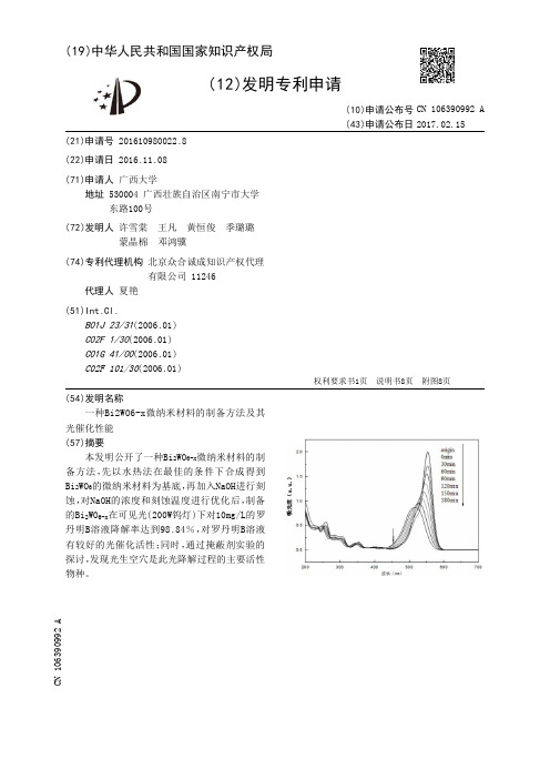
(19)中华人民共和国国家知识产权局(12)发明专利申请(10)申请公布号 (43)申请公布日 (21)申请号 201610980022.8(22)申请日 2016.11.08(71)申请人 广西大学地址 530004 广西壮族自治区南宁市大学东路100号(72)发明人 许雪棠 王凡 黄恒俊 季璐璐 蒙晶棉 邓鸿骥 (74)专利代理机构 北京众合诚成知识产权代理有限公司 11246代理人 夏艳(51)Int.Cl.B01J 23/31(2006.01)C02F 1/30(2006.01)C01G 41/00(2006.01)C02F 101/30(2006.01)(54)发明名称一种Bi2WO6-x微纳米材料的制备方法及其光催化性能(57)摘要本发明公开了一种Bi 2WO 6-x 微纳米材料的制备方法,先以水热法在最佳的条件下合成得到Bi 2WO 6的微纳米材料为基底,再加入NaOH进行刻蚀,对NaOH的浓度和刻蚀温度进行优化后,制备的Bi 2WO 6-x 在可见光(200W钨灯)下对10mg/L的罗丹明B溶液降解率达到98.84%,对罗丹明B溶液有较好的光催化活性;同时,通过掩蔽剂实验的探讨,发现光生空穴是此光降解过程的主要活性物种。
权利要求书1页 说明书8页 附图8页CN 106390992 A 2017.02.15C N 106390992A1.一种Bi 2WO 6-x 微纳米材料的制备方法,其特征在于,包括以下步骤:步骤一,Bi 2WO 6的制备:先在烧杯A中加入Bi(NO 3)3·5H 2O、蒸馏水和浓硝酸,在磁力搅拌器上搅拌溶液至完全溶解;同时将Na 2WO 4·2H 2O和蒸馏水在烧杯B中搅拌至完全溶解;将Na 2WO 4溶液逐滴滴加进A烧杯中,耗时20-30min;滴加完毕后,用HNO 3和NaOH溶液调节前驱液pH值为0.9-1.1,再搅拌12-18min;将上述溶液转移进水热反应釜中,满足填充度≤75%,置于烘箱,恒温120℃反应18-24h;反应完成后,将样品离心分离,分别多次用水洗、醇洗沉淀,再恒温70℃干燥18-21h,研磨可得Bi 2WO 6;步骤二,刻蚀反应:取强碱溶于去离子水,再加入Bi 2WO 6,调节磁力搅拌器至20-90℃,刻蚀0.5-6小时;反应完毕后,将样品离心分离,用60-70℃热水多次洗涤至溶液呈中性;再将固体置于烘箱中,恒温70℃干燥5-8h,即得到产品Bi 2WO 6-x 。
Microbiome中科院邓晔等...
Microbiome中科院邓晔等...推荐:江舜尧编译:晚香编辑:董小橙中科院资源与环境学院、中科院生态环境研究中心邓晔研究员等人于2019年5月4日在《Microbiome》上发表题目为《Unraveling the diversity of sedimentary sulfate-reducing prokaryotes (SRP) across Tibetan saline lakes using epicPCR》的文章。
该文章利用epicPCR技术揭示了西藏咸水湖沉积物中硫酸盐还原原核微生物(SRP)的多样性。
文章摘要硫酸盐的还原在整个生态圈中是一个重要的生物地球化学循环过程,然而,硫酸盐还原剂的主要分类群尚未完全确定。
本研究利用epicPCR(乳液,配对分离,PCR串联反应)技术对青藏高原咸水湖沉积物中硫酸盐还原原核生物(SRP)进行了系统发育进化研究。
对10个湖泊16S rRNA基因PCR扩增产物和融合了16S rRNA + dsrB 基因的epicPCR扩增产物分别测序,共检测到12519个OTUs和883个SRP-OTUs,其中变形杆菌门、厚壁菌门和拟杆菌门为优势门。
120个高丰度的SRP-OTUs(至少一个样品中>1%)属于17个门水平,其中仅有7个属于SRP门。
大多数来自整个微生物群落和SRPs的OTUs 都不是在超过一个以上的特定湖泊中检测到的,这表明其具有高度的地方特异性。
微生物群落和SRP的亚群体的α多样性表现出显著的正相关性。
采样前一个月的pH值和平均水温是整个微生物群落的环境决定因素,但是,平均水温和总氮量是SRP亚群体的主要环境驱动因素。
本研究发现西藏咸水湖中仍有许多未经证实的SRP,其中有许多可能是地方特异性的并适应特定的环境条件。
关键词:硫酸盐还原原核生物,功能分类多样性,epicPCR,西藏咸水湖文中主要图片说明图1 | 本研究中西藏咸水湖采样点地图。
参考文献
[1] Devaux P G, Horning M G, Horning E C. Benzyloxime derivatives of steroids – new metabolic profile procedure for human urinary steroids[J]. Anal Lett, 1971, 4(3): 151-160.[2] Oliver S G. Yeast as a navigational aid in genome analysis[J]. Microbiology, 1997, 143(5): 1483-1487.[3] Nicholson J K, Lindon J C, Holmes E. 'Metabonomics': understanding the metabolic responses of living systems to pathophysiological stimuli via multivariate statistical analysis of biological NMRspectroscopic data[J]. Xenobiotica, 1999, 29(11): 1181-1189.[4] Bales J R, Higham D P, Howe I, et al. Use of high-resolution proton nuclear magnetic resonance spectroscopy for rapid multi-component analysis of urine[J]. Clin Chem, 1984, 30(3): 426-432. [5] Lehnert W, Hunkler D. Possibilities of selective screening for inborn errors of metabolism using high-resolution proton-Fourier transform-NMR spectrometry[J]. Eur J Pediatr, 1986, 145(4):260-266.[6] Holmes E, Foxall P J D, Spraul M, et al. 750 MHz1H NMR spectroscopy characterization of thecomplex metabolic pattern of urine from patients with inborn errors of metabolism:2-hydroxyglutaric aciduria and maple syrup urine disease[J]. J Pharm Biomed Anal, 1997, 15(11): 1647-1659.[7] Khan A R, Rana P, Tyagi R, et al. NMR spectroscopy based metabolic profiling of urine and serumfor investigation of physiological perturbations during radiation sickness[J]. Metabonomic, 2011, 7(4): 583-592.[8] Bollard, M.E., Keun, Beckonert O, et al. Comparative metabonomics of differential hydrazine toxicity in the rat and mouse[J]. Toxicol Appl Pharmacol, 2005, 204(2): 135-151.[9] 李英帅,王琦,袁卓珺. 阳虚体质者血清和尿液的核磁共振代谢组学[J]. 高等学校化学学报,2011, 32(11): 2521-2527.[10] Kallweit E, Henning M, Groeneveld E. Application of NMR techniques on the body composition oflive animals. London, Elsevier, 1989.[11] Nicholson, J.K., Foxall, et al. 750 MHz1H and1H -13C NMR spectroscopy of human bloodplasma[J]. Anal Chem, 1995, 67(5): 793-811.[12] Liu M, Nicholson J K, Parkinson J A, et al. Measurement of biomolecular diffusion coefficients inblood plasma using two-dimensional1H-1H diffusion-edited total-correlation NMR spectroscopy[J].Anal Chem, 1997, 69(8): 1504-1509.[13] Nicholson J K, Wilson I D. High resolution proton magnetic resonance spectroscopy of biologicalfluids[J]. Prog Nucl Magn Reson Spectrosc, 1989, 21(4-5): 449-501.[14] Fan T W M. Metabolite profiling by one- and two-dimensional NMR analysis of complex mixtures[J]. Prog Nucl Magn Reson Spectrosc, 1996, 28: 161-219.[15] Qian B, Yan G Y, Deng P C, et al. NMR-based metabonomic study of the sub-acute toxicity of titanium dioxide nanoparticles in rats after oral administration[J]. Nanotechnology, 2010, 21(12): 125105.[16] Zhang L M, Ye Y F, Tang H R, et al. Systems responses of rats to aflatoxin B1 exposure revealedwith metabonomic changes in multiple biological matrices[J]. J Proteome Res, 2011, 10(2):614–623.[17] 许禄, 邵学广. 化学计量学, 北京, 科学出版社, 2005.[18] 边肇祺, 张学工. 模式识别(第二版), 北京, 清华大学出版社, 2000.[19] 周红光, 陈海彬, 王瑞平, 等. 代谢组学在中药复方研究中的应用[J]. 中国药理学, 2013,29(2):161-165.[20] 刘霞, 高红昌, 林东海, 等. 基于1H NMR 代谢组学方法分析马兜铃酸I 诱导的雌雄小鼠急性肾毒性[J]. 高等学校化学学报, 2010, 36(5): 927-932.[21] 苏梦翔, 高旋, 杭太俊, 等. 基于核磁共振代谢组学法研究雷公藤多苷片对大鼠代谢的影响[J]. 中国中药杂志, 2011, 11(36): 1149-1153.[22] Coen M, Ruepp S U, Lindon J C, et al. Integrated application of transcriptomics and metabonomicsyields new insight into the toxicity due to paracetamol in the mouse[J]. J Pharm Biomed Anal, 2004,35(1): 93-105.[23] Sehnaekenberg L, Beger R D, Dragan Y. NMR-based metabonomic evaluation of livers from ratschronically treated witht amoxifen, mestranol, and phenobarbital[J]. Metabolomies, 2005, 1(1): 87-94.[24] Raamsdonk L M, Teusink B, Broadhurst D, et al. A functional genomics strategy that uses Metabolome data to reveal the phenotype of silent mutations[J]. Nat Biotechnol. 2001, 19(1):45-50.[25] Bandy J G, Spurgeon D J, Svendsen C, et al. Earthworm species of the genus Eisenia can be phenotypically differentiated by metabolic profiling[J]. FEBS Lett, 2002, 521(1-3): 115-120. [26] Catchpole G S, Beckmann M, Enot D P, et al. Hierarchical metabolomics demonstrates substantialcompositional similarity between genetically modified and conventional potato crops[J]. NatlAcadSci USA, 2005, 102(40): 14458-14462.[27] Zhang X Y, Tang H R, Ji L N, et al. Human serum metabonomic analysis reveals progression axesfor glucose intolerance and insulin resistance statuses[J]. J Proteome Res, 2009, 8(11): 5188–5195.[28] Zhang H Y, Jia J M, Gao H C.1H NMR-based metabonomics study on serum of renal interstitialfibrosis rats induced by unilateral ureteral obstruction[J]. Mol Bio Syst, 2012, 8: 595–601. [29] Qi S W, Tu Z G, Dai Y, et al.1H NMR-based serum metabolic profiling in compensated anddecompensated cirrhosis[J]. World J Gastroenterol, 2012, 18(3): 285-290.[30] Rocha C M, Barros A S, Duarte I F, et al. Metabolic profiling of human lung cancer tissue by1Hhigh resolution magic angle spinning (HRMAS) NMR spectroscopy[J]. J Proteome Res, 2010, 9(1): 319–332.[31] He Q, Kong X, Wu G, et al. Metabolomic analysis of the response of growing pigs to dietary L-arginine supplementation[J]. Amino Acids, 2009, 37 (1):199 -208.[32] Martin J C, Canlet C, Delplanque B, et al.1H NMR metabonomics can differentiate the earlyatherogenic effect of dairy products in hyperlipidemic hamsters[J]. Atherosclerosis, 2009, 206(1): 127 -133.[33] 杨仓良. 毒药本草, 北京, 中国中医药出版社, 1993.[34] 周立国. 中药毒性机制及解毒措施, 北京, 人民卫生出版社, 2006.[35] 苑静. 砒霜的化学成分及药用的研究进展[J]. 河南化工, 2010, 27(2): 88.[36] 孙贵范. 深人研究慢性砷中毒的分子作用机制[J]. 中国地方病学杂志, 2004, 23(1): 1-2.[37] 周晓倩, 何云, 吴君. 砷中毒所致氧化应激与肝损伤[J]. 中国地方病学杂志, 2007, 26: 114-116.[38] 李富军, 皮静波, 孙贵范. 砷对作业工人脂质过氧化水平的影响[J]. 中国公共卫生学报, 1997,16(1): 58.[39] 刘连新, 朱安龙, 陈炜, 等. 三氧化二砷对原发性肝癌的作用及其机理研究[J]. 中华外科杂志, 2005, 1.[40] Zhu Q, Zhang J W, Zhu H Q, et al. Synergic effects of arsenic trioxide and c-AMP during acute promyelocytic leukemia cell maturation subtends a novel signaling cross-talk[J]. Blood, 2002, 99(3): 1014-1022.[41] Guillemin M C, Raffoux E, Vitoux D, et al. In vivo activation of cAMP signaling induces growth arrest and differentiation in acute promyelocytic leukemia[J]. J Exp Med, 2002,196(10): 1373-1380.[42] Ling Y H, Jiang J D, Holland J F, et al. Arsenic trioxide produces polymerization ofmicrotubules and mitotic rest before apoptosis in human tumor cell lines[J]. Mol Pharmacol, 2002, 62(3): 529.[43] Sanz M A, Grimwade D, Tallman M S, et al. Management of acute promyelocytic leukemia: recommendations from an expert panel on behalf of the European Leukemia Net[J]. Blood, 2009, 113(9): 1875-1891.[44] Kanzawa T , Kondo Y, Ito H, et al. Induction of autophagic cell death in malignant glioma cells byarsenic trioxide[J]. Cancer Research, 2003, 63(9): 2103-2108.[45] Nakagawa Y, Akao Y, Morikawa H, et al. Arsenic trioxide-induced apoptosis through oxidative stress in cells of colon cancer cell lines[J]. Life Sciences, 2002, 70(19): 2253-2269.[46] Sun R C, Board P G, Blackburn A C. Targeting metabolism with arsenic trioxide and dichloroacetatein breast cancer cells[J]. Molecular Cancer, 2011, 10(142): 1-15.[47] Crawford M A, Milne M D, Scribner B H. The effects of changes in acid-base balance on urinarycitrate in the rat[J]. J Physiol, 1959, 149: 413-423.[48] Phipps A N, Stewart J, Wright B, et al. Effect of diet on the urinary excretion of hippuric acid andother dietary-derived aromatics in rat. A complex interaction between diet, gut microflora and substrate specificity[J]. Xenobiotica, 1998, 28(5): 527-537[49] Delaney J, Neville W A, Swain A, et al. Phenylacetylglycine, a putative biomarker of phospholipidosis: its origins and relevance to phospholipid accumulation using amiodarone treatedrats as a model[J]. Biomarkers, 2004, 9(3): 271-290.[50] Daykin C A, Groenewegen A, Mulder T P J, et al. Nuclear magnetic resonance spectroscopic basedstudies of the metabolism of black tea polyphenols in humans[J]. J Agric Food Chem, 2005, 53(5): 1428-1434.[51] Wang Y L, Tang H R, Nicholson J K, et al. A metabonomic strategy for the detection of the metabolic effects of chamomile (Matricaria recutita L.) ingestion[J]. J Agric Food Chem, 2005,53(2): 191-196.[52] Wyss M, Kaddurah-Daouk R. Creatine and creatinine metabolism[J]. Physiol Rev, 2000, 80(3): 1107-1213.[53] Mudd S H, Poole J R. Labile methyl balances for normal humans on various dietary regimens[J].Metabolism, 1975, 24(6): 721-735.[54] Im, Y S, Chiang P K, Cantoni G L. Guanidoacetate methyltransferase. Purification and molecularproperties[J]. J Biol Chem, 1979, 254(21): 11047-11050.[55] Waterfield C J, Turton J A, Scales M D C, et al. Investigations into the effects of various hepatotoxic compounds on urinary and liver taurine levels in rats[J]. Arch Toxicol, 1993, 67(4): 244-254.[56] Clayton T A, Lindon J C, Everett J R., et al. Hepatotoxin-induced hypercreatinaemia andhypercreatinuria: their relationship to one another, to liver damage and to weakenednutritionalstatus[J]. Arch Toxicol, 2004, 78(2): 86-96.[57] 廖沛球, 薛蓉, 吴亦洁, 等. 给药硝酸镨后大鼠尿液和血清的核磁共振代谢组学研究[J]. 分析化学, 2012, 40(9): 1421-1428.[58] Foxalla P J D, Spraulb M, Farrant R D, et al. 750 MHz 1H and 1H-13C NMR spectroscopy of human blood plasma[J]. J Pharmaceut Biomed, 1993, 1(4–5): 267–276.[59] Pi J, Yamauchi H, Kumagai Y, et al. Evidence for Induction of Oxidative Stress Caused by ChronicExposure of Chinese Residents to Arsenic Contained in Drinking Water[J]. Environmental Health Perspectives, 2002, 110(4): 331-336.[60] Flora S J S. Arsenic-induced oxidative stress and itsreversibility following combined administrationof N-acetylcysteine and meso 2,3-dimercaptosuccinic acid in rats[J]. Clinical and Experimental Pharmacology and Physiology, 1999, 26(11): 865-869.[61] Kitchin K T, Ahmad S. Oxidative stress as a possible mode of action for arsenic Carcinogenesis[J].Toxicology Letters, 2003, 137(1-2): 3-13.[62] Griffin J L, Mann C J, Scott J, et al. Choline containing metabolites during cell transfection: an insight into magnetic resonance spectroscopy detectable changes[J]. FEBS letters, 2001, 509(2): 263-266.[63] Felig P. The glucose-alanine cycle[J]. Metabolism, 1973, 22(2): 179-207.[64] Wei Lai, Liao Pei Q, Wu H F, et al. Metabolic profiling studies on the toxicological effects of realgar in rats by1H NMR spectroscopy[J]. Toxicology and Applied Pharmacology, 2009, 234(3):314-325.[65] 王强玉, 王彦田. 砒石抑制肿瘤作用机制的研究进展[J]. 河北中医药学报, 2008, 23 (2): 43-45.[66] Kitchin K T. Recent advances in arsenic carcinogenesis: modes of action, animal model systems,and methylated arsenic metabolites[J]. Toxicol Appl Pharmacol, 2001 172 (3): 249-261.[67] Thomas D J, Styblo M, Lin S. The cellular metabolism and systemic toxicity of arsenic[J]. Toxicology and applied pharmacology, 2001, 176(2): 127-144.[68] Warburg O, Wind F, Negelein E. The metabolism of tumors in the body[J]. Journal of General Physiology, 1927, 8(6):519-530.刘昌孝、颜贤忠和许国旺以代谢物组学为参照介绍血液指纹图谱中医药信息学研究方法赵宏杰(吉林市中医院吉林吉林132011)雷钧涛(吉林医药学院吉林吉林132011)摘要:血液指纹图谱中医药信息学研究方法是一种新的中医药现代化研究方法,为方便大家了解和认识这一方法,特以代谢物组学为参照介绍了它的一些特点。
- 1、下载文档前请自行甄别文档内容的完整性,平台不提供额外的编辑、内容补充、找答案等附加服务。
- 2、"仅部分预览"的文档,不可在线预览部分如存在完整性等问题,可反馈申请退款(可完整预览的文档不适用该条件!)。
- 3、如文档侵犯您的权益,请联系客服反馈,我们会尽快为您处理(人工客服工作时间:9:00-18:30)。
Plant Cell,Tissue and Organ Culture49:149–151,1997.149 c1997Kluwer Academic Publishers.Printed in the Netherlands.Research noteMicropropagation of Sego LilyQian Hou1,John G.Carman&William A.VargaDepartment of Plants,Soils,and Biometeorology,Utah State University,Logan,UT84322-4820,USA;(1present address:United States Sugar Corporation,Research Dep.,P.O.Drawer1207,Clewiston,FL33440,USA; requests for offprints)Received05December1996;accepted in revised form08April1997Key words:bulb propagation,Liliaceae,Calochortus nuttallii T.&G.,plant tissue cultureAbstractThe Sego lily(Calochortus nuttallii T.&G.)produces showyflowers and is a wild species indigenous to the western United States.A3-to5-fold increase in shoots per month was achieved when basal sections of bulbs were cultured on Schenk and Hildebrandt medium containing88mM sucrose and8.9M6-benzyladenine(BA)and subcultured every28d on the same medium but containing88mM sucrose,2.2M BA,and1.0M indole-3-butyric acid.Roots formed from94%of shoots cultured at13C on medium containing88mM sucrose and2.7M-naphthalene acetic acid.Bulbs formed at the base of shoots cultured on medium containing263mM sucrose. Abbreviations:BA-6-benzyladenine;IBA–indole-3-butyric acid;MS–Murashige and Skoog;NAA–-naphthalene acetic acid;SH–Schenk and HildebrandtThe Sego lily,Calochortus nuttallii T.&G.,grows in the great basin region of the United States and is the State Flower of Utah.Its bulbs are edible and were used as emergency food for American Indians and ear-ly caucasian settlers.It produces showyflowers that range in color from white to cream,golden yellow,and mercial availability is limited because plants grown from seedflower only after several years,and production by offsets is slow(Cronquist et al.,1977). We report herein a micropropagation system for pro-ducing large numbers of plantlets and bulbs.Sego lily bulbs were collected in northern Utah in the late summer,disinfested in0.8%NaOCl for20min, rinsed four times in sterile water,and cut horizontally into three sections of equal width.Unless otherwise indicated,cultures were incubated at26C under a 16-h photoperiod provided by cool whitefluorescent lamps(60M m2sec1)on SH(Schenk and Hilde-brandt,1972)medium containing88mM sucrose.The media were adjusted to a pH of5.7prior to adding agar (7.0g l1,Difco Bacto agar),autoclaved(121C,104 kPa)for15min,and dispensed into200ml jars(20ml per jar)or500ml jars(40ml per jar).The jars were closed with plastic caps.Shoot induction from the top,middle,and basal sections of bulbs was evaluated using MS(Murashige and Skoog,1962),half strength MS(1/2MS),and SH (each with88mM sucrose)to which BA(2.2,8.9,and 22.2M)or-naphthalene acetic acid(NAA)(2.7, 10.7,and26.9M)were added.Sections fromfive bulbs per treatment were cultured.Each jar contained three sections from a single bulb(90bulbs total).The explants were recultured at28d to fresh medium.The cultures were scored28d later with zero indicating no shoots and three indicating many shoots.Bulb sections producing multiple shoots in the shoot initiation experiments were cut into pieces con-taining3to5shoots each.In one shoot multiplication experiment,the following four media were tested(each contained88mM sucrose):MS plus50ml l1coconut milk,MS plus2.2M BA and1.1M NAA,MS plus 2.2M BA and1.0M indole-3-butyric acid(IBA), and SH plus1.0M BA and1.0M IBA.Fourteen jars were used per treatment,and6to8explants were added150to each.After28d,numbers of new shoots and shoot height were recorded.Shoot vigor was scored using a scale of0to3with three indicating the best growth. Two media were tested in a second shoot multiplica-tion experiment(each contained88mM sucrose):SH alone and SH plus2.2M BA and1.0M IBA.Four jars were used per treatment(seven explants in each). After28d,multiplication rates were determined based on numbers of new shoots regenerated per explant. These cultures were subcultured every28d to fresh media.Cultures with many shoots were divided into sections containing3to5shoots each,transferred to growth regulator-free medium,and incubated for28d prior to use in rooting and bulb-induction experiments.The effects of NAA(0.5and2.7M),sucrose(88, 175,and263mM),and temperature(26and13C)on root initiation were evaluated in a factorial experiment (18explants per treatment).The explants of each treat-ment combination were incubated for28d,transferred to growth regulator-free SH medium(88mM sucrose) for28d,and scored for root development.Bulb formation was evaluated by transferring explants to six media(three explants per treatment): SH plus88mM sucrose and5g l1activated char-coal,SH plus263mM sucrose and5g l1activated charcoal,SH plus263mM sucrose and0.5M NAA, SH plus263mM sucrose and0.8M abscisic acid, and SH plus263mM sucrose.After28d at26C, explants were transferred to basal medium containing 263mM sucrose.After28d,percentages of shoots with bulbs were recorded on a per treatment basis. In another bulb-formation experiment,shoot numbers, shoot lengths,and dry weights(DW)of shoots and bulbs were recorded after sets of explants(15to19 per treatment)had incubated for28d on three media defined by sucrose level(58,117,and234mM).Each media also contained2.7M NAA and0.9M6-furfurylaminopurine.Rooted shoots(80per treatment)were planted in pots of sand,soil from a Sego lily collection site,equal volumes of perlite,vermiculite,bark,and peat moss, and equal volumes of perlite,vermiculite,and sandy loam soil.These were incubated at4C in a growth chamber for56d or misted(15sec every3min)for28 d at24C in a greenhouse.Bulbs were dried for14d at room temperature(RH40%)and planted in pots and subjected for56d to4C in a growth chamber (12-h photoperiod,300M m2s1)or planted to a Sego lily collection site(late fall).Seventeen bulbs dug from thefield and dried for14d were planted in the field as a control.Table1.Means(SE)by sucrose concentration for the tallest shoot per explant,number of shoots produced per explant,DW per shoot,and DW per bulb.All media contained2.7M NAA and 0.9M6-furfurylaminopurine.Data were ranked and subjected to Kruskal-Wallis analysis.Tallest Shoots per DW(mg) Sucrose(mM)n shoot(cm)explant(no.)Shoot Bulb5818 6.00.6a70.8a8910a243b117158.20.7a30.4b415b132b23419 3.50.3b30.5b365b608aMeans not followed by the same letter are significantly different (p0.05)according to Dunn’s test.We found that shoot formation was stimulated by BA but not NAA.More shoots formed from the basal sections of the bulbs(ranked at1.60.2SE),which contained the meristematic basal plate,than from the middle(ranked at0.80.2)or upper(ranked at 0.40.1)sections.No differences in shoot formation were observed among the nine treatment combinations defined by three BA levels(2.2,8.9,and22.2M)and three media(MS,1/2MS and SH).However,both the explants and the medium around the explants turned dark when MS and1/2MS media were used.This anomaly was rarely observed when SH medium was used.SH medium plus BA and IBA(ranked at2.50.2 SE)was superior to MS medium plus BA and NAA (ranked at1.60.2)or MS medium plus BA and IBA (ranked at1.10.2)in supporting shoot multiplica-tion.However,growth regulator-free MS medium plus coconut milk also produced favorable multiplication rates(ranked at2.00.2),though these cultures turned dark.SH medium with BA and IBA supported a28d shoot multiplication rate of3.3.There were20.3 1.3 (SE)shoots per jar of seven cultures at the beginning of the experiment followed by67.8 3.0shoots per jar at the end of the experiment.In contrast,growth regulator-free medium induced a multiplication rate of only1.4(23.3 1.1at the beginning and32.0 2.0 at the end of the experiment).Medium containing263mM sucrose inhibited root induction in our Sego lily cultures,and this negative effect was accentuated by NAA and low temperatures (Figure1).Roots from the in vitro-produced shoots were thicker and harder than roots fromfield-grown plants,possibly because of a prolonged exposure to exogenous auxin.Bulbs failed to form at the base of plantlets when sucrose levels were low(88mM)or when the media contained NAA.However,17%of shoots exposed to151 Figure1.Effects of NAA,sucrose,and temperature on root initiation of Sego lily shoots in vitro(n=18).The data were subjected to Chi squareanalysis of variance(sucrose and temperature main effects and the sucrose NAA and sucrose temperature interactions were significant). media containing263mM sucrose and no NAA pro-duced bulbs.Neither low temperatures nor the additionof abscisic acid or activated charcoal enhanced bulbletformation.While high sucrose levels in our experi-ments increased bulb DW,low sucrose levels increasedthe numbers,lengths,and DWs of shoots(Table1).Of320rooted shoots transferred to soil,only22(allfrom the low temperature treatment)survived for morethan two months.Soil substrate did not contribute tosurvivability,and none survived transfer to the green-house.Likewise,all of the bulbs produced in tissueculture died.Thirty-three of these were planted in potsand13were planted in thefield.In contrast,13of17bulbs collected from thefield(averaged2.5cm long,2.0cm wide)and replanted to thefield emerged thefollowing spring.The small size of in vitro-producedbulbs(averaged1.2cm long,0.7cm wide),accom-panied by rapid dehydration,which is common withbulbs produced in vitro(Le Nard and Chanteloube,1992),may have caused these results.While progresshas been made in producing rooted Sego lily shoots andbulbs in vitro,further research is required to identifyappropriate acclimatization procedures.AcknowledgementsThis research was supported by the Utah AgriculturalExperiment Station,Utah State University,Logan,UT84322.Approved as journal paper no.4493.ReferencesCronquist A,Holmgren AH,Holmgren NH,Reveal JL&HolmgrenPK(1977)Intermountainflora:Vascular plants of the inter-mountain west,USA,V ol.6.Columbia Univ.Press,NYLe Nard M&Chanteloube Fl(1992)In vitro culture of explantexcised from growing stems of Tulip(Tulipa gesneriana L.):Problems related to bud and bulblet formation.Acta Hort.325:435–440Murashige T&Skoog F(1962)A revised medium for rapid growthand bioassay with tobacco tissue cultures.Physiol.Plant.15:473–497.Schenk RU&Hildebrandt AC(1972)Medium and techniques forinduction and plant growth of monocotyledonous and dicotyle-donous plant cell cultures.Can.J.Bot.50:199–204。
