医学影像学专业英语4Aortic Dissection
影像医学专业英语
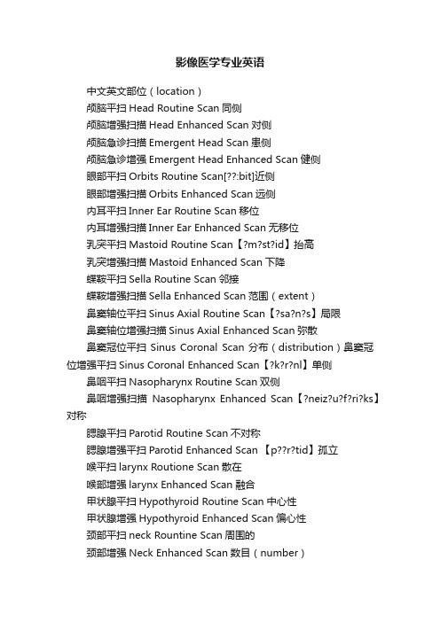
影像医学专业英语中文英文部位(location)颅脑平扫Head Routine Scan同侧颅脑增强扫描Head Enhanced Scan对侧颅脑急诊扫描Emergent Head Scan患侧颅脑急诊增强Emergent Head Enhanced Scan健侧眼部平扫Orbits Routine Scan[??:bit]近侧眼部增强扫描Orbits Enhanced Scan远侧内耳平扫Inner Ear Routine Scan移位内耳增强扫描Inner Ear Enhanced Scan无移位乳突平扫Mastoid Routine Scan【?m?st?id】抬高乳突增强扫描Mastoid Enhanced Scan下降蝶鞍平扫Sella Routine Scan邻接蝶鞍增强扫描Sella Enhanced Scan范围(extent)鼻窦轴位平扫Sinus Axial Routine Scan【?sa?n?s】局限鼻窦轴位增强扫描Sinus Axial Enhanced Scan弥散鼻窦冠位平扫Sinus Coronal Scan分布(distribution)鼻窦冠位增强平扫Sinus Coronal Enhanced Scan【?k?r?nl】单侧鼻咽平扫Nasopharynx Routine Scan双侧鼻咽增强扫描Nasopharynx Enhanced Scan【?neiz?u?f?ri?ks】对称腮腺平扫Parotid Routine Scan不对称腮腺增强平扫Parotid Enhanced Scan 【p??r?tid】孤立喉平扫larynx Routione Scan散在喉部增强larynx Enhanced Scan融合甲状腺平扫Hypothyroid Routine Scan中心性甲状腺增强Hypothyroid Enhanced Scan偏心性颈部平扫neck Rountine Scan周围的颈部增强Neck Enhanced Scan数目(number)肺栓塞扫描lung Embolism Scan单发胸腺平扫Thymus Rountine Scan多发胸腺增强Thymus Enhanced Scan增多胸骨平扫Sternum Rountine Scan减少胸骨增强Sternun Enhanced Scan大小(size)胸部扫描Chest Routine Scan大胸部薄层扫描High Resolution Chest Scan小胸部增强扫描Chest Enhanced Scan稳定胸部穿刺Chest Puncture Scan扩大轴位胸部穿刺Axial Chest Puncture Scan扩张上腹部平扫upper abdomen routine scan 膨胀中腹部平扫mid- abdomen routine scan 缩小上腹部增强扫描upper abdomen enhanced scan体积缩小中腹部增强扫描mid- abdomen enhanced scan 狭窄腹部穿刺Abdomen Puncture Scan闭塞轴位腹部穿刺Axial Abdomen Puncture Scan形状(shape)颈椎胸椎腰椎平扫C/T/L-Spine Routine Scan点状盆腔平扫/增强扫描Pelis Routine /Enhanced Scan斑点状骶髂关节扫描SI Joint Scan粟粒状肩关节扫描Shoulder Joint Scan结节状上肢软组织平扫/增强Upper Extremities Soft Tissue Scan团块状肘关节平扫Elow Joint Routine Scan圆形腕关节平扫Wrist Routine Scan卵圆形手部平扫Hand Routine Scan椭圆形髋关节平扫Hip Joint Routine Scan长方形膝关节平扫Keen Joint Routine Scan分叶形踝关节平扫Ankle Joint Routine Scan片状下肢软组织平扫增强Lower Extremities Soft Tissue Scan/Enhance条索状足部平扫foot routine scan线状血管造影和三维成像网状头部血管造影Head CT Angiography弧线形颈部血管造影Neck CT Angiography星状心脏冠脉造影Coronal Artery Angiography纠集心脏冠脉钙化积分Cardiac Calcium Scoring Scan舟状胸部血管造影Chest CT Angiography哑铃状腹部血管造影AbdomenCT Angiography不规则形上肢血管造影Upper Extremities CT Angiography细致下肢血管造影Lower Extremities CT Angiography粗糙五官三维成像3D Facial Scan变形胃部三维成像3D Stomach CT Scan增粗结肠三维成像3D Colon CT Scan增厚颈椎胸椎腰椎三维成像3D C/T/L -Spine变细肩关节三维成像3D Should Joint变平肘关节三维成像3D Elbow Joint边缘腕关节三维成像3D Wirst Joint轮廓髋关节三维成像3D Hip Joint光滑膝关节三维成像3D KneeJoint锐利踝关节三维成像3D Ankle Joint清晰模糊毛刺状分叶状密度,回声,信号密度透亮阴影不透光致密低密度混杂密度信号低信号高信号性质囊性空洞壁外壁渗出浸润实变增值实性结节状肿块纤维化钙化ipsilateralcontralateralaffected sideintact sideproximal sidedistal sidedeviation;shift;displacement nondisplaced elevation;descent;fallabutting;nextto;secondaryto localized,regional diffuseunilateralbilateralsymmetricasymmetricsolitaryscatteredconfluencecentraleccentricperipheralsolitarymultipledecreaselargesmallstabilityenlargementdilatationdistentionshrinkloss of volumestenosis,narrowingocclusion;obliteration,emphraxis punctual mottingmiliarynodularmasscircular,round,roudedovalellipseoblonglobulatedpathystripelinarreticularcurvilinearstellatecrowding,convergingboat-shaped;navicular;scaphoiddumb-bellfinecoarsedeformitythickenthickenthinningflattenedborder,margin,rim,edgeoutline,contoursmoothsharp,well-defined,well-circumscribed clear ,distinct hazy,indistinct,ill-defined,obscured spiculated multilobulateddensitylucency,transparentshadowhaziness,opacificationdensityhypodense,low densityhyperdense,high densitymixed densityhypointensityhyperintensitycystcavitationwallouter surface of the wall exudationinfiltration consolidationhyperplsiasolid nodule mass fibrosis calcification。
医学影像学专业英语Digestive system(4)
oesophageal varices: mainly caused by cirrhosis of the liver
x-ray appearances
Mucosa abnormality :Thickening, circuitous and disturbance.
Filling defects :longitudinal serpiginous.
Dividing line : not clear.
Contraction and relaxation is not enough good, barium is swallowed slowly.
Tension is lower.
x-ray appearances
Mucosa abnormality :Thickening, circuitous and disturbance.
Filling defects :longitudinal serpiginous.
Dividing line : not clear.
Contraction and relaxation is not enough good, barium is swallowed slowly.
– mucosa:destruction, discontinue or disappear
– filling defect – relaxation is limited, wriggling is weakening or disappeared. – lumens: stenosis, enlargement. – dividing line : clear
• Carcinoma of oesophagus and gastric fundus
医学影像学名词解释

医学影像学名词解释导言医学影像学是一门应用医学和物理学原理,运用不同的方法和技术来生成和解释人体内部结构和功能信息的学科。
通过各种影像技术,医学影像学为医生提供了一种非侵入性的手段来诊断和治疗疾病。
本文将对几个常见的医学影像学名词进行解释。
一、X射线摄影(Radiography)X射线摄影,也称为放射线摄影,是最常见和最常用的医学影像学技术之一。
它通过使用X射线穿透人体,然后在感光片或数字传感器上形成图像。
X射线摄影可用于检测骨折、肿瘤、肺部感染等疾病。
现代医学中广泛应用的数字化X射线技术(Digital Radiography)可以生成高质量的图像,并提供更方便的数据存储和传输。
二、计算机断层扫描(Computed Tomography, CT)计算机断层扫描(CT)是一种基于X射线的成像技术,它能够通过旋转的X射线束和敏感探测器来获取人体多个方向的横断面图像。
这些图像通过计算机进行处理和重建,形成一个连续的三维图像,可用于定位和评估肿瘤、脑出血、血管病变等疾病。
现代CT技术具有高分辨率和多功能性,能提供更准确的影像信息。
三、核磁共振成像(Magnetic Resonance Imaging, MRI)核磁共振成像(MRI)利用强磁场和无害的无线电波来生成人体内部的详细图像。
MRI能够提供高对比度的解剖结构和生理功能信息,并广泛应用于心脏、脑部、腹部、骨骼等部位的诊断中。
MRI技术在医学影像学领域中有着非常重要的地位,是一种无辐射、非侵入性的成像技术。
四、超声成像(Ultrasound Imaging)超声成像是一种使用高频声波来观察和诊断人体内部器官和结构的影像技术。
它通过声波在不同组织间的反射和回波来生成图像。
超声成像广泛应用于妇产科乃至心脏等各种领域,在妊娠期间的胎儿监测、器官肿瘤的识别和定位等方面具有重要作用。
五、正电子发射断层扫描(Positron Emission Tomography, PET)正电子发射断层扫描(PET)是一种核医学影像技术,通过记录和测量体内注射的放射性示踪剂产生的正电子和射线,来获得器官和组织的功能信息。
医学影像学名词中英对照

英中文名词对照第一章acromioclavicular oint肩锁关节air bronchogram支气管气像ankle joint踝关节ankylosis of joint关节强直arches of foot足弓biligrafin胆影葡胺bone age骨龄bone canaliculi骨小管bone cortex皮质骨bone deformity骨骼变形bone destruction骨质破坏bone lacuna骨陷窝bone lamella骨板bony articular surface骨性关节面bursa滑液囊calcification钙化carpal bones腕骨cavity空洞chondral calcification软骨钙化compact bone and spongy bone密质骨和松质骨degeneration of joint关节退行性变destruction of joint关节破坏diaphysis骨干digital subtraction angiography,DSA数字减影血管造影dislocation of joint关节脱位dual photon absorptiometry,DPA双光子吸收法dual X-ray energy absorptiometry,DXA双能X线吸收法elbow joint肘关节encapsulated effusion包裹性积液end plate终板epiphyseal line骨骺线epiphyseal plate骨骺板epiphysis骨骺----998 exudation渗出fibrotic lesion纤维性病变filling defect充盈缺损free pleural effusion游离性胸腔积液haemosiderosis含铁血黄素沉着Hafersian system哈弗系统haversian lamella哈氏骨板hilar dance肺门舞蹈hip joint髋关节hydropneumothorax液化胸hydroxyapatite crystal羟基磷灰石结晶hyperostosis/osteosclerosis骨质增生硬化intercondyloid eminence髁间隆起interlobar effusion叶间积液intermediate lamella骨间板internal and external circumfereutial lamella内、外环骨板interstitial pulmonary oedema间质性肺水肿intervertebral disc椎间盘intervertebral foramen椎间孔intervertebral space椎间隙intra-alveolar pulmonary oedema肺泡性肺水肿joint关节joint capsule关节囊joint cartilage关节软骨joint cavity关节腔joint space关节间隙knee joint膝关节lamellar bone层板骨left atrial enlargement左心房增大left ventricular enlargement左心室增大ligament韧带localized pleural effusion局限性胸腔积液looser zone假骨折线mass肿块medullary space骨髓腔metacarpal bones掌骨metaphysis干骺端metatarsal bones跖骨niche龛影obstructive atelectasis阻塞性肺不张---999 obstucive emphysema阻塞性肺气肿oral cholecystography口服胆囊造影ossification骨化ossification center骨化中心osteoblast成骨细胞osteoclast破骨细胞osteocyte骨细胞osteomalacia骨质软化osteonecrosis骨质坏死osteoporosis骨质疏松periosteal proliferation骨膜增生periosteal teaction骨膜反应periosteum and internal periosteum骨膜和骨内膜phalanges of fingers指骨phalanges lf toes趾骨pleural thickening,adhesion and calcification胸膜增厚、粘连及钙化pleural tumor胸膜肿瘤pneumothorax气胸proliferative lesion增殖性病变pulmonary hilar enlargement肺门增大pulmonary arterial hypertension肺动脉高压pulmonary arterial pleonaemia肺充血pulmonary hypertension肺高压pulmonary oligaemia肺少血pulmonary venous hypertension肺静脉高压pulmonary venous pleonaemia肺淤血quantitative computed tomography,QCT定量CT法right atrial enlargement右心房增大right ventricular enlargement右心室增大sequestrum死骨shoulder joint肩关节soft tissue mass软组织肿块soft tissue swelling软组织肿胀subpulmonary effusion肺下积液swelling of joint关节肿胀tarsal bones跗骨tibia tuberosity胫骨粗隆trabecula骨小梁V olkmann canal福尔克曼管woven bone非层板骨wrist腕关节--1000第二章anterior pararenal space肾旁前间隙aortopulmonary window level主肺动脉窗层面bone window骨窗CT angiography,CTA CT血管造影density resolution密度分辨力distal of the aortic arch level主动脉弓上层面dural sac硬膜囊dynamic contrast-enhanced imaging动态增强扫描electron beam CT,EBCT电子束CTfluid-fluid level液-液平面four-chamber level“四腔面”层面high resolution CT,HRCT高分辨力CT Hounsfield Unit HUintra/extra-capsular ligaments囊内外韧带Lateroconal fascia侧锥筋膜Left atrial level左心房层面Pericardial defect心包缺损Pericardial effusions心包渗出Pericardial neoplasm心包新生物Pericardial thickening and calcification心包增厚和钙化Pericardium心包perirenal space肾周间隙posterior pararenal space肾旁后间隙pulmonary artery level主肺动脉层面soft-tissue window软组织窗spatial resolution空间分辨力spiral CT螺旋CTaortic arch level主动脉弓层面ventricle level心室层面第三章1H31P MR spectroscopy1H和31P MR波谱成像3-D gradient echo imaging三维梯度回波成像技术annulus纤维环bone marrow骨髓chemical-shift imaging化学位移成像coronal view冠状位echo time TE回波时间---1001epidural fat硬膜外脂肪flow void phenomenon流空现象flow-related enhancement流动相关增强functional MRI,fMRI功能成像Gadolinium,Gd钆inversion tecovery,IR反转恢复技术laminar flow层流left ventricular outflow tract view左心室流出道体位left-anterior oblique view左前斜位long-axis长轴位longitudinal relaxation time,T1纵向弛豫时间magnetic tesonance angiography,MRA磁共振血管造影magnetic resonance urography,MRU磁共振尿路造影magnetization transfer with fast adiabatic trajectory磁化传递快速成像methemoglobin正铁血红蛋白MR angiography,MRA MR血管造影MRCP磁共振胰胆管成像Myocardial tagging心肌标记nucleus pulposus髓核phase contrast,PC相位对比法proton density,P质子密度proton relaxation enhancement effect质子弛豫增强效应proton weighted image,PWI质子密度加权像Repetition time,TR重得时间right anterior oblique view右前斜位right ventricular outflow tract view右心室流出道体位sagittal view矢状位short-axis短轴位T1weighted image,T1WI T1加权像T2weighted image,T2WI T2加权像Tagging标记time-of-flight,TOF时间飞跃法Transverse relaxation time,T2横向弛豫时间Transverse view横轴位Turbulent flow湍流water image水成像第四章acoustic shadow声影colour aliasing色彩倒错---1002colour doppler flow imaging,CDFI彩色多普勒血流显像colour scale彩阶continued wave doppler-ultrasoundcardiography,W-连续式多普勒超声心动图UCGduplex doppler ultrasound双功多普勒超声echocardiography or ultrasoundcardiography,UCG超声心动图grayscale灰阶harmonic谐波hemodynamics血流动力学motion mode M型motion mode ultrasoundcardiography,M-UCG M型超声心动图pulsed wave doppler-ultrasoundcardiography,PW-UCG脉冲式多普勒超声心动图real-time实时teal-time grayscale two-dimensional ultrasonic实时灰阶二维超声断面图tomographyteal-time spectral wave graphy实时频谱波图real-time two-dimensional colour doppler flow imaging实时二维彩色多普勒血流显像reverberations混响sampling volume,SV取样容积second harmonic二次谐波spectral aliasing频谱倒错tissue harmonic imaging,THI组织谐波成像transcranial Doppler,TCD经颅多普勒Ultrasonography,USG声像图ultrasound biomicroscopy,UBM超声生物组织显微镜第五章21-trisomy syndrome,Down syndrome21-三体综合征,唐氏综合征achondroplasia软骨发育不全acromegaly肢端肥大症acute gouty arthritis急性痛风性关节炎acute traumatic synovitis急性创伤性滑膜炎aneurysmal bone cyst动脉瘤样骨囊肿ankylosing spondylitis,AS强直性脊柱炎anterior marginal cartilage node,AMCN椎缘骨arachnodactyly蜘蛛脚样指bamboo spine竹节状脊柱Bence Jones protein凝溶蛋白bone bridge骨桥bone infarction骨梗死bone island骨岛bone marrow骨髓---1003bone tumor骨肿瘤burning injury烧伤burst fracture爆裂骨折cafe-au-lait patches咖啡色素斑center-edge angle C-E角Charcot joint夏科关节chondroblastoma成软骨细胞瘤chondroectodermal dysplasia软骨-外胚层发育异常chondroma软骨瘤chondromalacia patellae髌骨软化chondrosarcoma软骨肉瘤chordoma脊索瘤cleidocranial dysostosis颅锁骨发育不全cleidocranial dysplasia颅锁骨发育异常colles’s fracture柯莱斯骨折complete fracture完全骨折complete tear完全撕裂compression fracture压缩骨折compression or wedge fracture压缩或楔形骨折compressive erosions压迫性侵蚀congenital coax vara先天性髋内翻congenital dislocation of the hip先天性髋关节脱位congenital elevation of the scapula先天性肩胛高位症congenital malfomation先天畸形cri-chat syndrome,partial monosomy5p-syndrome猫叫综合征,5p-部分单体综合征anterior and posterior cruciate ligament injuries前、后交叉韧带损伤damatan sulfate,DS硫酸皮肤素degenerative osteoarthrosis退行性骨关节病degenerative spinal diseases脊椎退行性变development dysplasia of the hip,DDP髋关节发育异常disc herniation椎间盘突出disproportionate rhizomelic dwarfism肢根型侏儒dolichostenomelia细长指drawer test抽屉试验dyschondrosteosis软骨骨生成障碍ecchondroma外生性(皮质旁)软骨瘤electrical injury电击伤enchondroma内生性软骨瘤enthesopathy附丽病eosinophilic granuloma嗜酸性肉芽肿epiphyseal injury骨骺损伤---1004Ewing sarcoma尤文肉瘤external callus外骨痂extraskeletal Ewing sarcoma骨外尤文肉瘤facet syndrome小关节面综合征fallen fragment sign陷落征fatigue fracture疲劳骨折fibrodysplasia ossificans progressive进行性骨化性纤维结构不良fibrosarcoma of bone骨纤维网瘤fibrous bridge纤维桥fibrous callus纤维骨痂fibrous cortical defect纤维性骨皮质缺损fibrous dysplasia of bone骨纤维异常增殖症fracture骨折fragmental fracture粉碎性骨折Galeazzi fracture加莱阿齐骨折Gaucher disease高雪病giant cell tumor of bone骨巨细胞瘤giantism巨人症glomerular osteopathy肾小球性骨病gout痛风greenstick fracture青枝骨折haemangioma血管瘤Hand-Schüler-Christian disease韩-薛-柯病hemarthrosis关节积血hematoma血肿hemihypertophy偏身肥大hemivertebra半椎体hemophilic arthritis血友病性关节炎heparan sulfate,HS硫酸类肝素hereditary multiple exostosis遗传性多发性外生骨疣histocytosis X组织细胞增生症X human leukocyte antigen DR4,HLA-DR4白细胞表面相关抗原-DR4 hyperparathyroidism甲状旁腺功能亢进hypertrophic osteoarthropathy,HOA肥大性关节病hypervitaminosis A维生素A过多症hypervitaminosis D维生素D过多症hypovitaminosis D维生素D缺乏症idiopathic osteolysis特发性骨质溶解症incomplete fracture不完全骨折incomplete tear不完全撕裂internal callus内骨痂---1005 intramedullary osteosarcoma髓性骨肉瘤juvenile ankylosing spondylitis,JAS幼年强直性脊柱炎juvenile rheumatoid arthritis,JRA幼年类风湿性关节炎keratan sulfate,KS硫酸角质素Klinefelter syndrome克氏综合征lap seat-belt-type injuries完全带型损伤lateral collateral ligament complexes外侧副韧带复合体lead poisoning铅中毒letterer-Siwe disease勒-雪病leukemia白血病leprosy麻风病ligament injuries韧带损伤lipoma脂肪瘤liposarcoma脂肪肉瘤localized myositis ossificans局限性骨化性肌炎loose body游离体lumbar posterior marginal cartilage node,LPMN腰椎后缘软骨结节lymphangioma淋巴管瘤macromelia先天性巨肢症Madelung deformity马德隆畸形magic phenomenon魔角现象malignant osteoblastoma恶性成骨细胞瘤marble bone大理石骨marfan syndrome马方综合征marginal erosions边缘性侵蚀massive osteolysis大块骨溶解medial collateral ligament complexes内侧副韧带复合体melorheostosis蜡泪样骨病meniscal tears半月板撕裂Monteggia fracture蒙泰贾骨折mosaic pattern镶嵌状结构mucopolysaccharidosis,MPS粘多糖贮积症multiple chondroma多发性软骨瘤multiple osteochondromatosis多发性骨软骨瘤myeloma骨髓瘤myositis ossificans骨化性肌炎neuroarthropathy神经性关节病neurofibromatosis神经纤维瘤病Niemann-Pick disease尼曼-皮克病nonossifying fibroma非骨化性纤维瘤occult fracture陷匿骨折---1006ossifying fibroma骨化性纤维瘤osteitis condensans generalisata周身性致密性骨炎osteitis deformans畸形性骨炎,Paget病ostemoid osteoma骨样骨瘤osteoarthritis骨性关节炎osteoarthrosis deforms endemica大骨节病osteoblastoma成骨细胞瘤osteocartilagenous exostosis骨软骨性外生骨疣osteochondrodysplasias骨软骨发育异常osteochondritis dissecans剥脱性骨软骨炎osteochondroma骨软骨瘤osteochondrosis骨软骨炎osteochondrosis of carpal scaphoid腕舟状骨缺血坏死osteochondrosis of femoral head,Legg-Perthes disease股骨头骨骺缺血坏死osteochondrosis of lunate bone,Kienbock disease腕月骨缺血坏死osteochondrosis of metatarsal head跖骨头骨骺缺血坏死osteochondrosis of tarsal scaphoid,Kohler disease跗舟骨缺血坏死osteochondrosis of tibial tuberosity胫骨结节缺血坏死osteoclastoma破骨细胞瘤osteogenesis imperfecta成骨不全osteogenic sarcoma成骨肉瘤osteoma骨瘤osteoma in the craniofacial bone颅面骨骨瘤osteopathia condensans disseminata播散性致密性骨病osteopathia striata纹状骨病osteopetrosis石骨症osteophyte骨赘osteopoikilosis骨斑点症osteosarcoma骨肉瘤parosteal osteosarcoma骨旁骨肉瘤patellofemoral malalignment髌股关节对合关系异常pathological fracture病理骨折pigmented villonodular synovitis色素沉着绒毛结节性滑膜炎plasmacytoma浆细胞瘤progressive myositis ossificans进行性骨化性肌炎purulent osteomyelitis化脓性骨髓炎pyogenic arthritis化脓性关节炎remodeling改建renal osteodystrophy肾性骨营养不良renal osteopathy肾性骨病renal tubular osteopathy肾小管性骨病---1007 reticuloendotheliosis网状内皮细胞增生病reversed Colles fracture反柯雷骨折rheumatoid arthritis,RA类风湿性关节炎rheumatology风湿病学rickets佝偻病rotator cuff肩袖rotatory atlantoaxial subluxation旋转性寰枢关节半脱位sacralization腰椎骶化sclerosing osteomyelitis硬化性骨髓炎scoliosis脊柱侧弯scurvy坏血病sequestrum死骨seronegative spondylarthritides血清阴性脊椎关节病sickle-cell anemia镰状细胞贫血simple bone cyst骨囊肿skeletal metastases转移性骨肿瘤skip metastases跳跃性转移Smith fracture史密斯骨折soft tissue mass软组织肿块soft tissue swelling软组织肿胀solitary chondroma单发性软骨瘤solitary enchondroma单发性内生软骨瘤spinal osteochondrosis,Scheuermann disease,椎体骺板缺血坏死spinal stenosis椎管狭窄spondylolisthesis脊椎滑脱spondylolysis椎弓峡部不连stress fracture应力骨折stress radiography应力X线摄影subluxation半脱位surface osteosarcoma表面骨肉瘤syndesmophytes韧带赘synovial osteochondromatosis滑膜骨软骨瘤病synovial sarcoma滑膜肉瘤syphilis of bone骨梅毒tenosyovial giant cell tummor腱鞘巨细胞瘤thalassaemia地中海贫血tophi痛风结节transitional anomalies移行椎trauma of bone and joint骨与关节创伤traumatic fracture创伤性骨折traumatic rotatory atlantoaxial dislocation创伤性旋转性寰枢关节脱位--1008 trident hand三叉手tuberculosis of bone骨结核tuberculosis of joint关节结核tuberculosis of spine脊椎结核tumor-like disease瘤样病变Tumer syndrome杜纳综合征vertebral blocks阻滞椎vertebral coalition椎体融合vertebral osteochondrosis of primary ossification center椎体一次化骨中心缺血坏死vitamin C deficiency维生素C缺乏症xanthomatosis黄脂瘤病第六章abestosis石棉肺acquired immunodeficiency syndrome,AIDS艾滋病actinomycosis放线菌病acute military tuberculosis急性粟粒型肺结核agenesis and hypoplasia of the lung肺不发育和肺发育不全allergic pneumonia过敏性肺炎aluminum pneumoconiosis铝尘肺amyloidosis of lung肺淀粉样变性angiogram sign血管造影征anthracosis炭黑尘肺aspergillosis曲菌病bronchiectasis支气管扩张bronchogenic cyst支气管囊肿bronchopneumonia支气管肺炎broncho-pulmonary sequestration支气管肺隔离症butterfly sign蝶翼征cement pneumoconiosis水泥尘肺chronic bronchitis慢性支气管炎chronic pneumonia慢性肺炎coalworker pneumoconiosis煤工尘肺congenital bronchial cysts先天性支气管囊肿contusion of lung肺挫伤cryptococcosis陷球菌病cylindrical bronchiectasis柱状支扩dermoid cyst皮样囊肿diaphragmatic eventration膈膨升diaphragmatic hernia膈疝--1009electric are welder pneumoconiosis电焊工尘肺esophageal cyst食管囊肿foreign body of chest胸部异物foundry worker pneumoconiosis铸工尘肺Goodpasture syndrome肺-肾综合征graphite pneumoconiosis石黑尘肺halo sign晕轮征hamartoma错构瘤hematogenous pulmonary tuberculosis血行播散型肺结构(Ⅱ型) Hodgkin lymphoma霍奇金淋巴瘤Hodgkin disease,HD霍奇金病honeycomb lung蜂窝状肺hydropneumothorax液气胸inflammatory pseudotumor炎性假瘤interstitial pneumonia间质性肺炎intrathoracic goiter胸内甲状腺肿kaolin pneumoconiosis陶工尘肺Kaposi sarcoma卡波济肉瘤laceration and hematoma of lung肺撕裂伤与肺血肿laceratin of trachea and bronchus气管及支气管裂伤lipoma脂肪瘤lobar pneumonia大叶性肺炎loffler syndrome吕弗旨留综合征lung abscess肺脓肿lymphangioma淋巴管瘤lymphoma淋巴瘤mediastinal emphysema纵隔气肿mediastinal hematoma纵隔血肿mediastinal tumor纵隔肿瘤mediastinitis纵隔炎mesothelial cyst间皮囊肿mesothelioma of pleura胸膜间皮瘤mica pneumoconiosis metastatic tumor of pleura胸膜转移瘤mica pneumoconiosis云母尘肺neurogenic neoplasms神经源性肿瘤non Hodgkin lymphoma,NHL非霍奇金淋巴瘤pleural thickening,adhesion and calcification胸膜肥厚粘连和钙化pleuro-peritoneal hiatus hernia胸腹裂孔疝Pneumocomosis尘肺pneumomediastinum纵隔气肿pneumothorax气胸---1010primary complex原发综合征primary tuberculosis原发性肺结核(Ⅰ型) Pulmonary alveolar microlithiasis肺泡微石症Pulmonary alveolar proteinosis肺泡蛋白沉积症Pulmonary arterio-venous fistula肺动静脉瘘Pulmonary arterio-venous malformation,PAVM肺动静脉畸形Pulmonary connective tissue diseases肺结缔组织疾病Pulmonary edema肺水肿Pulmonary emboli肺栓塞Pulmonary infarcts肺梗死Pulmonary sequestration肺隔离症Pulmonary tuberculosis肺结核Pyothorax化脓性胸膜炎radiation pneumonitis放射性肺炎rheumatoid disease of the lung肺类风湿性病saccular bronchiectasis囊状支扩saroidosis结节病secondary pulmonary tuberculosis继发性肺结核(Ⅲ型) seminoma精原细胞瘤silicosis矽肺staphylococal pneumonia葡萄球菌肺炎subacute or chronic hematogenous disseminated pulmonary亚急性和慢性血行播散型肺结核tuberculosissystemic lupus erythomatosis,SLE系统性红斑狼疮talc pneumoconiosis滑石尘肺teratoma畸胎瘤thymoma胸腺瘤tramline sign轨道征traumatic diaphragmatic hernia外伤性膈疝tuberculosis of intrathoracic lymph nodes胸内淋巴结结核tuberculosis pleuritis结核性胸膜炎varicose bronchiectasis静脉曲张型支扩Wegner granuloma韦格肉芽肿第七章aberrant subclavian artery迷走锁骨下动脉aortic coarctation主动脉缩窄atrial septal defect,ASD房间隔缺损cardiomyopathy心肌病constrictive pericarditis缩窄性心包炎cor pulmonale肺源性心脏病--1011coronary artery stenosis冠状动脉狭窄Coronary heart disease冠心病dextrocardia镜面右位心dextroversion右旋心dilated cardiomyopathy扩张性心肌病Eisenmenger syndrome艾森曼格综合征hilar dance肺门舞蹈hypertensive heart disease高血压性心脏病hypertrophic cardiomyopathy肥厚性心肌病ischemic cardiomyopathy缺血性心肌病levoversion左旋心mitral stenosis二尖瓣狭窄patent ductus arteriosus,PDA动脉导管未闭pericardial cyst心包囊肿pericardial effusion心包积液pulmonary artery stenosis肺动脉狭窄restrictive cardiomyopathy限制性心肌病rheumatic heart disease风湿性心脏病right sided aorta右位主动脉弓tetralogy of Fallot法洛四联症total anomalous pulmonary venous drainage肺静脉完全性异位引流ventricular septal defect,VSD室间隔缺损第八章abdominal tuberculoisis腹部结核abscess of liver肝脓肿Achalasia贲门失弛缓症acute abdomen急腹症acute cholecystitis急性胆囊炎acute gastritis急性胃炎acute mechanical intestinal obstruction急性机械性小肠梗阻acute pancreatitis急性胰腺炎adenoma of the samall intestine小肠腺瘤adenomatous dysplasia腺瘤型异型增生adenomatous polyp腺瘤性息肉adenomyomatosis of gall bladder胆囊腺肌增生症advanced gastric cancer进展期胃癌allergic colitis过敏性结肠炎amoebic abscess of liver阿米巴性肝脓肿aperistalsis of the esophagus食管失蠕动症--1012 appendicoliths阑尾结石Barrett esophagus食管消化性溃疡benign tumor of duodenum十二指肠良性肿瘤benign tumors of the small intestine小肠良性肿瘤biligrafin胆影葡胺bright liver光亮肝Budd-Chiari Syndrome布加综合征carcinoid of the appendix阑尾类癌carcinoma of gallbladder胆囊癌cardiospasm贲门痉挛Caroli disease卡罗里病cavernous hemangioma of liver肝海绵状血管瘤cholangiocarcinoma胆管癌cholangiocelluar carcinoma胆管细胞癌cholecystitis胆囊炎cholelithiasis胆结石症cholesterinosis胆固醇沉积病chronic appendicitis慢性阑尾炎chronic cholecystitis慢性胆囊炎chronic gastritis慢性胃炎chronic hepatic schistosomiasis慢性血吸虫肝病chronic pancreatitis慢性胰腺炎cirrhosis of liver肝硬化colonic diverticulosis结肠憩室colonic polyps结肠息肉colorectal carcinoma结直肠癌comet tail sign慧星尾征Congenital anorectat anomalies先天性肛门直肠畸形Congenital esophageal atresia先天性食管闭锁Congenital hypertrophic pyloric stenosis先天性肥厚性幽门狭窄corrosive esophagitis腐蚀性食管炎crohn disease of the colon结肠克罗恩病CT angiography,CTA动脉造影CTCT arterial portography,CTAP门脉造影CTcystic tumor of pancrteas胰腺囊性肿瘤depressed type浅表凹陷型diffuse lesions of liver弥漫性肝病Diverticulitis憩室炎diverticulum of the small intestine小肠憩室double-layer echo双层回声duodenal cancer十二指肠癌---1013 duodenal diverticula十二指肠憩室duodenal sarcoma十二指肠肉瘤elevated type浅表隆起型esophageal carcinoma食管癌esophageal diverliculum食管憩室esophageal foreign bodies食管异物esophageal hiatus hernia食管裂孔疝esophageal spasm食管痉挛esophageal varices食管静脉曲张excavated type凹陷型familial polyposis家族性结肠息肉综合征fatty liver脂肪肝Flat type浅表平坦型focal nodular hyperplasia,FNH局灶性结节性增生foramen of Winslow小网膜囊温氏孔fungus abscess of liver霉菌性肝脓肿Gastric bezoar胃石gastric carcinoma胃癌gastric polyp胃息肉gastric varices胃静脉曲张gastric volvulus胃扭转gastrinoma胃泌素gastritis素瘤炎glucagonoma胰高血糖素瘤halo sign晕征hemochromatosis血色素沉着症hepatic adenoma肝腺瘤hepatocellular carcinoma肝细胞癌hepatolenticular degeneration肝豆状核变性Hirschsprung disease or aganglionosis of the colon先天性巨结肠hydatid disease of liver肝棘球蚴病hyperplastic polyp增生性息肉insolinoma胰岛素瘤intestinal obstruction肠梗阻Intussusception肠套叠ischemic colitis缺血性结肠炎juvenile polyposis幼年性结肠息肉综合征laceration of liver肝损伤Leiomyoma平滑肌瘤leiomyoma of esophagus食管平滑肌瘤leiomyoma of the small intestine小肠平滑肌瘤---1014 leiomyosarcoma of stomach胃平滑肌瘤lipoma脂肪瘤liver cell adenoma肝细胞腺瘤liver cyst肝囊肿lymphoma of the small intestine小肠淋巴瘤malabsorption syndrome吸收不良综合症malignant lymphoma of spleen脾恶性淋巴瘤malignant lymphoma of stomach胃恶性淋巴瘤malignant tumors of the small intestine小肠恶性肿瘤megaesophagus巨食管Mirrizzi syndrome米利兹综合症MR cholangio-pancreaticography,MRCP磁共振胰胆管成像mucous cystadenoma粘液性囊腺瘤multiple juvenile polyposis结肠多发性幼年息肉综合症neurinoma神经鞘瘤neurofibroma神经纤维瘤neurogenic tumor神经源性肿瘤niche龛影obstruction of biliary tract胆道梗阻oral cholecystography口服胆囊造影pancreatic carcinoma胰腺癌pancreatic cyst胰腺囊肿pancreatic islet cell tumor胰岛细胞瘤paralytic ileus麻痹性肠梗阻perforation of gastro-intestinal tract胃肠道穿孔peritoneal abscess腹腔脓肿peritoneal cavity腹膜腔peritoneal tumor腹腔肿瘤peritonitis腹膜炎percutaneous transhepaic cholangiography,PTC经皮肝穿刺胆管造影pyogenic abscess of the liver细菌性肝脓肿reflux esophagitis返流性食管炎ring sign环征rupture spleen脾破裂sarcoma of the stomach胃肉瘤secondary tumors of liver肝转移瘤serous cystadenoma浆液性囊腺瘤somatostainoma生长抑素瘤splenic cyst脾囊肿splenic hemangioma脾血管瘤splenic lymphangioma脾淋巴管瘤---1015 Strangulated intestinal obstruction绞窄性小肠梗阻Superficial type浅表型tadpole-tail sign蝌蚪尾征toxic dilatation of the colon结肠中毒扩张tracheophageal fistula气管食管瘘tuberculosis of mesenteric lymph node肠系膜淋巴结核tuberculosis of the colon结肠结核tuberculosis of the small intestine肠结核tuberculous peritonitis结核性腹膜炎ulcer of the stomach胃溃疡ulcerative colitis溃疡性结肠炎VIPoma舒血管肠肽瘤V olvulus of cecum盲肠扭转V olvulus of sigmoid colon乙状结肠扭转第九章abdominal aortic aneurysm腹主动脉瘤abdominal aortic dissection腹主动脉夹层adrenal myelolipoma肾上腺髓脂瘤adrenal Addison disease肾上腺型阿狄森病adrenal cyst肾上腺囊肿adrenal glands肾上腺adrenal hyperplasia肾上腺增生adrenal insufficiency diseases肾上腺功能低下性病变adrenal metastasis肾上腺转移瘤adrenal pheochromocytoma肾上腺嗜铬细胞瘤adrenal tuberculosis肾上腺结核adrenocortical carcinoma肾上腺皮质癌Angioleiomyolipoma血管平滑肌脂肪瘤bladder calculus膀胱结石bladder carcinoma膀胱癌cervical carcinoma宫颈癌chronic pyelonephritis慢性肾盂肾炎complicated cyst复杂性囊肿congenital abnormities of urinary system泌尿系统的先天性发育异常congenital anomalies of female reproductive tract女性生殖道先天性畸形congenital anomalies of inferior vena cava下腔静脉先天性异常cystic teratoma囊性畸胎瘤Cystitis膀胱炎duplication of kidney重复肾--1016ectopic kidney异位肾endometrial carcinoma子宫内膜癌horseshoe kidney马蹄肾hyperfunctioning adrenal diseases肾上腺功能亢进性病变idiopathic atrophy of adrenal glands特发性肾上腺萎缩contiuation下腔静脉中断并奇静脉/半奇静脉连续interrupted inferior cava with azygos/hemiazygosLymphoma淋巴瘤malrotation of kidney肾旋转异常mucinous cystadenocarcinoma粘液性囊腺癌mucinous cystadenoma粘液性囊腺瘤neuroblastoma成神经细胞瘤nonepithelial neoplasms非上皮性肿瘤nonfunctioning adrenal adenoma肾上腺非功能性皮腺瘤nonfunctioning adrenal cortical carcinoma肾上腺非功能性皮质癌nonfunctioning adrenal diseases肾上腺非功能性病变nontumoral and tumoral thromboses of the IVC下腔静脉非肿瘤性和肿瘤性血栓ovarian cyst卵巢囊肿pituitary Addison disease垂体型阿狄森病polycystic kidney disease肾多囊性病变prostate cancer前列腺癌prostatic hyperplasia前列腺增生pyelonephritis肾盂肾炎renal adscess肾脓肿renal agenesis肾缺如renal calculus肾结石renal carcinoma肾癌renal hypoplasia肾发育不全renal injuries肾外伤renal pelvic carcinoma肾盂癌renal tuberculosis肾结核serous cystadenocarcinoma浆液性囊腺癌serous cystadenoma浆液性囊腺瘤simple cyst of kidney肾单纯性囊肿testicular tumor睾丸肿瘤tuberculosis of urinary bladder膀胱结核tumor of retroperitoneal space腹膜后肿瘤tumor of ureter输尿管肿瘤tumor of urinary bladder膀胱肿瘤ureteral calculus输尿管结石ureteral trberculosis输尿管结核--1017Ureterocele输尿管膨出uterine leiomyoma子宫平滑肌瘤vanillylmandelic acid,VMA香草基扁桃酸vascular diseases of kidney肾血管性病变wandering kidney游走肾第十章acoustic neurinoma听神经瘤acute disseminated encephalomyelitis急性播散性脑脊髓炎Adrenoleukodystrophy肾上腺脑白质营养不良arachnoid cyst蛛网膜囊肿arterio-venous malformation,A VM动静脉畸形Astrocytoma星形细胞瘤brain abscess脑脓肿brain atrophy脑萎缩capillary telangiectasia毛细血管扩张症cavernous angioma海绵状血管瘤cerebral hydatidosis脑囊虫病cerebral heterotopic gray matter脑灰质异位cerebral cysticercosis脑包虫病cerebral infarction脑梗死cerebral vascular malformation脑血管畸形congenital hydrocephalus先天性脑积水contusion of brain脑挫伤Craniopharyngioma颅咽管瘤Craniostenosis狭颅症cryptic A TM隐匿性A TM Cytomegalovirus巨细胞包涵体病毒diffuse injury of brain弥漫性脑损伤Encephalomalacia脑软化encephalotrigeminal angiomatosis脑颜面血管瘤病Ependymitis室管膜炎Ependymoma室管膜瘤epidural hematoma硬膜外血肿flow-related enhancement流动相关增强Germinoma生殖细胞瘤Glioma胶质瘤hemorrhage of cerebrla vascular malformation脑血管畸形出血herpes simplex单纯疱疹hypertensive intracerebral hemorrhage高血压性脑出血---1018 hypoplasia of corpus callosum胼胝体发育不全infantile hydrocephalus婴儿性脑积水Intracerebral hematoma脑内血肿Intracranial aneurysm颅内动脉瘤Intracranial hematoma颅内血肿Intracranial hemorrhage颅内出血Intracranial hemorrhage of the newborn新生儿颅内出血Intracranial tuberculosis颅内结核contrsion and laceration of brain脑挫裂伤laceration of brain脑裂伤lacunar infarction腔隙性梗死lissencephaly无脑回畸形medulloblastoma髓母细胞瘤meningioma脑(脊)膜瘤meningocele脑膜膨出meningoencephalocele脑膜脑膨出metachromatic leukodystrophy,MLD异染性脑白质营养不良属罕见病metastatic tumor of the brain脑转移瘤multiple sclerosis,MS多发性硬化neonatal hypoxic-ischemic encephalopathy新生儿缺氧缺血性脑病neurocutaneous syndrome神经皮肤综合征neurofibromatosis神经纤维瘤病neuroglial tumors神经胶质瘤oligodendroglioma少突胶质细胞瘤olivopontocerebellar atrophy橄榄脑桥小脑萎缩pachygyria巨脑回畸形Parkinson disease帕金森病Pinealoma松果体瘤pituitary adenoma垂体瘤Polygyria小脑回畸形purulent meningitis小脓性脑膜炎rubella风疹schizencephaly脑裂畸形subdural fluid accumulation硬膜下积液subdural hematoma硬膜下血肿syringomyelia脊髓空洞症tectun脑顶盖toxoplasmosis弓形体病tuberculoma结核瘤tuberculous meningitis结核性脑膜炎tuberous sclerosis结节性硬化---1019 malformation of Galen vein大脑大静脉畸形venous angioma静脉性血管瘤venous malformation静脉畸形。
西医放射科术语英文翻译
西医放射科术语英文翻译以下是50个常见的西医放射科术语英文翻译:1. 放射学:Radiology2. 放射诊断:Radiologic Diagnosis3. 放射治疗:Radiation Therapy4. 影像学:Imaging5. X射线:X-ray6. 计算机断层扫描:Computed Tomography (CT)7. 核磁共振成像:Magnetic Resonance Imaging (MRI)8. 超声检查:Ultrasound Imaging9. 数字减影血管造影:Digital Subtraction Angiography (DSA)10. 正电子发射断层扫描:Positron Emission Tomography (PET)11. 单光子发射计算机断层扫描:Single Photon Emission Computed Tomography (SPECT)12. 荧光透视:Fluoroscopy13. X射线造影剂:X-ray Contrast Media14. CT灌注成像:CT Perfusion Imaging15. 分子影像学:Molecular Imaging16. 功能影像学:Functional Imaging17. 骨密度测量:Bone Density Measurement18. 乳腺摄影:Mammography19. 介入放射学:Interventional Radiology20. 放射性核素成像:Radioisotope Imaging21. 核医学:Nuclear Medicine22. 影像归档和通信系统(PACS):Picture Archiving and Communication System (PACS)23. 放射剂量:Radiation Dose24. 辐射防护:Radiation Protection25. 放射性衰变:Radioactive Decay26. 辐射单位:Radiation Units27. 图像重建算法:Image Reconstruction Algorithms28. CT值:CT Density Values29. MRI信号强度:MRI Signal Intensity30. X射线滤过器:X-ray Filters31. 影像增强器:Image Intensifiers32. 闪烁器:Scintillators33. 高压发生器:High-Voltage Generators34. 血管造影导管:Angiographic Catheters35. 放射性示踪剂:Radioactive Tracers36. 正电子药物:Positron-Emitting Radiopharmaceuticals37. 单光子药物:Single Photon-Emitting Radiopharmaceuticals38. SPECT显像剂:SPECT Imaging Agents39. CT灌注成像剂:CT Perfusion Imaging Agents40. MRI对比剂:MRI Contrast Agents41. 介入治疗设备:Interventional Therapy Equipment42. 数字乳腺X光机:Digital Mammography Machines43. X射线透视系统:X-ray Fluoroscopy Systems44. 放射治疗计划系统:Radiation Therapy Planning Systems45. 放射治疗设备:Radiation Therapy Equipment46. 质子治疗系统:Proton Therapy Systems47. 重离子治疗系统:Heavy Ion Therapy Systems48. 光子治疗系统:Photon Therapy Systems49. 三维打印在放射科的应用:3D Printing in Radiology Applications。
(完整版)医学影像专业英语

(1)To prospectively evaluate the effect of heart rate, heart rate variability, and calcification dual-source computed tomography (CT) image quality and to prospectively assess diagnostic accuracy of dual-source CT for coronary artery stenosis. by using invasive coronary angiography as the reference standard.前瞻性评价心率、心率变异性及钙化双源计算机断层扫描成像质量的影响及对冠状动脉狭窄的双源性冠状动脉狭窄诊断的准确性评价。
以侵入性冠状动脉造影为参照标准。
(2)Chest radiography plays an essential role in the diagnosis of thoracic disease and is the most frequently performed radiologic examination in the United States. Since the discovery of X rays more than a century ago, advances in technology have yieled numerous improvements in thoracic imaging. Evolutionary progress in film-based imaging has led to the development of excellent screen-film systems specifically designed for chest radiography.胸部X线摄影中起着至关重要的作用在胸部疾病的诊断,是最常用的影像学检查在美国。
医学影像学基本的英语名词(有音标)及知识重点一

放射诊断学(diagnostic radiology)[daɪəg'nɒstɪk] [reɪdɪ'ɒlədʒɪ]X线计算机体层成像(X-ray computed tomography,CT)[tə'mɒgrəfɪ]磁共振成像(magnetic resonance imaging,MRI)['rez(ə)nəns]正电子发射体层显像(positron emission tomography,PET)['pɒzɪtrɒn] [ɪ'mɪʃ(ə)n] 应用图像存档于传输系统(picture achieving and communication system,PACS)计算机辅助诊断(computer-aided diagnosis,CAD)计算机X线成像(computed radiography,CR)[,reidi'ɔɡrəfi]数字X线成像(digital radiography,DR)像素(pixel)['pɪks(ə)l; -sel]体素(voxel)[vɔk'səl]密度分辨力(density resolution)[rezə'luːʃ(ə)n] 空间分辨力(spatial resolution)窗技术(window technique)窗位(window level)窗宽(window width)部分容积现象(partial volume phenomenon)['pɑːʃ(ə)l] ['vɒljuːm]高分辨力CT(high resolution CT,HRCT)CT灌注成像(CT perfusion imaging,CTPI)脑血流量(cerebral blood flow,CBF)['serɪbr(ə)l; sə'riːbr(ə)l]脑血容量(cerebral blood volume,CBV)平均通过时间(mean transit time,MTT)达峰时间(time-to-peak,TTP)CT后处理:电影显示(cine display)、多平面重组(multiplanar reformation,MPR)、曲面重组(curved planar reformation,CPR)、三维显示技术:最大强度投影(maximum intensity projection,MIP)、最小强度投影(minimum intensity projection,minIP)、表面遮盖显示(surface shaded display,SSD)、容积再现技术(volume rendering technique,VRT)、CT仿真内镜(CT virtual endoscopy,CTVE)、组织透明投影(tissue transition projection,TTP)T1加权像(T1 weighted image,T1WI)T2加权像(T12weighted image,T2WI)质子密度加权像(proton density weighted image,PdWI)['prəʊtɒn]含钆(gadolinium,GD)[,gædə'lɪnɪəm]MRI序列:自旋回波序列(spin echo,SE)快速自旋回波序列(turbo SE,TSE,FSE)['tɜːbəʊ]梯度回波序列(gradient echo,GRE)反转恢复(inversion recovery,IR)平面回波成像(echo planar imaging,EPI )频率选择性脂肪抑制(frequency-selective fat-suppression)梯度回波序列上:同相位(in phase,IP)、反相位(opposed phase,OP)磁敏感加权成像(susceptibility weighted imaging,SWI)[sə,septɪ'bɪlɪtɪ]留空效应(flow void)流入相关增强(flow-related enhancement)磁共振血管成像(magnetic resonance angiography,MRA)['rez(ə)nəns][,ændʒɪ'ɒgrəfɪ]时间飞跃法(time of flight)相位对比法(phase contrast)MRI功能成像(functional MRI,fMRI)扩散加权成像(diffusion weighted imaging,DWI)灌注加权成像(perfusion weighted imaging,PWI)扩散张量成像(diffusion tensor imaging,DTI)显示脑白质纤维磁共振波谱(magnetic resonance spectroscopy,MRS)[spek'trɒskəpɪ] 化学位移(chemical shift)。
医学影像学专业英语Respiratory system

of the sternum.
Bulla formation: bulla are seen as thin-walled air-
focal pneumonia .
Radiographic appearances of pulmonary and pleural tuberculosis
The radiographic appearances can be considered under the following broad headings:
Increased retrosternal airspace on lateral film.
Decreased vascular markings in lung fields. The
lung marking are reduced bilatery and fan out in straight lines fron the hilum.
CXR signs of emphysema include:
Overexpanded lungs. If the lungs are enlarged you
should be able to count more than seven. Be careful however because you can sometimes count more than seven ribs in normal patients.
Part 4 Respiratory system
医学影像学常用英文词汇

医学影像学常用英文词汇[1.介绍]在医学影像学领域,了解常用的英文词汇对于交流和沟通至关重要。
本文档旨在提供一个涵盖医学影像学常用英文词汇的范本,供参考使用。
[2.影像学设备]2.1 X光设备- X-ray machine: X射线设备- Radiography: X射线照相术- fluoroscopy: X射线透视- computed Tomography (cT):计算机断层扫描- digital Radiography:数字化射线照相术2.2 核磁共振成像 (MRi)- Magnetic Resonance imaging (MRi):磁共振成像- gradient Magnet:梯度磁体- Rf coil:射频线圈- image Reconstruction:图像重建- diffusion Tensor imaging (dTi):扩散张量成像2.3 超声成像- Ultrasound:超声- Transducer:换能器- doppler effect:多普勒效应- a-mode Ultrasound: a模式超声- b-mode Ultrasound: b模式超声[3.影像学解剖学]3.1 头部和颅脑- Skull:颅骨- brn:脑- cerebrum:大脑- cerebellum:小脑- Ventricles:脑室3.2 胸部- Lungs:肺- Trachea:气管- diaphragm:横膈膜 - esophagus:食道 - heart:心脏3.3 腹部- Stomach:胃- Liver:肝脏- gallbladder:胆囊 - Pancreas:胰腺- kidneys:肾脏[4.影像学病理学]4.1 肿瘤- Tumor:肿瘤- benign:良性- Malignant:恶性 - Metastasis:转移 - Oncology:肿瘤学4.2 动脉疾病- atherosclerosis:动脉粥样硬化- aneurysm:动脉瘤- aortic dissection:主动脉夹层4.3 炎症与感染- inflammation:炎症- abscess:脓肿- cellulitis:蜂窝组织炎- Osteitis:骨炎- Pneumonia:肺炎[5.附件]本文档附带的附件包括:医学影像学常用英文词汇表。
医学影像学 英语

医学影像学英语IntroductionMedical Imaging is a branch of medicine that uses technology to capture images of internal organs, tissues, and structures inside the body. These images help medical professionals to diagnose, monitor, and treat medical conditions. Medical Imaging is a rapidly advancing field with new imaging technologies continually being developed to improve the accuracy, clarity, and speed of the images.Importance of Medical ImagingMedical Imaging plays a key role in modern medicine, allowing doctors and other medical professionals to see inside the body without the need for invasive procedures. The images produced by medical imaging techniques can be used to diagnose a wide range of conditions, including cancers, heart disease, brain disorders, and bone injuries. Medical Imaging can also be used to track the progress of treatment, monitor the development of a condition, and aid in surgical planning.Types of Medical Imaging TechniquesThere are several different types of Medical Imaging techniques, including:1. X-ray Imaging: X-ray Imaging uses electromagnetic radiation to produce images of internal structures in the body. The images produced by X-ray Imaging can be used to diagnose broken bones, pneumonia, tumors, and other conditions.2. Magnetic Resonance Imaging (MRI): MRI uses a magnetic field and radio waves to produce detailed images of internalstructures in the body. MRI is particularly useful for imaging soft tissues and can be used to diagnose brain and spinal cord disorders, tumors, and joint injuries.3. Computed Tomography (CT) Scan: CT scan uses X-rays to produce detailed cross-sectional images of internalstructures in the body. CT scans are useful for diagnosing cancers, bone injuries, and brain disorders.4. Ultrasound Imaging: Ultrasound uses high-frequency sound waves to produce images of internal structures in the body. Ultrasound is safe and painless and is commonly used to diagnose pregnancy, heart defects, and gallstones.5. Nuclear Medicine Imaging: Nuclear Medicine Imaging uses radioactive materials to produce images of internal structures in the body. This type of imaging can be used to diagnose cancers, heart conditions, and bone disorders.ConclusionMedical Imaging is an essential tool for modern medicine, allowing medical professionals to diagnose, monitor, andtreat medical conditions more accurately and efficiently. The field of Medical Imaging is an exciting and rapidly changing one, with new technologies continually being developed to improve the accuracy and clarity of the images produced. Today, Medical Imaging is an integral part of the diagnosis and treatment of many medical conditions, and its importance will only continue to grow in the future.。
医学影像专业英语范文.doc
(1)To prospectively evaluate the effect of heart rate, heart rate variability, and calcification dual-source computed tomography (CT) image quality and to prospectively assess diagnostic accuracy of dual-source CT for coronary artery stenosis. by using invasive coronary angiography as the reference standard.前瞻性评价心率、心率变异性及钙化双源计算机断层扫描成像质量的影响及对冠状动脉狭窄的双源性冠状动脉狭窄诊断的准确性评价。
以侵入性冠状动脉造影为参照标准。
(2)Chest radiography plays an essential role in the diagnosis of thoracic disease and is the most frequently performed radiologic examination in the United States. Since the discovery of X rays more than a century ago, advances in technology have yieled numerous improvements in thoracic imaging. Evolutionary progress in film-based imaging has led to the development of excellent screen-film systems specifically designed for chest radiography.胸部X线摄影中起着至关重要的作用在胸部疾病的诊断,是最常用的影像学检查在美国。
医学影像学专业英语Digestive system(2)
barium swallow: revealing the mucosae of middle and lower oesophagus
erect: used for middle and late stage of oesophageal varices
supine/prone:beneficial for early stage of oesophageal varices
gastric peptic ulcer
direct appearance: niche
acute period:
Hampton’s line: result from the edema of mucosa around the entrance of the niche
width: 1~2mm, smooth, transparent line
narrow neck sign: the entrance of niche is narrow.
collar sign
narrow neck sign
edema of mucosa
gastric peptic ulcer
direct appearance: niche
chronic period: converging of mucous folds
esophageal varices
early stage :
mucosae of distal esophagus are thickening, circuitousபைடு நூலகம்
the wall is like saw-tooth contraction and relaxation is normal, barium is swallowed
医学影像学英语
医学影像学英语Medical Imaging in EnglishMedical imaging is a crucial aspect of modern healthcare, providing valuable insights into the internal structures of the human body. In this article, we will explore the important role that medical imaging plays in the diagnosis and treatment of various medical conditions, as well as key terminologies and concepts related to the field.Types of Medical ImagingThere are several different modalities of medical imaging, each utilizing different technologies to produce images of the body. Some of the most common types of medical imaging include:1. X-ray: X-rays use electromagnetic radiation to create images of the bones and other dense structures within the body. They are commonly used to diagnose fractures, infections, and other conditions.2. Computed Tomography (CT): CT scans use a combination of X-rays and computer technology to create detailed cross-sectional images of the body. They are particularly useful for detecting internal injuries and tumors.3. Magnetic Resonance Imaging (MRI): MRI scans use a magnetic field and radio waves to generate detailed images of the body's soft tissues, such as muscles and organs. They are commonly used to diagnose conditions affecting the brain, spine, and joints.4. Ultrasound: Ultrasounds use high-frequency sound waves to create real-time images of the body's internal structures. They are often used tomonitor fetal development during pregnancy and to diagnose conditions affecting the abdomen and heart.Key Terminologies in Medical ImagingIn order to better understand medical imaging reports and discussions, it is important to be familiar with key terminologies commonly used in the field. Some essential terms include:1. Radiologist: A physician specially trained to interpret medical imaging studies and diagnose medical conditions based on the images.2. Contrast Agent: A substance that is injected into the body to enhance the visibility of certain structures on medical imaging studies.3. Radiopaque: A term used to describe substances that block X-rays and appear white on imaging studies, such as bone.4. Radiolucent: A term used to describe substances that do not block X-rays and appear dark on imaging studies, such as air.5. Tumor: An abnormal growth of tissue that may be benign (non-cancerous) or malignant (cancerous).Importance of Medical Imaging in HealthcareMedical imaging plays a crucial role in the diagnosis and treatment of a wide range of medical conditions. By providing detailed images of the body's internal structures, medical imaging allows healthcare providers to accurately diagnose and monitor conditions such as:- Fractures and other bone injuries- Tumors and other abnormalities in the body- Heart disease and other cardiovascular conditions- Brain injuries and neurological disorders- Abdominal conditions such as appendicitis and gallstonesIn addition to diagnosis, medical imaging is also used to guide surgical procedures, monitor the progress of treatments, and screen for early signs of disease. As technology continues to advance, medical imaging techniques are becoming increasingly sophisticated, offering more precise and detailed images than ever before.ConclusionIn conclusion, medical imaging is an essential tool in modern healthcare, providing valuable information that helps healthcare providers diagnose and treat a wide range of medical conditions. By understanding key terminologies and concepts related to medical imaging, patients and healthcare professionals can better communicate and collaborate to achieve optimal health outcomes. As technology continues to advance, the field of medical imaging will undoubtedly play an increasingly important role in the future of healthcare.。
放射科住配医师神经系统英语知识考核(40道单选,总分100)

放射科住配医师神经系统英语知识考核(40道单选,总分100)1、What's your diagnosis? [单选题] * Intramural hematomaPenetrating atherosclerotic ulcerAortic dissection(正确答案)Arterial aneurysmPDA(patent ductus arteriosus)。
2. Contrast-enhanced CT shows a ________ (arrow) of the ascendingaort. [单选题] *Intramural hematomaPenetrating atherosclerotic ulcer(正确答案)Aortic dissectionArterial aneurysmPDA(patent ductus arteriosus)3. Axial contrast-enhanced gated CT in a different patient shows a ______ in thedescending aorta only (arrows). [单选题] * dissection flap(正确答案)Penetrating atherosclerotic ulcercrescent hemorrhageatherosclerotic plaquefocal defect4.Pineal germinoma [单选题] *垂体瘤颅咽管瘤脑膜瘤松果体生殖细胞瘤(正确答案)听神经鞘瘤5.免疫缺陷 [单选题] *A.immunocompromisedB.Immunodeficiency(正确答案)C.ImmunocompetentD.immunosuppression6.Regardless of immune status, however, key imaging findings of primary central nervous system lymphoma are a periventricular location and _________ (hyperattenuating on CT, relatively hypointense on T2-weighted images, and reduced diffusivity). [单选题] *A.high cellularity(正确答案)B.low cellularityC.homogeneousD.heterogeneous7. What's your diagnosis?[单选题] *A. GBM Glioblastoma multiform(正确答案)B. LymphomaC. MeningeomaD. Metastatic tumorE. Medulloblastoma8. Peritumoral(瘤周的)edema of Lymphoma was marked and tumor invasion was common in the area of ______ [单选题] *A. interstitial edemaB. cytotoxic edemaC. vascular edema(正确答案)9.脑膜刺激征 [单选题] *A.dural tail signB.Meningeal hyperplasiaC.meningeal stimulation sign(正确答案)10. _________ is an autoimmune disease, causing focal immune cell infiltration (浸润) and demyelination (脱髓鞘) of the white-matter tracts within the brain and spinal cord. [单选题] *A. astrocytomaB. ependymomaC. multiple sclerosis(正确答案)D. lymphomaE. spinal cord infarct11. What's your diagnosis?[单选题] *A.GBM Glioblastoma multiformB.Lymphoma(正确答案)C.MeningeomaD.Metastatic tumor12.________ is by far the most common extra-axial tumor. It arises from arachnoid "cap" cells. [单选题] *A.GBM Glioblastoma multiformB.solitary fibrous tumorC.germinomaD.metastatic tumorE.Meningeoma(正确答案)13.以下分型正确的是 [单选题] *A.De Bakey: type I and Stanford: type AB.De Bakey: type II and Stanford: type BC.De Bakey: type II and Stanford: type A(正确答案)D.De Bakey: type III and Stanford: type A14.Which of the following diseases occurs in the pituitary gland? [单选题] *A. MedulloblastomaB. Rathke's clef cyst(正确答案)C. SchwannomaD. Ependymoma15.Most metastases are hematogenous and arise at the _______. [单选题] *A. Centrumsemioval (半卵圆中心)B. cerebellar hemispheresC. DuralD. gray-white junction(正确答案)E. cerebral hemisphere16.______metastases often feature marked edema, while______metastases may present as tiny enhancing foci that are apparent only on the post-contrast images. [单选题] *A. small LargerB. Larger small(正确答案)C. Larger LargerD. Small small17.Meningiomas can occur anywhere in the neuraxis, but are most commonly _________. [单选题] *A.supratentorial and parasagittal(正确答案)B.brain parenchymaC.cerebral ventricleD.subtentorial and parasagittal18.Medulloblastoma is a WHO grade Ⅳ tumor of small-blue-cell origin. It is one of the most common________brain tumors. [单选题] *A.femaleB.the agedC.maleD.adultE.Pediatric(正确答案)19.The sellar region (鞍区) is a site of various types of tumors._______is the most common and account for 10-15% of all intracranial tumors. [单选题] *A. Craniopharyngioma (颅咽管瘤)B. MedulloblastomaC. Rathke's clef cystD. SchwannomaE. Pituitary adenoma(正确答案)20. CT demonstrates a heterogeneous 2-cm mass within the fourth ventricle causing hydrocephalus. What's your diagnosis?[单选题] *A. astrocytomaB. SchwannomaC. multiple sclerosisD. ependymoma(正确答案)E. spinal cord infarct21 Cerebral hemorrhage is a type of brain stroke that is a common and severe brain complication in middle and elderly patients with ______ [单选题] *A. tuberculosisB. diabetesC. hypertension(正确答案)D. malignant tumorE. cerebral arteriosclerosis22. What's your diagnosis? [单选题] *A.Intramural hematomaB.cerebral arteriosclerosisC.DissectionD.cerebral aneurysmE.cerebral arteriovenous malformations(正确答案)23. What are the signs shown by the arrow?[单选题] *A.the hyperdense artery sign(正确答案)B.The insular ribbon signC.meningeal stimulation signD.dural tail signD.Black Hole Sign24. A cavernous malformation ( also called a cavernoma) is a ______with a very small but definite bleeding risk. [单选题] *A.vascular hamartoma(正确答案)B.HemorrhageC.cerebral arteriosclerosisD.cerebral aneurysmE.cerebral arteriovenous malformations25.What's your diagnosis?[单选题] *A. Craniopharyngioma (颅咽管瘤)B. MedulloblastomaC. Rathke's clef cyst(正确答案)D. SchwannomaE. Ependymoma26.What kind of disease is most likely to cause hemichorea(偏侧舞蹈)? [单选题] *A. tuberculosisB. diabetes(正确答案)C. hypertensionD. malignant tumorE. cerebral arteriosclerosis27. The most common primary tumors to cause parenchymal metastases_______ [单选题]A. lung cancer(正确答案)B. hepatocellular carcinomaC. OsteosarcomaD. renal carcinomaE. thyroid cancer28.Imaging appearance is dependent on the protein content of the cyst. [单选题] *The intra- cystic fluid may be isointense to CSF if low protein and ______ on Tl-weighted images if high protein.A.hyperintense(正确答案)B.hypointenseC. Isointense29. Medulloblastoma most commonly occurs in the midline in the_______ [单选题] *A.cerebellar vermins(正确答案)B. basal gangliaC.thalamusD.sellar region (鞍区)E.ventricle30. CT is highly sensitive for detection of _______ intracranial hemorrhage, which appears hyperattenuating relative to brain parenchyma and CSF with mass effect and edema. [单选题] *A. chronicB. hyperacute/acute(正确答案)C. subacute31.Choroid plexus tumors are ________ papillary neoplasms derived from choroid plexus epithelium.They are one of the more common supratentorial brain tumors in children younger than 2 years of age. [单选题] *A.Intraventricular(正确答案)B.cerebellar verminsC.basal gangliaD.ThalamusE.sellar region32. What's your diagnosis? [单选题] *A.cerebral infarctB.cerebral arteriosclerosisC.cerebral contusion and laceration(正确答案)D.cerebral aneurysmE.cerebral arteriovenous malformations33. Axial T2- (left image) and T1-weighted post-contrast MRI demonstrates a heterogeneously enhancing mass in the left cerebellopontine angle with an ice cream cone appearance that indents the cerebellar-pontine junction (yellow arrow) and protrudes into the internal acoustic meatus (red arrow). The porus acousticus is slightly widened (bluearrows).[单选题] *A. lymphomaB. MedulloblastomaC. astrocytomaD. Schwannoma(正确答案)E. Ependymoma34.儿童脉络丛乳头状瘤好发于 [单选题] *teral ventricle(正确答案)B.fourth ventricle35. MR imaging of hemorrhage is complex. The characteristics of blood products change on T1- and T2-weighted sequences as the iron in________ evolves through physiologic stages. [单选题] *A.ProteinB.red blood cellC.serumD. Hemoglobin(正确答案)36.What's your diagnosis? [单选题] *A. astrocytomaB. hemichorea(正确答案)C. multiple sclerosisD. lymphomaE. hemorrhage37. MRI shows characteristic " popcorn-like " (爆米花样) appearance of lobular mixed signal onTl- and T2-weighted images from blood products of various ages. There is a peripheral rim of hemosiderin which is dark on GRE and T2-weighted images . What'syour diagnosis? [单选题] *A.HemorrhageB.cerebral arteriosclerosisC.cavernous malformation(正确答案)D.cerebral aneurysmE.cerebral arteriovenous malformations38. CT images demonstrate a mildly hyperattenuating intraventricular lobular mass arising within the lateral body of the right lateral ventricle. What's your diagnosis?(儿童) [单选题] *A. choroid plexus tumorsB. MedulloblastomaC. Pituitary adenomaD. Schwannoma(正确答案)E. Ependymoma39. The most common locations of DAI(弥漫性轴索损伤) include the gray-white matter junction, ______, and the dorsolateral midbrain.The higher the grade, the worse the prognosis. [单选题] *A.the corpus callosum(正确答案)B.Centrumsemioval (半卵圆中心)C.cerebellar hemispheresD.sellar regionE.cerebral hemispheress40. What's your diagnosis?[单选题] *A. ependymomaB. medulloblastomaC. lymphomaD. SchwannomaE. hemangioblastoma(正确答案)。
(医学影像学)中英文对照学生翻译版

(医学影像学)中英⽂对照学⽣翻译版团队的⼒量 Strength of our team!湘雅医院2008级五年制临床医学、⿇醉医学及⼝腔七年制18组同学合作完成本⽂的翻译Double-Contrast Upper Gastrointestinal Radiography: A Pattern Approach for Diseases of the StomachAbstractThe double-contrast upper gastrointestinal series is a valuable diagnostic test for evaluating structural and functional abnormalities of the stomach. This article will review the normal radiographic anatomy of the stomach. The principles of analyzing double-contrast images will be discussed. A pattern approach for the diagnosis of gastric abnormalities will also be presented, focusing on abnormal mucosal patterns, depressed lesions, protruded lesions, thickened folds, and gastric narrowing.This article presents a pattern approach for the diagnosis of diseases of the stomach at double-contrast upper gastrointestinal radiography. After describing the normal appearance of the stomach on double-contrast barium studies and the principles ofdouble-contrast image interpretation, we will consider abnormal surface patterns of the mucosa, depressed lesions (erosions and ulcers), protruded lesions (polyps, submucosal masses, and other tumors), thickened folds, and gastric narrowing. 上消化道双重对⽐造影:⼀种⽤于胃部疾病诊断的成像⽅法摘要上消化道双重对⽐造影系列是⽤于评估胃部结构性和功能性病变的⼀种极有价值的诊断⽅法。
影像学 中英文名词解释

医学影像学名词解释Accessory lobe:additional pleura extending into the pulmonary segments, forming additional pulmonary lobe. The most commonly seen are azygos lobe in the inner zone superior to the right hilum, and inferior accessory lobe in the inner zone of inferior lobe.Air bronchogram sign : Because the air in the alveoli is replaced by exudates, while the air in the bronchus is not displaced and remain patent. This produces contrast between the air in the bronchial tree and the surrounding airless parenchyma.Ankylosis of joint:bony or fibrous tissues connect the articular surface. In plain film, it is characterized by a narrowed articular space. Whether the trabeculae pass through the articular space distinguishes bony or fibrous ankylosis.Artificial contrast:Those organs or spaces lack of natural contrast,can be renderde to be visible by means of contrast agents to create an artificial contrast.Bone destruction: localized absence of normal bone tissue and replaced by pathological tissues. Both the cortical and spongy bone are destructed because of either the absorption of bone tissues or the activation of osteoclasts by the pathological tissue. In plain film, it appears to be a decrease in bone density locally, absence of normal bone tissue, and probably worm-eaten or sievelike cortical bone.Cavity:formed as a result of the expulsion of necrotic tissues through bronchus. It can be devided into worm-eaten, thin-walled, and thick-walled cavities. often seen in TB, pulmonary abscess, and lung cancer.Codman’triangle: Codman’triangle is due to direct erosion of the already formed periosteal new bone by fast growing tumor.Colles’ fracture : The fracture line is within 2-3cm from the articular end of the radius, the distal fragment is displaced dorsally and radially and is often associated with fracture of the styloid process of the ulna and separation of the radioulnar joint.CTR: the ratio between maximal transverse diameter of the heart: summation of maximal diameter from left and right margin of the heart respectively to the mid line, and maximal width of the thorax: a horizontal line passing through the right diaphragmatic apex between inner edges of the thorax. maximum in adults: 0.5Degeneration of joint: degenerated and necrotic articular cartilage, replaced by fibrous tissues gradually. When the bony surface is involved, it can cause hyperostosis of the bone, which leads to rough articular surface, formation of osteophyte, and ossification of ligament. It is often seen in weight-bearing or frequently used joints.Destruction of bone: Bone tissue elimination caused by sclerotin partly substituted with pathologic organism. Roentgenologically,it shows osteolytic bone areas of decreased density and loss of bone structures.Double contour: On PA film, the right border of an enlarged left atrium may produce an extra shadow superimposed on the right cardiac border, giving a double contour.Early gastric cancer : Early gastric cancer is define as carcinoma limited to the mucosa and submucosa regardless of the presence or absence of lymph node involvement.Epiphyseal fracture: occurs in children’s long bone, for the epiphysis has not linked with metaphysic, so they may separate when there is an external force acting. In plain film, the epiphysis and metaphysis are not in the normal place, or the epiphyseal plate is broader than normal. The fracture line does not exist. Filling Defect: Filling defect is caused by a space occuping mass producing defect on the barium.Fracture: a complete/ incomplete break in the continuity of a bone or a cartilage. Incomplete fractures include crack ~ and greenstick ~. Complete fractures include transverse, oblique, vertical, spiral, fragmented, impacted, compression , and avulsion ~.Greenstick Fracture:Greenstick fracture occur almost exclusively during infancy and childhood. It is not easy for external force to cause the bone cortex complete break because of its pliant, so this kind of fracture showed buckling of the cortex without fracture lines or a transver fracture occur in the cortex, extending into the midport of the bone and then orienting along the longitudinal axis of the bone without disrupting the opposite cortex.Hilar dance: under fluorescence, there will be an obviously enhanced pulsation of the hilar arteries in pulmonary hypertension, seen in congenital heart diseases with left-to-right shunt.Hyperostosis osteoscleroses: osteosclerosis is abnormal hardening or increased density of bone on radiographsIntrapulmonary air containing space:pathological distension of physiological space in the lung. It appears to be a round translucency with a smooth wall about 1mm in X-rays. such as bullae and air containing bronchial cysts.Inverted S curve sign: PA film, atelectasis of the right superior lobe, elevated horizontal fissure, hilar mass, central bronchogenic carcinoma in the right superior lobeKerley line: pulmonary interstitial edema, formed due to thickening interalveolar septa in different area. A: stretching form the outer zone to the hilum obliquely, seen in acute LHF; B: in the costophrenic angle, 2-3cm long, stretches horizontally, seen in MS and chronic LHF; C: in the inferior field, netlike, seen in severe pulmonary venous hypertension.Kidney Autonephretomy :The caseous lesion of renal tuberculosis can produce calcification, and even result in calcification of entire kidney called autonephritomyLung markings: consisting of pulmonary a.,v., bronchi, and lymph tissues. In plain film, it appears to be branch like shadow radiating outward from the hilumand disappear with a gradual reduction in size.Niche: On profile, this unchanging collection of barium will project outside the confines of the stomach.Osteomalacia: Osteomalacia is a group of disorders resulting from inadequate or delayed mineralization of osteoid in mature cortical and spongy byne. The radiographic changes are characterized by general marked decrease of bone density, thick cortex, the normal outline of the bone is blurred.Osteonecrosis: Osteonecrosis occurs when metabolism of bone cells cease forever from local ischemia bone. The chief characteristic that is responsible for the radiographic definition of dead bone is its apparent increase in density. Osteoporosis: refers to a decrease in normal bone tissue per unit volume, in which mineral and organic matters decrease in proportion, leaving a qualitatively normal but quantitatively deficient bone tissue. The deficient bone becomes more fragile and more vulnerable to fractures. In plain film, it appears to be a decrease in bone density generally, thin and sparse trabeculae, wide intertrabecular space, and a thinner and stratiform cortical bone. It often occurs in the elderly, menopause in women, and other circumstances such as tumor, infection, endocrine disorders, etc.Osteosclerosis and Hyperostosis: refers to an increase in normal bone tissue per unit volume. In plain film, it appears to be an increase in bone density generally, with thickened cortex and trabeculae. The medullary space is narrowed or even vanished, and sometimes the cortical bone and spongy bone cannot be distinguished. It is usually seen in tumor, inflammation, and trauma.Pancoast’s tumor:peripheral bronchogenic carcinoma in the apex. can infiltrate into neighboring vertebrae and ribs, involves cervical sympathetic nerve and cause Horner’s syndrome.Periosteal reaction: when the periosteum is irritated pathologically, osteoblasts in the inner layer will be activated and produce sub-periosteal new bone. In plain film, it appears to be a high density shadow parallel to the cortex, with various patterns as linear, luminar, or lacelike. It usually indicates a destruction or injury of the bone.Pleural indentation: V-shaped or cordlike, dense shadow between the mass and pleura, contraction of scar tissue in tumor, adenocarcinoma, bronchioalveolar carcinomaPrimary complex:a combination of primary pulmonary tuberculous focus, hilar tuberculous lymphangitis and lymphadenitis. fomrs a typical dumbbell-like X-ray image.Primary complex tuberculosis; The combination of the primary pulmonary tuberculous focus, lymphangitis and intrathoracic lymphadenitis is known as the primary complex tuberculosis. It occurs chiefly in children.Schmorl’s nodule:Prolapse of the nucleus pulposus through the vertebral body endplate into the spongiosa of the vertebra, accompanied by responsivehyperostosis.Stirlin sign: There is a lack of barium retention in a diseased segment of ileum and caecum but with a column of barium remains on either side of the affected area. This phemonenon may result from spasm, organic constracture of a combination of both. It is suggestive of tuberculosis of intestine.Subpleural line:thickened adjacent interlobular septa connects together, dermatasclerosis, asbestosisThe third pathologic arch: It may form a separate arch between the pulmonary segment and the left ventricle ,due to enlargement of the atrial appendage. It is called the third pathologic arch. Tree-budded sign: bronchiolus, diffuse panbronchiolitis, bronchogenic dissemination流空效应:由于信号的采集需要一定的时间,快速流动的血液不产生或只产生极低的信号,与周围组织、结构间形成良好的对比,这种现象叫流空效应。
【临床医学】英文医学影像学考题及答案
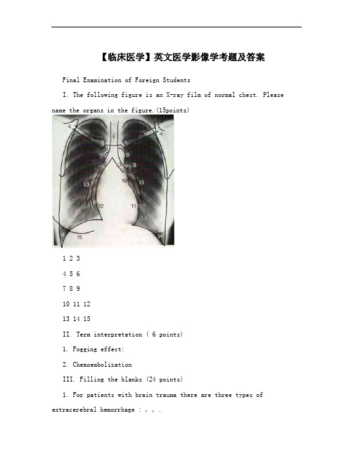
【临床医学】英文医学影像学考题及答案Final Examination of Foreign StudentsI. The following figure is an X-ray film of normal chest. Please name the organs in the figure.(15points)1 2 34 5 67 8 910 11 1213 14 15II. Term interpretation ( 6 points)1. Fogging effect:2. ChemoembolizationIII. Filling the blanks (24 points)1. For patients with brain trauma there are three types of extracerebral hemorrhage : , , .2. On X-ray film, there are four fundamental radiographic densities. Bones appear . Muscles appear . Fat appears .Air and gases appear .3. The indications of embolization conclude____________,______________, ______________, ________ ____, _______ ____, .4. The aim of inferior vena cava filter placement was to____________________. 5. Endovascular stent has two kinds of unfold types: _________ _and_______________.6. Angiography can be performed by ____________, _____________,_______________ technique.7. Give some example of non-vascular intervention:__________________,__________________, ______________________, ______________________.8. The main limitation of angioplasty was__________________________.IV. Answer the questions. (35 points)1. Please write down the common site of hypertensive cerebral hemorrhage and its CT appearance (10 points)2. Please write down CT and MRI appearance of meningioma. (14 points)3. What is the principle of transcatheter arterial infusion? (6 points)4. Please give an introduction of angioplasty and its main technique. (5 points)V. According to the clinical materials and radiologic examination , please write down the radiologic appearance and your impression. (20 points) 1.Case 1A 38-year-old man with a history of fever, cough, rusted sputum and left chest 9pain for one week. The white blood cell count was 12.0×10/L. Theposteroanterior and lateral view films as follows:2.Case 2A 34-year-old man, He suffered from low grade fever in the afternoon, cough, weakness for half a month. The findings of CT scan as follows:3.Case 3A-70-year-old femal with a history of colon carcinoma.4.Case 4A 38-year-old man with myasthenia gravis for half a year.5.Case5A 68-year-old female with a history of smoking for 30 years. The clinical symptom was cough, hemoptysis for two months.2002-2003 Final Examination of Foreign Students (Answers)I.1 trachea2 right main bronchus3 left main bronchus4 scapula5 clavicle6 sternum7 azygos vein8 aortic arch9 left pulmonary artery 10 upper left cardiac border 11 lower left cardiac border 12 right atrium 13 lower lobe arteries 14 lateral costophrenic angle 15 breast II.1. Fogging effect: During 2~3 weeks after cerebral infarction, the density of the infarcted lesion increased and became isodense. This is due to reduction in edema ,leakage of proteins from cell lysis and invasion of macrophage.2. Chemoembolization: A technique that combines transcatheterarterial infusion of chemotherapy drugs and embolization.III.1. epidural hemorrhage, subdural hemorrhage, subarachnoid hemorrhage2. black, gray-black, gray, white3. control of hemorrage, devascularization of tumors,. ablation of organ or tissue, varicocele,modification of blood flow, perigraft leak and back bleeding, following stent-graft,localization of small bowel angiodysplasia (choose 5)4. prevent the pummonary embolism5. balloon-expandble stent, self-expandable stent6.DSA, MRA, CTA7. percutaneous biopsy, percutaneous drainage & aspiration, transcatheter dilate nonvasclular lumen, percutaneous interstitial therapy8. restenosisIV.1.Very common reason is hypertension and the common site are basal ganglion and thalamus.Acute hemorrhage: hyperdense, 60-80Hu ( hemoglobin )Subacute hemorrhage: attenuation decreased with time (1.5 Hu per day);start periphery progress centrally; isodense between 1 to 6 weeks; peripheral enhancementChronic hemorrhage: hypodenseResidua of hemorrage: hypodense foci (37%); slitlike lesion (25%); calcification (10%); no abnormal (27%)2.CT: Sharp circumscribed round or smoothly lobulated mass;Connected with dura or inner table with a broad bas.Homogenous hyperdense (75%), isodense.Calcification 20~25%,diffuse, focal, sandlike, sunburst, globular, rimlike. Bone destructure or hyperosteogenySoft tissue mass in scalpEdema: non except for venous or sinus compressedNecrosis or cyst formation : seldom 8~25%Hemorrhage:uncommonEnhancement :obvious and uniform enhancementMRI: Isointense with gray matter on T1WI ,T2WI;CSF cleft and pial vessels around tumor; Flow void vessels within tumor;Enhanced rapidly and intensely,Dural tail sign. Dura adjacent to tumor show liner enhancemet. suggestive not specific for meningioma, also can be seen in schwannoma, gliblastoma multiforme, metastases3. Drugs administrated through veins were diluted, combined with protein, the level of drugs in the target organ is low, untarget organs was also affected, side effects was seriousTAI increase the level of drugs in the target organ, prolong the contact time with lesion, enhance the effectiveness, lower the side effects4. Ballon angioplasty. Endovascular stent. Atherectomy/thrombous removel. Laserangioplasty. Ultrasound angioplasty.V.1.case1: left lower lobe; confluent patchy opacities; obliterate the posterior left hemidiaphragm on the lateral view; impression: left lower lobe pneumonia2.case2: multiple(diffuse); small(military) nodules throughout both lungs; homogeneous density; impression: military tuberculosis3.case3: multiple nodules; bilateral lung fields; well-defined margins; homogeneous density; impression: pulmonary metastases4.case4: anterior mediastinum(anterior to the great vessels); mass; well-defined margins; soft tissue density; homogeneous density; impresson: thymoma5.case5: right hilum enlarged; the wall of the bronchus thickened; soft tissue density mass; mediastinal and right pulmonary artery involvement; obviously and heterogeneously enhancement; paratracheal and subcarinal lymph nodes enlarged; impression: right central bronchial carcinoma involved mediastinal and right pulmonary artery, mediastinal lymph nodes metastases。
医学影像专业英语总结
医学影像专业英语总结第一篇:医学影像专业英语总结By 南方医影像-枝枝Chest plain film/plain chest radiography 胸部平片Posteroanterior 后前位Left-lateral 左侧位Contour 轮廓Symmetric 对称 Lung field 肺野 Lung marking 肺纹理 Lesion 病变Lung hilar 肺门Mediastinum 胸廓Diaphragm 膈肌Rib 肋骨Round-shaped 类圆形的Mass 团块Post-basic segment 后基底段 Lobulated-edge 边缘分叶Well-defined margin 边界清楚ill-define margin 边缘不清vague margin Homogeneous attenuation 密度均匀Thoracic vertebraes 胸椎Obstructive atelectasis 阻塞性肺不张Sign of “recersal S” 反S征 Bilateral 双侧的 Cloud-shaped areas 大片密度增高区域Piece-like high attunuation 片状高密度 Pulmonary edema 肺水肿Node 结节Acute miliary tuberculosis 急性粟粒性肺结核 Anteroposterior abdomen plain film 腹部平片Supine overhead projection 仰卧前后位投照Radiopaque foreign body 不透光异物Stone 结石Liver 肝 gallbladder 胆 kidney 肾Bowel 肠Distension 扩张Free gas 游离气体Vertebrate and pelvis bone 腰椎和骨盆Plain film of pelvis 骨盆平片Acetabulun 髋臼Hip joint 髋关节Bone destruction 骨质破坏 Femoral head 股骨头The left hip joint space 左髋关节间隙 Osteoporosis 骨质疏松By 南方医影像-枝枝Anteroposterior elbow plain film 前后位肘关节平片Osteoslerosis 骨质硬化Hyperosteogeny 骨质增生 Humerus 肱骨 Ulna 尺骨 Radius 桡骨Periosteal reaction 骨膜反应Periosteal proliferation 骨膜增生(骨膜反应)Dislocated 脱位 Soft tissue软组织Tibia 胫骨Fibula 腓骨Cortex 皮质 Oblique fissure 斜行骨折线Fracture 骨折There is no obvious angle formation or abnormal removing of the breaking ends.骨折断端未见明显错位Femur 股骨 Metaphysis 干骺端 metaphyseal A longitude of 16 cm 长约16cm Slice-like 层状Linear 线状 Epiphysis 骨骺 Osteomyelitis 骨髓炎 Lower end 下端 Septa 分隔 Distend 膨胀、扩大Disrupted 中断Giant-cell tumor 巨细胞瘤 Needle-like 针状Tumor bone 肿瘤骨Osteosarcoma 骨肉瘤Upper gastrointestinal barlum meal examination and photogragh 上消化道钡餐造影摄片Folds 皱襞Esophagus 食管 Peristalsis 蠕动 Evacuation 排空Stomach 胃Niche 龛影crater Stenosis 狭窄Filling defect 充盈缺损Duodennal cap and loop 十二指肠球及肠圈 Mucosal folds 黏膜皱襞By 南方医影像-枝枝Gastric antrum 胃窦Coarse 粗糙的Nodular 结节状的Spasm 痉挛Antral gastritis 胃窦炎Pouch 囊袋 Diverticulum 憩室Deformed 变形Barium filled spot 钡斑 Mucous folds converging 黏膜皱襞聚集Palpation 触诊(加压)Peptic ulcer 消化性溃疡Duodenal bulb 十二指肠球部Lesser curvature of the stomach 胃小弯Barium-gas plane 气钡平面 Penetrating gastric ulcer 穿透性溃疡 Lumen 管腔 Gastric body 胃体Antrum 胃窦Stiff 僵硬的Cardia 贲门、心脏Fundus 胃底 Pyloric 幽门的 Colon结肠Oppressing 压迫Excrete 排泄、分泌 Interruption 中断Sigmoid 乙状结肠Transverse colon 横结肠 Thorn-like 小刺状的 Ulcerative colitis 溃疡性结肠炎Ascent colon 升结肠Bowel obstruction 肠梗阻Transverse image 轴位像 Plain CT scan CT平扫 Axial 轴位 8 mm slice apart 8 mm 层厚8mm,间隔8mm Brain parenchyma 脑实质Ventricle 脑室Subarchnoid cavity蛛网膜下腔Midline structures 中线结构 Circumferential 周围的External capsule 外囊Hypo-attenuation 低密度Hyper-attenuation 高密度 axial area 横截面积By 南方医影像-枝枝 Deformed 变形 Adjacent 邻近的 Deviated 移位Hematoma 血肿Pre-contrast transverse image平扫轴位像 Post-contrast scan 增强扫描 Kernel 中心(窗位?)Frontal part 额部 Predominantly 主要的 Wide-base 广基底 Cerebral flax 大脑镰Calcification 钙化Inner table 内板Meningioma 脑膜瘤Coronal 冠状的Orbit 眼眶MPR reconstruction MPR重建 Isoattenuating 等密度Prominent 凸出的、杰出的、显著的On arterial phase images 在动脉期Spotted enhancement点状强化Progressive enhancement 渐进性强化Cavernous hemangioma 海绵状血管瘤Temporal bone 颞骨Facial cannal 面神经管Internal auditory meatus 内听道Benign 良性的Nasopharynx 鼻咽Pharyngeal recess 咽隐窝Obliterate 消失、擦除Parapharyngeal space 咽旁间隙Ringed enhancement 环状强化 Invasion 发病、侵袭Metastasis 转移Sagittal image 矢状位Cervical 颈部的 cervical vertebra 颈椎 vertebrae Alignment 排列Curvature 曲度 Disci 椎间盘 Nerve root 神经根Sleeve 袖、套Lumbar spine 腰椎Ligament 韧带Disc herniation 椎间盘突出Exceed 超出 Epidural 硬膜外By 南方医影像-枝枝 Isthmus 峡部 Mildly 轻度的 Surge forward 向前 Spondylolisthesis 椎体前移 Osteosclerosis 骨质硬化 Marrow lumen 骨髓腔Heterogeneous 均匀Dysplasia 发育不良Fibrous dysplasia 纤维异常增殖症 Sternum 胸骨 CT value CT值 Cyst 囊肿Compage of thorax 胸廓 Trachea 气管 Bronchi 支气管 Through 通畅 Lymphadenectasis 淋巴结肿大 Air bronchogram sign 空气支气管征Carina of trachea 气管隆突Pneumonia 肺炎Apico-尖、顶Lobular 分叶 Spicule 毛刺 Biopsy 活检 Orifice 开口 Occlusion 闭塞Thymoma 胸腺瘤Configuration 形态Proportion 比例 Hepatic lobe 肝叶Hepatic parenchyma 肝实质Dilated 扩张 Spleen 脾脏 Retro-向后、后 Retroperitoneal 腹膜后 Artery phase、vein phase、delay phase动脉期、静脉期、延迟期Peripheral enhancement 周边强化 Portal vein 门静脉 Inferior vena cave 下腔静脉Centripetally 向心性地Cavernous hemangioma 海绵状血管瘤Heterogeneous 不均匀的Splenomegaly 脾大Hepatocarcinoma 肝癌Neoplastic 肿瘤的Thrombosis 血栓形成neoplastic thrombosis 癌栓By 南方医影像-枝枝Cirrhosis 硬化、肝硬化Cholecy 胆囊 Ectomy 切除术 cholecyectomy 胆囊切除术Pneumo-肺、呼吸、空气 pneumotosis积气Common bile duct 胆总管Dilation 扩张 Posterolateral 后外侧 Administration 行政、管理、处理 Contrast material 对比剂 Renal pelvis 肾盂 Renal calices 肾盏Hepatorenal recess 肝肾隐窝 Nephric 肾的 Perinephric space 肾周间隙 Gerota 肾Fascia 筋膜Gerota’s fascia 肾周筋膜Pancreatitis 胰腺炎 Mesenteric 肠系膜的 Superior mesenteric vein 肠系膜上静脉 CT endoscopy CT内窥镜 Greater curvature 胃大弯Gastroscopy 胃镜colonscopy 结肠镜MPR、SSD、VR、CTVE Cecum 盲肠 cecal 盲肠的Protrude 突出、凸出Tumor 肿瘤 carcinoma 癌Urinary bladder 膀胱 Uterus 子宫 Appendage 附件 Ureters 输尿管CTU VRT MIP Cystoscopy 膀胱镜Aorta 主动脉Ascending aorta 胸主动脉Cephalic 头部的brachio 臂brachiocephalic trunk 头臂干Proximal近端的、基部的Carotid 颈动脉的 Common carotid artery 颈总动脉 Endo-内 endomembrane 内膜 Tortuous 扭曲的、迂曲的Collateral 侧支Dissecting 夹层 aneurysm 动脉瘤 dissecting aneurysm 夹层动脉瘤 Thrombosis 血栓形成 Takayasu arteritis 多发性大动脉炎 Give rise to 引起 Embolism 栓塞 Iliac 髂的、回肠的 ileum 回肠 Common iliac artery 髂总动脉 Femoral 股 femoral artery 股动脉Popliteal 腘popliteal artery 腘动脉Peroneal 腓peroneal artery 腓动脉 By 南方医影像-枝枝Tibial 胫 tibial artery 胫动脉Right coronary artery 右冠状动脉Left anterior descending artery 左前降支 Left circumflex artery 左旋支 Plague 瘟疫、灾祸、斑块 soft plague 软斑块 Orientation 方位 High signal intensity 高信号 Gyrus 脑回 Infarction 梗死、缺血灶Parietal lobe 顶叶Subacute 亚急性的 subacute bleeding 亚急性出血Occupying effect 占位效应Posterior horn 后角Calcarine sulcus 距状沟 In coincidence with 与...一致Gray matter 灰质Splenium/genu/body of corpus callosum 胼胝体压部/膝部/体部Heterotopia 异位Subarachnoid 蛛网膜下的subarachnoid cavites 蛛网膜下腔Tonsil 扁桃体Cerebellum 小脑Occipital 枕骨的Cistern 池Cerbrospinal fluid 脑脊液Malformation 急性myelo-髓syringo-瘘管、洞Myelosyringosis 脊髓空洞症Sellae 鞍区Pituitary 垂体、粘液的Optic chiasma 视交叉Sponge sinus 海绵窦Craniopharyngeal duct颅咽管 Adenoma 腺瘤 Internal carotid 颈内动脉 Uniformly 均匀地、一致地Spectrum 范围、系列、波谱 spectroscopy 波谱 Cusp 峰Infra 以下Ento-内 entoplastron 内板Convexity 凸面Cranial 颅盖的、颅的Dural 硬脑膜的Dural mater硬脑膜 Creatine 肌酸 Meningioma 脑膜瘤 Left sidedness 左侧 Peduncle 脚、根、茎 bridge cerebellar peduncle region桥小脑区 Cork sign 瓶塞征By 南方医影像-枝枝 Brain stem 脑干 Acoustic 听觉的 Pontine 脑桥cerebellopontine angle 脑桥小脑角(桥小脑区)anterior pontine cistern 脑桥前池 Extrude 突入 Embed 包绕 Vertebral 椎的、椎骨的 vertebral artery 椎动脉 Ventricle 脑室 Corona radiate 放射冠 Screen pore 筛孔 Mass effect 占位效应 Malignant 恶性 Glioma 胶质瘤 Medullary 髓 velum 帆 inferior medullary velum 下髓帆Aqueduct 导水管Vermis 小脑蚓部 Blastoma-母细胞瘤 medullblastoma髓母细胞瘤hemangioblastoma 血管母细胞瘤Transparent 明显的、透明的Mural 壁的 mural tumor nodule 壁结节Clouding 片状的Lenticular 豆状的、透镜状的lenticular nucleus 豆状核 Caudate 尾的 caudate nucleus 尾状核 Precuneus 楔前叶Cingulate gyrus 扣带回Binding the history 结合病史 Manifestation 表现 appearence Hepatolenticular degeneration 肝豆状核变性Basiobasis 基底节Maxillary sinus 上颌窦Physio-curvature 生理曲度Bulging 膨胀、突出Strip 条状Spinal cord 脊髓Sclerosis 硬化Melanoma 黑色素瘤Project forward into 突入 Sphenoid sinus 蝶窦Clivus 斜坡Herniation 突出、疝出 Depletion 缺如Lumber lamina 腰椎椎板Spinous process 棘突Menigo-matter 脊膜Infiltrate 浸润Extensive 广泛的 Subchondral 软骨下的 Endplate 终板By 南方医影像-枝枝 Cone 锥 medullary cone 脊髓圆锥 Cork 塞住、抑制 Bifid 二分的、双裂的 bifid spine 脊柱裂 Menigomyelocele脊髓脊膜膨出Sacral 骶骨Proton 质子Blotch 斑点Archo 直肠Chordoma 脊索瘤Foramen 孔 intervertebral foramen 椎间孔 Neurogenic tumor 神经源性肿瘤 Placing upside down 倒置 Spinal meningima 脊膜瘤Raindrops 点滴状Teratoma 畸胎瘤 Cholecyst 胆囊Tumefacient 膨胀的、肿大的Uterine 子宫 Lacuna 缝隙、陷窝、管道Metra-archo lacuna 子宫直肠陷窝Uterine myoma 子宫肌瘤Split 分离 endometrium 内膜 Fundus 底部 Incisure 切迹 Cervix 宫颈 Septation 间隔 Metrodysplasia 子宫发育异常 bicorbate uterus 双角子宫Femoral head 股骨头 Cartilage 软骨 Weight-bearing surface 负重面Acetabulum 髋臼、关节腔Aseptic 无菌的Necrosis 坏死Meniscus 新月形、关节盘、凸透镜Lateral meniscus 外侧半月板Articular 关节的Fat-saturated 压脂Cruciate 十字的、交叉的cruciate ligament 交叉韧带 Tendon 腱 Bone matrix 骨质 Rupture 撕裂Mammary gland乳腺Axilla 腋窝Quadrant 象限Raio-hair sign 放射状毛刺征 Crab-feet sign 蟹足征By 南方医影像-枝枝Basilar artery 基底动脉Constriction 狭窄Dilatation 扩张 Spread area 走行区域 Initiation 起始 Siphon 虹吸Anastomosis 吻合Aneurysm 动脉瘤Void 无效的、空隙、排泄Flowing void effect 流空效应 Fog 烟雾 Moyamoya disease 烟雾病The lateral internal carotid artery angiogram 颈内动脉侧位像 The frontal internal carotid artery angiogram 颈内动脉正位像Angiography 血管造影Anesthesia 麻醉Catheter 导管 Catheterization 导管插入术 femoral ~股动脉插管Tip 尖端Decannulation 拔管Hemostasis 止血Ward 病房Course 走行、病程Sigmoid 乙状结肠 sigmoid sinus 乙状窦Occipital 枕骨 Tributary 属支Vascular 血管的Iohexol 碘海醇Sign of string beads 串珠征Tortuosity 扭曲Misty模糊的、烟雾状的Ophthalmic 眼的Meningeal 脑膜的 Collateral circulation 侧支循环The oblique vertebral artery angiogram 椎动脉斜位像The anterposterior vertebral artery angiogram 椎动脉正位像Saccular 囊状的Aforementioned 前述的Derive from 起源于Capillary 毛细血管Arteriae bronchiales 支气管动脉 Ondansetron Hydrochloride 欧贝 Dexamethasone 地塞米松Regafur 方克carboplatin 卡铂 mitomycin 丝裂霉素(化疗药物)Malaise 不适 Twisted 扭曲的 Reticular 网状的 Compatible with 符合 ~ tumor vessels 符合肿瘤血管By 南方医影像-枝枝 Inflexibility 僵直 Encirclement 包绕 Stain 染色Draining vein 引流静脉Fistulas 瘘Interventional treatment operation 介入治疗术Contrast medium 对比剂 Nidus 病灶 The signs of early filling and delayed evacuationon of contrast medium 早出晚归征 PV门静脉 Rim 边缘Tenuous 稀薄的、空洞的、纤细的 Interlobular artery 小叶间动脉Arcuate artery 弓形动脉 Shrunken 萎缩的Superior mesenteric artery 肠系膜上动脉Iodinated oil 乙碘油Sequentially 依次Withdraw 撤退 Winding 迂曲的Embolization 栓塞Iohexol deposits well 碘油沉积良好Dorsal 背部的stop bleeding bands 止血带Diluted 稀释的Meglumine diatrizoate 泛影葡胺Superficial vein 浅静脉 Retain 保留 Successively 依次地 Valve 瓣膜 Reflux 反流Varicose 静脉曲张的、迂曲扩张的 Dysfunction 功能不全第二篇:医学影像技术专业英语No significant differences were found between the area under the receiver operating characteristic curve of the digital and that of the conventional radiography method for almost all investigated criteria.The only exception(例外;异议)was mediastinal abnormalities, for which the digital method provided better results than the conventionvl method(P<0.05) Invasive breast cancer detection by mammography may be improved through attention to correct positioning.For CT of the brain, there are there standard reference lines;the orbitomeatal line, the Reid baseline, and the supraorbitomeatal line.The orbitomeatal line runs through the external canthus and the center of the external auditory meatus.This is the most widely used line, because acquisition of fewer sections is necessary to cover the entire brain.The Reid baseline runs though the inferior orbital wall and the superior border of the external auditory meatus.This line is paraller to the optic nerve and provides the best demonstration of the orditai contents.The supraorbitomeatal line runs through the superior orbital wall and the center of the external auditory meatus.This line approximately parallels the skull base.We developed reference lines for use at MR imaging that are analogous to the three standard reference lines used at CT, on the basis of anatomic landmarks that are visible on midsagittal MR images.These lines can be used to prescribe subsequent oblique axial sequences.During the past decade , many investigators have addressed the feasibility of picture archiving and communicationsystem(PACS)as an alternative to film-based radiology.To successfully implement the PACS, a number of factors such as film resolution, interpretation conditions, and a cost-effective viewer system must be taken into consideration.The most important clinical criterion for using the PACS technology is the ability to achieve acceptable accuracy when interpreting radiologic images at a soft-copy viewing workstation.Most previous reports have focused on a comparison of observer performance with soft-copy and hard-copy imajes, and such studies have been performed using chest radiography.However, observer performance in projection radiography of another anatomic section must also be compared to achieve that the PACS can completely replace film-based radiology.CT in an invaluable tool in the diagnosis of the chest diseases with high-resolution CT(HRCT)providion exquisite visualization of the pulmonary interstitium.However, CT examinations do not come without a cose..A report from the 2002 National Conference on Dose Reduction in CT indicates that CT now represents the largest single source of medical radiation exposure.Although CT constitutes only 15% of the total number of examinations in some university departments, it can account for 70% of the dose delivered.第三篇:专业英语总结幻灯片1 1.In-class practice 将下面与会议相关词语翻译为汉语λsponsor赞助者 Cover letter求职信λorganizer组织者Invitation letter邀请信λpreparatory committee筹备委员会λ organizing committee组织委员会λcontact person联系人Recommendation Letter推荐信λkeynote speaker主旨发言人λ participant与会者λ co-sponsors协办方λ honorary chairman荣誉主席λ program committee程序委员会λ(vice)chairman副主席λ members会员λ executive secretary行政秘书session chair分会场主席幻灯片2 λ Coffee or tea break with refreshments茶歇λ Dinner晚餐λLunch午餐λ Tour and sightseeing观光λ Banquet晚宴λ Reception接待会λ Welcome speech欢迎词λOpening remarks/address/speech开幕词λClosing remarks/address/speech闭幕词λKeynote speech/report主旨发言λ Paper session/paper presentation张贴λ Poster session海报展λ Demonstration说明λ Question and answer period/session问答环节幻灯片3 机场的英语标识 Airport 飞机场Airport lounges 机场休息室 Airport shuttle 机场班车 Arrivals 进港Check-in area(zone)办理登机区 Customers lounges 旅客休息室 Departures 出港Departure time 离港时间 Departure time on reverse 返航时间幻灯片4 机场的英语标识Destination on airport 到达机场Domestic flight 国内航班Emergency exit 安全出口 exit/out/way out 出口arrivals 进站(进港、到达)Exit to all routes 各通道出口 Flight connections 转机处Assistance/information/help point(desk)/inquiries 问讯处Left baggage 行李寄存 Lost property 失物招领Luggage from flight 到港行李幻灯片5 机场的英语标识Luggage pick-up/reclaim/ Luggage(baggage)claim 取行李Missing people help line 走失求助热线 Missing, police appeal forassistance 警察提供走失帮助No smoking except in designated area 除制定区域外,禁止吸烟 customs 海关Nothing(something)to declare 有(无)报关Passport control 入境检验 Queue here 在此排队reclaim belt 取行李传送带幻灯片6 机场的英语标识Seserved seating 预定的座位 Return fares 往返机票 Short stay 短暂停留 Stay close 跟紧international airport 国际机场 domestic airport 国内机场international terminal 国际候机楼international/dometic departure 国际/国内班出港 check-in 登机手续办理boarding pass/card 登机牌 gate/departure gate 登机口幻灯片7 机场的英语标识FLT NO(flight number)航班号 arriving form 来自。
- 1、下载文档前请自行甄别文档内容的完整性,平台不提供额外的编辑、内容补充、找答案等附加服务。
- 2、"仅部分预览"的文档,不可在线预览部分如存在完整性等问题,可反馈申请退款(可完整预览的文档不适用该条件!)。
- 3、如文档侵犯您的权益,请联系客服反馈,我们会尽快为您处理(人工客服工作时间:9:00-18:30)。
Types
DeBakey Type I
• Involves entire aorta
possible shock Asymmetric peripheral pulses Pulmonary edema
Imaging Findings
Chest films
• Mediastinal widening • Left paraspinal stripe • Displacement of intimal calcifications • Left pleural effusion • Displacement of endotracheal tube or
DeBakey Type II
• Least common
Ascending aorta only
DeBakey Type III
• Most common
Descending aorta only
Stanford Type A
• Ascending aorta involved
Over half develop aortic regurgitation(返流)
nasogastric tube
MRI
• Intimal flap • Slow flow or clot in false lumen
CT
• Intimal flap • Displacement of intimal calcification • Differential contrast enhancement of
Aneurysm defined by size criteria
In general, ascending aorta > 5 cm Descending aorta > 4 cm
Vessels involved with dissection
Any artery can be occluded Usually the right coronary and three
true versus false lumen
Computed tomography of the chest including CTA of the aorta is the investigation of choice for suspected aortic dissection.
The roles of CT in the evaluation of suspected aortic dissection are:
• False channel usually arises anterior in the ascending aorta and spirals to posterior and left lateral in descending aorta
• True channel is usually larger
Usually surgically
*
Cystic medial necrosis e.g.Marfan
’s
Usually surgically
*
DeBakey
Hypertensio
Type III Stanford Type Descending
n
Usually
(most
B
aorta only Atheroscler medically
Stanford Type B
• Ascending aorta NOT involved
Most dissections arise either just distal to the aortic valve or just distal to aortic isthmus
True versus false channel
*cGooaml ims too npr)event backward involvement of the aortic valve or ruoptsuirse into pericardium
Clinical
Sharp, tearing, intractable chest pain Previously hypertensive, now
common)
Stanford Type A
(ascending aorta involved)
Involves entire aorta
Stanford Type A
(ascending aorta
involved)
Ascending aorta onlyΒιβλιοθήκη Hypertensio n
Atheroscler osis
• Slower flow in false channel on MR
DeBakey Classificatio
n
Stanford Classification
Portion of Aorta
Involved
Common causes
RX
DeBakey Type I
DeBakey Type II (least
Aortic Dissection
3:1 male to female predominance
Over the age of 40
Hemorrhage in the media leading to either
• Tear in the weakened intima which breaks into the lumen, or
• Hemorrhage in the wall (less common)
• Hemorrhage separate media from adventitia
Predisposing factors
Hypertension (most commonly) Atherosclerosis Marfan’s syndrome Coarctation of the aorta Trauma (rare) Pregnancy (rare)
