人骨成型蛋白受体 1A BMP-R1A ELISA试剂盒北京驰明瑞使用说明书
SPRED1蛋白使用说明书

Instruction manual本产品仅供研究使用,不用于临床诊断。
本公司提供的电子版本说明书仅供参考,实验请以收到的纸质手册为准。
SPRED1, 1-444aa, Human, His tag, E.coli产品货号: TP04022第三版别名:Sprouty-related, EVH1 domain-containing protein 1, hSpred1, NFLS, spred-1描述:SPRED1 is a member of the Sprouty family of proteins and is phosphorylated by tyrosine kinase in response to several growth factors. This protein can act as a homodimer or as a heterodimer with SPRED2 to regulate activation of the MAP kinase cascade. Defects in this gene are a cause of neurofibromatosis type 1-like syndrome (NFLS). Recombinant human SPRED1 protein, fused to His-tag at N-terminus, was expressed in E.coli.配方:Liquid. In 20mM Tris-HCl (pH 8.0) containing 10% glycerol分子量:52.9 kDa (467aa)序列: MGSSHHHHHHSSGLVPRGSHMGSMSEETATSDNDNSYARVRAVVMTRDDSSGGWLPLGGSGLSSVTVFKVPHQEENG CADFFIRGERLRDKMVVLECMLKKDLIYNKVTPTFHHWKIDDKKFGLTFQSPADARAFDRGIRRAIEDISQGCPESKNEAE GADDLQANEEDSSSSLVKDHLFQQETVVTSEPYRSSNIRPSPFEDLNARRVYMQSQANQITFGQPGLDIQSRSMEYVQRQIS KECGSLKSQNRVPLKSIRHVSFQDEDEIVRINPRDILIRRYADYRHPDMWKNDLERDDADSSIQFSKPDSKKSDYLYSCGDE TKLSSPKDSVVFKTQPSSLKIKKSKRRKEDGERSRCVYCQERFNHEENVRGKCQDAPDPIKRCIYQVSCMLCAESMLYHC MSDSEGDFSDPCSCDTSDDKFCLRWLALVALSFIVPCMCCYVPLRMCHRCGEACGCCGGKHKAAG纯度:> 95% by HPLC浓度:0.5 mg/ml (determined by Bradford assay)内毒素:<1.0 EU per 1 ug of protein (determined by LAL method)存储: +4°C 保存 (1-2 周). 长期保存在-20°C或者-70°C. 避免反复冻融.1 / 1。
2X M5 HiPer SYBR Premix EsTaq plus (with Tli RNase

2X M5 HiPer SYBR Premix EsTaq plus (with Tli RNaseH)使用说明书产品名称单位货号2X M5 HiPer SYBR Premix EsTaq plus (with Tli RNaseH) 50 μl反应× 40 次MF788-T2X M5 HiPer SYBR Premix EsTaq plus (with Tli RNaseH) 50 μl反应× 200 次MF788-012X M5 HiPer SYBR Premix EsTaq plus (with Tli RNaseH) 50 μl反应× 400 次MF788-02【储存条件】长期保存,请置于-20˚C,有效期24个月。
经常使用,可置于4˚C保存至少六个月。
【产品简介】本产品采用Sybrgreen嵌合荧光法进行荧光定量的专用试剂。
制品中含有荧光定量反应的最适浓度Sybrgreen,是一种2×浓度的预混试剂,进行实验时,PCR反应液的配制十分方便简单。
制品中使用了antiTaq抗体的Hot Start法用DNA聚合酶,与荧光定量反应适合Buffer组合,可以有效抑制非特异性的PCR扩增,大大提高PCR的扩增效率,可以进行高灵敏度的荧光定量扩增反应。
本产品Buffer 经过改良,使反应特异性比SybrGreenPremix Ex Taq (withTli RNaseH)(货号MF787)更高。
另外,本产品中添加了Tli RNaseH (耐热性RNaseH),以cDNA作为模板进行PCR反应时,可以很好地抑制由于cDNA中残存mRNA 对PCR反应造成的阻害作用。
适合于快速荧光定量扩增反应,可以在宽广的定量区域内得到良好的标准曲线,对靶基因进行准确定量、检测,重复性好,可信度高。
【产品组份】MF788-T MF788-012x M5 HiPer SYBR Premix EsTaq plus (withTli RNaseH) * 1.0 ml 5x1mlROX Reference Dye(50×)** 40μl 200μlROX Reference Dye II(50×)** 40μl200μl*由以下组分预混而成:EsTaq plus,dNTP Mixture,Mg2+,Tli RNaseH,Sybrgreen。
人白介素1β (IL-1β)酶联免疫吸附测定试剂盒使用说明书
2022年修订第一版(本试剂盒仅供体外研究使用,不用于临床诊断!)产品货号:E-EL-H0149c产品规格:96T/48T/24T/96T*5Elabscience 人白介素1β(IL-1β)酶联免疫吸附测定试剂盒使用说明书Human IL-1β(Interleukin 1 Beta) ELISA Kit使用前请仔细阅读说明书。
如果有任何问题,请通过以下方式联系我们:销售部电话技术部电话************电子邮箱(销售)********************电子邮箱(技术)**************************网址:具体保质期请见试剂盒外包装标签。
请在保质期内使用试剂盒。
联系时请提供产品批号(见试剂盒标签),以便我们更高效地为您服务。
Copyright ©2021-2022 Elabscience Biotechnology Co.,Ltd. All Rights Reserved目录用途 (3)基本性能 (3)检测原理 (3)试剂盒组成及保存 (4)试验所需自备物品 (5)样品收集方法 (5)注意事项 (6)■ 试剂盒注意事项 (6)■ 样品注意事项 (6)样本稀释方案 (6)检测前准备工作 (7)操作步骤 (8)结果判断 (10)技术资源 (10)典型数据 (10)性能 (11)■ 精密度 (11)■ 回收率 (11)■ 线性 (11)声明 (12)Intended use (13)Character (13)Test principle (13)Kit components & Storage (14)Other supplies required (15)Sample collection (15)Note (16)■ Note for kit (16)■ Note for sample (16)Dilution Method (17)Reagent preparation (17)Assay procedure (18)Calculation of results (20)Technical resources (20)Typical data (20)Performance (21)■ Precision (21)■ Recovery (21)■ Linearity (21)Declaration (22)用途该试剂盒用于体外定量检测人 血清、血浆或其他相关生物液体中IL-1β浓度。
人骨成型蛋白受体1A(BMPR-1A)elisa试剂盒使用说明书

人骨成型蛋白受体1A(BMPR-1A)elisa试剂盒使用说明书Elisa kit规格:48孔配置/96孔配置标准品稀释液:1.5ml×1瓶酶标试剂:3 ml×1瓶(48)/6 ml×1瓶(96)【人骨成型蛋白受体1A(BMPR-1A) elisa试剂盒】本试剂仅供研究使用计算:以标准物的浓度为横坐标,OD值为纵坐标,在坐标纸上绘出标准曲线,根据样品的OD值由标准曲线查出相应的浓度;再乘以稀释倍数;或用标准物的浓度与OD值计算出标准曲线的直线回归方程式,将样品的OD值代入方程式,计算出样品浓度,再乘以稀释倍数,即为样品的实际浓度。
试剂盒组成:封板膜:2片(48)/2片(96)说明书:1份密封袋:1个标准品: 2700ng/L 0.5ml×1瓶0.5ml×1瓶 2-8℃保存酶标包被板: 1×48 1×96 2-8℃保存样品稀释液: 3ml×1瓶 6 ml×1瓶 2-8℃保存显色剂A液: 3ml×1瓶 6 ml×1瓶 2-8℃保存显色剂B液: 3ml×1瓶 6 ml×1瓶 2-8℃保存终止液: 3ml×1瓶6ml×1瓶 2-8℃保存浓缩洗涤液:(20ml×20倍)×1瓶(20ml×30倍)×1瓶 2-8℃保存实验原理:本试剂盒应用双抗体夹心法测定标本中人骨成型蛋白受体1A(BMPR-1A) 水平。
用纯化的人骨成型蛋白受体1A (BMPR-1A) 抗体包被微孔板,制成固相抗体,往包被单抗的微孔中依次加入中性粒细胞明胶酶相关脂质运载蛋白(BMPR-1A) ,再与HRP标记的中性粒细胞明胶酶相关脂质运载蛋白(BMPR-1A) 抗体结合,形成抗体-抗原-酶标抗体复合物,经过彻底洗涤后加底物TMB显色。
TMB在HRP酶的催化下转化成蓝色,并在酸的作用下转化成最终的黄色。
RayBio Mouse MIP-1 beta CCL4 IQELISA Kit 使用说明书
RayBio® Mouse MIP-1 beta/CCL4IQELISA KitCatalog #: IQM-MIP1bUser ManualLast revised April 6, 2023Caution:Extraordinarily useful information enclosedISO 13485 Certified3607 Parkway Lane, Suite 200Peachtree Corners, GA 30092 Tel: 1-888-494-8555 (Toll Free) or 770-729-2992, Fax: 770-206-2393Web: , Email: *******************RayBiotech, Inc.________________________________________ RayBio® Mouse MIP-1 beta/CCL4 IQELISA Kit ProtocolTable of ContentsSection Page # I.Introduction3II.Reagents3III.Storage3IV.Additional Materials Required4V.Reagent Preparation4VI.Assay Procedure5VII.Assay Procedure Summary8VIII.Calculation of ResultsA. Typical DataB. Sensitivity and Recovery 8 9 9IX.Troubleshooting Guide10I. INTRODUCTIONThe RayBio®I mmuno Q uantitative E nzyme L inked I mumuno S orbent A ssay (IQELISA) is an innovative new assay that combines the specificity and ease of use of an ELISA with the sensitivity of real-time PCR. This results in an assay that is simultaneously familiar and cutting edge and enables the use of lower sample volumes while also providing more sensitivity. The RayBio® Mouse MIP-1 beta/CCL4 IQELISA Kit is a modified ELISA assay with high sensitivity qPCR readout for the quantitative measurement of Human MIP-1 beta in serum, plasma, and cell culture supernatants. This assay employs an antibody specific for Human MIP-1 beta coated on a 96-well PCR plate. Standards and samples are pipetted into the wells and MIP-1 beta present in a sample is bound to the wells by the immobilized antibody. The wells are washed and a detection affinity molecule is added to the plates. After washing away unbound detection affinity molecule, primers and a PCR master mix are added to the wells and data is collected using qPCR. C t values obtained from the qPCR are then used to calculate the amount of antigen contained in each sample, where lower C t values indicate a higher concentration of antigen.II. REAGENTS1.MIP-1 beta Microplate (Item A)**: 96 well PCR plate coated with anti-Human MIP-1 beta.2.Wash Buffer I Concentrate (20x) (Item B): 25 ml of 20x concentrated solution.3.Standards (Item C): 2 vials of recombinant Human MIP-1 beta.4.Assay Diluent B (Item E): 15 ml of 5x concentrated buffer.5.Detection Affinity Reagent for MIP-1 beta (Item F): 2 vials of a 4x concentrated solutionof anti-Human MIP-1 beta affinity reagent.6.IQELISA Detection Reagent (Item G): 1 mL of a 10x concentrated stock.7.Primer Solution (Item I): 1.5 mL vial.8.PCR Master Mix (Item J): 1.4 mL vial.9.PCR Preparation buffer (Item K): 1mL vial of 10x concentrated buffer.10.Final Wash Buffer (Item L): 10 mL vial of 10x concentrated buffer.**The PCR plate used is a 0.2 mL, non-skirted 96-well plate (ThermoFisher, cat. no.:AB0600). Please ensure compatibility with your PCR machine prior to purchase. For additional information contact technical support (**************************).III. STORAGEMay be stored for up to 6 months at 2°to 8°C from the date of shipment. Standard (recombinant protein) should be stored at -20°C or -80°C (recommended at -80°C) after reconstitution. Opened PCR plate or reagents may be stored for up to 1 month at 2° to 8°C. Note: the kit can be used within one year if the whole kit is stored at -20°C. Avoid repeated freeze-thaw cycles.IV. ADDITIONAL MATERIALS REQUIRED1.Real-time PCR instrument, Bio-Rad recommended2.Precision pipettes to deliver 2 µl to 1 mL volumes.3.Adjustable 1-25 mL pipettes for reagent preparation.4.100 mL and 1 L graduated cylinders.5.Absorbent paper.6.Distilled or deionized water.7.Log-log graph paper or computer and software for data analysis.8.Tubes to prepare standard or sample dilutions.9.Heating block or water bath capable of 80°CV. REAGENT PREPARATION1.Bring wash buffer, samples, assay diluents, and PCR plate to room temperature (18 -25°C) before use. PCR master mix and Primer solution should be kept at 4°C at alltimes.2.Sample dilution: If your samples need to be diluted, 1x Assay Diluent B should be usedfor dilution of serum/plasma samples.Suggested dilution for normal serum/plasma: 2 fold*.*Please note that levels of the target protein may vary between different specimens.Optimal dilution factors for each sample must be determined by the investigator.3.Assay Diluent B should be diluted 5-fold with deionized water.4.Briefly spin the Detection Antibody vial before use. Add 25 µL of 1x Assay Diluent B intothe vial to prepare a detection antibody concentrate. Pipette up and down to mix gently (the concentrate can be stored at 4°C for 5 days). This concentrate should be diluted 80-fold with 1x Assay Diluent B and used in step 4 of the Assay Procedure.5.PCR preparation buffer should be transferred to a 15 mL tube and diluted with 9 mL ofdeionized or distilled water before use.6.Final Wash Buffer should be transferred to a 15 mL tube and diluted with 9 mL ofdeionized or distilled water for every 1 mL of 10x concentrate used before use.7.Preparation of standard: Preparation of standard: Briefly spin a vial of Standards. Add400 µl 1X Assay Diluent B into Standards vial to prepare a 25 ng/ml standard solution.Dissolve the powder thoroughly by a gentle mix. Add 20 µl of MIP-1beta standardsolution from the vial of Standards, into a tube with 480 µl 1X Assay Diluent B to preparea 1000 pg/ml standard solution. Pipette 200 µl 1X Assay Diluent B into each tube. Use the 1000 pg/ml standard solution to produce a dilution series (shown below). Mix each tube thoroughly before the next transfer. 1X Assay Diluent B serves as the zero standard (0 pg/ml).20.00 µL + 480 µL100 µL+ 200 µL100 µL+ 200 µL100 µL+ 200 µL100 µL+ 200 µL100 µL+ 200 µL100 µL+ 200 µL1000 pg/ml 333.333pg/ml111.111pg/ml37.037pg/ml12.346pg/ml4.115pg/ml1.372pg/mlpg/ml8.If the Wash Buffer Concentrate (20x) contains visible crystals, warm to room temperatureand mix gently until dissolved. Dilute 20 mL of Wash Buffer Concentrate into deionized or distilled water to yield 400 mL of 1x Wash Buffer.9.Prepare the IQELISA detection reagent by calculating how much will be needed. Thismay be accomplished by multiplying the number of wells to be assayed by the volume you plan to use per well. Once the volume of IQELISA detection reagent is known,prepare the reagent by diluting it 1:10 with deionized water and mixing thoroughly.VI. ASSAY PROCEDUREOptional Visual Aid: IQELISA [Good Laboratory Practice Guide]1.Bring all reagents and samples to room temperature (18 - 25°C) before use. It isrecommended that all standards and samples be run in triplicate. Partial plate runs may be accomplished by cutting the PCR plate into the desired number of strips using a pair of sturdy scissors, wire cutters, or shears. The remainder may be saved and used for a later date. If this is done, the PCR Plate Film should also be cut to a suitable size.2.Add 10-25 µL of each standard (see Reagent Preparation step 2) and sample intoappropriate wells. Volumes should be consistent between all wells, samples, andstandards. As little as 10 µL can be used if sample volume is limited, however thisincreases the chance of technical error. Ensure there are no bubbles present at thebottom of the wells. Dislodge any bubbles with gentle tapping or with a pipette tip being careful not to contact the sides or bottom of the well. Cover well and incubate for 1.5 -2.5 hours at room temperature.3.Discard the solution and wash 4 times with 1x Wash Solution. Wash by filling each wellwith Wash Buffer (100 µL) using a multi-channel Pipette or autowasher. Completeremoval of liquid at each step is essential to good performance. After the last wash,remove any remaining Wash Buffer by aspirating or decanting. Invert the plate and blot it against clean paper towels.4.Add 25 µL of prepared Detection Antibody (Reagent Preparation step 4) to each well.Incubate for 1 hour at room temperature with gentle shaking.5.Discard the solution. Repeat the wash as in step 3.6.Add 50 µL of prepared IQELISA detection reagent and incubate 1 hour with rocking(Reagent Preparation step 9)7.Discard the solution. Repeat the wash as in step 3, for a total of 6 washes.8.Add 75 µL of Final wash buffer to each well and incubate for 4 minutes with rocking.Remove the solution from each well and blot against paper towels.9.Add 75 µL of 1x PCR preparation buffer to each well and incubate for 10 seconds beforeremoving the buffer. Blot the plate after the buffer is removed to ensure completeremoval of the buffer.10.Add 10 µL of the Primer solution to each well of the plate. At this stage the plate can becovered and stored at -20°C for use the next day if needed.11.Add 10 µL of PCR Master Mix to each well and pipette thoroughly to mix the well (atleast 3x up and down).12.Cover the plate with the supplied PCR Plate Film, taking care to insure the film iscompletely and even pressed onto the plate, creating an air tight seal around each well of the plate.Optional Visual Aid: Sealing the plate [qPCR]13.Place the plate into a real-time PCR instrument using a FITC compatible wave length fordetection with the following settings for cycling1.2 minute activation at 95°C2.15 seconds 95°C denaturation3.25 seconds 60°C annealing/extension4.Repeat steps 2 and 3 34x**Optional: Include a melt curve to view potential plate contamination that can causehigh background and lower the sensitivity. This can be seen in the visual aid onYouTube.VII. ASSAY PROCEDURE SUMMARY1.Prepare all reagents, samples and standards as instructed.2.Add 25 µL standard or sample to each well. Incubate 1.5 - 2.5 hours at roomtemperature.3.Add 25 µL Detection Antibody to each well. Incubate 1 hour at room temperature.4.Add 50 µL of IQELISA Detection Reagent to each well. Incubate 1 hour5.Add 10 µL Primer solution and 10 µL of PCR master mix to each well6.Run real-time PCRVIII. CALCULATION OF RESULTSThe primary data output of the IQELISA kit is C t values. These values represent the number of cycles required for a sample to pass a fluorescence threshold. As the DNA is amplified additional fluorescent signal is produced, with each cycle resulting in an approximate doubling of the DNA. Therefore, higher levels of DNA (directly related to the amount of antigen in the sample) result in lower C t values.Calculate the mean C t for each set of triplicate standards, controls and samples. Subtract the C t value of each sample from the control to obtain the difference between the control and sample (Delta C t). Plot the values of the standards on a graph using a log scale for concentration on the x axis. This graph is the quickest way to visualize results, although not the most accurate. If this method is used the concentration of unknown samples can be estimated using a logarithmic line of best fit.The line of best fit will have an equation y = mln(x)+b, where y is the Delta C t value and x is the concentration. It may be helpful to use 5 significant figures for m and b to minimize rounding errors. To calculate the concentration of unknown sample this can be entered into Excel in the following format=EXP((y-b)/m))Where y is the Delta C t obtained during the assay, and b and m are obtained from the line of best fit.Alternatively, for a more accurate representation linear regression may be used. Both the Delta C t and Concentration can be transformed using a log base of 10, plotted on a graph as described above, along with a line of best fit (using a linear model). The equation of this line may be used to calculate the antigen concentration of unknown samples. This is the method used for the analysis spreadsheet for IQELISA available online.A. TYPICAL DATAThese data are for demonstration only. A standard curve must be run with each assay.B. SENSITIVITY and RECOVERYThe minimum quantifiable dose of MIP-1 beta is typically 0.97 pg/ml, however levels as lower than 0.97 pg/ml may be detected outside of the quantification range.Serum spike tests show recovery is 86% with a range from 80% to 91%.Intraplate CV is below 10% for all samples and Interplate CV is below 15%.X. TROUBLESHOOTING GUIDEThis product is for research use only.©2023 RayBiotech, Inc11。
碧云天生物技术 Human IL-1β ELISA Kit 说明书
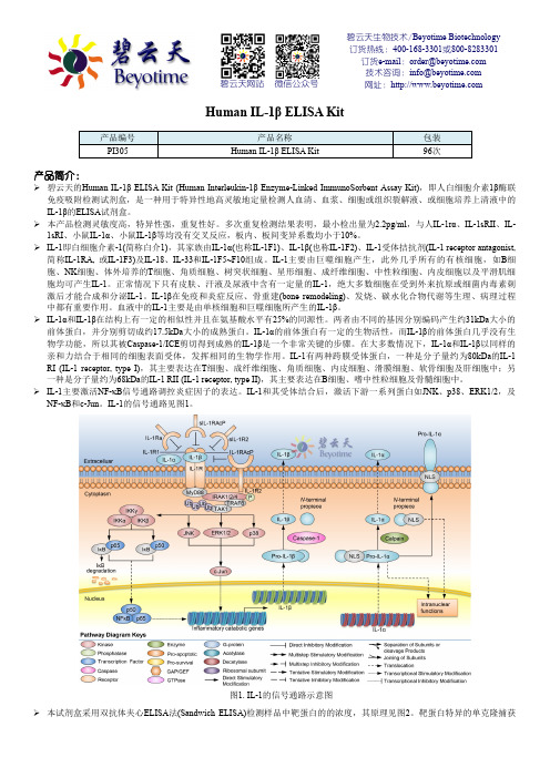
碧云天生物技术/Beyotime Biotechnology 订货热线:400-168-3301或800-8283301 订货e-mail :******************技术咨询:*****************网址:碧云天网站 微信公众号Human IL-1β ELISA Kit产品编号 产品名称包装 PI305Human IL-1β ELISA Kit96次产品简介:碧云天的Human IL-1β ELISA Kit (Human Interleukin-1β Enzyme-Linked ImmunoSorbent Assay Kit),即人白细胞介素1β酶联免疫吸附检测试剂盒,是一种用于特异性地高灵敏地定量检测人血清、血浆、细胞或组织裂解液、或细胞培养上清液中的IL-1β的ELISA 试剂盒。
本产品检测灵敏度高,特异性强,重复性好。
多次重复检测结果表明,最小检出量为2.2pg/ml ,与人IL-1r α、IL-1sRII 、IL-1sRI 、小鼠IL-1α、小鼠IL-1β等均没有交叉反应,板内、板间变异系数均小于10%。
IL-1即白细胞介素-1(简称白介1),其家族由IL-1α(也称IL-1F1)、IL-1β(也称IL-1F2)、IL-1受体拮抗剂(IL-1 receptor antagonist, 简称IL-1RA, 或IL-1F3)及IL-18、IL-33和IL-1F5~F10组成。
IL-1主要由巨噬细胞产生,此外几乎所有的有核细胞,如B 细胞、NK 细胞、体外培养的T 细胞、角质细胞、树突状细胞、星形细胞、成纤维细胞、中性粒细胞、内皮细胞以及平滑肌细胞均可产生IL-1。
正常情况下只有皮肤、汗液及尿液中含有一定量的IL-1,绝大多数细胞在受到外来抗原或细菌内毒素刺激后才能合成和分泌IL-1。
IL-1β在免疫和炎症反应、骨重建(bone remodeling)、发烧、碳水化合物代谢等生理、病理过程中都有重要作用。
试剂盒,人试剂盒,人基质裂解蛋白(MAT)ELISA试剂盒使用说明书

人基质裂解蛋白(MAT)ELISA试剂盒使用说明书本试剂仅供研究使用标本:血清或血浆樊克生物供应使用目的:本试剂盒用于测定人血清、血浆及相关液体样本基质裂解蛋白(MAT)含量。
试验原理:MAT试剂盒是固相夹心法酶联免疫吸附实验(ELISA).已知MAT浓度的标准品、未知浓度的样品加入微孔酶标板内进行检测。
先将MAT和生物素标记的抗体同时温育。
洗涤后,加入亲和素标记过的HRP。
再经过温育和洗涤,去除未结合的酶结合物,然后加入底物A、B,和酶结合物同时作用。
产生颜色。
颜色的深浅和样品中MAT的浓度呈比例关系。
基质裂解蛋白试剂盒,elisa试剂盒说明书,ELISA试剂盒,ELISA检测试剂盒,人elisa试剂盒说明书试剂盒内容及其配制试剂盒成份(2-8℃保存)96孔配置48孔配置配制96/48人份酶标板1块板(96T)半块板(48T)即用型塑料膜板盖1块半块即用型标准品:80ng/ml 1瓶(0.6ml)1瓶(0.3ml)按说明书进行稀稀空白对照1瓶(1.0ml)1瓶(0.5ml)即用型标准品稀释缓冲液1瓶(5ml)1瓶(2.5ml)即用型生物素标记的抗MAT抗体1瓶(6ml)1瓶(3.0ml)即用型亲和链酶素-HRP 1瓶(10ml)1瓶(5.0ml)即用型洗涤缓冲液1瓶(20ml)1瓶(10ml)按说明书进行稀释底物A 1瓶(6.0ml)1瓶(3.0ml)即用型底物B 1瓶(6.0ml)1瓶(3.0ml)即用型终止液1瓶(6.0ml)1瓶(3.0ml)即用型标本稀释液1瓶(12ml)1瓶(6.0ml)即用型自备材料1.蒸馏水。
2.加样器:5ul、10ul、50ul、100ul、200ul、500ul、1000ul。
3.振荡器及磁力搅拌器等。
安全性1.避免直接接触终止液和底物A、B。
一旦接触到这些液体,请尽快用水冲洗。
2.实验中不要吃喝、抽烟或使用化妆品。
3.不要用嘴吸取试剂盒里的任何成份。
操作注意事项1.试剂应按标签说明书储存,使用前恢复到室温。
人P21蛋白(P21)定量检测试剂盒(ELISA)使用说明书

本试剂盒只能用于科学研究,不得用于医学诊断。
人P21蛋白(P21)定量检测试剂盒(ELISA)使用说明书【试剂盒名称】人P21蛋白(P21)定量检测试剂盒(ELISA)【试剂盒用途】定量检测人血清、血浆及相关液体样本中P21蛋白(P21)的含量。
【检测原理】本试剂盒采用双抗体两步夹心酶联免疫吸附法(ELISA)。
将标准品、待测样本加入到预先包被人P21蛋白(P21)单克隆抗体透明酶标包被板中,温育足够时间后,洗涤除去未结合的成分,再加入酶标工作液,温育足够时间后,洗涤除去未结合的成分。
依次加入底物A、B,底物(TMB)在辣根过氧化物酶(HRP)催化下转化为蓝色产物,在酸的作用下变成黄色,颜色的深浅与样品中人P21蛋白(P21)浓度呈正相关,450nm波长下测定OD值,根据标准品和样品的OD值,计算样本中人P21蛋白(P21)含量。
【试剂盒组成】1酶标包被板12孔×8条7显色剂A液6mL2标准品0.3mL×6管8显色剂B液6mL320倍浓缩洗涤液25mL9终止液6mL4样本稀释液6mL10说明书1份5特殊稀释液6mL11封板膜2张6酶标试剂6mL12密封袋1个备注:标准品(1号→6号)浓度依次为:48、24、12、6、3、1.5ng/L【需要而未提供的试剂和器材】1、37℃恒温箱2、标准规格酶标仪3、精密移液器及一次性吸头4、蒸馏水5、一次性试管6、吸水纸【操作步骤】1、准备:从冰箱取出试剂盒,室温复温平衡30分钟。
2、配液:用蒸馏水将20倍浓缩洗涤液稀释成原倍的洗涤液。
3、加标准品和待测样本:取足够数量的酶标包被板,固定于框架上,分别设置标准品孔、待测样本孔和空白对照孔,记录各孔位置,在标准品孔中加入标准品50μL;待测样本孔中先加入待测样本10μL,再加样本稀释液40μL(即样本稀释5倍);空白对照孔不加。
4、温育:37℃水浴锅或恒温箱温育30min。
5、洗板:弃去液体,吸水纸上拍干,每孔加满洗涤液,静置1min,甩去洗涤液,吸水纸上拍干,如此重复洗板4次(也可用洗板机按说明书操作洗板)。
ABC公司 YTHDF1人源YTH结构域蛋白家族1 ELISA试剂盒说明书

Human YTH domain-containing family protein1ELISA Kit (YTHDF1)Catalog NO.:RK04130version:2.0*使用产品前,请务必阅读产品包装内说明书产品简介本试剂盒产品使用夹心法定量测定人血清、血浆、细胞培养上清或其它生物体液中YTHDF1的含量。
产品检测原理本实验采用双抗夹心ELISA法。
将抗YTHDF1抗体预先包被在酶标板上,向微孔中分别加入标准品和样品,样本中YTHDF1与固相载体上的抗体结合。
孵育后,未结合的样品在洗涤过程中会被洗去,加入抗YTHDF1的检测抗体,形成双抗体夹心。
洗去未结合的检测抗体,然后向微孔中加入酶结合物。
孵育和洗涤后,加入底物TMB。
样品中YTHDF1的含量与TMB反应的颜色深浅呈正相关,加入酸终止反应并测量吸光值。
根据梯度稀释的YTHDF1标准品的吸光值绘制标准曲线并求出样品的浓度。
试剂盒组分及保存条件未拆封的试剂盒可在2-8°C保存1年,已拆封的产品须在1个月内使用完。
组分规格货号拆封后保存条件抗体预包被酶标板Antibody Coated Plate 8×12RM17545将未使用的板条放回装有干燥剂的铝箔袋中,并重新密封,可在2-8°C存储1个月。
冻干标准品Standard Lyophilized 2支RM17546复溶后不建议再次使用。
浓缩生物素化抗体(100×)Concentrated Biotin Conjugate Antibody(100×)1×120ul RM17547可在2-8°C存储1个月。
浓缩链霉亲和素-HRP(100x)Streptavidin-HRP Concentrated(100×)1×120ul RM17548可在2-8°C存储1个月。
标准品/样本稀释液(R1)Standard/Sample Diluent (R1)1×20mLRM00023可在2-8°C 存储1个月。
TaKaRa BioMasher Standard 产品手册说明书
Cat. #9790A9791A Product ManualTaKaRa BioMasher Standard (Non-sterile)TaKaRa BioMasher Standard (Sterile)For Research UseTable of ContentsI. Description (3)II. Components (3)III. Storage (3)IV. Materials (3)V. Protocol (3)VI. Experimental Examples (4)VII. Related Products (4)I. DescriptionTaKaRa BioMasher Standard is a disposable microtube homogenizer designed to efficiently crush small amounts of biological samples for nucleic acid and/or protein extraction. This product is a set of a 1.5-ml microtubes and micro stir bars. The inner wall of the tube and the tip of the stir bar are textured allowing effective disruption of the sample. In addition, there is a lid to prevent splashing of the sample and reagents during sample processing. After the sample has been homogenized, the tube can be centrifuged to minimize sample loss.The TaKaRa BioMasher Standard is available either sterilized (Cat. #9791A) or nonsterilized (Cat.# 9790A).II. Components(1) TaKaRa BioMasher Standard (micro stir bar and microtube) 50(2) Stir bar grip (PESTLE GRIP) 1III. Storage Room temperatureKeep out of sun- and UV light.IV. MaterialsTaKaRa BioMasher StandardPolypropylene microtubes AutoclavablePolyacetal stir bar Not autoclavable*Silicon rubber stir bar grip Autoclavable* : The stir bar can not be autoclaved. If you require sterilization, please use Cat. #9791A. V. ProtocolNOTE: Wear a mask, gloves, lab coat, and safety goggles. If necessary, perform in a safety cabinet.1. Insert the micro stir bar into the end of the PESTLE GRIP.2. Add the sample to the microtube. Use less than 100 mg of sample. If necessary, add theextraction reagent (less than 250 μl).3. Insert the micro stir bar into the microtube. With the PESTLE GRIP, rotate and move themicro stir bar up and down, crushing the sample. If necessary, perform on ice.4. After crushing, disconnect the PESTLE GRIP and discard the micro stir bar. The PESTLEGRIP can be used repeatedly; store for future use.5. Close the lid of the microtube. The sample is ready for extraction.VII. Related ProductsRNAisoPlus (Cat. #9108/9109)*NucleoSpin® RNA II (Cat. #740955.10/.50/.250)NucleoSpin® RNA XS (Cat. #740902.10/.50/.250)NucleoSpin® Tissue (Cat. #740952.10/.50/.250)NucleoSpin® Tissue XS (Cat. #740901.10/.50/.250)Wide-Range DNA Ladder (100 - 2,000 bp) (Cat. #3427A/B)* : Not available in all geographic locations. Check for availability in your region.VI. Experimental ExampleTotal RNA extraction from mouse liver 100 mg of mouse liver was crushed using the TaKaRa BioMasher Standard (Sterile). As a comparison, 100 mg frozen mouse liver tissue was crushed with a mortar or with crusher beads. For all samples, total RNA was extracted using 1 ml RNAiso Plus (Cat. #9108/9109) according to the recommended protocol. Total RNA was quantified and analyzed on an 1% agarose gel.Results: Processing the samples using the TaKaRa BioMasher Standard had similar extraction efficiency as the mortar and bead crushing protocols.Crushing method total RNA (μg) A 260/A 2801.Mortar 360 1.702.Mortar 424 1.803.Crusher beads 530 1.924.Crusher beads 494 1.935.TaKaRa BioMasher Standard 411 1.896.TaKaRa BioMasher Standard4311.90M 125364(bp)2,000750Lane 1-6 Equal amounts of RNA M Wide-Range DNA Ladder (100 - 2,000 bp)NOTE : This product is for research use only. It is not intended for use in therapeutic or diagnostic procedures for humans or animals. Also, do not use this product as food, cosmetic, or household item, etc.Takara products may not be resold or transferred, modified for resale or transfer, or used to manufacture commercial products without written approval from TAKARA BIO INC.If you require licenses for other use, please contact us by phone at +81 77 543 7247 or from our website at .Your use of this product is also subject to compliance with any applicable licensing requirements described on the product web page. It is your responsibility to review, understand and adhere to any restrictions imposed by such statements.All trademarks are the property of their respective owners. Certain trademarks may not be registered in all jurisdictions.。
ELISA 检测试剂盒 说明书
本试剂盒只能用于科学研究,不得用于医学诊断小鼠小鼠((MouseMouse))肿瘤坏死因子可溶性受体肿瘤坏死因子可溶性受体ⅠⅠ(TNFsR-TNFsR-ⅠⅠ)ELISA检测试剂盒使用说明书检测原理试剂盒采用双抗体一步夹心法酶联免疫吸附试验(ELISA)。
往预先包被肿瘤坏死因子可溶性受体Ⅰ(TNFsR-Ⅰ)抗体的包被微孔中,依次加入标本、标准品、HRP标记的检测抗体,经过温育并彻底洗涤。
用底物TMB显色,TMB在过氧化物酶的催化下转化成蓝色,并在酸的作用下转化成最终的黄色。
颜色的深浅和样品中的肿瘤坏死因子可溶性受体Ⅰ(TNFsR-Ⅰ)呈正相关。
用酶标仪在450nm波长下测定吸光度(OD值),计算样品浓度。
样品收集、处理及保存方法1.血清:使用不含热原和内毒素的试管,操作过程中避免任何细胞刺激,收集血液后,3000转离心10分钟将血清和红细胞迅速小心地分离。
2.血浆:EDTA、柠檬酸盐或肝素抗凝。
3000转离心30分钟取上清。
3.细胞上清液:3000转离心10分钟去除颗粒和聚合物。
4.组织匀浆:将组织加入适量生理盐水捣碎。
3000转离心10分钟取上清。
5.保存:如果样本收集后不及时检测,请按一次用量分装,冻存于-20℃,避免反复冻融,在室温下解冻并确保样品均匀地充分解冻。
自备物品1.酶标仪(450nm)2.高精度加样器及枪头:0.5-10uL、2-20uL、20-200uL、200-1000uL3.37℃恒温箱操作注意事项1.试剂盒保存在2-8℃,使用前室温平衡20分钟。
从冰箱取出的浓缩洗涤液会有结晶,这属于正常现象,水浴加热使结晶完全溶解后再使用。
2.实验中不用的板条应立即放回自封袋中,密封(低温干燥)保存。
3.浓度为0的S0号标准品即可视为阴性对照或者空白;按照说明书操作时样本已经稀释5倍,最终结果乘以5才是样本实际浓度。
4.严格按照说明书中标明的时间、加液量及顺序进行温育操作。
5.所有液体组分使用前充分摇匀。
达优人 IL-1β ELISA 试剂盒说明书

产品信息和操作指南Human IL-1β预包被 ELISA kit Cat# : DKW12-1012-048 / DKW12-1012-096本试剂盒专用于科研,而非用于诊断Human IL-1βDKW12-1012目录产品简介 (1)知识背景 (1)试剂盒提供的试剂 (2)需要实验者自行准备的试剂与仪器 (2)注意事项 (3)试剂的配制 (5)操作过程 (7)结果分析 (9)试剂盒的保存 (9)操作步骤一览表 (10)参考文献 (11)ELISA测定中可能会出现的问题及解决方法 (12)预包被ELISA 试剂盒系列产品 (15)1、产品简介:达优®人IL-1β ELISA试剂盒是通过酶联免疫吸附技术,体外定量检测人血清、血浆、缓冲液或细胞培养液中的IL-1β,可同时检测天然的和重组的IL-1β。
本试剂盒为预包被板,“夹心一步”完成,整个过程孵育时间不超过4小时,洗涤6次,操作时间大大减少。
本试剂盒专用于科研,而非用于诊断。
使用前请仔细阅读说明书并检查试剂盒组分,若有任何疑问请与达科为生物工程有限公司联系,E-mail:*************.检测范围:500-15.6 pg/mL灵敏度:5 pg/mL重复性:板内、板间变异系数均<10%。
2、知识背景:白细胞介素-1(Interleukin-1,IL-1)家族蛋白包括IL-1α、IL-1β和IL-1受体拮抗物。
除了皮肤角质化细胞、一些上皮细胞和中枢神经系统的一些细胞之外,IL-1并不在健康个体的未经刺激的细胞中分泌。
生理条件下IL-1的浓度仅在10-12-10-14M之间。
然而,为了应对刺激(如炎症物质、感染、细菌性内毒素),巨噬细胞和各种其它类型的细胞的IL-1分泌量将大大增加(1-4)。
3、试剂盒提供的试剂:试剂规格配制Cytokine standard 2/1瓶* 干粉状,按瓶上说明操作Biotinylated antibody 2/1瓶* 1:50用Dilution buffer R(1×)稀释Streptavidin-HRP 2/1瓶* 1:100用Dilution buffer R(1×)稀释Dilution buffer R(1×) 3/2瓶* 即用型Washing buffer(50×)1瓶 150∶用蒸馏水稀释TMB 1瓶即用型Stop solution 1瓶即用型Precoated ELISA plate 8×12或8×6*即用型封板膜 2/1张* 即用型说明书1份*:96/48 Tests4、需要实验者自行准备的试剂与仪器:1.酶标仪(建议参考仪器使用说明提前预热)2.微量加液器及吸头:P10,P50,P100,P200,P1000 3.蒸馏水或去离子水4.全新滤纸5.旋涡振荡器和磁力搅拌器5、注意事项:1.试剂应按瓶上标签说明储存,使用前室温平衡20-30分钟。
人载脂蛋白 P ApoP ELISA试剂盒北京驰明瑞使用说明书

人载脂蛋白P ApoP使用说明书产品编号:本试剂盒仅供科研使用,不得用于临床及诊断使用!操作步骤1.取出试剂盒,于室温(20-25℃)放置15-30分钟。
实验过程应在室温(20-25℃)内进行。
2.取出酶标板,按照标准品的次序分别加入100μl的标准品溶液于空白微孔中。
3.空白微孔中加入100μl的样品,空白对照加入100μl的蒸馏水;4.在各孔中加入50μl的酶标记溶液;(不含空白对照孔)5.将酶标板用封口胶密封后,37℃孵育反应1小时;(在孵育箱中保持稳定的温度与湿度)6.充分清洗酶标板3-5次,保持各孔有充足的水压;(浓缩洗涤液以1:100的比例与蒸馏水稀释)7.酶标板洗涤后用吸水纸彻底拍干;8.各孔加入显色剂A、B液各50μl;(不含空白对照孔)9.20-25℃下避光反应10分钟;10.各孔加入50μl终止液,终止反应;结果判断1.30分钟内在波长450nm的酶标仪上读取各孔的OD值;2.百分结合率计算:设S0管计数为B0,各标准管或样品管计数为B,非特异管计数为NSB,则百分结合率计算公式如下:B/ B0=(B-NSB)/( B0-NSB)×100%3.logit计算:各标准点或样品管的logit值计算公式如下:logit=ln(B/ B0)/(1-B/ B0)4.将标准品的OD均值与标准品0点的OD均相除,为标准点的百分结合率,在log-logit坐标纸上绘图。
5.Log-logit双对数标准曲线:坐标纸上横轴从左至右第一个1-9表示为第一个10进位,第二个1-9表示为第二个10进位。
第三个1-9表示为第三个10进位。
坐标纸纵轴为百分比(1-99),即各标准吸光值的百分结合率。
取一条通过各点的直线。
要求尽可能多的点在线上,同时剩余的点均匀分布在直线的两边。
样品也同样由吸光值计算百分结合率,再从纵轴上的相应结合率找到直线上的点,此点对应的横坐标浓度即为样品的浓度,无须换算。
6.人工处理:以标准浓度取log值为横坐标,对应的logit值为纵坐标在普通坐标纸上或以标准浓度为横坐标,对应的B/B0为纵坐标在logit-log坐标纸上画出标准曲线(理想化时是一条直线)。
PAKAUTO操作说明
GTI血小板本身抗体检测试剂盒(GTI-PAKAUTO )注:按照每人份使用1条进行实验,阴性对照为1条,具体环节请见原始英文使用阐明书。
用去离子水稀释10x洗液,1倍体积Conc. Wash + 9倍体积去离子水。
在统计纸(RS)上标记检测格局。
1.用稀释液SD按照3:1比例稀释阴阳性对照、病人血清。
A)取由EDTA 抗凝的血样,离心(110-150rcf,10-15min ),得到富含血小板的血浆(PRP)。
B)将PRP移入一种干净的多聚丙烯管中,离心(1rcf, 5-10min),得血小板扣。
弃掉血浆。
C)向血小板扣中加入500ul 细胞重悬防腐液(CRP),用小枪头轻轻吹打混匀。
将此血小板悬浊液移入离心管中,用最少500ul CRP洗3次。
D)制备含1-5 x 10⁷血小板的血小板扣:用CRP重悬上步的血小板扣,将血小板的浓度调节为每微升悬液含10000-50000个血小板。
取1ml 此悬液入离心管,离心1rcf, 5min。
(或者直接取10-15ul 体积的血小板扣也可,此量足以制备血小板洗脱液)3.制备血小板对照A)向阳性血小板对照(PCP)、阴性血小板对照(NCP)中加入500ul CRP,室温放置最少10分钟使之水化。
轻轻混匀后离心1rcf, 5-10min。
吸干液体残留以得到干燥的血小板扣。
注意:最佳用新鲜的正常血小板制备正常血小板洗脱液,如果没有新鲜的血小板再用试剂盒中提供的干正常血小板。
4.制备洗脱液A)在制备洗脱液前要确保血小板已完全沉淀,且不混有残留的CRP。
B)向已制备的血小板扣中加入180ul 洗脱液(ES),枪头吹打混匀,室温放置2分钟。
离心1rcf, 5min。
C)快速将洗脱液转移到一种干净的离心管中,加180ul BS , 充足混匀,离心1rcf, 10min。
5.立刻进入下一步实验,或将血小板洗脱液放-80度冻存。
注意:切勿稀释血小板洗脱液。
6.向每个微孔内加300ul 用去离子水稀释好的洗液,室温放置5-10分钟。
人C反应蛋白(CRP)ELISA分析检测试剂盒使用说明书
人C反应蛋白(CRP)ELISA分析检测试剂盒使用说明书人C反应蛋白(CRP)ELISA分析检测试剂盒使用说明书引言:本文是针对人C反应蛋白(CRP)ELISA分析检测试剂盒的使用说明书,旨在帮助用户正确、便捷地操作该试剂盒,获取准确的检测结果。
请用户在使用前仔细阅读本说明书,并按照所列步骤进行操作。
1. 产品介绍1.1 人C反应蛋白(CRP)ELISA分析检测试剂盒是一种用于定量分析人体内C反应蛋白的试剂盒。
1.2 试剂盒内含有酶标板、标准品、检测样本预处理液、酶标抗体、酶标底物、洗涤缓冲液等。
2. 前期准备2.1 检测前,确保试剂盒在正确的保存条件下,并检查是否有损坏。
2.2 准备洗涤缓冲液,并按照说明稀释至所需浓度。
2.3 准备一次性使用的微量移液器和吸头。
2.4 样本处理前,确保样本已经充分解冻,并且清晰无杂质。
2.5 标准品的准备:按照说明书中的浓度进行稀释,得到一系列不同浓度的标准品。
3. 检测步骤3.1 样本预处理:取相应数量的样本和标准品,加入与试剂盒提供的检测样本预处理液等体积比例的去蛋白液,混合均匀,放置于室温下孵育一段时间。
3.2 洗涤:将试剂盒提供的洗涤缓冲液加入洗涤槽中,使用洗涤缓冲液洗涤酶标板中的洗涤孔,每孔洗涤3-4次,倾倒废液。
3.3 添加试剂:按照试剂盒中所标示的各组分体积,将样本、标准品和酶标抗体等逐步加入对应的酶标孔中。
3.4 孵育:酶标板封闭后,放置于恒温孵育箱中,按照说明书中所示的温度和时间进行孵育。
3.5 清洗:孵育结束后,将酶标板取出倒置,用洗涤缓冲液洗涤酶标孔,每孔洗涤3-4次,倾倒废液。
3.6 加底物:将试剂盒提供的酶标底物加入酶标孔中,按照说明书中所示的温度和时间进行反应,避免阳光直射。
3.7 反应终止:加入酶标终止液终止反应,避免经久暴露。
3.8 检测:使用酶标仪读取各孔的吸光度,记录相应数值。
4. 数据分析4.1 根据吸光度值,绘制标准曲线,使用标准曲线确定样本中的C 反应蛋白浓度。
人 BMP-2 ELISA试剂盒说明书
人BMP-2ELISA试剂盒Human BMP-2ELISA kitCat#:EHC172尊敬的客户,感谢您选用本公司QuantiCyto®ELISA系列产品。
本产品选用世界著名生产厂家的原料,采用专业体外诊断试剂生产技术制造,仅供科研使用。
本产品可检测血清、血浆、细胞培养上清液、灌洗液、尿液、羊水、腹水、脑脊液、胸腔积液、组织匀浆液等多种类型样本中天然和重组人BMP-2浓度,具体适用样本请咨询。
使用前请仔细阅读说明书并检查试剂组分。
如有疑问,请联系欣博盛生物科技有限公司。
Email:**********************,全国服务热线: 4006800892。
QuantiCyto ®EHC172Human BMP-22试剂盒组成:名称48T 96T 抗体预包被酶标板8×68×12冻干标准品5ng /支×2支5ng /支×3支标准品&标本通用稀释液12ml×1瓶20ml×1瓶浓缩生物素化抗体1支(规格见标签)1支(规格见标签)生物素化抗体稀释液10ml×1瓶16ml×1瓶浓缩酶结合物1支(规格见标签)1支(规格见标签)酶结合物稀释液10ml×1瓶16ml×1瓶浓缩洗涤液20×25ml×1瓶50ml×1瓶显色底物(TMB )6ml×1瓶12ml×1瓶反应终止液具腐蚀性6ml×1瓶12ml×1瓶封板胶纸3张6张产品说明书1份1份储存条件:未开封完整试剂盒4℃保存,请在保质期内使用。
已开封试剂盒抗体包被板条未用完的板条放回带拉链铝箔袋,封好口。
4℃条件下可储存1个月左右。
标准品冻干粉-20℃可储存6个月左右。
稀释后的标准品使用后请丢弃。
浓缩生物素化抗体浓缩液4℃可储存1个月左右,稀释后即用即弃。
浓缩酶结合物(避光)标准品&标本通用稀释液4℃条件下,可储存1个月左右。
- 1、下载文档前请自行甄别文档内容的完整性,平台不提供额外的编辑、内容补充、找答案等附加服务。
- 2、"仅部分预览"的文档,不可在线预览部分如存在完整性等问题,可反馈申请退款(可完整预览的文档不适用该条件!)。
- 3、如文档侵犯您的权益,请联系客服反馈,我们会尽快为您处理(人工客服工作时间:9:00-18:30)。
人骨成型蛋白受体1A BMP-R1A使用说明书产品编号:本试剂盒仅供科研使用,不得用于临床及诊断使用!操作步骤1.取出试剂盒,于室温(20-25℃)放置15-30分钟。
实验过程应在室温(20-25℃)内进行。
2.取出酶标板,按照标准品的次序分别加入100μl的标准品溶液于空白微孔中。
3.空白微孔中加入100μl的样品,空白对照加入100μl的蒸馏水;4.在各孔中加入50μl的酶标记溶液;(不含空白对照孔)5.将酶标板用封口胶密封后,37℃孵育反应1小时;(在孵育箱中保持稳定的温度与湿度)6.充分清洗酶标板3-5次,保持各孔有充足的水压;(浓缩洗涤液以1:100的比例与蒸馏水稀释)7.酶标板洗涤后用吸水纸彻底拍干;8.各孔加入显色剂A、B液各50μl;(不含空白对照孔)9.20-25℃下避光反应15分钟;10.各孔加入50μl终止液,终止反应;结果判断1.30分钟内在波长450nm的酶标仪上读取各孔的OD值;2.百分结合率计算:设S0管计数为B0,各标准管或样品管计数为B,非特异管计数为NSB,则百分结合率计算公式如下:B/ B0=(B-NSB)/( B0-NSB)×100%3.logit计算:各标准点或样品管的logit值计算公式如下:logit=ln(B/ B0)/(1-B/ B0)4.将标准品的OD均值与标准品0点的OD均相除,为标准点的百分结合率,在log-logit坐标纸上绘图。
5.Log-logit双对数标准曲线:坐标纸上横轴从左至右第一个1-9表示为第一个10进位,第二个1-9表示为第二个10进位。
第三个1-9表示为第三个10进位。
坐标纸纵轴为百分比(1-99),即各标准吸光值的百分结合率。
取一条通过各点的直线。
要求尽可能多的点在线上,同时剩余的点均匀分布在直线的两边。
样品也同样由吸光值计算百分结合率,再从纵轴上的相应结合率找到直线上的点,此点对应的横坐标浓度即为样品的浓度,无须换算。
6.人工处理:以标准浓度取log值为横坐标,对应的logit值为纵坐标在普通坐标纸上或以标准浓度为横坐标,对应的B/B0为纵坐标在logit-log坐标纸上画出标准曲线(理想化时是一条直线)。
根据待测样品的B/B0可以从坐标纸上查出样品的浓度值。
如果使用普通坐标纸,查出的数值应取反对数才是最后的浓度值。
7.自动处理:使用logit-log或四参数数据处理模式,由电脑自动计算得出结果。
8.敏感度:1.0ng/ml;9.图例Human Bone morphogenetic protein reportor 1A ELISA Kit96 TestsCatalogue Number:Store all reagents at 2-8°CFOR LABORATORY RESEARCH USE ONLY. NOT FOR THERAPEUTIC OR DIAGNOSTIC APPLICATIONS!PLEASE READ THROUGH ENTIRE PROCEDURE BEFORE BEGINNING!INTENDED USEThis BOGOO BMP-R1A ELISA kit is intended Laboratory for Research use only and is not for use in diagnostic or therapeutic procedures.The Stop Solution changes the color from blue to yellow and the intensity of the color is measured at 450 nm using a spectrophotometer. In order to measure the concentration of BMP-R1A in the sample, this BMP-R1A ELISA Kit includes a set of calibration standards. The calibration standards are assayed at the same time as the samples and allow the operator to produce a standard curve of Optical Density versus BMP-R1A concentration. The concentration of BMP-R1A in the samples is then determined by comparing the O.D. of the samples to the standard curve.PRINCIPLE OF THE ASSAYThe coated well immunoenzymatic assay for the quantitative measurement of serum BMP-R1A utilizes a monoclonal anti-BMP-R1A and a BMP-R1A-HRP conjugate. The assay asample and buffer are incubated together with anti-BMP-R1A antibody coated plate for sixty and washed. The diluted BMP-R1A-HRP conjugate is then added to each well and incubated. After the incubation period, the wells are decanted and washed three times. The wells are then incubated with a substrate for the enzyme. The product of the enzyme-substrate reaction forms a blue colored complex. Finally, a stopping solution is added to stop the reaction, which will then turn the solution yellow.The intensity of color is measured spectrophotometrically at 450nm in a microplate reader. The intensity of the color is inversely proportional to the BMP-R1A concentration since BMP-R1A from samples and BMP-R1A-HRP conjugate compete for the anti-BMP-R1A antibody binding site. Since the number of sites is limited, as more sites are occupied by BMP-R1A from the sample, fewer sites are left to bind BMP-R1A-HRP conjugate.Standards of known BMP-R1A concentrations are run concurrently with the samples being assayed and a standard curve is plotted relating the intensity of the color (Optical Density) to the concentration of BMP-R1A. The unknown BMP-R1A concentration in each sample is interpolated from this curve.REAGENTS PROVIDEDAll reagents provided are stored at 2-8° C. Refer to the expiration date on the label.1.MICROTITER PLATE 96 wells2.ENZYME CONJUGATE 6.0 mL 1 vial3.STANDARD.1 0ng/ml 1 vial4.STANDARD.2 50ng/ml 1 vial5.STANDARD.3 100ng/ml 1 vial6.STANDARD.4 250ng/ml 1 vial7.STANDARD.5 500ng/ml 1 vial8.STANDARD.6 1000ng/ml 1 vial9.SUBSTRATE A 6.0 mL 1 vial10.SUBSTRATE B 6.0 mL 1 vial11.STOP SOLUTION 6.0 mL 1 vial12.WASH SOLUTION x100 10 mL 1 vial13.Instruction 1MATERIALS REQUIRED BUT NOT SUPPLIED1.Microplate reader capable of measuring absorbance at 450 nm.2.Precision pipettes to deliver 2 ml to 1 ml volumes.3.Adjustable 10ml -100ml pipettes for reagent preparation.4.Adjustable 10ml -100ml pipettes for reagent preparation.5.100 ml and 1 liter graduated cylinders.6.Calibrated adjustable precision pipettes, preferably with disposable plastic tips. (A manifold multi-channelpipette is desirable for large assays.)7.Absorbent paper.8.37°C incubator.9.Distilled or deionized water.10.Data analysis and graphing software. Graph paper: linear (Cartesian), log-log or semi-log, or log-logit asdesired.11.Tubes to prepare standard or sample dilutions.ASSAY PROCEDURE1. 1 .Prepare all Standards before starting assay procedure (see Preparation Reagents). It is recommended thatall Standards and Samples be added in duplicate to the Microtiter Plate.Standards or Samples to2.First, secure the desired number of coated wells in the holder, then add 100 μL ofthe appropriate well of the antibody pre-coated Microtiter Plate.3.Add 50 μL of Conjugate to each well. Mix well. Complete mixing in this step is important. Cover andincubate for 1 hours at 37°C.4.Prepare Substrate Solution no more than 15 minutes before end of incubation (see Preparation of Reagents).5.Wash the Microtiter Plate using one of the specified methods indicated below:6.Manual Washing: Remove incubation mixture by aspirating contents of the plate into a sink or proper wastecontainer. Using a squirt bottle, fill each well completely with distilled or de-ionized water, then aspiratecontents of the plate into a sink or proper waste container. Repeat this procedure four more times for a totalof FIVE washes. After final wash, invert plate, and blot dry by hitting plate onto absorbent paper or papertowels until no moisture appears. Note: Hold the sides of the plate frame firmly hen washing the plate toassure that all strips remain securely in frame.7.Automated Washing: Aspirate all wells, then wash plate FIVE times using distilled or de-ionized water.Always adjust your washer to aspirate as much liquid as possible and set fill volume at 350 μL/well/wash(range: 350-400 μL). After final wash, invert plate, and blot dry by hitting plate onto absorbent paper paper towels until no moisture appears. It is recommended that the washer be set for a soaking time of 10seconds or shaking time of 5 seconds between washes.8.Add 50 μL Substrate A&B to each well. Cover and incubate for 15 minutes at 20-25°C.9.Add 50 μL of Stop Solution to each well. Mix well.10.Read the Optical Density (O.D.) at 450 nm using a microtiter plate reader within 30 minutes.BOGOO CALCULATION OF RESULTS1.Calculate the mean absorbance value A450 for each set of reference standards and samples.2.Divide the average A450 value for each standard, control and test sample by the average A450 of standard0and multiply by 100 to obtain %B/B0 for each sample.3.Prepare a standard curve by plotting the average absorbance of each standard versus the correspondingconcentrations of the standards on linear-log graph paper or the %B/B0 value for each standard versus the corresponding concentration of the standard on linear-log or logit-log graph paper. logit=ln(B/ B0)/(1-B/ B0)4.Any values obtained for diluted samples must be further converted by applying the appropriate dilution factorin the calculations.5.The standard density is a X, the B/ B0 is a Y, sitting to mark the paper in the logit- log up draw a standardcurve.According to the B/ B0 that need to be measured the sample can from sit to mark the density value that the paper looks up the sample up.6.The sensitivity by this assay is 1.0ng/ml7.The standard curve presented here is an example of the data typically produced with this kit; however, yourresults will not be identical to these.8.Standard curve。
