AOAC Official Method 964.02 testing disinfectants against against Pseudomonas aeruginosa
AOAC脂肪提取标准
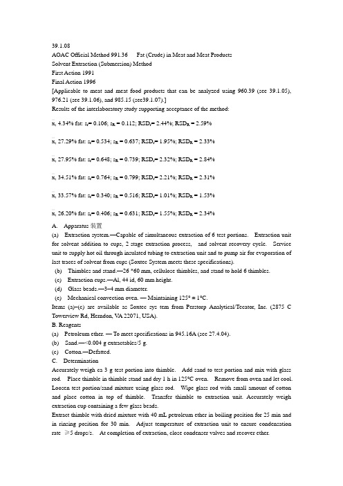
39.1.08AOAC Official Method 991.36 Fat (Crude) in Meat and Meat ProductsSolvent Extraction (Submersion) MethodFirst Action 1991Final Action 1996[Applicable to meat and meat food products that can be analyzed using 960.39 (see 39.1.05), 976.21 (see 39.1.06), and 985.15 (see39.1.07).]Results of the interlaboratory study supporting acceptance of the method:x—, 4.34% fat: s r= 0.106; s R = 0.112; RSD r= 2.44%; RSD R = 2.59%x—, 27.29% fat: s r= 0.534; s R = 0.637; RSD r= 1.95%; RSD R = 2.33%x—, 27.95% fat: s r= 0.648; s R = 0.739; RSD r= 2.32%; RSD R = 2.84%x—, 34.51% fat: s r= 0.764; s R = 0.799; RSD r= 2.21%; RSD R = 2.31%x—, 33.57% fat: s r= 0.340; s R = 0.516; RSD r= 1.01%; RSD R = 1.53%x—, 26.20% fat: s r= 0.406; s R = 0.631; RSD r= 1.55%; RSD R = 2.34%A. Apparatus装置(a) Extraction system.—Capable of simultaneous extraction of 6 test portions. Extraction unit for solvent addition to cups, 2-stage extraction process, and solvent recovery cycle. Service unit to supply hot oil through insulated tubing to extraction unit and to pump air for evaporation of last traces of solvent from cups (Soxtec System meets these specifications).(b) Thimbles and stand.—26 *60 mm, cellulose thimbles, and stand to hold 6 thimbles.(c) Extraction cups.—Al, 44 id, 60 mm height.(d) Glass beads.—3–4 mm diameter.(e) Mechanical convection oven. — Maintaining 125° ± 1°C.Items (a)–(c) are available as Soxtec sys tem from Perstorp Analytical/Tecator, Inc. (2875 C Towerview Rd, Herndon, V A 22071, USA).B. Reagents(a) Petroleum ether. — To meet specifications in 945.16A (see 27.4.04).(b) Sand.—<0.004 g extractables/5 g.(c) Cotton.—Defatted.C. DeterminationAccurately weigh ca 3 g test portion into thimble. Add sand to test portion and mix with glass rod. Place thimble in thimble stand and dry 1 h in 125°C oven. Remove from oven and let cool. Loosen test portion/sand mixture using glass rod. Wipe glass rod with small amount of cotton and place cotton in top of thimble. Transfer thimble to extraction unit. Accurately weigh extraction cup containing a few glass beads.Extract thimble with dried mixture with 40 mL petroleum ether in boiling position for 25 min and in rinsing position for 30 min. Adjust temperature of extraction unit to ensure condensation rate≥5 drops/s. At completion of extraction, close condenser valves and recover ether.Dry cup and contents 30 min in 125°C oven. Cool and weigh.D. CalculationsCalculate percent fat in test sample as follows:Fat content, % = (B -C) *100Awhere A = g test portion weight, B = g weight of extraction cup afterdrying, and C = g weight of extraction cup prior to extraction.Reference: J. AOAC Int. 75, 289(1992).39.1.07AOAC Official Method 985.15Fat (Crude) in Meat and Poultry 家禽Products Rapid Microwave-Solvent Extraction Method First Action 1985Final Action 1991A. Reagents and Apparatus(a) Automated solvent extractor.—Enclosed, self-contained, thermostatically controlled 恒温控制fat extraction and solvent recovery system with 0.5 mg fat sensitivity and 0–100% fat measurement range (CEM Corp., PO Box 200, Matthews, NC 28106, USA), or equivalent.(b) Methylene chloride.二氯甲烷—Reagent grade (Fisher Scientific Co., No. D-37) , or equivalent.(c) Glass fiber pads.玻璃纤维垫子—9.8 *10.2 cm rectangular 矩形and 11 cm round (CEM Corp.), or equivalent.(d) Microwave moisture analyzer.—0.2 mg H2O sensitivity, moisture/solids range of 0.1–99.9%,0.01% resolution分辨率. Includes automatic tare electronic balance, microwave drying system,and microprocessor digital computer control. Electronic balance pan is located inside drying chamber. (Balance sensitivity: 0.2 mg at 15 g capacity or 1.0 mg at 40 g capacity [CEM Corp., or equivalent].)B. DeterminationPrepare test samples as in 983.18 (see 39.1.01). Place 2 rectangular and one round glass fiber pad on balance pan inside microwave moisture analyzer, and tare. Remove rectangular pads and evenly spread ca 4 g well-mixed test portion onto rough side of one pad, cover with second pad, and place together with round pad on balance pan. Dry 3–5 min at 80–100% power, depending on product type. At end of drying cycle, remove from balance pan. Fold rectangular pads, with dried test portion, in half and place in automated solvent extractor chamber. Place round pad in recessed area at top of extractor chamber, close and latch lid. Start extraction cycle (test portion and rectangular pads are blended at this time with sufficient CH2Cl2to extract fat). After completion of extraction cycle, remove round pad with residue, and place on balance pan in microwave moisture analyzer. Redry pad and residue to constant weight (ca 30 s at 80–100% power) to re move residual solvent or moisture. Weight loss due to solvent extraction is converted to % fat by microprocessor and displayed on digital read out panel.Certain product classes require addition of adjustment factors to read out for accurate results, as follows: fresh meats, pre-blends, emulsions, cured/cooked meats, factor = 0.40; cooked sausages, factor = 0.80.Reference: JAOAC 68, 876(1985).39.1.06AOAC Official Method 976.21Fat (Crude) in Meat Rapid Specific Gravity MethodFirst Action 1976Final Action 1979A. Apparatus and Reagents(a) Foss-let fat analyzer.—Includes orbital shaker, specific gravity readout unit, solvent dispenser, reference standard oil (specific gravity at 23°C = 0.915; for periodic check of potentiometer calibration), stainless steel cup with cover and 8 mm bore brass hammer, pressure filtration device, and conversion chart (Foss Food Technology Corp.).(b) Drying agent.—Plaster of Paris (available locally through paint, hardware, or building supply dealers), 8 mesh Drierite, or an hydrous CaSO4.(c) Tetrachloroethylene.—Technical grade C2Cl4 (distributed locally through dry cleaning sup pliers or Fisher Scientific Co.,No. C-182).B. Deter mi na tionPrepare test sam ples as in 983.18 (see 39.1.01). Check calibration of Foss-let potentiometer daily by us ing C2Cl4 alone to set zero point.Use mixture of 22.5 g reference standard oil and 120 mL C2Cl4(specific gravity of mixture at 37° = 1.4763) to set 50% fat point at 850.0.Using either top-load or triple-beam balance with 0.1 g sensitivity, tare Foss-let cup after setting brass ham mer on itsspin dle. To an a lyze prod ucts con tain ing £60% fat, weigh 45.0 gtest sam ple into cup; for prod ucts con tain ing >60% fat, weigh22.5 g. Add ca 80 g Plas ter of Paris (or ca 60 g an hy drous CaSO4).Dis pense 120 mL C2Cl4 into cup. Press cover onto cup and in stall inor bital shaker. Set shaker timer for 2 min and turn unit on. Whileex trac tion pro ceeds, as sem ble pres sure fil tra tion de vice by plac inginto per fo rated base 7 cm fil ter pa per. To pro duce clear fil trate freeof mois ture drop lets (for very wet test sam ples), first place highre ten tion pa per, Whatman No. 50, or equiv a lent, and then phasesep a rat ing pa per, Whatman No. 1PS, or equiv a lent. Af ter 2 minex trac tion, re move cup from shaker, lift cover, and re move brassham mer from cup. Im merse cup in ice-water bath ca 0.4 min whilestir ring con tents with ther mom e ter to cool con tents from 47°–52°Cto ca 40°C. Wipe H2O from outer sur face of cup and pour con tentsinto as sem bled fil ter. Place pis ton at top of fil tra tion de vice andslowly press ex tract through mea sur ing sys tem. De press drain valvebut ton when ex tract ap pears in over flow tube and let cham ber drain;then re lease valve but ton. Re peat fill ing and drain ing 2 more timesun til 40–50 mL ex tract has flowed through, re tain ing fi nal 10 mLex tract in mea sur ing cham ber. Re move fil tra tion de vice, slideview ing lens into po si tion, ro tate con trol of read out po ten ti om e terclock wise un til hy drom e ter rises, and re cord read ing. Es tab lish thatex tract is at cham ber tem per a ture by re peat ing read ing 3–4 times.Av er age read ings and con vert into % fat by means of con ver sion chart. (Mul ti ply chart % fat by 2 if a 22.5 g por tion of high-fat product was used.)Ref er ences: JAOAC 58, 1182(1975); 60, 853(1977);68, 240(1985).。
微波,核磁共振法 水分,固形物,脂肪 快速测定仪 AOAC标准操作规程
食品与食品添加剂
JOURNAL OF AOAC INTERNATIONAL VOL. 88, NO. 1, 2005
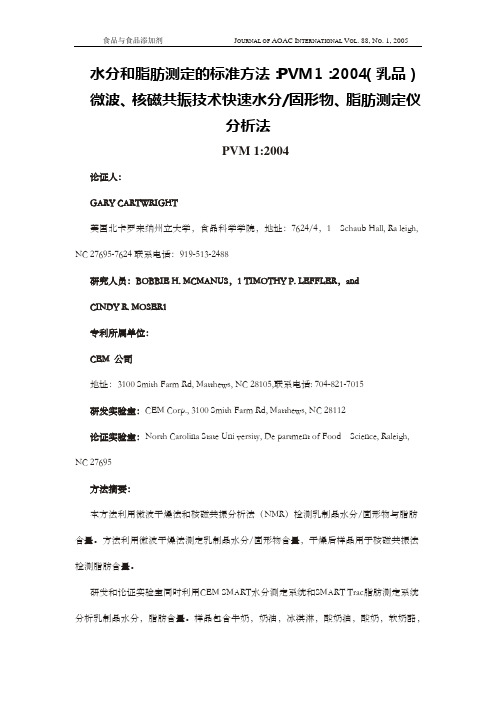
提示安装心脏起搏器或其他磁干扰仪器的人员与SMART Trac仪器磁元 件保持至少11英寸(0.3米)的距离。 2 本方法利用微波干燥法和快速NMR测定法来测定水分/固形物和脂肪含量, 特别是乳制品。本方法适用于大部分乳制品,测定范围宽泛。
5.缩写与专业术语 5.1 5.2 5.3 5.4 NMR——核磁共振 RF——随机频率 NCSU——北卡罗来州立大学 NIR——近红外
食品与食品添加剂
JOURNAL OF AOAC INTERNATIONAL VOL. 88, NO. 1, 2005
5.5 5.6
LR-NMR——低场核磁共振检测 DQCI——奶制品质量控制协会
6 方法原理 在上世纪中叶,人们开始探索NMR技术。通过观察静态磁场下,脉冲频率 使原子核吸收和释放随机频率能量的现象。核同位素发生NMR效应的频率依赖 于磁场的强度。 而这一现象是由原子核偶极磁矩和外加磁场共同影响的(这就是 核磁共振名字的由来,NMR并不涉及电离辐射过程)。 虽然许多元素的原子核都可以发生核磁共振信号,但是人们广泛应用 1H核 进行NMR实验,我们通常称之为“质磁共振”。核磁共振技术用于成像分析已 经有几十年的历史了,衍生出MRI核磁造影技术,MRI用于临床诊断已经超过20 年了。 SMART Trac系统利用NMR技术,LR-NMR测定方法用于工业质量控制也有 超过20年的历史了。大部分的LR-NMR测定法都是用的质磁共振。质磁共振技术 的主要区别于核磁共振的地方在于, 它是通过区别不同状态的氢核的含量来检测 样品成分的。 在核磁共振谱中,我们通过不同分子或相同分子不同部位的 1H核在磁场作 用下发生能量变化时微小的信号差异来区别不同的物质, 这是由于分子中电子分 布不一样的结构特点决定的,这导致了不同成分各自分子 1H核NMR频率微小的 不同(T2弛豫时间,也就是横向弛豫时间),这一现象导致样品中不同成分NMR 信号不同。这种现象被称之为化学位移效应。
关于AOAC简介
AOAC婴幼儿食品营养素检测 国际标准研讨会
受国际配方食品咨询委员会委托,AOAC(国际官定分析检测协会) 计划在今后 两年半时间内,对婴幼儿配方食品及成人营养品中优先考虑的至少20种营养素制 定AOAC国际标准。为此,在2010年9月和11月,AOAC分别举行了两次SPIFAN会议 (Stakeholder Panel on Infant Formula and Adult Nutritional),讨论了最 初的五个营养素检测方法,并指定了标准主持实验室和协同验证实验室。为推动 中国对婴幼儿食品营养检测国家标准的制修订能力建设,部分中国专家和企业代 表赴美国参加了工作会议。
分部是由AOAC总部批准成立的专门设在中国的分部组织, 旨在帮助来自分析化学、微生物及其他专业领域的中国会员更 多地了解AOAC;积极参加AOAC的各项活动;协助AOAC总部联系 中国会员;组织各项专业培训及教育活动等。
AOAC Official Methods SM
AOAC 逐步认可新的检测技术作为官方方法,如 LC-MS(液 质联用技术) 和 SPR(表面等离子共振技术)。 2007-至今,SPADA (PCR 检测微生物会议)
液相色谱-质谱联用仪:它结合了液相色谱仪有效分离热不稳性及高沸点 化合物的分离能力与质谱仪很强的组分鉴定能力。是一种分离分析复杂有 机混合物的有效手段。实现对复杂混合物更准确的定量和定性分析。而且 也简化了样品的前处理过程,使样品分析更简便。
质谱仪由以下几部分组成
数据控制和采集及供电系统 ┏━━━━┳━━━━━╋━━━━━━┓ 进样系统 离子源 质量分析器 检测接收器
于250 mL三角瓶中,固体试样需用约50 mL 45 ℃~50 ℃水使其溶解,加 入维生素D3内标1 mL(1 ug/mL) ,混合均匀。
AOCS脂肪酸检测方法

AOCS脂肪酸检测方法SAMPLING AND ANALYSIS OF COMMERCIAL FATS AND OILS AOCS Official Method Ce 1f-96Reapproved 1997 ? Revised 2001 Determination of cis-and trans-Fatty Acids in Hydrogenated and Refined Oils and Fats by Capillary GLCDEFINITIONThis method consists of the gas–liquid chromatography (GLC) conditions optimized to identify and quantify the trans fatty acid isomers in vegetable oils and fats (References, 1). The fatty acid methyl esters (FAME) of the sample are separated on a capillary gas chromatography column having a high-ly polar stationary phase, according to their chain length (CL), degree of (un)saturation, and geome-try and position of the double bonds [DB(s)].SCOPEThis method is specially designed to evaluate, by a single capillary GLC procedure, the level of trans isomers as formed during (high-temperature) refining or during hydrogenation of vegetable oils or fats (see Notes, 1 and 2). The method may also be used to report all other fatty acids, for example to obtain saturated fatty acid (SAFA), monounsaturated fatty acid (MUFA), and polyunsaturated fatty acid (PUFA) levels from the same sample and same analysis.APPARATUS1.Gas ch ro m at ograph—equipped with a cap i l l a ry injectionsystem (pre fe rably split mode, operated at a split ratio of ap p r ox i m a t e l y 1:100) and flame ionization detector (FID), capable of meeting the fo l l owing re q u i re m e n t s: injection port temperat u re, 250°C; detector temperat u re, 250°C; oven temperat u re conditions as given in Table 1.Typical results with these described conditions are show n in example ch ro m at ograms (Fi g u res 1–5).2.Column—highly polar stationary phase, such as one ofthe following:(a)CP?-Sil 88, 100 or 50 m×0.25 mm i.d., 0.20 μmfilm (Chrompack, Middelburg, The Netherlands).(b)SP-2650, 100 m ×.025 mm i.d., 0.20 μm fi l m(Supelco Inc., Bellefonte, PA, USA).(c)SP-2340, 60 m× 0.25 mm i.d., 0.2 μm fi l m(Supelco Inc.).(d)BPX-70, 120 m or 50 m×0.22 mm i.d., 0.25 μmfilm (SGE Inc., Austin, TX, USA).3.Recording instrument.4.Electronic integrator or chromatography software. REAGENTSUnless otherwise stat e d, use only re agents as specified in ISO 6353 (parts 2 and 3) (Refe rences, 2) if listed there; if not, then use re agents of re c og n i zed analytical grade and water of at least grade 3 as defined in ISO 3696 (Refe rences, 3).1.Carrier gas—helium, nitrogen, or hydrogen, GC quali-ty, dried, and oxygen removed by suitable filters.2.Internal standard (for calculating fatty acid data as mgper g oil)—tridecanoin, 5.0 mg/mL in chloroform. This solution is stable up to 1 week if stored in refrigerator in well sealed amber bottle. (See Notes, 3). PROCEDURE1.Sample preparation—(a)P r ep a r e the methyl esters from the tri g l y c e r i d e sf r om the oils or fats to be analy z e d, using theb o r on tri fl u o ride method as descri b e d, for ex a m-ple, in AOCS Official Method Ce 2-66 or IUPAC2.301 (References, 5 and 6).(b) B e f o r e test portions are taken from samples, thesamples should be mixed thoro u g h ly. Solid samplesshould be melted to ensure proper mixing.2.C h ro m at ograp hy—(a)Set up the gas ch ro m at ograph with the temperat u reand column as described in Table 1. Measure theave r age carrier gas linear velocity as indicated inTable 1, with a split ratio of ap p rox i m at e ly 1:100.(b)Inject 0.5 to 1 μL of the methyl esters (concentra-tion approximately 7 mg/mL) from the test sampleinto the gas ch r o m a t o graph. Compare the re s u l twith the example chromatograms (Figures 1–5). Ifthe sep a r ation obtained is not identical to theTable 1Proposed optimal GLC conditions for identification and quantification of trans isomers in refined and hydrogenated veg-etable oil samples (see References, 3).Stationary phase SP-2340SP-2560CP?-Sil 88BPX-70 Temperature conditions Isotherm 192°C Isotherm 170°C Isotherm 175°C Isotherm 198°C Column head pressure (kPa)125125130155Linear velocity of carrier gas (He)15 cm/sec16 cm/sec19 cm/sec17 cm/secPage 1of 6Page 2of 6SAMPLING AND ANALYSIS OF COMMERCIAL FATS AND OILS Ce 1f-96 ? Determination of cis-and trans-Fatty Acids in Hydrogenated and Refined Oils and Fatsexamplech r o m a t o grams, small ch a n g es in ove n t e m p e r at u r e may be re q u i r e d . If so, decrease or i n c r ease the oventemperat u r e with subsequent s t e ps of 1°C u ntil a good sep a r ation is obtained.These small corrections might be re q u i r ed to correct for batch differences between columns and i n s t r ument temperat u r e control and ge n e r a l l y fa l l within a ra n g e of only a few degrees (plus or minus) at maximum from the indicated value. The 20:1c peak will elute earlier relative to 18:3ccc if the oven temperature is increased (see Notes,4).3.Performance check—(a)If the GLC system is set up properly, the separa-tion obtained should allow identifi c a tion of the small amount of the nat u r a l l y present 18:1 11c i s isomer next to the 18:1 9cis peak in (high-temper-at u r e) re f ined oils such as soybean oil. The two 1 8:1c i s o m e r s should be cl e a r ly sep a r ated (see Figures 1–5).(b)The 20:1 nat u r al isomer should be positionedexactly between the last eluting 18:3 trans isomer (t r ans, cis, cis ) and the 18:3c c c (linolenic acid)peak in (high-temperature) refined oils.(c)If the separation is sufficient for this type of analy-sis, in (high-temperature) refined oils a small peak for the 18:1 t r a n s i s o m e r , two ap p r ox i m a t e l y e q u a l l y sized 18:2 t r a n s i s o m e r s, and 4 (some-times 5) 18:3 t r a n s i s o m e r s should be obtained (see Figures 1–5).(d)For partially hydrogenated oils and fats, the sepa-ration of the 18:1 13t r a n s and the 18:1 9c i s i s o m e r s should be visible on the ch r o m a t o gra m .This is required for an accurate peak split between cis and trans .4.Peak identification—(a )For (high-temperat u r e) re f ined oils and fats, thet ra n s i s o m e rs are limited in number because only ge o m e t r ical isomers, with the DB(s) on the same n a t u r al position,are fo r m e d . For C 1 8fatty acids these specific isomers are 18:1 9t ; 18:2 9c 1 2t and 9t 1 2c ; and 18:3 t c t , c c t , c t c , t c c 9, 12, 15-i s o m e rs (in some samples the 18:2 9t 12t and 18:3t t c i s o m e rs are found as well in ve ry small amounts).(b)For part i a l l y hy d r oge n a ted oils and fats the t ra n sDB-containing isomers are identified using the e q u i valent chain length (ECL) concept (Refe r -ences, 7; see Table 2). For accurate peak identifi-cation with this system, the ECL values have to be determined after suitable calibration with available cis and trans fatty isomer standards.SAMPLING AND ANALYSIS OF COMMERCIAL FATS AND OILS Ce 1f-96 ? Determination of cis-and trans- Fatty Acids in Hydrogenated and Refined Oils and FatsPage 3of 6Figure 4.Chromatogram of methyl esters from a physicall y refined rapeseed oil sample using 50 m ×0.25 mm ×0 .20μm CP?-Sil 88 column (Chr o m p a c k). The t r a n s fatty acid isomers are indicated in the chromatogram.Figure 5.Chromatogram of methyl esters from a high-tem -perature-refined rapeseed oil sa mple, using 50 m ×0.22 mm ×0.25 μm BPX-70 (SGE). The t ra n s fatty acid isomers are indicated in the chromatogram.SAMPLING AND ANALYSIS OF COMMERCIAL FATS AND OILS Ce 1f-96 ? Determination of cis-and trans- Fatty Acids in Hydrogenated and Refined Oils and Fats ArrayPage 4of 6SAMPLING AND ANALYSIS OF COMMERCIAL FATS AND OILS Ce 1f-96 ? Determination of cis-and trans- Fatty Acids in Hydrogenated and Refined Oils and FatsSAMPLING AND ANALYSIS OF COMMERCIAL FATS AND OILS Ce 1f-96 ? Determination of cis-and trans- Fatty Acids in Hydrogenated and Refined Oils and Fats2.D u r ing (high-temperat u r e) re f i n i n g, only ge o m e t r i c a li s o m e r s of the mono- and poly u n s a t u r ated fatty acidsare formed; that is, the DBs remain on the same, natur-al position. During hy d r oge n a tion, on the other hand, both positional and geometrical isomers are formed.3.If quantitation of fatty acids is re q u i r ed (mg/g), thei n t e r nal standard must be added prior to methy l a t i o n.The addition of a known quantity will allow the calcu-lation of fatty acid content by simple proportions. If ac o m p l e x mat e r ial is being examined for indiv i d u a lfatty acid content for labeling purposes, the intern a l s t a n d a r d should be added to the test sample befo r e extraction commences.4.The elution profile of the BPX-70 column [Apparatus,2(c)] is somewh a t diffe r ent; the 20:1c peak alway s elutes after the 18:3ccc peak using these conditions. REFERENCES1.This method parap h r ases one submitted by Dr. GuusS.M.J.E. Duch ateau of Unilever Research Lab o rat o ri e s,V l a a rd i n gen, The Netherlands, November 1995.2.ISO 6353, Reagents for Chemical Analysis, Pa r t 2(1983) and 3 (1987); Specifications.3.ISO 3696, Water for Analytical Lab o r at o r y Use—Specifications and Test Methods (1987).4.D u c h a teau, G. S. M.J.E., H.J. van Oosten, and M.A.Vasconcellos, Analysis of c i s- and t r a n s- F atty AcidI s o m e rs with Cap i l l a ry GLC in Hydroge n ated and Refi n e dV egetable Oils, J. Am. Oil Chem. Soc. 73:275 (1996).5.AOCS Official Method Ce 2-66, Preparation of MethylEsters of Long-Chain Fatty Acids.6.I U P AC, S t a n d a r d Methods for Analysis of Oils, Fat sand Derivat i ve s,B l a c k w ell Scientific Publ i c a t i o n s;IUPAC Method 2.301.7.J. Am. Oil Chem. Soc. 58:662 (1981).8.Th i r d Unilever interl a b o r at o r y test on the determ i n a-tion of low trans levels by capillary GC. Visser, R.G., P.A. Zandbelt, Y.S.J. V eldhuizen.9.G a r f i e l d, F.M., Quality Assurance Principles fo rAnalytical Laboratories, AOAC International, 1995.Page 6of 6。
AOAC_Official_Method_995
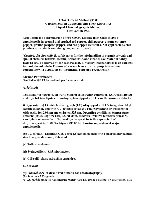
AOAC Official Method 995.03Capsaicinoids in Capsicums and Their ExtractivesLiquid Chromatographic MethodFirst Action 1995[Applicable for determination of 750-650000 Scoville Heat Units (SHU) of capsaicinoids in ground and crushed red pepper, chili pepper, ground cayenne pepper, ground jalapeno pepper, and red pepper oleoresins. Not applicable to chili powders or products containing oregano or thyme.](Caution: See Appendix B, safety notes for the safe handling of organic solvents and special chemical hazards-acetone, acetonitrile, and ethanol. See Material Safety Data Sheets, or equivalent, for each reagent. N-Vanillyi-nnonanamide is an extreme irritant; do not inhale. Dispose of waste solvents in an appropriate manner compatible with applicable environmental rules and regulations.)Method Performance:See Table 995.03 for method performance data.A. PrincipleTest sample is extracted in warm ethanol using reflux condenser. Extract is filtered and injected into liquid chromatograph equipped with UV or fluorescence detector.B. Apparatus (a) Liquid chromatograph (LC).--Equipped with I V integrator, 20 gL sample injector, and with UV detector set at 280 run. wavelength or fluorometer with excitation 280 nm and emission 325 nm. Operating conditions: temperature, ambient (20-25'C); flow rate, 1.5 mL/min., isocratic; relative retention times: N-vanillyl-n-nonanamide, 1.00; nordihydrocapsaicin, 0.90; capsaicin, 1.00; dihydrocapsaicin, 1.58. See Figure 995.03 for baseline separation of major capsaicinoids.(b) LC column.--Stainless, C18, 150 x 4.6 mm id, packed with 5 micrometer particle size. Use guard column, if desired.(c) Reflux condenser.(d) Syringe filter.- 0.45 micrometer.(e) C18 solid-phase extraction cartridge.C. Reagents(a) Ethanol-95% or denatured, suitable for chromatography(b) Acetone.-ACS grade.(c) LC mobile phase@Acetonitrile-water. Use LC grade solvents, or equivalent. Mix400 mL acetonitrile with 600 mL H20 containing 1% acetic acid (v/v). De-gas with helium or by other suitable technique.(d) N-Vanillyl-n-nonanamide standard solutions. -N-Vanillyln-nonanamide standard, 99%, is available as synthetic capsaicin from Penta International Corp., 50 Okner Pkwy, Livingston, NJ 07039. Keep solutions out of direct sunlight. (1) Standard solution A.-0. 15 mg/mL. Accurately weigh 75 mg N-vanillyl-n-nonanamide and transfer it into 500 mL volumetric flask. Dilute to volume with ethanol, and mix. Use standard solution A for analyzing all peppers except chili pepper.(2) Standard solution B.-0.015 mglmL. Transfer 10 mL standard solution A into I 00 mL volumetric flask, dilute to volume with ethanol, and mix. Use standard solution B when analyzing chili peppers.D. Extraction(a) Ground or crushed peppers.-Accurately weigh ca 25 g pepper into 500 ITIL boiling flask. Place 200 mL ethanol into same flask, add several glass beads, and attach flask to reflux condenser. Gently reflux test sample 5 h and then allow to cool. Filter 1-4 mL sample through 0.45 gm syringe filter into small glass vial. Use for LC analysis.(b) Red pepper oleoresins.-Accurately weigh 1-2 g oleoresin into 50 mL volumetric flask. Increase weight of sample, if total capsaicinoid concentration is < 1 %. Note: Do not allow any oleoresin to coat sides of flask.Add 5 mL acetone, C(b), to flask and swirl contents of flask until test sample is completely dispersed (no oleoresin can coat bottom of flask when turning flask neck at 45' angle). Add five 3-5 mL portions ethanol, swirling flask during each addition. Dilute contents of flask to volume with ethanol and mix well.Figure 995.03-Red pepper extract analyzed by (a) fluorescence detection, and (b) UV detection.Peak 1 = nordihydrocapsaicin; peak 2 = capsaicin; peak 3 = dihydrocapsaicinHold C18 solid-phase extraction cartridge over 25 mL volumetric flask or place cartridge on 10 mL glass syringe and hold over 25 mL volumetric flask. Transfer 5 mL solution from flask to cartridge or syringe. (Note: When using syringe, deliver solution to bottom of syringe so that sides of syringe are not coated with sample.) Pass aliquot through cartridge and collect in 25 mL flask. Wash cartridge 3 times with 5 mL ethanol, collecting washings in same flask. Dilute contents of flask to volume with ethanol and mix. Filter 1-4 mL solution through 0.45 micrometer syringe filter into small glass vial. Use for LC analysis.E. LC DeterminationInject 20 microliters standard solution B, C(d)(2), onto LC column, when analyzing chili peppers. When analyzing other matrices inject 20 microliters standard solution A, C(d)(1). Re-inject standard solution at intervals of 6 sample injections, or less.Inject in duplicate 20 microliter test sample from D onto LC column.After <30 sample injections, purge LC column 30 min with 100% acetonitrile at 1.5 mL/min flow rate. Use LC mobile phase, C(c), for further analysis.F. CalculationCapsaicinoids contain 3 major compounds: nordihydrocapsaicin (N), capsaicin (C), and dihydrocapsaicin (D). Calculate capsaicinoids as sum of these compounds [N + C + D; in Scoville Heat Units (SHU); 1 microgram total capsaicinoids/g = ca 15 SHU], as follows: (a) UV detection(1) Ground peppers and chili pepper.-N = (Pn/Ps) x (Cs/Wt) x (200/0.98) x 9300C = (Pc/Ps) x (Cs/Wt x (200/0.89) x 16100D = (Pd/Ps) x (Cs/Wt) x (200/0.93) x 16100where Pn, Pc, and Pd = average peak areas for nordihydrocapsaicin, capsaicin, and dihydrocapsaicin, respectively, from duplicate injections; Ps = average peak area of appropriate standard solution; Cs = concentration of standard solution, mg/mL; Wt = weight of test sample, g(2) Red pepper oleoresins:N= (Pn/Ps) x (Cs/Wt) x (250/0.98) x 9300C = (Pc/Ps) x (CS/Wt) x (250/0.89) x 16100D = (Pd/Ps) x (CS/Wt) x (250/0.93) x 16100(b) Fluorescence detection (1) Ground peppers and chili pepper.N= (Pn/Ps) x (Cs/Wt) x (200/0.92) x 9300C = (Pc/Ps) x (CS/Wt) x (200/0.88) x 16100D = (Pd/Ps) x (CS/Wt) x (200/0.93) x 16100(2) Red pepper oleoresins:N= (Pn/Ps) x (Cs/Wt) x (250/0.92) x 9300C = (Pc/Ps) x (CS/Wt) x (250/0.88) x 16100D = (Pd/Ps) x (CS/Wt) x (250/0.93) x 16100Reference: J. AOAC Int. (future issue).(c) 1996 AOAC INTERNATIONAL。
AOAC规定965
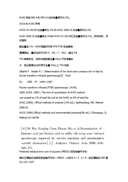
AOAC规定965.4和994.14法的定量限仅为5%。
AOCS和AOAC采用AOCS Cd 14d-99法定量限为1% 和AOAC 2000.10法定量限仅为5%AOAC 2000.10法定量限为5%和AOCS Cd 14d-99[]法定量限仅为1%。
研究发现,反式脂肪酸含量在1%~40%范围时利用ATR-FTIR测定具有高精确性,重现性好(RSDR 0.6%~7.4%),超过5%TFA有再现性;但反式脂肪酸含量5%以下时误差较大,因此推荐此法可用于含量5%以上TFA测定Ulberth F.Haider H J.Determination of low level trans unsatura tion in fats by fourier transform infrared spectroscopy[J].FoodSci..1992,57:1444—1447Fourier transform infrared (FTIR) spectroscopy ( AOAC,2000; AOCS, 1999 ). The limit of quantitation of AOCS methodwas quoted as 1% of total fat and by the AOAC as 5% of total fat,AOAC (2000). Official methods of analysis (17th ed.). Gaithersberg, MD. Method 2000.10.AOCS (1999).Official methods and recommended practices(5th ed.). Champaign, IL. Method Cd 14d-99.Predicted residual error sum of squares (PRESS) 预测残差平方和其校正模型的选择预测残差平方和(PRESS)分别为4~5,6~8;验证模型的SEP值为1.20~6.67Mirghani等测定了50个加标(MDA为0–60 μmol/kg oil)的棕榈油样品的FT-MIR,通过TBARS含量来间接定量MDA,分别利用PLS和PCR建立TBARS在0~0.25模型,,其R2和SEP分别为0.9414~0.9803,1.20~6.67。
AOAC_994.10(中文翻译)
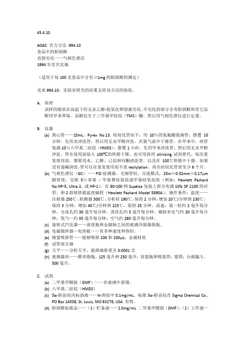
45.4.10AOAC 官方方法994.10食品中的胆固醇直接皂化——气相色谱法1994年首次实施(适用于每100克食品中含有≥1mg的胆固醇的测定)见表994.10,实验室研究的结果支持该方法的验收。
A.原理试样的脂质在高温下经无水乙醇-氢氧化钾溶液皂化。
不皂化的部分含有胆固醇和其它甾醇用甲苯萃取。
甾醇衍生于三甲基甲硅烷(TMS)醚,然后用气相色谱仪进行定量。
B.仪器(a)离心管——15mL,Pyrex No.13. 硅烷化管如下:用10%的氢氟酸装满管,静置10分钟。
先用水冲洗管,然后用无水甲醇冲洗。
在氮气流中干燥管。
在甲苯中,将管装满10%六甲基二硅烷(HMDS),静置1小时。
先用甲苯冲洗管,然后用无水甲醇冲洗。
管在使用前放入100℃的烘箱干燥。
也可用商用silinizing试剂替代。
每次重复使用前,都要用水,乙醇,己烷和丙酮清洗管,以及在100℃烘箱中干燥。
如果没有强碱清洗,管可以在重复使用而不需resilylation。
再次硅烷化管需至少6个月。
(b)气相色谱仪(GC)——FID检测器,毛细管柱,分流模式,25m×0.32mm×0.17μm膜厚度,交联5%苯基- 甲基聚硅氧烷或甲基硅氧烷胶(例如,Hewlett Packard No.HP-5, Ultra 2, 或HP-1),在80-100网Supelco包装上拆分充满10% SP 2100的衬管,和2斜坡烘箱温度编程(Hewlett Packard Model 5890A)。
操作条件:温度——注射器250℃、检测器300℃、分析柱190℃,保持2分钟;增加20℃/分钟到230℃,保持3分钟;增加40℃/分钟到225℃,保持25分钟。
流速:氦—柱约2毫升每分钟,分流孔约30毫升每分钟,清洗孔约3毫升每分钟,辅助补充气约20毫升每分钟;氢气—约35毫升每分钟;空气约280毫升每分钟。
(c)旋转式汽化器——浓度瓶和金属轴之间的玻璃冷凝器烧瓶。
食品真实性领域稳定同位素技术标准一览

食品真实性领域稳定同位素技术标准一览颁布年份方法产品组分仪器应用同位素1987OIV, recueil des méthodes d'analyse葡萄酒乙醇SNIF-NMR D/H,1990EC regulation 2676/90, annex 8葡萄酒乙醇SNIF-NMR D/H 1991AOAC method 991.41蜂蜜蜂蜜、蛋白质IRMS13C/12C 1992AOAC 992.09浓缩橙汁水IRMS18O/16O 1993CEN (TC174 N108, ENV 12140)果汁蔗糖IRMS13C/12C 1995AOAC Official method 995.17果汁乙醇SNIF-NMR D/H 1996OIV Resolution OENO 2/96葡萄酒水IRMS18O/16O 1997EC Regulation No. 822/97葡萄酒水IRMS18O/16O 1997CEN (TC174 N109, ENV 12141)果汁水IRMS18O/16O 1997CEN (TC174 N109, ENV 12142)果汁水IRMS D/H 1998AOAC 998.12蜂蜜蜂蜜、蛋白质IRMS13C/12C 1998BS DD ENV 13070-1998果汁果浆IRMS13C/12C 2000AOAC Official method 2000.19枫树蜜乙醇SNIF-NMR D/H 2000AOAC 44.5.17枫树糖浆糖IRMS13C/12C 2001OIV Resolution OENO 17/2001葡萄酒乙醇IRMS13C/12C 2002GBT18932.1蜂蜜蜂蜜、蛋白质IRMS13C/12C 2003EC No 440/2003,annex 2葡萄酒乙醇IRMS13C/12C 2004AOAC method 2004.01果汁、枫树蜜乙醇IRMS13C/12C 2005OIV Resolution OENO 7/2005起泡葡萄酒CO2IRMS13C/12C 2006AOAC method 2006.05香兰素香兰素SNIF-NMR D/H 2009OIV Resolution OENO 353/2009葡萄酒水IRMS18O/16O 2009OIV Resolution OENO 381/2009葡萄酒、烈性酒乙醇IRMS13C/12C 2010OIV Resolution OENO 343/2010葡萄酒甘油IRMS13C/12C。
维生素的检测现状及国际标准检测方法

36 食品安全导刊 2011年10月刊AnAlysis & test 分析与检测□ 左程丽 李端 拜发分析系统销售(北京)有限公司维生素的检测现状及国际标准检测方法维生素是各种生物生长和代谢所必需的,无处不在的营养卫士。
维生素帮助身体中的酶起催化作用,是我们每个人的健康要素。
人体一旦缺乏维生素,机体代谢会失去平衡,免疫力会下降,各种疾病就会趁虚而入,产生维生素缺乏症。
维生素检测背景市场上,食品添加剂像药品一样被标注上“剂量”。
一般的添加食品有婴儿配方食品、谷类产品和果汁,可以为日常饮食添加更多营养成分。
食品中添加的维生素必须进行严格控制,维生素在添加进食品之前就应通过检测,如果储存后再使用,则应重新检测。
European Directive 2002/46 EC制定了一系列关于食品添加剂标识及检测添加过维生素的食品中标注的维生素含量的法规。
我国国家标准GB5413-2010对婴幼儿配方食品与乳品中维生素含量也有明确的规定,标准中涉及的方法有微生物法及高效液相色谱法。
维生素传统测定方法大多步骤繁琐,检测周期长,操作费时,其中一些方法受干扰因素影响大,灵敏度较低,而在实际工作中往往要求测定结果准确、测定周期短、经济合算,这就导致对维生素快速分析方法的需求越来越迫切。
为此,德国拜发公司将继续秉承其研发宗旨,致力于追求简便、快速、准确的维生素分析检测方法。
维生素检测方法国际及国内维生素检测方法主要为微生物法及H P LC法,德国拜发R-Biopharm公司根据国际现行方法研发的配套检测产品能够使检测方法更简单省时,检测结果更准确可靠。
1.传统经典方法——微生物法微生物法根据维生素为细菌生长所必需的原理,以细菌繁殖程度或代谢产物定量该维生素含量,适用于检测多种衍生物的总和。
国家标准GB5413中检测维生素B 12、烟酸和烟酰胺、叶酸、泛酸、生物素及肌醇测定第一法(仲裁法) 均为等同采用AOAC的方法——微生物法。
AOAC 方法验证要求

AOAC官方方法的第一个动作®采用
官方方法委员会审查提交的合作研究报告、评论和相关文件,以确保遵守技术审查程序。提前通知的方法被认为是第一个行动发表在杂志的裁判是AOAC部分,在实验室管理。
随后,法行为由官方方法板被发表在AOAC的留意杂志裁判部分,在实验室管理。新®采用AOAC官方、完整的文本和实验室间合作研究的报告或摘要发表在AOAC国际期刊。所采用的方法被添加到《纲要》中,正式的分析方法。
®AOAC官方方法的改进
当有必要在现有的AOAC®进行修改
正式的方法、验证的程序和程度取决于修订的程度,无论是编辑、次要的还是实质性的。这些决定由总裁判和方法委员会作出。
申诉流程
对于一个AOAC官方方法或方法措施审查所有请求必须以书面形式提交。每个请求审查同一法;官方方法板然后对委员会总裁判方法的建议。
获得参与:可能的参与者列表可以得出通过个人接触,技术协会,贸易社团,文献检索,并在AOAC的杂志广告在裁判部分,实验室管理。被邀请参加实验室的人员应具备所使用的基本技术的经验;方法本身的经验不是选择的先决条件。
实验室必须认识到研究的重要性。投资大的正在测试的方法,这可能会对将要执行的方法的唯一协作研究。因此,对这种方法进行公正彻底的评价是很重要的。
AOAC官方方法最后行动的®采用
第一次行动AOAC®官方、有资格获得最后的交流状态后,他们已在文献中提供了至少2年。如果协会没有收到任何信息作为重要的问题在性能的方法,一般建议采用该方法作为裁判的最终作用和方法在AOAC的杂志裁判部分上市,在实验室管理,所以有兴趣的可以提交意见和数据,如果需要的话。
总裁判
总裁判由主题线下的合适方法委员会。总裁判负责一个广阔的研究领域(例如,肥料;水果及其制品;饲料中霉菌毒素的药物;)和坐标,引导一批副裁判工作的具体方法的广泛的主题范围内的活动。每个总裁判评论各自的AOAC官方、章和推荐新的副裁判的话题。反过来,每个总裁判与副裁判一起讨论方法开发的概念;审查副裁判的报告;建议对方法采取适当的行动;并编写一份年度报告,向指定区域内的科学问题提交方法。
AOAC 30.1.23A AOAC Official Method 995.13-国外标准规范

30.1.23AAOAC Official Method995.13Carbohydrates in Soluble(Instant)CoffeeAnion-Exchange Chromatographic Methodwith Pulsed Amperometric DetectionFirst Action1995(Applicable for determination of free and total carbohydrates[ex-cept total fructose,which is degraded]in soluble[instant]coffee.) See Tables995.13A–J for the results of the interlaboratory study supporting the acceptance of the method.A.PrincipleFree carbohydrates.—Coffee is dissolved in H2O.Solution is fil-tered through C18disposable cartridge,and then through0.2µm membrane filter.Filtered solution is injected onto LC system.Car-bohydrates are separated on pellicular anion-exchange column and measured by pulsed amperometric detector.Total carbohydrates.—Coffee is hydrolyzed with1M HCl.Solu-tion is filtered and then passed through cation-exchange disposable cartridge in the Ag form to neutralize solution and to eliminate the Cl anion prior to injection onto LC system.B.Apparatus(a)Balance.—Analytical,weighing to0.1mg.(b)Flasks.—250mL round-bottom.(c)Volumetric flasks.—100and1000mL.(d)Pipets.—Delivering200–1000µL and5µL;with disposable tips.(e)Cylinders.—50and1000mL,tall-form,graduated.(f)Funnels.—Analytical,60°C.(g)Vacuum filtering system.—Aspirator with regulating device. System should include:heavy-walled filtering flask with ground cone neck,1L;funnel with ground glass joint,300mL;aluminum assembly clip;connection with vacuum outlet;filter holder,47mm id;and low-water extractable membrane filters,0.2µm porosity, 47mm diameter.(h)Filter papers.—Qualitative,folded,medium fast.(i)C18cartridges.—Octadecylated silica(ca10%C);6mL car-tridge volume;capable of holding500mg test portion;disposable.Con-dition and use cartridges according to manufacturer’s instructions. (j)Cation-exchange cartridges.—Styrene-based resin,Ag form, 1.8–2.0milliequivalent capacity/cartridge;disposable.Condition and use cartridges according to manufacturer’s instructions. (k)Membrane filters.—0.2µm porosity,25mm diameter;dis-posable;polypropylene.(l)Water bath.—Maintaining98±2°C.(m)Liquid chromatograph(LC).—Metal-free,compatible with 300mM NaOH.Operating conditions:mobile phase,isocratic(see Table995.13for mobile phase conditions);column temperature, ambient;flow rate,1.0mL/min;post-column solvent,300mM NaOH at0.6mL/min;detector settings,optimum parameters as pro-vided by e with computing integrator.(n)LC column.—250×4mm id;packed with polystyrene divinylbenzene substrate(10µm diameter)agglomerated with microbead quaternary amine functionalized latex(350nm diame-ter);5%cross-linked.(o)Pulsed amperometric detector.—With gold electrode.Fill reference cell with300mM NaOH.Select the detector range to avoid saturation of the major peak in chromatogram.(p)Guard column.—50×4mm id,packed with the same mate-rial as analytical column,(n).(q)Post-column solvent delivery system.—Compatible with 300mM NaOH.C.ReagentsUse18MΩ⋅cm demineralized H2O throughout.(a)Sodium hydroxide.—50%(w/w)aqueous solution,contain-ing minimum amount of Na2CO3and Hg.Do not shake or stir solu-tion before use.(b)Hydrochloric acid solution.—1.00M standard volumetric so-lution(83.3mL HCl/L).(c)Eluent A.—18MΩ⋅cm demineralized water.Filter through0.2µm membrane filter.Degas by sparging with He20–30min.Pre-pare fresh eluent A daily.(d)Eluent B.—300mM NaOH.Pipet15.6mL50%NaOH into 985mL eluent A.Degas by sparging with He20–30min.Eluent B is stable2days if stored at room temperature under He.(Note:It is crit-ical to remove dissolved CO2from eluents.Carbonate acts as strong “pusher”on LC column,resulting in drastic reduction in resolution.)(e)Carbohydrates standard solutions.—(1)Standard stock solu-tions.—1mg/mL aqueous stock solutions of arabinose,fructose, fucose,galactose,glucose,mannose,rhamnose monohydrate, ribose,xylose,sucrose,and mannitol.Weigh100mg each carbohy-drate to the nearest0.1mg into separate100mL volumetric flasks, dissolve in H2O,and dilute to volume with H2O.(2)Mixed carbohy-drates standard solution.—Further dilute and mix carbohydrates stock solutions to reach carbohydrate concentrations similar to those found in nonhydrolyzed or hydrolyzed soluble coffee test solutions. Pass diluted carbohydrates standard solution through0.2µm mem-brane filter prior to injection onto LC column.(Note:If resolution of rhamnose from arabinose is difficult to achieve,do not add rhamnose to mixed standard solution.)D.Isolation of CarbohydratesUse soluble coffee as is without grinding or homogenization.(a)Free carbohydrates.—Weigh300mg coffee to the nearest0.1mg into100mL volumetric flask.Add70mL H2O and shake until dissolution is complete.Dilute solution to volume with H2O.Filter 5–10mL solution through C18cartridge.Discard the first1mL.Pass fil-trate through0.2µm membrane filter prior to LC injection.(b)Total carbohydrates.—Weigh300mg coffee to the nearest0.1mg into100mL volumetric flask.Add50mL1.00M HCl and swirl.Place flask in boiling water bath for2.5h.(Note:Always keep the level of solution in flask below that of H2O in bath.)Swirl flaskTable995.13Conditions of mobile phase for determinationof free and total carbohydrates in solublecoffee by anion-exchange chromatographicmethod with pulsed amperometric detection Time,min Eluent A,%Eluent B,%01000(start acquisition) 50.01000(stop acquisition) 50.10100(start cleanup) 65.00100(stop cleanup) 65.11000(start re-equilibrium) 80.01000(stop re-equilibrium)Test sample N a Mean,%RSD r RSD R r R 17(2)0.024 1.6590.0010.041 1′11(0)0.1797.0500.0350.021 211(1)0.0600.91580.0020.10 2′11(0)0.1517.6460.0320.20 311(0) 1.5 2.89.90.120.44 3′11(1) 1.85 2.2180.120.94 411(0)0.619 3.6240.0630.42 4′11(0)0.782 4.6210.100.45 59(1)0.1928.2340.0450.18 5′11(0)0.30011370.0910.32 68(1)0.0597.1490.0120.083 6′10(0)0.1799.8500.050.25 a N=No.of laboratories after removal of outliers(in parentheses).Table995.13B Interlaboratory study results of the determination of free arabinose in soluble coffeeTest sample N a Mean,%RSD r RSD R r R 111(0)0.891 3.7140.0920.035 1′11(0) 3.54 6.6210.66 2.1 29(2) 1.320 1.6 5.10.0600.19 2′11(0) 4.83 3.3170.45 2.4 311(0)0.464 3.8110.0490.14 3′11(0) 4.76 3.1130.42 1.7 411(0)0.7477.3120.150.25 4′11(0) 4.54 4.618.40.59 2.4 510(1)0.505 4.47.70.0630.11 5′9(2) 4.08 3.0 4.90.340.56 611(0)0.629 4.18.90.0730.16 6′11(0) 3.79 5.7200.61 2.2a N=No.of laboratories after removal of outliers(in parentheses).Table995.13C Interlaboratory study results of the determination of free galactose in soluble coffeeTest sample N Mean,%RSD r RSD R r R 111(0)0.562 3.0 5.30.0470.084 1′10(1)17.88.18.9 4.1 4.8 211(0)0.339 4.18.00.0390.077 2′10(1)18.5 2.312 1.2 6.2 311(0)0.1919.8130.0530.070 3′11(0)8.08 2.78.00.6208.0 411(0)0.438 5.98.30.0740.10 4′11(0)13.3 3.913 1.8 5.9 59(2)0.475 3.1 4.10.0410.055 5′9(2)18.40 1.77.50.87 3.9 611(0)0.362 5.0120.0510.13 6′10(1)17.7 4.38.5 2.2 4.3a N=No.of laboratories after removal of outliers(in parentheses).Test sample N a Mean,%RSD r RSD R r R 111(0)0.1059.9210.0290.062 1′11(0)0.6848.7170.170.32 29(1)0.0421020.00.0120.024 2′10(1)0.8267.4220.170.50 310(1) 2.04 2.5 6.20.140.360 3′11(0)16.6 5.924.0 2.811.0 410(1) 1.66 4.1 6.10.190.29 4′11(0) 4.38 3.8240.47 2.9 510(1)0.18610210.0530.11 5′11(0) 1.95 5.7130.310.69 611(0)0.18610240.0520.12 6′11(0) 1.027.9140.230.40 a N=No.of laboratories after removal of outliers(in parentheses).Table995.13E Interlaboratory study results of the determination of free mannose in soluble coffeeTest sample N a Mean,%RSD r RSD R r R 111(0)0.583 4.9240.0800.40 1′10(1)17.9 5.811 2.9 5.7 210(0)0.1558.2360.0130.16 2′11(0)14.4 2.615 1.1 6.2 311(0)0.470 4.2180.0560.23 3′11(0) 2.60 2.0140.15 1.0 411(0)0.3297.0190.06517 4′11(0) 5.60 3.0150.48 2.4 510(1)0.277 4.2400.0330.31 5′11(0)7.65 2.8110.60 2.3 611(0)0.991 3.8170.110.48 6′10(0)19.1 2.222 1.212.00 a N=No.of laboratories after removal of outliers(in parentheses).Table995.13F Interlaboratory study results of the determination of free fructose in soluble coffeeTest sample N a Mean,%RSD r RSD R r R 19(0)0.17117310.0820.15 1′6(0)0.18924680.130.37 28(1)0.05421340.0320.052 39(2) 3.62 2.9180.30 1.9 3′9(0) 2.01 5.7720.32 4.1 410(1) 3.12 2.9180.26 1.6 4′7(1) 1.37 5.5700.21 2.7 510(0)0.2829.0450.0720.36 5′7(0)0.24420590.140.41 68(2)0.460 5.6160.0670.2 6′9(1)0.3637.3680.0750.70 a N=No.of laboratories after removal of outliers(in parentheses).Test sample N a Mean,%RSD r RSD R r R 17(2)0.07315740.0310.15 27(l)0.04528600.0350.077 58(0)0.10215980.0430.28 55(1)0.08016180.0360.041 67(l)0.1209.1810.0310.28 a N=No.of laboratories after removal of outliers(in parentheses).Table995.13H Interlaboratory study results of the determination of free xylose in soluble coffeeTest sample N a Mean,%RSD r RSD R r R 18(2)0.09723380.0630.10 29(0)0.1469.8200.0400.084 311(0) 1.86 4.6230.240.42 411(0)0.736 3.7280.0770.56 57(0)0.02925230.0200.023 511(0) 1.837.4220.38 1.2 69(0)0.13314240.0530.090 a N=No.of laboratories after removal of outliers(in parentheses).Table995.13I Interlaboratory study results of the determination of free sucrose in soluble coffeeTest sample N a Mean,%RSD r RSD R r R 210(0)0.149 4.8380.0200.16 310(1) 1.32 1.8100.0660.370 410(1)0.746 6.8120.140.25 511(0)0.18115420.0770.21 69(1)0.158 3.4330.0150.15 a N=No.of laboratories after removal of outliers(in parentheses).Table995.13J Interlaboratory study results of the determination of free fucose in soluble coffeeTest sample N a Mean,%RSD r RSD R r R 27(1)0.01620.0460.0090.021 38(0)0.0547.0670.0110.10 47(1)0.03623770.0230.074 57(1)0.011 5.4450.0020.014 66(1)0.02510360.0070.025 a N=No.of laboratories after removal of outliers(in parentheses).by hand every30min.Cool to room temperature under tap water. Dilute solution to100mL with H2O and filter through folded filter paper.Pass3mL filtrate through cation-exchange cartridge.Discard the first1mL.Filter neutralized solution through0.2µm membrane filter prior to LC injection.E.LC DeterminationInject equal volumes(10–20µL)of standard,C(e),and test solu-tions from D(a)or(b)onto LC column.[Note:Retention times and resolution tend to vary from column to column.Start clean-up and re-equilibration sequence only when the last monosaccharide (ribose)has been eluted.It may be necessary to perform2–3injec-tions of carbohydrates standard solution or to increase the re-equilibrium time in order to achieve a good separation of glucose, sucrose,and xylose.]Under normal conditions,approximate retention times of carbo-hydrates are:mannitol,4min;fucose,7min;rhamnose,15min;arabinose,16min;galactose,22min;glucose,25min;sucrose, 27min;xylose,30min;mannose,32min;fructose,40min;and ribose,43min.See Figure995.13for chromatogram of mixed carbo-hydrates standard solution.F.CalculationsCalculate concentration of carbohydrate in sample solution as follows:Carbohydrate,%=(R1/R2)×(W0/V0)×(V/W)×100 where R1=peak response of carbohydrate in test solution;R2=peak response of carbohydrate in carbohydrate standard solution;W0= mass of carbohydrate in the standard solution,mg;V0=total volume of the standard solution,mL;V=volume of standard solution,mL; and W=weight of test portion,mg.Express results as percent free or percent total carbohydrates (as is).References:J.AOAC Int.78,768(1995);79,1400(1996). Revised:June2000Figure995.13—HPAE-PAD chromatogram of mixed carbohydrates standard solution:mannitol,15m g;fucose,15m g/mL;rhamnose,35m g/mL;arabinose,40m g/mL;galactose,50m g/mL;glucose,55m g/mL;sucrose,45m g/mL; xylose,55m g/mL;mannose,45m g/mL;fructose,90m g/mL;and ribose,90m g/mL.。
AOAC官方方法994.10
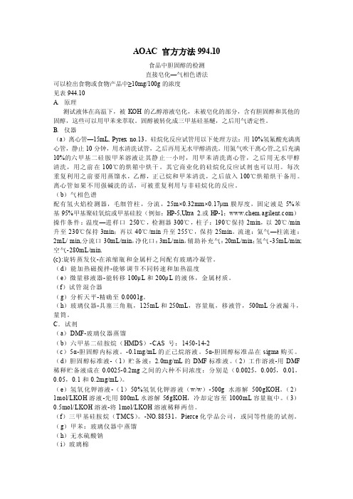
AOAC 官方方法994.10食品中胆固醇的检测直接皂化—气相色谱法可以检出食物或食物产品中≥10mg/100g的浓度见表944.10A.原理测试液体在高温下,被KOH的乙醇溶液皂化,未被皂化的部分,含有胆固醇和其他的固醇,这些可以用甲苯来萃取。
固醇被转化成三甲基硅基醚,之后用气谱定性。
B.仪器(a)离心管—15mL,Pyrex no.13。
硅烷化反应试管用以下处理方法:用10%氢氟酸充满离心管,静止10分钟,用水清洗试管,之后再用无水甲醇清洗。
用氮气吹干离心管,之后充满10%的六甲基二硅胺甲苯溶液让其静止一小时,用甲苯清洗离心管,之后用无水甲醇清洗,用之前在100℃的烘箱中烘干。
其它商业化的硅烷化反应试剂也可以用。
每次重复利用之前要用蒸馏水,乙醇,正己烷和甲苯清洗,之后放入100℃烘箱烘干备用。
离心管如果不用强碱洗的话,可被重复利用与非硅烷化的反应。
(b)气相色谱配有氢火焰检测器,毛细管柱,分流,25m×0.32mm×0.17μm膜厚度,固定液是5%苯基95%甲基聚硅氧烷或甲基硅胶(例如:HP-5,Ultra 2.或HP-1;www.chem.agilent.c om)操作条件:温度—进样口250℃,检测器300℃,柱子:190℃保持2min,以20℃/min 升至230℃保持3min;再以40℃/min升至255℃,保持25min。
流速:氦气—柱流速:2mL/ min,分流口30mL/min,净化口:3mL/min,辅助补充气:20mL/min;氢气-35mL/min;空气-280mL/min.(c):旋转蒸发仪-在浓缩瓶和金属杆之间配有玻璃冷凝管。
(d)能加热磁搅拌-能够调节不同转速和加热温度(e)微量移液器-能转移100μL和200μL的液体,金属材质。
(f)试管混合器(g)分析天平-精确至0.0001g。
(h)玻璃仪器-具塞三角瓶,125mL和250mL,容量瓶,移液管,500mL分液漏斗,量筒。
AOAC标准目录

AOAC电子版标准目录一、AOAC方法1. AOAC Official Method 993.31.Phosphorus (Available) in Fertilizers. Direct Extraction Method2. AOAC Official Method 993.31.Nitrogen (Total) in Fertilizers. Combustion Method.3. AOAC Official Method 995.01.Dirthianon in Technical Products.and Fornmulations.4. AOAC Official Method 995.14.Methomy in insecticidal Formulations.Reversed-phase liquid Chromatographic Method5.AOAC Official Method 993.02.bentazon in pesticide formulations liquid Chromatographic Method6.AOAC Official Method 995.02.Cyfiuthrin in Pesticide formulations liquid Chromatographic Method7.AOAC Official Method 993.01 Phosphamidon in Technical and Formulated Products8.AOAC Official Method 996.03 Acephate in Technical and Soluble Powder Formulations9.AOAC Official Method 995.08.Atrazine in Water Magnetic Particle lmmunoassay10.AOAC Official Method 2000.05. Determination of Glyphosate and AminomethyphonicAcid(ACMPA)in Crops11.AOAC Official Method 993.15 1,2-Dibromoethane and 1,2-Dibromo-3-chloropropane in Water12.AOAC Official Method 994.19 Total nitrogen in Urine Pyrochemiuminescence Method13.AOAC Official Method 995.21 Yeast and mold counts in Foods hydrophobic Grid Membranefilter14.AOAC Official Method 996.02 Coliform Count in Dairy Products15.AOAC Official Method 2000.15 Rapid enumeration of Coliforms in Foods Dry RehydratableFilm Method16.AOAC Official Method 2000.13 Reveal for E.coli O157:H7 Test System in selected foods17.AOAC Official Method 2000.14 Reveal for E.coli O157:H7 Test in selected foods andEnvironmental Swabs18.AOAC Official Method 993.06 Staphylococal enterotoxins in selected foods19.AOAC Official Method 995.12 Staphylococcus autrus lsolated from foods latexAggluTINATION Test Method20.AOAC Official Method 2001.05 Petrifilm S.aureus count Platr Method for the RapidEnumeration of Staphylococcus aureus in Seleced Foods21.AOAC Official Method 993.10 Clistridium Perfringrns from Shellfish lron Milk Method22.AOAC Official Method 995.20Salmonella in Raw,Highly contaminated Foods and poultryFeed23.AOAC Official Method 993.08 Salmonella in foods24.AOAC Official Method 993.07 Sa;monella Cocoa and Chocolate motility Enrichment onmodified Sem-Solld25.AOAC Official Method 995.07 Salmonella in dried Milk Products Motility Enrichment onModified Sem-Solld26.AOAC Official Method 994.04 Salmonella in drt Foods Refrigerated Pre-Enrichmenr27.AOAC Official Method 2000.07 Salmonella in Fooods Rapid Cocorimetric28.AOAC Official Method 2000.07 Salmonella in Fooods with a Low Microbial Load Detecition29.AOAC Official Method 2001.07 Salmonella in Selected Foods lmmumno-ConcentrationSalmonella (ICS)30.AOAC Official Method 2001.08 Salmonella in Selected Foods lmmumno-ConcentrationSalmonella (ICS)31. AOAC Official Method 2001.09 Salmonella in Selected Foods lmmumno-ConcentrationSalmonella (ICS)32. AOAC Official Method 2001.08 Salmonella in Selected Foods lmmumno-ConcentrationSalmonella (ICS)33.AOAC Official Method 993.12 Listeria monceytogenes in milk ang dairy Products34.AOAC Official Method 993.09 Listeria in Dairy Pruducts, Seafoods, and Meats. ColorimetricDeoxyribonucleic Acid Hybridization Method ( GENE-TRAK Listeria Assay)35. AOAC Official Method 994.03. Listeria monocytogenes in Dairy Pruducts, Seafoods, andMeats. Colorimetric Deoxyribonucleic Acid Hybridization Method (Listeria-Tek)36.AOAC Official Method 994.06. Vibrio vulnificus. Gas Chromatographic dentification Methodby Microbial Fatty Acid profile37.AOAC Official Method 993.32.Multiple sulfonamide Residues in Raw Bovine MilkLiquid Chromatographic Method First Action 199338.AOAC Official Method 2001.14 Determination of Nitrogen(Total)in Cheese kieldahl Method39.AOAC Official Method 993.O5 l-Malic/Total Malic Acid Ratio in Apple Juice40.AOAC Official Method 995.06 D-Malic Acid in Apple Juice Liquid Chromatographic Method41. AOAC Official Method 994.11 Benzoic Acid in Orange Juice Liquid ChromatographicMethod42.AOAC Official Method 995.17 Beet Sugar in fruit Juices43.AOAC Official Method 993.20 Lodine Value of Fates and Oils Wijs(Cyclohexane-AceticSolvent)Method44.AOAC Official Method 994.02 Lead in Edible Oils and Fats Direct Graphite Furnace45.AOAC Official Method 994.18 Mon-and Diglycerides in Fats and Oils Gas ChromatographicMethod46.AOAC Official Method 2000.17 Determination of Trace Glucose and Fructose Determinationof Trace Glucose and Fructose in Raw Cane Sugar47.AOAC Official Method 994.09 Glucoamylase Activity in Lndustrial Enzyme Preparations48.AOAC Official Method 995.11 Phosphorus (total)in Foods Colorimetric Method49.AOAC Official Method 2001.13 Determination of Vitamin A(Retinol)in Foods LiquidChromatography50.AOAC Official Method 2000.11 Polydextrose in Foods lon Chromotography51.AOAC Official Method 2000.01 Determination of 3-Chloro-1,2-Propanediol in Foods andFood Ingredients52.AOAC Official Method 993.16 Total Aflato xins(B1,B2,and G1)in Corn Enzyme-Linkedlmmunosorbent Assay Method53.AOAC Official Method 993.17 Aflatoxins in Corn and Peanuts Thin-Layer ChromatographicMethod54. AOAC Official Method 994.08 Aflatoxins in Corn,Almonds, brazil Nuts,Peanuts,andPeanuts,and Pistachio Nuts55.AOAC Official Method 2000.16 Aflatoxin B1 in Baby Foood lmmmunoaffinty Column HplcMethod56.AOAC Official Method 2000.08 Aflatoxin M1in Liquid Milk lmmmunaffinity Column byLiquid Chromatography57.AOAC Official Method 995.15 Fumonisins B1,B2,and B3in Corn liquid ChromatographicMethod58.AOAC Official Method 2001.04 Determination of Fumonisins B1,and B2,in Corn and CornFlakes59.AOAC Official Method 2001.06 Determination of Total Fumonisins in Corn Competitive ofDirecet Enzyme-Linked Immunosorbent Assay60.AOAC Official Method 2000.09 Ochratoin A in Roaseted Coffee Immunoaffinity ColumnHPLC Method61.AOAC Official Method 2000.03 Ochratoin A in Barley Immunoaffinity by Column HPLC62.AOAC Official Method 2001.01 Determination of Ochratoxin A in Wine and Beer63.AOAC Official Method 995.10 Patulin in Appple Juice Liquid Chromatoraphic Method64.AOAC Official Method 2000.02Patulin in Clear and Cloudy Apple Juices and Apple Puree65.AOAC Official Method 994.01 Zearlenone in Corn Wheat,and Feed Enzyme LinkedImmunosorbent (Agri-screen)Method66.AOAC Official Method 993.03 Nitrate in Baby Foods spectrophotometric Method67.AOAC Official Method 999.12 Taurine in Pet Food68.AOAC Official Method 996.16 Selenium in Feeds and Premixes69.AOAC Official Method 999.13 Ethoxyquin in Feeds Liquid Chromatographic Method70.AOAC Official Method 999.16 Sulfamethazine in Animal Feeds71.AOAC Official Method 997.04 Monensin in Premix and Animal Feeds LiquidChromatographic Method72.AOAC Official Method 998.02 Neomycin in Feeds Stahl Microbiological Agar73.AOAC Official Method 997.01 Tebuconazole in Fungicide and Technical formulations74.AOAC Official Method 997.14 Thiodicarb in Technical Products and Formulations75.AOAC Official Method 999.04 Determination of Chlorothalonil and Hexachlorobenzene76.AOAC Official Method 997.07 N-octyl Bicycloheptene Dicarboximide (MGK 264),Pyrethrinsand Piperonyl Butoxide (PB)77.AOAC Official Method 996.12 Glyphosate in Water-Soluble Granular Formulations78.AOAC Official Method 997.12 Imidacloprid in Liquid and Solid Formulations Reversed-PhaseLiquid Chromatographic Method79.AOAC Official Method 999.17 Lead and Cadmium Extracted from Ceramic Foodware80.AOAC Official Method 999.10 Lead,Cadmium,Zinc, Copper,and lron in foods81.AOAC Official Method 999.11 Determination of Lead,Cadmium,Copper, Iron,and Zincin Foods82.AOAC Official Method 997.15 Lead in Sugars Graphite Furnace Atomic Absorption Method83.AOAC Official Method 2000.04 Iodine-131 in Milk Radiochemical Separtion Method84.AOAC Official Method 998.11 Screening Test for Nitrate in Forages With a Test Strip85.AOAC Official Method 997.02 Yeast and Mold Cunts in Foods Dry Rehydratable Film Method(Petrifilm TM Method)86.AOAC Official Method 996.09 Escherichia coli O157:H7 in Selected Foods VisualImmunoprecipitate Assay (VIP TM)87.AOAC Official Method 996.10 Enterohemorrhagic Escherichia coli(EHEC)O157:H7Detection in Selected Foods88.AOAC Official Method 997.11Escherichia coli O 157:H7 Counts in Foods HydrophobicGrid Membrane Filter (ISO-GRID)Method Using89.AOAC Official Method 996.08 Salmonella in Foods Enzyme-Linked ImmunofluoressentAssay90.AOAC Official Method 997.16 LOCA TE Enzyme-Linked Immmunosorbent Assay forldentification of Salmonella in Foods91.AOAC Official Method 998.09 Salmonella in Foods Coloric Polyclonal Enzyme92.AOAC Official Method 999.09 Vlp for Salmonella for the Detection of Motile and Non-MotileSalmonella in All Foods93.AOAC Official Method 995.22 Listeria in Foods Colorimetric Polyclonal Enzyme94.AOAC Official Method 996.14 Detection of Listeria Monocytogenes and Related ListeriaSpecies in Selected Foods and from Environmental surfaces95.AOAC Official Method 997.03 Detection of Listeria Monocytogenes and Related ListeriaSpecies in Selected Foods and from Environmental surfaces96.AOAC Official Method 999.06 Listeria in Foods Enzyme-Linked ImmunofluorescentAssay(ELFA)97.AOAC Official Method 995.09 Chortetracycline,Oxytetracycline,and Tetracycline in Edible Animal Tissues98.AOAC Official Method 997.09 Nitrogen in Beer,Wort,and Brewing Grains Protein (Total)by Calculation99.AOAC Official Method 996.11 Starch (Total)in Cereal Products Amylonglucosidass-α-Amylase Method100.AOAC Official Method 995.04 Multiple Tetracline Residues in Milk Metal Chelate Affinity-Liquid Chromatographic Method101.AOAC Official Method 998.04 Neutral Lactase (β-Galactosidase)Activity in Industrial Enzyme Preparations102.AOAC Official Method 999.05 Naringin and Neohesperidin in Orange Juice103.AOAC Official Method 998.03 Aflatoxins in Peanuts Aflatonxins in Peanuts Alternative BF Method104.AOAC Official Method 999.07 Aflatoxin B1 and Total Aflatoxins in Peanut Butter,Pistachio Paste,Fig Paste,and Paprika Powder105.AOAC Official Method 997.05 Taurine in Powdered Milk and Powdered Infant Formulae 106.AOAC Official Method 995.05 Vitamin D in Infant Formulas and Enteral Products Liquid Chromatographic Method107.AOAC Official Method 999.15 Vitamin K in Milk and Infant Formulas Liquid Chromatographic Method108.AOAC Official Method 986.07 Fensulfothion in Chromatographic Method。
AOAC 996.06 Fat in Food鱼油的检测方法1
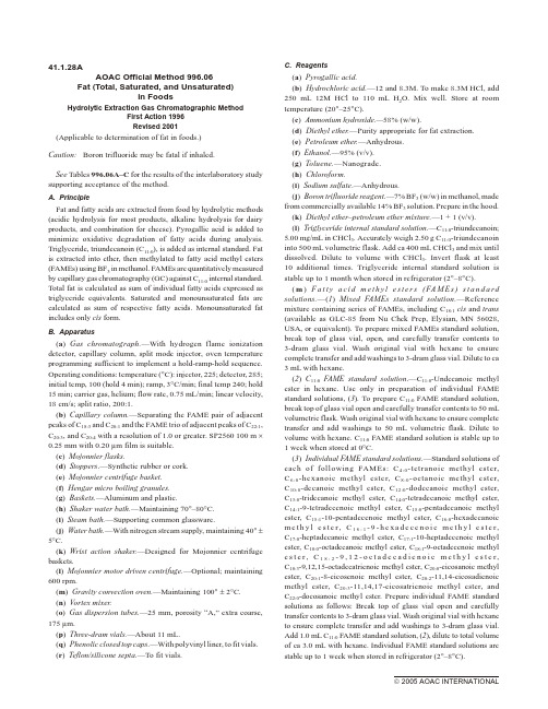
(d) Diethyl ether.—Purity appropriate for fat extraction.
(e) Petroleum ether.—Anhydrous.
(f) Ethanol.—95% (v/v).
(g) Toluene.—Nanograde.
(c) Mojonnier flasks. (d) Stoppers.—Synthetic rubber or cork. (e) Mojonnier centrifuge basket. (f) Hengar micro boiling granules. (g) Baskets.—Aluminum and plastic. (h) Shaker water bath.—Maintaining 70°–80°C. (i) Steam bath.—Supporting common glassware. (j) Water bath.—With nitrogen stream supply, maintaining 40° ± 5°C. (k) Wrist action shaker.—Designed for Mojonnier centrifuge baskets. (l) Mojonnier motor driven centrifuge.—Optional; maintaining 600 rpm. (m) Gravity convection oven.—Maintaining 100° ± 2°C. (n) Vortex mixer. (o) Gas dispersion tubes.—25 mm, porosity “A,” extra coarse, 175 mm. (p) Three-dram vials.—About 11 mL. (q) Phenolic closed top caps.—With polyvinyl liner, to fit vials. (r) Teflon/silicone septa.—To fit vials.
最新的AOAC凯氏氮测定公认标准方法(2001) (1)
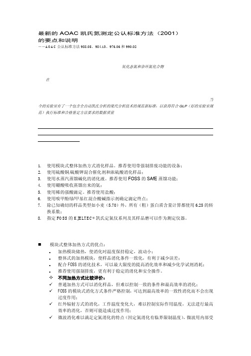
最新的AOAC 凯氏氮测定公认标准方法(2001)的要点和说明――AOAC 公认标准方法988.05、984.13、976.06和990.02由于凯氏定氮法经过100多年的发展,各种相关标准很多,且有些方法对其公认的参比性提出挑战,认为凯氏定氮法存在缺陷,不能测定所有的氮(如氧化态氮和杂环氮化合物)且精度不是最好等,作为参比标准方法有点混乱。
在2001年,AOAC 经过了验证,最后确定了以模块式加热消化、蒸汽蒸馏、硼酸吸收、盐酸滴定、指示剂指示颜色终点法的凯氏定氮方法学作为定氮技术参比试验条件,在欧美的14个权威实验室(FOSS 的凯氏定氮仪技术规范完全符合选定标准,因此成为唯一指定的可选试验仪器,FOSS 公司的实验室也是其中的一个)开展了凯氏定氮法的升级完善认证试验,形成新版(2001年)的AOAC 最新定氮标准,使当今的实验室有了一个包含全自动凯氏分析的现代分析技术的规范新标准,以获得符合GLP (好的实验室规范)执行标准和合格鉴定方法要求的数据质量。
**所有AOAC 公布的标准都经过协作验证试验,并且所有对应的验证试验报告都同时另刊发表,公开供需要者查询。
AOAC 公布的方法也因其严谨的科学性和公开且明确的科学态度而得到全世界的公认,获得最权威的参比方法的美誉。
新的凯氏定氮法标准要点:1.使用模块式整体加热方式消化样品。
推荐使用带强制排废功能的设备; 2.使用硫酸铜/硫酸钾混合催化剂和浓硫酸消化样品; 3.使用水蒸汽蒸馏碱化的消化液。
推荐使用FOSS 的SAfE 蒸馏功能; 4.使用硼酸吸收蒸馏出来的氨; 5.使用稀的强酸滴定。
推荐使用盐酸; 6.使用嗅甲酚绿/甲基红混合酸碱指示剂确定滴定终点; 7. 除已知确切的样品类型如小麦(5.70)外,所有(粗)蛋白质含量计算都使用6.25的转换系数;8. 指定FOSS 的KJELTEC®凯氏定氮仪系列及其样品磨可以作为测定仪器。
新的凯氏定氮法方法学简要说明:模块式整体加热方式的优点:加热模块储热,使消化时温度保持稳定,波动小;整体式的加热模块,使样品消化条件一致化,有利于减少误差;配合FOSS 的消化技术,可以最大限度的提高消化效率和减少化学试剂消耗; 推荐使用强制排废,更有利于稳定的消化和安全操作。
AOAC 官方方法999.03 食品中总果聚糖的测定中文翻译

AOAC 官方方法999.03 食品中总果糖的测定酶/分光光度法1999年第一次执行(适用于食品中果糖的测定。
不适用于高度解聚的果糖,无论是酸的还是酶的。
)支持方法验收的实验室间研究结果见表999.03。
A.原理用热水提取产物以溶解果聚糖。
将等份的提取物用特定的蔗糖酶处理以将蔗糖水解成葡萄糖和果糖,并用纯淀粉降解酶的混合物将淀粉水解成葡萄糖。
所有还原糖用碱性硼氢化物还原成糖醇。
果聚糖用纯化的果聚糖酶(外切-菊粉酶加内切-菊粉酶)水解成果糖和葡萄糖,并且这些糖通过β-羟基苯甲酸酰肼(PAHBAH)方法测量用于还原糖。
B.装置设备(a)研磨机。
(b)热板。
带磁力搅拌器。
(c)水浴.保持40±0.1℃。
(d)沸水浴。
(e)涡旋混合器。
(f)pH计。
(g)停止计时器。
(h)滤纸。
(i)真空烘箱。
用于干燥果糖标准品。
(j)分光光度计。
在410nm下操作。
(k)移液管。
用一次性吸头输送100和200μL。
或者,可以使用机动手持式分液器。
(l)正位移移液器。
(m)玻璃试管。
(n)容量瓶。
(0)聚丙烯容器。
C.试剂所有试剂应具有分析纯度等级。
(a)马来酸钠缓冲液。
100mM。
pH 6.5。
将11.6g马来酸溶于900mL蒸馏水中,用2M NaOH(8.0g NaOH / 100mL)将pH调节至6.5,并用水稀释至体积1L容量瓶中。
储存在4℃。
(b)乙酸钠缓冲液。
100mM。
pH 4.5。
将5.8 mL冰醋酸(1.05 g / mL)吸取到900 mL蒸馏水中。
使用1M NaOH调节至pH 4.5并用水稀释至1L。
储存在4°C。
(c)对羟基苯甲酸酰肼(PAHBAH)还原糖分析试剂.(1)溶液A.-在磁力搅拌器上,在250mL烧杯中加入10g PAHBAH至60mL水中。
搅拌浆液并加入10mL浓HCl。
用蒸馏水调节至200 mL并在室温(约22°C)下储存。
溶液稳定至少2年。
(2)溶液B.-将24.9g柠檬酸三钠二水合物加入500mL蒸馏水中并搅拌溶解。
黄曲霉毒素检测
❖ 最新国标方法“免疫亲和柱法”较好的解决了上面的不足,该种 方法可以采用配套的荧光仪进行检测,也可以和高效液相色谱 法结合,解决标准物污染和操作过程繁复等问题。同时该种方 法有着较好的权威性和通用性,且为国外多个组织认可,被列 为标准方法,如:
❖ 美国公职分析化学家协会(AOAC)
❖ 美国农业部联邦谷物检测中心(FGIS) 国际纯粹与应用化 学协会(IUPAC)
这些方法或多或少都具有如下不足之处: ❖ (1)、在操作过程中,需要使用剧毒的黄曲霉毒素M1作为标定标准物
,对操作人员造成巨大的沾 ❖ 污危险,而且黄曲霉毒素M1标准物质购买十分困难。 ❖ (2)、操作过程烦琐、复杂、时间长,劳动强度大。 ❖ (3)、仪器设备昂贵、笨重、操作复杂,难以实现现场快速分析。 ❖ (4)、灵敏度较差,重复性很难得到满意结果。
A C
黄曲霉毒素分布范围广
黄曲霉毒素存在于土壤、动植物、各种坚果(特 别是花生和核桃中)中。日常检验发现在大豆、谷物 、玉米、通心粉、调味品、牛奶、奶制品、食用油等 制品中也经常发现黄曲霉毒素(其中以花生和玉米污 染最严重)。
毒性极强
黄曲霉毒素进入人体后主要经消化道吸 收,大部分分布 在肝脏、肾脏。因此主要引 发肝癌、胃癌等,还可以诱发骨癌、肾癌、 直肠癌、乳腺癌、卵巢癌等。黄曲霉毒素如 不连续摄入,一般不在体内积蓄。一次摄入 后约1周将大部分排出。只有严重霉变的粮食 才可能会含有大量毒素,导致急性毒性。
金标试纸法
金标试纸法是利用单克隆抗体而设计的固相免疫分析法,可一 步式检测AFT。一步式AFT快速检测纸法可在5~10 min完成对样 品中AFB1的定性测定,具有简单、快速、灵敏度高等特点,无须仪 器设备配合测定,检测既可在实验室中进行,也可在农场、饲料混 合车间等进行实地测定,对黄曲霉毒素定性检测准确度在85 %以 上,灵敏度为4 ng/mL ,可测出样品中20 ng/g的黄曲霉毒素[10]。 Sun Xiu lan等合成一种对AFB1具有特异性的抗体金标探针,该纳 米金标探针用于免疫色谱法对AFB1分析,完成一个样品分析所需 要的时间少于10 min,比ELISA少6~10 min,在标准品的对照下,最 低检测限可达2.5ng/mL
- 1、下载文档前请自行甄别文档内容的完整性,平台不提供额外的编辑、内容补充、找答案等附加服务。
- 2、"仅部分预览"的文档,不可在线预览部分如存在完整性等问题,可反馈申请退款(可完整预览的文档不适用该条件!)。
- 3、如文档侵犯您的权益,请联系客服反馈,我们会尽快为您处理(人工客服工作时间:9:00-18:30)。
6.2.06AOAC Of f i c ial Method 964.02Testing Dis in fec tants againstPseu do mo nas aeruginosaUse-Dilution MethodFirst Ac t ion 1964Revised 2006(Applicable to testing disinfectants with H2O to determine maximum dilutions effective for practical disinfection. These microbiological methods are technique-sensitive methods in which careful adherence to the method with identified critical control points, good microbiological techniques, and quality controls is required for proficiency and validity of results. These methods have been validated using distilled H2O only without soil challenge; see A(c) for detailed information on H2O.)Notes: (1) All manipulations of the test organism are required to be performed in accordance with biosafety practices stipulated in the institutional biosafety regulations. Use the equipment and facilities indicated for the test organism. For recommendations on safe handling of microorganisms, refer to the CDC/NIH Biosafety in Microbiological and Biomedical Laboratories manual.(2) Disinfectants may contain a number of different active ingredients, such as heavy metals, aldehydes, peroxides, and phenol. Personal protective clothing or devices are recommended during the handling of these items for purpose of activation dilution, or efficacy testing. A chemical fume hood or other containment equipment may be employed when appropriate during performing tasks with concentrated products. The study analyst may wish to consult the Material Safety Data Sheet for the specific product/active ingredient to determine best course of action.(3) References to water (H2O) mean reagent grade, except where otherwise specified. (4) Commercial dehydrated media made to conform to the specified recipes may be substituted.A. Reagents(a) Culture media for stock and test cultures.—(1) Nutrient broth.—Boil 5 g beef extract (Difco; paste or powder), 5 g NaCl, and 10 g peptone (Anatone, peptic hydrolysate of pork tissues, manufactured by American Laboratories, Inc., Omaha, NE 68127, USA) in 1 L H2O 20 min and dilute to volume with H2O; adjust to pH 6.8 ± 0.1. (If colorimetric method is used, adjust broth to give dark green with bromothymol blue.) Filter through paper (Whatman No. 4, or equivalent), place 10 mL portions in 20 ´ 150 mm test tubes, and steam sterilize 20 min at 121°C. Use this broth for daily transfers of test cultures.(2) Synthetic broth.—Solution A.—Dissolve 0.05 g L-cystine, 0.37 g DL-methionine, 0.4 g L-arginine×HCl, 0.3g DL-histidine, 0.85 g L-lysine×HCl, 0.21 g L-tyrosine, 0.5 g DL-threonine, 1.0 g DL-valine, 0.8 g L-leucine, 0.44 g DL-isoleucine, 0.06 g glycine, 0.61 g DL-serine, 0.43 g DL-alanine, 1.3 g L-glutamic acid×HCl, 0.45 g L-aspartic acid, 0.26 g DL-phenylalanine, 0.05 g DL-tryptophan, and 0.05 g L-proline in 500 mL H2O containing 18 mL 1 N NaOH.Solution B.—Dissolve 3.0 g NaCl, 0.2 g KCl, 0.1 g MgSO4×7 H2O, 1.5 g KH2PO4 4.0 g Na2HPO4 0.01 g thiamine×HCl, and 0.01 niacinamide in 500 mL H2O.Mix Solutions A and B, final pH should be 7.1 ± 0.1, dispense in 10 mL portions in 20 ´ 150 mm tubes, and steam sterilize 20 min at add 0.1 mL sterile 10% glucose solution per tube. Grow cultures with tube slanted.(3) Nutrient agar.—Dissolve 1.5% Bacto agar (Difco) in nutrient broth and adjust to pH 7.2–7.4 (blue-green with bromothymol blue) or in synthetic broth, tube, steam sterilize, and slant.(4) Subculture media.—Use (i), (ii), or (iii).(i) Nutrient broth.—Described in (a)(1).(ii) Fluid thioglycolate medium U SP.—Mix 0.5 g L-cystine 0.75 g agar, 2.5 g NaCl, 5.5 g glucose×H2O, 5.0 g H2O soluble yeast extract, and 15.0 g pancreatic digest of casein with 1 L H2O. Heat on water bath to dissolve, add 0.5 g Na thioglycolate or 0.3 g thioglycolic acid, and adjust with 1 N NaOH to pH 7.1 ± 0.2. If filtration is necessary, reheat without boiling and filter hot through moistened filter paper. Add 1.0 mL freshly prepared 0.1% Na resazurin solution, transfer 10 mL portions to 20 ´ 150 mm tubes, and steam sterilize 20 min at 121°C. Cool at once to 25°C and store at 20–30°C, protected from light.(iii) Letheen broth.—Dissolve 0.7 g lecithin (American Lecithin Co., Oxford, CT 06478, USA) and 5.0 g polysorbate 80 (Tween 80, or equivalent) in 400 mL hot water, and boil until clear. Add 600 mL solution of 5.0 g beef extract (Difco; paste or powder), 10 g peptone (Anatone), (a)(1), and 5 g NaCl in H2O and boil 10 min. Adjust with 1 N NaOH and/or 1 N HCl to pH 7.0 ± 0.2 and filter through coarse paper; transfer 10 mL portions to 20 ´ 150 mm tubes, and steam sterilize 20 min at 121°C.(iv) Other subculture media.—Use (4)(ii) with 0.7 g lecithin (Alcolec Granules, American Lecithin Co.) and 5.0 g polysorbate 80 (Tween 80, or equivalent) added; or suspend 29.8 g dehydrated fluid thioglycolate medium (Difco), 0.7 g lecithin, and 5.0 g polysorbate 80 in 1 L H2O, and boil until solution is clear. Cool, dispense in 10 mL portions in 20 ´ 150 mm tubes, and steam sterilize 20 min at 121°C. Store at 20–30°C. Protect from light.(5) Cystine Trypticase Agar.—BBL Microbiology Systems. Suspend 29.5 g in 1 L H2O. Heat gently with frequent agitation and boil ca 1 min or until solution is complete. Transfer 10 mL portions to 20 ´ 150 mm tubes, and steam sterilize 15 min at 12 lb pressure. Cool in upright position and store £25 days at 20–30°C. Use for monthly (30 ±2 days) transfer of stab stock cultures of P. aeruginosa PRD 10 (ATCC 15442).(b) Test organism, Pseudomonas aeruginosa.—PRD 10 ATCC 15442. Every 10 to 12 months, initiate new stock cultures from lyophilized culture obtained directly from a reputable supplier (ATCC or equivalent). Initiate cultures according to supplier recommendations or equivalent. For stock cultures, stab inoculate CTA (inoculum taken from the initial reconstituted culture) and incubate at 36 ± 1°C for 48 ± 2 h. Every 30 ± 2 days inoculate a new set of stock culture tubes from a current stock culture tube. Incubate the new stock cultures 36 ± 1°C for 48 ± 2 h. Following incubation, store the cultures at 2–5°C for 30 ± 2 days. Repeat cycle for 1 year.(c) Sterile water.—Prepare stock supply of H2O in 1 L flasks with closures, sterilize 20 min at 121°C, and use to prepare dilutions of the test substance. Use reagent grade water. Reagent grade water should be free of substances that interfere with analytical methods. Any method of preparation of reagent grade water is acceptable provided that the requisite quality can be met. Reverse osmosis, distillation, and deionization in various combinations all can produce reagent grade water when used in the proper arrangement. See Standard Methods for the Examination of Water and Wastewater for details on reagent grade water.(d) Sodium hydroxide solution.—Approximately 1 M (4%). (For cleaning metal carriers before use.)B. Apparatus(a) Pipets and glassware.—(1) Volumetric pipets and volumetric flasks of various volumes for disinfectant preparation.(2) Test tubes.—For disinfectant, autoclavable 25 ´ 150 mm or 25 ´100 mm (Bellco Glass Inc., Vineland, NJ 08360-3493, USA); reusable or disposable 20 ´ 150 mm (for cultures/subcultures). Cap with closures before sterilizing.Sterilize all glassware 2 h in hot air oven at 180°C or steam sterilize for a minimum of 20 min at 121°C with drying cycle. (b) Water bath.—Constant temperature, relatively deep water bath capable of maintaining 20 ± 1°C.(c) Racks or other tube holding device.—Any convenient style.(d) Transfer loops and hooks or equivalent.—(1) Transfer loop.—Make 4 mm id single loop at end of 50–75 mm (2–3 in.) Pt or Pt alloy wire No. 23 B&S gage or 4 mm loop fused on 75 mm (3 in.) shaft (available from Johnson Matthey, West Chester, PA 19380, USA). Fit other end in suitable holder. Bend loop at 30° angle with stem. V olumetric transfer devices may be used instead of transfer loops. (2) Hooks.—Make 3 mm right angle bend at end of 50–75 mm nichrome wire No. 18 B&S gage. Have other end in suitable holder.(e) Carriers.—Polished stainless steel cylinders (penicillin cups), 8 ± 1 mm od, 6 ± 1 mm id, length 10 ± 1 mm, of type 304 stainless steel, SS 18-8 (S&L Aerospace Metals, Maspeth, NY 11378, USA, or Fisher Scientific, e.g., Cat. No. 7-907-5 as of January 2006). Discard carriers that are visibly damaged (dull, chipped, dented, or gouged).Before the stainless steel carriers can be used for use-dilution testing, each individual carrier must be screened biologically. This is accomplished by performing an AOAC use-dilution test on each carrier using a 48–54 h old culture of Staphylococcus aureus (ATCC 6538) and 500 ppm alkyl dimethyl benzyl ammonium chloride with alkyl chain distribution C14, 50%, C12, 40%, C16, 10% (e.g., BTC-835 Stepan Co., Northfield, IL 60093, USA). Dilute chemical in water to 500 ppm and use as the test disinfectant. Test at 20 ± 1°C with a 10 min exposure period. Discard those carriers giving positive results. In subsequent testing of samples, carriers in tubes showing growth must be rescreened and may not be reused unless screen tests result in no growth.(f) Positive displacement pipet.—With corresponding sterile tips able to deliver 10 m L.(g) Timer.—Any certified timer that can display time in seconds.(h)Petri dishes.—Matted with 2 layers of S&S No. 597 or Whatman No. 2, 9 cm filter paper.C. Operating Technique(a) Carrier preparation.—Visually screen carriers. Discard cylinders that are visibly damaged (dull, chipped, dented, or gouged). Soak carriers overnight (approximately 12 h) in 1 N NaOH, rinse several times with tap water. Collect a portion of the last rinse water and add 2–3 drops of 1% phenolphthalein; if any NaOH remains phenolpthalein turns pink indicating need for additional rinsing. Continue to rinse carriers until addition of phenolpthalein does not produce color change, then rinse twice more with H2O. Place cleaned carriers in 25 ´ 150 mm test tubes hold at room temperature up to 3 months; then, reclean and sterilize prior to use.(b) Test culture preparation.—Initiate test culture by inoculating a 10 mL tube (20 ´ 150 mm) of nutrient broth or synthetic broth from a stock CTA slant. Transfer one 4 mm id loopful (or use a 10 m L certified transfer loop a calibrated positive displacement pipet to deliver 10 m L) of inoculum from the stock culture into the broth. Make at least 3 consecutive 24 ± 2 h transfers (use one 4 mm id loopful, or a 10 m L certified transfer loop, or a calibrated positive displacement pipet to deliver 10 m L) in 10 mL nutrient broth or synthetic broth incubated at 36 ± 1°C. Up to 30 ± 2 total transfers are allowed. If only one of the consecutive 24 h transfers has been missed, it is not necessary to repeat the previous 3-day sequence prior to the inoculation of the 48–54 test culture. For this final subculture step, inoculate for the test procedure, a sufficient number of 25 ´ 150 mm tubes containing 20 mL nutrient or synthetic broth; incubate 48–54 h at 36 ±1°C. Do not shake 48–54 h test culture. The pellicle from the 48–54 h cultures must be removed from the broth before mixing on a V ortex mixer either by decanting the liquid aseptically into a sterile tube or by gently aspirating the broth away from the pellicle using a pipet. Any disruption of the pellicle resulting in dropping, or breaking up of the pellicle in culture before or during its removal renders that culture unusable in the use-dilution test. This is extremely critical because any pellicle fragment remaining will result in uneven clumping and layering of organism on the cylinders, allowing unfair exposure to disinfectant and causing false positive results.Using a V ortex-style mixer, mix nutrient broth test cultures 3–4 s and let stand 10 min at room temperature before continuing. Remove the upper portion of each culture, leaving behind any debris or clumps, and transfer to a sterile flask; pool cultures in the flask and swirl to mix. Aliquot 20 mL portions into sterile 25 ´ 150 mm test tubes.Using a sterile hook, aseptically transfer 20 carriers prepared as above into each of the tubes containing the test culture. Drain the water from the carriers by tapping them against the side of the tube before transferring. Multiple carriers may be transferred on a single wire hook. The test culture must completely cover the carriers. If a carrier is not covered, gently shake the tube, or reposition the carrier within the tube with a sterile wire hook. Be sure to inoculate a sufficient number of carriers for the test. (Alternately, the water may be siphoned off the carriers and the 20 mL test culture added directly to the carriers without transferring.)After 15 ± 2 min contact period, remove carriers using flamed nichrome wire hook; shake carrier vigorously against side of the tube to remove excess culture, and place on end in vertical position in sterile Petri dish matted with 2 layers of Whatman No. 2 (or equivalent) filter paper, making sure that carriers do not touch to prevent improper drying. Place no more than 12 carriers in a Petri dish. Carriers that touch or fall over cannot be used for testing and must be removed and recleaned. Once all of the carriers have been transferred, cover and place in incubator at 36 ± 1°C and let dry40 ± 2 min. Inoculated carriers must be used on day of preparation.(c) Disinfectant sample preparation.—Equilibrate water bath and allow it to come to 20 ± 1°C or the temperature specified (±1°C). Prepare the disinfectant dilutions within 3 h of performing the assay unless test parameters specify otherwise. Ready-to-use products are tested as received; no dilution is required.Aseptically prepare disinfectant samples as directed. Prepare all dilutions with sterile standardized volumetric glassware. For diluted products, use ³1.0 mL or 1.0 g of sample disinfectant to prepare the use-dilution to be tested. Use v/v dilutions for liquid products and w/v dilutions for solids. Round to 2 decimal places toward a stronger product. Dispense 10 mL aliquots of the diluted disinfectant or ready-to-use product into 25 ´ 100 mm (or 25 ´ 150 mm) test tubes, one tube per carrier. Place tubes in the equilibrated water bath for approximately 10 min to allow test solution to come to specified temperature.(d) Test procedure.—After the required drying time, carriers are sequentially transferred from Petri dish to test tubes containing disinfectant at appropriate intervals. Use a certified timer to time transfers. Modify intervals to accommodate exposure times other than 10 min. (Note: Proper execution of transfer step is one of the most critical technique-sensitive areas of method. False positives will result if sides of tube are touched.)One carrier is added per tube. Immediately after placing carrier in the test tube, briefly swirl tube before placing it back in the bath. The carrier must be deposited in the tube within ±5 s of the prescribed drop time. Using alternating hooks, flame the hook and allow it to cool after each carrier transfer. When lowering the carriers into the disinfectant tubes, neither the carrier itself nor the tip of the wire hook can touch the interior sides of the tube. Individual manipulation of carriers is required; the use of semi-automated ring carrier is prohibited. (Note: Above step is one of the most critical, technique-sensitive areas of method. False positives can result from transfer of live organisms to sides of tubes due to contact or aerosol formation.) If the side is touched, mark or note the tube; the tube is not counted if it yields a positive result.After the carriers have been deposited into the disinfectant, and the exposure time is complete, the carriers are then transferred in a sequentially timed fashion into the subculture tubes containing the appropriate neutralizer (10 mL in 20 ´ 150 mm tubes). The carrier is removed from the disinfectant tube with a sterile hook, tapped against the interior sides of the tube to remove the excess disinfectant and transferred into the subculture tube. Flame hook after each carrier transfer. The remaining carriers are moved into their corresponding subculture tubes at the appropriate time. As with the transfers to the disinfectant tubes, transfers into subculture tubes should be within ±5 s of the actual transfer. Contact of the carrier to the interior sides of the subculture tube during transfer should be avoided as much as possible.After the carrier is deposited in the subculture tube, recap the subculture tube and shake thoroughly. Place subculture tubes into 36 ± 1°C incubator and incubate for 48 ± 2 h.The subculture medium (primary subculture tube) must serve as a suitable neutralizer for the test substance as well as an adequate growth medium which must be confirmed in advance or concurrently with the use-dilution test. Report results as + (growth), or – (no growth) as determined by presence or absence of turbidity. Growth in tubes should be checked by Gram stain to ensure that no contamination is present. Check ³20% of positive tubes. In the event that there are positive carriers present in the test, the test may be repeated in order to confirm the outcome. Once the results are recorded, it is important that the carriers be reprocessed before use in another study.Note: If a secondary subculture tube is deemed necessary to achieve neutralization, then transfer carrier from the primary tube to a secondary tube of sterile medium after a minimum of 30 ± 5 min from the end of the initial transfer. Transfer the carriers using a sterile wire hook to a second subculture tube containing 10 mL of the appropriate subculture medium which may contain a suitable neutralizer. Move the carriers in order but the movements do not have to be timed. Thoroughly shake the subculture tubes after all of the carriers have been transferred. Incubate both the primary and secondary subculture tubes 48 ± 2 h at 36 ± 1°C. Record the results from both tubes (a carrier set) after this time.(e) Viability controls.—On the day of testing, place 2 dried inoculated carriers into separate tubes containing 10 mL neutralizing subculture broth. Incubate tubes for 48 ±2 h at 36 ± 1°C. Positive growth in each tube validates test system viability.(f) Verification of positive carriers.—Positive carriers are examined for test organism by inoculating onto TSA and selective media. Incubate TSA and selective media plates 18–24 h at 36 ± 1°C. Examine plates for colonial morphology characteristic to the test organism (conforming to the morphology in Bergeys manual). Growth from subculture media should be checked by Gram stain. Any suitable identification can also be done. Maximum dilution of germicide which kills test organism on 10 carriers in 10 min interval represents presumed maximum safe use-dilution for practical disinfection.Note: While killing in 10 of 10 replicates specified provides reasonably reliable index in most cases, killing in 59 of 60 replicates is necessary for confidence level of 95%.References:J. Bacteriol. 49, 526(1945).Am. J. Vet. Res. 9, 104(1948).JAOAC36, 466(1953); 70, 318(1987);71, 117(1988); 72, 116(1989).ASTM International Method E 1054, Standard TestMethods for Evaluation of Inactivators ofAntimicrobial Agents.Standard Methods for the Examination of Water andWastewater (2005) 21st Ed., American Public HealthAssociation, Washington, DC, USA.Biosafety in Microbiological and BiomedicalLaboratories (1999) 4th Ed., U.S. Department ofHealth and Human Services, Public Health Service,Centers for Disease Control and Prevention andNational Institutes of Health.Additional GuidanceThe information provided in this section is not considered a component of the official test; rather it serves as procedural guidance to augment use-dilution testing of specific antimicrobial products and specific test conditions as the need arises.A. Neutralization ConfirmationA neutralization confirmation test must be performed in advance or in conjunction with the use-dilution test. Historical use of neutralizer media for specific active ingredients may also be taken in consideration. A neutralization confirmation procedure must demonstrate the recovery of a low level (e.g., 10–100 CFU) of the test organism in the subculture media. For example:(a) At the conclusion of the incubation period, randomly select at least one negative tube for each 10 tubes tested to be used in neutralization confirmation. Dilute a 24–48 h culture of the test organism using phosphate buffer dilution water to achieve100–1000 CFU/mL. Add 0.1 mL diluted suspension to each tube. Confirm number of cells in the suspension in duplicate by pour plate or spread plates. Incubate tubes and plates for 48 ± 2 h at 36 ± 1°C. Count colonies on plates to determine inoculum level. Examine tubes for growth. Growth in tubes indicates effective neutralization.(b) In a separate assay to simulate actual test conditions, expose a sterile carrier to the test material and transfer to subculture medium (or both primary and secondary tubes if used in the efficacy test) as in the test procedure. Immediately following the transfer, inoculate the tube(s) with 10–100 CFU/tube of the specified culture and incubate 48 ±2 h at 36 ±1°C. Confirm number of cells in the suspension in duplicate by pour plate or spread plates. Count colonies on plates to determine inoculum level. Examine tubes for growth. Growth in tubes indicates effective neutralization.(c) See also ASTM Method E 1054.B. Quantitation of Test Organism on CarriersPerform count on at least 3 carriers for each set of carriers used in testing. Place each dried inoculated carrier into 10 mL neutralizing broth (e.g., letheen broth). Recover bacteria from the carrier by sonication for 30 s to 5 min or mixing on a Vortex mixer for 30 s. Alternatively, carriers may be pooled in neutralizing broth at the same 1 carrier per 10 mL ratio prior to sonication or mixing on a V ortex mixer. Prepare serial dilutions in phosphate buffer dilution water and plate in duplicate using standard plating procedures. Incubate at 36 ± 1°C for 24–48 h and record the number of colonies.C. Hard W aterFor products requiring hard water, see960.09E (see 6.3.03). D. Organic BurdenFor one-step cleaner disinfectants, V ortex the 48–54 h test culture to mix. Allow culture to stand for ³10 min before using. For a 5% preparation, pipet 19 mL culture and 1 mL organic soil/serum into a 25 ´ 150 mm test tube (this volume will allow testing of 20 carriers), mix, or use 9.5 mL culture and 0.5 mL organic load/serum (this volume will allow testing of 10 carriers).。
