MALAT 1 A long non-coding RNA and its important 3 end functional motif in colorectal cancer metastas
malat1名词解释
malat1名词解释
MALAT1,全称为Metastasis Associated Lung Adenocarcinoma Transcript 1,也被称为NEAT2,是一种非编码RNA(Non-coding RNA)。
它是哺乳动物中最保守的LncRNA之一,长度为8779nt,定位于染色体。
在正常组织细胞核中,MALAT1的丰度较高。
自2003年被首次报道以来,MALAT1在多种肿瘤组织中表达上调,被广泛认为对肿瘤细胞的增殖、迁移、侵袭和凋亡有促进作用,因此通常被视为促癌基因。
然而,在乳腺癌中,MALAT1表现出了与通常不同的功能特性,成为了一个抑癌基因。
在乳腺癌小鼠模型和人乳腺癌细胞的研究中,敲除MALAT1反而促进乳腺癌的肺部转移,而恢复表达MALAT1则可以抑制转移。
此外,MALAT1通过结合Hippo信号通路核心转录因子TEAD,阻止其与共激活因子YAP的结合,进而抑制VEGFA等促转移基因的表达。
以上内容仅供参考,如需更多信息,建议查阅关于MALAT1的相关学术论文。
non-coding RNA
引言:RNA(ncRNA)是近年来发现的一类能转录但不编码蛋白质且具有特定功能的RNA小分子。
功能基因组学的飞速发展将越来越多的目光引向了对非编码转录产物功能的研究。
在人的转录组中,存在着一类长度大于200nt,但并不具备编码蛋白质功能的基因转录产物,即长非编码RNA(long noncoding RNA,lncRNA)。
相比于小分子RNA,它们仍是目前基因组转录产物中较为陌生的部分。
但其强大的生物学功能,例如在肿瘤发生发展中的作用、比较近缘物种及寻找雄性功能基因等等反面的重要作用,已经引起了科学界的极大重视。
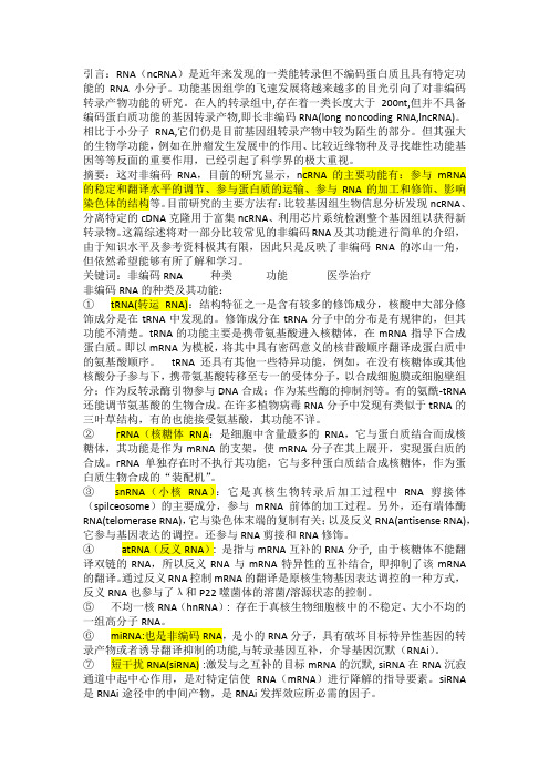
摘要:这对非编码RNA,目前的研究显示,ncRNA的主要功能有:参与mRNA 的稳定和翻译水平的调节、参与蛋白质的运输、参与RNA的加工和修饰、影响染色体的结构等。
目前研究的主要方法有:比较基因组生物信息分析发现ncRNA、分离特定的cDNA克隆用于富集ncRNA、利用芯片系统检测整个基因组以获得新转录物。
这篇综述将对一部分比较常见的非编码RNA及其功能进行简单的介绍,由于知识水平及参考资料极其有限,因此只是反映了非编码RNA的冰山一角,但依然希望能够有所了解和学习。
关键词:非编码RNA 种类功能医学治疗非编码RNA的种类及其功能:①tRNA(转运RNA):结构特征之一是含有较多的修饰成分,核酸中大部分修饰成分是在tRNA中发现的。
修饰成分在tRNA分子中的分布是有规律的,但其功能不清楚。
tRNA的功能主要是携带氨基酸进入核糖体,在mRNA指导下合成蛋白质。
即以mRNA为模板,将其中具有密码意义的核苷酸顺序翻译成蛋白质中的氨基酸顺序。
tRNA还具有其他一些特异功能,例如,在没有核糖体或其他核酸分子参与下,携带氨基酸转移至专一的受体分子,以合成细胞膜或细胞壁组分;作为反转录酶引物参与DNA合成;作为某些酶的抑制剂等。
有的氨酰-tRNA还能调节氨基酸的生物合成。
在许多植物病毒RNA分子中发现有类似于tRNA的三叶草结构,有的也能接受氨基酸,其功能不详。
non-coding RNA——现代分子生物学课程ppt课件
分类
非编码RNA 从长度上来划分可以分为3类: 小于50 nt,包括microRNA,siRNA,piRNA; 50 nt到500 nt,包括rRNA,tRNA,snRNA,snoRNA, SLRNA,SRPRNA 等等; 大于500 nt,包括长的mRNA-like 的非编码RNA,长的不 带polyA 尾巴的非编码RNA等等。
回收50-110nt(fractionalⅠ)和 110-500nt (fractional) → Poly(A)聚合酶加尾 → cDNApSPORTI → PCR → 高密度陈列 → 杂交 除去高丰度已知的小RNA → 未杂交的cDNA测序。
.
蛋白质-RNA复合物分离法
分离蛋白质-RNA复合物,除去蛋白 质,纯化RNA分子,反转录cDNA , 克隆、测序、结果分析。所得RNA 的cDNA分子必须进行Northern杂交, 除去断裂的小RNA。
cDNA克隆策略可鉴别已知的高丰度非编码RNA,如 tRNAs或小核糖体RNA。
.
实时荧光定量(RT-PCR技术)
实时荧光定量PCR技术是一种高通量、灵 敏的基因表达检测技术,常用于蛋白编码 基因表达检测,也被广泛地应用于 microRNA或其他非编码RNA的表达检测。
通过该技术,可定量监测目的基因的表达 情况,筛选生物学功能相关的非编码RNA。
发现,越来越多的研究表明RNA在遗传方面的重要作 用。
.
Non-coding RNA(非编码RNA)
非编码RNA是指不编码蛋白质的RNA。其 中包括rRNA,tRNA,snRNA,snoRNA 和microRNA 等多种已知功能的 RNA,还 包括未知功能的RNA。这些RNA的共同特 点是都能从基因组上转录而来,但是不翻 译成蛋白,在RNA 水平上就能行使各自的 生物学功能了。
Activation of LTBP3 Gene by a Long Noncoding RNA (lncRNA) MALAT1 Transcript in Mesenchymal Stem Cell
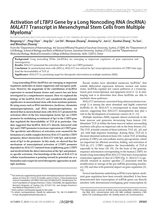
Activation of LTBP3Gene by a Long Noncoding RNA(lncRNA) MALAT1Transcript in Mesenchymal Stem Cells from Multiple Myeloma*Received for publication,April9,2014,and in revised form,August27,2014Published,JBC Papers in Press,September3,2014,DOI10.1074/jbc.M114.572693 Bingzong Li‡1,Ping Chen‡1,Jing Qu§1,Lei Shi§,Wenyue Zhuang¶,Jinxiang Fu‡,Jun Li‡,Xiaohui Zhang‡,Yu Sun‡, and Wenzhuo Zhuang§2From the‡Department of Haematology,the Second Affiliated Hospital of Soochow University,Suzhou215006,the§Department of Cell Biology,School of Biology and Basic of Medical Science,Soochow University,Suzhou215123,and the¶Department of Molecular Biology,Medical Ecsomatics College of Beihua University,Jinlin132013,ChinaBackground:Long noncoding RNAs(lncRNAs)are emerging as important regulators of gene expression and tumorigenesis.Results:MALAT1promoted the activation effect of Sp1on LTBP3promoter.Conclusion:In mesenchymal stem cells(MSCs),MALAT1was critical for Sp1transcriptional activation of LTBP3that regu-lated the bioavailability of TGF-.Significance:MALAT1is a promising target for therapeutic intervention in multiple myeloma(MM).Long noncoding RNAs(lncRNAs)are emerging as important regulatory molecules in tumor suppressor and oncogenic path-ways.However,the magnitude of the contribution of lncRNA expression to normal human tissues and cancers has not been investigated in a comprehensive manner.Here we explored the biology of the lncRNA MALAT1and considered the potential significance in mesenchymal stem cells from myeloma patients. By using assays such as RNA interference,luciferase,chromatin immunoprecipitation,and RNA immunoprecipitation,we showed that in mesenchymal stem cells MALAT1promoted the activation effect of the key transcription factor Sp1on LTBP3 promoter by modulating recruitment of Sp1to the LTBP3gene that regulated the bioavailability of TGF-in particular.Our data suggested that lncRNA MALAT1directly interacted with Sp1and LTBP3promoter to increase expression of LTBP3gene. The specificity and efficiency of activation were ensured by the formation of a stable complex between MALAT1and the LTBP3 promoter,direct interaction of MALAT1with Sp1,and recruit-ment of Sp1to the promoter.In this study,we showed that the mechanism of transcriptional activation of LTBP3promoter depended on MALAT1initiated from neighboring gene LTBP3 and involved both the direct interaction of the Sp1and promot-er-specific activation.Our knowledge of the role of MALAT1in cellular transformation is pointing toward its potential use as a biomarker and a target for novel therapeutic approaches in mul-tiple myeloma.Recent studies have identified numerous lncRNAs3that exhibit differential expression between normal and tumor states.lncRNAs regulate key cancer pathways at a transcrip-tional,post-transcriptional,and epigenetic level(1–5).A chal-lenging task is to determine how these lncRNAs are able to modulate those pathways.MALAT1(metastasis-associated lung adenocarcinoma tran-script1)is among the most abundant and highly conserved lncRNAs(6–8).MALAT1is overexpressed in many human cancers,suggesting that MALAT1misregulation may play a role in the development of numerous cancers(6,9–13). Multiple myeloma(MM)expands almost exclusively in the bone marrow and generates devastating bone lesions(14). Abundant TGF-within the bone marrow milieu surrounding myeloma cells plays an important role in the bone lesions(15). The TGF-family consists of three isoforms,TGF-1,-2,and -3,with high sequence homology.Among them,TGF-1is the most abundant isoform in bone(16).Latent TGF--binding proteins(LTBPs)are extracellular matrix glycoproteins,which are important for efficient secretion,folding,and activation of TGF-s(17).LTBP3regulates the bioavailability of TGF-especially in the bone(18,19).On the basis of the genomic sequence information corresponding to the human LTBP3,we found a long noncoding RNA MALAT1that had a transcription orientation opposite to that of LTBP3(Fig.1).MALAT1is spe-cifically retained in nuclear speckles(7)associated with the modification or storage of the pre-mRNA processing machin-ery(20)and thus with potential impact on gene regulation(21, 22).Several mechanisms underlying ncRNA transcription-medi-ated gene regulation have been recently identified.It has been demonstrated that transcription of ncRNA genes can directly interfere with downstream/overlapping gene transcription by*This work was supported by grants from the National Natural Science Foun-dation of China(81172256,81102466,and81272631),China Postdoctoral Science Foundation(2014M551650and2014M550307),Postdoctoral Sci-ence Foundation of Jiangsu Province,China(1302028B),the Applied Basic Research Programs of Suzhou City(SYS201132),and Jiangsu Province’sKey Medical Department in2011.1These authors contributed equally to this work.2To whom correspondence should be addressed.Tel.:86-512-65880107;E-mail:zhuangwenzhuo@.3The abbreviations used are:lncRNA,long noncoding RNA;ncRNA,noncod-ing RNA;MSC,mesenchymal stem cell;MM,multiple myeloma;LTBP, latent TGF--binding protein;HD,healthy donor;chr,chromosome(s).THE JOURNAL OF BIOLOGICAL CHEMISTRY VOL.289,NO.42,pp.29365–29375,October17,2014©2014by The American Society for Biochemistry and Molecular Biology,Inc.Published in the U.S.A.altering transcription factor binding at the yeast SER3,FLO11,and IME4loci (23–25),and the Drosophila Ubx locus (26).Because MALAT1is highly conserved among mammals and predominantly localizes to nuclear speckles,we hypothesize that it plays a critical role in the LTBP3gene transcription.Our data suggested that lncRNA MALAT1directly interacted with Sp1and LTBP3promoter to increase expression of LTBP3gene.lncRNAs can regulate the transcriptional process through a range of mechanisms.However,the pervasive transcription of promoters anticipates a core role for long ncRNA in regulating the process of transcription (27,28).Here we showed that the lncRNA MALAT1was critical for Sp1transcriptional activa-tion of the LTBP3.MALAT1could recruit the transcription factor Sp1on the LTBP3promoter.The specificity and effi-ciency of activation were ensured by the formation of a stable complex between MALAT1and the LTBP3promoter,direct interaction of MALAT1with Sp1,and recruitment of Sp1to the promoter.Sp1forms a complex with MALAT1,which can bridge Sp1to the LTBP3promoter.Our results demonstrated that the transcription factor Sp1could bind directly and specif-ically to MALAT1,which was expressed in the MSCs from myeloma patients at high levels.MALAT1specifically cooper-ated with Sp1to increase the transcriptional activity of LTBP3in MSCs.A stable complex containing MALAT1,Sp1,and the Sp1consensus site of LTBP3suggested that MALAT1activated transcriptional activity by directly influencing Sp1activity.Our analysis revealed an RNA-dependent mechanism of transcrip-tional activation.It is a dynamic process dependent on the pro-duction of the regulatory transcript from the nearby gene.Highly specific and stable triplex structures formed by lncRNA with the transcription factor and the promoter may well con-tribute to promoter targeting and activation in vivo .These data provided an unexpected lncRNA/transcription factor-based strategy to integrate transcriptional programs.We also high-light the possibility of using this unique RNA as a promising target for therapeutic intervention in multiple myeloma.EXPERIMENTAL PROCEDURESPatients and Subjects—Twenty-five patients with newly diag-nosed MM (14males and 11females,aged 48–83years)and 5normal subjects (2males and 3females,aged 47–63years)were recruited in this study (Table 1).All samples were obtained at the Second Affiliated Hospital of Soochow University accord-ing to institutional rmed consent was obtained according to procedures approved by the Ethics Committee of the Affiliated Hospital of Soochow University.All volunteers signed the informed consent form (ICF).Clinical and biologic information (sex,age at diagnosis,treatment,the level of MALAT1,LTBP3expression)was available for all patients.MSC Isolation and Culture—Isolation and culture of bone marrow-derived MSCs were performed as described previously (29,30).Briefly,mononuclear cells were separated by a Ficoll-Paque gradient centrifugation (specific gravity 1.077g/ml;Nycomed Pharma AS,Oslo,Norway)and depleted of hemato-poietic cells and myeloma cells using CD45,GlyA,CD34,and CD138micromagnetic beads (Miltenyi Biotec Inc.,Auburn,CA).Culture medium was Dulbecco’s modified Eagle’s medi-um/Ham’s F-12medium (DF12;Gibco Life Technologies,Paisley,UK)containing 2%FCS (Life Technologies),1ϫinsu-lin-transferrin-selenium (Life Technologies),109M dexameth-asone (Sigma),104M ascorbic acid 2-phosphate (Sigma),10ng/ml EGF (R&D Systems,Minneapolis,MN),10ng/ml plate-let-derived growth factor BB (PDGF-BB;R&D Systems),100units/ml penicillin,and 1000units/ml streptomycin (LifeTech-FIGURE 1.Genomic organization of human LTBP3and MALAT1.MALAT1and LTBP3genes are located at chromosome 11q13.1.They are separated by ϳ32.0kb and are transcribed in opposite directions.TABLE 1Characterization of myeloma patientsClinical stage of patients was evaluated according to the Durie-Salmon Staging System.M,male;F,female.Patient Sex Age (in years)Clinical stage Para protein Percentage of monoclonalplasma cells Bone lesions 1F 59IIIB IgG-79.0Yes 2F 65IIIA IgG-44.0Yes 3M 69IIA IgG-33.0Yes 4F 46IIIB IgG-28.0Yes 5F 77IIIA IgG-14.0Yes 6M 73IIIB IgG-93.0Yes 7F 44IIIA IgA-28.0No 8F 59IIIA IgG-19.0Yes 9M 75IIIA IgG-58.0Yes 10F 66IIIA Nonsecreted 90.0Yes 11F 46IIIA IgG-21.0No 12M 59IIIA IgG-31.0Yes 13F 59IIIA IgA-25.0Yes 14F 73IIIA IgG-57.0Yes 15M 50IIIA -Light chain 16.5Yes 16M 61IIIB IgG-18.5No 17F 58IIIA IgG-16.0Yes 18M 71IIIA IgG-28.0Yes 19F 61IIA IgG-42.0Yes 20F 62IIIA -Light chain 35.0Yes 21M 71IIIA IgA-78.0No 22M 62IIIA IgG-14.0Yes 23M 61IIIA Nonsecreted 67.0Yes 24M 67IIIA IgG- 5.0Yes 25M64IIIAIgG-16.0YeslncRNA MALAT1Transcript in Mesenchymal Stem Cellsnologies).Cells were cultured at37°C and5%CO2humidifiedatmosphere.Culture medium were changed every3–4days. Once adherent cells were more than80%confluent,they were detached with0.125%trypsin and0.01%EDTA and replanted at a1:3dilution under the same culture conditions.Cells from the second and third passage were analyzed for further study. Cells were regular tested for mycoplasma contamination using MycoAlert mycoplasma detection kit(Lonza,Rockland,ME). RNA Isolation and Quantitative Real-time Polymerase Chain Reaction—RNA was prepared using the EASYspin RNA extrac-tion kit following the manufacturer’s protocol.mRNAs were assayed using the TaqMan microRNA and gene expression assays(Applied Biosystems,Foster City,CA),respectively. GAPDH was used as reference gene.The Ct value of each target gene was normalized against the Ct value of the reference gene(Cttarget ϪCtGAPDH).ELISA—Levels of transforming growth factor-1(TGF-1) were assessed by ELISA kits(R&D Systems)according to the man-ufacturer’s instructions.MSCs(4ϫ105cells/ml)were plated in complete medium,and the supernatants were harvested.Levels of TGF-1were determined by subtracting the value from medium alone and then normalized according to cell number.Small Interfering RNAs—siRNAs targeting MALAT1were obtained from GenePharma.Sequences of the siRNAs were as follows:si MALAT1-1,5Ј-GAAUUCCGGUGAUGCGAGU-3Ј; si MALAT1-2,5Ј-GGCAAUAGAGGCCCUCUAA-3Ј;siSp1-1, 5Ј-AGCCUUGAAGUGUAGCUAU-3Ј;siSp1-2,5Ј-GGUAG-CUCUAAGUUUUGAU-3Ј.siRNA universal negative control (GenePharma)that does not target any human gene product was used as a negative control.Antibodies—The following antibodies were used:LTBP3 (Santa Cruz Biotechnology,Santa Cruz,CA,sc-390913),Sp1 (Abcam,ab13370),and-actin(Millipore,Temecula,CA). Lentiviral Constructs—The sequence of MALAT1was syn-thesized and subcloned into lentivector-transferred plasmid pCDH-CMV-MCS-EF1-coGFP to generate pCDH-CMV-MALAT1-EF1-coGFP.The recombinant vector pCDH-CMV-MALAT1-EF1-coGFP or the control vector pCDH-CMV-MCS-EF1-coGFP was triple-transfected with the packaging vectors psPAX2and pMD2.G into293T cells by calcium chlo-ride to produce the lentivirus.MSCs were infected with control or MALAT1-expressing lentivirus.The expression of MALAT1 in cells was determined by quantitative PCR.Luciferase Assay—Genomic DNA from293T was used to amplify the human LTBP3promoter region that was cloned into the pGL3-basic luciferase vector(Promega)by using SacI and XhoI sites(primers listed in Table1).IK-2,Sp1,GATA-2, and MZF1binding site mutations were introduced into the (Ϫ2000/ϩ200)construct to disrupt the binding elements and to generate the mutIK-2construct,Sp1mut construct, mutGATA-2construct,and mutMZF1construct using QuikChange site-directed mutagenesis kit(Stratagene),respectively(prim-ers listed in Table2).MSCs were transiently transfected with different LTBP3pro-moter reporter constructs and Renilla luciferase plasmid(Pro-mega)as a normalization control using Lipofectamine2000 (Invitrogen).Firefly and Renilla luciferase activities were mea-sured consecutively by using the Dual-Luciferase assay(Pro-mega)48h after transfection.ChIP Assays—Cells were first cross-linked with2%parafor-maldehyde for10min at37°C and sonicated,and then the DNA-protein complexes were isolated with a ChIP assay kit (Millipore,catalog number17-371)according to the manufac-turer’s instructions with antibodies against Sp1(Millipore)The precipitated DNA was quantified by real-time PCR with prim-ers listed in Table2.The results were normalized relative to the input control.RNA Immunoprecipitation—RNA immunoprecipitation assay was performed with an RNA-binding protein immunoprecipita-tion kit(Millipore,catalog number17-700)according to the man-ufacturer’s instructions with antibodies against Sp1(Millipore). Quantitative PCR for RNA was performed using TaqMan univer-sal PCR mix as described above.The results were normalized rel-ative to the input control.DNA Pulldown Assay—The pulldown assay,which was sim-ilar to RNA pulldown assay,was performed as described previ-ously with modifications(31).Biotinylated oligonucleotides were synthesized that corresponded to LTBP3promoter sequences that include putative Sp1transcription factor bind-ing sites(5Ј-Biotin-CTCCCCGCCCCGCCCCGCC-3Ј)or its mutation(5Ј-Biotin-CTCCCCTTCCCGCCCCGCC-3Ј). Double-stranded oligonucleotides were prepared by mixing equal amounts of forward(biotin-labeled)and reverse oligo-nucleotide at100°C for1h and then slow cooling to room temperature for30min.One milligram of whole-cell lysates from MM-MSCs was incubated with3g of biotinylated oligonucleotides for1h at25°C;complexes were isolated with streptavidin-agarose beads(Invitrogen).The RNA present in the pulldown material was detected by quantitat-ive RT-PCR analysis.Statistical Analysis—All quantitative data presented are the meanϮS.D.from at least three samples per data point.The Pearson product-moment correlation coefficient(r)was used to establish the association of the two variables.The difference was considered statistically significant when pϽ0.05.A p value of0.05was used as the boundary of statistical significance.All statistical tests were two-sided.RESULTSExpression of MALAT1and LTBP3,as Well as TGF-1Secre-tion in MSCs from Bone Marrow of Myeloma Patients and Healthy Donors—To explore the biology of MALAT1and con-sider the potential significance in MM,we examined the expression of MALAT1in MSCs from MM patient samples and healthy donors.The expression of MALAT1was assessed by quantitative real-time PCR.Consistently higher expression level of MALAT1was found in MSCs from all25patient sam-ples relative to that from healthy donors(pϽ0.001).Under the same conditions,quantitative real-time PCR showed that LTBP3was more highly expressed in MSCs from all25 myeloma patients when compared with that from healthy donors(pϽ0.001)(Fig.2A).The changes in protein levels of LTBP3in cells as mentioned above were further confirmed by the immunoblotting analysis(Fig.2C).To determine the bio-logical source of TGF-1in MSCs,the synthesis and secretionlncRNA MALAT1Transcript in Mesenchymal Stem Cellsof TGF-1by MSCs(Fig.2B)were measured by ELISA. TGF-1was significantly higher in MSCs from myeloma patients than that from healthy donors(pϽ0.01).Because both MALAT1and LTBP3were augmented in in MSCs from myeloma patients,to examine any correlation between MALAT1and LTBP3,we performed Pearson correlation coef-ficient analysis in both healthy donors and myeloma patients.A positive correlation between MALAT1and LTBP3was observed in MSCs from healthy donors and myeloma patients(rϭ0.7670,pϽ0.0001,Fig.2D).Knockdown of the MALAT1Decreased LTBP3Transcription and TGF-1Secretion in MM-MSCs—To examine the func-tional significance of MALAT1overexpression in MSCs from myeloma patients,we knocked down the MALAT1using two independent siRNAs against MALAT1.MM-MSCs1,MM-MSCs2, MM-MSCs4,and MM-MSCs5,which correspond to patient1, patient2,patient4,and patient5,respectively,were used in the analyses.MM-MSCs1,MM-MSCs2,MM-MSCs4,and MM-MSCs5were treated with control siRNA or with siRNA target-ing MALAT1.As shown in Fig.3,knockdown of the MALAT1 decreased LTBP3transcription(pϽ0.01).A dramatic reduc-tion of TGF-1secretion was also observed in MALAT1knock-down MM-MSCs1,MM-MSCs2,MM-MSCs4,and MM-MSCs5by ELISA assays(pϽ0.01).A similar result was obtained with another MALAT1siRNA targeting a different sequence of MALAT1.Specific MALAT1siRNA caused aTABLE2Primers used in this studylncRNA MALAT1Transcript in Mesenchymal Stem Cellsmarked decrease in LTBP3mRNA levels,suggesting that LTBP3expression is dependent on MALAT1function.Overexpression of MALAT1Increased LTBP3Transcription and TGF-1Secretion in HD-MSCs—To further elucidate the functional connection between MALAT1and LTBP3,we used a lentiviral vector system to efficiently overexpress MALAT1inMSCs from healthy donors (HD-MSCs).HD-MSCs1and HD-MSCs2,which correspond to healthy donor 1and healthy donor 2,respectively,were used in the analyses.MALAT1expression was quantitated by real-time PCR 48h after infec-tion.MALAT1was overexpressed more than 11-and 14-fold,respectively (Fig.4A ).As shown in Fig.4,B and C ,overexpres-FIGURE 2.Different expression of MALAT1and LTBP3,as well as different TGF-1secretion between MSCs from bone marrow of myeloma patients and those from healthy donors.A ,MALAT1and LTBP3transcript levels were determined by quantitative real-time PCR analysis of MSCs from bone marrow of myeloma patients and healthy donors.GAPDH was measured for an internal control and used to normalize the data.B ,supernatant TGF-1secreted by the cultured MSCs was determined by ELISA assay as described under “Experimental Procedures.”Levels of TGF-1were determined by subtracting the value from medium alone and then normalized according to cell number.The error bars represent three independent ELISA readings.C ,LTBP3was determined by immunoblotting in MSCs from bone marrow of myeloma patients and healthy donors.D ,correlation between MALAT1and LTBP3in MSCs from healthy donors and myeloma patients;r ϭ0.7670,p Ͻ0.0001.FIGURE 3.Knockdown of the MALAT1by RNA interference decreased LTBP3transcription and TGF-1secretion.MM-MSCs1,MM-MSCs2,MM-MSCs4,or MM-MSCs5was transfected with two independent siRNA against MALAT1or with control siRNA (siControl )for 48h.A ,quantitative real-time PCR analysis was performed to determine MALAT1and LTBP3levels,with GAPDH used as an internal normalization control.Error bars represent mean ϮS.D.B ,LTBP3levels were determined by immunoblotting.C ,TGF-1was quantified by ELISA.lncRNA MALAT1Transcript in Mesenchymal Stem Cellssion of MALAT1increased LTBP3transcription(pϽ0.01).Anincrease of TGF-1secretion was also observed in MALAT1-overexpressing HD-MSCs1(HD-MSCs1-OM)and HD-MSCs2(HD-MSCs2-OM)by ELISA assays(pϽ0.01)(Fig.4D).Thesedata suggested that MALAT1may play a specific role in LTBP3transcriptional activation in MSCs.MALAT1Cooperates with Sp1to Increase LTBP3Transcription—The close relationship between MALAT1and the LTBP3led usto ask whether MALAT1may function by regulating the tran-scriptional activity of the LTBP3.To test this hypothesis,weconstructed reporter plasmids carrying various genomicsequences around the transcription start site of the LTBP3andsubjected them to luciferase assay.A significant reduction inpromoter activity was detected when the region fromϪ600to Ϫ400was deleted,indicating that it contains important regula-tory elements for the basal expression of LTBP3(pϽ0.01)(Fig.5A).Further definition of this region is necessary to determinethe precise transcription factor that is involved in theMALAT1-mediated interaction.Because conserved regions ina given gene promoter are expected to contain regulatory ele-ments,we focused on the highly conserved region(Fig.5B).A bioinformatics search identified a previously reported high score Sp1consensus site(32).In addition,conserved IK-2, GATA-2,and MZF1elements were identified in this region.To define the MALAT1active region,we used the different muta-tion reporters to assay for the effect of MALAT1knockdown on the activity of different transcription factors.Luciferase assay using a construct mutated for IK-2,Sp1,GATA-2,and MZF1 consensus sequence revealed that Sp1region was both neces-sary and sufficient for the activity.These findings also sug-gested that Sp1was a key transcription factor of LTBP3through directly binding to the core promoter element.MALAT1 knockdown had a repressive effect on the LTBP3promoter. This effect was observed only when IK-2,GATA-2,and MZF1 consensus sequences were mutated,respectively.The con-struct mutated for Sp1did not have the same effect(Fig.5C) (pϽ0.01).To provide further confirmation,we examined lucif-erase activity in MSCs2cotransfected with two independent siRNA against Sp1or with control siRNA and empty luciferase reporter,LP8reporter construct,or LP8reporter construct with mutated Sp1binding site.We found that mutating Sp1 binding sites or knocking down Sp1reduced the luciferase activity of the LP8reporter vector.Knocking down Sp1did not influence the luciferase activity of LP8reporter with mutated Sp1binding site(Fig.5D)(pϽ0.01).These data demonstrated that MALAT1cooperated with Sp1but not with IK-2,GATA-2, or MZF1to increase the LTBP3transcription in MSCs.These results strongly suggested that MALAT1-mediated activation was Sp1-dependent.Four Sp1binding sites were detected at the LTBP3locus in ChIP-Seq experiments from UCSC Genome Browser(Table3). To validate the binding between these Sp1binding sites of LTBP3and Sp1,we performed ChIP to pull down endogenous Sp1associated with the Sp1binding site at LTBP3locus and demonstrated by quantitative PCR analysis with primers spe-cific to Site1,Site2,Site3,and Site4(binding regions indicated in ChIP-Seq experiments from UCSC Genome Browser)and Site5(binding region indicated in Fig.5C).As shown in Fig.5E, the endogenous Sp1binding site of LTBP3pulldown by Sp1was specifically enriched in Site5but not in Site1,Site2,Site3,or Site4in MSCs.The Interactions of Sp1with Sp1Consensus Site at the LTBP3 Promoter Were Controlled by lncRNA MALAT1—To prove that MALAT1directly regulates LTBP3transcription by recruiting Sp1to the LTBP3promoter,we performed CHIP assay.MM-MSCs7,MM-MSCs8,MM-MSCs10,and MM-MSCs12,which correspond to patient7,patient8,patient10,and patient12, respectively,were used in the analyses.MM-MSCs7,MM-MSCs8,MM-MSCs10,and MM-MSCs12were treated with control siRNA or with siRNA targeting MALAT1.Indeed,we found that endogenous Sp1directly interacts with the core ele-ment of the LTBP3promoter in MSCs,as demonstrated by CHIP assay.Moreover,we observed a marked decrease in the occupancy of the LTBP3promoter by Sp1in MALAT1knock-down cells when compared with control knockdown cells and untreated cells(Fig.6,A–C)(pϽ0.01).These data suggested that recruitment of Sp1to the LTBP3promoter to cause gene-specific activation was directed by lncRNA MALAT1tetheredFIGURE4.Overexpression of MALAT1increased LTBP3transcription and TGF-1secretion.HD-MSCs1or HD-MSCs2was infected with control or MALAT1-expressing lentivirus for48h.A and B,quantitative real-time PCR analysis was performed to determine MALAT1(A)and LTBP3(B)levels,with GAPDH used as an internal normalization control.Error bars represent meanϮS.D.C,LTBP3levels were determined by immunoblotting.D,TGF-1 was quantified by ELISA.lncRNA MALAT1Transcript in Mesenchymal Stem CellsFIGURE 5.Identification of MALAT1core element in the LTBP3promoter.A ,luciferase reporter activity of promoter constructs of LTBP3on chromosome 11q13.1in MSCs2cells.Schematic presentation of LTBP3promoter deletion constructs is shown on the left .The arrow above construct LP1indicates the position of the transcription start site ϩ1.Transcriptional activity of the corresponding constructs is indicated on the right .Luciferase activities were normalized by Renilla luciferase activities.Values represent mean ϮS.D.from three experiments.B ,conservation track in UCSC Genome Browser.The LTBP3promoter locus from chr 65326100to 65326300contains regions of conservation subtracks.The subtracks display phyloP scores (in blue and red )The subtracks display phyloP scores (in blue and red ).The phyloP tracks measure conservation (with positive scores,shown in blue ),whereas the phyloP tracks also indicate accelerated evolution (with negative scores,shown in red ).C ,relative luciferase activity of LP8reporter construct.Transcription factor binding sites are indicated (blue boxes :1,IK-2;2,Sp1;3,GATA-2;and 4,MZF1.Deletions introduced into the LP8construct are shown as yellow X showing abolition of the promoter activity.EV ,empty vector.D ,luciferase activity in MSCs2cotransfected with two independent siRNA against Sp1or with control siRNA and empty luciferase reporter,LP8reporter construct,or LP8reporter construct with mutated Sp1binding site.Data are presented as the relative ratio of firefly luciferase activity to Renilla luciferase activity.lncRNA MALAT1Transcript in Mesenchymal Stem CellsFIGURE 6.lncRNA MALAT1in recruitment of the Sp1on LTBP3promoter in vivo .A ,schematic representation of the primer sets specific for LTBP3promoter.B ,MSCs were untreated or treated with control siRNA or with siRNA targeting MALAT1,and ChIP assays were performed with the Sp1antibody and the indicated PCR primers.The binding of Sp1to the LTBP3promoter was detected by quantitative real-time PCR.The results were normalized to the input control.All data are shown as mean ϮS.D.C ,representative analysis of PCR products derived by ChIPs,which were separated on an agarose gel.D ,RNA immunopre-cipitation using Sp1antibody followed by quantitative real-time PCR for MALAT1in MSCs transfected with two independent MALAT1siRNA or control siRNA revealed specific interaction between Sp1and MALAT1.E ,MM-MSCs lysates were incubated with biotinylated Sp1consensus site DNA probe;after pulldown,MALAT1was extracted and assessed by quantitative RT-PCR.The error bars represent mean ϮS.D.of three independent experiments.TABLE 3Sp1binding sites detected at the LTBP3locus in ChIP-Seq experiments from UCSC Genome BrowserSite Chromosome Chromosome start Chromosome end Name Score Site1chr116530813065308450Sp1161Site2chr116531392165314257Sp1126Site3chr116531904565319381Sp1187Site4chr116532471765325053Sp1293lncRNA MALAT1Transcript in Mesenchymal Stem Cellsto the Sp1consensus site of LTBP3.MALAT1acted as a co-fac-tor to modulate transcription factor Sp1activity.To strengthen the evidence for RNA-dependent activation of transcription factor Sp1in vivo,we then performed an RNA immunoprecipitation assay and showed that immunoprecipi-tation of Sp1from MM-MSCs7,MM-MSCs8,MM-MSCs10,or MM-MSCs12contained MALAT1transcript.Knockdown of MALAT1caused weak interaction between Sp1and MALAT1 (Fig.6D).The specific association between MALAT1and the Sp1consensus site of LTBP3was further validated by pulldownof endogenous MALAT1using in vitro biotinylated Sp1con-sensus site DNA probe(Fig.6E)(pϽ0.01).The data strengthen the evidence for the interaction of the Sp1with MALAT1. These results clearly implicated that lncRNA MALAT1posi-tively regulates LTBP3transcription by recruiting Sp1to the LTBP3promoter.Recent studies of transcription factors that bind DNA and RNA with distinct roles have been reported(33, 34).In our studies,we showed that Sp1,an important transcrip-tion factor,known to bind and activate promoters,cooperated with the lncRNA MALAT1,resulting in an increase of LTBP3 transcription.DISCUSSIONThe notion that ncRNAs can have numerous molecular functions is rapidly evolving(35,36).Although long ncRNAs represent a large class of transcriptional units and appear to be evolutionarily conserved,their specific roles in critically mod-ulating regulated gene expression remain poorly understood. With the recent recognition of lncRNAs flanking many genes (4,37–39),a central issue is to fully understand their potential roles in regulating the process of transcription,possibly through different mechanisms(26,28,33,40–43).Among a small number of genes that were dysregulated in adult MALAT1knock-out mice,many were MALAT1neigh-boring genes,including LTBP3,thus indicating a potential reg-ulatory role of MALAT1gene transcription(44)Our data showed that in MSCs,lncRNA MALAT1regulated transcrip-tion of nearby protein-coding gene LTBP3that regulates the bioavailability of TGF-in particular.MALAT1affected tran-scriptional activity by cooperation and transcription factor complex formation.MALAT1bound with the Sp1transcrip-tion factor and enhanced Sp1activity in the LTBP3target. Knockdown of MALAT1using specific siRNAs significantly decreased LTBP3transcription,indicating required functions of this coactivator on this gene.The interaction of transcription factors,in particular the bicoid homeodomain protein with both DNA and RNA,has been reported previously(33,34,45), raising the possibility of a direct interaction between the Sp1 protein and the MALAT1ncRNA.By using assays such as lucif-erase,chromatin immunoprecipitation,and RNA immunopre-cipitation,we showed that MALAT1had a critical function in a mechanism of promoter-specific transcriptional activation. MALAT1functioned as an Sp1transcriptional coactivator.Our data suggested that the Sp1-MALAT1complex stabilized the interaction between Sp1and Sp1consensus sequences to increase transcriptional activity.A model of one possible mech-anism is shown in Fig.7.In this model,MALAT1bound to Sp1 in a DNA-independent manner,and the entire complex bound to Sp1consensus sequences.We propose that the lncRNA MALAT1acted as specific transcriptional coactivators.Tran-scriptional coregulators,including coactivators and corepres-sors,are required for regulating programs of gene expression in a transcription factor-and gene-specific manner(46,47). MALAT1recruited the binding and action of the transcription factor Sp1to the promoter to increase expression of the adja-cent gene.Sp1bound both DNA and RNA during the cooper-ative interaction.Here we suggested the model in which lncRNA MALAT1serves as a molecular“ligand”for a specific transcription factor,Sp1.This in turn permitted gene-specific Sp1-MALAT1interactions resulting in increase of LTBP3tran-scription.It is tempting to speculate that other transcription factors exert functional roles on gene transcription by being analogously recruited to the transcription units through gene-specific lncRNAs.Myeloma cells enhance bone resorption and suppress bone formation(14).TGF-plays a role in the suppression of bone formation in MM bone lesions.The inhibition of TGF-signal-ing causes not only an enhancement of bone formation but also a suppression of MM cell growth(15).Here we showed that in MSCs,MALAT1promoted the activation effect of the key tran-scription factor Sp1on LTBP3promoter by modulating recruit-ment of Sp1to the LTBP3gene that regulates the bioavailability of TGF-in particular.Our growing knowledge of the role of MALAT1is pointing toward their potential use as biomarkers and targets for novel therapeutic approaches in MM.Broadly, our data highlight combinational actions of transcription factor and lncRNA.An ultimate goal should be the use of cis-and trans-acting lncRNAs as both diagnostic markers and RNA-based targets for the treatment of human diseases.To reach this end,a fuller understanding of the functional versatility of lncRNAs as key regulators of genome expression in physiolog-ical conditions and diseases will be required. REFERENCES1.Khaitan,D.,Dinger,M.E.,Mazar,J.,Crawford,J.,Smith,M.A.,Mattick,J.S.,and Perera,R.J.(2011)The melanoma-upregulated long noncoding RNA SPRY4-IT1modulates apoptosis and invasion.Cancer Res.71, 3852–38622.Kotake,Y.,Nakagawa,T.,Kitagawa,K.,Suzuki,S.,Liu,N.,Kitagawa,M.,and Xiong,Y.(2011)Long non-coding RNA ANRIL is required for the PRC2recruitment to and silencing of p15INK4B tumor suppressor gene.Oncogene30,1956–19623.Gupta,R.A.,Shah,N.,Wang,K.C.,Kim,J.,Horlings,H.M.,Wong,D.J.,Tsai,M.C.,Hung,T.,Argani,P.,Rinn,J.L.,Wang,Y.,Brzoska,P.,Kong,B., Li,R.,West,R.B.,van de Vijver,M.J.,Sukumar,S.,and Chang,H.Y.(2010) Long non-coding RNA HOTAIR reprograms chromatin state to promote cancer metastasis.Nature464,1071–10764.Carninci,P.,Kasukawa,T.,Katayama,S.,Gough,J.,Frith,M.C.,Maeda, FIGURE7.A model proposing that MALAT1and Sp1form a complex,ulti-mately affecting transcription of the LTBP3gene.lncRNA MALAT1Transcript in Mesenchymal Stem Cells。
沉默MALAT1基因对蜂毒素诱导HepG2细胞增殖和凋亡的影响
沉默MALAT1基因对蜂毒素诱导HepG2细胞增殖和凋亡的影响赵斌;吴毓婷;黄成;吕雄文;李俊【摘要】Aim To investigate the effects of silencing MALAT1 gene on cell proliferation inhibition and apop-tosis induced by Melittin in human hepatocellular car-cinoma HepG2 cells. Methods The inhibitory rate of cell proliferation treated with Melittin in HepG2 cells was examined by MTT assay. Apoptotic rate was detec-ted by flow cytometry. The MALAT1 expression level in HepG2 cells was measured by qPCR. Specific siR-NAs were utilized to silence MALAT1 expression. The rates of cell proliferation inhibition and apoptosis in HepG2 cells treated with siRNA and Melittin were compared with those of Melittin alone. Results Melit-tin significantly suppressed the growth of HepG2 and induced cell apoptosis in a dose-dependent manner. Compared with normal liver cell lines, MALAT1 was highly expressed in HepG2 cells ( P<0. 05 ) . The ex-pression of MALAT1 in HepG2 cells was inhibited by Melittin, and the inhibitory rate increased with the in-crease of concentration. The rates of cell proliferation inhibition and apoptosis in HepG2 cells treated with siRNA and Melittin were significantly higher than those treated merely with Melittin. Conclusion Melittin can reduce the expression of MALAT1 and silencing MALAT1 can effectively promote proliferation inhibi-tion and apoptosis in HepG2 cells induced by Melittin.%目的:探讨沉默HepG2细胞株中MALAT1基因对蜂毒素诱导的细胞增殖抑制和凋亡的影响。
白藜芦醇调控长链非编码RNAMALAT1抗动脉粥样硬化
白藜芦醇(resveratrol,Rev)是一类天然多酚化 合物,主要 存 在 于 葡 萄、红 酒 和 传 统 药 材 虎 杖 的 根 中。研究发现,Rev具有广泛的生物学功能,包括抗 炎、抗氧化、抗凋亡、抗肿瘤等作用[1]。动脉粥样硬 化(atherosclerosis,AS)是引发心血管疾病的主要原 因,Rev具有良好的抗 AS作用。众所周知,Rev可 以多靶点改善内皮功能障碍,抑制平滑肌细胞的增
收稿日期:2018-09-18,修回日期:2018-11-19 基金项目:黑龙江省 自 然 科 学 基 金 资 助 项 目 (NoQC2016112);国 家
自然科学基金资助项目(No81503069) 作者简介:贾雪凌(1993-),女,硕士生,研究方向:病理学与病理生
理学,Email:381406414@qq.com; 范玉华(1983-),男,博士,副教授,研究方向:药理学,共 同第一作者,Email:fyh198306@126.com; 宋印利(1967-),男,硕士,教授,硕士生导师,研究方向: 病理学与 病 理 生 理 学,通 讯 作 者,Email:syl3127@126. com
中国药理学通报 ChinesePharmacologicalBulletin 2019Jan;35(1):35~40
·35·
网络出版时间:2018-12-1115:00 网络出版地址:http://kns.cnki.net/kcms/detail/34.1086.r.20181210.0951.020.html
殖和迁移,减少巨噬细胞浸润和泡沫细胞的形成,以 及减少脂质堆积等,发挥预防和治疗 AS的作用[1]。 AS是一类以炎性浸润和脂质积聚为特征的慢 性炎症性疾病,其中,动脉壁中持续的巨噬细胞积聚 是发病机制的基础。人们发现,在患者动脉斑块中 存在着大量的巨噬细胞,这些巨噬细胞是由内膜中 单核细胞分 化 而 来 的,通 过 分 泌 促 炎 细 胞 因 子 (如 IL6、IL1β、TNFα),吸 收 胆 固 醇、脂 质 形 成 泡 沫 细 胞,以及引发细胞凋亡等,促进 AS斑块的形成、发 展和破裂[2-3]。 长链非编码 RNA(longnoncodingRNA,lncR NAs)是一类由 RNA聚合酶 II转录,长度超过 200 个核苷酸,且 缺 乏 蛋 白 编 码 能 力 的 内 源 性 非 编 码 RNA。过去,人 们 认 为 lncRNAs是 转 录 过 程 中 的 “噪音”,近年来发现它在心血管疾病的发生和发展 中具有重要的意义。人类肺腺癌转移相关转录本 1 (metastasisassociated in lung denocarcinoma tran script1,MALAT1)是目前研究最为深入的一种高度 丰富和保守的 lncRNAs,其长度约为8000nt,定位 于染色体 11q13[4]。据报道,MALAT1参与多种心 血管 疾 病 的 病 理 生 理 过 程[5-6]。 Han等[7]发 现, MALAT1在糖 尿 病 AS大 鼠 的 巨 噬 细 胞 中 表 达 增 高,提示 MALAT1与 AS密切相关。尽管有研究表 明,氧 化 型 低 密 度 脂 蛋 白 可 通 过 促 进 MALAT1转 录,进而增强巨噬细胞中的脂质摄取[8],但 MALAT1 在 AS中的确切作用机制仍尚不明确,有待进一步 研究。由 于 Rev的 潜 在 抗 动 脉 硬 化 作 用,以 及 MALAT1参与糖尿病 AS发生发展过程,因此,本研 究旨在探讨 Rev调控 MALAT1发挥抗 AS作用的机 制,并阐明 MALAT1在 AS中的潜在作用,为指导临 床用药提供理论依据。 1 材料与方法 1.1 材料 1.1.1 实验动物 8周龄 ApoE-/-小鼠,♂,体质 量(27±3)g,购 自 南 京 君 科 生 物 工 程 有 限 公 司 [SCXK(苏)20160010]。所用动 物 饲 养 在 标 准 实 验动物房内,温度(23±1)℃,湿度 55% ~60%,可 自由 摄 食 饮 水。 ApoE-/- 小 鼠长链非编码 RNAMALAT1抗动脉粥样硬化
长链非编码RNA MALAT1在肝细胞癌发生发展中的作用
长链非编码RNAMALAT1在肝细胞癌发生发展中的作用孙永康,颜学波,朱泽民,申鼎成,谢智钦,赵志坚,唐才喜中南大学湘雅医学院附属株洲医院肝胆胰脾外科,湖南株洲412007摘要:肝细胞癌(HCC)具有发病率高、生存率低、治疗效果不良、发病机制复杂等特点。
近年来,多项研究证实lncRNAMALAT1在HCC中表达上调,具有促进HCC细胞增殖、侵袭和转移的作用,并且在HCC的诊断、预后及治疗方面具有临床指导意义。
总结了lncRNAMALAT1在HCC中的研究现状,探讨其表达模式、作用机制以及在预测和监测HCC发展中的临床意义,以期深入了解lncRNAMALAT1在介导HCC发展中的作用。
lncRNAMALAT1将有望成为HCC诊断和预后的潜在生物标志物,并在将来应用于临床靶点治疗。
关键词:癌,肝细胞;RNA,长链非编码;肺腺癌转移相关转录子1中图分类号:R735.7 文献标志码:A 文章编号:1001-5256(2021)03-0704-05Roleoflongnon-codingRNAMALAT1inthedevelopmentandprogressionofhepatocellularcarcinomaSUNYongkang,YANXuebo,ZHUZemin,SHENDingcheng,XIEZhiqin,ZHAOZhijian,TANGCaixi.(DepartmentofHepatobiliaryandPancreatosplenicSurgery,AffiliatedZhuzhouHospital,XiangyaMedicalCollege,CentralSouthUniversity,Zhuzhou,Hunan412007,China)Abstract:Hepatocellularcarcinoma(HCC)hasthefeaturesofhighincidencerate,lowsurvivalrate,poortreatmentoutcome,andcomplexpathogenesis.Inrecentyears,manystudieshaveshownthatlongnon-codingRNA(lncRNA)MALAT1isupregulatedinHCCandcanpromotetheproliferation,invasion,andmetastasisofHCCcells,anditcanalsoguidethediagnosis,prognosticevaluation,andtreatmentofHCCinclinicalpractice.ThisarticlereviewsthecurrentstatusofresearchonlncRNAMALAT1inHCCanddiscussesitsexpressionpattern,mechanismofaction,andclinicalsignificanceinpredictingandmonitoringtheprogressionofHCC,soastogainadeepunder standingoftheroleoflncRNAMALAT1intheprogressionofHCC.ItispointedoutthatlncRNAMALAT1isexpectedtobecomeapotentialbiomarkerforthediagnosisandprognosticevaluationofHCCandmaybeusedasatherapeutictargetinclinicalpractice.Keywords:Carcinoma,Hepatocellular;RNA,LongNoncoding;Metastasis-AssociatedLungAdenocarcinomaTranscript1DOI:10.3969/j.issn.1001-5256.2021.03.041收稿日期:2020-09-08;修回日期:2020-10-20基金项目:湖南省自然科学基金项目(2020JJ5998);湖南省科技计划项目经费资助(2016SK4006)作者简介:孙永康(1993—),男,从事肝胆胰疾病临床与基础研究通信作者:唐才喜,tcx0826@163.com 肝细胞癌(HCC)是消化系统常见的恶性肿瘤,已成为全球癌症相关死亡的第二大原因[1]。
长链非编码RNA-MALAT1在缺氧诱导的肾小管上皮细胞及肾脏纤维化组织中的表达情况
蛋白表达情况;建立大鼠单侧输尿管结扎(UUO)模型,比较 UUO 组和对照组中 lncRNA-MALAT1 的 mRNA 和蛋白表
达情况;选取 IgA 肾病(Lee 氏芋~吁级)患者的肾活检组织,比较其与正常组织中 lncRNA-MALAT1 的 mRNA 表达情
况。结果 与对照组比较,缺氧组 lncRNA-MALAT1 的 mRNA 及蛋白的表达均增加(P约0.01)。与对照组比较,UUO 组
- 14 -
临床医学研究与实践 2021 年 7 月第 6 卷第 19 期
多 种 疾 病 中 发 挥 重 要 作 用 [8-10]。肺 腺 癌 转 移 相 关 转 录 因 子 1(MALAT1)是一种由 8 779 个核苷酸组成的 lncRNA[11]。 既往本课题组研究了 lncRNA-MALAT1 在高糖诱导的人 腹膜间皮细胞转分化和纤维化中的作用12],并且有研究显 示,lncRNA-MALAT1 与宫颈癌[13]、卵巢癌[14]、甲状腺癌[15]、 前列腺癌 等 [16] 肿瘤的发生、发展、浸润、转移有关。那么 lncRNA-MALAT1 在肾脏纤维化中是否也发挥着一定的 作用呢?缺氧可导致肾小管上皮细胞发生 EMT,而单侧 输尿管结扎(UUO)模型是经典的肾脏纤维化模型[17],IgA 肾病以(Lee 氏芋~吁级)肾脏活检组织纤维化较为明显。 为此,本研究首先采用缺氧刺激肾小管上皮细胞,建立大 鼠 UUO 模型,然后收集患者的肾脏活检组织,提取肾小管 上皮细胞及肾脏组织中的 RNA 并检测 lncRNA-MALAT1 的表达情况,具体内容报道如下。
712100, China)
ABSTRACT: Objective To study the expression of long non-coding RNA-metastasis-associated lung adenocarcinoma transcript 1 (lncRNA-MALAT1) in renal tubular epithelial cells induced by hypoxia and renal fibrosis tissue. Methods After HK-2 cells stimulated by hypoxia and normoxia for 72 h, the mRNA and protein expressions of lncRNA-MALAT1 in the cells of two groups were compared. The unilateral ureteral ligation (UUO) model of rats was established to compare the mRNA and protein expressions of lncRNA-MALAT1 in UUO group and control group. The mRNA expression of lncRNAMALAT1 in renal biopsy tissues of patients with IgA nephropathy (Lee's 芋-郁 grade) and normal tissues was compared. Results Compared with the control group, the mRNA and protein expressions of lncRNA -MALAT1 increased in the hypoxia group (P<0.01). Compared with the control group, the mRNA and protein expressions of lncRNA-MALAT1 in the UUO group increased (P <0.05). Compared with adjacent normal renal tissues, lncRNA -MALAT1 mRNA expression increased in renal biopsy tissues of patients with IgA nephropathy (Lee's 芋 - 郁 grade) (P <0.05). Conclusion The expression of lncRNA -MALAT1 upregulate in hypoxia stimulation, UUO model and IgA nephropathy cells, and can be used as a biomarker to predict renal EMT and fibrosis. KEYWORDS: long non -coding RNA; metastasis -associated lung adenocarcinoma transcript 1; epithelial -mesenchymal transition; renal fibrosis
长链非编码RNA_MALAT1_对氧糖剥夺
第 49 卷第 4 期2023年 7 月吉林大学学报(医学版)Journal of Jilin University(Medicine Edition)Vol.49 No.4Jul.2023DOI:10.13481/j.1671‐587X.20230402长链非编码RNA MALAT1对氧糖剥夺/复氧诱导的人脑微血管内皮细胞血管生成的影响高昶1,2, 刘燕3, 杨皓翔4, 张翠翠5(1. 济宁医学院精神卫生学院行为医学重点实验室,山东济宁272067;2. 济宁医学院研究生处,山东济宁272067;3. 济宁医学院精神卫生学院精神医学研究院,山东济宁272013;4. 济宁医学院附属医院神经内科,山东济宁272029;5. 济宁医学院附属医院重症医学三科,山东济宁272029)[摘要]目的目的:探讨长链非编码RNA(lncRNA)肺腺癌转移相关转录本1(MALAT1)对氧糖剥夺/复氧(OGD/R)诱导的人脑微血管内皮细胞(HBMECs)血管生成的影响,并阐明其潜在的分子机制。
方法方法:采用生物信息学方法预测MALAT1、不均一核糖核蛋白K(hnRNPK)和血管内皮生长因子A(VEGFA)的结合位点。
HBMECs进行OGD/R处理构建脑缺血细胞模型,分为对照组、OGD/R模型组、OGD/R+siNC组、OGD/R+沉默MALAT1(OGD/R+siMALAT1)组和OGD/ R+siMALAT1+过表达VEGFA(OGD/R+siMALAT1+VEGFA)组。
采用小干扰RNA(siRNA)沉默MALAT1的表达,采用pcDNA载体构建VEGFA过表达载体,将构建的siMALAT1和pcDNA VEGFA载体分别或同时转染至HBMECs中。
利用管形成实验检测各组细胞血管形成能力,Western blotting法检测各组细胞中VEGFA蛋白表达水平。
6周龄健康雄性C57BL/6 J小鼠20只,随机分为假手术组、大脑中动脉闭塞(MCAO)模型组、MCAO+NC空载体(MCAO+NC)组和MCAO+过表达MALAT1(MCAO+MATAL1)组,每组5只,除假手术组外,其余各组小鼠利用线栓法构建MCAO小鼠模型,MCAO+NC组和MCAO+MATAL1组小鼠右脑室内分别注射NC空载体和MALAT1过表达载体。
MALAT1在细胞增殖凋亡和纤维化中的研究进展
称核富集丰富转录物 2( noncoding nuclear - enriched
abundant transcript 2ꎬNEAT2) ꎬ首次发现于非小细胞
肺癌( non - small cell lung cancer) 中ꎬ是一类转录本
长度超过 8 000 bp 的 LncRNAꎬ展现出了高度的保守
药物治 疗 的 应 用 进 展 [ J] . 世 界 临 床 药 物ꎬ2014ꎬ35
(4) :193 - 197.
[ 收稿日期] 2019 - 07 - 15
现代中西医结合杂志 Modern Journal of Integrated Traditional Chinese and Western Medicine 202T1 的调控机制
码蛋 白 质ꎬ 大 多 数 基 因 转 录 产 物 为 非 编 码 RNAs
胞的增殖与转移、参与神经突触的发生、调节新生的
两种:长度小于 200 bp 为短链非编码 RNAs ( small
等ꎬ提示其具有促进细胞增殖与分化的能力ꎮ 本篇
RNAs( long ncRNAsꎬLncRNAs)
循环系统中作用机制的最新研究作一系统归纳ꎮ
[1]
ꎮ ncRNAs 在基因
而是 以 RNA 的 形 式 在 多 种 层 面 上 发 挥 生 物 学 功
能
[2]
ꎮ 近来许多研究关注到长链非编码 RNAs( Ln ̄
cRNAs) 的分子功能ꎬ包括对前体 RNAs 的加工、基
因表达转录的调节和转录后修饰ꎬLncRNAs 还作为
性ꎬ定位于人染色体 11q13. 1
[4]
ꎮ MALAT1 属于基
因间转录本ꎬ全长的 MALAT1 定位于核小点( nucle ̄
Long Non-coding RNA MALAT1在喉癌中的作用及意义的开题报告
Long Non-coding RNA MALAT1在喉癌中的作用
及意义的开题报告
一、研究背景
喉癌是一种常见的恶性肿瘤,其发病率不断上升,严重影响患者的
生活质量和寿命。
长非编码RNA(long non-coding RNA,lncRNA)是一类长度超过200 nt的RNA分子,与多种细胞过程相关,包括反式转录、染色质修饰、转录后调节和蛋白质稳定性等。
近年来,研究表明,LncRNA在多种癌症中具有重要的生物学功能。
其中,MALAT1是一种广
泛存在于正常组织中的lncRNA,在喉癌中可能起到重要的作用。
因此,
研究MALAT1在喉癌中的作用及意义具有很高的科学价值和临床意义。
二、研究目的
探究MALAT1在喉癌中的表达模式、调控机制及其对喉癌的生物学
功能的影响,为喉癌的治疗和预后评估提供理论依据。
三、研究内容和方法
1.样本收集和处理:收集喉癌患者组织及其癌旁正常组织,并进行RNA提取和纯化处理。
2.实验设计:采用qRT-PCR检测MALAT1在喉癌组织中的表达水平,并采用免疫组化、原位杂交、Western blot分析MALAT1的亚细胞定位、调控机制及其与其他基因的相互作用等。
3.生物学功能分析:利用体内及体外实验验证MALAT1对喉癌增殖、侵袭、迁移、凋亡及凯伦西的影响。
四、预期成果和意义
预计可以得到MALAT1在喉癌中的表达模式、调控机制及其对喉癌
的生物学功能的影响,并探究MALAT1在喉癌预后评估及治疗中的潜在
价值,为喉癌的早期预测、个性化治疗及预后评估提供理论基础,并进一步拓展LncRNA在肿瘤领域的应用和研究。
长链非编码RNA MALAT1影响口腔鳞状细胞癌侵袭的实验研究
长链非编码RNA MALAT1影响口腔鳞状细胞癌侵袭的实验研究刘速;周旋;王晓非;岳恺;段远胜;何清华;王佳鑫;司海山;王旭东【摘要】Objective:To investigate the effect of metastasis-associated lung adenocarcinoma transcript 1 (MALAT1) in modulat-ing the effects of oral squamous cell carcinoma (OSCC) invasion. Methods:Real-time polymerase chain reaction was employed to de-tect the expression of MALAT1 in samples of OSCC post-radical resection, normal oral mucosa samples, and oral squamous cell lines. MALAT1-siRNA was transfected into TSCCa human tongue squamous cell carcinoma cell lines. Cell proliferation was determined by methyl-thiazolyl-tetrazolium reduction assay. Cell migration and invasive ability were evaluated by scratch test and transwell assay. The expression of proteins that regulated invasion and apoptosis were examined using Western blot assay. Immunofluorescence assay was used to detect changes in epithelial-mesenchymal transition (EMT)-associated proteins in the cells. Tumor-bearing nude mouse models were established by subcutaneous implantation of TSCCa cells. Immunohistochemistry was used to detect up-regulation of proliferating cell nuclear antigen (PCNA) and matrix metalloproteinase-2/9 (MMP-2/9). Results:MALAT1 expression was significantly higher in OSCC than in normal tissues (P<0.05). MALAT1 expression was inhibited by transfecting MALAT1-siRNA. After MALAT1 expres-sion was down-regulated in TSCCa cells, proliferation was inhibited and invasion was attenuated, showing significant differences com-paredwith the cells transfected with scrambled siRNA and control cells (P<0.05). Expression of N-cadherin and MMP-2/9 were down-regulated in the cells after MALAT1 was knocked down. Tumor growth was significantly slowerin the MALAT1-siRNA group than in the control groups. IHC indicated that PCNA and MMP-2/9 expression of tumor tissues were significantly inhibited in MALAT1-siR-NA group. Conclusion:MALAT1 is over-expressed in human OSCC. MALAT1 reduction can inhibit the proliferation and invasion of OSCC cells. Furthermore, MALAT1 may promote OSCC invasion and metastasis by modulating EMT.%目的:探讨肺腺癌转移相关转录因子1(metastasis-associated lungadenocarcinoma transcript 1,MALAT1)对口腔鳞状细胞癌侵袭能力的影响。
长链非编码 RNA MALAT1的研究进展
长链非编码 RNA MALAT1的研究进展宋铁峰;袁颖;王会琴;庄春雨;王楠;张同存【摘要】长链非编码 RNA(Long noncoding RNAs,lncRNAs)是一类转录本长度超过200 nt 的 RNA 分子,它们并不编码蛋白质,而是以 RNA 的形式在转录水平、转录后水平、表观遗传学修饰等多种层面上调控基因的表达。
肺癌转移相关转录本1(Metastasis-associated lung adenocarcinoma transcript 1,MALAT1)首先在非小细胞肺癌被发现,并引起学者关注。
MALAT1定位于染色体11q13.1,具有高度保守性。
近年来,研究发现,MALAT1能特异性招募 SR 蛋白家族成员,并参与表观遗传调控以及细胞周期调控等;MALAT1高表达于多种肿瘤中,促进肿瘤细胞的增殖、转移和侵袭。
此外,最近发现 MALAT1在血管生成过程中发挥重要作用。
主要对 MALAT1的特性、作用机制,以及在肿瘤和血管系统的作用作一系统综述。
%Long non-coding RNAs(lncRNAs)are the ones with transcripts that are longer than 200 nt. They do not encode proteins, but regulate the expression levels of the gene as a RNA molecular at transcriptional, post-transcriptional and epigenetic modification. Metastasis-associated lung adenocarcinoma transcript 1(MALAT1)was firstly discovered in non-small cell lung cancer, and has caused the concerns from scholars. MALAT1 located on chromosome 11q13.1 and was highly conserved. In recent years, studies have shown that MALAT1 specifically recruited SR protein family members, was involved in epigenetic regulation and cell cycle regulation. MALAT1, which is highly expressed in many human tumors, promotes tumor proliferation, invasion and metastasis. In addition, recently it is found that MALAT1 plays animportant role in angiogenesis. This article systematically reviewed the characteristics and mechanism of MALAT1, as well as its biological function in tumor and vascular systems.【期刊名称】《生物技术通报》【年(卷),期】2016(000)001【总页数】9页(P20-28)【关键词】长链非编码 RNA;MALAT1;作用机制;肿瘤;血管生成【作者】宋铁峰;袁颖;王会琴;庄春雨;王楠;张同存【作者单位】天津科技大学生物工程学院分子药理学和分子生物学研究室,天津300457;天津科技大学生物工程学院分子药理学和分子生物学研究室,天津300457;天津科技大学生物工程学院分子药理学和分子生物学研究室,天津300457;天津科技大学生物工程学院分子药理学和分子生物学研究室,天津300457;天津科技大学生物工程学院分子药理学和分子生物学研究室,天津300457;天津科技大学生物工程学院分子药理学和分子生物学研究室,天津300457【正文语种】中文近年来,长链非编码RNA(Long noncoding RNAs,lncRNAs)的研究进展迅猛,已成为分子生物学领域的最新研究热点。
长链非编码RNA MALAT1在头颈肿瘤中的研究进展
长链非编码RNA MALAT1在头颈肿瘤中的研究进展刘慢慢;邵晓琳【期刊名称】《口腔颌面外科杂志》【年(卷),期】2017(27)1【摘要】长链非编码RNA肺腺癌转移相关转录本1(MALAT1)被发现在肿瘤发生发展中具有重要作用.本文结合国内外最新研究报道,阐述了MALAT1的结构与功能及其在头颈肿瘤中的表达和可能的作用机制.MALAT1与多种头颈肿瘤的发生发展密切相关,有望成为头颈肿瘤早期诊断及靶向治疗的新途径.%It has been found that metastasis-associated lung adenocarcinoma transcript 1 (MALAT1), one of long non-coding RNA(lncRNA), plays an important role in the development of tumor. This article reviewed the structure and func-tion of MALAT1, as well as the expression and mechanism in head and neck cancer. MALAT1 may serve as a new method for early diagnosis and target therapy of head and neck cancer.【总页数】5页(P51-55)【作者】刘慢慢;邵晓琳【作者单位】中国医学科学院北京协和医学院,北京协和医院口腔科,北京 100730;中国医学科学院北京协和医学院,北京协和医院口腔科,北京 100730【正文语种】中文【中图分类】R782;R739.8【相关文献】1.长链非编码RNA在头颈肿瘤中的研究进展 [J], 易翔2.长链非编码RNA MALAT1在肿瘤中的研究进展 [J], 孔令平;周旋(综述);张仑(审校)3.长链非编码RNA MALAT1在子宫颈癌中的研究进展 [J], 周文; 夏月; 谭碧波; 陈昌贤; 莫凌昭4.长链非编码RNA作为一种非侵入性标志物在肝细胞癌中应用的研究进展 [J], 吕欣然;曹莉莉5.长链非编码RNA MALAT1在肿瘤诊治中的研究进展 [J], 李悄;李国政;张博;王伟因版权原因,仅展示原文概要,查看原文内容请购买。
长链非编码RNA MALAT1在骨肉瘤细胞中的表达变化及其对细胞侵袭能力的影响
长链非编码RNA MALAT1在骨肉瘤细胞中的表达变化及其对细胞侵袭能力的影响王涛;刘一泽;冯健洲;周禹伯;申德伟;王勇【摘要】目的探讨长链非编码RNA肺腺癌转移相关转录物1(lncRNA MALAT1)在骨肉瘤细胞中的表达变化及其对骨肉瘤细胞侵袭能力的影响.方法①采用qRT-PCR法检测骨肉瘤MG-63细胞及正常成骨细胞系hFOB 1.19中的lncRNA MALAT1表达.②将MG-63细胞随机分为MALAT1沉默组和MALAT1非沉默组,分别转染MALAT1-siRNA质粒及con-siRNA质粒;转染48 h,采用qRT-PCR法检测lncRNA MALAT1表达,Transwell侵袭实验检测细胞侵袭能力,Western blotting法检测ROCK1蛋白表达.③将MG-63细胞随机分为对照组、MALAT1沉默组、MALAT1沉默+ROCK1对照组及MALAT1沉默+ROCK1过表达组,分别转染con-siRNA质粒、MALAT1-siRNA质粒、MALAT1-siRNA+pcDNA质粒、MALAT1-siRNA+oe-ROCK1质粒;转染48 h检测各组ROCK1蛋白表达和细胞侵袭能力.结果①MG-63细胞lncRNA MALAT1相对表达量高于hFOB 1.19细胞(P<0.01).②MALAT1沉默组lncRNA MALAT1相对表达量、细胞侵袭能力及ROCK1蛋白相对表达量均低于MALAT1非沉默组(P均<0.01).③对照组、MALAT1沉默+ROCK1过表达组ROCK1蛋白表达及细胞侵袭能力均高于MALAT1沉默组、MALAT1沉默+ROCK1对照组(P均<0.01).结论骨肉瘤细胞lncRNA MALAT1表达升高;lncRNA MALAT1可能通过调控ROCK1表达而参与骨肉瘤细胞侵袭.【期刊名称】《山东医药》【年(卷),期】2018(058)032【总页数】4页(P1-4)【关键词】骨肉瘤;长链非编码RNA;肺腺癌转移相关转录物1;细胞侵袭;ROCK1【作者】王涛;刘一泽;冯健洲;周禹伯;申德伟;王勇【作者单位】沈阳医学院附属中心医院,沈阳110024;沈阳医学院附属中心医院,沈阳110024;沈阳医学院附属中心医院,沈阳110024;沈阳医学院附属中心医院,沈阳110024;沈阳医学院附属中心医院,沈阳110024;沈阳医学院附属中心医院,沈阳110024【正文语种】中文【中图分类】R738.1骨肉瘤是青少年最常见的恶性骨肿瘤之一,好发于长骨干骺端[1,2]。
长链非编码RNA MALAT1在口腔鳞状细胞癌患者血浆中的表达及意义

长链非编码RNA MALAT1在口腔鳞状细胞癌患者血浆中的表达及意义王军;赵思语;欧阳少波;廖岚【期刊名称】《实用口腔医学杂志》【年(卷),期】2018(34)2【摘要】目的: 探讨长链非编码RNA 肺腺癌转移相关转录本1(MALAT1) 在口腔鳞癌患者血浆中的表达及临床意义.方法:纳入70 例口腔鳞癌患者, 50 例健康对照者;采用实时荧光定量PCR 的方法检测2 组受试者血浆中MALAT1 的表达.分析15例口腔鳞癌患者手术前后血浆中MALAT1 的表达变化.结果: 口腔鳞癌患者血浆MALAT1 的表达水平显著高于健康对照组(P< 0. 001) ,且与患者TNM 分期、肿瘤分化程度及淋巴结转移显著相关(P< 0. 05) .患者术后血浆MALAT1 表达显著降低(P< 0. 001) .血浆MALAT1 诊断口腔鳞癌的曲线下面积(AUC) 为0. 814,敏感度和特异度分别为87. 43%和72. 00%.结论:血浆MALAT1 有望作为口腔鳞癌辅助诊断指标.%Objective: To investigate the expression of long non-coding RNA (lncRNA) metastasis-associated lung adenocarcinoma transcript 1 (MALAT1) in plasma of patients with oral squamous cell carcinoma (OSCC) and its clinical significance. Methods: 70 OSCC patients and 50 healthy controls were included. The relative expression of MALAT1 in plasma was examined by quantitative realtime PCR. The expression of MALAT1 in plasma in 15 OSCC patients was analyzed retrospectively 30 days after operation. Results: The expression level of MALAT1 in plasma of OSCC patients was significantly higher than that of healthy controls(P< 0. 001).The expression level of MALAT1 in OSCC patients was significantly correlated with TNM stage, tumor differentiation and lymph node metastasis(P< 0. 05). After operation the expression level of MALAT1 in plasma of OSCC patients was significantly decreased(P< 0. 001). The AUC of the diagnosis of OSCC with MALAT1 was 0. 814, and the sensitivity and specificity were 87. 43% and 72. 00% respectively. Conclusion: MALAT1 can be used as an auxiliary diagnostic marker for OSCC.【总页数】5页(P188-192)【作者】王军;赵思语;欧阳少波;廖岚【作者单位】330006,南昌大学第二附属医院口腔颌面外科;南昌大学附属口腔医院修复科,江西省口腔生物学重点实验室;南昌大学附属口腔医院修复科,江西省口腔生物学重点实验室;南昌大学附属口腔医院修复科,江西省口腔生物学重点实验室【正文语种】中文【中图分类】R739.8【相关文献】1.长链非编码RNA MALAT1影响口腔鳞状细胞癌侵袭的实验研究 [J], 刘速;周旋;王晓非;岳恺;段远胜;何清华;王佳鑫;司海山;王旭东2.口腔鳞状细胞癌组织特征长链非编码RNA(lncRNA)中CCAT2的表达及作用分析 [J], 刘英;李连伟3.长链非编码RNA肌动蛋白纤维相关蛋白1-反义RNA1在口腔鳞状细胞癌中的表达及其相关功能 [J], 耿玉东; 王树斌; 卢泰青; 滕薇4.长链非编码RNA COL11A1-208在口腔鳞状细胞癌中的表达及临床意义 [J], 蒋英英;秦星;张建军;陈万涛5.长链非编码RNA AFAP1-AS1在口腔鳞状细胞癌中的表达及临床意义 [J], 付丛;张行炜因版权原因,仅展示原文概要,查看原文内容请购买。
长链非编码RNA MALAT1与子宫颈癌
长链非编码RNA MALAT1与子宫颈癌石丹丽;王佳丽;唐艳萍(综述);庞小芬;莫凌昭(审校)【摘要】长链非编码RNA( long non-coding RNA,lncRNA)是一类转录本长度超过200 nt 的RNA 分子,从表观遗传学调控、转录调控、转录后调控等层面实现对基因表达的调控,在肿瘤发生发展中的作用及其机制成为研究的热点。
肺腺癌转移相关转录本1( metastasis-associated lung adenocarcinoma transcript 1,MALAT1)属lncRNA家族重要成员,研究发现其与子宫颈癌发病机制相关,可能促进子宫颈癌发生,有望成为子宫颈癌诊断和治疗的新靶点。
本文就 lncRNA MALAT1在子宫颈癌中的研究现状作一综述。
【期刊名称】《中国癌症防治杂志》【年(卷),期】2016(008)006【总页数】4页(P397-400)【关键词】子宫肿瘤;长链非编码RNA;肺腺癌转移相关转录本1;作用机制;基因表达【作者】石丹丽;王佳丽;唐艳萍(综述);庞小芬;莫凌昭(审校)【作者单位】530021 南宁广西医科大学附属肿瘤医院妇瘤科; 广西医科大学研究生院;530021 南宁广西医科大学附属肿瘤医院妇瘤科; 广西医科大学研究生院;广西医科大学附属肿瘤医院实验研究部;530021 南宁广西医科大学附属肿瘤医院妇瘤科;530021 南宁广西医科大学附属肿瘤医院妇瘤科【正文语种】中文【中图分类】R737.33子宫颈癌是妇科最常见的恶性肿瘤之一,在我国其发病率逐渐上升并呈年轻化趋势[1]。
尽管人乳头状瘤病毒,如HPV16、HPV18等持续感染是子宫颈癌重要的致病因素[2],但其发病仍是一个多因素、多步骤的复杂过程,基因异常调控在其中发挥重要作用。
目前在临床诊断和治疗上仍缺乏监测子宫颈癌复发、转移及判断预后的可靠指标。
随着基因芯片技术的迅速发展,近年来长链非编码RNA (long non-coding RNA,lncRNA)在肿瘤发生、发展中的作用及其机制成为研究的热点。
长链非编码RNAMALAT1在冠状动脉粥样硬化性心脏病患者外周血清中的表达及临床意义
长链非编码RNAMALAT1在冠状动脉粥样硬化性心脏病患者外周血清中的表达及临床意义陆强;赵建荣【期刊名称】《岭南心血管病杂志》【年(卷),期】2017(023)005【摘要】目的探讨长链非编码RNA(long non-coding RNAs,lncRNAs)MALAT1在冠状动脉粥样硬化性心脏病(冠心病)患者外周血清中的表达水平及其临床意义.方法采用实时定量聚合酶链反应检测122例冠心病患者(冠心病组)和48例冠状动脉造影检查阴性人群(对照组)血清lncRNA MALAT1表达水平;将冠心病组患者分为未接受他汀类药物治疗亚组和接受他汀类药物治疗亚组,分析冠心病患者血清lncRNA MALAT1表达水平差异.采用单因素及多因素分析影响冠心病患者临床预后的危险因素.结果与对照组相比,冠心病组血清lncRNA MALAT1的表达水平明显升高,差异具有统计学意义(P<0.05);冠心病未接受他汀类药物治疗亚组血清lncRNA MALAT1表达水平高于接受他汀类药物治疗亚组,差异具有统计学意义(P<0.05).此外,单因素分析发现患者的年龄、吸烟、低密度脂蛋白胆固醇(LDL-C)浓度、高密度脂蛋白胆固醇(HDL-C)浓度、糖尿病、原发性高血压(高血压)及血清MALAT1的表达是冠心病的危险因素(P<0.05);进一步多因素分析发现高血压、低密度脂蛋白胆固醇浓度及血清MALAT1表达水平是冠心病的独立危险因素(P<0.05).结论冠心病患者外周血清中lncRNA MALAT1表达水平升高;他汀类药物可降低冠心病患者血清lncRNA MALAT1表达水平.lncRNA MALAT1可能是潜在的冠心病预后预测的血清标志物.【总页数】5页(P506-510)【作者】陆强;赵建荣【作者单位】上海瑞金医院卢湾分院心内科上海200020;上海瑞金医院卢湾分院心内科上海200020【正文语种】中文【中图分类】R541.4【相关文献】1.长链非编码RNA-ROR在慢性心力衰竭患者血清中的表达及临床意义 [J], 张立臻;刘淑珍;马军;万大国2.长链非编码RNA HOTAIR在食管鳞癌患者血清中的表达及临床意义 [J], 王宇;薛誉;王嫣;董欣;李苏宜3.长链非编码RNA ZFAS1在胃癌患者血清中的表达及临床意义 [J], 张艳;施英娟;申娴娟;王峰;鞠少卿4.长链非编码LncRNA Malat1在糖尿病肾病患者血清中的表达及临床意义 [J], 周连吉;吴标良;杨大伟;王民登;郭星荣;欧丽娜5.长链非编码RNA ELFN1-AS1在结直肠癌患者血清中的表达及临床意义 [J], 董利阳;徐小卫;郑婷婷;刘佳梦;毛朝明因版权原因,仅展示原文概要,查看原文内容请购买。
长链非编码RNA MALAT-1与泌尿系肿瘤关系的研究进展

长链非编码RNA MALAT-1与泌尿系肿瘤关系的研究进展郭少君;田跃军;粟东林;张学丽【期刊名称】《现代泌尿外科杂志》【年(卷),期】2017(22)1【摘要】肺腺癌转移转录本1(MALAT-1)是一种长度约8 000核苷酸、缺乏开放的阅读框架、无蛋白编码功能的LncRNA分子.MALAT-1可改变SR蛋白分布发挥选择性剪切作用;可通过Pc2和Sp1蛋白在转录和转录后水平参与基因的表达调控,可参与Wnt/β-catenin、PI3K/Akt等分子信号转导通路,可调节MYB相关蛋白B(B-MYB)和异质核糖核蛋白c(hnRN PC)等分子蛋白而影响细胞周期.近来研究结果也证实MALAT-1作为一个促癌基因,与泌尿系肿瘤的发生、发展密切相关.本文着重论述MALAT-1与泌尿系肿瘤的关系,希望为其早期诊断和治疗提供一个新靶点.【总页数】4页(P65-68)【作者】郭少君;田跃军;粟东林;张学丽【作者单位】兰州大学第二医院泌尿外科研究所,甘肃省泌尿系疾病研究重点实验室,甘肃省泌尿系统疾病临床医学中心,甘肃兰州730030;兰州大学第二医院泌尿外科研究所,甘肃省泌尿系疾病研究重点实验室,甘肃省泌尿系统疾病临床医学中心,甘肃兰州730030;兰州大学第二医院泌尿外科研究所,甘肃省泌尿系疾病研究重点实验室,甘肃省泌尿系统疾病临床医学中心,甘肃兰州730030;兰州大学第二医院泌尿外科研究所,甘肃省泌尿系疾病研究重点实验室,甘肃省泌尿系统疾病临床医学中心,甘肃兰州730030【正文语种】中文【中图分类】R737.1【相关文献】1.长链非编码RNA MALAT-1在肿瘤中的作用及其机制研究进展 [J], 黄劲龙;沈建箴2.长链非编码RNA CCAT2与消化系统肿瘤关系的研究进展 [J], 鲁首男;姜兴明;苏志雷;符稳;崔占昆;邰升3.长链非编码RNA与肿瘤关系的研究进展 [J], 迟洪辉;邱志刚4.长链非编码RNA CCAT1与消化系统恶性肿瘤关系的研究进展 [J], 赵金璐;李国东;刘明5.长链非编码RNA-UCA1与miRNAs在泌尿系统肿瘤中的研究进展 [J], 宋莉平;王宇因版权原因,仅展示原文概要,查看原文内容请购买。
- 1、下载文档前请自行甄别文档内容的完整性,平台不提供额外的编辑、内容补充、找答案等附加服务。
- 2、"仅部分预览"的文档,不可在线预览部分如存在完整性等问题,可反馈申请退款(可完整预览的文档不适用该条件!)。
- 3、如文档侵犯您的权益,请联系客服反馈,我们会尽快为您处理(人工客服工作时间:9:00-18:30)。
INTERNATIONAL JOURNAL OF ONCOLOGY 39: 169-175, 2011Abstract. The human metastasis associated lung adeno-carcinoma transcript 1 (MALAT-1) as a long non-coding RNA known to be misregulated in many people who are detected with cancer. Our earlier studies found that MALAT-1 plays a pivotal role in colorectal cancer (CRC) metastasis. In this study, we analyzed the MALAT-1 gene in five fragments. We employed the sequencing process to identify MALAT-1 mutations in the following types of samples: CRC cells (SW620, SW480), normal colorectal tissues, and primary CRC tissues. We were successful in detecting the following mutations: fragment 5434 nt-6951 nt of the MALAT-1 was mutated in SW620 cells, while fragments 5434 nt-6951 nt and 6918 nt-8441 nt of MALAT-1 were mutated in SW480 cancer cells and primary CRC tissues. We over-expressed five fragments of MALAT-1 in the CRC cell line SW480; simultaneously ensuring that MALAT-1 had low expression. Our data illustrated that one of the 5 fragments (6918 nt-8441 nt) located at the 3' end of MALAT-1 plays a pivotal role in the biological processes of cell proliferation, migration and invasion. Based on these observations, we can infer that the 3' end of MALAT-1 is an important biological motif in the invasion and metastasis of CRC cells. We have successfully presented the first evidence that mutations were found on the long non-coding RNA MALAT-1 in CRC. Moreover, long non-coding RNA MALAT-1 has an important biological motif located at the 3' end of MALAT-1 (6918 nt-8441 nt) in CRC. Our study gives a new direction to research primarily focused on exploring the molecular mechanisms occurring during the invasion and metastasis of CRC. IntroductionCoding RNA and non-coding RNA (ncRNA) constitute the two major families of RNA. As compared with microRNA, long non-coding RNA research is gaining attention. In this study, several interesting facts were revealed through the sequencing of the human genome. This study was successful in discovering the following facts: there are only ~20,000 protein-coding genes, representing <2% of the total genomic sequence (1). It is still not clear if this pervasive transcription should be considered as completely useless (transcriptional ‘noise’) (2-4) or if non-coding RNAs (ncRNAs) with important functions exist (5). Some data indicate that these non-coding RNAs (ncRNAs) perform several significant functions, such as transcription interference, induction of chromatin remodeling and histone modifications, modulating alternative splicing patterns, generating endo-siRNAs, modulating protein activity, playing a role in structure or organization, altering protein localization, and acting as a small RNA precursor (6). CRC is one of the most common malignant tumors, with an increasing rate of incidence that is reported throughout the world. Metastasis is the main cause for the failure of the treat-ment (7). We cannot exactly identify individuals who are more prone to metastasis on the basis of their clinical and histo- pathological findings, including the molecular markers that are known (8,9). We have not been successful in gaining a thorough understanding of the early molecular mechanisms (10-14). Current treatment methods are not capable of deciphering key genes and signaling cascades involved in the metastasis of human CRC thus remaining elusive. The human metastasis associated lung adenocarcinoma transcript 1 (MALAT-1) is basically defined as follows: MALAT-1 is a long non-coding RNA consisting of more than 8,000 nt expressed from chromosome 11q13. It has high expression in many cancer types, and it is related to metastasis. Because MALAT-1 lacks significant open reading frame, it cannot translate protein in vitro (15). Furthermore, the MALAT-1 transcript is associated with human lung adenocarcinoma metastasis: it is a long non-coding RNA that is known to be mis-regulated in many types of cancers detected in humans (16). It has been demon-strated that high expression of this gene is related to CRC metastasis (17).In this study, MALAT-1 was divided into five segments, each overlapping and covering the entire length of the MALAT-1.MALAT-1: A long non-coding RNA and its important 3' end functional motif in colorectal cancer metastasisChUAN XU*, MINhUI YANG*, JIE TIAN*, XIAOYAN WANg and ZUGUO LIDepartment of Pathology, Nanfang hospital, Southern Medical University,Guangzhou 510515, Guangdong Province, P.R. ChinaReceived December 21, 2010; Accepted February 17, 2011DOI: 10.3892/ijo.2011.1007Correspondence to: Dr ZuGuo Li, Department of Pathology,Nanfang hospital, Southern Medical University, Guangzhou 510515,P.R. ChinaE-mail: zuguole@*Contributed equallyKey words: long non-coding RNA, MALAT-1, colorectal cancer,metastasis, mutation, function moti fXU et al: MALAT-1 IN COLORECTAL CANCER METASTASIS 170We used an RT-touchdown PCR to amplify these five fragments of the MALAT-1, and the mutations were identified on the basis of sequence analysis. For conducting further characterization of the role of these five segments of MALAT-1 in CRC invasion, we constructed an over-expression vector. In addition, functional experiments were performed using human CRC SW480 cell line - a low in vitro metastatic potential cell line.Materials and methodsNucleic acid extraction and amplification by RT-touchdown PCR. RNA was extracted from CRC cell lines SW620 and SW480, CRC tissues samples, and normal colorectal tissues samples using TRIzol Reagent (Invitrogen, Germany). One microgram from each sample was used for the complete procedure of cDNA synthesis. Because the RNA was available in limited amounts, cDNA was synthesized and amplified in accordance with the RT-touchdown PCR (all primer sequences are given in Table II). Amplification of gAPDH served as a loading control. PCR conditions that were employed in the process are briefly stated: 1 min at 95˚C, 20 sec at 95˚C, 20 sec at 62˚C, and 1 min at 72˚C. PCR was amplified for 2 PCR cycles with 1˚C lower annealing temperature, until 50˚C, 20 sec at 95˚C, 20 sec at 50˚C, 1 min at 72˚C, 11 cycles, and 3 min at 72˚C, a total of 35 cycles. The PCR amplified products were subjected to electrophoreses on a 1% agarose gel.CDNA cloning and sequencing. The PCR products were purified from CRC cell lines SW620 and SW480, CRC tissues samples and normal colorectal tissues were cloned into a standard vector (PTA2 Cloning, Toyobo). Eight positive clones from each reaction were randomly selected and grown overnight in 2 ml of LB medium. Then, these plasmids were isolated. The inserted fragments of MALAT-1 were identified and analyzed using restricting enzyme digestion. Furthermore, individual clones were sequenced using standard procedures of dye terminator chemistry on a 3700 Sequencer (Applied Biosystems). Sequences were compared to sequences in GeneBank using the Blast program at the NCBI. After conducting sequencing successfully, the five fragments of the MALAT-1 gene were sub-cloned in the pEGFP-C1 vector (Clontech, USA); then these recombinant eukaryotic expression vectors were constructed using the gene recombination technique for construction of the recombinant plasmids pEgFP-C1-MALAT-1/fragments. Eight positive clones were confirmed on the basis of restricting enzyme digestion and sequence analysis.Cell culture and transfection. SW620 and SW480 cells were maintained in RPMI medium 1640 supplemented with 10% FBS, 2 mM L-glutamine, penicillin (100 units/ml), and strepto-mycin (100 µg/ml) in a humidified atmosphere of 5% CO2 and is maintained at a temperature of 37˚C. Cells were plated in growth medium without antibiotics at a density of 50-60%; this was done a day before conducting transfection. In the human colon cancer SW480 cell line, transfection of these five pEgFP-C1-MALAT-1/fragment vectors was conducted using Lipofectamine 2000 (Invitrogen, Germany) according to the manufacturer's protocol for 48 h. Fluorescent microscopy was used for observing expression of gFP transfected cells.Real-time quantitative polymerase chain reaction (RT-QPCR). After transfection, 1 µg total of RNA was reverse transcribed using SuperScript reverse transcriptase (Takara) and oligo (dT) primers. The resulting cDNA samples were amplified by real-time PCR using primers specific for MALAT-1/2997 nt-5541 nt, MALAT-1/202 nt-1880 nt, MALAT-1/5434 nt-6951 nt, MALAT-1/ 6918 nt-8441 nt, MALAT-1/1821 nt-3247 nt, and gAPDH (the primer sequences are given in Table III). Amplification of gAPDH served as a loading control. The experiments were performed in triplicate.Flow cytometry assay. After transfection, cells were collected when they were in an exponential growth phase. Single-cell suspensions containing 1x10⁶ cells were fixed using 75% alcohol. The cell cycle was monitored using propidium iodide (PI) as it could successfully stain the nuclei. Finally, the fluorescence of DNA-bound PI in cells was measured using a FACScan flow cytometer (BD Biosciences): the results obtained using this device were then analyzed with ModFit 3.0 software (Verity Software House, Topsham, ME). The experiments were performed in triplicate.In vitro cell growth assay. The cells were prepared at a concentration of 1x10⁴ cells/ml, respectively. Aliquots (100 µl) were dispensed in a 96-well microtiter plates. The cells were incubated for 1, 2, 3, 4, 5 and 6 days, respectively. Three-(4.5-dimethylthiazol-2-yl)-2.5-diphenyltetrazolium bromide (MTT) assay was performed by adding 20 µl of MTT (5 mg/ml; Promega, Madison, WI, USA) for 4 h. When MTT incubation was completed, the supernatants were removed. Then, 150 ml of dimethyl sulfoxide (DMSO Sigma, St. Louis, MO, USA) as added to each well. After 15 min, the absorbance (OD) of each well was measured and the value was recorded using a microplate reader set at a wavelength of 570 nm. The experiments were performed in triplicate.Plate clone formation assay. From each treatment group, nearly 1x10² cells were added to each well (3 cm in diameter) of a 6-well culture plate. Each cell group consisted of three wells. After incubating at 37˚C for 12 days, the cells were washed twice with PBS and stained with Giemsa solution. The number of colonies containing ≥50 cells was counted under a microscope [plate clone formation efficiency = (number of colonies/number of cells inoculated) x100%]. These experiments were performed in triplicate.Tumor cell invasion assay. Warm serum-free medium was added to the top chamber of the cell invasion chamber (Chemicon, Temecula, CA, USA); thus rehydration of the ECM layer was performed for 2 h at room temperature. Tumor cells in serum-free medium (300 µl containing 1x10⁵ cells) were added to the top chamber. The bottom chamber was prepared using 10% FBS that serves as a chemo-attractant. After incubating for 48 h, we removed the non-invasive cells using a cotton swab. The cells that had migrated through the membrane and adhered to the lower surface of the membrane were fixed with methanol and stained with haematoxylin. ForINTERNATIONAL JOURNAL OF ONCOLOGY 39: 169-175, 2011171quantification, the cells were counted under a microscope in5 predetermined fields at a magnification of x200. The experi-ments were performed in triplicate.Statistical analysis. The SPSS statistical software package,version 13.0 (Abbott Laboratories, North Chicago, IL, USA) wasused for conducting the statistical analyses. RT-QPCR assay,flow cytometry assay, plate clone formation assay, and tumorcell invasion assay were tested using one-way ANOVA. In allsamples, P≤0.05 was considered to be statistically significant.In vitro cell growth assay was tested using factorial designANOVA.ResultsIdentification of MALAT-1 mutation. Because MALAT-1 isvery long, it was incised into five fragments (Fig. 1A). In thisstudy, it was discovered that humans and mice had highlyhomologous sequence of MALAT-1 as far as the fragments6528 nt-7960 nt and 8152 nt-8336 nt of MALAT-1 (Fig. 1B) wereconcerned. This observation was in good agreement with theresults of bioinformatics analysis. By sequencing SW480 weexplored the mutation of the five fragments of MALAT-1 inthe following types of samples: CRC SW620 and SW480 celllines (these had different metastatic potential), normal colorectaltissues and primary CRC tissues. First, RNA was extracted fromthe SW620 and SW480 CRC cell lines, normal tissues, andprimary CRC tissues. Then, the five fragments of MALAT-1were amplified by RT-touchdown PCR (Fig. 2A-C); the PCRproducts were cloned subsequently. Finally, TA cloned bacteriawere sequenced. After conducting sequencing analysis andcomparison with normal tissues, we found that the fragment 5434 nt-6951 nt of the MALAT-1 was mutated in SW620, whereas the other two fragments 5434 nt-6951 nt and 6918 nt- 8441 nt of the MALAT-1 were mutated in SW480 cancer cells and the primary CRC tissues. Table I gives an overview of all mutations in the CRC cells and tissues.Construction of eukaryotic expression vector for 5 fragments of MALAT-1 and its expression in human CRC SW480 cells. In order to generate 5 recombinant expression vectors, the cDNA sequences of the 5 MALAT-1 fragments, derived from the normalcolorectal mucosa tissue, were cloned into eukaryotic expression Figure 1. The location the five fragments and species homologous fragments of MALAT-1. (A) Schematic representation of the location of five fragments of MALAT-1. The red bar shows the function motif of MALAT-1. Below, the size of five fragments of MALAT-1. (B) Red bars represent the highly conser-vative domain in the seed-pairing sequence of MALAT-1 between human (has) and mouse (Mmu).Table I. Mutations found in MALAT-1 in CRC-derived cell lines and CRC tissues.Gene Mutation location Cell lines or cancer tissues MALAT-1/5434 nt-6951 nt (6559 nt-6560 nt)delAA SW620 cell line(6600 nt)G→A(6365 nt)T→CMALAT-1/5434 nt-6951 nt (6560 nt)delT SW480 cell line MALAT-1/6918 nt-8441 nt (7592 nt)T→C SW480 cell line(7930 nt)C→T(8349 nt)delTMALAT-1/5434 nt-6951 nt (6560 nt)delA Primary colorectal cancer tissues MALAT-1/6918 nt-8441 nt (8349 nt)delT Primary colorectal cancer tissuesXU et al : MALAT-1 IN COLORECTAL CANCER METASTASIS172vector pEGFP-C1. 5 recombinant plasmids were transfected into human CRC SW480 cells using Lipofectamine-2000: these were then identified with real-time PCR (Q-PCR). This study was conducted on the human CRC SW480 cells. The data obtained in this study suggest that the five fragments of MALAT-1 exhibit over-expression with real-time quantitative PCR (Fig. 3).Identifying the biological behavior of the five fragments of MALAT-1 in human CRC SW480 cells. To confirm the role of MALAT-1 in the biological processes of proliferation, invasion and metastasis of CRC, 5 fragments of MALAT-1 expression vectors were over-expressed in CRC cells SW480 in vitro . In this study, we were successful in detecting their biological function in proliferation, invasion and metastasis. As shown in the cell cycles, the cellular content in the G2 phase of the 5 segments was increased remarkably in the SW480 CRC cell line. In addition, the cellular content was the highest in the G2 phase of fragment 6918 nt-8441 nt (Fig. 4A) (P<0.01). Comparing the plate colony formation efficiency in the 5 fragments of the MALAT-1, we discovered that the formed colonies expressed these 5 segments of MALAT-1 in SW480 cells in a significantly different manner. It is interesting to note that the formed colonies expressing fragment 6918 nt-8441 nt were maximal (Fig. 4B) (P<0.001). In addition, MTT assay results consistently agree with the cell cycles and plate colony formation efficiency. Similarly, SW480 cells expressing fragment 6918 nt-8441 nt have the fastest proliferation (Fig. 4C) (P<0.001). To investigate the effect of MALAT-1 expression on CRC cell invasion, we conducted a Matrigel invasion assay. When compared with those transfected with the control, it was found that the quantitative analysis measuring the ability of cancer cell invasion showed that: the invasion activity was significantly increased in SW480 cells when the over-expressed 6918 nt-8441 nt fragment was located at the 3' end of MALAT-1 (Fig. 4D) (P<0.001).Table II. RT-touchdown PCR primers of five fragments of human MALAT-1.gene Primer sequences (3'-5')Amplificationsize (nt)MALAT-1/202 nt-1880 nt For: gTATATgAgCTCACCATggCCATTCCAggTggTggTATTTAg 1679Rev: GAACTCGGGCCACCATGCTCCAAATTGTTTCATCCTACCACTC MALAT-1/1821 nt-3247 nt For: CgACgCgAgCTCACCATgACTAAACgCAgACgAAAATggAAAg 1427Rev: TCGCTCGGGCCCACCATGACAAACTTGCCATCTACTATT MALAT-1/2997 nt-5541 nt For: TggCCTgAgCTCACCATgTggTTggTAAAAATCCgTgAggTCg 2545Rev: TATATAGGGCCCACCATGTAGGGAAGGGGTCAGGAGAAAGTG MALAT-1/5434 nt-6951 nt For: CATgACgAgCTCACCATgAgTTTAACTTgCATCTgCAgTATTg 1518Rev: CTGCAGGGCCCACCATGTTGAAGACTGTTGCTTGTTTGGAA MALAT-1/6918 nt-8441 nt For: ACCTCCgAgCTACCATgCAAgAAACATTCCAAACAAgCAACAg 1524Rev: AGTCATGGGCACCATGCCTGAAGACAGATTAGTAGTCAAAGCAFigure 2. PCR products of the five fragments of MALAT-1 (fragment 1- fragment 5) by RT-touchdown PCR in human CRC cell lines (SW620 and SW480) and tumor tissues. (A) RT-touchdown PCR of the five segments of MALAT-1 gene in human CRC cell line SW620; gAPDH was amplified as control. (B) RT-touchdown PCR of the five segments of MALAT-1 gene in human CRC cell line SW480; gAPDH was amplified as control. (C) RT- touchdown PCR of the five segments of MALAT-1 gene in human normal colorectal tissue and human CRC tissue; gAPDH was amplified as control. All results are representative of three separate experiments.INTERNATIONAL JOURNAL OF ONCOLOGY 39: 169-175, 2011173DiscussionMALAT-1 (also known as HCN, NEAT2, PRO2853 and NCRNA00 047) is located at chromosome 11q13.1: it is a poly-adenylated non-coding RNA (ncRNA) of ~8 kb (15). The aberrant expression of ncRNA has an important role in the process of cancer development (18). MALAT-1 is over-expressed in meta-stasis of the following types of cancer: non-small cell lung cancer, endometrial stromal sarcoma, and hepatocellular carcinoma (15,16). MALAT-1 is also expressed in some normal human tissues (15).MALAT-1 ncRNA lacks credible open reading frames: it does not contain a valid Kozak sequence, thereby indicating that the likelihood of translation is quite impossible (15). Major part of MALAT-1 is present in an unspliced form. Significantsignal pathways have not yet been observed outside the nucleus (19). The function of this long ncRNA is not known. Its abundance and aberrant expression in many cancers suggests that it plays a pivotal role in the development of cancer (20).On the basis of previous reports, several intracellular functions of ncRNA have been proposed. First, the long ncRNA plays an important role in pre-mRNA metabolism; it establishes an intimate association with SC35 splicing domains that are located within the mammalian nucleus (19). Second, it is concentrated in nucleoli as a riboregulator, controlling expression of its targeted gene (16). Third, it has been identified as a regulator of tumor suppressor proteins (e.g. PTB-associated splicing factor (PSF) (20). In recent studies, a highly conserved tRNA-like small RNA of 61 nucleotides, originating from the MALAT-1 locus (7519 nt to 7576 nt), was found exclusively in the cytoplasm. The 3' end processing mechanism is significant, because it is through that mechanism of a single gene locus yields both a stable nuclear-retained non-coding RNA with a short poly (A) tail-like moiety, and a small tRNA-like cytoplasmic RNA (hMALAT-1: 7519 nt-7542 nt; mMALAT-1: 6691 nt-6714 nt) (21). This 61 nt transcript is included in the 6918 nt-8441 nt (1524 nt) transcript that was found exclusively in our study. We suggest that this special structure can determine its very important function.As predicted by bioinformatics analysis, we found a highly homologous sequence of MALAT-1 in both humans and mice and this sequence was located at their 3' end region of MALAT-1. Conventional theory states that homologous gene sequences of species are highly conserved in the biological evolution process. DNA sequences with functional importance are often conserved among species through negative selection (22-25). however, some studies have illustrated that a group of ultra-conserved ncRNAs is altered in the case of people detected with cancer (26).In our study, based on the results of sequencing analysis, we deduce that the MALAT-1 RNA transcripts are mutated not only in human CRC cell lines (SW620 and SW480), but also in primary CRC tissues. Furthermore, our findings suggest that fragment 5434 nt-6951 nt of the MALAT-1 is mutated inthe SW620 colorectal cell line with high metastatic potential.Figure 3. mRNA relative expression levels of the five segments of MALAT-1 after transfection and over-expression of the five segments of MALAT-1 in the human CRC cell line SW480. Each bar represents the mean ± SD. Representative data from three independent experiments are shown.Table III. Real-time PCR primers of five fragments of human MALAT-1.genePrimer sequence (3'-5')Amplification size (nt)MALAT-1/202 nt-1880 nt For: TCATACCTAACCAggCATAACA 206Rev: AAGTGCTCACAAGGCAAATC MALAT-1/1821 nt-3247 nt For: CgCATTTACTAAACgCAgAC 133Rev: TCTCTATTCTTTTCTTCGCC MALAT-1/2997 nt-5541 nt For: gTTACTCTTTTTTCCCCCCACCCCC 125Rev: TTCTCCCCCACCCTCTCTCTTCCCT MALAT-1/5434 nt-6951 nt For: gTTTgTCTgTAgTTCAgTgTTgggg 128Rev: TAAAGTTACATTCGTTCTTCCGCTC MALAT-1/6918 nt-8441 nt For: CCAATgCTCTTCAgTAgggTCA 141Rev: ACCAGGAGTGCCAACCACCXU et al : MALAT-1 IN COLORECTAL CANCER METASTASIS174On the other hand, fragments 5434 nt-6951 nt and 6918 nt- 8441 nt of the MALAT-1 are mutated in SW480 colorectal cell line with low metastatic potential. Moreover, fragments 5434 nt- 6951 nt and 6918 nt-8841 nt of the MALAT-1 are mutated in primary CRC tissues. These mutations focus on species homo-logous fragments 6528 nt-7960 nt and 8152 nt-8336 nt of the MALAT-1. Our observations suggest that these mutations occurred consistently in CRC cells and tissues. This implies that these consistent mutations are not random mutations in unstable human CRC cells; instead, specific alterations play an important role in CRC development. On the basis of these observations, we speculate that these mutations occur in relatively conserved and species homologous fragments of MALAT-1; therefore,they possibly relate to CRC development and metastasis.Figure 4. The proliferation effect and invasion ability of five segments of MALAT-1 after the over-expression of the five segments of MALAT-1 in the human CRC cell line SW480. (A) Analysis of the cell cycles of the transfected cells for the five segments of MALAT-1 by flow cytometry in the human CRC cell line SW480. (B) Analysis of the plate colony's formation efficiency of transfected cells for the five segments of MALAT-1 in human CRC cell line SW480. (C) MTT analysis of the proliferation rate of mock and the five fragments of MALAT-1 transfected cells in human CRC cell line SW480. Each value represents the mean ± SD of the absorbance value (OD) for cells. The difference in the proliferation ability between the control and these five segments of MALAT-1 over-expression-treated SW480 cells was significant (*P<0.001). All results are representative of three separate experiments. (D) Representative images of invaded SW480 cells are shown at magnification x200. The difference in invasion ability between the control and segment 6918 nt-8441 nt of MALAT-1 over-expression-treated SW480 cells was significant (*P<0.001). Each of the three independent experiments was performed in duplicate.INTERNATIONAL JOURNAL OF ONCOLOGY 39: 169-175, 2011175MALAT-1 is up-regulated in placenta percreta and is strongly associated with trophoblast-like cell invasion in vitro (27). MALAT-1 can promote cell migration and invasion. It also possibly plays an important role in cell invasion and metastasis. In this study, these roles have been confirmed. In order to determine the functional motif of the MALAT-1 in human CRC, 5 fragments of MALAT-1 mRNA transcripts were over-expressed in the human CRC cell line SW480 by the gene recombination technique. MALAT-1 mRNA transcripts originating from normal colorectal tissue were used for this purpose. We found that fragment 6918 nt-8441 nt, located at the 3' end of the MALAT-1, played an important role in the following biological processes: cell proliferation, migration, and invasion of human colorectal malignancies. Therefore, we suggest that this motif of the 3' end MALAT-1 gene (6918 nt- 8841 nt) may be an important functional motif of the MALAT-1. In addition, this gene could possibly play a role in the develop-ment and metastasis in CRC.On the basis of the data obtained in this study, we hypo-thesize that in the structure of long non-coding RNA MALAT-1, there are different functional motifs involved in the develop-ment and metastasis of CRC. The biological motif of MALAT-1, playing a pivotal role in metastasis of CRC, is generally located at the post-median of MALAT-1. The domain may have one, two, or even multiple motifs. Fragment 6918 nt- 84411 nt of MALAT-1 is one of the most important regulation motifs. Its primary function is to promote cell proliferation and invasion. however, as mutations occur in this fragment, MALAT-1 will lose the ability to promote cell proliferation, invasion, and metastasis. In addition, as in a normal state, the fragment 5434 nt-6951 nt plays a pivotal role in maintaining cell's normal biological character and function. Because of muta-tion in this fragment, the MALAT-1 will lose normal regulation function, thereby promoting cancer development. Because MALAT-1 has a significant role in CRC development and metastasis, MALAT-1 can possibly be used as a target for future drug development, and for the purpose of early diagnosis and prognosis of human CRC. Further research is needed to identify the signal pathways involved in the pathogenesis and metastasis of human CRC that are supposedly regulated by MALAT-1.AcknowledgementsWe are grateful to Professor Y.Q. Ding and the researchers at the Key Laboratory of Molecular Tumor Pathology, located in guangdong, China for providing assistance in our experiments. We also thank the Department of Bioinformatics at Southern Medical University of Guangzhou, China for their technical support. This study was supported by the National Natural Science Foundation (NNSF 30770976) of China.References1. Thigpen KG: International sequencing consortium. Environ Health Perspect 112: A406, 2004.2. Wang J, Zhang J, Zheng H, et al: Mouse transcriptome: neutral evolution of ‘non-coding’ complementary DNAs. Nature 431: 757, 2004.3. Struhl K: Transcriptional noise and the fidelity of initiation by RNA polymerase II. Nat Struct Mol Biol 14: 103-105, 2007.4. Ebisuya M, Yamamoto T, Nakajima M and Nishida E: Ripples from neighbouring transcription. Nat Cell Biol 10: 1106-1113, 2008.5. Mattick JS: RNA regulation: a new genetics? Nat Rev Genet 5: 316-323, 2004.6. Wilusz JE, Sunwoo H and Spector DL: Long noncoding RNAs: functional surprises from the RNA world. Genes Dev 23: 1494-1504, 2009.7. Christofori g: New signals from the invasive front. Nature 441: 444-450, 2006.8. Beahrs OH: Staging of cancer of the colon and rectum. Cancer 70: 1393-1396, 1992.9. Duffy MJ, van Dalen A, haglund C, et al: Tumour markers in colorectal cancer: European Group on Tumour Markers (EGTM) guidelines for clinical use. Eur J Cancer 43: 1348-1360, 2007.10. Fearon ER and Vogelstein B: A genetic model for colorectal tumorigenesis. Cell 61: 759-767, 1990.11. Bernards R and Weinberg RA: A progression puzzle. Nature 418: 823, 2002.12. Vogelstein B and Kinzler KW: Cancer genes and the pathways they control. Nat Med 10: 789-799, 2004.13. Takayama T, Miyanishi K, hayashi T, Sato Y and Niitsu Y: Colorectal cancer: genetics of development and metastasis. J gastroenterol 41: 185-192, 2006.14. Cardoso J, Boer J, Morreau H and Fodde R: Expression and genomic profiling of colorectal cancer. Biochim Biophys Acta 1775: 103-137, 2007.15. Ji P, Diederichs S, Wang W, et al: MALAT-1, a novel noncoding RNA, and thymosin beta4 predict metastasis and survival in early-stage non-small cell lung cancer. Oncogene 22: 8031-8041, 2003.16. Lin R, Maeda S, Liu C, Karin M and Edgington TS: A large noncoding RNA is a marker for murine hepatocellular carcinomas and a spectrum of human carcinomas. Oncogene 26: 851-858, 2007.17. Chang JL, Li Zg, Wang XY and Yang MH: Detection of p53, MALAT1, ki-67 and β-catenin mRNA expression and its significance in molecular diagnosis of colorectal carcinoma. World Chinese J Digestol 16: 3849-3854, 2008.18. Perez DS, Hoage TR, Pritchett JR, et al: Long, abundantly expressed non-coding transcripts are altered in cancer. Hum Mol genet 17: 642-655, 2008.19. Hutchinson JN, Ensminger AW, Clemson CM, Lynch CR, Lawrence JB and Chess A: A screen for nuclear transcripts identifies two linked noncoding RNAs associated with SC35 splicing domains. BMC genomics 8: 39, 2007.20. Garen A and Song X: Regulatory roles of tumor-suppressor proteins and noncoding RNA in cancer and normal cell functions. Int J Cancer 122: 1687-1689, 2008.21. Wilusz JE, Freier SM and Spector DL: 3' end processing of a long nuclear-retained noncoding RNA yields a tRNA-like cytoplasmic RNA. Cell 135: 919-932, 2008.22. Smith NG, Brandstrom M and Ellegren h: Evidence for turnover of functional noncoding DNA in mammalian genome evolution. genomics 84: 806-813, 2004.23. Pennacchio LA, Ahituv N, Moses AM, et al: In vivo enhancer analysis of human conserved non-coding sequences. Nature 444: 499-502, 2006.24. Sironi M, Menozzi g, Comi gP, Cagliani R, Bresolin N and Pozzoli U: Analysis of intronic conserved elements indicates that functional complexity might represent a major source of negative selection on non-coding sequences. Hum Mol genet 14: 2533-2546, 2005.25. Pollard KS, Salama SR, King B, et al: Forces shaping the fastest evolving regions in the human genome. PLoS genet 2: E168, 2006.26. Calin GA, Liu CG, Ferracin M, et al: Ultraconserved regions encoding ncRNAs are altered in human leukemias and carcinomas. Cancer Cell 12: 215-229, 2007.27. Tseng JJ, hsieh YT, hsu SL and Chou MM: Metastasis associated lung adenocarcinoma transcript 1 is up-regulated in placenta previa increta/percreta and strongly associated with trophoblast-like cell invasion in vitro. Mol hum Reprod 15: 725-731, 2009.。