Effect of Ascorbic Acid and Thiamine
中医药治疗胰腺纤维化的临床对策及研究进展

·综述·DOI: 10.3969/j.issn.1001-5256.2023.09.034中医药治疗胰腺纤维化的临床对策及研究进展纪晓丹1,龚彪1,李兴佳1,吕婵1,徐莹21 上海中医药大学附属曙光医院消化科,上海 201203;2 上海中医药大学教学实验中心,上海 201203通信作者:徐莹,******************(ORCID: 0000-0002-4645-3094)摘要:胰腺纤维化是慢性胰腺炎疾病发展不可逆的主要病理变化,目前临床针对胰腺纤维化的治疗仍缺乏疗效确切的药物。
本文总结了近年关于中医药治疗胰腺纤维化的临床策略及研究进展。
中医辨证胰腺纤维化涉及到的脏腑有肝、胆、脾、胃;病理因素与火、瘀血、痰湿相关;中药提取物抗胰腺纤维化的相关研究涉及的药物类别包括健脾类、化湿类及化瘀类等,中药方剂治疗胰腺纤维化的相关机制信号通路主要是干预胰腺星状细胞的激活。
以上研究为中医药对胰腺纤维化的预防、干预及防治并发症的深入探索提供了参考。
关键词:胰腺炎,慢性;纤维化;中医药疗法基金项目:国家自然基金青年科学基金项目(82004162);上海市青年科技英才扬帆计划(20yf1449500);上海中医药大学附属曙光医院“四明青年基金”(SGKJ-201924)Application of traditional Chinese medicine in treatment of pancreatic fibrosis:Clinical strategies and research advancesJI Xiaodan1,GONG Biao1,LI Xingjia1,LYU Chan1,XU Ying2.(1. Department of Gastroenterology,Shuguang Hospital Affiliated to Shanghai University of Traditional Chinese Medicine,Shanghai 201203,China;2. Teaching and Experiment Center of Shanghai University of Traditional Chinese Medicine, Shanghai 201203, China)Corresponding author: XU Ying,******************(ORCID: 0000-0002-4645-3094)Abstract:Pancreatic fibrosis is the main irreversible pathological change during the progression of chronic pancreatitis, and at present,there is still a lack of effective drugs for the treatment of pancreatic fibrosis in clinical practice. This article summarizes the application of traditional Chinese medicine (TCM) in the treatment of pancreatic fibrosis in recent years from the aspects of clinical strategies and research advances. The TCM syndrome differentiation of pancreatic fibrosis involves the liver,gallbladder,spleen,and stomach,and pathological factors are associated with fire,blood stasis,and phlegm dampness. The research on the anti-pancreatic fibrosis effect of TCM extracts mainly involves spleen-strengthening,dampness-resolving, and blood stasis-resolving drugs, and intervention against the activation of pancreatic stellate cells is the main signaling pathway involved in the mechanism of TCM prescriptions in the treatment of pancreatic fibrosis. The above studies provide a reference for in-depth research on the application of TCM in the prevention and intervention of pancreatic fibrosis and the prevention and treatment of related complications.Key words:Pancreatitis, Chronic; Fibrosis; Traditional Chinese Medicine TherapyResearch funding:National Natural Science Fund for Youth (82004162); Shanghai Young Science and Technology Talents Sailing Program (20yf1449500);“Siming Youth Fund” of Shuguang Hospital affiliated to Shanghai University of Traditional Chinese Medicine (SGKJ-201924)胰腺纤维化是慢性胰腺炎疾病发展的主要病理变化[1],在临床上针对慢性胰腺炎的治疗主要以改善疼痛、预防其急性发作,纠正胰腺内外分泌功能不全及防治并发症为主[2]。
葡萄酒中英文对照

bulk 原酒bottled wine 瓶装酒original(import bottled wine) 原装酒红葡萄品种:Cabernet Sauvignon(France):赤霞珠Cabernet Franc(France):品丽珠Cabernet Gernischt(France) :蛇龙珠Carignan:佳利酿Sinsaut(France) :神索Gamay(France) :佳美Grenache(Spain) :歌海娜Merlot(France) :梅鹿辄Petit Verdot (France) :味尔多Pinot Noir(France) :黑比诺Ruby Cabernet(America) :宝石解百纳Sangiovese(Italy) :桑娇维塞Syrah(France) :西拉Zinfandel(America) :增芳德Muscat Hamburg:玫瑰香Saperavi(Former Soviet Union):晚红蜜Zinfandel 仙粉黛Malbec 玛尔贝克白葡萄品种:Aligote(France) :阿里高特Chardonney(France) :霞多丽Chenin Blanc(France) :白诗南Traminer(Germany) :琼瑶浆Italian Riesling:贵人香Grey Risling:灰雷司令White Riesling(Germany) :白雷司令Muller-Thurgau(germany) :米勒Muscat Blanc:白麝香Pinot Blanc(France:)白品乐Pinot Noir 黑品诺Sauvignon Blanc(France) :长相思Selillon(France) :赛美蓉Silvaner(Germany) :西万尼Ugni Blanc(France) :白玉霓Folle Blanche(France) :白福尔Colombard(France) :鸽笼白Long Yan(China,Changcheng):龙眼Rkatsiteli (Former Soviet Union):白羽Syrah (Shiraz) 西拉染色品种:Alicante Bouschet(France) :紫北塞Yan 73(China,Changyu) :烟73Yan 74(China,Changyu) :烟74葡萄分类Vitaceae:葡萄科Vine:葡萄树American Vine:美洲种葡萄Franco-american:欧美杂交种Hybrid:杂交品种Wild Grape(Vine):野生葡萄Cultivar:栽培品种Wine Grape:酿酒葡萄Table Grape:鲜食葡萄Seedless Grape:无核(籽)葡萄Grape(Vine) Variety:葡萄品种葡萄酒分类Dry red wine:干红葡萄酒Semi-dry wine:半干葡萄酒Dry white wine:干白葡萄酒Rose wine:桃红葡萄酒Sweet wine:甜型葡萄酒Semi-sweet wine:半甜葡萄酒Still wine:静止葡萄酒Sparkling wine:起泡葡萄酒Claret:新鲜桃红葡萄酒(波尔多产)Botrytised wine:贵腐葡萄酒Fortified wine:加强葡萄酒Flavored wine:加香葡萄酒Brut wine:天然葡萄酒Carbonated wine:加气起泡葡萄酒Appetizer wine( Aperitif):开胃葡萄酒Table wine:佐餐葡萄酒Dessert wine:餐后葡萄酒Champagne:香槟酒Vermouth:味美思Beaujolasis:宝祖利酒Mistelle:密甜尔Wine Cooler:清爽酒Cider:苹果酒Brandy:白兰地Fruit brandy:水果白兰地Pomace Brandy:果渣白兰地Grape brandy:葡萄白兰地Liquor(Liqueur):利口酒Gin:金酒(杜松子酒)Rum:朗姆酒Cocktail:鸡尾酒Vodka:伏特加Whisky:威士忌Spirit:酒精,烈酒Cognac(France) :科尼亚克白兰地(法)Armagnac(France) :阿马尼亚克白兰地(法)Sherry(Spain) :雪莉酒(西班牙)Port(Portuguese) :波特酒(葡萄牙)BDX:波尔多红酒葡萄酒品尝Taste:品尝Clarity:清澈、透明Transparent:透明的Sensation;感觉Bitter Flavors:苦味Off-flavor, Off-smell, Odour:异味Stemmy:果梗味Reduction Smell:还原味Oxidative Smell:氧化味Harmony:协调性Odour:气味Olfactory:嗅觉的Scent:植物香气Aroma:果香Bouquet:酒香Body:酒体Perception:感觉Amber:琥珀色的Ruby:宝石红色Tawny:黄褐色Violet:紫罗兰色Pink:紫红色Brown:褐色的Round:圆润的Full:完整的、丰满的Harmonious:协调的Supple:柔顺的Soft:柔软的Smooth:平滑的Mellower:醇美的Lively:充满活力的Rich:饱满的,馥郁的Fine:细腻的Fresh:清新的Well-balanced:平衡良好的Subtle:微妙的, 精细的Velvety:柔软的、温和的、柔顺的Fragrant:芳香的、香气幽雅的Flowery:花香的Syrupy:美妙的、甜美的Mellow:甘美的、圆润的、松软的Luscious:甘美的、芬芳的Tranquil:恬静的Spicy:辛辣的Tart:尖酸的Harsh,Hard:粗糙的Lighter:清淡的、轻盈的Thin:单薄的Flat:平淡的Unbalanced:不平衡的Spoiled,Unsound:败坏的Fuller:浓郁的Vinous:酒香的Coarse:粗糙的、粗劣的Piquant:开胃的、辛辣的Tart:尖酸的、刻薄的Astringent:收敛的、苦涩的Conflict:不和谐的Stale:走味的,沉滞的Dull:呆滞的、无活力的Sulphur Taste:硫味Hydrogen Sulphide odour:硫化氢味Taste of Lees:酒泥味Mousiness:鼠臭味Corked Taste,Corkiness,Corky:木塞味ouldy Taste,Musty Taste:霉味Cooked Taste:老化味Resinous:树脂味Casky (Woody )Taste:橡木味,木味Smoke Taste:烟熏味Metallic Flavour:金属味Earthy Taste:泥土味Herbaceous Taste:青草味After Taste:后味葡萄酒欣赏与服务Wine Bar:酒吧Sommelier:斟酒服务员Label:酒标Water Jar:斟酒壶Wine Funnel:斟酒漏斗Decanter:细颈玻璃壶Beverage:饮料Soft Drink:软饮料Tumbler:大酒杯、酒桶Palate:味觉、鉴赏力Bouquet:香味Ice-Bucket:冰桶Fruity:果味的Subside:沉淀物酿酒微生物Yeast:酵母Wild yeast:野生酵母Yeast hulls:酵母菌皮Dry activity yeast:活性干酵母Bacteria:细菌Malolactic bacteria(MLB):乳酸菌Lactic acid bacteria(LAB):乳酸菌Acetic acid bacteria:醋酸菌Spoilage yeast:败坏酵母生理生化过程Transpiration:蒸腾作用Evaporation:蒸发Photosynthesis:光合作用Maillard Reaction :麦拉德反应Veraison:转色期Saturation:饱和Alcoholic fermentation(AF):酒精发酵Stuck (Sluggish)Fermentation:发酵停滞Primary Fermentation:前发酵,主发酵Secondary Fermentation;二次发酵Heterofermentation:异型发酵Malolactic fermentation (MLF):苹果酸-乳酸发酵Malo-Alcohol Fermentation (MAF):苹果酸-酒精发酵Methode Charantaise:夏朗德壶式蒸馏法Maceration Carbonique :CO2浸渍发酵Whole bunch fermentation :CO2浸渍发酵Beaujolasis method:宝祖利酿造法Unareobic fermentation:厌氧发酵法Thermovinification:热浸渍酿造法Charmat method:罐式香槟法Enzymatic browning:酶促褐变Acetification:酸败Ageing:陈酿Sur lies:带酒脚陈酿Esterify:酯化Saccharify:糖化Liquefy:溶解、液化Bottle ageing:瓶内陈酿Amelioration:原料改良Chaptalization:加糖Distillation:蒸馏Fractional Distillation:分馏Rectification:精馏Clarification:澄清设备Filtrate(filtration):过滤Two-way Pump:双向泵Screw Pump:螺杆泵Centrifuge:离心机Distillation:蒸馏Heat Exchanger:热交换器Crusher:破碎机Destemer:除梗机Presser:压榨机Atmosphere Presser:气囊压榨机Screw Presser:连续压榨机Filter:过滤机Bottling Line:灌装线Plate Filtration(filter):板框过滤(机)Vacuum Filtration(filter):真空过滤(机)Depth Filtration(filter):深层过滤(机)Cross Filtration(filter):错流过滤(机)Membrane Filtration(filter):膜过滤(机)Sterile Filtration(filter):除菌过滤(机)Pocket Filtration(filter):袋滤(机)Rotary Machine:转瓶机Pomace Draining:出渣Blending:调配Racking:分离(皮渣、酒脚)Decanting:倒灌(瓶)Remuage:吐渣Fining:下胶Deacidification:降酸Pump over:循环Skin Contact:浸皮(渍)Mix colors:调色Oxidative Ageing Method:氧化陈酿法Reducing Ageing Method:还原陈酿法Stabilization:稳定性Ullage:未盛满酒的罐(桶)Headspace:顶空NTU:浊度Receiving bin:接收槽Corkscrew:开瓶器Distilling Column:蒸馏塔Condenser:冷凝器Heat Exchanger:热交换器Cork:软木塞Cellar:酒窖Wine Showroom:葡萄酒陈列室Optical Density(OD):光密度Metal Crown Lid:皇冠盖Blanket:隔氧层Pasteurisation:巴斯德杀菌法原料、病虫害、农药Grape Nursery:葡萄苗圃Graft:嫁接苗Scion:接穗Seedling:自根苗Disease:病害Botrytis:灰霉病Downy Mildew:霜霉病Powdery Mildew:白粉病Fan Leaf:扇叶病毒病Anthracnose:炭疽病Mild Powder:灰腐病Black Rotten:黑腐病Noble rot:贵腐病Pearls:皮尔斯病Phylloxera:根瘤蚜Nematode:线虫Bird Damage:鸟害Pest:昆虫Lime Sulphur:石硫合剂Nursery:营养钵Herbicide:除草剂Pesticide:杀虫剂Fungicide:真菌剂Bordeaux mixture:波尔多液Microclimate:微气候Variety:品种Cluster:果穗Rachis:穗轴Scion:接穗Rootstock:砧木Grafting:嫁接学科名词Enology:葡萄酒酿造学Pomology:果树学Vinification:葡萄酒酿造法Wine-making:葡萄酒酿造Ampelography:葡萄品种学Viniculture:葡萄栽培学Wine Chemistry 葡萄酒化学Enologist,Winemaker:酿酒师Vintage:年份Inoculation(inoculum):接种(物)MOG(material other than grapes):杂物Terpene:萜烯Terpenol:萜烯醇萄酒酿酒辅料Betonite:膨润土(皂土)Kieselgur ,diatomite:硅藻土Capsule:胶帽Tin Plat、Foil:锡箔Pigment:颜料、色素Casein:酪蛋白Pectin:果胶酶Silica gel:硅胶Gelatin:明胶Isinglass:鱼胶Egg white:蛋清Albumen:蛋白Blood powder:血粉理化指标Total acid:总酸Titrable acid:滴定酸Residul sugar:残糖Carbon dioxide:二氧化碳Sugar-free extract:干浸出物Volatile acid:挥发酸Sulfur dioxide:二氧化硫Total sulfur dioxide:总二氧化硫Free sulfur dioxide:游离二氧化硫Copper(Cu):铜Iron(Fe):铁Potassium:钾(K)Calcium(Ca):钙Sodium(Na):钠物质名词Methanol:甲醇High Alcohol:高级醇Polyalcohol:多元醇Ethyl acetate:乙酸乙酯Flavonol:黄酮醇Glycine:甘油Calcium Pectate:果胶酸钙Ochratoxin:棕曲霉毒素Butanol:丁醇Isobutanol:正丁醇Gastric Acid:胃酸Propanone:丙酮Acetic Acid:乙酸Formic Acid:甲酸,蚁酸Phospholipids:磷脂Amino Acid:氨基酸Fatty Acid:脂肪酸Carbonic Acid:碳酸Carbohydrate:碳水化合物Fixed Acid:固定酸Tartaric Acid:酒石酸Malic Acid:苹果酸Citric Acid:柠檬酸Lactic Acid:乳酸Succinic Acid:琥珀酸Sorbic acid:山梨酸Ascorbic acid:抗坏血酸Benzyl acid:苯甲酸Gallic acid:没食子酸Ferulic Acid:阿魏酸Pcoumaric acid:香豆酸Glucose, Dextrose ,Grape Sugar:葡萄糖Fructose, Fruit Sugar:果糖Cane Sugar, Short Sweetening:蔗糖Polysaccharides:水解多糖Starch :淀粉Amylase:淀粉酶Foam:泡沫Protein:蛋白质Mercaptan:硫醇Thiamine:硫胺(VB1)Ammonium Salt:铵盐Melanoidinen:类黑精Glycerol:甘油,丙三醇Copper citrate:柠檬酸铜Copper sulphate:硫酸铜Hydrogen sulphide:硫化氢Oak (barrel):橡木(桶)Catechins:儿茶酚Low Flavour Threshold:香味阈值Maillard Reaction:美拉德反应Volatile Phenols:挥发性酚Vanillan:香子兰Vanillin:香草醛,香兰素Linalool:里那醇,沉香醇Geroniol:牻牛儿醇,香茅醇Pyranic acid:丙酮酸Furan Aldehydes:呋喃醛Eugenol:丁香酚Guaiacol:愈创木酚Carbohydrate Degradation Products:碳水化合物降解物Cellulose:纤维素Hemicellulose:半纤维素Hemicellulase:半纤维素酶Maltol:落叶松皮素Oak Lactone:橡木内酯Hydrolysable Tannins:水解单宁Ellagitannins:鞣花单宁Proanthocyanidin:原花色素Relative Astringency(RA):相对涩性Lagic Acid:鞣花酸Polypetide Nitrogen:多肽氮Oxido-reduction Potential:氧化还原电位Condenced Phenols:聚合多酚Poly-phenols:多酚PVP(P):聚乙烯(聚)吡咯烷酮Anthocyanin:花青素Alcohol, ethanol:乙醇Invert Sugar 转化糖Oxygen:氧气Ester:酯类物质Nitrogen:氮气Aroma:果香Virus:病毒Bacteriophage:噬菌体Body:酒体Byproduct:副产物Potassium Bitartrate(KHT):酒石酸氢钾Potassium Sorbate:山梨酸钾Diammonium Phosphate:磷酸氢二铵Potassium Meta-bisulfite(K2S2O5):偏重亚硫酸钾Tannin:单宁Oak tannins:橡木丹宁Undesired (Excessive )Tannins:劣质单宁Desired tannins:优质单宁Enzyme:酶Laccase:漆酶Polyphenol Oxidase(PPO):多酚氧化酶β-glucosidase:β-葡(萄)糖苷酶β-glucanase:β-葡聚糖酶Mannoproteins:甘露糖蛋白Lees:酒泥Chateau:酒庄Bulk wine、Raw wine:原酒Hygiene:卫生Activated carbon:活性碳Currant:茶蔗子属植物、无核小葡萄干Raspberry:木莓、山莓、覆盆子、悬钩子葡萄酒等级法国:A.O.C:法定产区葡萄酒V.D.Q.S:优良产区葡萄酒V.D.P:地区餐酒V.D.T:日常餐酒葡萄酒营养物质名词Nutrition:营养素Free Amino Nitrogen(FAN):游离氨基酸氮Sterol:甾醇Vitamin:维生素Tocopherol:VE,生育酚Thiamine:VB1,硫胺素Flavin:黄素Riboflavin:VB2,核黄素Nicotinic Acid:烟酸葡萄酒分析Determination:检测Titration:滴定Dilute:稀释Litmus Paper:石蕊试纸Reagent:试剂Goggle:护目镜Flask:烧瓶Beaker:烧杯(带倾口)Distilled Water:蒸馏水Hydrometer:液体比重计Refractometer:手持糖量仪High Performance Liquid Chromatography (HPLC):高效液相色谱Paper Chromatography:纸层析法Specific Gravity:比重Sodium Hydroxide:氢氧化钠(NaOH)Potassium Hydrogen Phthalate:邻苯二甲酸氢钾Phenolphthalein:酚酞Pipette:移液管Erlenmeyer Flask:锥形烧瓶Activated Charcoal:活性碳Whatman Filter Paper:沃特曼滤纸PH-meter:PH计Titration End-point:滴定终点Buffer Solution:缓冲液Potassium Hydrogen Tartrate:酒石酸氢钾Calibrate:校准Electrode:电极Starch Indicator:淀粉指示剂Sulphuric Acid:硫酸Pyrex Beaker:耐热烧杯Potassium Iodide:碘化钾(KI)Sodium Thiosulphate:硫代硫酸钠(NaS2SO3)Hydrogen Peroxide:过氧化氢(H2O2)Orthophosphoric Acid:正磷酸Methyl-red:甲基红Ebullioscope(Ebullimeter):酒精计Thermometer:温度计Pycnometer:比重瓶Formic Acid:甲酸(蚁酸)Sodium Formate:甲酸钠Bromophenol Blue:溴酚蓝Agar Plating:琼脂平板培养基Chocolate Agar:巧克力琼脂Corn Meal Agar:玉米粉琼脂Egg Albumin Agar:卵蛋白琼脂Glycerin Agar:甘油琼脂Malt Agar:麦芽汁琼脂(培养基)Nutrient Agar:营养琼脂Plain Agar:普通琼脂Starch Agar:淀粉琼脂Potato-dextrose Agar(P.D.A):土豆-葡萄糖培养基Autoclave:高压锅,灭菌锅Petri Dishes:灭菌盘Low-magnification Microscope:低倍显微镜Micro-loop:接种环Micro-needle:接种针Alcohol Lamp:酒精灯葡萄酒病害Copper Casse:铜破败病Ferric Casse:铁破败病Proteinic Casse:蛋白质破败病Blue Casse:蓝色破败病White Casse:白色破败病Oxidasic Casse:氧化酶破败病Micobial Disease:细菌病害Mannitic Disease:甘露醇病。
水溶性维生素

吸收与代谢
• 主要在小肠吸收 • 在肝脏经磷酸化成硫胺素磷酸盐
– TPP:硫胺素焦磷酸酯 79% – TMP:硫胺素单磷酸酯 11% – TTP:硫胺素三磷酸酯 5%
• 抗硫胺素因子
–硫胺素酶(鱼类肠道、蕨类植物) –多羟基酚类物质(红色甘蔗、茶、咖啡、 黑加仑)
生理功能
• 是物质代谢和能量代谢的关键性物质 基础
维生素C
• 公元1271年威尼斯商人马可· 波罗 • 14世纪初《马可· 波罗游记》在欧洲面世后 • 1497年夏天,奉葡萄牙国王之命,达枷玛率队穿越印度洋, 开辟通往神秘东方的新航线,160名水手中的100人相继病倒 并很快死去 • 在达枷玛之后的 300 年间,神秘病症仍在西方世界流窜,无 数人因此而丧命,人们称这种令人毛骨悚然的病症为坏血病
• 食物来源
– 广泛存在于动物与植物性食物中 – 奶类和肉类提供相当数量的Vit B2 – 谷类和蔬菜是我国居民Vit B2的主要来源
食物来源
动物性食物(肝、 肾、心多) 真菌类 紫菜 奶类 蛋类
RIN: 男性1.4mg/d 女性1.2mg/d
维生素B3 (尼克酸)
200多年前的欧洲阿尔卑斯山区玉米产地,流行着一种 可怕的癞皮病(又名“糙皮病”);
膳食参考摄入量与食物来源
• AI (μg/d) :成人 2.4 孕妇 2.6 • 食物来源:
乳母 2.8
– 来源于动物食品,主要食物来源为肉类、动物 内脏、鱼、禽、贝壳类及蛋类 – 乳及乳制品含有少量 – 植物性食品中不含Vit B12
维生素C (抗坏血酸)
• 又 名 抗 坏 血 酸 ( ascorbic acid ) , 己 糖 醛 酸 (hexueonic acid),自然界中存在L型和D型两种, D型无生物活性 • 食物中Vit C分为还原型与脱氢型 • 水溶性维生素 • 结晶状态稳定,水溶液易氧化 • 空气、热、光、碱、铜、铁不稳定 • 维生素C,又名抗坏血酸历史上还曾被称为) • 抗坏血酸在植物和很多动物体内可以合成,但人类、 灵长目动物和豚鼠等因为体内缺乏古洛糖酸内酯氧 化酶,自身不能合成维生素C,必须从膳食中获得
抗坏血酸抗肿瘤作用机制的研究进展

始分解,失去一个电荷阴性的电子,形成抗坏血酸自由基
(Asc一),这个活性电子还原一个蛋白质.核心金属离子 (protein.centered metal),称之为过渡态金属(如铜、铁),将 3价铁还原为2价铁离子,在此过程中,会形成具有高度活性 的氧离子。再与细胞外液中的氢离子聚合产生心O:(图2)一
Jun-wen,Email:oujunwen@yahoo.com.cn
as
a
【Abstract】In
cancer
foreign countries,ascorbic acid(vitamin C)has been used
not
complementary drug in
treatments for many years.Although it has
18
图2抗坏lflL酸在细胞外液产生过氧化氧(H。O二)的假设机棚a1
3.2.2.3
H20:更喜欢肿瘤细胞
肿瘤细胞对H20:的易感性考虑与以下几个方面有关: (1)癌细胞自身抗氧化酶(如H20:酶、超氧化物歧化酶)减 少123-241,清除H20:的能力较正常细胞差;(2)细胞内过渡态金 属活性的增强阎;(3)肿瘤细胞以无氧糖酵解代谢为主,需要 大量的葡萄糖提供能量,为此癌细胞会增加GLUT载体的数量 以满足自身能量的需要,而药理浓度的抗坏血酸只能依赖 GLUT转运体进入细胞内,与葡萄糖共同使用GLUT载体转运,
学研究显示,假如健康人体细胞每天受到10'单位的氧化冲
击,就可能导致20余种DNA氧化损伤。体内的修复系统只能 修复99%的DNA损伤,仍有1%的损伤被保留下来。因此,细 胞DNA氧化损伤和突变可随年龄增长而逐步累积。抗坏血酸 在生理学浓度下作为强大的抗氧化剂可以通过清除潜在的诱 导畸变的活性氧簇,防止细胞内DNA的氧化损伤和损伤蓄积。 而且,它还能增强淋巴细胞对氧自由基损伤的防御作用,提高
生化词汇

Biochemistry> 冈崎片段oncogene <生物化学Biochemistry> 癌基因,原癌基因one carbon unit <生物化学Biochemistry> 一碳单位operator <生物化学Biochemistry> 操纵基因operon <生物化学Biochemistry> 操纵子orotic acid <生物化学Biochemistry> 乳清酸ossification <生物化学Biochemistry> 成骨作用oxaloacetic acid <生物化学Biochemistry> 草酰乙酸oxidases <生物化学Biochemistry> 氧化酶类oxidative phosphorylation <生物化学Biochemistry> 氧化磷酸化oxidoreductase <生物化学Biochemistry> 氧化还原酶palindrome <生物化学Biochemistry> 回文结构pancreatic lipase <生物化学Biochemistry> 胰脂肪酶pantothenic acid <生物化学Biochemistry> 遍多酸pentose <生物化学Biochemistry> 戊糖pentose phosphate pathway <生物化学Biochemistry> 磷酸戊糖途径pepsin <生物化学Biochemistry> 胃蛋白酶pepsinogen <生物化学Biochemistry> 胃蛋白酶原peptide <生物化学Biochemistry> 肽peptide bond <生物化学Biochemistry> 肽键peptidyl site <生物化学Biochemistry> 肽基位或P位peroxidase <生物化学Biochemistry> 过氧化物酶phenylalanine <生物化学Biochemistry> 苯丙氨酸phosphatidic acid <生物化学Biochemistry> 磷脂酸phosphogluconate <生物化学Biochemistry> 磷酸葡萄糖酸phospholipase <生物化学Biochemistry> 磷脂酶plasmid <生物化学Biochemistry> 质粒polycistron <生物化学Biochemistry> 多作用子polypeptide <生物化学Biochemistry> 多肽porphyrin <生物化学Biochemistry> 卟啉precipitation <生物化学Biochemistry> 沉淀preproalbumin <生物化学Biochemistry> 前清蛋白原primary structure <生物化学Biochemistry> 一级结构primase <生物化学Biochemistry> 引发酶primer <生物化学Biochemistry> 引物glucogenic amino acid <生物化学Biochemistry> 生糖氨基酸glucokinase <生物化学Biochemistry> 葡萄糖激酶gluconeogenesis <生物化学Biochemistry> 糖(原)异生作用glutamic acid <生物化学Biochemistry> 谷氨酸glutaminase <生物化学Biochemistry> 谷氨酰胺酶glutamine <生物化学Biochemistry> 谷氨酰胺glutathione <生物化学Biochemistry> 谷胱甘肽glycerol <生物化学Biochemistry> 甘油glycine <生物化学Biochemistry> 甘氨酸glycogen <生物化学Biochemistry> 糖原glycogen phosphorylase <生物化学Biochemistry> 糖原磷酸化酶glycogen synthase <生物化学Biochemistry> 糖原合成酶glycolysis <生物化学Biochemistry> 糖酵解guanosine <生物化学Biochemistry> 鸟苷helicase <生物化学Biochemistry> 解链酶(解旋酶)heme <生物化学Biochemistry> 血红素heteroduplex <生物化学Biochemistry> 杂化双链hexokinase <生物化学Biochemistry> 己糖激酶histamine <生物化学Biochemistry> 组胺histidine <生物化学Biochemistry> 组氨酸housekeeping gene <生物化学Biochemistry> 管家基因hybridization <生物化学Biochemistry> 杂交hydrogen bond <生物化学Biochemistry> 氢键hydrolase <生物化学Biochemistry> 水解酶类hydroperoxidases <生物化学Biochemistry> 氢过氧化酶类hydrophobic bond (hydrophobic interaction) <生物化学Biochemistry> 疏水键hydroxyapatite <生物化学Biochemistry> 羟磷灰石hydroxymethylglutaryl CoA cleavage enzyme <生物化学Biochemistry> HMG CoA裂解酶hydroxymethylglutaryl CoA synthetase <生物化学Biochemistry> HMG CoA合酶Hydroxyproline <生物化学Biochemistry> 羟脯氨酸acceptor site <生物化学Biochemistry> 受位acetone <生物化学Biochemistry> 丙酮activator <生物化学Biochemistry> 激活蛋白,激活剂,活化物adenine (A) <生物化学Biochemistry> 腺嘌呤adenosine <生物化学Biochemistry> 腺苷aerobic dehydrogenase <生物化学Biochemistry> 需氧脱氢酶alanine <生物化学Biochemistry> 丙氨酸albumin <生物化学Biochemistry> 白蛋白,清蛋白allopurinol <生物化学Biochemistry> 别嘌呤醇allosteric effect <生物化学Biochemistry> 别构(位)效应allosteric enzyme <生物化学Biochemistry> 变构酶,别位酶allosteric regulation <生物化学Biochemistry> 别构调节amine <生物化学Biochemistry> 胺aminoacyl site <生物化学Biochemistry> A位,氨酰基位anticodon <生物化学Biochemistry> 反密码子arginine <生物化学Biochemistry> 精氨酸ascorbic acid <生物化学Biochemistry> 抗坏血酸(维生素C)asparagine <生物化学Biochemistry> 天冬酰胺aspartic acid <生物化学Biochemistry> 天冬氨酸asymmetric transcription <生物化学Biochemistry> 不对称转录attenuator <生物化学Biochemistry> 衰减子base <生物化学Biochemistry> 碱基base pairing <生物化学Biochemistry> 碱基配对bile pigment <生物化学Biochemistry> 胆色素biotin <生物化学Biochemistry> 生物素biotransformation <生物化学Biochemistry> 生物转化calcitriol <生物化学Biochemistry> 1,25二羟胆骨化醇(钙三醇)calcium dependent protein kinase <生物化学Biochemistry> Ca依赖性蛋白激酶,蛋白激酶C(C 激酶)Calmodulin <生物化学Biochemistry>carbohydrate <生物化学Biochemistry> 糖carnitine <生物化学Biochemistry> 肉毒碱catalase <生物化学Biochemistry> 触酶,过氧化氢酶cephalin <生物化学Biochemistry> 脑磷脂de novo synthesis <生物化学Biochemistry> 从头合成degradation <生物化学Biochemistry> 降解denaturation <生物化学Biochemistry> 变性deoxycholic acid <生物化学Biochemistry> 脱氧胆酸deoxyribonucleotide <生物化学Biochemistry> 脱氧核糖核苷酸dialysis <生物化学Biochemistry> 透析dihydroxyacetone phosphate <生物化学Biochemistry> 磷酸二羟丙酮disulfide bond <生物化学Biochemistry> 二硫键DNA polymerase <生物化学Biochemistry> DNA聚合酶domain <生物化学Biochemistry> 域,结构域,功能区donor site <生物化学Biochemistry> 给位double helix <生物化学Biochemistry> 双螺旋effector <生物化学Biochemistry> 效应器,效应物elongation <生物化学Biochemistry> 延长endopeptidase <生物化学Biochemistry> 内肽酶enhancer <生物化学Biochemistry> 增强子enolphosphopyruvate <生物化学Biochemistry> 磷酸烯醇式丙酮酸enzyme <生物化学Biochemistry> 酶essential amino acid <生物化学Biochemistry> 必需氨基酸essential fatty acid <生物化学Biochemistry> 必需脂肪酸exon <生物化学Biochemistry> 外显子exopeptidase <生物化学Biochemistry> 外肽酶fat <生物化学Biochemistry> 脂肪feedback inhibition <生物化学Biochemistry> 反馈抑制作用feritin <生物化学Biochemistry> 铁蛋白ferrochelatase <生物化学Biochemistry> 亚铁螯合酶folic acid <生物化学Biochemistry> 叶酸free fatty acid <生物化学Biochemistry> 游离脂肪酸free radicals <生物化学Biochemistry> 自由基fructose diphosphatase <生物化学Biochemistry> 果糖二磷酸酶gene cloning <生物化学Biochemistry> 基因克隆gene expression <生物化学Biochemistry> 基因表达gene library <生物化学Biochemistry> 基因文库gene transfer <生物化学Biochemistry> 基因导入,转基因genetic code <生物化学Biochemistry> 遗传密码genetic engineering <生物化学Biochemistry> 基因工程genetic recombination <生物化学Biochemistry> 基因重组genome <生物化学Biochemistry> 染色体基因,基因组globin <生物化学Biochemistry> 珠蛋白hypocalcemia <生物化学Biochemistry> 低钙血症induction <生物化学Biochemistry> 诱导initiator codon <生物化学Biochemistry> 起动信号,起始密码子intermediary metabolism <生物化学Biochemistry> 中间代谢ionic bond <生物化学Biochemistry> 离子键isocitrate dehydrogenase <生物化学Biochemistry> 异柠檬酸脱氢酶isoleucine <生物化学Biochemistry> 异亮氨酸isomerase <生物化学Biochemistry> 异构酶类isozyme <生物化学Biochemistry> 同工酶jaundice <生物化学Biochemistry> 黄疸ketogenic amino acid <生物化学Biochemistry> 生酮氨基酸key enzyme <生物化学Biochemistry> 关键酶kinase <生物化学Biochemistry> 激酶lactate <生物化学Biochemistry> 乳酸盐lecithin <生物化学Biochemistry> 卵磷脂leucine <生物化学Biochemistry> 亮氨酸ligase <生物化学Biochemistry> 连接酶linoleate <生物化学Biochemistry> 亚油酸linolenate <生物化学Biochemistry> 亚麻酸lipoic acid <生物化学Biochemistry> 硫辛酸lipoid <生物化学Biochemistry> 类脂lipoprotein <生物化学Biochemistry> 脂蛋白lithocholic acid <生物化学Biochemistry> 石胆酸lyases <生物化学Biochemistry> 裂合酶类malate <生物化学Biochemistry> 苹果酸malate aspartate shuttle <生物化学Biochemistry> 苹果酸天冬氨酸穿梭metabolic regulation <生物化学Biochemistry> 代谢调节mitogen activated protein kinase <生物化学Biochemistry> 分裂原活化蛋白激酶mixed function oxidase <生物化学Biochemistry> 混合功能氧化酶molecular cloning <生物化学Biochemistry> 分子克隆molecular disease <生物化学Biochemistry> 分子病monooxygenase <生物化学Biochemistry> 单加氧酶monooxygenase system <生物化学Biochemistry> 单加氧酶体系nicotinamide <生物化学Biochemistry> 烟酰胺,尼克酰胺nitrogen balance <生物化学Biochemistry> 氮平衡pyruvate carboxylase <生物化学Biochemistry> 丙酮酸羧化酶pyruvate dehydrogenase complex <生物化学Biochemistry> 丙酮酸脱氢酶复合体pyruvate kinase <生物化学Biochemistry> 丙酮酸激酶recombinant DNA <生物化学Biochemistry> 重组DNAgenetic engineering <生物化学Biochemistry> 基因工程regulatory gene <生物化学Biochemistry> 调节基因renaturation <生物化学Biochemistry> 复性repair <生物化学Biochemistry> 修复replication <生物化学Biochemistry> 复制repression <生物化学Biochemistry> 阻遏residue <生物化学Biochemistry> 残基respiratory chain <生物化学Biochemistry> 呼吸链restriction endonuclease <生物化学Biochemistry> 限制性内切核酸酶retinol <生物化学Biochemistry> 视黄醇(维生素A)reverse transcriptase <生物化学Biochemistry> 逆转录酶reverse transcription <生物化学Biochemistry> 逆转录作用salting out <生物化学Biochemistry> 盐析salvage pathway <生物化学Biochemistry> 补救(重新利用)途径screening <生物化学Biochemistry> 筛选secondary structure <生物化学Biochemistry> 二级结构semiconservative replication <生物化学Biochemistry> 半保留复制sense strand <生物化学Biochemistry> 有意义链sequence <生物化学Biochemistry> 序列serine <生物化学Biochemistry> 丝氨酸signal recognition particle <生物化学Biochemistry> 信号肽识别颗粒silencer <生物化学Biochemistry> 抑制子simple protein <生物化学Biochemistry> 单纯蛋白质specificity <生物化学Biochemistry> 特异性splicing <生物化学Biochemistry> 剪接作用squalene <生物化学Biochemistry> 鲨烯stage specificity <生物化学Biochemistry> 阶段特异性stercobilinogen <生物化学Biochemistry> 粪胆素原stress <生物化学Biochemistry> 应激structural gene <生物化学Biochemistry> 结构基因substrate <生物化学Biochemistry> 作用物substrate level phosphorylation <生物化学Biochemistry> 作用物(底物)水平磷酸化subunit <生物化学Biochemistry> 亚单位,亚基succinate dehydrogenase <生物化学Biochemistry> 琥珀酸脱氢酶supersecondary structure <生物化学Biochemistry> 超二级结构Taurine <生物化学Biochemistry> 牛磺酸telomerase <生物化学Biochemistry> 端粒酶telomere <生物化学Biochemistry> 端区(端粒)template strand <生物化学Biochemistry> 模板链termination <生物化学Biochemistry> 终止terminator <生物化学Biochemistry> 终止子terminator codon <生物化学Biochemistry> 终止信号thiamine <生物化学Biochemistry> 硫胺素(维生素B1) threonine <生物化学Biochemistry> 苏氨酸thymidine <生物化学Biochemistry> 胸苷,胸腺嘧啶核苷thymine (T) <生物化学Biochemistry> 胸腺嘧啶tocopherol <生物化学Biochemistry> 生育酚proalbumin <生物化学Biochemistry> 清蛋白原processing <生物化学Biochemistry> 加工proenzyme <生物化学Biochemistry> 酶原proline <生物化学Biochemistry> 脯氨酸promoter <生物化学Biochemistry> 启动基因(启动子),催化剂prosthetic group <生物化学Biochemistry> 辅基protease <生物化学Biochemistry> 蛋白酶pyridoxal <生物化学Biochemistry> 吡哆醛pyridoxamine <生物化学Biochemistry> 吡哆胺。
三氧化二砷抗实体瘤作用及其机理的研究现状

三氧化二砷抗实体瘤作用及其机理的研究现状【摘要】用三氧化二砷治疗急性早幼粒细胞白血病效果显着,近年来国内外多项研究表明三氧化二砷对多种恶性实体瘤也有强大的抗癌作用,其作用机制十分复杂,主要有:诱导肿瘤细胞凋亡、分化;抑制肿瘤细胞增殖;直接损伤DNA;抑制肿瘤血管生成;抑制肿瘤转移;影响机体免疫等。
通过综述三氧化二砷的临床抗实体瘤的应用现状及可能的作用机理,有助于指导其临床的应用及进一步探讨其作用机理。
【关键词】三氧化二砷;实体瘤;机理三氧化二砷(Arsenic trioxide, As2O3,ATO)是中药砒霜的主要成分,但它又是一种剧毒物质。
20 世纪 70 年代我国学者首先使用三氧化二砷治疗复发性和难治性急性早幼粒细胞性白血病(APL),并取得了 52%~92% 的完全缓解[1]。
由此受到国内外学者的广泛关注,近年来研究表明 ATO 除了能治疗血液系统疾病,对多种恶性实体瘤如胃癌、食管癌、肝癌等也有强大的抗癌作用,中国食品药品监督管理局已经批准 ATO 用于 APL 和肝脏肿瘤的治疗,这些进展极大促进了其应用于治疗各种恶性肿瘤的基础研究和临床实践。
本文作者将该药在实体瘤方面的应用及机理的的研究进展综述如下。
1 ATO 对实体瘤的治疗作用1.1 消化系统肿瘤项颖等报道,ATO 治疗16 例中晚期肝癌患者,有效率 18.6%。
朱安龙等尝试以动脉和静脉途径给予 ATO,在累计观察的67 例不能手术切除的原发性肝癌患者中,完全缓解率 4.5%,部分缓解率35.8%,绝大多数患者自觉症状改善。
2004 年 9 月,我国食品药品监督局已经正式批准 ATO 注射液可用于晚期原发性肝癌的治疗。
涂水平等在三氧化二砷诱导胃癌细胞凋亡的实验研究中发现 ATO 诱导胃癌细胞凋亡的作用与 ATO 的浓度和时间存在相关性。
邓志华等发现 ATO 对胃癌、结肠癌、胰腺癌细胞均具有明显的抑制肿瘤细胞生长和诱导凋亡的作用。
沈忠英等通过研究发现 ATO 可作为治疗食管癌的辅助药物。
维生素类咀嚼片参考配方

Multivitamin Chewable Tablets for Children1.FormulationVitamin A acetate dry powder....................7.0 g500,000 i.u./g (BASF)Thiamine mononitrate (BASF)....................1.2 gRiboflavin (BASF)......................................1.2 gNicotinamide..........................................20.0 gPyridoxine hydrochloride (BASF)................1.8 gCyanocobalamin 0.1% dry powder............6.5 g(BASF)Ascorbic acid, powder (BASF).................60.0 gVitamin D3acetate dry powder100,000 i.u./g (BASF)...............................5.0 gVitamin E acetate ..................................31.0 gdry powder SD 50 (BASF)Sorbitol, crystalline [10].........................200.0 gSucrose, crystalline...............................200.0 gKollidon VA 64 [1]...................................20.0 gAerosil 200 [4]..........................................1.0 gOrange flavour, dry powder.....................30.0 gRaspberry flavour, dry powde....................6.0 gPassion fruit flavour, dry powder................3.0 gCyclamate sodium....................................2.0 g2.Manufacturing (Direct compression)Mix all ingredients, pass through a 0.8 mm sieve and press with medium to high compression force (20 kN).2.9Tablet formulations (Lab Scale)3.Tablet propertiesWeight.................................................575 mgDiameter...............................................12 mmForm...................................................biplanarHardness................................................100 NDisintegration..........................................7 minFriability...................................................0.2%Multivitamin Instant Granules(2–4 RDA of Vitamins)1.FormulationI.Vitamin A+D dry powder 250,000+ 50,000 I.U./g CWD (BASF)....................200 gThiamine mononitrate (BASF).....................26 gRiboflavin (BASF).......................................33 gNicotinamide...........................................110 gPyridoxine hydrochloride (BASF).................22 gCalcium D-pantothenate (BASF)...............150 gCyanocobalamin 0.1%...............................66 ggelatin coated (BASF)Ascorbic acid powder (BASF)................1,150 gVitamin E acetate dry powder...................210 gSD 50 (BASF)Sucrose, finely ground........................20,000 gKollidon CL-M [1]..................................5,000 gOrange flavour......................................1,000 gII.Kollidon VA 64 [1].................................2,000 gEthanol or Isopropanol.....................approx. 7 l2.ManufacturingPass mixture through a 0.8 mm sieve and granulate with solution II in the fluidized bed. Fill 6–12 g of the granules in sachets.If the technology of a fluidized bed is not available, the dry powders ofvitamin A, E and B12should be added after the granulation of the othercomponents.3.AdministrationSuspend 6–12 g (= 1 sachet) in a glass of water corresponding to 2–4 RDA of vitamins.4.4Formulation of granules, dry syrups and Iyophylisates (Lab scale)4.Properties of the suspensionThe multivitamin suspension is prepared prior to application by shaking the granules with water. The uniform, yellow suspension thus obtainedshows no sedimentation over a period of some hours. The redispersi-bility is very easy.5.Stability (after 12 months, 20–25 °C, HPLC)Vitamin C..................................................91%Calcium pantothenate.......................not testedAll other vitamins (95)Vitamin A + Vitamin D3+ Vitamin CChewable Tablets for Children (2,000 i.u. + 200 i.u. + 30 mg) 1.FormulationVitamin A + D3dry powder .......................4.0 g500,000 + 50,000 i.u./g (BASF)Ascorbic acid, powder (BASF).................33.0 gSucrose, crystalline...............................300.0 gSorbitol, crystalline [10].........................300.0 gMannitol...............................................300.0 gLudipress [1]........................................300.0 gStearic acid [7].........................................5.0 gSaccharin sodium.....................................0.1 gCyclamate sodium..................................30.0 gFlavour mixture (Firmenich)......................30.0 gPolyethylene glycol 6000, powder [6].......20.0 g2.Manufacturing (Direct compression)Pass all components through a 0.8 mm sieve, mix and press with high compression force.3.Tablet propertiesWeight..............................................1,290 mgDiameter...............................................16 mmForm...................................................biplanarHardness................................................107 NDisintegration..........................................7 minFriability...................................................0.4%2.9Tablet formulations (Lab Scale)2.9Tablet formulations (Lab Scale)Vitamin A Chewable Tablets(100,000 i.u.)1.FormulationVitamin A acetate dry powder...................350 g325,000 i.u./g (BASF)Mannitol..................................................350 gKollidon VA 64 [1]......................................25 gMagnesium stearate (Merck)........................5 gAerosil 200 [4].............................................3 g2.Manufacturing (Direct compression)Mix all components, pass through a 0.8 mm sieve and press with medium compression force.3.Tablet propertiesWeight.................................................750 mgDiameter...............................................12 mmForm...................................................biplanarHardness................................................111 NDisintegration........................................24 minFriability................................................< 0.1%Vitamin B5(Calcium D-Pantothenate)Chewable Tablets(600 mg)1.FormulationCalcium D-Pantothenate (BASF)....................610 gSorbitol, crystalline [10].................................150 gAvicel PH 101 [5]..........................................140 gKollidon CL [1]...............................................30 gPolyethylene glycol 6000, powder [6]..............50 gFlavours..........................................................q.s.2.Manufacturing (Direct compression)Pass all components through a 0.8 mm sieve, mix and press with low compression force.3.Tablet propertiesWeight......................................................987 mgDiameter....................................................12 mmForm........................................................biplanarHardness..................................................>150 NDisintegration (water)..................................19 minFriability.....................................................<0.3%4.RemarkPerhaps the addition of Kollidon CL is not needed.2.9Tablet formulations (Lab Scale)2.9Tablet formulations (Lab Scale)Vitamin C (Ascorbic acid) Chewable Tablets (100 mg, 500 mg, 1,000 mg)1.FormulationAscorbic acid, powder (BASF).....................42.2%Microcristalline cellulose,.............................28.3%e.g. Avicel PH 101 [5]Sucrose, powder........................................13.0%Sucrose, crystalline.......................................8.0%Kollidon VA 64 [1].........................................2.4%Cyclamate sodium........................................2.4%Polyethylene glycol 6000, powder [6].............2.0%Orange flavour + strawberry flavour (2+1).......1.2%Aerosil 200 [4]..............................................0.2%Saccharin sodium.........................................0.1%2.Manufacturing (Direct compression)Mix all components, pass through a 0.8 mm sieve and press to tabletswith medium to high compression force.3.Tablet propertiesVitamin C content / Tablet100 mg500 mg1000 mg Weight250 mg1250 mg2500 mgDiameter8 mm15 mm20 mmForm biplanar biplanar biplanarHardness157 N>100 N>150 NDisintegration (water)15 min>15 min14 minFriability<0.1%0.8%0.6%4.RemarkThis formulation also is mentioned in “Standardzulassungen für Fertig-arzneimittel”, Deutscher Apothekerverlag, 1988.2.9Tablet formulations (Lab Scale)Vitamin C (Ascorbic Acid + Ascorbate) Chewable Tablets(500 mg)1.FormulationsNo. 1No. 2 Ascorbic acid, crystalline (BASF)..................500 g100 gSodium ascorbate, crystalline (BASF)...................–450 gSorbitol, crystalline [10]..............................1,100 g264 gSucrose, crystalline.............................................–200 gSucrose, powder.................................................–200 gDextrose......................................................300 g–Polyethylene glycol 6000, powder [6].............100 g60 gMagnesium stearate [2]...................................10 g 3 gAerosil 200 [4]................................................10 g 4 gSaccharin sodium...............................................– 1 gCyclamate sodium..........................................10 g–Orange flavour...............................................30 g20 g 2.Manufacturing (Direct compression)Pass all components through a 0.8 mm sieve, mix and press with medium to high compression force.3.Tablet propertiesNo. 1No. 2 Weight...................................................2,080 mg1,295 mgDiameter....................................................20 mm16 mmForm........................................................biplanar biplanarHardness..................................................>150 N126 NFriability.......................................................0.7%0.7%2.9Tablet formulations (Lab Scale)Vitamin C (Ascorbic acid) Chewable Tablets (500 mg) with Sucrose1.FormulationAscorbic acid (BASF)....................................500 gSucrose, crystalline......................................850 gAvicel PH 101 [5]..........................................575 gKollidon VA 64 [1]...........................................60 gMagnesium stearate [2]..................................15 g2.Manufacturing (Direct compression)Pass all components through a 0.8 mm sieve, mix and press withmedium compression force.3.Tablet propertiesWeight...................................................2,000 mgDiameter....................................................20 mmForm........................................................biplanarHardness.....................................................130 NDisintegration...........................................>20 minFriability.......................................................0.5%2.9Tablet formulations (Lab Scale)Vitamin C (Ascorbic acid) Chewable Tablets with Dextrose(100 mg)1.FormulationsNo. 1No. 2I.Ascorbic acid, crystalline..............................105 g—Ascorbic acid, EC coated 97.5% (Merck)..............–110 gDextrose......................................................500 g500 g II.Kollidon 90 F [1]...............................................4 g 4 g Water and/or isopropanol.........................30–50 g30–50 g III.Polyethylene glycol 6000, powder [6]................6 g 6 g2.Manufacturing (Wet granulation)Granulate mixture I with solution II (in a fluidized bed), sieve, add III and press with high compression force.3.Tablet propertiesNo. 1No. 2 Weight......................................................620 mg620 mgDiameter....................................................12 mm12 mmForm........................................................biplanar biplanarHardness.....................................................150 N> 100 NDisintegration.............................................10 min not testedFriability.....................................................<0.1%0.1%4. Chemical stability (40 °C, closed)0 Months 3 Months 6 Months Formulation No. 1100%100%100% Formulation No. 2100%92%93%5.Remarks1.If no fluidized bed is available water should be avoided as granulationsolvent.2.The use of coated ascorbic acid does not increase the stability.Vitamin C (Ascorbic Acid) Chewable Tablets with Fructose(120 mg)1.FormulationAscorbic acid, powder (BASF).......................120 gFructose......................................................500 gLudipress [1]................................................200 gAvicel PH 101 [5]..........................................100 gKollidon VA 64 [1]...........................................15 gAerosil 200 [4]..................................................4 gPolyethylene glycol 6000, powder [6]..............35 g2.Manufacturing (Direct compression)Pass all components through a 0.8 mm sieve, mix and press with highcompression force.3.Tablet propertiesWeight......................................................970 mgDiameter....................................................12 mmForm........................................................biplanarHardness.....................................................222 NDisintegration (water)....................................9 minFriability..........................................................0%Vitamin E Chewable Tablets(100 mg)1.FormulationsNo. 1No. 2No. 3 Vitamin E acetate SD 50............200 g200 g200 g(BASF)Ludipress [1]....................................–493 g–Sorbitol, crystalline [10].............390 g––Mannitol...................................100 g––Dicalcium phosphate [9],..................––400 ggranulated with 5% Kollidon 30Aerosil 200 [4]..............................7 g7 g 4 gMagnesium stearate [2].................3 g––2.Manufacturing (Direct compression)Mix all components, pass through a 0.8 mm screen and press with high compression force.3.Tablet propertiesNo. 1No. 2No. 3 Weight...................................711 mg727 mg624 mgDiameter.................................12 mm12 mm12 mmForm.....................................biplanar biplanar biplanarHardness.................................106 N102 N68 NDisintegration..........................12 min15 min17 minFriability.......................................0%0%0%4.RemarkThese tablets could be commercialized in Europe as dietary foodbecause all components are allowed for this application.Vitamin E Chewable Tablets(200 mg)1.FormulationVitamin E acetate dry powder................400.0 gSD 50 (BASF)Ludipress [1]........................................200.0 gAerosil 200 [4]........................................10.0 gSaccharin sodium.....................................0.1 g2.Manufacturing (Direct compression)Mix all components, pass through a 0.8 mm sieve and press with high compression force.3.Tablet propertiesWeight.................................................610 mgDiameter...............................................12 mmForm...................................................biplanarHardness..................................................67 NDisintegration (water)...........................>30 minFriability.....................................................0%4.RemarkThese tablets could be commercialized in Europe as dietary foodbecause all components are allowed for this application.2.9Tablet formulations (Lab Scale)Vitamin E Chewable Tablets(400 mg)1.FormulationVitamin E acetate dry powder...................800 gSD 50 (BASF)Ludipress [1]...........................................790 gAerosil 200 [4]...........................................20 gFlavours.....................................................q.s.2.Manufacturing (Direct compression)Pass all components through a 0.5 mm sieve, mix and press with high compression force.3.Tablet propertiesWeight..............................................1,665 mgDiameter...............................................20 mmForm...................................................biplanarHardness................................................108 NDisintegration (water)..........................> 30 minFriability.....................................................0%4.RemarkThese tablets could be commercialized in Europe as dietary foodbecause all components are allowed for this application.Supplier and address Excipients[1]BASF AG Cremophor®productsDepartment MER Kollicoat®products67056 Ludwigshafen, Kollidon®productsGermany Ludipress®Lutrol®productsPropylene glycol Pharma or Sicovit®BASF subsidiary in the Soluphor®Pcountry concerned Solutol®HS 15[2]Bärlocher GmbH Calcium arachinate80992 Munich, Germany Magnesium stearate[3]Cerestar GmbHDüsseldorferstrasse 191Potato starch47809 Krefeld, Germany Corn starch[4]Degussa AGGB Industry + Fine ChemicalsPostfach 134563457 Hanau, Germany Aerosil®200[5]FMC Corp. Food +PharmaceuticalProducts735 Market Street Avicel®productsPhiladelphia, PA 19103, USA Ac-Di-Sol®[6]Hüls AGPostfach45674 Marl, Germany Polyethylene glycol 6000, powder [7]Mallincrodt Inc.P.O. Box 5439675 McDonnel BoulevardSt. Louis, MO 63134, USA Stearic acid[8]Meggle Milchindustrie GmbHPostfach 40Lactose Monohydrate D 2083512 Wasserburg, Germany Tablettose®[9]Rhône-Poulenc15, Rue Pierre PaysB.P. 5269660 Collonges-au Mont d’Or, Dicalcium phosphate, CaHPO4 France(DI-TAB®)[10]Riedel-de-Haen AGWunstdorferstrasse 40Sorbitol, crystalline30926 Seelze, Germany Talc。
高良姜素对缺氧诱导因子-1α诱导胃癌细胞上皮间质转化的作用机制

网络出版时间:2023-05-0917:01:08 网络出版地址:https://kns.cnki.net/kcms/detail/34.1086.R.20230508.1429.014.html高良姜素对缺氧诱导因子 1α诱导胃癌细胞上皮间质转化的作用机制贺文煜1,张海明1,余 涛1,李 丹2(1.华中科技大学同济医学院附属武汉中心医院中西医结合肿瘤科,湖北武汉 430014;2.武汉大学人民医院药学部,湖北武汉 430060)收稿日期:2022-11-15,修回日期:2023-02-10基金项目:国家自然科学基金资助项目(No81704023);武汉市卫生健康委医学科研项目(NoWZ20C27)作者简介:贺文煜(1986-),女,硕士,主治医师,研究方向:中西医结合防治肿瘤,通信作者,E mail:cathy86327@163.comdoi:10.12360/CPB202209092文献标志码:A文章编号:1001-1978(2023)05-0839-05中国图书分类号:R284 1;R341;R341 31;R394;R735 2;R845 22摘要:目的 探讨高良姜素(galangin,GLA)对缺氧微环境下胃癌的作用研究。
方法 采用CoCl2诱导胃癌SGC 7901细胞株,体外模拟缺氧肿瘤微环境。
使用CCK 8试剂,检测细胞活性;通过Transwell实验分析癌细胞侵袭能力;并通过蛋白印迹和逆转录PCR检测相关基因表达水平。
结果 研究显示,低中高浓度的GLA(50、100、150μmol·L-1)呈剂量依赖性抑制CoCl2诱导胃癌SGC 7901细胞活性;且此浓度梯度对CoCl2干预后的细胞侵袭也呈现明显抑制作用(P<0 01);深入研究发现,随着GLA浓度的提高,CoCl2诱导引起的HIF 1α蛋白水平的升高被抑制,而HIF 1αmRNA水平未受影响;同时,上皮间质转化相关基因Snail和Vimentin的表达随GLA剂量增加而明显增加(P<0 05),而E cadherin表达明显下降(P<0 01)。
生化英文词汇

Biochemistry> 冈崎片段oncogene ————癌基因,原癌基因one carbon unit ————一碳单位operator ————操纵基因operon ————操纵子orotic acid ————乳清酸ossification ————成骨作用oxaloacetic acid ————草酰乙酸oxidases ————氧化酶类oxidative phosphorylation ————氧化磷酸化oxidoreductase ————氧化还原酶palindrome ————回文结构pancreatic lipase ————胰脂肪酶pantothenic acid ————遍多酸pentose ————戊糖pentose phosphate pathway ————磷酸戊糖途径pepsin ————胃蛋白酶pepsinogen ————胃蛋白酶原peptide ————肽peptide bond ————肽键peptidyl site ————肽基位或P位peroxidase ————过氧化物酶phenylalanine ————苯丙氨酸phosphatidic acid ————磷脂酸phosphogluconate ————磷酸葡萄糖酸phospholipase ————磷脂酶plasmid ————质粒polycistron ————多作用子polypeptide ————多肽porphyrin ————卟啉precipitation ————沉淀preproalbumin ————前清蛋白原primary structure ————一级结构primase ————引发酶primer ————引物glucogenic amino acid ————生糖氨基酸glucokinase ————葡萄糖激酶gluconeogenesis ————糖(原)异生作用glutamic acid ————谷氨酸glutaminase ————谷氨酰胺酶glutamine ————谷氨酰胺glutathione ————谷胱甘肽glycerol ————甘油glycine ————甘氨酸glycogen ————糖原glycogen phosphorylase ————糖原磷酸化酶glycogen synthase ————糖原合成酶glycolysis ————糖酵解guanosine ————鸟苷helicase ————解链酶(解旋酶)heme ————血红素heteroduplex ————杂化双链hexokinase ————己糖激酶histamine ————组胺histidine ————组氨酸housekeeping gene ————管家基因hybridization ————杂交hydrogen bond ————氢键hydrolase ————水解酶类hydroperoxidases ————氢过氧化酶类hydrophobic bond (hydrophobic interaction) ————疏水键hydroxyapatite ————羟磷灰石hydroxymethylglutaryl CoA cleavage enzyme ————HMG CoA裂解酶hydroxymethylglutaryl CoA synthetase ————HMG CoA合酶Hydroxyproline ————羟脯氨酸acceptor site ————受位acetone ————丙酮activator ————激活蛋白,激活剂,活化物adenine (A) ————腺嘌呤adenosine ————腺苷aerobic dehydrogenase ————需氧脱氢酶alanine ————丙氨酸albumin ————白蛋白,清蛋白allopurinol ————别嘌呤醇allosteric effect ————别构(位)效应allosteric enzyme ————变构酶,别位酶allosteric regulation ————别构调节amine ————胺aminoacyl site ————A位,氨酰基位anticodon ————反密码子arginine ————精氨酸ascorbic acid ————抗坏血酸(维生素C)asparagine ————天冬酰胺aspartic acid ————天冬氨酸asymmetric transcription ————不对称转录attenuator ————衰减子base ————碱基base pairing ————碱基配对bile pigment ————胆色素biotin ————生物素biotransformation ————生物转化calcitriol ————1,25二羟胆骨化醇(钙三醇)calcium dependent protein kinase ————Ca依赖性蛋白激酶,蛋白激酶C(C激酶) Calmodulin <生物化学Biochemistry>carbohydrate ————糖carnitine ————肉毒碱catalase ————触酶,过氧化氢酶cephalin ————脑磷脂de novo synthesis ————从头合成degradation ————降解denaturation ————变性deoxycholic acid ————脱氧胆酸deoxyribonucleotide ————脱氧核糖核苷酸dialysis ————透析dihydroxyacetone phosphate ————磷酸二羟丙酮disulfide bond ————二硫键DNA polymerase ————DNA聚合酶domain ————域,结构域,功能区donor site ————给位double helix ————双螺旋effector ————效应器,效应物elongation ————延长endopeptidase ————内肽酶enhancer ————增强子enolphosphopyruvate ————磷酸烯醇式丙酮酸enzyme ————酶essential amino acid ————必需氨基酸essential fatty acid ————必需脂肪酸exon ————外显子exopeptidase ————外肽酶fat ————脂肪feedback inhibition ————反馈抑制作用feritin ————铁蛋白ferrochelatase ————亚铁螯合酶folic acid ————叶酸free fatty acid ————游离脂肪酸free radicals ————自由基fructose diphosphatase ————果糖二磷酸酶gene cloning ————基因克隆gene expression ————基因表达gene library ————基因文库gene transfer ————基因导入,转基因genetic code ————遗传密码genetic engineering ————基因工程genetic recombination ————基因重组genome ————染色体基因,基因组globin ————珠蛋白hypocalcemia ————低钙血症induction ————诱导initiator codon ————起动信号,起始密码子intermediary metabolism ————中间代谢ionic bond ————离子键isocitrate dehydrogenase ————异柠檬酸脱氢酶isoleuc ine ————异亮氨酸isomerase ————异构酶类isozyme ————同工酶jaundice ————黄疸ketogenic amino acid ————生酮氨基酸key enzyme ————关键酶kinase ————激酶lactate ————乳酸盐lecithin ————卵磷脂leucine ————亮氨酸ligase ————连接酶linoleate ————亚油酸linolenate ————亚麻酸lipoic acid ————硫辛酸lipoid ————类脂lipoprotein ————脂蛋白lithocholic acid ————石胆酸lyases ————裂合酶类malate ————苹果酸malate aspartate shuttle ————苹果酸天冬氨酸穿梭metabolic regulation ————代谢调节mitogen activated protein kinase ————分裂原活化蛋白激酶mixed function oxidase ————混合功能氧化酶molecular cloning ————分子克隆molecular disease ————分子病monooxygenase ————单加氧酶monooxygenase system ————单加氧酶体系nicotinamide ————烟酰胺,尼克酰胺nitrogen balance ————氮平衡pyruvate carboxylase ————丙酮酸羧化酶pyruvate dehydrogenase complex ————丙酮酸脱氢酶复合体pyruvate kinase ————丙酮酸激酶quaternary structure ————四级结构recombinant DNA————重组DNAgenetic engineering ————基因工程regulatory gene ————调节基因renaturation ————复性repair ————修复replication ————复制repression ————阻遏residue ————残基respiratory chain ————呼吸链restriction endonuclease ————限制性内切核酸酶retinol ————视黄醇(维生素A)reverse transcriptase ————逆转录酶reverse transcription ————逆转录作用salting out ————盐析salvage pathway ————补救(重新利用)途径screening ————筛选secondary structure ————二级结构semiconservative replication ————半保留复制sense strand ————有意义链sequence ————序列serine ————丝氨酸signal recognition particle ————信号肽识别颗粒silencer ————抑制子simple protein ————单纯蛋白质specificity ————特异性splicing ————剪接作用squalene ————鲨烯stage specificity ————阶段特异性stercobilinogen ————粪胆素原stress ————应激structural gene ————结构基因substrate ————作用物substrate level phosphorylation ————作用物(底物)水平磷酸化subunit ————亚单位,亚基succinate dehydrogenase ————琥珀酸脱氢酶supersecondary structure ————超二级结构Taurine ————牛磺酸telomerase ————端粒酶telomere ————端区(端粒)template strand ————模板链termination ————终止terminator ————终止子terminator codon ————终止信号tertiary structure ————三级结构thiamine ————硫胺素(维生素B1) threonine ————苏氨酸thymidine ————胸苷,胸腺嘧啶核苷thymine (T) ————胸腺嘧啶tocopherol ————生育酚proalbumin ————清蛋白原processing ————加工proenzyme ————酶原proline ————脯氨酸promoter ————启动基因(启动子),催化剂prosthetic group ————辅基protease ————蛋白酶pyridoxal ————吡哆醛pyridoxamine ————吡哆胺。
世界卫生组织儿童标准处方集

WHO Model Formulary for ChildrenBased on the Second Model List of Essential Medicines for Children 2009世界卫生组织儿童标准处方集基于2009年儿童基本用药的第二个标准目录WHO Library Cataloguing-in-Publication Data:WHO model formulary for children 2010.Based on the second model list of essential medicines for children 2009.1.Essential drugs.2.Formularies.3.Pharmaceutical preparations.4.Child.5.Drug utilization. I.World Health Organization.ISBN 978 92 4 159932 0 (NLM classification: QV 55)世界卫生组织实验室出版数据目录:世界卫生组织儿童标准处方集基于2009年儿童基本用药的第二个标准处方集1.基本药物 2.处方一览表 3.药品制备 4儿童 5.药物ISBN 978 92 4 159932 0 (美国国立医学图书馆分类:QV55)World Health Organization 2010All rights reserved. Publications of the World Health Organization can be obtained fromWHO Press, World Health Organization, 20 Avenue Appia, 1211 Geneva 27, Switzerland (tel.: +41 22 791 3264; fax: +41 22 791 4857; e-mail: ******************). Requests for permission to reproduce or translate WHO publications – whether for sale or for noncommercial distribution – should be addressed to WHO Press, at the aboveaddress(fax:+41227914806;e-mail:*******************).世界卫生组织2010版权所有。
活萃司、身心能量饮

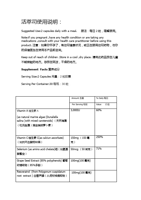
活萃司使用说明:Suggested Use:2 capsules daily with a meal. 服法:每日2粒,随餐服用。
Note:if you pregnant ,have any health condition or are taking any medications ,consult with your health care practitioner before using this product. 注意:如果你怀孕了,有任何健康状况,或正在服用任何药物,与你的保健医生在使用本产品前咨询。
Keep out of reach of children .Store in a cool ,dry place. 请将此药品放在儿童不能接触的地方。
存放在阴凉,干燥的地方。
Supplement Facts营养成分Serving Size:2 Capsules用量:2粒胶囊Serving Per Container:30每甁:30粒Other lngredients:Vegetable capsule,microcrystallinecellulose,magnesium stearate,silica. 其他成分:植物胶囊,微晶纤维素,硬脂酸镁,二氧化硅。
Made in the USA Exclusively for. 美国制造的专供。
网址:Interush Media Inc.,Irvine,Califomia,USA92612迅联网媒体公司,尔湾,加利福尼亚,USA92612Visit us online at:请访问我们的网站:身心能量饮使用说明:For best results:Drink on an empty stomach 2 hours after meals,or 15 minutes before eating 最好的效果是:在饭后2小时或是吃饭前15分钟空腹喝。
12种维生素注射剂说明书

12种维生素注射剂说明书在当今的医学界里,维生素注射剂是常用的一种治疗手段。
维生素注射剂不但可以补充身体的营养,还可以提高免疫力,缓解疼痛等一系列作用。
下面,我们就来看一下12种维生素注射剂的说明书。
1. 维生素A注射剂维生素A注射剂含有维生素A醋酸酯(Retinyl Acetate),它能有效地保护眼睛和皮肤,并对人体的免疫系统、角质化和造血系统有很大帮助。
一般来说,每次注射剂量为3,000-10,000国际单位(IU)。
2. 维生素B1注射剂维生素B1注射剂含有硝酸硫胺素(Thiamine Nitrate),它能有效地帮助身体代谢碳水化合物和脂肪,并预防恶性贫血。
一般来说,每次注射剂量为50-100毫克(mg)。
3. 维生素B2注射剂维生素B2注射剂的主要成分是核黄素(Riboflavin),它可以帮助身体吸收蛋白质和铁,并促进身体的生长和维持正常的视觉功能。
一般来说,每次注射剂量为3-10毫克(mg)。
4. 维生素B3注射剂维生素B3注射剂含有烟酸(Niacin),它可以促进新陈代谢,提高能量和促进心血管健康。
一般来说,每次注射剂量为50-100毫克(mg)。
5. 维生素B5注射剂维生素B5注射剂的主要成分是泛酸(Pantothenic Acid),它可以帮助身体代谢蛋白质、脂肪和碳水化合物,同时还可以增加身体能量和缓解贫血。
一般来说,每次注射剂量为50-100毫克(mg)。
6. 维生素B6注射剂维生素B6注射剂含有吡哆醇(Pyridoxine),它可以帮助身体代谢蛋白质、脂肪和碳水化合物,同时还可以增强免疫系统和促进伤口愈合。
一般来说,每次注射剂量为50-100毫克(mg)。
7. 维生素B12注射剂维生素B12注射剂含有氰钴胺(Cyanocobalamin),它可以提高红细胞生成、预防贫血、增加身体能量,同时还有助于神经系统的正常发育。
一般来说,每次注射剂量为1,000-2,000微克(μg)。
比卡鲁胺联合紫杉醇对雄激素受体阳性三阴性乳腺癌MDA-MB-231细胞的增殖抑制作用

㊃论著㊃比卡鲁胺联合紫杉醇对雄激素受体阳性三阴性乳腺癌MDA⁃MB⁃231细胞的增殖抑制作用丁钥 许焱 丁丽 朱小泉 张永强DOI:10.3877/cma.j.issn.1674-0807.2018.03.002作者单位:100730北京医院国家老年医学中心肿瘤内科通信作者:张永强,Email:zhyq95@ 【摘要】 目的 探讨比卡鲁胺(BIC)联合化疗药物紫杉醇(PTX)对雄激素受体(AR)阳性的三阴性乳腺癌MDA⁃MB⁃231细胞的增殖抑制作用及可能的作用机制㊂方法 采用CCK⁃8试剂盒观察不同浓度的BIC (0.1㊁1.0㊁10.0μmol /L)和PTX(0.1㊁1.0㊁10.0㊁100.0㊁1000.0㊁10000.0nmol /L)以单药及不同联合给药方式处理后,对MDA⁃MB⁃231细胞增殖的抑制作用㊂细胞增殖抑制率比较采用单因素方差分析㊂组间两两比较采用LSD 法㊂选取10nmol /L PTX 及10nmol /L DMSO 分别处理MDA⁃MB⁃231细胞样品(各3个)72h,采用生物信息学方法分析样品的相关基因表达芯片数据,采用校正t 检验筛选出差异基因㊂结果 使用不同浓度的BIC 分别处理MDA⁃MB⁃231细胞24㊁48㊁72h 后,各组MDA⁃MB⁃231细胞增殖抑制率在不同时间点差异均有统计学意义(F =4.124㊁8.189㊁4.139,P =0.037㊁0.004㊁0.032)㊂BIC 10.0μmol /L 组MDA⁃MB⁃231细胞增殖抑制率在48h 最高,为(12.9±5.5)%㊂不同浓度的PTX 分别处理MDA⁃MB⁃231细胞24㊁48㊁72h 后,不同浓度组MDA⁃MB⁃231细胞增殖抑制率在不同时间点差异均有统计学意义(F =8.407㊁47.432㊁14.907,P 均<0.001)㊂PTX 在48h 时对MDA⁃MB⁃231细胞的半数抑制浓度(IC 50)为5380.0nmol /L㊂5000.0nmol /L PTX 单药或联合不同浓度(0.1㊁1.0㊁10.0μmol /L)的BIC 同时处理MDA⁃MB⁃231细胞48h 后,5000.0nmol /L PTX 单药处理组与3个实验组中细胞增殖抑制率分别为(53.2±2.7)%㊁(53.2±3.1)%㊁(51.7±3.4)%㊁(51.0±2.3)%,组间差异无统计学意义(F =0.831,P =0.492)㊂采用5000.0nmol /L PTX 和10.0μmol /L BIC 以不同的序贯方式联合给药处理MDA⁃MB⁃231细胞(PTX 24h +BIC 24h 组㊁BIC 24h +PTX 24h 组㊁PTX 48h +BIC 24h 组㊁BIC 48h+PTX 24h 组),并用5000.0nmol /L PTX(PTX 48h 组)和10.0μmol /L BIC(BIC 48h 组)单药处理及同时联合给药(PTX 48h+BIC 48h 组)分别处理MDA⁃MB⁃231细胞后,各组间细胞增殖抑制率差异有统计学意义(F =241.466,P <0.001)㊂其中,两两比较结果显示,PTX 24h +BIC 24h 组细胞增殖抑制率为(72.9±1.9)%,高于BIC 24h +PTX 24h 组的(42.9±1.7)%(P <0.001),PTX 48h 组的(60.9±3.7)%(P <0.001)和PTX 48h +BIC 48h 组的(60.3±4.1)%(P <0.001)㊂PTX 处理组中有EGR1㊁FST㊁FOS㊁IL8㊁IL6㊁RPL27A 及CA27个基因的表达量与DMSO 处理组比较,差异均有统计学意义(t =18.647㊁10.336㊁10.098㊁9.683㊁9.408㊁9.050㊁8.001,P 均<0.050)㊂结论 通过先PTX 再BIC 的序贯联合给药方式较单药及其他联合给药方式能够更有效抑制AR 阳性三阴性乳腺癌细胞MDA⁃MB⁃231的增殖,两者间可能存在协同作用㊂【关键词】 乳腺肿瘤; 雄激素受体拮抗剂; 化学疗法; 联合药物疗法; 早期生长反应蛋白质1【中图法分类号】 R737.9 【文献标志码】 A Inhibitory effect of bicalutamide and paclitaxel on proliferation of androgen receptor⁃positive triple negative breast cancer MDA⁃MB⁃231cells Ding Yue ,Xu Yan ,Ding Li ,Zhu Xiaoquan ,Zhang Yongqiang.Department of Medical Oncology ,Beijing Hospital /National Center of Gerontology ,Beijing 100730,China Corresponding author :Zhang Yongqiang ,Email :zhyq95@【Abstract 】 Objective To investigate the inhibitory effect of bicalutamide (BIC)combined withpaclitaxel(PTX)on the proliferation of androgen receptor(AR)⁃positive triple negative breast cancer MDA⁃MB⁃231cells and its possible mechanism.Methods The CCK⁃8kit was used to determine the effect of BIC (0.1,1.0,10.0μmol/L)and PTX at different concentrations(0.1,1.0,10.0,100.0,1000.0, 10000.0nmol/L)in monotherapy or in sequential combination of both on the proliferation of MDA⁃MB⁃231 cells.Inhibition rate was compared using one⁃way analysis of variance.The pairwise comparison was performed using the LSD method.MDA⁃MB⁃231cells were treated with10nmol/L PTX and10nmol/L DMSO respectively for72h.Three cell samples were taken in each group to analyze the relevant gene expression profiling in array using a bioinformatic method.The adjusted t test was used to screen out differential genes. Results After MDA⁃MB⁃231cells were treated with different concentrations of BIC for24,48and72h, respectively,the inhibition rates of MDA⁃MB⁃231cells were statistically different at different time points(F= 4.124,8.189,4.139,P=0.037,0.004,0.032).The inhibition rate of MDA⁃MB⁃231cells reached the highest[(12.9±5.5)%]at48h after the treatment of10.0μmol/L BIC.The inhibition rates of MDA⁃MB⁃231cells were significantly different at different time points(F=8.407,47.432,14.907,P<0.001)after the treatment of PTX at different concentrations.The half inhibitory concentration(IC50)of PTX in MDA⁃MB⁃231 cells at48h was5380.0nmol/L.After48h treatment of5000.0nmol/L PTX alone or combined with0.1, 1.0,10.0μmol/L BIC,the inhibition rate of MDA⁃MB⁃231cells was(53.2±2.7)%,(53.2±3.1)%, (51.7±3.4)%,(51.0±2.3)%in PTX monotherapy group and three experimental groups,respectively, indicating no significant difference(F=0.831,P=0.492).MDA⁃MB⁃231cells were treated with sequential combination of5000.0nmol/L PTX and10.0μmol/L BIC(PTX24h+BIC24h group,BIC24h+PTX24h group,PTX48h+BIC24h group,BIC48h+PTX24h group),the monotherapy with5000.0nmol/L PTX (PTX48h group)or10.0μmo/L BIC(BIC48h group)and the synchronous combined therapy of PTX and BIC(PTX48h+BIC48h group),respectively.The result showed that there was a statistically significant difference in inhibition rate(F=241.466,P<0.001).The result of pairwise comparison showed that the inhibition rate in PTX24h+BIC24h group was(72.9±1.9)%,significantly higher than(42.9±1.7)% in BIC24h+PTX24h group(P<0.001),(60.9±3.7)%in PTX48h group(P<0.001)and(60.3±4.1)%in PTX48h+BIC48h group(P<0.001).There was a significant difference in the expression of seven genes(EGR1,FST,FOS,IL8,IL6,RPL27A and CA2)between PTX⁃treated group and DMSO⁃treated group(t=18.647,10.336,10.098,9.683,9.408,9.050,8.001,all P<0.050) Conclusions Sequential administration of PTX and BIC can inhibit the proliferation of AR⁃positive triple negative breast cancer MDA⁃MB⁃231cells more effectively compared with the monotherapy and other combination methods.The two drugs may have the synergistic effect.【Key words】 Breast neoplasms; Androgen receptor antagonists; Chemotherapy; Combination modality therapy; Early growth response protein1 乳腺癌是女性常见恶性肿瘤之一,近年发病率持续上升㊂其中,三阴性乳腺癌(triple negative breast cancer,TNBC)是一种特殊的乳腺癌分子亚型,占15%~20%,ER㊁PR及HER⁃2表达均为阴性,不适合内分泌治疗和抗HER⁃2靶向治疗,因此,化疗是主要的系统治疗手段㊂雄激素是人类性激素的一种,在乳腺癌的发生㊁发展中起着重要作用[1]㊂临床研究显示:雄激素受体(androgen receptor,AR)阳性的TNBC患者可通过影响AR信号通路而从抗雄激素治疗中获益,即AR很可能是AR阳性TNBC患者新的治疗靶点[2⁃3]㊂但是,目前鲜见关于抗雄激素治疗与化疗联合应用的相关报道㊂在本研究中,笔者选择已被证实为AR阳性的TNBC细胞系MDA⁃MB⁃231为实验对象[4⁃6],观察抗雄激素药物比卡鲁胺(bicalutamide,BIC)联合化疗药物紫杉醇(paclitaxel, PTX)对MDA⁃MB⁃231细胞增殖作用的影响,并采用生物信息学分析探讨PTX在TNBC治疗中可能的作用机制㊂材料与方法一㊁实验试剂与仪器PTX注射液购自华北制药股份有限公司(生产批号:FPW1607001);BIC购自北京百灵威科技有限公司(生产批号:LB40Q78);DMEM㊁胰蛋白酶㊁胎牛血清为美国Gibco公司产品;CCK⁃8试剂盒为日本同仁化学研究所产品;酶标仪购自瑞士Tecan公司㊂二㊁细胞培养MDA⁃MB⁃231细胞由北京协和医院细胞库提供,用CCK⁃8试剂盒检测MDA⁃MB⁃231细胞的增殖情况,取对数生长期的MDA⁃MB⁃231细胞,用含10%胎牛血清的DMEM培养液调节细胞数为3.5×10⁴/ml,每孔100μl,接种到96孔板㊂将96孔板置于37℃,5%CO2培养箱中过夜,待细胞贴壁稳定后,进行药物实验㊂三㊁实验方法1.单药处理细胞使用不同浓度的BIC(0.1㊁1.0㊁10.0μmol/L)和不同浓度的PTX(0.1㊁1.0㊁10.0㊁100.0㊁1000.0㊁10000.0nmol/L)分别干预MDA⁃MB⁃231细胞24㊁48和72h;空白对照组加入等量的DMEM培养基㊂2.同时联合给药处理细胞使用5000.0nmol/L PTX分别联合0.1㊁1.0㊁10.0μmol/L BIC同时处理MDA⁃MB⁃231细胞作为联合给药组,以5000.0nmol/L PTX单药处理作为对照组㊂根据以上实验结果确定后续实验中BIC与PTX的最佳药物作用浓度及作用时间㊂3.BIC与PTX以不同联合给药方式处理细胞联合给药组(实验组)分别采用同时给药(PTX 48h+BIC48h组,PTX与BIC同时作用于细胞48h)㊁序贯吸弃给药(PTX24h+BIC24h组,即先给予PTX作用24h后,吸弃,再给予BIC24h; BIC24h+PTX24h组,即先给予BIC作用24h 后,吸弃,再给予PTX24h)和序贯非吸弃给药(PTX48h+BIC24h组,即先给予PTX作用24h 后,不吸弃,再给予BIC24h;BIC48h+PTX24h 组,即先给予BIC作用24h后,不吸弃,再给予PTX 24h)方式;对照组采用PTX(PTX48h组)和BIC (BIC48h组)单独给药方式处理细胞㊂以上实验每组均设置6个复孔㊂药物处理后,吸弃上清液,每孔加入100μl CCK⁃8试剂(按照CCK⁃8体积/培养基体积=1/10比例稀释),震荡混匀后放入培养箱中继续培养2h,用酶标仪检测吸光度(D值),波长为450nm㊂重复3次试验后,通过同一时间实验组与空白对照组所测得的吸光度值计算各实验组的细胞增殖抑制率:细胞增殖抑制率(%)=(D空白对照组-D实验组)/D空白对照组×100%㊂四㊁差异基因筛选本研究所用到的芯片数据平台为Affymetrix公司的GPL15207,基因表达谱芯片数据来自由Lasham等[7]提交的芯片GSE60964系列㊂选取10nmol/L PTX处理72h的MDA⁃MB⁃231细胞3个样品(编号为GSM1494631㊁GSM1494632㊁GSM1494633)为实验组,溶剂(DMSO)处理72h的MDA⁃MB⁃231细胞3个样品(编号为GSM1494628㊁GSM1494629㊁GSM1494630)作为对照组,通过对此6个样品的基因表达谱芯片数据进行分析处理㊂采用Bioconductor的Impute包(version1.48.0)处理下载的基因芯片,K近邻分类算法补充芯片原始数据的缺省值,多个探针对应相同基因进行归一化处理,采用Bioconductor 的Limma包(v3.30.13)提取差异基因,R studio 1.0.136软件采用校正的t检验进行显著性分析㊂差异基因的筛选条件为P<0.050㊂五㊁统计学分析采用SPSS20.0统计软件对实验结果数据进行分析处理,结果用⎺x±s表示,细胞增殖抑制率比较采用单因素方差分析,组间两两比较采用LSD法㊂P< 0.050为差异具有统计学意义㊂结 果一㊁BIC和PTX单药对MDA⁃MB⁃231细胞增殖的影响使用0.1㊁1.0㊁10.0μmol/L BIC分别处理MDA⁃MB⁃231细胞24㊁48㊁72h后,结果显示,不同浓度组MDA⁃MB⁃231细胞增殖抑制率在不同时间点差异有统计学意义,具体统计数据见表1㊂10.0μmol/L 组在24㊁48㊁72h时细胞增殖抑制率均大于0.1μmol/L组(P均<0.050)㊂10.0μmol/L组在48h的细胞增殖抑制率达到最大值,为(12.9±5.5)%㊂因此,在后续联合给药实验中选择BIC浓度为10.0μmol/L,药物作用时间为48h㊂使用不同浓度的PTX分别处理MDA⁃MB⁃231细胞24㊁48㊁72h后,各浓度组MDA⁃MB⁃231细胞增殖抑制率在不同时间点差异均有统计学意义,具体统计数据见表2㊂根据48h时不同浓度的PTX处理后细胞增殖抑制率结果,PTX在48h时对MDA⁃MB⁃231细胞的半数抑制浓度(50%inhibitoryconcentra⁃tion,IC50)为5380.0nmol/L㊂为方便稀释药物浓度,本实验在随后的联合用药中采用PTX浓度为5000.0nmol/L㊂表1 不同浓度比卡鲁胺作用不同时间后各组MDA⁃MB⁃231细胞增殖抑制率比较(%,⎺x±s)组别实验次数作用时间24h48h72h0.1μmol/L组33.9±4.1 2.4±3.1-0.9±4.8 1.0μmol/L组33.9±1.8 8.0±4.5a1.8±5.5 10.0μmol/L组38.1±2.3ab12.9±5.5a7.5±5.4a F值4.1248.1894.391P值0.0370.0040.032 注:a与0.1μmol/L组比较,P<0.050;b与1.0μmol/L组比较,P< 0.050表2 不同浓度紫杉醇作用不同时间后各组MDA⁃MB⁃231细胞增殖抑制率比较(%,⎺x±s)组别实验次数作用时间24h48h72h0.1nmol/L组310.0±6.229.0±2.159.5±5.51.0nmol/L组317.5±6.923.7±5.754.5±4.4 10.0nmol/L组322.9±7.929.2±4.158.5±4.4 100.0nmol/L组328.0±6.940.0±2.864.4±4.2 1000.0nmol/L组330.5±4.342.4±1.666.0±3.6 10000.0nmol/L组328.6±7.248.0±2.072.9±1.8 F值8.40747.43214.907 P值<0.001<0.001<0.001二㊁5000.0nmol/L PTX与不同浓度BIC联合给药对MDA⁃MB⁃231细胞增殖的影响作用48h后,对照组(PTX单药组)与3个联合给药组(5000.0nmol/L PTX与0.1㊁1.0㊁10.0μmol/L BIC联合给药)的细胞增殖抑制率分别为(53.2±2.7)%㊁(53.2±3.1)%㊁(51.7±3.4)%和(51.0±2.3)%,组间差异无统计学意义(F=0.831,P= 0.492)㊂与单药PTX相比,PTX与BIC同时联合给药,不论BIC浓度高低,并不增强其对MDA⁃MB⁃231细胞的增殖抑制作用㊂三㊁5000.0nmol/L PTX与10.0μmol/L BIC以不同序贯方式联合给药对MDA⁃MB⁃231细胞增殖的影响各组细胞增殖抑制率比较,差异有统计学意义(F=241.466,P<0.001,表3)㊂其中,两两比较结果显示,PTX24h+BIC24h组细胞增殖抑制率为(72.9±1.9)%,高于BIC24h+PTX24h组的(42.9±1.7)%(P<0.001),PTX48h组的(60.9±3.7)%(P<0.001)和PTX48h+BIC48h组的(60.3±4.1)%(P<0.001)㊂对比2种序贯联合给药方式,在相同的用药顺序下,细胞增殖抑制率差异均无统计学意义[PTX24h+BIC24h组与PTX48h+ BIC24h组:(72.9±1.9)%比(75.8±1.8)%,P= 0.521;BIC24h+PTX24h组与BIC48h+PTX24h 组:(42.9±1.7)%比(47.0±3.1)%,P=0.290]㊂表3 比卡鲁胺与紫杉醇不同给药方式处理后MDA⁃MB⁃231细胞增殖抑制率比较(%,⎺x±s)组别实验次数增殖抑制率PTX48h组360.9±3.7 PTX48h+BIC48h组360.3±4.1 PTX24h+BIC24h组372.9±1.9 PTX48h+BIC24h组375.8±1.8 BIC24h+PTX24h组342.9±1.7 BIC48h+PTX24h组347.0±3.1 BIC48h组325.0±0.8F值241.466P值<0.001 注:PTX指紫杉醇;BIC指比卡鲁胺;PTX48h组与PTX48h+BIC48h 组㊁PTX24h+BIC24h组与PTX48h+BIC24h组㊁BIC24h+PTX24h 组与BIC48h+PTX24h组比较,P>0.050,其余两两比较,P均<0.001四㊁差异基因筛选结果实验组(PTX处理组)中有7个基因EGR1㊁FST㊁FOS㊁IL8㊁IL6㊁RPL27A及CA2的表达量与对照组(DMSO处理组)相比,差异均有统计学意义,具体统计数据见表4㊂图1为相关基因的表达量热图㊂注:蓝色代表DMSO处理组,红色代表紫杉醇处理组;表达量高的基因趋近红色,表达量低的基因趋近于绿色;DMSO指二甲基亚砜图1 7个差异基因在紫杉醇和DMSO处理后的MDA⁃MB⁃231细胞中的表达量热图讨 论乳腺癌作为女性最常见的恶性肿瘤,严重威胁着女性的身体健康㊂与非TNBC相比,TNBC因其临床过程更具侵袭性㊁更早发生远处转移㊁生存期更短表4摇紫杉醇和DMSO处理后MDA⁃MB⁃231细胞中差异基因表达比较(⎺x±s)组别样本数差异基因EGR1FST FOS IL8IL6RPL27A CA2DMSO处理组35.798±0.1038.267±0.0885.984±0.0734.703±0.2765.653±0.0315.060±0.0453.772±0.095紫杉醇处理组37.160±0.0319.027±0.0656.681±0.0505.910±0.0596.261±0.0535.687±0.0784.428±0.109 t值18.64710.33610.0989.6839.4089.0508.001P值<0.0010.0230.0230.0240.0240.0280.048 注:DMSO为二甲基亚砜且治疗手段少而成为乳腺癌治疗的难点与热点[8⁃11]㊂化疗是TNBC目前主要的系统治疗手段,但到目前为止最佳化疗药物和方案尚未确定,有证据支持采用含蒽环类和紫杉类方案[12]㊂PTX是TNBC化疗的首选药物,通过抑制有丝分裂达到杀伤肿瘤细胞的作用[12]㊂研究显示,AR在不同的乳腺癌亚型中表达比例不同,在luminal A型㊁luminal B型和HER⁃2过表达型乳腺癌中阳性率相对较高,而在基底细胞样乳腺癌中的表达率则相对较低,表明AR在乳腺癌中的表达与ER㊁PR及HER⁃2的表达有关,提示AR可作为乳腺癌潜在的治疗靶点[13⁃14]㊂一项临床2期研究表明,在ER阴性㊁PR 阴性㊁AR阳性且经多线治疗后的转移性乳腺癌患者中使用BIC,6个月的临床获益率为19%,中位无进展生存期为12周[15]㊂另一项临床2期研究采用新一代AR拮抗剂恩杂鲁胺分别作用于雄激素驱动基因阳性和雄激素驱动基因阴性的AR阳性TNBC患者,结果显示雄激素驱动基因阳性组的中位OS达到20个月,显著高于雄激素驱动基因阴性组(8个月)[16]㊂因此,AR拮抗剂有可能作为AR阳性TNBC患者的一种新治疗手段,但是,目前就AR拮抗剂与现有化疗手段如何结合尚无相关研究㊂因此,进一步研究抗雄激素治疗对AR阳性TNBC的有效性,并探讨抗雄激素药物与化疗药物如何更好地联合使用及其可能机制有着重要的现实意义㊂本研究采用CCK⁃8试剂盒检测了BIC与PTX 在不同给药剂量及不同给药方式下对MDA⁃MB⁃231细胞增殖的抑制作用㊂研究结果发现,BIC单药和PTX单药对MDA⁃MB⁃231细胞均有明显的抑制作用,且随着药物浓度的增加对细胞的增殖抑制作用增强,提示2种药物对AR阳性TNBC抗雄激素治疗的有效性和可行性㊂其中,PTX对细胞的增殖抑制作用强且持久,而BIC对细胞的抑制作用相对偏弱且时间较短,提示抗雄激素治疗需连续给药㊂BIC 和PTX联合给药时,不同给药方式对MDA⁃MB⁃231细胞的增殖抑制效果不同㊂当PTX和BIC同时联合给药时,BIC并不能提高PTX对MDA⁃MB⁃231细胞的抗肿瘤效果,这与临床上ER阳性乳腺癌抗雌激素治疗与化疗同时联合使用并不能提高疗效的结果一致㊂另外,先给PTX后给BIC的序贯给药方式明显优于同时给药方式,也明显优于BIC+PTX的序贯给药方式㊂在给药顺序相同时,联合序贯吸弃与不吸弃2种给药方式对细胞的增殖抑制作用差异无统计意义,但相较于先PTX后BIC的序贯不吸弃给药方式,序贯吸弃给药方式缩短了化疗药物的作用时间,减轻了化疗药物的不良反应㊂2017年ASCO会议报道,对ER阳性或PR阳性㊁HER⁃2阴性转移性乳腺癌一线化疗后疾病得到控制的患者,接受ER下调剂氟维司群维持治疗可使患者的无进展生存期达到19.5个月[17]㊂这种在ER阳性或PR阳性的患者中使用先化疗后用内分泌治疗的用药方式,与本实验在AR阳性的TNBC细胞中先用PTX化疗杀伤增殖活跃的肿瘤细胞,后用BIC内分泌治疗取得明显疗效的联合给药方式具有一定相似性㊂因此,先给予PTX化疗,在疾病控制情况下选择AR拮抗剂维持治疗可作为AR阳性TNBC的治疗策略之一㊂笔者比较了PTX与DMSO处理后MDA⁃MB⁃231细胞的基因表达谱芯片数据,发现了7个差异基因㊂其中,EGR1是属于多基因家族的锌指转录因子,能与下游多种基因的启动子区域相结合,调节相应基因的转录,从而参与细胞的生长㊁增殖㊁分化及凋亡等生物学活动[18⁃19]㊂研究发现,在前列腺癌细胞中,EGR1过表达能够引起AR受体相关通路的激活,增强AR从细胞质向细胞核中的转运[20];同时,在PTX耐药的乳腺癌MCF⁃7细胞中,EGR1能够通过增加MDR1基因的转录而导致药物抵抗[21]㊂由此可知,EGR1的表达与PTX的治疗㊁耐药及AR的表达和通路激活关系密切㊂笔者推测,PTX序贯BIC联合用药时的强协同效应可能与EGR1表达上调有关㊂在以AR为靶点的TNBC抗雄激素治疗以及联合用药中,EGR1可能发挥着重要的桥梁作用,但具体的作用机制尚需进一步研究㊂参 考 文 献[1] Cochrane DR,Bernales S,Jacobsen BM,et al.Role of the androgenreceptor in breast cancer and preclinical analysis of enzalutamide[J].Breast Cancer Res,2014,16(1):R7.[2] Gucalp A,Tolaney S,Isakoff SJ,et al.Phase II trial of bicalutamidein patients with androgen receptor⁃positive,estrogen receptor⁃negative metastatic breast cancer[J].Clin Cancer Res,2013,19(19):5505⁃5512.[3] Traina TA,Miller K,Yardley DA,et al.Enzalutamide for the treatmentof androgen receptor-expressing triple-negative breast cancer[J].J Clin Oncol,2018,36(9):884-889.[4] Lehmann BD,Bauer JA,Chen X,et al.Identification of human triple⁃negative breast cancer subtypes and preclinical models for selection of targeted therapies[J].J Clin Invest,2011,121(7):2750⁃2767.[5] Neve RM,Chin K,Fridlyand J,et al.A collection of breast cancercell lines for the study of functionally distinct cancer subtypes[J].Cancer Cell,2006,10(6):515⁃527.[6] 赵晶,付丽.乳腺癌的分子分型[J/CD].中华乳腺病杂志(电子版),2009,3(2):35⁃40[7] Lasham A,Mehta SY,Fitzgerald SJ,et al.YB⁃1affects response topaclitaxel in TNBCs by modulation of EGR1[EB/OL].[2017⁃09⁃02].https:///geo/query/acc.cgi?acc= GSE60964.[8] Celis J,Gromova I,Gromov P,et al.Molecular pathology of breastapocrine carcinomas:A protein expression signature specific for benign apocrine metaplasia[J].FEBS Lett,2006,580(12):2935⁃2944.[9] Nguyen PL,Taghian AG,Katz MS,et al.Breast cancer subtypeapproximated by estrogen receptor,progesterone receptor,and HER⁃2 is associated with local and distant recurrence after breast⁃conserving therapy[J].J Clin Oncol,2008,26(14):2373⁃2378. [10]Haffty BG,Yang Q,Reiss M,et al.Locoregional relapse and distantmetastasis in conservatively managed triple negative early⁃stage breast cancer[J].J Clin Oncol,2006,24(36):5652⁃5657. [11]Dent R,Trudeau M,Pritchard KI,et al.Triple⁃negative breastcancer:clinical features and patterns of recurrence[J].Clin Cancer Res,2007,13(15):4429⁃4434.[12]Liedtke C,Mazouni C,Hess KR,et al.Response to neoadjuvanttherapy and long⁃term survival in patients with triple⁃negative breast cancer[J].J Clin Oncol,2008,26(8):1275⁃1281. [13]Choi JE,Kang SH,Lee SJ,et al.Androgen receptor expressionpredicts decreased survival in early stage triple⁃negative breast cancer [J].Ann Surg Oncol,2015,22(1):82⁃89.[14]Isola JJ.Immunohistochemical demonstration of androgen receptor inbreast cancer and its relationship to other prognostic factors[J].J Pathol,1993,170(1):31⁃35.[15]Seitz S,Buchholz S,Schally AV,et al.Triple negative breast cancersexpress receptors for LHRH and are potential therapeutic targets for cytotoxic LHRH⁃analogs,AEZS108and AEZS125[J].BMC Cancer, 2014,14(1):847.[16]Traina TA,Yardley DA,Schwartzberg LS,et al.Overall survival(OS)in patients(Pts)with diagnostic positive(Dx+)breast cancer: Subgroup analysis from a phase2study of enzalutamide(ENZA),an androgen receptor(AR)inhibitor,in AR+triple⁃negative breast cancer(TNBC)treated with0-1prior lines of therapy[EB/OL].[2017⁃10⁃20]./doi/abs/10.1200/JCO.2017.35.15_suppl.1089.[17]Wang S,Xu F,Ouyang Q,et al.Fulvestrant as maintenance therapyafter first-line chemotherapy in postmenopausal hormone receptor-positive,HER2-negative advanced breast cancer patients(FANCY):A prospective,multicenter,single arm phase II study[EB/OL].[2017⁃10⁃20]./doi/abs/10.1200/JCO.2016.34.15_suppl.e12012.[18]Lee KH,Kim JR.Hepatocyte growth factor induced up⁃regulations ofVEGF through Egr⁃1in hepatocellular carcinoma cells[J].Clin Exp Metastasis,2009,26(7):685⁃692.[19]Abdulkadir SA,Qu Z,Garabedian E,et al.Impaired prostatetumorigenesis in Egr1⁃deficient mice[J].Nat Med,2001,7(1): 101⁃107.[20]Yang SZ,Abdulkadir SA.Early growth response gene1modulatesandrogen receptor signaling in prostate carcinoma cells[J].J Biol Chem,2003,278(41):39906⁃39911.[21]Tao W,Shi JF,Zhang Q,et al.Egr⁃1enhances drug resistance ofbreast cancer by modulating MDR1expression in a GGPPS⁃independent manner[J].Biomed Pharmacother,2013,67(3):197⁃202.(收稿日期:2017⁃10⁃31)(本文编辑:刘军兰)丁钥,许焱,丁丽,朱小泉,等.比卡鲁胺联合紫杉醇对雄激素受体阳性三阴性乳腺癌MDA⁃MB⁃231细胞的增殖抑制作用[J/CD].中华乳腺病杂志(电子版),2018,12(3):135⁃140.。
维生素和无机盐

VitD2原:麦角固醇
VitD3原:7-脱氢胆固醇
麦角固醇 → VitD2
胆固醇 → 7-脱氢胆固醇 → VitD3(紫外照射)
VitD3在血液中运输:与维生素D结合蛋白(vitamin D binding protein, DBP)相结合而运输。
(一)维生素B1形成辅酶焦磷酸硫胺素
N
CH2 N+
CH3
OO
H3C
N
NH2HC
S
CH2 CH2 O P O P OH
OH OH
硫胺素(thiamine)
焦磷酸硫胺素(thiamine pyrophosphate, TPP)
第29页,此课件共86页哦
(二)维生素B1在糖代谢中具有重要作用
➢ TPP是α-酮酸氧化脱羧酶的辅酶,也是转酮酶 的辅酶
引起,主要发生在婴儿,特别是早产儿。新生儿 缺维生素E可引起贫血。
临床上常用维生素E治疗先兆流产及习惯性流 产。
第23页,此课件共86页哦
四、维生素K
(一)维生素K是2-甲基1,4-萘醌的衍生物
天然形式:
K1-----植物甲萘醌或叶绿醌
K2-----肠道细菌的产物
人工合成:K3
O CH 3
H
O
3
维生素K1 (植物甲萘醌)
第12页,此课件共86页哦
(三)维生素A缺乏或过量摄入均引起疾病
缺乏症:
1.夜盲症 2.干眼病
3.对感染性疾病的敏感性增加
过多则可引起中毒症状:头痛、恶心、 共济失调;肝细胞损伤和高脂血症;长骨增 厚、高钙血症、软组织钙化以及皮肤干燥、 脱屑和脱发等。
第13页,此课件共86页哦
生长抑素类似物奥曲肽对人胃癌p53和Ras蛋白表达的影响

生长抑素类似物奥曲肽对人胃癌p53和Ras蛋白表达的影响田蕾;李舒【摘要】Objective To investigate the inhibitory effect of somatostatin (SST) analogue octreotide on human gastric cancer. Methods Fifty gastric cancer patients were randomly assigned into 2 equal groups. The patients in the control group received no medication before surgical resection of gastric cancer, and those in octreotide group were given daily subcutaneous injection of 100 μg octreotide for 7 days before the surgery. The resected specimens were examined histologically and the expressions of p53 and Ras protein were detected by immunohistochemistry. Results Compared with the control group, gastric cancer tissue in octreotide group showed significantly increased necrosis (P< 0.05) and enhanced proliferation of fibrous tissues (P<0.05) with lowered expressions of p53 and Ras protein (P<0.05). Conclusion Octreotide can inhibit the expressions of p53 and Ras and suppress the growth of the human gastric cancer.%目的探讨生长抑素类似物奥曲肽对人胃癌生长的抑制作用.方法 50例胃癌患者随机分为对照组25例,术前不用药物;奥曲肽组25例,术前奥曲肽100μg皮下注射,1次/d,共7d.术中切除标本送组织学研究.免疫组化法检测胃癌组织p53和Ras蛋白表达情况.结果与对照组比较,奥曲肽组胃癌组织坏死差异有统计学意义(P<0.05),纤维组织增生明显(P<0.05),p53和Ras蛋白表达有统计学意义(P<0.05).结论奥曲肽通过抑制人体内胃癌p53和Ras蛋白的表达,从而发挥抑制胃癌细胞生长的作用.【期刊名称】《南方医科大学学报》【年(卷),期】2011(031)007【总页数】4页(P1245-1248)【关键词】胃癌;奥曲肽;p53;Ras【作者】田蕾;李舒【作者单位】辽宁医学院附属第一医院消化内科,辽宁,锦州,121001;辽宁医学院附属第一医院消化内科,辽宁,锦州,121001【正文语种】中文【中图分类】R735.2胃癌是最常见的恶性肿瘤之一,在我国,胃癌的死亡率占所有恶性肿瘤死亡的23.02%,居各类癌死亡的第一位。
幽门螺杆菌感染

幽门螺杆菌感染本刊的这一栏目首先提供一份强调某一常见临床问题的病例摘要,然后提出支持各种策略的证据。
如果有正式指南,作者接下来会对其进行综述。
文章以作者的临床建议结尾。
一名来自东欧的32岁女性移民因持续性上腹痛和腹胀接受检查。
之前的检查显示,全血细胞计数和全面的代谢检查结果正常,乳糜泻血清学检查结果呈阴性。
幽门螺旋杆菌IgG血清检测结果呈阳性。
患者接受了为期10日的每日2次,每次20 mg奥美拉唑、1 g阿莫西林和500 mg克拉霉素治疗,但症状持续存在。
您将如何进一步检查和治疗该患者?临床问题幽门螺旋杆菌感染是全球范围内均可见到的一种常见且通常持续终生的感染1。
研究提示,不同地区的感染率不同,但由于人口增长,以及再次感染和因根除不成功导致的复发,感染者数量在过去30年中保持稳定,甚至有所增加2。
较差的社会经济状况是幽门螺杆菌感染的危险因素2,因为较差的社会经济状况与较拥挤的居住条件相关,而拥挤的居住条件有利于幽门螺杆菌的家庭内传播3。
此外还有内镜检查导致的医源性感染4。
虽然大多数感染者无症状,但我们已经发现幽门螺杆菌感染与数种疾病直接相关,尤其是消化性溃疡病和非溃疡性消化不良。
已经有证据(在下文中综述)表明,通过治疗根除幽门螺杆菌可降低这两种疾病的风险5-7,但其中有关非溃疡性消化不良的数据不太统一。
胃癌也与感染幽门螺杆菌密切相关。
日本的一项研究表明,在感染幽门螺杆菌的消化性溃疡、消化不良或胃不典型增生患者中,有2.9%发生了胃癌(7.8年的平均随访期间),而在未感染幽门螺杆菌的上述疾病患者中未发现胃癌8。
根据强有力的证据,世界卫生组织(WHO)已将幽门螺杆菌归类为导致胃腺癌的1类致癌物9,10。
除日本之外,因幽门螺杆菌感染导致胃癌发病率增加的区域包括中东、东南亚、地中海地区、东欧、中美洲和南美洲。
在世界上幽门螺杆菌感染率高的地区(例如东欧和东亚)长大,如今居住在美国或西欧的移民患胃癌的风险也增加。
静脉营养液

靜脈營養液,添加物的相容性和安定性財團法人仁愛綜合醫院黃柔輔林俊杰藥師前言:靜脈營養液的添加物(包括電解質、微量元素和維生素)可能產生不相容性的沉澱和各組成間的化學反應。
在電解質方面最常見的是鈣、磷沉澱的問題(1),而會影響鈣、磷沉澱的因素有很多,如:pH值、溫度、氨基酸的來源、鈣和磷所使用的鹽類…等等。
在維生素方面最常見的是維生素C的氧化(2)、維生素B1、B2的不安定(3),必須適當的保存,以防止失效問題。
而微量元素方面也曾報告過有沉澱的問題,如iron phosphate salts(2,4),還有copper會和cysteine產生copper cysteinate的沉澱(2,5)。
因此,調配或保存TPN溶液時,需避免這些反應的產生,發揮PN溶液的最高效應。
以下會就電解質、微量元素、維生素三方面來討論,考慮它們加入PN溶液的安定性和相容性。
A)電解質方面:所有PN給予必須包括一些必須的電解質,給予時會考慮病人的狀況、年齡、腎功能和其他疾病等因素,但PN溶液可添加多少電解質,則必須小心。
1994年,美國FDA曾經報導由於磷酸鈣之沉澱,造成2名病患死亡和至少2名以上的急性呼吸窘迫症的案例(6),因此在添加電解質時,必須考慮會不會造成沉澱,一價的離子並不會造成沉澱,而會發生沉澱的是二價或三價的離子,尤其是鈣和磷。
而沉澱的發生不是在添加的過程,就是發生在調配完成後(和時間放置多久有關);因此必須小心添加的順序和調配後的保存。
以下為影響鈣、磷沉澱的因素:1. 溶液的pH值在溶液中,磷酸鹽是以三價的磷酸根(PO43-)、二價的磷酸氫根(HPO42-)和一價的磷酸二氫根(H2PO4-)存在(如圖一),這三個離子會維持一個平衡的狀態,但是PO43-要在強鹼(pH值很大時)的情況下才會存在,所以在PN溶液中只可能以HPO42-及H2PO4-存在;而H2PO4-和鈣離子相對的溶解度較大,溶解度為1800mg/dL;但HPO42-和鈣離子卻非常不可溶,溶解度只有30mg/dL,因此HPO42-較會和鈣離子產生沉澱。
脂溶性维生素缺乏诊断详述

脂溶性维生素缺乏诊断详述*导读:脂溶性维生素缺乏症状的临床表现和初步诊断?如何缓解和预防?维生素A1.维生素A是视觉细胞内感受弱光的物质-视紫红质的组成成分在视觉细胞内由 11-顺视黄醛(retinal)与不同的视蛋白(opsin)组成视色素。
在感受强光的锥状细胞内有视红质、视青质及视蓝质,杆状细胞内有感受弱光或暗光的视紫红质。
当视紫红质感光时,视色素中的11一顺视黄醛在发生的光异构作用下转变成全反视黄醛,并与视蛋白分离而失色。
全反式视黄醛可经异构酶作用缓慢地重新异化成为11一顺视黄醛,但大部分被还原成全反视黄醇,经血流至肝变成11一顺视黄醇,而后再随血流返回视网膜氧化成11-顺视黄醛,合成视色素。
在这个视循环中会造成部分全反视黄醛分解成无用的物质,所以要经常补充维生素A。
其他视色素的感光过程与视紫红质相同。
在维生素A缺乏时,必然引起11-顺视黄醛的补充不足,视紫红质合成减少,对弱光敏感性降低,日光适应能力减弱,严重时会发生"夜盲症"。
2.维生素A也是维持上皮组织的结构与功能所必需的物质当维生素A缺乏时,可引起上皮组织干燥、增生和角质化,产生干眼病、皮肤干燥、毛发脱落等。
3.其它作用维生素A能促进粘多糖、糖蛋白及核酸的合成,因而能促进机体的生长。
维生素D具有生物活性的 1,25-(OH)2-D3的靶细胞是小肠粘膜、肾及肾小管。
主要的作用是促进钙及磷的吸收,有利于骨的生成、钙化。
当缺乏维生素D时,儿童可发生佝偻病,成人引起软骨病。
维生素E1.维生素E是体内最重要的抗氧化剂,能避免脂质过氧化物的产生,保护生物膜的结构与功能。
机体内的自由基具有强氧化性,如超氧阴离子自由基(O2-)、过氧化物自由基(ROO·)及羟基自由基(OH·)等。
维生素E的作用在于捕捉自由基形成生育酚自由基,生育酚自由基又可进一步与另一自由基反应生成非自由基产物--生育醌。
硒作为谷脱甘肽过氧化酶的必需因子,通常认为是对抗过氧化作用的第二道防线。
烟酸对杂交鲟幼鱼生长性能、肌肉品质和抗氧化功能的影响

中国水产科学 2020年4月, 27(4): 406-413 Journal of Fishery Sciences of China研究论文收稿日期: 2019-08-27; 修订日期: 2019-09-18.基金项目: 现代农业产业技术体系专项资金(CARS-46); 国家自然科学基金面上项目(31772854); 国家自然科学基金青年科学基金项目(31602178).作者简介: 吴金平(1989–), 男, 硕士, 助理研究员, 从事水产动物营养与饲料研究. E-mail: wujinping@ 通信作者: 陈细华, 研究员. E-mail: chenxh@ DOI: 10.3724/SP.J.1118.2020.19234烟酸对杂交鲟幼鱼生长性能、肌肉品质和抗氧化功能的影响吴金平, 杜浩, 陈细华, 褚志鹏, 阮瑞, 金佳利, 乔新美, 刘伟中国水产科学研究院长江水产研究所, 农业农村部淡水生物多样性保护重点实验室, 湖北 武汉430223摘要: 为评价饲料中添加不同水平的烟酸对杂交鲟(Acipenser schrenckii ♂×A. baeri ♀)幼鱼生长性能、肌肉成分、肌肉质构和抗氧化性能的影响, 在基础饲料中分别添加0 mg/kg 、30 mg/kg 、120 mg/kg 、1200 mg/kg 的烟酸配制成4种实验饲料, 投喂初始体重(54.41±0.79) g 的杂交鲟幼鱼84 d, 实验分为4组, 每组3个重复, 每个重复15尾鱼。
结果表明: 饲料添加不同水平的烟酸对杂鲟幼鱼增重率有显著影响(P<0.05), 0 mg/kg 组增重率显著低于30 mg/kg 、120 mg/kg 、1200 mg/kg 烟酸组(P<0.05), 各添加组之间无显著差异(P>0.05)。
各组成活率、饲料系数和肝体比不受饲料烟酸的影响(P>0.05)。
- 1、下载文档前请自行甄别文档内容的完整性,平台不提供额外的编辑、内容补充、找答案等附加服务。
- 2、"仅部分预览"的文档,不可在线预览部分如存在完整性等问题,可反馈申请退款(可完整预览的文档不适用该条件!)。
- 3、如文档侵犯您的权益,请联系客服反馈,我们会尽快为您处理(人工客服工作时间:9:00-18:30)。
Ann.Occup.Hyg.,V ol.51,No.6,pp.563–569,2007ÓThe Author2007.Published by Oxford University Presson behalf of the British Occupational Hygiene Societydoi:10.1093/annhyg/mem036 Effect of Ascorbic Acid and ThiamineSupplementation at Different Concentrations onLead Toxicity in LiverCHUNHONG WANG1*,JIANCHENG LIANG1,CHUNLIAN ZHANG1, YONGYI BI1,XIANGLIN SHI2and QUN SHI11Department of Toxicology,School of Public Health,Wuhan University,DongHu Road115,Wuhan 430071,People’s Republic of China;2Institute for Nutritional Sciences,SIBS,CAS,Shanghai20032, People’s Republic of ChinaReceived16May2007;infinal form11June2007Objective:To investigate the effect of ascorbic acid[vitamin C(VC)]on liver damage param-eters in the lead-exposed mice,when given in combination with thiamine[vitamin B1(VB1)]atdifferent concentrations.Methods:Sixty-six male mice were randomly assigned into11groups(n56).Mice in GroupI were supplied with only the tap water as the drinking water;mice in Group II were providedwith a tap water containing0.2%lead acetate;mice in Group III–XI were given different doseof VC(140,420,1260mg kg21bw)and VB1(10,30,90mg kg21bw)according to333factorialdesign by oral gavages,along with ingestion of0.2%lead acetate.After42test days,DNA dam-age of liver cells was assessed using single-cell gel electrophoresis.The apoptosis rate of livercells was determined byflow cytometry.The activities of superoxide dismutase(SOD)and glu-tathione peroxidase(GSH-Px)in blood and the level of reduced glutathione(GSH)in liver cellswere measured based on individual biochemical reactions.Results:Compared with the Group I,sub-chronic lead ingestion(Group II)resulted in a sig-nificant decrease of Hb,GSH-P X,SOD in blood and GSH level in liver cells;lead exposure in-duced also a significant increase in DNA damage and apoptosis of liver cells(P<0.05).Supplementation with VC and VB1,however,reversed these effects.The best effective combi-nation was VC(420mg kg21bw)and VB1(10mg kg21bw),followed by the combination of VC(420mg kg21bw)and VB1(30mg kg21bw).But no reversion was shown in the combination ofthe highest combination of VC(1260mg kg21)and VB1(90mg kg21).Conclusions:Thesefindings strongly indicated that combination of VC and VB1can lessenthe damage to liver cells from oxidative stress induce by lead,but the antioxidant effects aredependent on their concentrations.Keywords:ascorbic acid;DNA damage;lead;liver;thiamineINTRODUCTIONLead-induced oxidative stress in blood and other soft tissues has been postulated to be one of the possible mechanisms of lead-induced toxic effects(Pande et al.,2001).Disruption of pro-oxidant/antioxidant balance might lead to the tissue injury.It was re-ported that lead increased the level of lipid peroxida-tion(Upasani et al.,2001)and brain thiobarbituric acid-reactive substances and altered the antioxidant defense system(Adanaylo and Oteiza,1999).Similar effects were also reported in the hepatic tissues (Sandhir and Gill,1995).A number of recent studies confirmed the possible involvement of reactive oxy-gen species(ROS)in lead-induced toxicity(Gurer and Ercal,2000).Several antioxidant enzymes and molecules have been used to evaluate lead-induced oxidative damage in animal and human studies.Reduced glutathione (GSH)and glutathione disulfide(GSSG)concentra-tions,as well as modifications in superoxide dismu-tase(SOD)activity are the most frequently used markers in tissues or in blood.*Author to whom correspondence should be addressed.Tel/fax:þ86-27-68758648;e-mail:chwang027@563 by guest on March 7, 2011 Downloaded fromBased on the observation that free radical was gen-erated during the pathogenesis processes induced by lead exposure,it was presumed that supplementation of antioxidants could be an alternative method for chelation therapy (Flora et al.,2003).Specifically,ascorbic acid,the known chelating agent with anti-oxidant features,was widely reported with the capability of protecting cells from oxidative stress (Ramanathan et al.,2002;Patra and Swarup,2004).Besides,additional protective effect of vitamin C (VC)on cell apoptosis was reveled in a recent study (Gruss-Fischer and Fabian,2002).Thiamine,the endogenous –SH containing mole-cule,was recognized as one of the protective agent for lead exposure (Ghazaly,1991).Additional inves-tigations have confirmed that the simultaneous administration of vitamin B 1(VB 1)and VC is more efficient in protecting or treating the experimental lead intoxication than either of them individually (Flora and Tandon,1986;Dhawan et al.,1988).Although the investigations about prevention of lead toxicity utilizing VC and VB 1are fairly well de-fined and previously been considered,reports on the impacts of simultaneous administration with VC and VB 1at different concentrations on lead exposure are novel.Moreover,some authors have raised questions pertaining to the potential risk for side effects of with intake of large doses of vitamins such as VC or tocopherol (Aiguo,2001).In fact,some people in China are intaking vitamin supplement largely.These also deserve deeper investigations.It was pre-sumed that these vitamins might act as pro-oxidant for the introduction of apoptotic cells in the presence of oxygen and the generation of H 2O 2.This study was therefore conducted to investigate whether the combinations of VC and VB 1at various concentra-tions are able to protect the target organs (liver)from lead-associated oxidative stress.MATERIALS AND METHODSAnimals and experimental designAnimal used were Kunming albino male mice (n 566)at the initial age of 5–6weeks,weighting 15–4g.Animals were purchased from the Labora-tory Animal Center of Wuhan University,China.An-imals were accommodated in polypropylene cages in the laboratory animal house,with a 12-h dark/light cycles,at the temperature of 20–25°C and relative humidity of 30–70%.Mice were provided with the standard rodent feeds procured from the Laboratory Animal Center of Wuhan University.Water was sup-plied ad lib.After 7days acclimatization period,mice were randomly assigned into 11groups (n 56).Mice in Group I served as negative control and received tap water alone;mice in Group II served as positive con-trol and received 0.2%lead acetate in drinking water;mice in Group III–XI were given different doses of VC (140,420,1260mg kg À1bw)and VB 1(10,30,90mg kg À1bw)according to 3Â3factorial design by oral gavage daily,along with intaking of the 0.2%lead acetate as shown in Table 1.All experimental animals were under the described treatments for con-tinuous 42days.For each mice,the average water consumption was 5.6ml day À1,so total lead intake was about 470.4mg during experiment.At the end of treatment,blood samples were col-lected in heparinized vials and the liver was removed and kept in a container with the ice jacket for follow-ing analysis.Biochemical analysisActivity of SOD and GSH-Px in blood.The SOD activity in blood was determined using the method modified from the colorimetric NADH–phenazine-methosulphate–nitroblue tetrazolium formazan inhi-bition reaction assay and was expressed in U g À1of hemoglobin (Kakkar et al.,1984).The glutathione peroxidase (GSH-Px)activity in blood erythrocyte hemolysates was measured using the coupled method described by Paglia and Valen-tine (Paglia and Valentine,1967;Hatanaka et al.,2000),with the t -butylhydroperoxide as the sub-strate.The enzyme activity was expressed in interna-tional units per gram of hemoglobin.Assay of GSH content in liver.About 0.2g liver was trimmed in ice-cold saline (0.85%w/v NaCl)and gently homogenized in the ice-cold 1.17%KCl of the 0.10mol l À1phosphate buffer (pH 7.4)at a ra-tio of 10%(w/v),using a glass Potter-type homoge-nizer at 500–800rpm.The homogenates were filtered through a muslin cloth and were centrifuged at 20000g for 30min at 4°C to obtain the supernatants.The GSH was measured in deproteinized superna-tant fractions from liver homogenates,employing 0.04%5,59-dithiobes-(2-nitrobenzoicacid)in 10%Table 1.The dose of lead acetate and vitamin in different groups Group Leadacetate (%)VC(mg kg À1bw)VB 1(mg kg À1bw)I 000II 0.200III 0.214010IV 0.214030V 0.214090VI 0.242010VII 0.242030VIII 0.242090IX 0.2126010X 0.2126030XI0.2126090564 C.Wang et al.by guest on March 7, 2011 Downloaded fromsodium citrate and recording absorption at 412nm on a spectrophotometer (Meng and Bai,2004).Assessment of DNA damageThe single-cell gel electrophoresis assay (SCGE)or comet assay was used for the detection of sin-gle-strand DNA breaks and reparation in individual cells.The method was modified from that reported by Wang et al.,(2006)as follows.Preparation of liver cell suspensions.A liver specimen was cut into pieces with the addition of a phosphate-buffered saline (PBS)solution,followed by a cleaning through a filter.The cellular suspension was then centrifuged at 1000rpm for 10min;the supernatant was discarded and the cell pellets were resuspended in an EP tube.The concentration of sus-pension was adjusted to 106–107cells ml À1for each sample.SCGE assay.Slides were cleaned with acid wash and spread with 40l l of 0.6%agarose,then stood for $5min at room temperature for the gel to set.Onto the first gel layer,a mix of a 20-l l live cell sus-pension and a 80-l l 1.1%low-melting agarose was laid.The slide was then immersed in a cold lysis buffer (2.5mol l À1NaCl,100mmol l À1Na 2EDTA,10mmol l À1Tris,pH 10,1%Triton X-100and 10%dimethyl sulfoxide)for at least 1h at 4°C.At completion of those procedures,slides were arranged on the electrophoresis chamber and fresh prepared electrophoresis buffer (1mmol l À1Na 2EDTA and 300mmol l À1NaOH,pH 12)was added to the cham-ber.After rinsing the slides with PBS for $20min,electrophoresis was performed at 25V ,300mA for 30min.All procedures were performed in the ab-sence of strong light.At the completion of the elec-trophoresis,slides were neutralized by washing with a 0.4-mol l À1Tris–HCl buffer (pH 7.5)for three times,5min each.Slides were then covered with 40l l of 0.1mg ml À1propidium iodide (PI)for the DNA ing a fluorescence microscope (Olympus,Japan)configured with the wavelengths of excitation (510–560nm)and filtration (690nm),100cells from the randomly selected filed were counted and the photograph was taken in each slide.The images were scored for each sample using an image analysis software system (IMI 1.0).Parameters for assessing DNA single-strand breaks included the percentage of cells with tail and tail length by visual estimation.Apoptosis analysisHepatic cells apoptosis were determined with the appearance of the DNA fragments in the apoptotic cells using flow cytometry (FCM).The method used for the detection of the DNA condensation and frag-mentation was modified from the subdiploid DNA peak assay described by Iavicoli et al.,(2001).Ge-netic materials can be bound by the DNA bindingfluorochrome,such as PI and ethidium bromide.Vi-able cells resist the intake of the fluorochromes due to the integration of the cell membrane.Cells stained with these fluorescence indicate the occurrences of the late-stage apoptosis or the appearances of necro-sis.On the DNA histogram figures acquired from the FCM,the diploid DNA peak (G1cells)depressed in apoptotic cells and the subdiploid DNA peak (SD peak)appeared ahead of the G1peak.The ap-pearance of an SD peak represents cells apoptosis.Necrosis cells do not have SD peak,but it exhibits consecutive small DNA fragment curves.Subdiploid DNA peak assay:(i)Aliquot 1$5Â106cells in suspension and centrifuge at 500rpm for 5min.Discard the supernatant;(2)Fix cells in 70%alcohol (À20°C)for 2$24h until analysis;(3)Add PI to a final concentration of 20l g ml À1.Mix well,and let stand at 4°C for 30min in order to allow the dye to equilibrate;(4)Analyze with a flow cytometer with an argon–ion laser tuned to 488nm.Record the light emitted into the red fluores-cence photomultiplier (PMT)using a 620-nm long pass filter in front of the PMT and (5)Analyze the percentage of SD peak which represents the percent-age of apoptotic cells.Statistical analysisThe analyses were performed with the SPSS 11.5software package.The analysis of variance was ap-plied for the comparison of the means of the different treatment groups.Post hoc testing was performed for inter-group comparison applying the least significant difference.Values of P ,0.01and P ,0.05were considered to be significant.RESULTSBiochemistryChronic lead ingestion (Group II)resulted in a sig-nificant decrease in blood Hb,GSH-P X ,SOD and in liver GSH levels after 42days of treatment (Table 2).Supplementation with VC and VB 1,however,re-versed these effects.Moreover,the biochemistry pa-rameters investigated showed no significant differences in Group III,VI and VII with that of the negative control (Group I)(P .0.05).DNA damage of liver cellsAs shown in Table 3and Fig.1A,B,animals in the negative control (Group I)exhibited low but measur-able levels of DNA migration in liver cells assessed by the comet assay.The analysis of the data showed that lead intake increased percentage of cells with tail and tail length.Significant difference was observed in animals in positive control (Group II)after 6weeks of lead exposure in comparison with the neg-ative control (P ,0.01).Combined administrationEffect of ascorbic acid and thiamine supplementation 565by guest on March 7, 2011 Downloaded fromwith VC and VB 1resulted in a significant reduction in the tail length and in the rate of DNA damage,es-pecially in Group VI and VII,in comparison with that of Group II (P ,0.05).From Table 3,we observed the lower values of DNA damage at Group VI.The percentage of liver apoptotic cells was shown in Table 3and Fig.2.In the negative control group (Group I),only about 5%apoptotic cells were de-tected.After lead intake,the percentage of liver apo-ptotic cells (11.9%)in positive control (Group II)was significantly higher than that in Group I (P ,0.01).Except Group XI,the percentage of liver apo-ptotic cells was markedly decreased in all other groups administered with combinations of VC and VB 1,as compared with that of Group II (P ,0.01or P ,0.05).In consistent with the finding in bio-chemistry,the percentage of apoptotic cells in Group VI and VII was also lowest (5.8%,5.2%).The Olive tail length resulted from SCGE in Table 3showed similar reversing effect induced by the sup-plementation of vitamins.Factorial analysis showed that the best effective combination was 420mg kg À1bw VC and 10mg kg À1bw VB 1,the second best was 420mg kg À1bw VC and 30mg kg À1bw VB 1.However,the combination of the highest dose VC (1260mg kg À1)and VB 1(90mg kg À1)had no effect on lead toxicity of oxidative damage (Fig.3).DISCUSSIONLead poisoning has been among the most studied health problems over the years.One of the ways thatTable 2.Changes of blood Hb,GSH-P X ,SOD and liver GSH levels in mice Group Blood GSH in liver (l g/100mg,wet)Hb (g dl À1)GSH-Px (U g À1,Hb)SOD (U mg À1,Hb)I (control)15.47–1.06241.5–24.923.23–1.92233.4–29.4II (lead)11.42–1.09b 193.0–23.9a17.51–2.29b173.1–27.3b III 14.14–0.98d 224.5–25.0c 21.08–2.13d 210.8–21.3d IV 13.14–1.16b,d 216.4–24.619.26–1.98b 200.0–22.0V 12.27–0.85b 207.5–27.4a 18.88–2.06b 191.1–31.9b VI 14.98–1.08d 238.2–25.7d 22.76–2.50d 226.3–29.7d VII 14.20–0.91d 213.2–29.4c 21.96–2.69d 217.4–33.7c VIII 13.72–0.91b,d 220.6–22.420.24–2.31d 205.2–21.2IX 12.75–1.39b 211.3–25.519.54–2.43b 193.2–20.3b X 12.00–1.43b 203.0–22.0b 18.58–1.90b 183.4–27.7b XI11.71–1.38b197.0–24.6b17.88–1.72b181.7–23.3ba P ,0.05orbP ,0.01compared with control group (Group I);cP ,0.05or dP ,0.01compared with only lead-exposed group (Group II).Table 3.The results of comet assay and apoptosis analysis in liver cells of different groups Group Cells with tail (%)Tail length (l m)Olive tail length (l m)Apoptosis rate (%)I (control)15.715.1–8.7 5.0–3.6 4.9–0.9II (lead)67.2b53.4–14.5b18.3–6.1b11.9–1.0b III 28.6b,d 22.6–9.4a,d 8.6–4.3a,d 6.9–0.8a,d IV 38.2b,d 27.5–10.3b,d 10.5–4.9b,d 8.1–0.6b,d V 44.6b,d 34.1–11.5b,d 11.8–5.2b,d 9.7–0.8b,c VI 19.2d 19.0–8.8d 5.9–3.7d 5.2–0.6d VII 21.6a,d 20.8–9.1d 6.7–4.1d 5.8–0.8d VIII 36.4b,d 25.8–9.6b,d 9.4–4.6b,d 7.5–0.9a,d IX 41.0b,d 30.4–10.8b,d 10.8–4.8b,d 8.9–0.9b,c X 48.8b,d 36.6–11.7b,d 13.4–5.5b,d 10.4–0.8b,c XI56.0b,d41.6–13.3b,d15.1–5.8b,d11.1–0.8ba P ,0.05orbP ,0.01compared with control group (Group I);cP ,0.05or dP ,0.01compared with only lead-exposed group (Group II).566 C.Wang et al.by guest on March 7, 2011 Downloaded fromlead exerts its toxic effects is through oxidative stress that may be an important contributor to the negative pathogenesis observed after lead exposure.This study clearly showed that chronic lead inges-tion results in a significant decrease in blood Hb,GSH-P X ,SOD and in liver GSH levels.Under nor-mal conditions,cells possess enzymatic and non-enzymatic defenses to cope with free radicals,such as SOD,GSH-Px,catalase and GSH (Shalana et al.,2005).Oxidative damage,however,may occur when antioxidant potential is decreased and/or when oxidative stress is increased.Some investigators had indicated that lead exposure increased the production of ROS in the liver (Patra et al.,2001;Pande and Flora,2002).Patra reported that lead exposure is in-deed associated with significant increases in lipid peroxide level in the liver.Free radical-induced oxidative damage has been implicated in the pathogenesis of a number of injury and disease states.We have previously found that ROS played a pivotal role in apoptosis of testis cells in lead-exposed mice (Wang et al.,2006).In the pres-ent study,it was exhibited that a significant increase in DNA damage and apoptosis in liver cells occurred via ROS as it played a very important role in apopto-sis induction under both physiological and pathologi-cal conditions.Interestingly,mitochondria were both the source and target of ROS.ROS,whichwasFig.1.DNA damage of liver cells.(A)Liver cell of tail was observed obviously after mice treated lead for 6weeks (Â400).(B)Length of tail cell was decreased significantly at the Group VI (VC 420mg kg À1bw and VB 110mg kg À1bw (Â400).Fig.2.The percentage of liver apoptotic cells (FCM,stained by PI).(A)Control group;(B)only lead-exposed;(C)Group VI (VC 420mg kg À1bw and VB 110mg kg À1bw (Â400)and (D)Group VII (VC 420mg kg À1bw and VB 130mg kg À1bw (Â400).Effect of ascorbic acid and thiamine supplementation 567by guest on March 7, 2011 Downloaded frompredominantly produced in the mitochondria,led to the free radical attack of membrane phospholipids and loss of mitochondrial membrane potential,which caused the intermembrane proteins,such as cyto-chrome c,to be released out of the mitochondria and ultimately triggered caspase-3activation.Caspase-3activation led to DNA breakage,nuclear chromatin condensation and cell apoptosis (Li et al.,2006;Wang et al.,2006).After coadministrated with vitamins,the lead tox-icity was normalized effectively.The levels of GSH-P X ,SOD in blood and GSH in the liver had a significant increase when compared with lead-exposed group (P ,0.01).Namely,free radicals in-duced by lead may oxidize GSH and the vitamin treatment reversed lead-induced decreases in GSH levels.It is well known that ascorbic acid directly re-acts with lipid hydroperoxides and restores the anti-oxidative potency of a -tocopherol.Production of GSH is considered to be the first line of defense against oxidative damage and free radical generation where GSH functions as a scavenger and a cofactor in metabolic detoxification of ROS (Tandon et al.,2003).As discussed previously,ROS played a piv-otal role in apoptosis.So lead-exposed mice co-treated with ascorbic acid could directly reacts with lipid hydroperoxides or/and increase the levels of GSH and then prevented liver destruction from apoptosis.The exact mechanism of thiamine in antagonizing lead toxicity has not been clearly elucidated until now.It might be attributed to the formation of com-plexes between thiamine and lead followed by its excretion.Thiamine also has been found to protect against lead-induced lipid peroxidation in rat liver and kidney (Senapati et al.,2000).It may scavenge O 2 Àand OH directly and thus affect the cellular re-sponse to oxidative stress (Jung and Kim,2003).Anna and Wig ło (2006)reported that thiamine may act as a potent antioxidant as it scavenges free radi-cals such as 2,2-azinobis(3-ethylbenzthiazoline-6-sulfonic acid)radical cation relatively slowly.Its radical-scavenging activity,expressed as the trolox equivalent antioxidant capacity value,is comparable to such natural antioxidants as some carotenoids.The fact that simultaneous supplementation of ascor-bic acid and thiamine ameliorated the liver damage in lead-treated mice,indicating that prevention was at-tributed to apoptotic inhibition by increased levels of GSH-P X ,SOD and GSH.At the highest dose of ascorbic acid and thiamine,the results differed from the low and middle doses.When compared with the only lead exposure group,the combination of high dose VC (1260mg kg À1)and VB 1(90mg kg À1)had no effect on lead toxicity of oxidative damage.We also previously found that this combination caused more severe injury in testis than that in lead-treated group (Wang et al.,2006).Although ascorbic acid was a well-known antioxi-dant,it might possess pro-oxidant property under certain conditions.When the concentration of ascor-bic acid is higher than normal,ascorbic acid level still kept normal in plasma by way of self-metabolism indeed continued to act as antioxidant scavenging free radicals.However,excessive concentration led to accumulation of ascorbic acid,and then an in-creased level of lipid peroxidation (Childs et al.,2001).The pro-oxidant character of ascorbic acid was due to its high reactivity with iron (Halliwell and Gutteridge,1999).Ascorbic acid,as an electron donor of chemical reaction both in intracellular and extracellular states,provided an electron to Fe 3þwhile the concentration of ascorbic acid was exces-sive.The reduction potentials of Fe 3þand ascorbic acid easily allow for the formation of the ascorbateradicals and Fe 2þiron,which generated O 2À,OH Àand H 2O 2.The free radicals increased lipid peroxida-tion,oxidize GSH and subsequently promoted liver cell apoptosis.Additionally,thiamine is a soluble vi-tamin and has been assumed to be generally non-toxic mainly due to the ready excretion of excess thiamine from the body.In many multi-vitamin supplements for human and mice that were commercially available at present,.100mg of thiamine per 1kg of body weight were added for everyday intake (Franca et al.,2001;Zhou et al.,2003).Thus,we thought that VC had no effect on lead toxicity of oxidative damage at higherdose.Fig.3.Factorial analysis for Olive tail length.(A)Joint action between VC and VB 1and (B)joint action between VB 1and VC.568 C.Wang et al.by guest on March 7, 2011 Downloaded fromCONCLUSIONIn summary,combination of VC and VB 1can lessen the damage to liver cells from oxidative dam-age induce by lead,but the antioxidant effects are de-pendent on their concentrations.FUNDINGHubei Hygienical Administration (Project No.WJ01551);outstanding Youth Fund (Project No.2003)of School of Medical,Wuhan University,People’s Republic of China;Philip Morris USA,Inc.;Philip Morris International.Acknowledgements—The authors thank the postgraduates in our laboratory for their technical help and Katherine L.Meeker in Lombardi Cancer Center of Georgetown University for revi-sion of manuscript.REFERENCESAdanaylo V ,Oteiza P.(1999)Lead intoxication:antioxidant defence and oxidative stress in rat brain.Toxicology;135:77–85.Aiguo M.(2001)The experimental study of the antioxidant ac-tivities of vitamin C,E administrated with different levels in Guinea pigs and p Med;51:418–23.Anna G,Wig ło S.(2006)Antioxidant activity of water soluble vitamins in the TEAC (trolox equivalent antioxidant capac-ity)and the FRAP (ferric reducing antioxidant power)as-says.Food Chem;96:131–6.Childs A,Jacobs C,Kaminski T.(2001)Supplementation with vitamin C and N-acetyl-cysteine increases oxidative stress in humans after an acute muscle injury induced by eccentric ex-ercise.Free Radic Biol Med;31:745–53.Dhawan M,Kachru DN,Tandon S.(1988)Influence of thia-mine and ascorbic acid supplementation on the antidotal ef-ficacy of thiol chelators in experimental lead intoxication.Arch Toxicol;62:301–4.Flora S,Pande M,Mehta A.(2003)Beneficial effect of com-bined administration of some naturally occurring antioxi-dants (vitamins)and thiol chelators in the treatment of chronic lead intoxication.Chem Biol Interact;145:267–80.Flora S,Tandon S.(1986)Preventive and therapeutic effects of thiamine,ascorbic acid and their combination in lead intox-ication.Acta Pharmacol Toxicol;58:374–8.Franca D,Souza A,Almeida K et al .(2001)B vitamins induce an antinociceptive effect in the acetic acid and formaldehyde models of nociception in mice.Eur J Pharmacol;421:157–64.Ghazaly K.(1991)Influence of thiamine on lead intoxication,lead deposition in tissue and lead hematological responses of Tilapia p Biochem Physiol;100:417–21.Gruss-Fischer T,Fabian I.(2002)Protection by ascorbic acid from denaturation and release of cytochrome C,alteration of mitochondrial membrane potential and activation of mul-tiple caspases induced by H 2O 2,in human leukemia cells.Biochem Pharmacol;63:1325–35.Gurer H,Ercal N.(2000)Can antioxidant be beneficial in the treatment of lead poisoning?Free Radic Biol Med;29:927–45.Halliwell B,Gutteridge J.(1999)Free radicals in biology and medicine.New York:Clarendon Press.pp.25–7.Hatanaka N,Nakaden H,Yamamoto Y .(2000)Selenium kinet-ics and changes in glutathione peroxidase activities in pa-tients receiving long-term parenteral nutrition and effects of supplementation with selenite.Nutrition;16:22–6.Iavicoli I,Carelli G,Sgambato A.(2001)Lead inhibits growth and induces apoptosis in normal rat fibroblasts.Altern Lab Anim;29:461–9.Jung I,Kim J.(2003)Thiamine protects against paraquat-induced damage:scavenging activity of reactive oxygen species.Environ Toxicol Pharmacol;15:19–26.Kakkar P,Das B,Vishwanathan P.(1984)A modified spectro-photometric assay of superoxide dismutase.Indian J Bio-chem Biophys;21:130–2.Li J,Tang Q,Li Y et al .(2006)Role of oxidative stress in the apoptosis of hepatocellular carcinoma induced by combina-tion of arsenic trioxide and ascorbic acid.Acta Pharmacol Sin;27:1078–84.Meng Z,Bai W.(2004)Oxidation damage of sulfur dioxide on testicles of mice.Environ Res;96:298–304.Paglia DE,Valentine WN.(1967)Studies on the quantitative and characterization of erythrocyte glutathione peroxidase.J Lab Clin Med;70:158.Pande M,Flora S.(2002)Lead induced oxidative damage and its response to combined administration of lipoic acid and succimers in rats.Toxicology;177:187–96.Pande M,Mehta A,Pant B et al .(2001)Combined administra-tion of a chelating agent and an antioxidant in the prevention and treatment of acute lead intoxication in rats.Environ Tox-icol Pharmacol;9:173–84.Patra R,Swarup D.(2004)Effect of antioxidant ascorbic acid,L-methionine on tocopherol alone or along with chelator on cardiac tissue of lead-treated rats.Veterinarski Arch;74:235–44.Patra R,Swarup D,Dwivedi S.(2001)Antioxidant effects of tocopherol,ascorbic acid and L-methionine on lead induced oxidative stress to the liver,kidney and brain in rats.Toxicol-ogy;162:81–8.Ramanathan K,Balakumar B,Panneerselvam C.(2002)Effects of ascorbic acid and alpha tocopherol on arsenic induced oxidative stress.Hum Exp Toxicol;21:675–80.Sandhir R,Gill K.(1995)Effect of lead on lipid peroxidation in liver of rats.Biol Trace Elem Res;8:91–7.Senapati S,Dey S,Dwivedi S.(2000)Effect of thiamine hydro-chloride on lead induced lipid peroxidation in rat liver and kidney.Vet Hum Toxicol;42:236–7.Shalana M,Mostafab M,Hassounab M.(2005)Amelioration of lead toxicity on rat liver with Vitamin C and silymarin supplements.Toxicology;206:1–15.Tandon S,Singh S,Prasad S et al .(2003)Reversal of cadmium induced oxidative stress by chelating agent,antioxidant or their combination in rat.Toxicol Lett;145:211–7.Upasani C,Khera A,Balaraman R.(2001)Effect of lead with Vitamins E,C,or spirulina on malondialdehyde:conjugated dienes and hydroperoxides in rats.Indian J Exp Biol;39:70–4.Wang C,Zhang Y ,Liang J.(2006)Impacts of ascorbic acid and thiamine supplementation at different concentrations on lead toxicity in testis.Clin Chim Acta;370:82–8.Zhou Y ,Peng Y ,Deng Z et al .(2003)Survey on outbreak of vitamin B1deficiency.J Chenzhou Med Coll;5:22–3.Effect of ascorbic acid and thiamine supplementation 569by guest on March 7, 2011 Downloaded from。
