Synthesis of 2-pyridinamines and their alkyl derivatives from 2-cyanopyridines
吡啶二羧酸根敏化LaF3∶Tb发光纳米粒子的制备及对DNA的测定
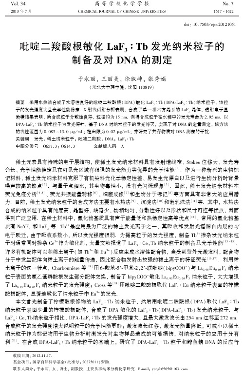
Vol.34高等学校化学学报No.72013年7月 CHEMICAL JOURNAL OF CHINESE UNIVERSITIES 1617~1622 doi:10.7503/cjcu20121051吡啶二羧酸根敏化LaF 3∶Tb 发光纳米粒子的制备及对DNA 的测定于永丽,王丽美,徐淑坤,张秀娟(东北大学理学院,沈阳110819)摘要 采用水热法合成了水溶性良好的吡啶二羧酸根(DPA)敏化LaF 3∶Tb(DPA⁃LaF 3∶Tb)纳米粒子,该粒子的发光强度大且光学性能稳定.X 射线衍射分析表明,合成了单一相六方晶系的LaF 3晶体;透射电子显微镜结果表明,所合成粒子分散性良好,粒径约为15nm.测得合成粒子在水相中的发光寿命为2.95ms.以DPA⁃LaF 3∶Tb 纳米粒子为发光探针,基于DNA 对纳米粒子的发光猝灭,实现了对DNA 的定量测定,该方法的线性范围为0.083~13.0μg /mL;检出限为0.02μg /mL;并研究了共存物质对DNA 测定的干扰.关键词 发光;稀土纳米粒子;吡啶二羧酸;DNA;LaF 3∶Tb 中图分类号 O657.3;O614.3 文献标志码 A 收稿日期:2012⁃11⁃17.基金项目:国家自然科学基金(批准号:20875011)资助.联系人简介:于永丽,女,博士,副教授,主要从事纳米分析化学研究.E⁃mail:yongli6565@ 稀土元素具有特殊的电子层结构,使稀土发光纳米材料具有发射谱线窄㊁Stokes 位移大㊁发光寿命长㊁光学性能稳定及在可见光区域有很强的发光能力等优异的光学性能[1].作为一种新兴的生物标记材料,稀土发光纳米材料克服了有机染料光化学稳定性差㊁易发生光漂白以及进行生物分析时背景噪声较高的缺点[2].与量子点相比,其生物毒性小,没有光闪烁现象[3].因此,稀土发光纳米材料在荧光免疫分析[4,5]㊁荧光共振能量转移[6]㊁细胞成像[7]和生物分子标记[8]等方面具有非常大的应用潜力.目前,稀土发光纳米粒子的合成方法主要有水热法[9]㊁沉淀法[10]和微乳液法[11]等.其中,水热法合成的纳米粒子具有纯度高㊁晶型好㊁缺陷少㊁物相均匀㊁分散性好以及形状和尺寸可控等优点,因而得到广泛应用.在稀土材料中,氟化物基质具有声子能量低和热稳定性高等优点[12],常用的氟化物基质有NaYF 4和LaF 3等.Tb 3+是应用最为广泛的稀土发光离子之一,其吸收和发射光谱源自内层的4f 电子跃迁,由于吸收系数小,所以发光强度很弱.为提高粒子的发光强度,制备Tb 3+掺杂发光纳米粒子时通常同时掺杂Ce 3+作为敏化剂,大量文献报道了LaF 3∶Ce,Tb 纳米粒子的制备及光学性能[13~15].许多有机配体可以和稀土离子(如Tb 3+和Eu 3+)反应生成水溶性配合物,当受到紫外光激发时,配合物分子中发生配体向稀土离子的能量传递,因此配合物发射出较强的稀土离子的特征荧光[16,17].利用稀土离子的这一特点,Charbonnière 等[18]用6⁃羧基⁃5′⁃甲基⁃2,2′⁃联吡啶(bipyCOO -)与La 0.95Eu 0.05F 3纳米粒子表面的氨乙基磷酸根发生部分配体交换,制备了bipyCOO -敏化La 0.95Eu 0.05F 3纳米粒子,大大增强了La 0.95Eu 0.05F 3纳米粒子的发光强度;Cross 等[19]用吡啶二羧酸根取代LaF 3∶Eu 纳米粒子表面的柠檬酸根配体,显著地敏化了纳米粒子中Eu 3+的发光.本文首先制备了柠檬酸根修饰的LaF 3∶Tb 纳米粒子,然后用吡啶二羧酸根(DPA)取代LaF 3∶Tb纳米粒子表面少量的柠檬酸根配体,合成了DPA 敏化的LaF 3∶Tb(DPA⁃LaF 3∶Tb)发光纳米粒子.与LaF 3∶Ce,Tb 纳米粒子相比,DPA⁃LaF 3∶Tb 的发光强度增大,且最大激发波长由254nm 红移至272nm.合成粒子的发光强度增大说明粒子的光学性能更好;激发波长红移,激发光能量降低,可减小以稀土纳米粒子作为标记物用于生物分析时激发光对生物样品造成的可能损伤,对纳米粒子的应用十分有利[20].在合成DPA⁃LaF 3∶Tb 纳米粒子的基础上,研究了DPA⁃LaF 3∶Tb 粒子和鲱鱼精DNA 的反应行为.实验结果表明,DNA 对DPA⁃LaF 3∶Tb 纳米粒子的发光产生猝灭,当DNA 浓度在一定范围时,粒子的发光强度与DNA 的浓度之间符合修正的Stern⁃Volmer 方程,据此建立了DPA⁃LaF 3∶Tb 纳米发光探针测定DNA 的新方法,为稀土发光纳米材料的制备及应用开辟了新的思路.1 实验部分1.1 试剂与仪器La 2O 3和Tb 4O 7(北京有研稀土新材料股份有限公司);Ce(NO 3)3㊃6H 2O㊁柠檬酸钠㊁NH 4F 和浓氨水均为分析纯;三羟甲基氨基甲烷(Tris,纯度99.9%)(上海国药集团化学试剂有限公司);2,6⁃吡啶二羧酸(纯度99%,阿拉丁试剂公司);鲱鱼精DNA(美国Sigma 公司).实验用水为三次蒸馏水.Cary Eclipse 荧光分光光度计(美国瓦里安公司);UV⁃2100型双光束紫外⁃可见分光光度计(北京瑞丽分析仪器有限公司);Spectrum one 傅里叶变换红外光谱仪(美国PE 公司);Tecnai G 220型透射电子显微镜(美国FEI 公司);Rigaku /Dmax⁃r B 型X 射线衍射仪(荷兰PANalytical 公司);TGA /DSC 1型同步热分析仪(瑞士Mettler⁃toledo 公司).1.2 实验方法1.2.1 DPA⁃LaF 3∶Tb 纳米粒子的制备 将一定体积0.10mol /L 柠檬酸钠溶液置于锥形瓶中,在磁力搅拌下滴加一定体积浓度均为0.10mol /L 的La(NO 3)3/Tb(NO 3)3混合溶液(体积比9∶1),再滴加一定体积1.00mol /L 的NH 4F 溶液[n (柠檬酸钠)∶n (稀土总量)∶n (氟化氨)=3∶5∶20].用浓氨水调节溶液的pH 值为7.0,将其转移至硝化罐中,置于烘箱中在120℃下加热4h.反应结束后,自然冷却至室温,即制得澄清透明的柠檬酸根修饰的LaF 3∶Tb 纳米粒子(以下简称LaF 3∶Tb 纳米粒子)溶液.移取一定体积上述LaF 3∶Tb 纳米粒子溶液于锥形瓶中,向其中加入一定体积0.020mol /L DPA 溶液(二者摩尔比为2.5∶1),调节溶液pH 值为4.0.将混合溶液置于振荡器中振荡反应5min,制得DPA⁃LaF 3∶Tb 纳米粒子溶液.向溶液中加入乙醇使粒子沉淀析出并离心分离,将沉淀分散在水中,得到纯化的DPA⁃LaF 3∶Tb 纳米粒子溶液,溶液外观呈透明状.1.2.2 基于DPA⁃LaF 3∶Tb 纳米粒子的发光猝灭测定DNA 向8个比色管中分别加入1000μL 浓度为4.2×10⁻³mol /L 的DPA⁃LaF 3∶Tb 粒子溶液和800μL 浓度为1.50mol /L 的NaCl 溶液,再分别加入不同体积(5~780μL)浓度为100μg /mL 的DNA 标准溶液,最后用pH =8.0的Tris⁃盐酸缓冲溶液定容至6.00mL,摇匀,放置15min.在546nm 处测定以上溶液的发光强度I ,以lg I 对DNA 的浓度c 作图绘制标准曲线.2 结果与讨论2.1 DPA⁃LaF 3∶Tb 纳米粒子的制备及表征图1示出了所合成的DPA⁃LaF 3∶Tb 纳米粒子粉体的X 射线衍射谱图.与LaF 3的XRD标准谱图Fig.1 XRD pattern of as⁃prepared particles (JCPDF 卡号:01⁃074⁃1324)相比,合成样品衍射峰的位置及强度与标准谱图相吻合,且无杂峰出现,表明实验合成了单一相LaF 3晶体,该晶体属于六方晶系.图1中在2θ角为25.0°,27.6°,44.0°,50.8°和52.8°处出现较强的衍射峰,可分别指认为LaF 3的(110),(111),(300),(302)和(221)晶面衍射峰.根据图中衍射峰的数据,采用Scherrer 方程计算得到合成的DPA⁃LaF 3∶Tb 粒子的平均晶粒度为14nm.图2是合成的DPA⁃LaF 3∶Tb 纳米粒子的TEM 照片.可见,粒子形貌为类球形,分散性较好,粒度较均匀,粒径约为15nm,与XRD 测试计算所得的晶粒度相吻合.考察了DPA⁃LaF 3∶Tb 粒子的发光性能,图38161高等学校化学学报 Vol.34 Fig.2 TEM image of DPA⁃LaF 3∶Tbnanoparticles Fig.3 Excitation (a )and emission (b )spectra of DPA⁃LaF 3∶Tb colloidal solution and UVabsorption spectrum of DPA solution (c )谱线a 和b 分别是DPA⁃LaF 3∶Tb 胶体溶液的激发和发射谱图.为研究DPA⁃LaF 3∶Tb 粒子的发光机理,扫描了DPA 的紫外吸收光谱(图3谱线c ).可见,DPA⁃LaF 3∶Tb 粒子的激发波长和DPA 的紫外吸收峰波长完全相同,最大激发波长为272nm.这说明DPA⁃LaF 3∶Tb 粒子的激发是由于DPA 对紫外光的吸收产生,同时也证明在LaF 3∶Tb 粒子表面结合了DPA.从谱线b 可以看出,DPA⁃LaF 3∶Tb 粒子分别在490,546,586和622nm 处出现了4个发射峰,这是Tb 3+的特征发射峰,分别归属于Tb 3+离子电子的5D 4⁃7F 6,5D 4⁃7F 5,5D 4⁃7F 4和5D 4⁃7F 3跃迁.DPA 中含有氮原子和羧基氧原子,是一类多功能有机配体,可与稀土离子发生键合作用[19,21],本实验中DPA 与LaF 3∶Tb 粒子表面的稀土离子结合,从而修饰在粒子表面.在DPA⁃LaF 3∶Tb 纳米粒子中,DPA 接受紫外光的能量,产生了Tb 3+的特征发射.即在DPA⁃LaF 3∶Tb 纳米粒子中,发生了DPA 向Tb 3+的能量传递,从而产生了Tb 3+的发光光谱.已有文献报道,以Tb 3+为激活剂合成LaF 3发光材料时,大多掺杂Ce 3+作为敏化剂[13~15].本实验对合成LaF 3∶Ce,Tb 纳米粒子的实验条件进行了优化,在最优条件下制备了LaF 3∶Ce,Tb 纳米粒子.另外,分别合成了LaF 3∶Tb,DPA⁃LaF 3∶Tb 和DPA⁃LaF 3∶Ce,Tb 纳米粒子,合成时4种纳米粒子中Tb 3+的掺杂量相同(Tb 3+和稀土离子总量的摩尔比为1∶10),以上4种胶体溶液的发光光谱如图4所示.Fig.4 Excitation (A )and emission (B )spectra of DPA⁃LaF 3∶Tb (a ),LaF 3∶Ce ,Tb (b ),DPA⁃LaF 3∶Ce ,Tb (c )and LaF 3∶Tb (d )colloidal solutions由图4(A)可见,DPA⁃LaF 3∶Tb 纳米粒子的最大激发波长为272nm,与另外两种纳米粒子的最大激发波长254nm 相比,发生了明显的红移,表明激发光能量有所降低,当将该粒子作为标记物用于生物分析时,可以减小激发光对生物样品造成的可能损伤.图4(B)为DPA⁃LaF 3∶Tb(272nm 激发)㊁LaF 3∶Ce,Tb(254nm 激发)和DPA⁃LaF 3∶Ce,Tb(254nm 激发)的发射谱图.可见,DPA⁃LaF 3∶Tb 纳米粒子的最大发光强度分别是LaF 3∶Ce,Tb 和DPA⁃LaF 3∶Ce,Tb 纳米粒子的1.65倍和1.85倍,表明DPA⁃LaF 3∶Tb 纳米粒子的发光性能最好.由于LaF 3∶Tb 粒子的激发和发射均来自Tb 3+电子的f ⁃f 跃迁,发光强度很弱,其激发和发射谱图见图4中谱线d 所示.在图4(A)中,LaF 3∶Ce,Tb 纳米粒子的激发峰位于254nm,该激发峰是由于Ce 3+离子的外层电子9161 No.7 于永丽等:吡啶二羧酸根敏化LaF 3∶Tb 发光纳米粒子的制备及对DNA 的测定发生从4f 向5d 能级的跃迁所致.Ce 3+离子吸收紫外光后,将能量传递给Tb 3+离子,从而产生Tb 3+离子的发射光谱[22].DPA⁃LaF 3∶Ce,Tb 纳米粒子有2个激发峰,来自于Ce 3+和DPA 对紫外光的吸收,分别位于254和272nm.由图4(B)可见,DPA⁃LaF 3∶Tb 粒子的发光强度比LaF 3∶Ce,Tb 粒子的大,说明DPA 作为敏化剂比Ce 3+更有效;DPA⁃LaF 3∶Tb 粒子的发光强度比DPA⁃LaF 3∶Ce,Tb 粒子的大,这可能是因为在DPA⁃LaF 3∶Ce,Tb 粒子中有部分DPA 与Ce 3+结合,减弱了DPA 向Tb 3+的能量传递所致.DPA 作为敏化剂,在合成DPA⁃LaF 3∶Tb 纳米粒子时其加入量会影响合成粒子的发光强度,DPA 加入量对合成粒子发光强度的影响见图5.可见,随着LaF 3∶Tb 粒子和DPA 摩尔比的减小(实验中固定LaF 3∶Tb 粒子的加入量,逐渐增加DPA 用量),产物的发光强度增大,这是因为DPA 和柠檬酸根竞争占据LaF 3∶Tb 粒子表面,增大DPA 的浓度对其与粒子的结合有利.当LaF 3∶Tb 粒子与DPA 摩尔比小于2.5后,产物的发光强度基本不变.因此,实验选择LaF 3∶Tb 粒子和DPA 的最佳摩尔比为2. 5.Fig.5 Effect of the amount of DPA on the lumines⁃cent intensity of DPA⁃LaF 3∶Tb nanoparticles Fig.6 FTIR spectra of DPA⁃LaF 3∶Tb (a )andLaF 3∶Tb (b )powderDPA⁃LaF 3∶Tb 和LaF 3∶Tb 纳米粒子的红外谱图如图6所示.由图6谱线b 可见,在3438cm -1处有一宽而强的吸收峰,为缔合的O H 伸缩振动的吸收带,来自样品中未烘干的水分子;1610和1385cm -1处的谱峰分别为 COO -的对称和反对称伸缩振动峰,说明在LaF 3∶Tb 纳米粒子表面包覆了柠檬酸根.采用差热热重分析测得LaF 3∶Tb 粒子表面柠檬酸根的质量分数为10%.图6谱线a 和b 的吸收峰位置完全相同,说明DPA⁃LaF 3∶Tb 粉体表面主要是柠檬酸根包覆,只有少量DPA 存在,所以图6谱线b 中未出现DPA 的红外吸收峰.实验中合成DPA⁃LaF 3∶Tb 粒子时加入的DPA 和LaF 3∶Tb 的摩尔比为2.5∶1,DPA⁃LaF 3∶Tb 粉体的红外谱图中未出现DPA 的红外吸收峰,说明合成时加入的DPA 大部分存在于溶液中.只有当反应溶液中DPA 的浓度达到一定数值时,DPA 才能替换已经包覆在稀土粒子表面的柠檬酸根,结合在粒子的表面.为用于生物分析,合成粒子必须具有良好的水溶性.实验制备的DPA⁃LaF 3∶Tb 纳米粒子水溶性良好,粒子溶液外观呈透明状.测得合成粒子胶体溶液的发光寿命为2.95ms,远大于生物分子和量子点的荧光寿命,可用于进行时间分辨荧光分析[7].2.2 基于DPA⁃LaF 3∶Tb 纳米粒子的发光猝灭测定DNAFig.7 Emission spectra of DPA⁃LaF 3∶Tb (a )andDPA⁃LaF 3∶Tb+DNA (b )制备的DPA⁃LaF 3∶Tb 粒子具有良好的水溶性且表面修饰了有机配体,因此可将其直接用于生物分析.向DPA⁃LaF 3∶Tb 溶液体系中加入DNA 前后胶体溶液的发射光谱图如图7中谱线a 和b 所示.可见,加入DNA 对发射峰的位置没有影响,但对纳米粒子的发光强度有明显的猝灭.为探讨DNA 猝灭DPA⁃LaF 3∶Tb 纳米粒子发光的原因,考察了DNA 加入量对DPA 溶液吸收光谱的影响.结果表明,随着DNA 浓度的增大,DPA 在272nm 的吸光度降低,发生减色效应,表明DPA 与DNA 发生了嵌入作用[23,24].由于嵌入的DPA 分子中吡啶环的π*空轨道与碱基的π轨道发生偶0261高等学校化学学报 Vol.34 合,偶合后的π*轨道因部分填充电子,使π→π*跃迁几率减小,从而产生减色效应.由于DPA⁃LaF 3∶Tb 纳米粒子发光的能量来自于DPA,当DPA 和DNA 发生嵌入作用,进而脱离LaF 3∶Tb 纳米粒子表面,必然导致DPA⁃LaF 3∶Tb 纳米粒子的发光猝灭.向一系列DPA⁃LaF 3∶Tb 纳米粒子溶液中加入不同量的DNA,DNA 对粒子发光强度的猝灭符合修正的Stern⁃Volmer 方程[25]:lg(I 0/I )=1+K sv [Q],式中,I 0和I 分别为加入猝灭剂前后粒子的发光强度,[Q]为猝灭剂DNA 的浓度,K sv 为Stern⁃Volmer 猝灭常数.以lg I 对DNA 的浓度作图得到测定DNA 的标准曲线(n =8).标准曲线的线性回归方程为lg I =2.93-0.0161c ,线性相关系数R =0.9975,线性范围为0.083~13.0μg /mL.对于不加DNA 的空白体系平行测定11次,计算得到方法的检出限为0.02μg /mL(3σ).对浓度为5.00μg /mL 的DNA 溶液平行测定11次,相对标准偏差为0.11%.综上可知,该分析方法的线性范围较宽,检测的灵敏度较高.为考察所建立分析方法的选择性,测定了一些常见物质对体系发光强度的影响,结果列于表1.可见,当这些物质浓度小于实验浓度时,其干扰程度小于±5%,说明此时对DNA 的测定没有干扰.Table 1 Effects of coexistance substances on the determination of DNA *Coexistance substanceConcentration /(mol㊃L -1)Relative error(%)Coexistance substance Concentration /(μg㊃L -1)Relative error(%)Ni 2+1×10⁻⁷-4.7Adenine,guanine 0.1 1.6,-1.6Cd 2+4×10⁻⁷ 2.5Urea 0.5 1.0Cu 2+1×10⁻⁶-2.4BSA,histidine 8-2.8,-4.7Fe 3+,Zn 2+2×10⁻⁶ 4.8,-3.2Glycine 20-1.7Ca 2+,H 2PO -41×10-5 4.3,-3.1Glutamate,lysine 40-3.6,-1.9Mg 2+2×10⁻⁴-3.3Glucose 1000 1.4K +,NO -35×10-3 4.7,4.7HCO -33×10-24.7 *The concentration of DPA⁃LaF 3∶Tb nanoparticles is 7.0×10⁻⁴mol /L;the concentration of DNA is 8.00μg /mL.为验证本方法的实用性,配制了合成样品进行测定.合成样品含有DNA(4.00μg /mL)㊁BSA(2.0μg /mL)㊁赖氨酸(2.0μg /mL)㊁谷氨酸(2.0μg /mL)㊁腺嘌呤(0.02μg /mL)㊁鸟嘌呤(0.02μg /mL)㊁葡萄糖(5.0×10-5mol /L)㊁KNO 3(5.0×10⁻⁵mol /L)㊁MgCl 2(2.0×10⁻⁶mol /L)㊁CaCl 2(5.0×10⁻⁷mol /L)㊁尿素(5.0×10-8mol /L)㊁FeCl 3(3.0×10⁻⁸mol /L)和ZnCl 2(2.0×10⁻⁸mol /L).以DPA⁃LaF 3∶Tb 纳米粒子为发光探针测定合成样品中DNA 含量,5次测定结果的平均值为3.97μg /mL,相对标准偏差为2.8%.低标和高标回收率分别为104%和95.6%,说明本法较为准确可靠.3 结 论采用水热法合成了水溶性良好的DPA⁃LaF 3∶Tb 发光纳米粒子.与LaF 3∶Ce,Tb 粒子相比,DPA⁃LaF 3∶Tb 粒子的发光强度增大,激发波长明显红移.采用XRD,TEM 和FTIR 对合成粒子进行了表征,结果表明合成了单一相六方晶系的LaF 3,粒子的分散性良好,粒径约为15nm.以DPA⁃LaF 3∶Tb 纳米粒子为发光探针,基于DNA 对纳米粒子的发光猝灭,实现了对DNA 的定量测定.本实验合成稀土纳米粒子的反应原料价廉易得,反应条件温和,实验方法简单快速.制备的DPA⁃LaF 3∶Tb 纳米粒子具有良好的光学性能和生物相容性,在生物大分子的分析中具有良好的应用前景.在后续的研究中,将尝试采用激发波长在近紫外或可见光区的有机配体对合成的稀土粒子进行敏化,得到对近紫外或可见光响应的纳米粒子,并将其用于细胞成像和生物分析研究.参 考 文 献[1] Zhang C.,Sun L.,Zhang Y.W.,Yan C.H.,J.Rare Earths ,2010,28,807 819[2] Lin Z.B.,Su X.G.,Zhang J.H.,Jin Q.H.,Chin.J.Anal.Chem .,2002,30(2),237 241(林章碧,苏星光,张家骅,金钦汉.分析化学,2002,30(2),237 241)[3] Wang X.,Li Y.D.,mun .,2007,43,2901 2910[4] Wang M.,Hou W.,Mi C.C.,Wang W.X.,Xu Z.R.,Teng H.H.,Mao C.B.,Xu S.K.,Anal.Chem .,2009,81,8783 1261 No.7 于永丽等:吡啶二羧酸根敏化LaF 3∶Tb 发光纳米粒子的制备及对DNA 的测定8789[5] Juntunen E.,Myyryläinen T.,Salminen T.,Soukka T.,Pettersson K.,Anal.Biochem.,2012,428,31 38[6] Zhai H.,Yu Y.L.,Cheng L.Y.,Xu S.K.,Wang N.Z.,Acta Chimica Sinica,2011,69(10),1205 1210(翟晗,于永丽,成利艳,徐淑坤,王乃芝.化学学报,2011,69(10),1205 1210)[7] Wu J.,Ye Z.Q.,Wang G.L.,Yuan J.L.,Talanta,2007,72,1693 1697[8] Nichkova M.,Dosev D.,Gee S.J.,Hammock B.D.,Kennedy I.M.,Anal.Chem.,2005,77,6864 6873[9] Zeng J.H.,Su J.,Li Z.H.,Yan R.X.,Li Y.D.,Adv.Mater.,2005,17,2119 2123[10] Xiao Y.,Gao Z.Y.,Wu D.,Jiang Y.,Liu N.,Jiang K.,Chem.Res.Chinese Universities,2011,27(6),919 923[11] Wang G.F,Qin W.P.,Wei G.D.,Wang L.L.,Zhu P.F.,Ryongjin K.,Zhang D.S.,Ding F.H.,Zheng K.Z.,J.FluorineChem.,2009,130,158 161[12] Wang F.,Zhang Y.,Fan X.P.,Wang M.Q.,J.Mater.Chem.,2006,16,1031 1034[13] Wang L.Y.,Li Y.D.,Chem.Eur.J.,2007,13,4203 4207[14] Zhao G.Y.,Sun L.D.,Yan C.H.,Chem.J.Chinese Universities,2011,32(3),635 640(赵光耀,孙聆东,严纯华.高等学校化学学报,2011,32(3),635 640)[15] Wang Z.L.,Quan Z.W.,Jia P.Y.,Lin C.K.,Luo Y.,Chen Y.,Fang J.,Zhou W.,O’Connor C.J.,Lin J.,Chem.Mater.,2006,18,2030 2037[16] Shen L.,Shi M.,Shi E.X.,Du Y.K.,Li F.Y.,Huang C.H.,Chem.J.Chinese Universities,2006,27(8),1413 1417(沈莉,石梅,石恩娴,杜玉扣,李富友,黄春辉.高等学校化学学报,2006,27(8),1413 1417)[17] Ocaňa J.A.,Callejón M.,Barragán F.J.,Eur.J.Pharmacol.,2001,13,297 301[18] Charbonnière L.J.,Rehspringer J.L.,Ziessel R.,Zimmermann Y.,New J.Chem.,2008,32,1055 1059[19] CrossA.M.,Stanley May P.,van Veggel F.C.J.M.,Berry M.T.,J.Phys.Chem.C,2010,114,14740 14747[20] Wu S.J.,Duan N.,Zhu C.Q.,Ma X.Y.,Wang M.,Wang Z.P.,Biosensors and Bioelectronics,2011,30,35 42[21] Liu C.B.,Xiang L.,Li X.X.,Wen H.L.,Chem.J.Chinese Universities,2006,27(12),2256 2259(刘崇波,向丽,李新新,温辉梁.高等学校化学学报,2006,27(12),2256 2259)[22] Guo H.,Zhang T.,Qiao Y.,Zhao L.,Li Z.,J.Nanosci.Nanotechnol.,2010,10,1913 1919[23] Li J.F.,Shi W.T.,Han X.Y.,Dong C.,Choi Martin M.F.,Chem.Res.Chinese Universities,2012,28(4),614 619[24] Ding Y.,Ren X.X.,Zhuang C.X.,Le X.Y.,Chinese J.Inorg.Chem.,2009,25(4),4656 662(丁杨,任祥祥,庄楚雄,乐学义.无机化学学报,2009,25(4),656 662)[25] Sun J.F.,Liu L.H.,Ren C.L.,Chen X.G.,Hu Z.D.,Microchim.Acta,2008,163,271 276Synthesis of Dipicolinate Sensitized LaF3∶Tb Nanoparticlesand Their Application in the Determination of DNAYU Yong⁃Li*,WANG Li⁃Mei,XU Shu⁃Kun,ZHANG Xiu⁃Juan(College of Science,Northeastern University,Shenyang110819,China)Abstract Lanthanide nanoparticles have been used in bioanalysis due to their excellent luminescence proper⁃ties.Herein,dipicolinate(DPA)sensitized LaF3∶Tb(DPA⁃LaF3∶Tb)nanoparticles with good water solubili⁃ty were synthesized by hydrothermal method,which emit strong and stable luminescence.X⁃Ray diffraction (XRD)pattern showed that the particles were single⁃phase LaF3crystals in hexagonal system,and transmis⁃sion electron microscopy(TEM)image showed a good dispersion of DPA⁃LaF3∶Tb nanoparticles with nearly uniform size of about15nm.The lifetime of the particles in water was measured2.95ms.It was found that DNA could quench effectively the luminescence of the particles.Based on the luminescence quench,the de⁃termination of DNA was realized with the linear range of0.083~13.0μg/mL and the detection limit of0.02μg/mL.Moreover,the interference of coexistence substances to the determination of DNA was studied. Keywords Luminescence;Lanthanide nanoparticles;Dipicolinate;DNA;LaF3∶Tb(Ed.:N,K) 2261高等学校化学学报 Vol.34 。
Histone Methylation by PRC2 Is Inhibited by Active Chromatin Marks
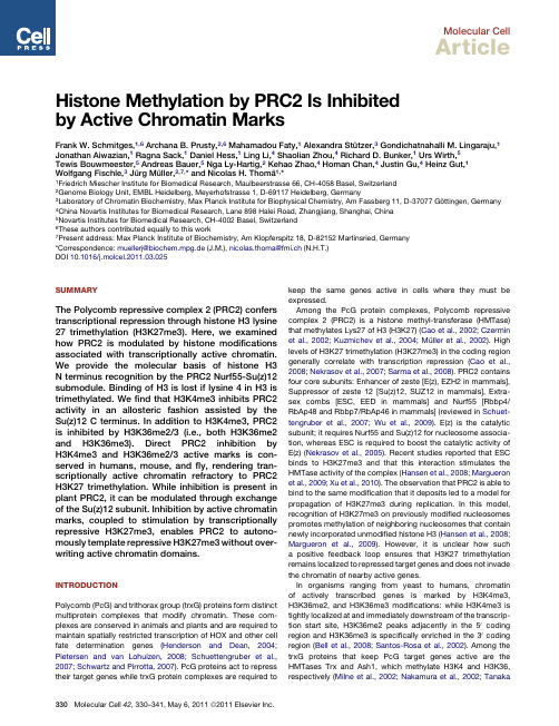
Molecular CellArticleHistone Methylation by PRC2Is Inhibitedby Active Chromatin MarksFrank W.Schmitges,1,6Archana B.Prusty,2,6Mahamadou Faty,1Alexandra Stu¨tzer,3Gondichatnahalli M.Lingaraju,1 Jonathan Aiwazian,1Ragna Sack,1Daniel Hess,1Ling Li,4Shaolian Zhou,4Richard D.Bunker,1Urs Wirth,5Tewis Bouwmeester,5Andreas Bauer,5Nga Ly-Hartig,2Kehao Zhao,4Homan Chan,4Justin Gu,4Heinz Gut,1 Wolfgang Fischle,3Ju¨rg Mu¨ller,2,7,*and Nicolas H.Thoma¨1,*1Friedrich Miescher Institute for Biomedical Research,Maulbeerstrasse66,CH-4058Basel,Switzerland2Genome Biology Unit,EMBL Heidelberg,Meyerhofstrasse1,D-69117Heidelberg,Germany3Laboratory of Chromatin Biochemistry,Max Planck Institute for Biophysical Chemistry,Am Fassberg11,D-37077Go¨ttingen,Germany4China Novartis Institutes for Biomedical Research,Lane898Halei Road,Zhangjiang,Shanghai,China5Novartis Institutes for Biomedical Research,CH-4002Basel,Switzerland6These authors contributed equally to this work7Present address:Max Planck Institute of Biochemistry,Am Klopferspitz18,D-82152Martinsried,Germany*Correspondence:muellerj@biochem.mpg.de(J.M.),nicolas.thoma@fmi.ch(N.H.T.)DOI10.1016/j.molcel.2011.03.025SUMMARYThe Polycomb repressive complex2(PRC2)confers transcriptional repression through histone H3lysine 27trimethylation(H3K27me3).Here,we examined how PRC2is modulated by histone modifications associated with transcriptionally active chromatin. We provide the molecular basis of histone H3 N terminus recognition by the PRC2Nurf55-Su(z)12 submodule.Binding of H3is lost if lysine4in H3is trimethylated.Wefind that H3K4me3inhibits PRC2 activity in an allosteric fashion assisted by the Su(z)12C terminus.In addition to H3K4me3,PRC2 is inhibited by H3K36me2/3(i.e.,both H3K36me2 and H3K36me3).Direct PRC2inhibition by H3K4me3and H3K36me2/3active marks is con-served in humans,mouse,andfly,rendering tran-scriptionally active chromatin refractory to PRC2 H3K27trimethylation.While inhibition is present in plant PRC2,it can be modulated through exchange of the Su(z)12subunit.Inhibition by active chromatin marks,coupled to stimulation by transcriptionally repressive H3K27me3,enables PRC2to autono-mously template repressive H3K27me3without over-writing active chromatin domains.INTRODUCTIONPolycomb(PcG)and trithorax group(trxG)proteins form distinct multiprotein complexes that modify chromatin.These com-plexes are conserved in animals and plants and are required to maintain spatially restricted transcription of HOX and other cell fate determination genes(Henderson and Dean,2004; Pietersen and van Lohuizen,2008;Schuettengruber et al., 2007;Schwartz and Pirrotta,2007).PcG proteins act to repress their target genes while trxG protein complexes are required to keep the same genes active in cells where they must be expressed.Among the PcG protein complexes,Polycomb repressive complex2(PRC2)is a histone methyl-transferase(HMTase) that methylates Lys27of H3(H3K27)(Cao et al.,2002;Czermin et al.,2002;Kuzmichev et al.,2004;Mu¨ller et al.,2002).High levels of H3K27trimethylation(H3K27me3)in the coding region generally correlate with transcription repression(Cao et al., 2008;Nekrasov et al.,2007;Sarma et al.,2008).PRC2contains four core subunits:Enhancer of zeste[E(z),EZH2in mammals], Suppressor of zeste12[Su(z)12,SUZ12in mammals],Extra-sex combs[ESC,EED in mammals]and Nurf55[Rbbp4/ RbAp48and Rbbp7/RbAp46in mammals](reviewed in Schuet-tengruber et al.,2007;Wu et al.,2009).E(z)is the catalytic subunit;it requires Nurf55and Su(z)12for nucleosome associa-tion,whereas ESC is required to boost the catalytic activity of E(z)(Nekrasov et al.,2005).Recent studies reported that ESC binds to H3K27me3and that this interaction stimulates the HMTase activity of the complex(Hansen et al.,2008;Margueron et al.,2009;Xu et al.,2010).The observation that PRC2is able to bind to the same modification that it deposits led to a model for propagation of H3K27me3during replication.In this model, recognition of H3K27me3on previously modified nucleosomes promotes methylation of neighboring nucleosomes that contain newly incorporated unmodified histone H3(Hansen et al.,2008; Margueron et al.,2009).However,it is unclear how such a positive feedback loop ensures that H3K27trimethylation remains localized to repressed target genes and does not invade the chromatin of nearby active genes.In organisms ranging from yeast to humans,chromatin of actively transcribed genes is marked by H3K4me3, H3K36me2,and H3K36me3modifications:while H3K4me3is tightly localized at and immediately downstream of the transcrip-tion start site,H3K36me2peaks adjacently in the50coding region and H3K36me3is specifically enriched in the30coding region(Bell et al.,2008;Santos-Rosa et al.,2002).Among the trxG proteins that keep PcG target genes active are the HMTases Trx and Ash1,which methylate H3K4and H3K36, respectively(Milne et al.,2002;Nakamura et al.,2002;Tanaka330Molecular Cell42,330–341,May6,2011ª2011Elsevier Inc.et al.,2007).Studies in Drosophila showed that Trx and Ash1 play a critical role in antagonizing H3K27trimethylation by PRC2,suggesting a crosstalk between repressive and activating marks(Papp and Mu¨ller,2006;Srinivasan et al.,2008).In this study we investigated how PRC2activity is modulated by chromatin marks typically associated with active transcrip-tion.We found that the Nurf55WD40propeller binds the N terminus of unmodified histone H3and that H3K4me3 prevents this binding.In the context of the tetrameric PRC2 complex,wefind that H3K4me3and H3K36me2/3(i.e.,both H3-K36me2and H3-K36me3)inhibit histone methylation by PRC2in vitro.Dissection of this process by usingfly,human, and plant PRC2complexes suggests that the Su(z)12subunit is important for mediating this inhibition.PRC2thus not only contains the enzymatic activity for H3K27methylation and a recognition site for binding to this modification,but it also harbors a control module that triggers inhibition of this activity to prevent deposition of H3K27trimethylation on transcription-ally active genes.PRC2can thus integrate information provided by pre-existing histone modifications to accurately tune its enzymatic activity within a particular chromatin context.RESULTSStructure of Nurf55Bound to the N Terminusof Histone H3Previous studies reported that Nurf55alone is able to bind to histone H3(Beisel et al.,2002;Hansen et al.,2008;Song et al., 2008;Wysocka et al.,2006)but not to a GST-H3fusion protein (Verreault et al.,1998).By usingfluorescence polarization(FP) measurements,we found that Nurf55binds the very N terminus of unmodified histone H3encompassing residues1–15(H31–15) with a K D of$0.8±0.1m M but does not bind to a histone H319–38 peptide(Figure1A).Crystallographic screening resulted in the successful cocrystallization of Nurf55in complex with an H31–19peptide.After molecular replacement with the known structure of Nurf55(Song et al.,2008),the initial mF oÀDF c differ-ence map showed density for H3residues1–14in bothNurf55molecules in the crystallographic asymmetric unit. Figures1B–1E show the structure of H31–19bound to Drosophila Nurf55,refined to2.7A˚resolution(R/R free=20.1%and25.0%, Table1;Figure S1A,available online).The H3peptide binds to theflat surface of the Nurf55WD40propeller(Figure1B),subse-quently referred to as the canonical binding site(c-site)(Gaudet et al.,1996).The H3peptide is held in an acidic pocket(Figures 1C and1E)and traverses the central WD40cavity in a straight line across the propeller(Figure1B).Nurf55binds the H3peptide by contacting H3residues Ala1, Arg2,Lys4,Ala7,and Lys9.Each of these residues forms side-chain specific contacts with the Nurf55propeller(Figures 1D and1E).The bulk of the molecular recognition is directed toward H3Arg2and Lys4.Ala1sits in a buried pocket with its a-amino group hydrogen bonding to Nurf55Asp252,which recognizes andfixes the very N terminus of histone H3.The neighboring Arg2is buried deeper within the WD40propeller fold,with its guanidinium group sandwiched by Nurf55residues Phe325and Tyr185(Figure1D).H3Lys4binds to a well-defined surface pocket on Nurf55located on blade2,near the central cavity of the propeller.Its3-amino group is specifically coordi-nated by the carboxyl groups of Nurf55residues Glu183andGlu130and through the amide oxygen of Asn132(Figure1E).Lys9is stabilized by hydrophobic interactions on the WD40surface while having its3-amino group held in solvent-exposedfashion(Figure1D).Ser10of histone H3marks the beginningof a turn that inverses the peptide directionality.Histone H3residues Thr11–Lys14become progressively disordered andare no longer specifically recognized.No interpretable densitywas observed beyond Lys14.Taken together,Nurf55specificallyrecognizes an extended region of the extreme N terminus ofhistone H3(11residues long,700A˚2buried surface area)in thecanonical ligand binding location of WD40propeller domains. Structure of the Nurf55-Su(z)12Subcomplex of PRC2 The H3-Nurf55structure prompted us to investigate how Nurf55might bind histone tails in the presence of Su(z)12,its interactionpartner in PRC2(Nekrasov et al.,2005;Pasini et al.,2004).Asafirst step we mapped the Nurf55-Su(z)12interaction in detailby carrying out limited proteolysis experiments on reconstitutedDrosophila PRC2,followed by isolation of a Nurf55-Su(z)12subcomplex.Mass spectrometric analysis and pull-down exper-iments with recombinant protein identified Su(z)12residues73–143[hereafter referred to as Su(z)1273–143]as sufficient forNurf55binding(Figures S1C and S1D).Crystals were obtained when Drosophila Nurf55and Su(z)12residues64–359were set up in the presence of0.01%subtilisinprotease(Dong et al.,2007).After data collection,the structurewas refined to a maximal resolution of2.3A˚(Table1).Molecularreplacement with Nurf55as search model provided clear initialmF oÀDF c difference density for a13amino acid-long Su(z)12 fragment spanning Su(z)12residues79–91(Figures2A–2C).Thefinal model was refined to2.3A˚(R/R free=17.5%/20.9%)and verified by simulated annealing composite-omit maps(Fig-ure S1B).The portion of Su(z)12involved in Nurf55binding willhenceforth be referred to as the Nurf55binding epitope(NBE).The Su(z)12binding site on Nurf55is located on the side of thepropeller between the stem of the N-terminal a helix(a1)andthe PP loop(Figures2A and2B).Binding between Su(z)12andNurf55occurs mostly through hydrophobic interactions in anextended conformation.The interaction surface betweenNurf55and the NBE is large for a peptide,spanning around800A˚2.Sequence alignment between Su(z)12orthologs revealsthat the NBE is highly conserved(53%identity and84%similarity)in animals and in plants(Figure2E).With the exceptionof Su(z)12Arg85,the majority of the conserved Su(z)12NBEresidues engage in hydrophobic packing with Nurf55(Figures2B and2C).Together with the Su(z)12VEFS domain and theC2H2zincfinger(C5domain)(Birve et al.,2001),the NBE consti-tutes the only identifiable motif in Su(z)12found conserved in allSu(z)12orthologs.The NBE binding site on Nurf55has previously been shown tobe occupied by helix1of histone H4(Figure2D)(Murzina et al.,2008;Song et al.,2008),an epitope not accessible in assemblednucleosomes(Luger et al.,1997).Nurf55binds H4and theSu(z)12NBE epitope in a different mode,and importantly,withopposite directionality(Figure2D).The detailed comparison ofthe Nurf55-Su(z)12structure with that of H4bound to Nurf55Molecular CellAllosteric PRC2Inhibition by H3K4me3/H3K36me3Molecular Cell42,330–341,May6,2011ª2011Elsevier Inc.331strongly suggests that binding of Su(z)12(NBE)and of H4(helix 1)are mutually exclusive (Figure 2D).We therefore refer to the Su(z)12and H4binding site on Nurf55as the S/H -site.Su(z)12fragments that include the NBE have poor solubility by themselves and generally require Nurf55coexpression for solu-bilization.However,we were able to measure binding of a chem-ically synthesized Su(z)1275–93peptide to Nurf55by isothermal titration calorimetry (ITC)and found that the peptide was bound with a K D value of 6.7±0.3m M in a 1:1stoichiometry (Fig-ure 2F).Pull-down experiments with recombinant proteinandFigure 1.Crystal Structure of Nurf55in Complex with a Histone H31–19Peptide(A)Nurf55binds to an H31–15peptide with an affinity of $0.8±0.1m M as measured by FP.It has similar affinity for an H31–31peptide (2.2±0.2m M)but no binding can be detected to an H319–38peptide.(B)Ribbon representation of Nurf55-H31–19.Nurf55is shown in rainbow colors and H31–19is depicted in green.The peptide is bound to the c -site of the WD40propeller.(C)Electrostatic surface potential representation (À10to 10kT/e)of the c -site with the H3peptide shown as a stick model in green.(D)Close-up of the c -site detailing the interactions between Nurf55(yellow)and the H31–19peptide (green),with a water molecule shown as a red sphere.(E)Schematic representation of interactions between the H31–19peptide (green)and Nurf55(yellow).Molecular CellAllosteric PRC2Inhibition by H3K4me3/H3K36me3332Molecular Cell 42,330–341,May 6,2011ª2011Elsevier Inc.streptavidin beads suggest that Su(z)12residues 94–143harbor an additional Nurf55binding site not visible in the structure (Figure S1E).Su(z)12144–359,lacking the N-terminal 143residues,no longer binds to Nurf55.The NBE (residues 79–93)and the region adjacent to the NBE (residues 94–143)are thus required for stable interaction with Nurf55.The extended NBE was found enriched after limited proteolysis and in subsequent gel filtration runs coupled with quantitative mass spectrometry (Figure S1C).As the NBE was the only fragment visible after structure determi-nation,we conclude that it represents the major Su(z)12interac-tion epitope for Nurf55binding.The Nurf55-Su(z)12Complex Binds to Histone H3In order to study the potential interdependence of the identified Nurf55binding sites we compared binding of Nurf55and Nurf55-Su(z)12to the histone H3N terminus.FP experiments showed similar affinities for binding of a histone H31–15peptide to Nurf55(K D $0.8±0.1m M;Figure 1A)and a Nurf55-Su(z)1273–143complex (K D $0.6±0.1m M;Figure 2G).Importantly,mutation of Nurf55resi-dues contacting H3via its c -site drastically reduced binding to an H31–15peptide (Figure S2A),demonstrating that the Nurf55-Su(z)1273–143complex indeed binds the H31–15peptide through the c -site.We conclude that the presence of Su(z)12is compatible with Nurf55binding to H3via its c -site and that the two binding interactions are not interdependent.The observation that the Su(z)12NBE occupies the same Nurf55pocket that was previously shown to bind to helix 1of histone H4prompted us to test whether the Su(z)1273–143-Nurf55complex could still bind to histone H4.We performed pull-down experiments with a glutathione S-transferase (GST)fusion protein containing histone H41–48(Murzina et al.,2008)and found that H4stably interacted with isolated Nurf55but not with Su(z)1273–143-Nurf55(Figure 2H).In PRC2,the presence of Su(z)12in the Nurf55S/H -site therefore precludes binding to helix 1of histone H4.H3Binding by Nurf55-Su(z)12Is Sensitive to the Methylation Status of Lysine 4We next investigated how posttranslational modifications of the H3tail affect binding to the Nurf55-Su(z)1273–143complex.Modi-fications on H3Arg2,Lys9,and Lys14did not change affinity of Nurf55-Su(z)12for the modified H31–15peptide (Figures S2B and S2D).In contrast,peptides that were mono-,di-,or trimethylated on Lys4were bound with significantly reduced affinity exhibiting K D values of 17±3m M (H3K4me1),24±3m M (H3K4me2),and >70m M (H3K4me3),respectively (Figure 2I).The FP binding data were independently confirmed by ITC measurements (Figures S2C–S2F).Together,these findings are in accord with the structural data,which show that H3K9and H3K14are being held with their 3-amino moiety solvent-exposed,while the H3K4side chain is tightly coordinated (Figure 1E).The additional methyl groups on the H3K43-amino group are expected to progressively decrease affinity because of increased steric clashes within the H3K4binding pocket.H3K27Methylation by PRC2Is Inhibited by Histone H3K4me3MarksWe then examined the effect of H3K4me3modifications,which are no longer retained by Nurf55-Su(z)12,on the catalytic activity of PRC2.In a first set of experiments,we determined PRC2steady-state parameters on histone H31–45peptide substrates that were either unmodified or methylated at Lys 4.We observed similar K M values for H3and H3K4me3peptides of 0.84±0.21m M and 0.36±0.07m M,respectively (Figure 3A),and similar K M values for SAM (5.42±0.65m M for H3and 10.04±1.56m M for H3K4me3).The turnover rate constant k cat ,however,was 8-fold reduced in the presence of H3K4me3:2.53±0.21min -1for unmodified H3and 0.32±0.08min -1in the presence of H3K4me3(Figure 3A).While substrate binding is largely unaf-fected,turnover is thus severely inhibited in the presence of H3K4me3.This behavior,which results in a k cat /K M specificity constant of 7.83103M -1s -1(unmodified H3)compared to 0.533103M -1s -1(H3K4me3),is consistent with heterotrophic allosteric inhibition of the PRC2HMTase triggered by the pres-ence of the H3K4me3.To investigate the effect of the H3K4me3modification on PRC2activity in the context of nucleosomes,we reconstituted mononucleosomes with a trimethyllysine analog (MLA)at Lys4in H3(referred to as H3Kc4me3;Figure S3A)(Simon et al.,2007).We found that total H3K27methylation (measured by incorporation of 14C-labeled methyl groups)was substantially impaired on H3Kc4me3-containing nucleosomes compared to wild-type nucleosomes (Figures S3B and S3C).We used western blot analysis to monitor how levels of H3K27mono-,di-,and trimethylation were affected by the H3Kc4me3modifica-tion.While H3K27me1formation was reduced by more than 50%on H3Kc4me3nucleosomes compared to unmodified nucleosomes (Figures 3B and 3C),H3K27dimethylationandTable 1.Crystallographic Data and Refinement StatisticsNurf55–Su(z)12Nurf55–H31–19Space Group P212121P212121theses.Molecular CellAllosteric PRC2Inhibition by H3K4me3/H3K36me3Molecular Cell 42,330–341,May 6,2011ª2011Elsevier Inc.333titrant H3:S/H-siteN CNCSu(z)12 79-91Histone H4 31-41PP-loophelix α10.00.51.0 1.52.0 2.5-16.00-14.00-12.00-10.00-8.00-6.00-4.00-2.000.00-2.00-1.50-1.00-0.500.00020406080100120Time (min)µc a l /s e cMolar RatioK C a l /M o l e o f I n j e c t a n tK D = 6.7 ± 0.3 µMtitrant: H31-15DEFGH ICA B protein concentration [µM]Nurf55-Su(z)12protein concentration [µM]00.20.40.60.81.000.20.40.60.81.00.010.11101000.010.1110100Nurf55Nurf55-Su(z)12unmod K4me1K4me2K4me3f r a c t i o n b o u n df r a c t i o n b o u n d G ST -H 41-48 + N u r f 55G S T -H 41-48 + N u r f 55-S u (z )1273-143N u r f 55(m o c k c o n t r o l )N u r f 55-S u (z )1273-143(m o c k c o n t r o l )Nurf55GST-H41-48Su(z)1273-143i np ut i n p u t i n p u t i n p u t G S T -p u l l d o w n G S T -p u l l d o w n G S T -p u l l d o w n G S T -p u l l d o w n Figure 2.Crystal Structure and Characterization of Nurf55in Complex with the Su(z)12Binding Epitope for Nurf55(A)Ribbon representation of Nurf55-Su(z)12.Nurf55(rainbow colors)depicts the WD40domain nomenclature and Su(z)12is shown in magenta.The S/H -site is marked by a dashed box.(B)Detailed interactions of Su(z)12(magenta)with the S/H -site (yellow).Water molecules are depicted as red spheres.(C)Schematic representation of interactions between Su(z)12(magenta)and Nurf55(yellow).(D)Overlay of the backbone trace of Su(z)12(magenta)and the H4helix a 1(orange)(Song et al.,2008)in the S/H -site.(E)Alignment of the Su(z)12NBE with sequences from Drosophila melanogaster (dm,Q9NJG9),mouse (mm,NP_954666),human (hs,AAH15704),Xenopus tropicalis (xt,BC121323),zebrafish (dr,BC078293),and the three Arabidopsis thaliana (at)homologs Fis2(ABB84250),EMF2(NP_199936),and VRN2(NP_567517).Identical residues are highlighted in yellow.(F)ITC profile for binding of a Su(z)1275–93peptide to Nurf55.Data were fitted to a one-site model with stoichiometry of 1:1.The derived K D value is 6.7±0.3m M.(G)Binding of H31–15to Nurf55(0.8±0.1m M)and Nurf55-Su(z)1273–143(0.6±0.1m M)measured by FP.Molecular CellAllosteric PRC2Inhibition by H3K4me3/H3K36me3334Molecular Cell 42,330–341,May 6,2011ª2011Elsevier Inc.trimethylation were impaired by more than 80%by using H3Kc4me3nucleosomes (Figure 3C).In order to ascertain that inhibition of PRC2is indeed due to trimethylation of the aminogroup in the lysine side chain,and not due to the use of the MLA,we performed HMTase assays on H3K4me3-containing nucleosomes generated by native peptide ligation (Shogren-Knaak et al.,2003)and on H3Kc4me0and H3K4A nucleosomes.H3K27mono-,di-,and trimethylation was comparably inhibited on H3K4me3and on H3Kc4me3-containing nucleosomes,but was not affected by H3Kc4me0and H3K4A (Figures S3D and S3E).We conclude that H3K4me3specifically inhibits PRC2-mediated H3K27methylation with the most pronounced inhibi-tory effects observed for H3K27di-and trimethylation.We next tested whether the H3K4me3modification affects PRC2nucleosome binding.In electrophoretic mobility shift assays (EMSA),we found that PRC2binds unmodified or H3Kc4me3-modified nucleosomes with comparable affinity (Fig-ure S4A).Even though binding of Nurf55to the N terminus of ABC571141712290286571141712290286unmodified H3Kc4me3H3K27me3H4dmPRC2 [nM]H3K27me2H4H3K27me1H4H3K27me1H3K27me3H3K27me2dmPRC2 + unmod H3dmPRC2 + H3Kc4me3r e l a t i v e H M T a s e a c t i v i t ySubstrate Apparent Km of SAM (µM)Apparent Km of peptide (nM)k cat (min )-1k cat /Km (M S )-1-1H31-45 -biotin H3K4me31-45 -biotin5.42 ± 0.6510.04 ± 1.56355 ± 74.60.32 ± 0.080.53 x 10836 ± 207 2.53 ± 0.217.8 x 1033Figure 3.HMTase Activity of PRC2Is In-hibited by H3K4me3Marks(A)HMTase assay with PRC2and H31–45-biotin peptides measuring the concentration of SAH produced by the enzymatic reaction.When an H3K4me3-modified peptide is used,the specificity constant (k cat /K M )is drastically reduced,indicative of heterotrophic allosteric inhibition.(B)Western blot-based HMTase assay by using recombinant Drosophila mononucleosomes (571nM)and increasing amounts of PRC2.HMTase activity was monitored with antibodies against H3K27me1,H3K27me2,or H3K27me3as indicated;in each case the membrane was also probed with an antibody against unmodified histone H4to control for equal loading and western blot processing.Deposition of K27di-and trime-thylation is drastically reduced when nucleosomes are used that carry a H3Kc4me3modification.(C)Quantification of HMTase activity of Drosophila PRC2(286nM)on unmodified and H3Kc4me3-modified nucleosomes by quantitative western blotting.histone H3is almost 100-fold reduced byH3K4me3(Figure 2I and Figure S2F),inter-action of the Nurf55c -site with H3K4does not seem to make a detectable con-tribution to nucleosome binding by PRC2in this assay.Consistent with the allosteric mechanism of H3K4me3inhibition that we had observed in the peptide assays (Figure 3A),inhibition of the PRC2HMTase activity by H3K4me3-containingnucleosomes is not caused by impaired nucleosome binding,but is rather the consequence of reduced catalytic turnover.H3K4me3Needs to Be Present on the Same Tail as K27to Inhibit PRC2We then assessed whether inhibition of the PRC2HMTase activity by H3K4me3requires the K4me3mark to be located on the substrate nucleosome (in cis ),or whether it could also be trig-gered if the H3K4me3modification was provided on a separate peptide (in trans ).We performed HMTase assays on unmodified oligonucleosomes in the presence of increasing amounts of a histone H31–15peptide trimethylated at K4(H31–15-K4me3)(Figure 4A).Addition of the H31–15-K4me3peptide did not affect PRC2HMTase activity at peptide concentrations as high as $200m M.When testing H31–19-unmodified peptide in controls at comparable concentrations,we did observe concentration-dependent PRC2inhibition (Figure 4A),probably because of substrate competition at large peptide excess.As H3K4me3-(H)GST pull-down assay with recombinant GST-H41–48and Nurf55and Nurf55-Su(z)1273–143proteins.GST-H41–48is able to bind Nurf55alone but in the Nurf55-Su(z)1273–143complex the binding site is occupied by Su(z)12(left panel).Control pull-downs with GST beads and either Nurf55or Nurf55-Su(z)1273–143alone showed no unspecific binding (right panel).(I)Binding of different H31–15peptides to Nurf55-Su(z)1273–143measured by FP.While unmodified H3is bound with 0.8±0.1m M affinity,methylation of Lys 4drastically reduces binding affinity (17±3m M for K4me1,24±3m M for K4me2,and >70m M for K4me3).Molecular CellAllosteric PRC2Inhibition by H3K4me3/H3K36me3Molecular Cell 42,330–341,May 6,2011ª2011Elsevier Inc.335modified peptides did not show this competitive behavior,we conclude that PRC2is not inhibited by H3K4me3in trans and that H3K4me3and unmodified H3peptides are probably bound to PRC2in a different fashion.Analogously,we saw no inhibition when testing the effect of H3K4me3in trans by using peptides as substrates (Figure S4B ).Taken together,our findings strongly argue that H3K4me3only inhibits PRC2if present on the same tail that contains the H3K27target lysine (in cis ).Previous studies reported that addition of H3K27me3peptides in trans enhances H3K27methylation of oligonucleosomes by human PRC2through binding to the EED WD40domain (Margueron et al.,2009;Xu et al.,2010).We tested whether addition of H3K27me3peptides in trans would stimulate H3K27methylation by PRC2on H3Kc4me3-modified nucleosomes.We observed that the inhibitory effect of H3Kc4me3-containing nucleosomes can,at least in part,be overcome through addition of high concentrations of H3K27me3peptides (Figure 4B and Figure S4C).PRC2is therefore able to simultaneously integrate inhibitory (H3K4me3)and activating (H3K27me3)chromatin signatures and adjust its enzymatic activity in response to the surrounding epigenetic environment.PRC2Inhibition of H3K4me3Is Conserved in Mammalian PRC2Our results with Drosophila PRC2prompted us to investigate to what extent inhibition by H3K4me3is an evolutionarily conserved mechanism.H3K27methylation by human and mouse PRC2on nucleosome substrates carrying H3Kc4me3modifications was also strongly inhibited,comparable to the inhibition observed for Drosophila PRC2(Figure 5A and Figure S5A).The Su(z)12Subunit Codetermines Whether PRC2Is Inhibited by H3K4me3In Arabidopsis thaliana ,three different E(z)homologs combined with three Su(z)12homologs have been described.The distinct PRC2complexes in plants harboring the different E(z)or Su(z)12subunits are implicated in the control of distinct developmental processes during Arabidopsis development (He,2009).In this study we focused on PRC2complexes con-taining the E(z)homolog CURLY LEAF (CLF).We expressed and reconstituted the Arabidopsis PRC2complex comprising CLF,FERTILIZATION INDEPENDENT ENDOSPERM (FIE,a homolog of ESC),EMBRYONIC FLOWER 2(EMF2,a homolog of Su(z)12),and MULTICOPY SUPPRESSOR OF IRA (MSI1,a homolog of Nurf55).We found that CLF indeed functions as a H3K27me3HMTase (Figure 5B).Moreover,H3K27methylation by the CLF-FIE-EMF2-MSI1complex on nucleosome arrays containing H3Kc4me3was inhibited (Figure 5B)in a manner comparable to human or Drosophila PRC2.We next tested a related Arabidopsis PRC2complex again composed of CLF,FIE,and MSI1but containing the Su(z)12homolog vernalization 2(VRN2)instead of EMF2.The VRN2protein is specifically implicated as a repressor of the FLC locus,thereby controlling flowering time in response to vernalization (reviewed in Henderson and Dean,2004).The CLF-FIE-MSI1-VRN2complex was active on unmodified nucleosomes but,strikingly,it was not inhibited on H3Kc4me3-modified nucle-osomes (Figure 5C).Substitution of a single subunit (i.e.,EMF2by VRN2)thus renders the complex nonresponsive to the H3K4methylation state.While PRC2inhibition by H3K4me3appears hardwired in mammals and flies,in which only a single-Su(z)12ortholog is present,Arabidopsis inhibition can be enabled or disabled through exchange of the Su(z)12homolog.The Su(z)12C Terminus Harboring the VEFS Domain Is the Minimal Su(z)12Domain Required for Activation and Active Mark InhibitionThe importance of the Su(z)12subunit in active mark H3K4me3inhibition prompted us to map the Su(z)12domains required for inhibition.Previous findings showed that E(z)or E(z)-ESC in the absence of Su(z)12is enzymatically inactive (Nekrasov et al.,2005).Moreover,the VEFS domain (Birve et al.,2001)was found to be the major E(z)binding domain (Ketel et al.,2005).We reconstituted mouse PRC2complexes containing EZH2,EED,and either SUZ12C 2H 2domain +VEFS (residues 439–741)or SUZ12VEFS alone (residues 552–741).Both of these minimal complexes were active in HMTase assays on nucleosomes (Figure 5D)but with lower activity than that of the full PRC2complex.We therefore focused on formation ofAB26511030206H3K27me3H4peptides in trans[µM]H3K27me2H4H3K27me1H4H3 unmod H3K4me3H3K36me326511032062651103206n o e n z ym e26511030H3K27me3H4peptides in trans[µM]H3K27me2H4H3K27me1H4H3K27me3n oe n z y m eFigure 4.PRC2Activity Is Not Inhibited by H3K4me3Peptides in trans(A)Western blot-based HMTase assay by using unmodified 4-mer oligonu-cleosomes (36nM)and increasing amounts of H3peptides added in trans .Enzyme concentration was kept constant at 86nM.Western blots were pro-cessed as described in Figure 3B.HMTase activity is inhibited by an unmodified H31–19peptide (left),but not by H3K4me3-or H3K36me3-modified peptides.(B)HMTase assay with H3Kc4me3-modified oligonucleosomes (36nM),86nM PRC2,and H3K27me3peptide in trans .Western blots were processed as described in Figure 3B.HMTase activity of PRC2can be stimulated by the H3K27me3peptide even on inhibiting substrate leading to increased levels of H3K27di-and trimethylation.Molecular CellAllosteric PRC2Inhibition by H3K4me3/H3K36me3336Molecular Cell 42,330–341,May 6,2011ª2011Elsevier Inc.。
酶促选择性合成阿糖胞苷氨基酸衍生物
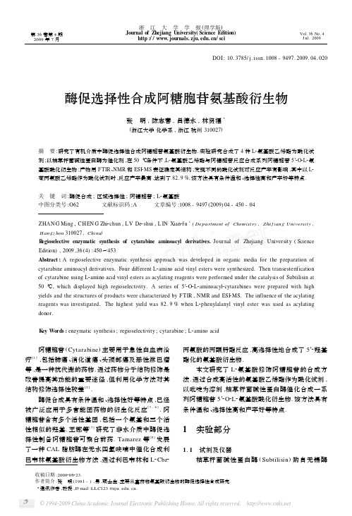
第36卷第4期2009年7月浙 江 大 学 学 报(理学版)Journal of Zhejiang U niversity(Science Edition)http :///sciVol.36No.4J ul.2009收稿日期:2008206223.作者简介:张 明(1981-),男,硕士生,主要从事药物氨基酸衍生物的酶促选择性合成研究.3通讯作者,教授,E 2mail :LL C123@.DOI :10.3785/j.issn.1008-9497.2009.04.020酶促选择性合成阿糖胞苷氨基酸衍生物张 明,陈志春,吕德水,林贤福3(浙江大学化学系,浙江杭州310027)摘 要:研究了有机介质中酶促选择性合成阿糖胞苷氨基酸衍生物.实验研究合成了4种L 2氨基酸乙烯酯为酰化试剂;以枯草杆菌碱性蛋白酶为催化剂,在50℃条件下,L 2氨基酸乙烯酯与阿糖胞苷反应合成系列阿糖胞苷5′2O 2L 2氨基酸酰化衍生物;产物用FTIR 、NMR 和ESI 2MS 表征确定其结构;发现不同的酰化试剂对反应产率有影响,其中以L 2苯丙氨酸乙烯酯作为酰化试剂时,反应产率最高,达到了82.9%.该方法具有条件温和、选择性高和产率好等特点.关 键 词:酶促合成;区域选择性;阿糖胞苷;L 2氨基酸中图分类号:O62 文献标识码:A 文章编号:1008-9497(2009)04-450-04ZHAN G Ming ,CH EN G Zhi 2chun ,L V De 2shui ,L IN Xian 2f u 3(De partment of Chemist ry ,Zhej iang Universit y ,H angz hou 310027,China )R egioselective enzym atic synthesis of cytarabine aminoacyl derivatives.Journal of Zhejiang University (Science Edition ),2009,36(4):450~453Abstract :A regioselective enzymatic synthesis approach was developed in organic media for the preparation of cytarabine aminoacyl derivatives.Four different L 2amino acid vinyl esters were synthesized.Then transesterification of cytarabine using L 2amino acid vinyl esters as acylating reagents were performed under the catalysis of Subtilisin at 50℃,which high regioselectivity.A series of 5′2O 2L 2aminoacyl 2cytarabines were prepared with high yields and the structures of products were characterized by FTIR ,NMR and ESI 2MS.The influence of the acylating reagents was investigated.The highest yield was 82.9%when L 2phenylalanyl vinyl ester was used as acylating donor.K ey Words :enzymatic synthesis ;regioselectivity ;cytarabine ;L 2amino acid 阿糖胞苷(Cytarabine )主要用于急性白血病治疗[1],包括肺癌、消化道癌、头颈部癌及恶性淋巴瘤等,是一种抗代谢的药物.通过药物分子结构修饰是改善提高其功能的重要途径,但利用化学方法对其结构修饰选择性较差[2].酶促合成具有条件温和、选择性好等特点,已经被广泛应用于多官能团药物的衍生化反应[3-6].阿糖胞苷含有多个活性基团,包括一个氨基和三个活性相似的羟基.王娜等[7]研究了非水介质中酶促选择性制备阿糖胞苷可聚合前药.Tamarez 等[8]发展了一种CAL 脂肪酶在无水四氢呋喃中催化合成利巴韦林氨基酸衍生物方法,通过利巴韦林和L 2Cbz 2丙氨酸的丙酮肟酯反应,高选择性地合成了5′2羟基酯化的氨基酸衍生物.本文研究了L 2氨基酸修饰阿糖胞苷的合成方法.通过合成高活性的氨基酸乙烯酯作为酰化试剂,以吡啶为溶剂,枯草杆菌碱性蛋白酶催化合成一系列阿糖胞苷5′2O 2L 2氨基酸酰化衍生物.该方法具有条件温和、选择性高和产率好等特点.1 实验部分1.1 试剂及仪器枯草杆菌碱性蛋白酶(Subtilisin )购自无锡酶制剂厂;阿糖胞苷为深圳信泰精细化工公司生产;其他试剂均为分析纯.化合物熔点在X24数字显示显微熔点测定仪上测定;红外光谱在Nicolet2Nexas F TIR670傅利叶变换红外光谱仪上测定;核磁在Bruker Advance DMX500型核磁共振仪(500M Hz)上测定;质谱在Bruker Esquire3000ESI2MS上测定;旋光度由WZZ22S自动旋光仪测定.1.2 酰化试剂L2氨基酸乙烯酯的制备酰化试剂的制备参考文献[9]中的方法.称取0.05mol N2Boc2L2氨基酸加入盛有200mL(2.2 mol)醋酸乙烯酯、0.5g醋酸汞的500mL圆底烧瓶中.60℃恒温水浴中加热并搅拌,滴加0.25mL浓硫酸,反应24h.反应完毕后柱层析分离产物,洗脱剂为石油醚/乙酸乙酯(体积比5/1).产物通过F TIR和1H2NMR进行结构表征.N2Boc2L2苯丙氨酸乙烯酯(a):[α]25D+9.5(c1, CHCl3);1H2NMR(CDCl3,δ):7.32-7.13(m, 6H),4.98(br,1H),4.93(d,1H,J=14Hz),4.67 (br,1H),4.65(d,1H,J=6Hz),3.13(m,2H), 1.42(s,9H).IR(neat,cm-1):3373,3090,1760, 1717,1647.N2Boc2L2丙氨酸乙烯酯(b):[α]25D+15.1(c1, CHCl3);1H2NMR(CDCl3,δ):7.25(dd,1H,J=6 Hz;J=14Hz),5.09(br,1H),4.94(d,1H,J=14 Hz),4.63(d,1H,J=6Hz),4.37(t,1H,J=6.5 Hz),1.49-1.41(m,12H).IR(neat,cm-1):3362, 3093,1761,1715,1647.N2Boc2L2缬氨酸乙烯酯(c):[α]25D+12.9(c1, CHCl3);1H2NMR(CDCl3,δ):7.22(dd,1H,J=6 Hz;J=14Hz),5.01(dbr,1H,J=8.5Hz),4.91 (d,1H,J=14Hz),4.60(d,1H,J=6Hz),4.26 (dd,1H,J=4.5Hz,J=9Hz),2.15(m,1H),1.41 (s,9H),0.95,0.88(d,6H,J=7Hz).IR(neat, cm-1):3363,3093,1758,1716,1647.N2Boc2L2亮氨酸乙烯酯(d):[α]25D+6.7(c1, CHCl3);1H2NMR(CDCl3,δ):7.25(dd,1H,J= 6.0Hz;J=14Hz),4.95(d,1H,J=14Hz),4.93 (br,1H),4.63(d,1H,J=6Hz),4.37(dd,1H,J= 9Hz,J=14Hz),1.74(m,1H),1.63(m,1H), 1.54(m,1H),1.44(s,9H),0.96,0.94(d,6H,J= 7Hz).IR(neat,cm-1):3362,3093,1759,1715, 1647.1.3 阿糖胞苷氨基酸衍生物的酶促合成50mL锥形瓶中加入阿糖胞苷122mg(M=243,0.5mmol,C9H13N3O5),10mL无水吡啶,0.2g 枯草杆菌碱性蛋白酶,再分别加入氨基酸乙烯酯(2 mmol),50℃恒温振荡培养箱中反应,转速250r・min-1.反应结束后柱层析分离提纯产物,洗脱剂为乙酸乙酯/甲醇/水(体积比27/2/1).产物通过F TIR、1H2NMR、13C2NMR和MS等分析手段进行表征,并确定其结构.5′2O2羧基2(N2Boc2L2苯丙氨酸酯)2阿糖胞苷(1a):1H2NMR(DMSO2d6,δ):7.46(d,1H,J=7.5 Hz),7.25(m,6H),7.12(br,1H),7.02(br,1H), 6.09(d,1H,J=3.5Hz),5.67(d,1H,J=7.5 Hz),5.57(br,2H),4.31-4.17(m,3H),3.97-3.89(m,3H),3.02(dd,1H,J=4.5Hz,J=13.5 Hz),2.86(dd,1H,J=5Hz,J=13.5Hz),2.00(q, 1H,J=7Hz),1.32(s,9H).13C2NMR(DMSO2d6,δ):172.5,166.1,156.0,155.6,143.4,138.1, 129.6,128.8,126.9,93.1,86.7,82.2,78.8,77.2, 74.8,65.0,55.8,36.8,28.6,28.3.IR(K Br, cm-1):3356,3030,2970,1736,1693,1706, 1603,1591.ESI2MS(m/z):491.1[M+H]+.5′2O2羧基2(N2Boc2L2丙氨酸酯)2阿糖胞苷(1b):1H2NMR(DMSO2d6,δ):7.44(d,1H,J=7.5 Hz),7.32(d,1H,J=7Hz),7.14(br,1H),7.05 (br,1H),6.08(d,1H,J=3.5Hz),5.67(d,1H,J =7.5Hz),5.59(br,2H),4.33(dd,1H,J=8.5 Hz,J=3Hz),4.20(dd,1H,J=3.5Hz,J=8.5 Hz),4.03- 3.91(m,3H),3.88(br,1H),1.37, 1.32(s,9H),1.24(d,3H,J=7.5Hz).13C2NMR (DMSO2d6,δ):173.7,166.1,156.8,155.6,143.3, 93.1,86.8,82.3,78.7,77.2,74.7,64.8,49.5, 28.7,17.4.IR(K Br,cm-1):3356,3219,2978, 2887,1736,1698,1645.ESI2MS(m/z):437.1[M +Na]+.5′2O2羧基2(N2Boc2L2缬氨酸酯)2阿糖胞苷(1c):1H2NMR(DMSO2d6,δ):7.46(d,1H,J=7.5 Hz),7.19(d,1H,J=9Hz),7.13(br,1H),7.05 (br,1H),6.08(d,1H,J=3.5Hz),5.64(d,1H,J =7.5Hz),5.59(br,2H),4.39(dd,1H,J=8.5 Hz,J=3Hz),4.16(dd,1H,J=3.5Hz,J=8.5 Hz),3.94(m,2H),3.86(m,2H),2.00(q,1H,J= 7Hz),1.37(s,9H),0.87(d,6H,J=7Hz).13C2 NMR(DMSO2d6,δ):172.6,166.1,156.3,155.6, 143.3,93.0,87.0,82.4,78.8,77.2,74.7,64.7, 59.9,30.2,28.7,19.4,18.8.IR(K Br,cm-1): 3356,3219,2970,2877,1736,1693,1648.ESI2154 第4期张 明,等:酶促选择性合成阿糖胞苷氨基酸衍生物MS (m/z ):465.1[M +Na ]+.5′2O 2羧基2(N 2Boc 2L 2亮氨酸酯)2阿糖胞苷(1d):1H 2NMR (DMSO 2d 6,δ):7.44(d ,1H ,J =7.5Hz ),7.27(d ,1H ,J =9Hz ),7.15(br ,1H ),7.05(br ,1H ),6.08(d ,1H ,J =3.5Hz ),5.65(d ,1H ,J =7.5Hz ),5.58(br ,2H ),4.33(dd ,1H ,J =8Hz ,J =11Hz ),4.18(dd ,1H ,J =4Hz ,J =12Hz ),3.99(m ,2H ),3.92(m ,1H ),3.87(m ,1H ),1.63(m ,1H ),1.52(m ,1H ),1.44(m ,1H ),1.37(s ,9H ),0.86(d ,3H ,J =6Hz ),0.83(d ,3H ,J =6Hz ).13C 2NMR (DMSO 2d 6,δ):173.6,166.1,156.1,155.6,143.3,93.1,86.8,82.2,78.7,77.2,74.8,64.7,52.5,40.3.2,28.7,24.8,19.4,18.8.IR (K Br ,cm -1):3356,3219,2968,2877,1736,1693,1647.ESI 2MS (m/z ):479.1[M +Na ]+.2 结果与讨论2.1 L 2氨基酸乙烯酯的合成在非水介质酶促酯化反应中,人们通常采用活化的酰基供体来进行反应.乙烯酯是当前在酶促酯交换反应中较好,也是用得比较多的酰基供体[4,10].本文以4种含不同侧链的L 2氨基酸(L 2苯丙氨酸、L 2丙氨酸、L 2缬氨酸、L 2亮氨酸)为原料,在催化剂作用下与乙酸乙烯酯反应,合成了新颖的高活性酰化试剂L 2氨基酸乙烯酯.其中L 2苯丙氨酸是典型的芳香族氨基酸;L 2丙氨酸是结构最简单的天然氨基酸;而3种脂肪族氨基酸L 2丙氨酸、L 2缬氨酸和L 2亮氨酸的侧链残基由小到大,用于考察酰化试剂位阻的不同对酶促反应的影响.L 2氨基酸乙烯酯通过F TIR 和1H 2NMR 进行结构表征.1H 2NMR 中7.25,4.93,4.65pp m 证明乙烯酯中端基-OC H =CH 2的NMR 信号,IR 中3093,1647cm -1确认-C H =C H 2的特征吸收;1760cm -1是C =O 的特征吸收;合成产物的旋光度测定结果如实验部分所示,表明合成a 2d 产物是预期的L 2氨基酸乙烯酯.2.2 酶促选择性合成阿糖胞苷氨基酸衍生物研究是以枯草杆菌蛋白酶为催化剂,在吡啶介质和50℃条件下,用实验室合成的4种氨基酸乙烯酯为酰化试剂,酶促合成阿糖胞苷氨基酸衍生物,合成反应方程式如图1所示.产物通过F TIR 、1H 2NMR 、13C 2NMR 和ESI 2MS 等分析手段进行表征,数据如实验部分所示.图1 阿糖胞苷氨基酸衍生物的酶促选择性合成Fig.1 Regioselective enzymatic synthesis of cytarabin aminoacyl derivatives 虽然阿糖胞苷分子结构中含有一个氨基和3个活性相似的羟基都可以参与反应,而以枯草杆菌碱性蛋白酶为催化剂的反应中,根据TL C 点板及产物结构表征结果可以确定反应得到单一产物.为了研究判断羟基酰化位置,阿糖胞苷及其衍生物的糖环部分碳13C 2NMR 信号的化学位移如表1所示.从表中可以看出,阿糖胞苷的L 2苯丙氨酸衍生物1a 的C 25′的化学位移从61.3移至低场65.0,C 24′的化学位移从85.3移向高场82.2.这样就可以确定1a 为底物糖环上的5′2羟基酰化的衍生物[11],因为化合物的某个碳上羟基被酰化时,将导致该碳的13C 2NMR 信号的化学位移向低场移动,同时引起相邻碳13C 2NMR 信号的化学位移向高场移动.用同样的方法可判断化合物1b 21d 的酰化取代也发生在阿糖胞苷5′2羟基.1H 2NMR 数据也证实了酶促合成的阿糖胞苷氨基酸衍生物是伯羟基酰化衍生物.因此,在无水吡啶中,枯草杆菌碱性蛋白酶催化阿糖胞苷氨基酸衍生化反应具有很高的选择性.表1 阿糖胞苷及其衍生物糖环部分碳谱化学位移Table 1 Chemical shifts of13C 2NMR of sugar moiety ofcytarabine and it ′s derivativesCarbon 11a 1b 1c 1d 1′86.386.786.887.086.82′76.977.277.277.277.23′75.374.874.774.774.84′85.382.282.382.482.25′61.365.064.864.764.7254浙江大学学报(理学版) 第36卷 2.3 L2氨基酸乙烯酯的结构对反应影响本文考察了4种含有不同侧链基团的氨基酸乙烯酯作为酰化试剂参与酶促合成反应,实验结果和物性数据如表2所示.表中结果显示,4种氨基酸乙烯酯都具有较高的反应活性,反应产率在65.9%~82.9%.酰化试剂中含有不同氨基酸残基对反应产率有一定影响,其中以L2苯丙氨酸乙烯酯作为酰化试剂时,反应产率最高,达到了82.9%.L2苯丙氨酸乙烯酯作为酰化试剂反应效果较好可能是由于含有芳香基团而有利于其与酶活性中心的结合;对比3种脂肪族氨基酸乙烯酯,可以看出氨基酸残基侧链基团的位阻大小对反应产率影响不是很显著.表2 酶促合成阿糖胞苷氨基酸衍生物的产率及产物的理化性质Table2 Y ields of enzymatic reaction and the products′properties酰化试剂产物反应时间/d产率1熔点/℃旋光度2 N2Boc2L2苯丙氨酸1a382.9114~116+52.4 N2Boc2L2丙氨酸1b378.6129~130+59.8 N2Boc2L2缬氨酸1c365.9111~113+58.2 N2Boc2L2亮氨酸1d374.078~80+49.9 1:过柱产率;2:[α]25D(c1,DMSO)3 结 论研究以阿糖胞苷为底物,4种L2氨基酸乙烯酯为活性酰化试剂,在非水介质中酶促选择性合成了阿糖胞苷的氨基酸衍生物.利用F TIR、1H2NMR、13C2NMR和ESI2MS等手段对产物结构进行表征.发现在无水吡啶中,枯草杆菌碱性蛋白酶催化反应选择性的发生在阿糖胞苷糖环的5′2羟基上.研究了4种氨基酸乙烯酯酰化试剂对反应的影响,发现以苯丙氨酸乙烯酯作为酰化试剂时反应最佳,产率达到82.9%.所得到阿糖胞苷衍生物以及相应前药性质正在系统研究中.参考文献(R eferences):[1] PALLAVICINI M G.Cytosine arabinoside:molecular,pharmacokinetic and cytokinetic considerations[J].Pharm acol Ther,1984,25:2072238.[2] TOBIAS S C,BORCH R F.Synthesis and biologicalevaluation of a cytarabine phosphoramidate prodrug[J].Mol Pharm aceutics,2004,1:1122116.[3] FERRERO M,GO TOR V.Biocatalytic selective mod2ifications of conventional nucleosides,carbocyclic nu2cleosides,and c2Nucleosides[J].Chem R ev,2000,100:431924347.[4] W AN G Y F,LALONDE J J,MOMON G AN M,et al.Lipase2catalyzed irreversivle transesterifications using enolesters as acylating reagents2preparative enantioselectiveand regioselective syntheses of alcohols,glycerol deriva2tives,sugars,and organometallics[J].J Am Chem Soc, 1988,110:720027205.[5] YU H L,XU J H,L U W Y,et al.Environmentallybenign synthesis of natural glycosides using apple seed meal as green and robust biocatalyst[J].J Biotechnol, 2008,133:4692477.[6] IN TRA A,BA VA A,NASINI G,et al.Regioselectiveenzymatic acylation of polyhydroxylated sesquiterpenoids [J].J Mol C atal B2E nzym,2004,29:95298.[7] WAN G Na,CH EN Zhi2chun,L U De2shui,et al.Controllable selective synthesis of a polymerizable pro2 drug of cytarabine by enzymatic and chemical methods [J].Bioorg Med Chem Lett,2005,15:406424067. [8] TAMAREZ M,MOR GAN B,WON G G S K,et al.Pilot2scale lipase2catalyzed regioselective acylation ofribavirin in anhydrous media in the synthesis of a novel prodrug intermediate[J].Org Process R es Dev,2003, 7:9512953.[9] BO YER V,STANCH EV M,FAIRBAN KS A J,etal.Ready protease2catalyzed synthesis of carbohydrate2 amino acid conjugates[J].Chem Commun,2001,12: 190821909.[10] KIT A Y,T A KE BE Y,MURAT A K,et al.Convenientenzymatic resolution of alcohols using highly reactive,nonharmful acyl donors,12ethoxyvinyl esters[J].J OrgChem,2000,65:83288.[11] YOSHIMO TO K,ITAN TAN I Y,TSUDA Y.13C2Nuclear magnetic resonance spectra of O2acylglucose.Additivity of shift parameters and its application tostructure elucidations[J].Chem Pharm Bull,1980,28:206522074.(责任编辑 涂 红)354 第4期张 明,等:酶促选择性合成阿糖胞苷氨基酸衍生物。
烟酰胺单核苷酸(NMN)的活性与化学制备的研究进展
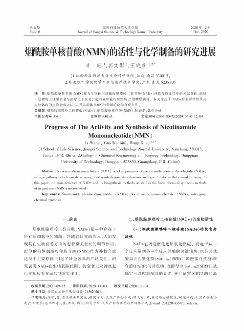
2020年12月Dec. 2020第6期Issue 6江西科技师范大学学报Journal of Jiangxi Science & Technology Normal University烟酰#单核&酸(NMN )的活性与化学制备的硏究进展李 旺A ,郭文彬A ,王晓季A"(1.江西科技师范大学生命科学学院,江西南昌330013;2.东莞理工学院化学工程与能源技术学院,广东东莞523830)摘要:烟酰胺单核/酸(NM N )作为生物体内烟酰胺腺瞟吟二核/酸(NAD+)补救合成途径中的关键前体,能够一定程度上延缓衰老与治疗由于衰老引起的老年退行性疾病、2型糖尿病等%本文综述了 NAD+的主要活性及其生物体内的生物合成方法,以及其前体NMN 的最新的化学合成方法。
关键词:烟酰胺腺瞟吟二核/酸(NAD+);烟酰胺单核/酸(NMN );抗衰老;化学合成中图分类号:06-1文献标识码:A 文章编号:2096-854X (2020)06-0112-04Progress of The Activity and Synthesis of NicotinamideMononucleotide (NMN )Li Wang 1, Guo Wenbin 1, Wang Xiaoji 1,2,*(1.School of Life Science, Jiangxi Science and Technology Normal University, Nanchang 330013,Jiangxi, P.R. China; 2.College of Chemical Engineering and Engergy Technology, DongguanUniversity of Technology, Dongguan 523830, Guangdong, P.R. China )Abstract : Nicotinamide mononucleotide (NMN ) is a key precursor of nicotinamide adenine dinucleotide (NAD+)salvage pathway, which can delay aging, treat senile degenerative diseases and type 2 diabetes that caused by aging. In this paper, the main activities of NAD+ and its biosynthesis methods, as well as the latest chemical synthesis methods of its precursor NMN were reviewed.Key words : Nicotinamide adenine dinucleotide (NAD +); Nicotinamide mononucleotide (NMN ); anti -aging;chemical synthesis—、前言烟酰胺腺8吟二核/酸(NAD+ "是一种存在于 所有活细胞中的辅酶。
卟啉化合物的合成
柱进行层析 ,产率为 915 %。微波合成卟啉在我国 发展较快 ,研究表明微波作用的时间与强度 、反应体 系溶剂及催化剂的选择 、反应试剂的组成及用量等 均对四苯基卟啉的合成有较大的影响 ,经过改进 ,微 波合成卟啉的产率有较大的提高 ( > 30 %) [29 ,30] 。 112 八乙基卟啉的合成
卟啉类化合物的合成方法归纳起来主要有两 种 : (1) 由非卟啉前体合成卟啉 ; (2) 卟啉化合物的官 能团修饰 。非卟啉前体合成卟啉的方法按照缩合成 环方式的不同大致可以分为 : (1) 4 个吡咯单体直接 缩合环化生成卟啉 ,适用于图 1 中化合物 1 和 2 ; (2) 模块法 ,适用于图 1 中化合物 3 —10 。合成方法和路 线的选择取决于目标卟啉分子的结构特点和性质 。
中 ,以三氟化硼和乙醚络合物催化 ,整个反应分两步 进行 ,先得到卟啉合成的中间体 (porphyrinogen ,卟啉 原 ———Dolphin 等[13] 曾证实了这一反应中间体的存 在) ,然后 , 以二氯二腈基苯醌 (DDQ) 或四氯苯醌 (TCQ) 将卟啉原氧化得到最终产物卟啉 ,从而使反 应可以在常温下进行 。由于 Lindsey 法的反应温度 较低 ,一般不会产生焦油状副产物 ,目标产物的分离 提纯较容易 ;同时温和的反应条件也允许反应物先 经过化学修饰 ,连接上一些敏感基团 ,平均产率可达 30 % —40 %。但该反应浓度低 ,以吡咯计仅为 10- 2 molΠL ,且最大反应容积为 1L ,放大后则效果不好 。 1994 年 ,Lindsey 等[14] 研究了在高浓度下的反应 ( ≥ 011molΠL) ,实验采用一步法 ,即将原料 、氧化剂 、催 化剂同时加入 ,最后产率可达 10 % —20 % ; 采用两 步法 , 即先加催化剂 , 反应后再加氧化剂 , 产率为 20 % —30 % , (图 3) 。Lindsey 法经过改良 ,还能得到 立体位阻较大的苯基 2 ,52位有取代基的四芳基卟 啉[12] ,其 非 共 平 面 的 构 型 表 现 出 不 同 的 光 学 性 能[15] 。四 (2 ,4 ,62三甲基苯基) 卟啉[16] 就是这样一 个例子 ,合成时在 BF3 中加入乙醇作为协同催化剂 , 产率达 30 %。Llama 等[17] 合成 TPP 时加入了过渡金 属盐 ,这一改良使产率高达 68 % ,而且可以做到比 传统 Lindsey 法的反应液浓度高 。
Pyrabactin的合成及植物生长调节活性
Synthesis and Plant Growth Regulation Activity of Pyrabactin
ZHOU Fan, DUAN Liu-sheng
(College of Agriculture and Biotechnology, China Agricultural University, Beijing 100193, China)
2 生物活性测定
对所合成的目标化合物pyrabactin进行了室内生物活 性试验,采用单子叶植物小麦种子(京411)和双子叶植物绿 豆种子(绿宝石)做发芽试验,测定种子发芽调节活性。
2.1 试验方法 种子处理:将小麦种子(京411)和绿豆种子(绿宝石)用
2%次氯酸钠消毒15 min,用灭菌水冲洗干净。 发芽试验:使用0(对照)、1、10、100、1 000 mg/L的
589。谱图与6-氯化亚砜的用量
1.2.3 2,6-二氯苯并 唑酮的合成
考察了氯化亚砜的用量对氯化结果的影响。按1.2.3
在上步反应混合液中,滴加氯化亚砜90 g,之后加热 至120 ℃反应6 h,减压回收邻二氯苯,余物经减压蒸馏 得浅黄色油状液体,室温下很快固化,产物重81.2 g,收率
Key words: pyrabactin; synthesis; plant growth regulating activity
脱落酸(ABA)是已经发现的五大激素之一,能调控 植物的多种生理活动[1],比如加速叶片的脱落,诱导休眠, 抑制发芽和关闭气孔,并且在面对干旱和低温等逆境胁 迫时做出响应[2-3]。由于脱落酸成本高,稳定性差,所以人 们一直在研究与脱落酸有类似活性的物质[4]。
据 文 献 报 道 :p y r a b a c t i n 可 以 特 异 性 激 活 模 式 植 物 拟南芥的ABA受体,即和start蛋白的PYR/PYL家族中的
多硫代二酮哌嗪类化合物的研究进展_李莉媛
. 161 .收稿日期:2012-03-15基金项目:山东省自然科学基金(ZR2010HM057);国家863课题基金(2011AA09070104)作者简介:李莉媛,女,生于1985年,在读博士研究生,主要从事真菌活性代谢产物的提取、分离和鉴定。
E-mail:2001angel007@。
*通讯作者,顾谦群,女,硕士,教授,博士生导师。
E-mail:guqianq@文章编号:1001-8689(2013)03-0161-14多硫代二酮哌嗪类化合物的研究进展李莉媛 朱天骄 李德海 顾谦群*(中国海洋大学 医药学院 海洋药物教育部重点实验室,青岛 266003)摘要:多硫代二酮哌嗪(Epipolythiodioxopiperazines ,ETPs )是一类重要的主要由真菌产生的活性次级代谢产物,其结构特征为二酮哌嗪母核中含有硫桥;ETPs具有广泛的生物活性,例如:抗增殖、细胞毒、免疫抑制、抗病毒以及抗菌等。
大量研究表明,分子中的硫桥结构是ETP类化合物保持活性的关键药效基团。
据统计,截至2011年底,从天然界中共分离得到126个ETP 类化合物。
本文根据其结构中组成氨基酸以及修饰的不同将ETP类化合物分为15种结构类型,并就其来源、结构、生物活性、生物合成及其构效关系研究进行综述。
关键词:多硫代二酮哌嗪;真菌;生物活性;构效关系;生物合成中图分类号:R978 文献标识码: AProgress in the research of epipolythiodioxopiperazinesLi Li-yuan, Zhu Tian-jiao, Li De-hai and Gu Qian-qun(Key Laboratory of Marine Drugs, Chinese Ministry of Education, School of Medicine and Pharmacy,Ocean University of China, Qingdao 266003)Abstract Epipolythiodioxopiperazines (ETPs) are an important class of biologically active compounds mainly produced by fungi. ETPs are characterized by a bridged polysulfide piperazine ring. These metabolites have been found to possess a wide variety of biological activities, including antiproliferative, cytotoxic, immunomodulatory, antiviral, antibacterial and antifungal activities. The crucial structural element responsible for the observed biological properties has been shown to be the sul fi de linkage. According to statistic, up to the end of 2011, 126 ETPs have been isolated from fungi and lichens. In this article, 15 different kinds of ETPs are known according to the diversity of the amino acids of the the core ETP moiety and the modi fi cations of these amino acids. We review the researches on the origins, structures, biological activities, biosynthesis and structure-activity relationships of ETPs.Key words Epipolythiodioxopiperazines; Fungi; Bioactivity; Structure-activity relationship; Biosynthesis 中国抗生素杂志2013年3月第38卷第3期多硫代二酮哌嗪类化合物(e p i p o l y t h i o -dioxopiperazines, ETPs)是一类重要的生物活性化合物,主要由真菌代谢产生,其结构中都含有带硫桥的二酮哌嗪母核。
邻苯二酚类化合物的合成及作为核酸交联剂的生物活性研究——武汉大学综合化学实验实验报告
邻苯二酚类化合物的合成及作为核酸交联剂的生物活性研究(武汉大学化学与分子科学学院,武汉430072)摘要:本实验合成了两个邻苯二酚类化合物,分别检测它们对不同二级结构核酸的交联活性。
实验的活性检测部分既包含对双链核酸的交联活性检测,又包含对G-四链体的交联活性检测,分别通过琼脂糖凝胶电泳和聚丙烯酰胺凝胶电泳监测其交联活性。
最后用圆二色谱测G-四链体的半解链温度,并用MBTH显色反应证实邻苯二醌中间体的产生,进一步证实了交联的发生。
关键词:邻苯二酚类核酸交联剂G-四链体中图分类号:O 643 文献标识码:A0 引言交联具有重要的生物学意义。
组成DNA 长链的碱基,糖环和磷酸基团中含有大量的富电子的N,P 原子,O,这些原子容易受到亲电试剂的进攻。
利用这一点,在特定的药物小分子上接上亲电基团,这些亲电基团进攻富含电子的N,O,P原子从而生成稳定的共价键。
从而改变了DNA 的结构组成,达到阻碍DNA 复制和转录的目的。
这样的过程被称为DNA 的烷基化。
而这些小分子就被称为DNA 的烷基化试剂。
有很多研究表明,在生物体中的DNA 的被烷基化后,生物体会有一个自生修复的机制。
被烷基化的DNA 可以通过生物体内的DNA 修复酶得到部分修复。
使得这样的DNA 损伤不至于影响到生物体内遗传信息的传递和细胞的死亡。
但是如果药物分子带的烷基化基团增多,对DNA 双链烷基化效果更好,也就使DNA修复酶对DNA 的修复作用变弱。
通常我们把这些带有两个或者以上的烷基化试剂叫做DNA 的交联剂。
EcoR I是Ⅱ型限制性内切酶[1],EcoR I以同型二聚体作用,两个EcoR I分子沿着DNA 识别序列的相对方向排列,进入DNA分子内部对碱基发挥作用。
EcoR I分子内部特异性氨基酸与DNA识别序列GAATTC形成氢键。
一旦结合,限制酶内部的其他残基催化水解反应,利用水破坏识别序列内部DNA双链中每条链的磷酸二酯键[2]。
本实验将其用作对pBR322质粒DNA进行酶切,得到线性的DNA分子有利于通过凝胶电泳研究化合物的交联模式。
两种苝二酰亚胺衍生物的合成及光物理性质研究_赵英杰
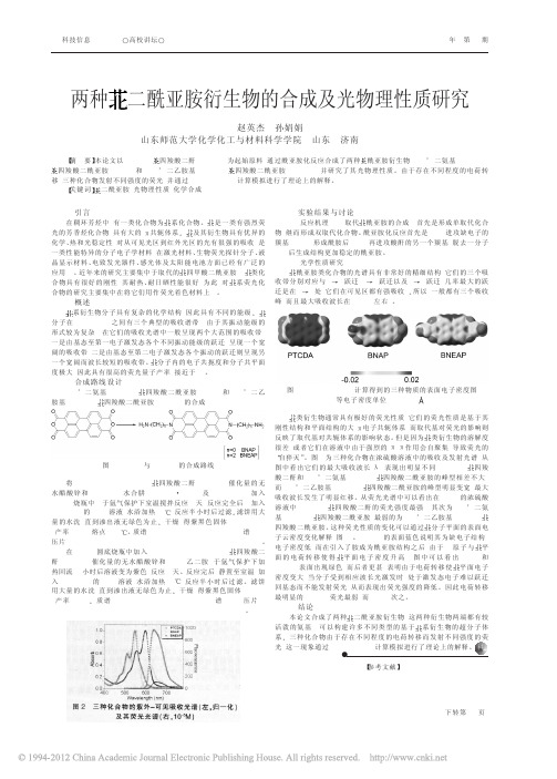
系衍生物分子具有复杂的化学结构, 因此具有不同的能级。 分子在 350- 600nm 之间有三个典型的吸收谱带, 由 于 其 振 动 能 级 的 形式较为复杂, 在它们的吸收光谱中一般呈现两个大范围的吸收带, 一是由基态至第一电子激发态各个不同振动能级的跃迁, 呈现一个宽 阔的吸收带, 二是由基态至第二电子激发态各个振动的跃迁则呈现另 一个宽阔而波长较短的吸收带。 分子内的电子共扼度和分子共平面 度极大, 因此具有很高的荧光量子产率( 接近于 1) 。
【参 考 文 献 】
[ 1] L.Schmidt - Mende, A.Fechtenkotter, K.Mullen etal. Self - organized discotic
liquid crystals for high - efficiency organic photovoltaics [J].Science. 2001, 293:
酰亚胺类化合物的光谱具有非常好的精细结构, 它们的三个吸 收带分别对应与 0→1 跃迁, 0→2 跃迁以及 0→3 跃迁 , 几 率 最 大 的 跃 迁是在 0→1 处, 它们在可见区都有强吸收[7]。所以, 一般都有三个吸收 峰, 而且最大吸收波长在 530nm 左右[8]。
图 3 B3LYP /6- 31G 计算得到的三种物质的表面电子密度图 ( 等电子密度单位 0.0004 e !-3)
3.合成路线设计 N,N’- 二氨基- 3,4,9,10- 四 羧 酸 二 酰 亚 胺 ( BNAP) 和 N,N’- 二 乙 胺基- 3,4,9,10- 四羧酸二酰亚胺( BNEAP) 的合成:
4.实验结果与讨论 4.1 反应机理 N- 取代 酰亚胺的合成, 首先是形成单取代化合 物, 继而形成双取代化合物。酰亚胺化反应首先是- NH2 进攻缺电子的 羰基( C=O) , 形成酰胺后, - NH 再进攻酸酐的另一个羰基, 脱去一分子 H2O 后生成结构更加稳定的酰亚胺。 4.2 光学性质研究
二烯丙基二硫化物诱导肝癌细胞凋亡的研究

二烯丙基二硫化物诱导肝癌细胞凋亡的研究目的:研究大蒜提取物二烯丙基二硫化物(DADS)诱导肝癌细胞凋亡过程的影响作用,探讨DADS抗肿瘤的可能机制。
方法:MTT法检测细胞活性及流式细胞术检测细胞凋亡率。
结果:与未处理的对照组相比,随着用药浓度增高,细胞生长抑制率逐渐增高,即细胞活性下降(P<0.01)。
结论:DADS能诱导肝癌细胞发生细胞凋亡,为肿瘤治疗方面的临床用药奠定基础。
标签:二烯丙基二硫化物;人肝癌细胞株HepG2;细胞凋亡。
近年来,植物来源药物越来越受到重视,充分利用我国药用植物资源,开发新的抗肿瘤药物,已成为肿瘤研究领域的重要内容。
大蒜为百合科植物蒜的鳞茎,目前对大蒜的研究在国内外已经成为热点。
大量研究证实,大蒜有防肿瘤、抗肿瘤作用,其中主要的生物活性物质是大蒜所含有的含硫化合物,现已测得含硫成分多达30余种,DADS就是其中的一种脂溶性的有效成分。
已有研究实,DADS 对CNE2人鼻咽癌细胞、HepG2人肝癌细胞生长有抑制作用[1]。
本实验我们进行了DADS诱导肝癌细胞凋亡的研究,现报告如下:1材料与方法1.1材料高糖型DMEM培养基购自美国Gibco公司;新生小牛血清购自杭州四季青公司;DMSO、MTT均为Sigma公司产品;DADS为Fluka公司产品。
DADS用DMSO配制,处理细胞时,DMSO的最终体积小于或等于0.1%。
1.2方法1.2.1细胞培养人肝癌细胞株HepG2 由南华大学肿瘤研究所提供,置DMEM 培养液中,加10%小牛血清,于37℃5% CO2培养箱中培养。
隔天换液一次。
细胞经传代和收获,取对数生长期细胞进行实验。
1.2.2实验分组①溶媒对照组:二甲亚砜(DMSO)即DADS 0μmol/L;②DADS 浓度梯度组:DADS(20、50、100μmol/L)。
1.2.3MTT 法(四甲基偶氮唑盐微量酶反应比色法) 测定细胞活性活细胞内线粒体琥珀酸脱氢酶能催化四甲基偶氮唑盐(MTT)形成蓝色甲臜。
氨基酸卟啉衍生物的合成

浙江大学硕t-毕业论文
Abstract
This dissertation includes two parts:(1)The total synthesis of TAPP—Valine and TAPP·SerZ—PAG,and(2)Investigation on the phosphorylation reaction of aniline
serving as the model compound of TAPP.
Part 1:Adler’S strategy was used to the synthesis of meso—Tetra
(4-nitrophenyl)porphyrin(TNPP)through Cyclolization of 4-nitrobenzaldhyde
浙江大学 硕士学位论文 氨基酸卟啉衍生物的合成 姓名:金晓敏 申请学位级别:硕士 专业:化学·有机化学 指导教师:吴健
20020601
巍江大学硕士毕业论文
摘要
本文主要由两部分组成:(1)带有缬氨酸侧链的卟啉TAPP.Val和带有B一葡 萄糖基丝氨酸侧链的卟琳TAPP—SerZ。PAO的全合成;(2)以苯胺作为1-APP的 模拟物对卟啉化合物的磷酰化反应进行了初步探索。
be widely used in many fields such as biology,material,polymer and SO on.
B-D—pentacetyl glucose was prepared from the D-glucose.Based on this,a
perfect synthesis process of 3-O-12’,3’,4’,6’-tetra(O—acetyl)一B-D—glucopyranosyl]- N-benzyloxycarbonyl—L—Serine(PAG-Ser-Z)through the reaction of 8
ICHM7(step4)基因毒性杂质评估和控制◆中英

ASSESSMENT AND CONTROL OF DNA REACTIVE(MUTAGENIC) IMPURITIES IN PHARMACEUTICALS TOLIMIT POTENTIAL CARCINOGENIC RISK为限制潜在致癌风险而对药物中DNA活性(诱变性)杂质进行的评估和控制M7Current Step 4 versiondated 23 June 2014This Guideline has been developed by the appropriate ICH Expert Working Group and has been subject to consultation by the regulatory parties, in accordance with the ICH Process. At Step 4 of the Process the final draft is recommended for adoption to the regulatory bodies of the European Union, Japan and USA.M7Document History 文件历史The document is provided "as is" without warranty of any kind. In no event shall the ICH or the authors of the original document be liable for any claim, damages or other liability arising from the use of the document.The above-mentioned permissions do not apply to content supplied by third parties. Therefore, for documents where the copyright vests in a third party, permission for reproduction must be obtained from this copyright holder.ASSESSMENT AND CONTROL OF DNA REACTIVE (MUTAGENIC) IMPURITIES IN PHARMACEUTICALS TO LIMIT POTENTIALCARCINOGENIC RISK为限制潜在致癌风险而对药物中DNA活性(诱变性)杂质进行的评估和控制ICH Harmonised Tripartite GuidelineICH三方协调指南Having reached Step 4 of the ICH Process at the ICH Steering Committee meeting on 5 June 2014, this Guideline is recommended for adoption to the three regulatory parties to ICHASSESSMENT AND CONTROL OF DNA REACTIVE (MUTAGENIC) IMPURITIES IN PHARMACEUTICALS TO LIMIT POTENTIALCARCINOGENIC RISK为限制潜在致癌风险而对药物中DNA活性(诱变性)杂质进行的评估和控制1. INTRODUCTION概述The synthesis of drug substances involves the use of reactive chemicals, reagents, solvents, catalysts, and other processing aids. As a result of chemical synthesis or subsequent degradation, impurities reside in all drug substances and associated drug products. While ICH Q3A(R2): Impurities in New Drug Substances and Q3B(R2): Impurities in New Drug Products (Ref. 1, 2) provides guidance for qualification and control for the majority of the impurities, limited guidance is provided for those impurities that are DNA reactive. The purpose of this guideline is to provide a practical framework that is applicable to the identification, categorization, qualification, and control of these mutagenic impurities to limit potential carcinogenic risk. This guideline is intended to complement ICH Q3A(R2), Q3B(R2) (Note 1), and ICH M3(R2): Nonclinical Safety Studies for the Conduct of Human Clinical Trials and Marketing Authorizations for Pharmaceuticals (Ref. 3).原料药合成牵涉到使用活性化学物质、试剂、溶剂、催化剂和其它工艺助剂,导致在所有原料药及其制剂中会残留有化学合成或其降解产物、杂质。
专业英语翻译练习

英文题目翻译
Synthesis, structure and tyrosinase (酪氨酸酶) inhibition of natural phenols derivatives 天然多酚衍生物的合成、结构和酪氨酸 酶抑制作用
英文题目翻译
中药寡糖的药理作用与机制研究进展
Progress on the Pharmacological Action and Mechanism of Oligosaccharides from TCM (Traditional Chinese Medicine)
Alkaline amylase ['æmɪleɪz] is one of alkaline enzymes with optimum pH in the alkaline range, and it could keep stability and efficiently hydrolyze starch under alkaline conditions. Alkaline amylase finds wide applications in textile, detergent, pharmaceutical, food and other fields. Alkaline amylases could be produced by alkaliphilic microorganisms. In this work, the advances of alkaline amylase production and applications were reviewed.
英文句子翻译
本文总结分析了近几年该类化合物构 效关系的研究进展,期望为黄酮类衍生
含双Cyclen阳离子脂质的合成及与DNA的相互作用
因素. 然而 , 基因治疗要作为一种常规的治疗方法还有很多问题需要解决 , 中很重要的一点就是寻 其 找 高效 低毒 的转 基 因载体 .基 因载体 可分 为病 毒载 体 和非 病 毒 载体 ,病 毒载 体尽 管 转 染效 率 高 , 但 是存在免疫原性 、 致癌性及宿主 D A插入整合等弊端 , 而限制 了其应用 .非病毒载体无免疫原 N 从 J
中性 脂 质 D P O E形成 的脂 质体 对 D A的包裹 能力 ,发现 其 已具备 了作 为基 因载体 的潜在 性 . N
Байду номын сангаас1 实验 与 结 果
1 1 仪 器与试 剂 .
H N R和 ” M rkr V I4 0M z M CN RBue A - H 核磁共振波谱仪 , TR 1P I0 F I 6 C型傅里叶变换红外光谱 仪 ; 分辨 质谱 ( M ) 用 B krD hnc i T F型质谱 仪 测定 .柱 层析 硅胶 H( 岛海 洋化 工 厂 高 HR S 采 o r e aois o O B 青
性 , 于 大规模 生产 , 转染 效 率低 于病 毒 载体 .在 所有 的非 病 毒 载体 中 ,由于 阳离 子脂 质 体 具 有较 便 但
低的免疫原性 , 容易合成及表征 , 结构明确 , 利于大规模生产和包裹大片段的 D A分子,已被作为一 N 种 重要 的基 因载 体广 泛研究 j .自 18 97年 Fl e 等 首 次将脂 质 体用 作 基 因转 染 载体 以来 ,已经设 eg r n 计合成了多种脂质体 , 但这些脂质体尚不能满足临床应用 的要求 ,因此探寻更加高效低毒的阳离子脂 质体基因载体仍非常重要. 已有研究 证明,以多胺基团作为亲水性头部 的阳离子脂质体较含有单个胺或季铵盐头部的阳
干扰素α-2b蛋白的制备与纯化
干扰素α-2b蛋白的制备与纯化Production and PurificationInterferon α-2b protein学科专业:微生物与生化药学研究生:李强指导教师:***天津大学药物科学与技术学院二零一三年六月独创性声明本人声明所呈交的学位论文是本人在导师指导下进行的研究工作和取得的研究成果,除了文中特别加以标注和致谢之处外,论文中不包含其他人已经发表或撰写过的研究成果,也不包含为获得天津大学或其他教育机构的学位或证书而使用过的材料。
与我一同工作的同志对本研究所做的任何贡献均已在论文中作了明确的说明并表示了谢意。
学位论文作者签名:签字日期:年月日学位论文版权使用授权书本学位论文作者完全了解天津大学有关保留、使用学位论文的规定。
特授权天津大学可以将学位论文的全部或部分内容编入有关数据库进行检索,并采用影印、缩印或扫描等复制手段保存、汇编以供查阅和借阅。
同意学校向国家有关部门或机构送交论文的复印件和磁盘。
(保密的学位论文在解密后适用本授权说明)学位论文作者签名:导师签名:签字日期:年月日签字日期:年月日中文摘要干扰素(IFNs)是宿主细胞在受到病原体例如病毒、微生物、寄生虫、肿瘤细胞的侵染后产生并释放的。
干扰素属于糖蛋白的一种,具有干扰病毒在宿主体内复制的能力,它对于提高宿主免疫系统对病毒的识别和抵抗起到十分重要的作用,现在被广泛的运用在病毒引起的疾病和肿瘤的治疗中。
本实验中的研究对象是干扰素α-2b,它属于干扰素α,同样具有抗病毒、抗肿瘤等作用。
现代已经广泛的应用于肿瘤和癌症的治疗中。
但是天然态的干扰素α-2b产量稀少,这样就使得需要这种药物治疗的病人承受巨大的经济负担,那么提高干扰素蛋白的产量就是摆在现在科研人员面前的一道难题。
运用重组DNA技术,人们已经可以用E coli表达系统高效表达干扰素α-2b 基因,干扰素α-2b蛋白以包涵体的形式存在,这样就需要将包涵体蛋白变性、复性来得到具有生物活性的干扰素α-2b,然而这一步中极易形成聚集体和错误折叠,这个问题已经是影响干扰素大量生产的瓶颈问题。
吡咯烷二硫代氨基甲酸铵晶体结构
吡咯烷二硫代氨基甲酸铵晶体结构英文回答:The crystal structure of pyrrolidine-2-thione ammonium formate is a fascinating topic to explore. This compound consists of two main components: pyrrolidine-2-thione and ammonium formate. Pyrrolidine-2-thione is a five-membered ring containing four carbon atoms and one sulfur atom. Ammonium formate, on the other hand, is a salt composed of ammonium cations (NH4+) and formate anions (HCO2-).In the crystal structure, the pyrrolidine-2-thione molecules and ammonium formate ions are arranged in a regular pattern. The pyrrolidine-2-thione molecules form a lattice structure, with each molecule connected to its neighboring molecules through weak intermolecular forces, such as hydrogen bonding. The ammonium formate ions are located within the lattice, filling the spaces between the pyrrolidine-2-thione molecules.One interesting feature of the crystal structure is the presence of hydrogen bonding between the pyrrolidine-2-thione molecules and the ammonium formate ions. Hydrogen bonding occurs when a hydrogen atom is attracted to an electronegative atom, such as oxygen or nitrogen. In this case, the hydrogen atoms in the ammonium cations form hydrogen bonds with the oxygen atoms in the formate anions. This interaction helps to stabilize the crystal structure and contributes to the overall stability of the compound.Another important aspect of the crystal structure is the arrangement of the atoms within the pyrrolidine-2-thione molecule. The carbon atoms in the ring are bonded to each other and to the sulfur atom, forming a stable and rigid structure. This arrangement allows the molecule to maintain its shape and contributes to the overall stability of the crystal structure.In summary, the crystal structure of pyrrolidine-2-thione ammonium formate is characterized by the arrangement of pyrrolidine-2-thione molecules and ammonium formate ions in a regular pattern. The hydrogen bonding between thesecomponents contributes to the stability of the crystal structure. The arrangement of atoms within the pyrrolidine-2-thione molecule also plays a crucial role in maintaining the overall stability of the compound.中文回答:吡咯烷二硫代氨基甲酸铵的晶体结构是一个值得探索的有趣话题。
双(邻苯二腈)类化合物的合成
双(邻苯二腈)类化合物的合成钟西站;李忠芳;许国峰【摘要】Two kinds of diphthalates nitrile compounds 4′-(1,4-phenylenebis(oxy))diphthaloni-trile (Compound A),and 4,4′-{[propane-2,2-diylbis(4,1-phenylene)]bis(oxy)}diphthalonitrile (compound B)were obtained by using hydroquinone and 2,2-bis(4-hydroxyphenyl)propane to react with 4-nitro phthalates nitrile in alkaline media at a nitrogen atmosphere,respectively.The efforts of reaction conditions on yields were investigated,and the optimum reaction conditions were obtained:alkaline condition was anhydrous potassium carbonate distracted in DMF,the mo-lar ratios of reactants were 2.4∶1 (compound A)and 2.3∶1 (compound B),optimal reaction temperature was 55o C and reaction time was 6h .UV-Visible spectroscopy and Infrared (IR) were used to characterize A and B,and the products were obtained successfully.%以对苯二酚和双酚A为原料,分别与4-硝基邻苯二腈在氮气保护的碱性环境下,合成了两种双(邻苯二腈)类化合物:4,4′-(对苯二氧)双(邻苯二腈)(化合物 A)和4,4′-{[丙烷-2,2-二基双(1,4-对苯)]双氧代}双(邻苯二腈)(化合物B).考察了不同反应条件对产物产率的影响,得到了最佳制备工艺条件:碱性环境为分散在DMF中的无水碳酸钾,反应物摩尔配比为2.4∶1(化合物A)和2.3∶1(化合物B),反应温度为55℃,反应时间为6 h.对化合物A 和B分别进行紫外光谱和红外光谱表征,确定得到了目标产物.【期刊名称】《山东理工大学学报(自然科学版)》【年(卷),期】2016(030)004【总页数】4页(P1-4)【关键词】双邻苯二腈;产率;最佳工艺探讨【作者】钟西站;李忠芳;许国峰【作者单位】山东理工大学化学工程学院,山东淄博 255049;山东理工大学化学工程学院,山东淄博 255049;山东理工大学化学工程学院,山东淄博 255049【正文语种】中文【中图分类】O625.67酞菁作为一类重要的功能材料,被广泛应用于高档颜料,电化学、光电化学领域的催化剂,化学传感器等领域[1-4].金属聚合酞菁具有较高的共轭结构和电化学稳定性,并且对氧分子还原表现出良好的催化活性,因此,聚合酞菁类化合物的制备及其性能研究受到人们的重视[5].多邻苯二腈作为合成聚合酞菁的主要中间体也就越来越受到人们的关注[6-9]. 邻苯二腈的工业生产通常采用亚胺合成二腈的方法[10].本文报道了一种制备分子中含有多个邻苯二腈化合物的方法,以对苯二酚或双酚A 的酚羟基在碱性环境下,与4-硝基邻苯二腈中硝基相邻的碳原子亲核取代为反应原理,得到了两种不同的双邻苯二腈化合物A 4,4′-(对苯二氧)双邻苯二腈和化合物B 4,4′-{[丙烷-2,2-二基双(1,4-对苯)]双氧代}双邻苯二腈.其中化合物B未见文献报道.1.1 仪器与试剂仪器:红外分光光度计IR5700,美国Nicolet公司(采用KBr压片,测试范围为4000~400cm-1,扫描64圈);紫外分光光度计UV-3600 ,日本Shimadzu公司(无水乙醇作溶剂);数字熔点仪WRS-1B,上海精密科学仪器有限公司.试剂:实验所用的4-硝基邻苯二腈(上海生农生化制品有限公司)、对苯二酚(天津市凯通化学试剂有限公司)、双酚A(天津市巴斯夫化工有限公司)、N, N-二甲基甲酰胺、无水碳酸钾、氢氧化钠、氢氧化钾、无水乙醇等均为国药集团分析纯试剂.1.2 实验步骤在装有电动搅拌、回流冷凝管和温度计的250mL三颈烧瓶中,加入0.01mol (1.10g)对苯二酚,0.02mol (2.76g)无水碳酸钾和100mL N,N-二甲基甲酰胺.氮气保护下搅拌一段时间后加入0.02mol (3.46g) 4-硝基邻苯二腈,加热,反应温度控制在55oC,反应6h,观察反应液颜色变化.停止加热,反应液呈现黄色,稍作冷却将反应液转移至盛有冰水的烧杯中,析出黄色沉淀,布氏漏斗抽滤,用去离子水洗滤饼至中性,无水乙醇重结晶三次,干燥后得2.54g化合物A,产率58.15%,熔点265.3oC~265.8oC.化合物B的合成方法同上,只是将对苯二酚替换成双酚A,产率65%.熔点195.9oC~196.4oC. 反应方程式如图1所示.酚羟基在碱性环境下生成ArO-,具有较强的亲核能力. 4-硝基邻苯二腈中与硝基相连的碳易受亲核试剂的进攻,易发生亲核取代反应.其反应受介质的碱性、原料的摩尔配比、反应温度、反应时间等条件的影响.以化合物A的合成为例,考察了不同的碱、反应物摩尔配比、反应温度以及反应时间对产率的影响.2.1 化合物A合成工艺的探讨2.1.1 不同的碱对产率的影响实验过程中,考察了不同的碱介质对反应产率的影响,结果见表1.其中碱性介质的用量均为0.02mol.表1表示不同碱介质对反应产率的影响.由表中可以看出,使用无水K2CO3作碱性介质的产率最高,达到58.15%,其次是KOH,产率为42.7%,NaOH作介质时的产率最小,只有40.5%.在使用NaOH作介质时,反应液的颜色呈现深褐色,颜色较深.当更换介质为KOH后,反应液的颜色变浅,呈现棕色.使用无水K2CO3作碱性介质时,溶液呈现深黄色.分析原因可知,当使用NaOH和KOH作介质时,由于其碱性较强,会使反应中生成的酚氧负离子浓度过大而易发生氧化,使副反应增强,颜色加深.同时,随着实验中使用介质本身碱性的增强,反应液颜色逐渐加深说明若介质本身的碱性过强,会使反应过程中易发生氧化副反应而使产率降低. 2.1.2 反应物摩尔配比对产率的影响根据化学反应方程式计算,两种反应物4-硝基邻苯二腈和对苯二酚之间的摩尔配比为2∶1.为了确保对苯二酚利用率达到最大并保证两端的酚羟基都能被取代,实验中加入稍过量的4-硝基邻苯二腈,反应物摩尔配比对产率的影响结果如图2所示. 由图2可知,当摩尔配比为2∶1时,产率为56%.随着两种反应物摩尔配比的增加,产物的产率随之升高.在4-硝基邻苯二甲腈和对苯二酚的比例达到2.4∶1的时候,产品产率达到最大的58%.继续增大摩尔配比发现产率没有增加.在比例达到2.8∶1的时候产率反而出现下降趋势.由此得到两种反应物之间的最佳配比为2.4∶1.2.1.3 反应温度对产率的影响在两种反应物摩尔配比为2.4∶1的条件下考察反应温度对产品产率的影响,结果如图3所示.由图3可以看出,当温度从45℃升高到55℃的过程中,产率随温度升高有明显增大,表明升高反应温度会在一定程度上使反应的产率提高,原因是适当的升高温度会促进酚羟基在碱性环境下生成ArO-,有利于亲核取代反应的进行.当温度超过55℃或更高后产率会出现明显的下降,这是由于酚羟基在温度升高后容易被氧化而发生副反应,导致产率下降.由此可以确定,实验过程中的最佳反应温度为55℃.2.1.4 反应时间对产率的影响前述实验结果中确定了最佳碱性环境为溶解在DMF中的无水碳酸钾,最佳摩尔为2.4∶1,最佳反应温度为55oC,考察反应时间对产率的影响,结果如图4所示.从图中可以看出,随着反应时间的延长,产率先是有着明显增大,后来增大减缓,在反应时间到6h的时候产率基本达到最高为58%,再增加反应时间产率变化较小,由此可以确定最佳反应时间为6h.综合以上考察因素,可以确定反应工艺条件为:碱性环境为溶解在DMF中的无水碳酸钾,反应物摩尔配比在2.4∶1,反应温度在55oC,反应时间为6h时反应产率最高,达到58%.同样,对化合物B的最佳制备工艺条件作考察,分别对不同的碱,反应物摩尔配比,反应温度和反应时间进行探讨,可确定反应最佳工艺条件为:碱性环境为溶解在DMF中的无水碳酸钾溶液,反应物摩尔比在2.3∶1,温度55oC,反应时间为6h时,反应产率最高,达到65%.2.2 化合物A和B的紫外光谱分析以无水乙醇为溶剂,室温下两种化合物A,B和反应原料4-硝基邻苯二腈的紫外吸收光谱如图5所示. 曲线a在265nm和309nm处出现了苯环的吸收峰,曲线c在270nm和306nm处也存在吸收峰,a相对于c在这两个位置的吸收峰都发生了红移,这是由于原料中的硝基被供电子基团取代所导致.曲线b也同样的在265nm和309nm处出现了吸收峰,相比于曲线c也发生了红移,原因也是产物中结构中引入了供电子基团.2.3 化合物A和B的红外光谱分析采用KBr压片法,分别对4-硝基邻苯二腈、化合物A和化合物B进行红外光谱测试,测试范围为4000~400cm-1,结果如图6所示.由图可知,曲线a、b、c在2235cm-1处都有氰基(C≡N) 伸缩振动吸收峰;在1714cm-1,1487cm-1处都有明显的苯环的特征吸收峰.曲线a中,1536cm-1和1355cm-1处为4-硝基邻苯二腈中N=O键的伸缩振动的吸收峰,发生反应后,这两处峰在产物曲线b和c中都消失,说明反应过后硝基已被取代.曲线b在1194cm-1和1244现了芳醚键的不对称伸缩振动吸收峰,曲线a在这两个位置无吸收峰,由此可以确认得到了化合物A. 曲线c在1201cm-1和1255cm-1处相对于曲线a出现了两个吸收峰,这是由于反应过程中也生成了芳醚键,由此可以确定得到了化合物B.从曲线c中可以看出,在2975cm-1处出现了甲基的吸收峰,说明双酚A成功与4-硝基邻苯二腈发生反应,最终得到目标产物.以对苯二酚,双酚A和4-硝基邻苯二腈为原料,成功制备了两种双邻苯二腈化合物A 4,4′-(对苯二氧)双邻苯二腈和化合物B 4,4′-{[丙烷-2,2-二基双(1,4-对苯)]双氧代}双邻苯二腈.通过实验可得,合成化合物A的最佳工艺条件:碱性环境为分散在DMF中的无水碳酸钾,4-硝基邻苯二腈和对-苯二酚最佳摩尔配比为2.4∶1,最佳反应温度为55oC,最佳反应时间为6h,最大产率可达58%;合成化合物B的最佳工艺条件:碱性环境为溶解在DMF中的无水碳酸钾,4-硝基邻苯二腈和对苯二酚最佳摩尔配比为2.3∶1,最佳反应温度为55oC,最佳反应时间为6h,最大产率可达65%.对两种化合物和原料进行紫外、红外光谱分析可以确定,成功得到了两种化合物.【相关文献】[1]Mugadza T, Nyokong T. Synthesis, characterization and application of monocarboxy-phthalocyanine-single walled carbon nanotube conjugates in electrocatalysis[J]. Polyhedron, 2011,30(11):1820-1829.[2]Marais E, Klein R, Antunes E, et al. Photocatalysis of 4-nitrophenol using zinc phthalocyanine complexes[J]. Journal of Molecular Catalysis A: Chemical, 2007,261(1):36-42.[3]施云芬, 施云波, 孙墨杰, 等. 酞菁钯-聚苯胺修饰声表面波传感器及含磷毒气的检测[J]. 半导体学报, 2008,29(5):998-1002.[4]Kumar A, Brunet J, Varenne C, et al. Tetra-tert-butyl copper phthalocyanine-based QCM sensor for toluene detection in air at room temperature[J]. Sensors and Actuators B:Chemical, 2015,210(0):398-407.[5]Li Z, Yang J, Xu G, et al. Non-precious cathode electrocatalyst for magnesium air fuel cells: Activity and durability of iron-polyphthalocyanine absorbed on carbon black[J]. Journal of Power Sources, 2013,242(0):157-165.[6]Artar M S, Dede E, Gümü S, et al. Metallo Phthalocyanines b earing 2-Isopropyl-6-methylpyrimidin-4-yloxy Substituents: Synthesis, Characterization, Aggregation Behavior, Antioxidant and Antibacterial Activity, and Electronic Properties[J]. Zeitschrift für anorganische und allgemeine Chemie, 2014,640(10):1953-1959.[7]Çelebi M, Arrtar M S, Okumus V, et al. Metallo phthalocyanines bearing (2-((2-hydroxyethyl) (p-tolyl)amino)ethoxy) and (2-((2-phenoxyethyl) (p-tolyl)amino)ethoxy)phthalonitrile substituents: Synthesis, characterization, aggregation behavior, antioxidant and antibacterial activity[J]. Synthetic Metals, 2014,195:154-161. [8]apok , Gut A, Nowakowska M. Synthesis and spectroscopic properties of 5-tert-butyl-3-(trifluoromethyl)phthalonitrile: a novel precursor for the synthesis of phthalocyanines[J]. Tetrahedron Letters, 2013,54(33):4 388-4 391.[9]黄新, 赵福群, 李忠玉, 等. α-含氧取代酞菁的聚集性质研究[J]. 高等学校化学学报,2007,28(3):487-491.[10]赵宝中, 尹彦冰, 王静波, 等. 取代邻苯二腈的合成[J]. 分子科学学报, 2004,20(1):38-42.。
含哌嗪基团咔唑衍生物的合成及其对质子的荧光传感行为
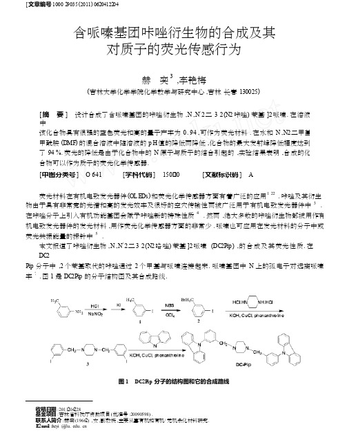
[ 文章编号1000 29035 (2011) 0620412204含哌嗪基团咔唑衍生物的合成及其对质子的荧光传感行为奕3赫,李艳梅(吉林大学化学学院化学教学与研究中心,吉林长春130025)[ 摘要] 设计合成了含哌嗪基团的咔唑衍生物,N ,N′2二3 2(N2咔唑) 苯基]2哌嗪. 在溶液中该化合物具有很强的蓝色荧光和高的量子产率为0 . 94 ,可作为荧光材料. 在水和N ,N2二甲基甲酰胺(DMF) 的混合溶液中随溶液的p H 值的降低而降低,化合物的最大发射峰降低幅度达到了94 %. 荧光的降低是由于化合物中的N 原子与质子的结合引起的,实验结果表明,合成的化合物可以作为质子的荧光化学传感器.[ 关键词] 合成;咔唑;荧光;发光材料;传感器;质子[ 中图分类号] O 641[ 学科代码] 150·20[ 文献标识码] A荧光材料在有机电致发光器件(OL E Ds) 和荧光化学传感器方面有着广泛的应用1 22 . 咔唑及其衍生物由于具有非常宽的光谱和高的发光效率及很好的空穴传输性而被广泛用于有机电致发光器件中 3 . 在咔唑分子上引入有机功能基团会赋予咔唑新的特殊性质 4 . 然而,绝大多数的咔唑衍生物都被用作有机电致发光器件的发光材料,用作荧光化学传感器方面的非常少. 哌嗪也可应用在发光材料的分子中或荧光共振能量的探针中 5 .本文报道了咔唑衍生物,N ,N′2二3 2(N2咯唑) 苯基]2哌嗪(DC2Pip ),的合成及其荧光性质.在DC2Pip 分子中,2 个苯基取代的咔唑通过2 个甲基与哌嗪连接起来. 哌嗪基团中N 上的孤电子对远离哌嗪6率7. 图1 是DC2Pip 的分子结构图及其合成路线.图1 DC2P ip 分子的结构图和它的合成路线收稿日期:2011204228基金项目:吉林省科技厅资助项目( 批准号:20090598) .联系人简介: 赫奕(19662) ,女,副教授,主要从事有机和有机/ 无机杂化材料研究.E2m a il :heyi @jlu. edu. cn413第6 期赫奕,等:含哌嗪基团咔唑衍生物的合成及其对质子的荧光传感行为1 实验部分1. 1 仪器与试剂仪器:Varian Unity Inova S pectrom eter ( 500 MHz) 核磁共振波谱仪,V arian C ary E clip s e 荧光光谱仪, Varian C ary 500 紫外可见光谱仪,Sartori ous P B210 p H 计测量.试剂:所用试剂均为试剂级产品.二甲苯使用之前采用氢化钙回流除水后蒸出备用.1. 2 化合物1 的合成间甲基碘苯1 :在搅拌和冰水浴下将亚硝酸钠(2 . 28 g ,33 mm ol) 的水溶液滴加到间甲基苯胺(3 . 21g ,30 mm ol) 和盐酸(6 m ol/ L ,30 m L) 的混合溶液中,然后将碘化钾(5 . 48 g ,33 mm ol) 水溶液加入到上面的反应混合物中.在室温下反应10 h 后,用氯仿萃取反应混合物,水洗氯仿溶液,最后用无水硫酸钠干燥. 柱色谱分离( 石油醚) 得2 . 9 g 产品(13 . 3 mm ol) ,产率35 %. 1 H NMR (δ,C DCl3 ) : 7 . 58 ( 1 H , s) ,7156~7 . 50 (1 H ,d) ,7 . 18~7 . 14 (1 H ,d) ,7 . 04~6 . 98 (1 H ,m) ,2 . 33 (3 H ,s) .间溴甲基碘苯(2) :将含有间甲基碘苯1 (6 . 79 g ,31 mm ol) 、N2溴代丁二酰亚胺( 6. 10 g ,34 mm ol) 和2 ,2′2偶氮二异丁氰(0 . 094 g ,0 . 57 mm ol) 的四氯化碳溶液加热至回流3 h. 冷却到室温后将固体滤出,柱色谱分离(石油醚) 得到浅黄色固体产物6 . 8 g (23 mm ol) ,产率75 %. 1 H NMR (δ,C DCl3 ) : 7175 ( 1 H , s) , 7165~7 . 60 (1 H ,d) ,7 . 40~7 . 30 (1 H ,d) ,7 . 15~6 . 98 (1 H ,t) ,4 . 40 (2 H ,s) .N ,N′2二( 间2碘2苯甲基) 哌嗪(3) : 将含有间溴甲基碘苯2 (4 . 65 g ,15 . 7 mm ol) 、一水合哌嗪盐酸盐(1 . 38 g ,7 . 8 mm ol) 、氢氧化钾(4 . 39 g ,78 . 26 mm ol) 、氯化亚铜(0 . 23 g ,2 . 35 mm ol) 和1 ,102啡络啉( 0. 46g ,2135 mm ol) 的二甲苯溶液(35 ) 在搅拌下加热回流10 h. 冷却至室温后将固体滤出,溶液倒入50 m L 水中.用乙醚萃取,用水洗乙醚溶液后用无水硫酸镁干燥乙醚溶液.柱色谱分离( V(乙酸乙酯) / V ( 石油醚) = 1∶6) 得淡黄色固体3 . 68 g (7 . 1 mm ol) ,产率91 %. 1 H NMR (δ,C DCl3) :7 . 68 (2 H ,s) ,7 . 60~7 . 55 (2 H ,d) , 7 . 28 (2 H ,d) ,7 . 04~6 . 98 (2 H ,t) ,3 . 44 ( 4 H ,s ) ,2 . 46 (8 H ,s) .N ,N′2二3 2(N2咯唑) 苯基]2哌嗪( D C2Pip ): 将含有N , N′2二( 间2碘2苯甲基)哌嗪3 ( 2 . 46 g , 4 .77 mm ol) 、咔唑(1 . 98 g ,12 mm ol) 、氢氧化钾(0 . 78 g ,13 . 9 mm ol) 、氯化亚铜(0 . 055 g ,0 . 55 mm ol) 和1 ,102啡络啉(0 . 11 g ,0 . 55 mm ol) 的二甲苯加热回流12 h 后冷却至室温. 滤出固体后溶液倒入60 m L 水中,用乙醚萃取. 乙醚溶液用水洗后无水硫酸钠干燥.柱色谱分离( V (乙酸乙酯) / V ( 石油醚) = 1∶5) 得白色固体1111 g (1 . 86 mm ol) ,产率39 %. 1 H NMR (δ,C DCl3) :8 . 15~8 . 12 (4 H ,d) ,7 . 58~7 . 50 (4 H ,t) ,7 . 46~7 . 37 (12 H ,m) ,7 . 30~7 . 24 (4 H ,m) ,3 . 62 (4 H ,s) ,2 . 60 (8 H ,s) .2 结果与讨论2. 1 DC2Pip 的吸收光谱和发射光谱图2 是在H2O/ DMF (体积比为4∶1) 的混合溶液中浓度为1 ×1025m ol ·L - 1 的DC2Pip 的吸收光谱和发射光谱(激发波长为294 nm) . DC2Pip 的吸收光谱中有3 个吸收峰,分别位于300 ,332 和346 nm. 300 nm 的吸收峰来源于咔唑的吸收,是由于苯基咔唑基团中π2π3 跃迁引起的,而332 和346 nm 的吸收峰是由咔唑基团本身的吸收引起的8 .在294 nm 光的激发下,DC2Pip (1 ×1025 m ol·L - 1) 在H2O/ DMF (体积比为4∶1) 混合溶液中具有强烈的蓝色荧光,荧光光谱见图2 .总共有4 个发射峰,最大强度的发射峰位于405 nm ,另外3 个发射峰分别位于352 ,368 nm 和428 nm. 发射的荧光主要来源于苯基取代的咔唑基团,哌嗪对荧光发射可能有一定的贡献9 210 . 这是由于哌嗪上有2 个孤电子对,它们远离哌嗪环的中心,因而与咔唑基团有一定的共轭作用7. 荧光分子把吸收的光能转变成发射光的能力可以用荧光量子效率(ΦF) 来评估.采用硫酸喹咛414 分 子 科 学 学 报第 27 卷 (ΦF = 0 . 55) 为标准测得 DC 2Pip 具有很高的量子效率11 ,达到了 0 . 94 ,这一结果表明 DC 2Pip 具有很强的荧光发射效率 ,可以作为荧光发射材料 .2 . 2 质子对 DC 2Pip 的吸收光谱和发射光谱的影响在 H 2O/ DMF (体积比为 4∶1) 的溶液中 ,DC 2Pip 的吸收光谱和荧光光谱与溶液的 p H 值有很大的关系 . DC 2Pip 的浓度为 1 ×1025 m ol ·L - 1 ,采用高浓度的盐酸和氢氧化钠溶液来调节溶液的 p H 值 . 为了在加入盐酸溶液或氢氧化钠溶液时不改变 DC 2Pip 的浓度 ,盐酸和氢氧化钠溶液的最大加入量不超过 DC 2Pip 溶液体积的 3 %. 采用氢氧化钠溶液调节 DC 2Pip 溶液的 p H 值至 10 . 89 ,然后连续加入微小体积的盐酸 溶液来调低溶液的 p H 值 . 在每一次加入盐酸充分搅拌后测量溶液的荧光光谱. 从 p H 值 10 . 89~3 . 38 , DC 2Pip 溶液的荧光光谱随 p H 值的变化光谱见图 3 . 从图 3 中很容易看出 ,p H 值强烈影响 DC 2Pip 溶液的 荧光光谱强度 ,荧光强度随 p H 值的降低亦即质子的不断加入而明显下降.图 2 DC 2Pip ( 1 ×1025 mo l ·L - 1 ) H 2 O / DMF( 体积比为 4∶1) 混合溶液中的吸收光谱和发射光谱( 激发波长 294 nm) . 图 3 在 p H 值为 10. 89~3. 38 范围内 , D C 2P ip 在 H 2 O / DMF ( V/ V ,4∶1) 混合溶液中的荧光 光谱随 p H 值的降低而变化的趋势.在实验的 p H 值为 10 . 89~3 . 38 范围内 ,荧光发射峰强度随 p H 值的降低而下降. 在连续滴加盐酸溶 液的过程中 ,发射峰位臵没有发生明显的移动 ,而酸度的增加导致了荧光强度明显的降低 ,最大降幅达 到了 94 % ,荧光发射被近乎淬灭 . DC 2Pip 溶液的荧光强度可以被可逆的调节. 当 p H 值达到 3 . 38 后 ,氢 氧化钠碱溶液被连续的加入来调节上述溶液的 p H 值 ,我们发现荧光强度随碱溶液的不断加入而逐渐 增加了. 所以我们认为 DC 2Pip 溶液能够呈现 2 种不同的状态 ,在酸性溶液中荧光是关闭的而在碱性溶 液中荧光是打开的. 这些变化来源于咔唑和哌嗪基团上的 N 原子的“质子化”和“去质子化”,2 种行为分 子引起荧光的淬灭和增强 . 实验结果表明 DC 2Pip 可以用来作为质子的荧光化学传感器来监控溶液的 p H 值 .为了考察 N 原子的质子化对荧光强度的影响 ,我们绘制了 DC 2Pip 在 p H 值为 12 . 6~1 . 30 范围内 405 nm 发射峰强度随 p H 值的变化曲线 ,见图 4 . 曲线随 p H 值的变化过程中出现 2 个拐点 ,分别位于 p H 值 10 . 5~7 . 0 和 p H 值 3 . 0~1 . 5 范围 ,表明荧光强度随 p H 值的降低是由 2 种 N 原子的质子化引起的 , 即咔唑和哌嗪基团中的不同 N 原子的质子化12 .为了进一步说明质子化对 DC 2Pip 光谱性质的影响 ,我们也考察了在 H 2O/ DMF (体积比为 4∶1) 的混 合溶液中 p H 值为 10 . 89~4 . 57 范围内 DC 2Pip 的吸收光谱随 p H 值的变化情况 . 与荧光的变化相似 ,DC 2 Pip 的吸收光谱强度随 p H 值的下降而减小 ,但降低幅度比荧光降低小很多 ,见图 5 . 在 p H 值由 10 . 89~ 4157 的变化范围 ,吸收光谱的强度降低了 40 % ,表明质子化作用降低了 DC 2Pip 分子对激发光的吸收 ,进 而引起荧光强度的降低 . 荧光光谱随 p H 值的变化趋势与紫外光谱的变化趋势相同 ,表明在激发状态下 分子中没有质子转移12 .实验结果表明 DC 2Pip 荧光强度的降低是由分子中 N 原子的质子化引起的 ,说明 DC 2Pip 的荧光光 谱依赖于溶液的 p H 值 . 荧光强度和 p H 值的这种依赖关系是典型的 p H 值传感器的特点 ,说明 DC 2Pip415 第 6 期 赫 奕 ,等 :含哌嗪基团咔唑衍生物的合成及其对质子的荧光传感行为 可以用于质子即 p H 值的传感体系中.图 4 浓度为 1 ×10 - 5 mol ·L - 1 的 DC 2Pip 在 H 2O / DMF 混合溶液中的荧光滴定曲线. 图 5 在 H 2 O / DMF ( 体积比为 4∶1) 混合溶液中 ,DC 2Pip 随 p H 值变化的吸收光谱曲线.3 结论本文合成了含有哌嗪的咔唑衍生物 ,该化合物在溶液中具有很强的荧光 ,荧光强度随 p H 值的变化 而发生明显的增强或减弱 . 结果表明 ,它可以作为质子的荧光化学传感器 ,这种分子的合成丰富了质子 即 p H 值传感器的设计模式 .[ 参 考 文 献]TA NG C W ,V AN S L Y K E S A. J . Appl Phys Le tt ,1987 ,51 (12) :9132915 . 靳瑞发 ,包洪政. J . 分子科学学报 ,2009 ,25 (2) :1272131 . SH AO H X ,CHEN X P ,WA NG Z X ,et al . J . J Lumin ,2007 ,127 (2) :3492354 . LOI SEA U F ,CA MPAG N A S , H A MEUR LAI NE A ,e t al . J . J A m Chem S oc ,2005 ,127 (32) :11352211363 . MA LL I CK A , H A LDAR B ,SENG UPTA S ,et al . J . J Lumin ,2006 ,118 (2) :1652172 . NI U Y Y , H O U H W ,WEI Y L ,et al . J . I norg Chem C ommun. 2001 ,4 (7) :3582361 . G U D , Y ANG G C , HE Y ,et al . J . Synth Met ,2009 ,159 (23224) :249722501 . WA NG X J ,FENG L H ,CHEN Z B. J . Spectrachimica A ct a Part A :M olecular and Biom olecular Spectr osc opy ,2008 ,71 (4) :143321437 . SHIH P I ,CHI A NG C H ,DIX IT A K ,et a l . J . O rgan Lett ,2006 ,8 (13) :279922802 . PARK T J ,J EO N S J ,PARK J J ,et al . J . Thin S olid Films ,2008 ,517 (2) :8962900 . CROSB Y G A ,DE MAS J N. J . J Phys Chem ,1971 ,75 (8) :99121024 . SAL E H N ,A L 2SO UD Y A ,N AU W M. J . Spectrachim Acta A :M olecular and Biom olecular Spectr oscopy ,2008 ,71 (3) :8182822 .123456789101112 The synthesis of piperazine 2conta ining carb azole derivate andits sensing perf o rmance to protonHE Y i 3 ,L I Y an 2m ei( Chemical Education Reseanh Centre ,C ollege of Chemistry ,Jilin University ,Changchun 130024 ,China )Abstract :A novel piperazine 2containing carbaz ole derivative ,N ,N ′2di 3 2(N 2carbaz ole ) benz yl 2piperazine (DC 2Pip ) , was designed and prepared. T he absorpti on and fluorescence m axim a of DC 2Pip were observed at 300 and 405 nm ,re 2 spectively. DC 2Pip em its strong blue fluorescence and has a high quantum yield (ΦF = 0. 94) in soluti on ,which indi 2 cates that DC 2Pip can be used as fluorescence em itting m aterials. T he effect of p H o n the fluorescence characteristics of DC 2Pip in H 2O/ DMF (4∶1 ,V / V ) soluti on has also been investigated ,the intensity of fluorescence decreases with the decrease of p H value ,these results shows that DC 2Pip offers potential as optical sens or for protons.K ey w or d s :synthesis ;carbaz ole ;luminescence ;em itting m aterial ; s ensor ;proton。
二苯基乙二胺的合成
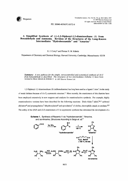
OUU'-I.U % U . 3 ~ [ ~ I ) I U . 3 / / U
A Simplified Synthesis of (+)-l,2-Diphenyi-l,2-diaminoethane (1) from Benzaldehyde and Ammonia. Revision of the Structures of the Long-Known Intermediates "Hydrobenzamide" and "Amarine"
8633
"hydrobenzamide" was thereby shown to be 2 (Figure 1 and Scheme 2) and that of amarine hydrochloride to be 3.HC1 (Figure 2 and Scheme 2). The stereospecific formation of 3 from 2 can readily be accounted for as a wellprecedented disrotatory pericyclic closure of an intermediate 2,4-diazapentadienyl anion. The conversion of 3 to 4 under vigorous basic conditions is also simply explained in terms of equilibration via deprotonation of 3 to form a delocalized benzylic anion and subsequent protonation to give 4, with reversibility at each step. Because of the ready availability of 4 from benzaldehyde and ammonia via intermediates 2 and 3, we sought a simple method for conversion of 4 into (+)-l,2-diphenyl-l,2-diaminoethane (1). As expected for a cyclic amidine, 4 is resistant to acid-catalyzed hydrolysis even at reflux, because of the stability of the conjugate acid. The amidinium ion derived from 4 also resists reduction by borohydride-type reagents. However, reduction of 4 by aluminum amalgam in moist THF occurred readily to give the corresponding imidazolidine which upon treatment with aqueous acid and subsequent workup provided (+)-1 in 85% yield. The specific procedure follows.