自发性2型糖尿病啮齿类动物模型研究概况
2型糖尿病大鼠模型的建立及其糖代谢特征分析

引言:
2型糖尿病是一种常见的代谢性疾病,其发病率逐年上升。该病的发生与遗 传、环境、生活方式等多种因素有关。为了深入研究2型糖尿病的发病机制和治 疗方法,建立可靠的动物模型是至关重要的。大鼠作为常用的实验动物,具有易 饲养、易繁殖、费用低等优点,被广泛应用于医学研究中。
在本次演示中,我们通过腹腔注射链脲佐菌素的方法建立2型糖尿病大鼠模 型,并对其糖代谢特征进行分析。
谢谢观看
实验组(n=18)360±406.5±0.810.6±2.7*
注:与对照组比较,*P<0.05。
3、胰岛素抵抗与胰岛细胞功能
与对照组相比,实验组大鼠的HOMA-IR显著升高(P<0.05),而ISI显著降低 (P<0.05),表明实验组大鼠存在胰岛素抵抗和胰岛细胞功能受损。结果如表2 所示。
与对照组相比,实验组大鼠的体重和血糖水平均显著升高(P<0.05),而对 照组大鼠的体重和血糖水平无明显变化(P>0.05)。结果如表1所示。
表1:两组大鼠体重与血糖水平 比较(x±s)
组别体重(g)FPG(415±355.0±0.67.8±1.9
表2:两组大鼠胰岛素抵抗与胰 岛细胞功能比较(x±s)
组别HOMA-IRISI
对照组(n=19) 1.9±0.416.4±3.8
实验组(n=18)3.6±0.89.5±2.7
注:与对照组比较,*P<0.05。
4、讨论
本研究成功建立了2型糖尿病大鼠模型,并发现模型大鼠的体重、血糖水平 和胰岛素抵抗指数均显著高于对照组,而胰岛细胞功能指数显著低于对照组。这 些结果表明,建立的2型糖尿病大鼠模型符合临床2型糖尿病的特征,为研究该病 的发病机制和治疗提供了可靠的动物模型。然而,建模过程中可能受到某些因素 影响,如STZ剂量、注射途径等。
型糖尿病动物模型研究进展
Ⅱ型糖尿病动物模型研究进展摘要: 糖尿病是以高血糖为主要标志的内分泌代谢性疾病,是严重威胁人类健康的主要慢性病之一,而Ⅱ型糖尿病占糖尿病总数的90%~95%左右。
建立合适的Ⅱ型糖尿病动物模型是阐明其发病机制的前提条件。
因此,该文综述了目前国内外糖尿病研究中常用的动物模型,对发展新型Ⅱ型糖尿病动物模型的研究提供参考价值。
关键词:Ⅱ型糖尿病;动物模型;模型构建Research Progress about the Construction of Type ⅡDiabetic Animal ModelLIU Shu—YunAbstract: Diabetes mellitus,the endocrine and metabolic diseases,is characterized by hyperglycemia. It is one of the most prevalent chronic diseases that threat to human health,and type 2 diabetes accounted for 90% -95% of the total diabetes. The animal model of type 2 diabetes provide the important precondition to many scholars in study of the pathogenesis and mechanism of diabetes.Therefore,this article reviews a number of animal models of T2DM commonly used according to the articles that have been published both inside country and abroad,which will provide reference for the development of type II diabetic animal models.Key Words:Type Ⅱ Diabetes Mellitus, Animal model,Model construction糖尿病( Diabetes mellitus,DM) 是以高血糖为主要标志的内分泌代谢性慢性疾病,其严重威胁着人类健康。
II型糖尿病(胰岛素非依赖)动物模型

II型糖尿病(胰岛素非依赖)动物模型
1、自发性糖尿病模型(1)非肥胖型实验动物:PO大鼠、中国地鼠、GK大鼠、NSY鼠模型特点:高血糖,胰岛素抵抗,与人类II型糖尿病发病症状相似,NSY鼠有年龄依赖特征。
适用研究:非肥胖II型糖尿病研究获取方法:直接购买。
(2)肥胖型实验动物:ZDF大鼠、OLETF大鼠、ob/ob小鼠、db/db小鼠、KK小鼠模型特点:肥胖、糖尿病特征,同时伴有高血脂、高血压、脂肪肝、糖肾等并发症。
适用研究:肥胖、II型糖尿病以及所引起的各种并发症获取方法:直接购买。
2、诱发性糖尿病模型(1)饮食诱导实验动物:DIO小鼠模型特点:持续高脂饮食诱导的肥胖小鼠。
适用研究:肥胖、II型糖尿病研究获取方法:C57BL/6小鼠持续高脂饮食饲喂14周左右,检测糖尿病相关指标。
3、转基因模型实验动物:MKR小鼠、MODY2小鼠模型特点:靶向确定基因的小鼠模型,研究分子机制时有优势。
适用研究:II型糖尿病研究获取方法:自己构建转基因小鼠或直接定制购买。
自发性2型糖尿病动物模型
自发性2型糖尿病动物模型
自发性2型糖尿病动物模型
糖尿病的患病率呈逐年升高的趋势,已成为全球重点关注的公共性卫生问题.糖尿病动物模型可以部分模拟人的糖尿病状态,广泛应用于对糖尿病病因与治疗方法方面的研究与分析.非人灵长类动物在生理学、遗传学及生物学等特性方面与人类极相似,是生命科学及生物医药研究与开发方面是不可替代的实验动物.文章旨在对自发糖尿病动物模型,特别是非人灵长类糖尿病动物模型进行综述.
作者:万玉玲刘晓明 WAN Yu-ling LIU Xiao-ming 作者单位:广东省昆虫研究所,广州,510260 刊名:医学综述ISTIC英文刊名:MEDICAL RECAPITULATE 年,卷(期):2008 14(14) 分类号:Q95 关键词:非胰岛素依赖性糖尿病 2型糖尿病动物模型非人灵长类动物。
2型糖尿病大鼠模型中对胰岛素升高机制的初步探讨的开题报告
2型糖尿病大鼠模型中对胰岛素升高机制的初步探讨
的开题报告
糖尿病是一种由胰岛素分泌不足或组织胰岛素不敏感导致的高血糖症。
其中,2型糖尿病占据了大多数病例。
研究2型糖尿病的动物模型,是了解疾病机制以及开发新的治疗手段的重要途径。
本研究主要以2型糖尿病大鼠模型为研究对象,初步探讨了该模型中胰岛素升高的机制。
首先,我们将从实验上确定2型糖尿病大鼠模型。
该模型通常采用高脂饮食喂养鼠类,再注射低剂量链脲佐菌素(STZ)诱导胰岛素抵抗。
通过检测餐后血糖水平和胰岛素抵抗指标,确认模型建立成功。
其次,我们将通过检测血浆中多个激素的含量,探讨该模型中胰岛素升高的机制。
预计在2型糖尿病大鼠模型中,血浆中胰岛素样生长因子-1(IGF-1)和瘦素等激素的含量将显著升高。
该结果将进一步提示这些激素在模型的胰岛素升高中具有重要作用。
最后,我们将利用分子生物学技术,探究这些激素对相关蛋白的表达及信号通路的调节。
尤其是探究IGF-1和胰岛素样生长因子-1受体(IGF-1R)在该模型中的变化情况以及分析IGF-1R信号通路的影响。
这将为更深入地理解疾病机制提供参考。
本研究的实验方案和结果,将加深我们对2型糖尿病的理解和治疗方法的研究。
2型糖尿病鼠类模型的研究进展

2型糖尿病鼠类模型的研究进展高秀莹,周迎生【摘要】【摘要】小鼠、大鼠糖尿病模型对基础与临床防治研究十分重要,不同的研究目标对应不同的动物模型载体。
本文就目前常用的2型糖尿病鼠类模型的构建、主要疾病特征及应用等进行评述,为研究者了解、选择适合的动物模型提供参考。
【期刊名称】中国实验动物学报【年(卷),期】2014(000)004【总页数】6【关键词】【关键词】 2型糖尿病;动物模型;鼠类综述·进展糖尿病已成为全球性的公共健康问题,据IDF统计2013年全球糖尿病患者达3.82亿,预计至2035年全球糖尿病患者将增至5.92亿[1]。
在我国,糖尿病成为继肿瘤、心脑血管病之后的第三大严重危害人们健康的慢性疾病,2010年我国糖尿病患者已达9240万[2],其中超过90%的患者为2型糖尿病。
2型糖尿病的发病机制尚未明确,建立一种既符合人类2型糖尿病发病特点,又稳定、实用的动物模型在2型糖尿病研究中起着至关重要的作用。
鼠类作为目前应用最广的糖尿病动物模型,因其体积小、生长周期短、经济易得、易于实现基因修饰等,较其他种属有着无可比拟的优势。
目前2型糖尿病鼠类模型主要分为三大类:自发性2型糖尿病模型、诱发性2型糖尿病模型、转基因/基因敲除2型糖尿病模型。
本文对近年来国内外较常用的2型糖尿病鼠类模型构建、主要疾病特征、确立标准及其应用进行概述,为研究者提供参考。
1 2型糖尿病鼠类模型分类1.1 自发性2型糖尿病模型该模型动物未经过任何有意识的人工处置,多数采用有自发性糖尿病倾向的近交系纯种动物,按照饲养条件喂养,自发成模,最接近人类疾病的发病过程。
该模型可分为肥胖自发性2型糖尿病模型和非肥胖自发性2型糖尿病模型,因2型糖尿病患者多伴肥胖,故以前者应用居多。
1.1.1 肥胖自发性2型糖尿病模型常用的肥胖自发性糖尿病模型包括单基因遗传背景的ob/ob小鼠、db/db小鼠、Zucker糖尿病肥胖大鼠(Zucker diabetic fatty rat,ZDF)和多基因背景的KK/Ay小鼠、OLETF大鼠。
糖尿病小鼠模型的构建
糖尿病小鼠模型的构建I型糖尿病模型方法:模型的诱导采用多次小剂量链脲佐菌素(STZ)给药法(MLDSTZ),雄性BALB/c小鼠,随机分为3组:模型组、阴性对照组、阳性对照组。
模型组小鼠连续5天腹腔注射STZ溶液(60mg/kg),注射前禁食8h,不禁水。
注射时将STZ溶于0.1mol/L柠檬酸钠缓冲液(pH4.5),避光冰上配置,溶解后尽量在30min完成注射。
阴性对照组腹腔注射柠檬酸缓冲液,正常对照组不作处理。
II型糖尿病模型目前,构建II型糖尿病动物模型的方法有很多种,其中自发性糖尿病动物模型、转基因/基因敲除糖尿病动物模型、单纯应用STZ所致糖尿病模型、STZ 与饮食协同作用所致II型糖尿病动物模型等应用较多。
1、自发性糖尿病动物模型自发性II型糖尿病动物模型主要是啮齿类,其最大优点是疾病的发生、发展与人类的很相似,因此在研究II型糖尿病的生理、病理及有关临床药物研发等方面有重要价值。
此类动物模型包括小鼠、大鼠、地鼠,其中小鼠包括KK-Ay、ob/ob、db/db等单基因突变鼠、新西兰肥胖小鼠和NSY小鼠:大鼠包括GK大鼠、Zucker大鼠和OLETF大鼠等;地鼠则以中国地鼠为主。
2、转基因构建糖尿病模型以实验方法敲入外源基因或敲除内源基因,在染色体组内稳定整合并能遗传给后代的一类动物称为转基因动物。
转基因糖尿病动物模型主要是通过基因技术,按照自己的意愿,控制实验动物的特定基因及其表达,使动物表现为一定的遗传性状。
主要利用基因定位与基因转移,由于转基因为随机整合,所以在子代观察到得效果取决于成功整合发生的位点和拷贝的数量,转基因的表达效果也因动物体系不同而有所差异。
3、单纯应用STZ所致糖尿病模型使用不同剂量(60mg/kg\45mg/kg)的STZ静脉注射,注射前禁食8h,不禁水。
注射时将STZ溶于0.1mol/L柠檬酸钠缓冲液(pH4.5),避光冰上配置,溶解后尽量在30min完成注射。
自发性2型糖尿病小鼠发病早期认知功能的研究
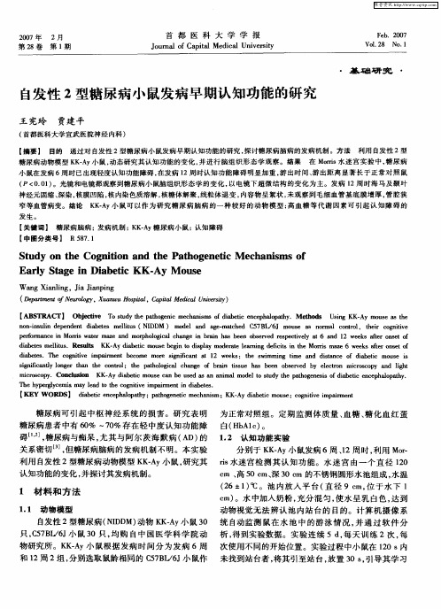
i r c p .Co cu i n K Ay d a e i u e C s d a i l d e o su y t e p to e e i o ib t n e h o a h . m c so y o n l so K- ib t mo s a b u e a a ma mo lt t d h ah g n ss fda ei e c p a p t y c n e s n n c l e h p r l c mi y l a o t e c g i v mp i e ti i b ts y eg y e a ma e d t o t e i ar n n da ee . h n i m
e f m n ei M r sw t a ea  ̄h o a c a g n ban h b e b evd rs t e t n 2 w e sat n e f p r r a c or a rm z d mo d gc h n e i ri a e n o s re cp ci l a 6 a d 1 ek f ro sto o n i e n i l s e vy e
W a g Xi l g i in ig n a i 。JaJa pn n n
( e r etfNu l X aw o il Cpt eil n ei ) Dp t n o er o am o  ̄, un uHs t , ail d a U irt pa a M c v sy
2型糖尿病动物模型研究概况
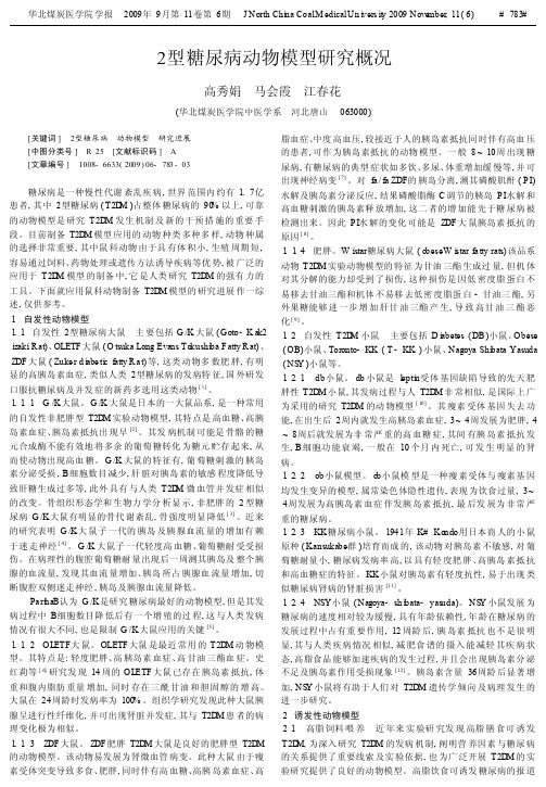
2型糖尿病动物模型研究概况高秀娟马会霞江春花(华北煤炭医学院中医学系河北唐山063000)[关键词]2型糖尿病动物模型研究进展[中图分类号]R25[文献标识码]A[文章编号]1008-6633(2009)06-783-03糖尿病是一种慢性代谢紊乱疾病,世界范围内约有1.7亿患者,其中2型糖尿病(T2D M)占整体糖尿病的90%以上,可靠的动物模型是研究T2D M发生机制及新的干预措施的重要手段。
目前制备T2D M模型应用的动物种类多种多样,动物种属的选择非常重要,其中鼠科动物由于具有体积小,生殖周期短,容易通过饲料、药物处理或遗传方法诱导疾病等优势,被广泛的应用于T2D M模型的制备中,它是人类研究T2D M的强有力的工具。
下面就应用鼠科动物制备T2D M模型的研究进展作一综述,仅供参考。
1自发性动物模型1.1自发性2型糖尿病大鼠主要包括G/K大鼠(Goto-K ak2 izaki R at)、OLETF大鼠(O tsuka Long Evans To kushi ba F atty R at)、Z DF大鼠(Zuke r d i abe ti c fatty R a t)等,这类动物多数肥胖,有明显的高胰岛素血症,类似人类2型糖尿病的发病特征,国外研发口服抗糖尿病及并发症的新药多选用这类动物[1]。
1.1.1G/K大鼠。
G/K大鼠是日本的一大鼠品系,是一种常用的自发性非肥胖型T2D M实验动物模型,其特点是高血糖、高胰岛素血症、胰岛素抵抗出现早[2]。
其发病机制可能是骨骼的糖元合成酶不能有效地将多余的葡萄糖转化为糖元贮存起来,从而使动物出现高血糖。
G/K大鼠的特征有,葡萄糖刺激的胰岛素分泌受损,B细胞数目减少,肝脏对胰岛素的敏感程度降低导致肝糖生成过多等,此外具有与人类T2D M微血管并发症相似的改变。
骨组织形态学和生物力学分析显示,非肥胖的2型糖尿病G/K大鼠有明显的骨代谢紊乱,骨强度明显降低[3]。
2型糖尿病动物模型中西医研究进展

三、诱导性2 型糖尿病动物模型
• 诱导性2 型糖尿病动物模型是通过物理、化学、生物 等致病因素人工诱发出具有糖尿病特征的动物模型。 • 1.高脂饲料诱导,此类模型可表现为高体重、高血脂、 高胰岛素血症和糖耐量增高等胰岛素抵抗的特征 • 例如:葛学美用脂肪占总热能的45.5%的高脂饮食成 功地诱发了C57BL /6J小鼠产生2型糖尿病,并可使 小鼠血清胰岛素水平不断提高,血糖升高,小鼠体重 超常,伴血脂明显差异 • 邬云红以脂肪热量比为65% 的高脂饲料喂养Wistar 大鼠20 周,出现空腹血清胰岛素、空腹血糖明显升 高,胰岛素敏感指数明显降低。
2. 药物诱导:链脲佐菌素( streptozotcin,STZ) 是 目前使用最广泛的糖尿病动物模型化学诱导剂, 它对一些种属的动物胰岛β细胞有选择地破坏,可 以使猴、狗、羊、兔、大鼠、小鼠等实验动产生1 型或2 型糖尿病. 有研究表明:给小鼠腹腔注射STZ 65mg/kg ,可致 空腹血糖明显上升,不同剂量和不同时期给予 STZ 可造成不同严重程度的2 型糖尿病; 用STZ 处理的新生大鼠在成年后将呈现典型的2 型糖尿 病表现。 乔凤霞和申竹芳给地鼠多次腹腔注射STZ 40 mg/kg , 结果动物中大部分血糖、血清甘油三脂和胆固醇 均升高。
二、转基因性2 型糖尿病动物模型
• 在动物原来遗传背景的基础上,通过改变某种基因的表达 水平以建立人类疾病的动物模型。 • 胰岛素受体底物- 2 基因敲除( IRS -2)小鼠可出现显著 的葡萄糖耐量受损,胰岛素抵抗和胰岛素分泌不足,从而 引发2 型糖尿病 • 此外,IRS -1 和β细胞葡萄糖激酶( GK) 双基因敲除( GK -IRS -1 )小鼠,IR - /IRS - 1 双基因敲除杂合体小鼠 均可出现高血糖,并随着年龄发展为显性糖尿病。
2型糖尿病动物模型研究进展

Ⅱ型糖尿病动物模型研究进展摘要: 糖尿病是以高血糖为主要标志的内分泌代谢性疾病,是严重威胁人类健康的主要慢性病之一,而Ⅱ型糖尿病占糖尿病总数的90%~95%左右。
建立合适的Ⅱ型糖尿病动物模型是阐明其发病机制的前提条件。
因此,该文综述了目前国内外糖尿病研究中常用的动物模型,对发展新型Ⅱ型糖尿病动物模型的研究提供参考价值。
关键词:Ⅱ型糖尿病;动物模型;模型构建Research Progress about the Construction of TypeⅡ Diabetic Animal ModelLIU Shu—Yun(Lab of Transplant Engineering and Immunology West China Hospital,Sichuan University 2013224070006)Abstract: Diabetes mellitus,the endocrine and metabolic diseases,is characterized by hyperglycemia. It is one of the most prevalent chronic diseases that threat to human health,and type 2 diabetes accounted for 90% -95% of the total diabetes. The animal model of type 2 diabetes provide the important precondition to many scholars in study of the pathogenesis and mechanism of diabetes.Therefore,this article reviews a number of animal models of T2DM commonly used according to the articles that have been published both inside country and abroad,which will provide reference for the development of type II diabetic animal models.Key Words:Type Ⅱ Diabetes Mellitus, Animal model,Model construction糖尿病( Diabetes mellitus,DM) 是以高血糖为主要标志的内分泌代谢性慢性疾病,其严重威胁着人类健康。
2型糖尿病相关三基因过表达小鼠模型研究中期报告
2型糖尿病相关三基因过表达小鼠模型研究中期报告
本研究来自XXX大学,旨在研究2型糖尿病的发病机制及相关基因的作用。
本中期报告将重点介绍三种基因过表达小鼠模型的构建和部分实验结果。
首先,本研究选取了三个与2型糖尿病相关的基因:葡萄糖转运蛋白2(GLUT2)、胰岛素样生长因子1(IGF1)和肌酸激酶(CKM)。
通过基因克隆和转染技术,将这三种基因分别构建成过表达小鼠模型。
同时,还建立了野生型小鼠作为对照组。
通过实验室多次验证和筛选,本研究最终选取了三个不同的过表达小鼠模型,分别为GLUT2过表达小鼠、IGF1过表达小鼠和CKM过表达小鼠。
本研究还测定了这三种基因表达水平的差异以及其对血糖水平的影响。
实验结果显示,GLUT2过表达小鼠的血糖水平明显高于野生型小鼠组,IGF1过表达小鼠和CKM过表达小鼠的血糖水平也有所上升,但相对于GLUT2过表达小鼠组,增长速度较缓慢。
同时,本研究还进行了胰岛组织切片和生化指标的检测等进一步实验,以深入了解这三种基因对2型糖尿病的作用。
总之,本研究成功构建了三种基因过表达小鼠模型,并初步研究了它们对血糖水平的影响。
未来将继续以这三个模型为基础,探究基因调控对2型糖尿病发病机制的影响,为该疾病的治疗和预防提供新思路。
2型糖尿病大鼠模型研究概况

2型糖尿病大鼠模型研究概况【摘要】目的:综述近年来2型糖尿病(T2DM)大鼠模型的研究进展及对其优缺点进行评价和未来同类模型的展望。
方法:主要对T2DM大鼠模型的建立技术和方法进行综合性评价。
结果:T2DM大鼠模型目前可以分为自发性T2DM 和实验性T2DM模型,且仍有较大发展空间。
结论:经过综合评价研究,认为各种建模方法均有优缺点,目前较认可的是实验性T2DM大鼠模型,因价格低廉,造模方便而广受欢迎,但仍缺乏一定的造模标准。
【关键词】2型糖尿病;动物模型;研究概况随着经济社会的发展,人们的饮食结构、生活方式等发生了很大改变,糖尿病发病率显著上升,尤其T2DM占了较高比例,大概占了糖尿病发病率的90%。
T2DM是因人体胰岛素分泌相对不足或靶细胞对胰岛素敏感性降低继而引发糖、蛋白质、脂肪和水电解质等代谢紊乱所导致的疾病。
患者典型表现为三多一少,即多饮、多食、多尿表现,同时还伴有身体消瘦、疲乏、烦躁、口渴等临床症状。
选择一些合适的动物模型进行动物试验成了我们研究糖尿病的良好途径,我们可以从中比较一些糖尿病药物的作用效果以及其药动学的特点,在临床用药上对评价某套治疗方案的可行性及预后等具有十分重要的参考意义。
目前研究的临床T2DM动物模型主要集中在大鼠上,这可能是由于大鼠作为T2DM动物模型相对较稳定且与人T2DM表现相似的优点。
因此我们在下面综述近几年来国内外有关临床T2DM大鼠模型研究的情况。
总体上来说,目前临床T2DM研究的大鼠模型主要分为两类,一类是自发性T2DM大鼠模型,另一类则是实验性T2DM大鼠模型,考虑到成本及方便程度,目前以后者居多。
1 实验性T2DM大鼠模型1.1 单纯高脂高糖引发的T2DM 在试验中,通过较长时间给予大鼠过量的高糖高脂饮食,发现能够诱导出较满意的T2DM大鼠模型,从而能为进一步研究奠定良好的基础。
目前认为其机理可能是高糖高脂饲料会导致胰岛B细胞超负荷,进而使胰岛细胞发生损伤、萎缩甚至死亡,胰岛的功能因此下降,继而建立起伴胰岛素抵抗的T2DM模型。
2型糖尿病动物模型的研究进展

01 摘要
目录
02 引言
03 研究现状
05
人工模拟2型糖尿病 动物模型
04
自发性2型糖尿病动 物模型
06
基因工程2型糖尿病 动物模型
目录
07 其他类型2型糖尿病 动物模型
09 研究不足
08 研究方法与成果 010 参考内容
摘要
2型糖尿病是一种常见的内分泌代谢疾病,严重影响全球公共健康。为了深 入探讨其发病机制和治疗方案,研究者们建立了各种2型糖尿病动物模型。本次 演示综述了近年来2型糖尿病动物模型的研究进展,包括自发糖尿病模型、人工 模拟糖尿病模型、基因工程糖尿病模型和其他类型糖尿病模型,分析了这些模型 的特点、应用和局限性,并探讨了今后研究的方向和意义。
三、型糖尿病动物模型的实验设 计与结果分析
在建立型糖尿病动物模型时,研究者们通常会进行一系列实验设计,包括测 定动物的体重、血糖、胰岛素水平等生化指标,以及进行组织病理学检查等。根 据实验结果,研究者们可以分析型糖尿病的发生机制、探讨疾病的干预措施并评 估药物治疗的效果。
例如,在肥胖诱导的型糖尿病动物模型中,通过给予高脂饮食并测定动物的 血糖、胰岛素水平等指标,研究者们发现高脂饮食可引起肥胖小鼠的胰岛素抵抗 和β细胞功能衰物模型在研究型糖尿病的发病机制和防治策略中发挥了重要作用。 现有的肥胖诱导、遗传诱导和化学药物诱导的型糖尿病动物模型都具有一定特点 和应用范围。然而,仍需进一步完善现有动物模型的种类和品质,以适应不同研 究方向的需求。
例如,可以探索更加精准的基因编辑技术,构建具有人源性基因突变的型糖 尿病动物模型,以深入研究基因突变与糖尿病的关系。另外,在未来的研究中, 可以结合新型技术如代谢组学、蛋白质组学等,从多角度探讨型糖尿病的发病机 制和药物作用机制。加强干细胞治疗、免疫治疗等新型治疗手段的研究,为型糖 尿病的治疗提供更多可能性。
自发性Ⅱ型糖尿病模型db/db小鼠生物学特性研究
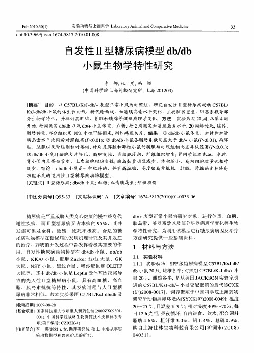
自发 性 I型糖 尿病 模 型 d /b I bd 小 鼠生 物 学特 性研 究
李 娜, 张 周, 冯 颖 ( 中国科 学院上 海 药物研 究 所, 海 2 10 ) 上 0 2 3 [ ] 目的 以 C 7 LKs—b+表 型正 常 小 鼠为对 照组 ,研 究 自发 性 I型糖尿 病动物 C 7 L 摘要 5 B / Jd / I 5B / KJ b b s d/ 小鼠的体生长曲线、糖代谢曲线 、 — d 血清胰 岛素水平变化、主要脏器重量 、 脏器系数等部 分 生物 学特性 ,并探讨 其肝 脏 、 肾脏 和胰 腺 等组织 病理 学 变化 。方法 实验 为期 2 O周, 第 4周 从 开 始, 周 测定 d/b以及 d/ 鼠体 重 、血糖 , 2周 测定血 清胰 岛素 水 平,O周龄 处 死, 器、 每 bd b+小 每 2 脏 脂肪 称 重, 部分 组 织用 1% 中性 甲醛 固定 , 0 制作 病理 切 片。结 果 ① d/b小 鼠体 重 、血 糖和血 清 bd
方 法 研 究 提 供 ~ 些 基础 资料 。
1 材 料 与方 法
11 实验材 料 .
大 鼠、NS Y小 鼠、黑 线仓 鼠、嗜 沙肥 鼠和 OL T EF
111 实验动 物 ..
S F级糖 尿病 模型 C 7 LKs—b P 5B / J / d
大 鼠等 ,其 中 d /b小 鼠是 L pi 体基 因缺 陷导 bd et n受 致 的先 天 性 I型 糖 尿 病 小 鼠 ,具 有 高 血 糖 、 高血 I
d b小 鼠 2 0只 , 雌雄 各半 ; 对照 组 C 7 L K Jd / 5 B / s—b+小
鼠2 O只, 雌雄 各 半, 是从 美 国 J ACKS 实验 室 引 ON
Ⅱ型糖尿病动物模型的构建

方 法 。动物 模 型一直 以来 是研 究疾 病发 病 、 预防、 诊 断、 治 疗 的实验 材 料 。建 立 一种 既符 合 人 类 Ⅱ型 糖
鼠、 地鼠, 其 中小 鼠包 括 K K . A y 、 o b / o b 、 d b / d b等 单
基 因 突变 鼠、 新 西 兰 肥 胖小 鼠 和 N S Y小 鼠 ; 大 鼠包
2 动 物模 型 的构 建
2 . 1 自发性 糖尿病 动物 模型
自发性 Ⅱ型糖 尿 病 动 物 模 型 主要 是 啮齿 类 , 其
最 大优 点是 疾病 的发 生 、 发 展 与人类 的很 相似 , 因此
在研 究 Ⅱ型糖 尿病 的 生 理 、 病 理 及 有 关 临 床药 物研 发 等方面 有重 要 价值 。此类 动 物 模 型 包 括小 鼠 、 大
2 0 1 3年 4月
中 国 实验 动 物 学 报
AC TA LAB0RAT ORI UM ANI MAL I S S CI E NTI A S I NI C A
Ap r i l , 2 01 3 Vo 1 . 2 l No . 2
第2 l 卷
第 2期
Ⅱ型 糖 尿 病 动 物模 型 的构 建
c omp l i c a t i o ns .I n t h i s pa pe r ,t y p e I I d i a b e t i c a n i ma l mo de l s a n d t he i r c o n s t r u c t i o n we r e r e v i e we d,wh i c h wi l l p r o v i d e i m-
p o r t a n t r e f e r e n c e or f t he d e v e l o pme n t o f t y pe I I d i be a t i c a n i ma l mo de l s .
糖尿病和糖尿病视网膜病变相关的自然发病啮齿动物模型
糖尿病和糖尿病视网膜病变相关的自然发病啮齿动物模型【摘要】糖尿病是一种复杂的异质性疾病,目前世界各地有超过一亿人感染此病,造成极为严峻社会和经济问题。
自然发病的糖尿病啮齿动物模型关于糖尿病发病机制,糖尿病并发症和增加糖尿病危险性的遗传或环境阻碍等的研究有着无可比拟的重要作用。
咱们综述了与糖尿病视网膜病变相关的自然发病啮齿动物模型的发生进展和其特性特点。
【关键词】啮齿模型;糖尿病;糖尿病视网膜病变Abstract Diabetes is a complex and heterogeneous disorder presently affecting more than 100 million people worldwide and causing serious socio economic problems. Spontaneous rodent models of diabetes mellitus have proved invaluable in understanding the pathogenesis, complications, and genetic or environmental influences that increase the risks of diabetes. We have reviewed here in the development and characterization of spontaneous rodent models that displayed most features commonly associated with diabetic retinopathy.KEYWORDS: rodent model; diabetes mellitus; diabeticretinopathyINTRODUCTIONDiabetes mellitus is a term that is used loosely to describe a complex and heterogeneous disorder simply characterized by hyperglycemia (elevated blood sugar levels). Diabetes mellitus has occurred in humans at least 4000 years. In 1966, it was proposed that the same may be true in animals, particularly those living in association with humans, whether as domestic animals or as animals bred in the laboratory[1].An animal model is defined as "a living organism with an inherited, naturally acquired, or induced pathological process that in one or more respects closely resembles the same phenomenon occurring in man". Animal models of diabetes mellitus can be caused by anti insulin serum, pancreatectomy, glucose infusion,βcytotoxic agents, and viruses; or caused by diabetogenic nutritional and hormonal factors. Some models spontaneously develop non insulin dependent or insulin dependent diabetes[2]. Selective inbreeding has produced several strains of animal that are considered reasonable modelsof type 1 diabetes (T1D), type 2 diabetes (T2D) and related phenotypes such as obesity and insulin resistance. Apart from their use in studying the pathogenesis of the disease and its complications, all new treatments for diabetes, including islet cell transplantation and preventative strategies, are initially investigated in animals[3]. Here we review the spontaneous rodent models of diabetes and their application on retinal research.SPONTANEOUS MODELS OF TYPE 1 DIABETESThere are several spontaneous rodent models of type 1 diabetes, two of which have been extensively studied: the Bio Breeding(BB) rat and the non obese diabetic (NOD) mouse[4].NOD Mouse The NOD mouse was developed by selectively breeding offspring from a laboratory strain that in fact was first used in the study of cataract development [5,6]. Insulitis is present when the mice are 4 5 weeks old, followed by subclinical βcell destruction and decreasing circulating insulin concentrations. Frank diabetes typically presents between 12 and 30 weeks of age. An autoimmune lesion involvinglymphocytic infiltration and destruction of the pancreatic βcells leads to hypoinsulinemia, hyperglycemia, ketoacidosis, and death. There is a larger gender difference with 90% of females and 20% of males developing diabetes in NOD mice [7].Type 1 diabetes is a polygenic disease. Both in human and in NOD mouse type 1 diabetes, the primary susceptibility gene is located within the MHC [8]. NOD mouse represents probably the best spontaneous model used in genetic and immunologic studies and seems particularly analogous to human type 1 diabetes. It has provided not only essential information on type 1 diabetes pathogenesis, but also valuable insights into mechanisms of immunoregulation and tolerance. Importantly, it allows testing of immuno intervention strategies potentially applicable to man [9,10].Type 1 Bio Breeding Rat Models The Bio Breeding(BB) rat was first recognized in the Bio Breeding Laboratories in 1974[11]. It is extremely useful for studying both spontaneous diabetic prone (BBDP) and induced diabetic resistant (BBDR) diabetes and associated diabetic complications [12]. As in human type 1 diabetes, the syndrome in BB rats is characterized byhyperglycemia, lymphocytic insulitis, and the presence of antibodies to islet cell surface molecules. In common with the NOD mouse, the pancreatic islets are subjected to an immune attack with T cells, B cells, macrophages and natural killer cells being recruited to the insulitis[13,14]; the susceptibility gene is located within the MHC [15]. A variety of auto antibodies, including glutamic acid decarboxylase (GAD), have been reported in both BB rats and the NOD mouse, although it remains far from clear which, if any, of these are primary autoantigens[1618].It is believed that the development of diabetes in the model is secondary to a cell mediated autoimmune process and may have implications for the pathophysiology of type 1 diabetes in humans. BBDP rats are prone to the long term complications of diabetes such as neuropathy and retinopathy that occur in this model are very similar to complications in human diabetics [19].SPONTANEOUS MODELS OF TYPE 2 DIABETESNumerous spontaneous rodent models have been used to model various defects of human type 2 diabetes, such as OtsukaLong Evans Tokushima fatty (OLETF), Goto Kakizaki (GK), Akita mice and Akimba mice that showed the some characteristics of these animal models same as human.OLETF Rat Selective breeding for more than 20 generations has led to the generation of a spontaneously diabetic strain of Long Evans rats that displays polyuria, polydipsia, and slight obesity [1,20]. The OLETF rat develops hyperphagia and insulin resistance between 12 and 24 weeks of age, and mild obesity, hyperglycemia, and hyperinsulinemia between 20 and 28 weeks of age. By 40 weeks of age, the diabetic rats are hypoinsulinemic and exhibit defects in insulin secretion [21]. Obese OLETF rats are unable to control individual meal size due to the loss of cholecystokinin A receptors [22,23]. These rats have proven useful in studying the effects of exercise and diet on the development of type 2 diabetes, to test the efficacy of antidiabetic agents, and to study the complications of diabetes [2426].GK Rat The GK rat is a widely accepted model for research in type 2 diabetes. The GK rat was created by selective breeding of Wistar rats for oral glucose intolerance[27]. There are atleast two loci responsible for high blood glucose in GK rats[28]. Males and females become diabetic at weaning age, most likely due to an over all inherent lack of normal beta cell mass. Diabetes in the GK rat is characterized by fasting hyperglycemia, impaired secretion of insulin in response to glucose, and hepatic and peripheral insulin resistance. Late onset complications such as retinopathy, microangiopathy, neuropathy, and peripheral nephropathy have been described in the literature [29,30].Type 2 Bio Breeding Rat Models This model has been extremely useful for studying both spontaneous diabetic prone (BBDP) and induced diabetic resistant (BBDR) diabetes and associated diabetic complications[12]. In this strain, diabetes is manifested by lymphopenia,obesity,hyperinsulinemia, and autoimmune diabetes. Islets from obese rats reveal beta cell hyperplasia, and diabetes develops due to a combination of insulin resistance and autoimmune insulitis. So the BBZDP/Wor rat is often complicated by the presence of both type 1 and type 2 diabetes characteristics [31,32].Akita Mice Akita mice, a model of spontaneous earlyonset diabetes mellitus, are from the C57BL/6 background with a dominant mutation in the Ins2 gene, which results in a loss of beta cell function and failed insulin secretion 4 6 weeks postpartum. Symptoms in Akita mice include hyperglycemia, hypoinsulinemia, polydipsia, and polyuria, beginning around 34 weeks of age. The diabetic phenotype is more severe and progressive in the male than in the female. Obesity or insulitis does not accompany diabetes[33]. The mean lifespan of diabetic male mice on the C57BL/NJcl background (305 days) was significantly shorter than that of nondiabetic males in another colony of the same strain (690 days). Akita mice will serve as an excellent substitute for mice made insulin dependent diabetic by treatment with alloxan or streptozotocin [34,35].AKimba Mice The AKimba mice have been produced through crossing homozygous Kimba mice with Akita mice in Lions Eye institutes (LEI) in Australia. Kimba is a transgenic mouse model of retinal neovascularization. The AKimba mouse demonstrates features exhibited by the Akita as well as the Kimba mice [36].DIABETIC RETINOPATHYThe frequency of diabetic retinopathy increases proportionally to the duration of diabetes and blood glucose control. Microaneurysms are the earliest clinically visible manifestation of background retinopathy. Additional microvascular abnormalities result from significant vascular occlusion and characterize the preproliferative retinopathy stage. Approximately 50% of patients who reach the preproliferative stage will progress to proliferative retinopathy within 15 months[37].Most present diabetic rodent models can be used to study the initial or latent phase diabetic retinopathy. Matsuura showed that the peak latency of oscillatory potential (OP), the earliest electroretinographic manifestation of diabetic retina, was prolonged and retinal acellular capillaries and pericyte ghosts, the characteristic morphological changes in early diabetic retinopathy were not accelerated in OLETF rat [38]. Thinning of inner nuclear layers and outer retina were observed[35,39]. These observations suggested that retinal neuronal changes takes place prior to the angiopathic diabetic changes in diabetic rodents. Retinal complications including increased vascular permeability, thicker basement membranes,caliber irregularity, narrowing, tortuosity and loop formations of capillaries in these animals were similar to those seen in diabetic patients[35,40].It was reported that apoptosis of retinal microvascular cell (RMC) was increased and oxidative stress promoted the apoptosis of RMC in diabetic GK rats, similar to that in diabetic patients. Furthermore, a combination of vitamins C and E and an advanced glycation end products inhibitor mostly inhibited this increased apoptosis and ameliorated diabetic retinopathy. It indicated that apoptosis of RMC was a good marker of the progression of diabetic retinopathy in GK rats [41]. Vascular endothelial growth factor (VEGF) and hypoxia inducible factor1 (HIF1) levels in ocular tissue of GK rats [42] and NOD mice [43] were evaluated by ELISA and immunohistochemical studies. Increased VEGF and HIF 1 production in certain ocular tissue, similar to that in humans, are observed quite early. Lower levels of glutathione and normal endothelial/ pericyte ratio in GK rat retina indicated that impaired glucose metabolism may influence one of the defense mechanisms for oxidative stress and that decreased glutathione levels occur prior to morphological signs of pericyte loss and/or endothelialcell proliferation in this diabetic animal model [44].Few diabetic rodent models can present features of the advanced diabetic retinopathy. BBZDR/Wor rats progress to late stages of preproliferative retinopathy (PPDR) but do not demonstrate proliferative (PDR) aspects of the disease[45,46]. The AKimba mouse was hyperglycemic and developed retinopathy resembled the late stages of PPDR and the stage of PDR. The retinopathy includes increased permeability, capillary dropout, retinal non perfusion, vein beading, hemorrhage, neovascularisation and retinal detachment.In a word, spontaneous rodent models of diabetes mellitus have proved invaluable in understanding diabetes and diabetic retinopathy. The work comparing and contrasting type 1 and type 2 diabetic rodent models should help elucidate detailed molecular mechanisms behind diabetic complications, and help lead to the development of better therapeutics to treat diabetes and diabetic retinopathy.【参考文献】1 Willy JM, Abdullah S. Animal models of diabetes. In: Conn PM, ed. Sourcebook of models for biomedical research,Humana Press. Vol 1. ed. Totowa, New Jersey, US 2020:6516562 Frederick ES, Richard JT. Ethical issues involved in the development of animal models for type I diabetes. ILAR Journal 1993;35(1):123 Rees DA, Alcolado JC. Animal models of diabetes mellitus. Diabetic Medicine 2005;22(4):3593704 Sai P, Gouin E. Spontaneous animal models for insulindependent diabetes (type 1 diabetes). Vet Res 1997;28(3):223 2295 Li ZG, Zhang W, Anders AA. Alzheimer like changes in rat models of spontaneous diabetes. Diabetes 2007;56(7):1817 18246 Makino S, Kunimoto K, Muraoka Y, Mizushima Y, Katagiri K, Tochino Y. Breeding of a non obese diabetic strain of mice. Exp Anim 1980;29(1):1137 Atkinson M, Leiter EH. The NOD mouse model of insulindependent diabetes: as good as it gets? Nat Med 1999;5(6): 601 6048 Todd JA. Genetic analysis of type 1 diabetes using whole genome approaches. Proc Natl Acad Sci USA 1995;92(19): 856085659 Meagher C, Sharif S, Hussain S, Cameron MJ, Arreaza GA, Delovitch TL. Cytokines and chemokines in the pathogenesis of murine type 1 diabetes. Adv Exp Med Biol 2003;520:13313810 Yoon JW, Jun HS. Cellular and molecular pathogenic mechanisms of insulin dependent diabetes mellitus. Ann N Y Acad Sci 2001;928:200 21111 Eizirik DL, Mandrup PT. A choice of death the signaltransduction of immune mediated βcell apoptosis. Diabetologia 2001;44(12):2115 213312 Eiselein L, Schwartz HJ, Rutledge JC. The challenge of type 1 diabetes mellitus. ILAR J 2004;45(3):23123613 Bone AJ, Hitchcock PR, Gwilliam DJ, Cunningham JM, Barley J. Insulitis and mechanisms of disease resistance: studies in an animal model of insulin dependent diabetes mellitus. J Mol Med 1999;77(1):576114 Lally FJ, Ratcliff H, Bone AJ. Apoptosis and disease progression in the spontaneously diabetic BB/S rat. Diabetologia 2001;44(3):32032415 Ramanathan S, Poussier P. BB rat lyp mutation and type 1 diabetes. Immunol Rev 2001;184:16117116 Yoon JW, Yoon CS, Lim HW. Control of autoimmune diabetes in NOD mice by GAD expression or suppression in βcells. Science 1999;284:1183118717 Bortel R, Waite DJ, Whalen BJ, Todd D, Leif JH, Lesma E, Moss J, Mordes JP, Rossini AA, Greiner DL. Levels of Art2+ cells but not soluble Art2 protein correlate with expression of autoimmune diabetes in the BB rat. Autoimmunity 2001;33(3):199 21118 Mackay IR, Bone A, Tuomi T, Elliott R, Mandel T, Karopoulos C, Rowley MJ. Lack of autoimmune serological reactions in rodent models of insulin dependent diabetes mellitus. J Autoimmun 1996;9(6):70571119 H nninen A, Hamilton WE, Kurts C. Development of new strategies to prevent type 1 diabetes: the role of animal models. Ann Med 2003;35(8):54656320 Shima K, Zhu M, Mizuno A. Pathoetiology and prevention of NIDDM lessons from the OLETF rat. J Med Invest 1999;46(34):12112921 Kawano K, Hirashima T, Mori S, Saitoh Y, Kurosumi M, Natori T. Spontaneous long term hyperglycemic rat with diabetic complications. Otsuka Long Evans Tokushima Fatty (OLETF) strain. Diabetes 1992;41(11):1422142822 Bi S, Moran TH. Actions of CCK in the controls of food in take and body weight: Lessons from the CCK A receptor deficient OLETF rat.Neuropeptides 2002;36:17118123 Bi S, Moran TH. Response to acute food deprivation in OLETF rats lacking CCK A receptors. Physiol Behav 2003; 79(45):65566124 Yamamoto M, Jia DM, Fukumitsu KI, Imoto I, Kihara Y, Hirohata Y, Otsuki M. Metabolic abnormalities in the genetically obese and diabetic Otsuka Long Evans Tokushima Fatty rat can be prevented and reversed by alpha glucosidase inhibitor. Metabolism 1999;48(3):34735425 Sima AA, Sugimoto K. Experimental diabetic neuropathy: An update. Diabetologia 1999;42(7):77378826 Ziegler D. Treatment of diabetic polyneuropathy: Update 2006. Ann N Y Acad Sci 2006;1084(11): 25026627 Kim CS, Sohn EJ, Kim YS, Jung DH, Jang DS, Lee YM, Kim DH, Kim JS. Effects of KIOM79 on hyperglycemia and diabetic nephropathy in type 2 diabetic Goto Kakizaki rats. J Ethnopharmacol 2007;111(2): 24024728 Fakhrai Rad H, Nikoshkov A, Kamel A, Fernstr m M, Zierath JR, Norgren S, Luthman H, Galli J. Insulin degrading enzyme identified as a candidate diabetes susceptibility gene in GK rats. Hum Mol Genet 2000;9(14):2149215829 Sato N, Komatsu K, Kurumatani H. Late onset of diabetic nephro pathy in spontaneously diabetic GK rats. Am J Nephrol 2003;23(5):33434230 Nobrega MA, Fleming S, Roman RJ, Shiozawa M, Schlick N, Lazar J, Jacob HJ. Initial characterization of a rat model of diabetic nephropathy. Diabetes 2004;53(3):73574231 Mordes JP, Bortell R, Blankenhorn EP, Rossini AA, Greiner DL. Rat models of type 1 diabetes: Genetics, environment and autoimmunity. ILARJ 2004;45(3):27829132 Vernet D, Cai L, Garban H, Babbitt ML, Murray FT, Rajfer J, Gonzalez Cadavid NF. Reduction of penile nitric oxide synthase in diabetic BB/WORdp (type I) and BBZ/WORdp (type II) rats with erectile dysfunction. Endocrinology 1995;136(12):5709571733 Choeiri C, Hewitt K, Durkin J, Simard CJ, Renaud JM, Messier C. Longitudinal evaluation of memory performance and peripheral neuropathy in the Ins2C96Y Akita mice. Behav Brain Res 2005;157(1):313834 Gastinger MJ, Singh RS, Barber AJ. Loss of cholinergic and dopaminergic amacrine cells in streptozotocin diabetic rat and Ins2Akita diabetic mouse retinas. Invest Ophthalmol Vis Sci 2006;47(7):3143315035 Barber AJ, Antonetti DA, Kern TS, Reiter CE, Soans RS, Krady JK, Levison SW, Gardner TW, Bronson Ins2Akita mouse as a model of early retinal complications in diabetes. Invest Ophthalmol Vis Sci 2005;46(6):2210221836 Tee LB, Penrose MA, O'Shea JE, Lai CM, Rakoczy EP, Dunlop SA. VEGF induced choroidal damage in a murine model of retinal neovascularisation. Br J Ophthalmol 2020;92(6):832 83837 Aiello LM. Perspectives on diabetic retinopathy. AmJ Ophthalmol 2003;136(1):12213538 Matsuura T, Yamagishi S, Kodama Y, Shibata R, Ueda S, Narama long evans tokushima fatty (OLETF) rat is not a suitable animal model for the study of angiopathic diabetic retinopathy. Int J Tissue React 2005;27(2):596239 Lu ZY, Bhutto IA, Amemiya T. Retinal changes in Otsuka long evans Tokushima Fatty rats (spontaneously diabetic rat) possibility of a new experimental model for diabetic retinopathy. Jpn J Ophthalmol 2003;47(1):283540 Noritake M, Imran AB, Tsugio A. Retinal capillary changes in Otsuka Long Evans Tokushima Fatty Rats (spontaneously diabetic strain). Ophthalmic Res 1999;31(5):358 36641 Yatoh S, Mizutani M, Yokoo T, Kozawa T, Sone H, Toyoshima H, Suzuki S, Shimano H, Kawakami Y, Okuda Y, Yamada N. Antioxidants and an inhibitor of advanced glycation ameliorate death of retinal microvascular cells in diabetic retinopathy. Diabetes Metab Res Rev 2006;22(1):384542 Sone H, Kawakami Y, Okuda Y, Sekine Y, Honmura S, Matsuo K, Segawa T, Suzuki H, Yamashita K. Ocular vascular endothelial growth factor levels in diabetic rats are elevated before observable retinal proliferative changes. Diabetologia 1997;40(6):72673043 Li C, Xu Y, Jiang D, Hong W, Guo X, Wang P, Li expression of HIF 1 in the early diabetic NOD mice. Yan Ke Xue Bao 2006;22(2):10711144 Agardh CD, Agardh E, Hultberg B, Qian Y, Ostenson glutathione levels are reduced in Goto Kakizaki rat retina, but are not influenced by aminoguanidine treatment. Current Eye Research 1998;17(3):25125645 Ellis EA, Guberski DL, Hutson B, Grant MB. Time course of NADH oxidase, inducible nitric oxide synthase and peroxynitrite in diabetic retinopathy in the BBZ/WOR rat. Nitric Oxide 2003;6(3): 29530446 Frank RN, Schulz L, Abe K, Iezzi variation indiabetic macular edema measured by optical coherence tomography. Ophthalmology 2004;111(2):211217。
- 1、下载文档前请自行甄别文档内容的完整性,平台不提供额外的编辑、内容补充、找答案等附加服务。
- 2、"仅部分预览"的文档,不可在线预览部分如存在完整性等问题,可反馈申请退款(可完整预览的文档不适用该条件!)。
- 3、如文档侵犯您的权益,请联系客服反馈,我们会尽快为您处理(人工客服工作时间:9:00-18:30)。
[收稿日期]20100118(006)[基金项目]北京市教育委员会共建项目[作者简介]李娟娥,博士,研究方向为中医药对糖尿病及其并发症的防治,E-mail :lizhuan_1980@ [通讯作者]*刘铜华,教授,博士生导师,Tel :(010)64286642,E-mail :thliu@·综述·自发性2型糖尿病啮齿类动物模型研究概况李娟娥,王磊,秦灵灵,刘铜华*(北京中医药大学,北京100029)[摘要]人们筛选成功并保存下来的自发性的T2DM 动物模型主要是啮齿类,由于这类动物模型的最大优点是其疾病的发生、发展与人类的很相似,因此在研究T2DM 生理、病理及临床新药等方面有着非常大的应用价值。
目前,此类动物模型主要有小鼠、大鼠和沙鼠模型,其中小鼠有单基因突变小鼠(ob /ob ,db /db ,KK-ay )、新西兰肥胖小鼠和NSY 小鼠,大鼠包括GK 大鼠、OLETF 大鼠和ZUCKER 大鼠等。
该文对各个模型的发病机制、生理和病理特点等方面进行了阐述,以利于此类模型在研究T2DM 方面的应用。
[关键词]2型糖尿病;啮齿类;动物模型[中图分类号]R 285.5[文献标识码]A[文章编号]1005-9903(2010)06-0267-05A Review on Spontaneous Models of Type 2Diabetes Mellitus in RodentsLI Juan-e ,WANG Lei ,QIN Ling-ling ,LIU Tong-hua *(Beijing University of Chinese Medicine ,Beijing 100029,China )[Abstract ]The spontaneous models of T2DM which were successful screened and conserved have been mainly in rodents.The occurrence and development of T2DM in this models is similar to mankinds ,so they have important application value in the study of T2DM ,such as physiology ,pathology and new clinical medicines.Nowdays ,these models mainly include signle gene mutation mouse (ob /ob ,db /db ,KK-ay ),NZO and NSY mouse ,GK ,OLETF and ZUCKER rats ,Psammomys Obesus.In order to improve the application of these models for T2DM ,the characteristics of each model about mechanisms ,physiology and pathology were described in this article.[Key words ]type 2diabetes mellitus ;rodents ;animal model2型糖尿病占糖尿病群体的90%以上。
目前对其发生机制的大多数认识来源于对自发性胰岛素依赖或胰岛素抵抗的糖尿病动物模型的分析。
自发性2型糖尿病动物(spontaneous animal of T2DM )是指未经任何有意识的人工处置,在自然情况下所发生糖尿病的实验动物。
这类动物模型的最大优点就是疾病的发生、发展与人类T2DM 的疾病很相似,其应用价值很高。
目前人们筛选成功并能够作为种系保存下来的自发性糖尿病动物模型主要是啮齿类动物模型。
1小鼠模型1.1单基因突变小鼠导致小鼠易患T2DM 的单基因突变包括小鼠瘦素基因(Lep )、瘦素受体基因(Lepr )和agouti (A )黄色基因突变。
这类基因突变可致小鼠发生肥胖征候群,但在人类由于Lep 和Lepr 基因座突变引起的极度肥胖非常罕见,所以在一定程度上并不能准确反映人类最常见的由饮食诱导的肥胖型的T2DM 。
较适合的对照鼠为+/+的同窝或同系小鼠。
1.1.1ob /ob 小鼠1950年,Ingalls 发现一株近亲繁殖的小鼠食欲亢进、过度肥胖、高血糖;研究后发现是由定位于第6号染色体ob 基因(Lep ob )隐性突变引起[1],1994年证实ob /ob 鼠中ob 基因呈突变形式,且为纯合子[2]。
该鼠由于不能产生正常的瘦素而表现为肥胖,从断奶时即开始发胖,肥胖随着周龄逐渐加重,终生表现食欲旺盛;3 4周龄发展为高胰岛素血症,7月龄水平最高[2]。
肌肉和肝脏均存在多种胰·762·岛素信号通路传导系统的损害[3]。
有报道[4]称经瘦素治疗后30%的体重下降归因于能量消耗。
在早期高血糖时就对机械和热刺激有显著的敏感性[5],慢性高血糖和肥胖可能导致早期感觉神经性耳聋[6]。
Lep ob基因突变的致糖尿病作用与种系遗传背景关系密切。
B6背景下的自发突变中存在胰岛细胞持续增生肥大代偿作用,而在SM/J背景下没有观察到这种代偿,因而发生严重的糖尿病。
携带Lep ob突变的B6小鼠多年来作为预防肥胖和轻度高血糖的干预治疗模型。
1.1.2db/db小鼠1966年Jackson实验室发现C57BL/KsJ 品系小鼠自发出现的瘦素受体基因(db),定位于4号染色体上[7],呈常染色体隐性遗传。
db/db小鼠的缺陷主要为瘦素受体的转录异常,遂致瘦素作用不足,故对瘦素治疗不敏感,需应用大剂量瘦素方能奏效。
db/db小鼠纯合体大概3到4周龄开始变胖,血浆胰岛素在10 14d、血糖在4到8周开始升高,该模型具有典型的“三多”症状。
一些特征在BKS纯合子可以观察到,包括持续的血糖升高、严重的胰岛β细胞功能衰竭和约在10月龄死亡;BKS背景的小鼠血糖在5到33周表现为持续的升高,而B6背景的在15周以后开始恢复正常。
前者的血脂水平与月龄有关,14周时甘油三酯、HDL、VLDL+LDL比以后的水平低,而后者则与月龄无关。
该鼠可见外周神经和心肌病变;伤口愈合延迟、代谢率增加;雌性纯合子卵巢激素生成减弱[8]。
与ob/ob小鼠不同的是该鼠可发生明显的肾病。
该小鼠多年来作为预防肥胖、轻度高血糖和糖尿病肾病的干预治疗模型。
1.1.3KK-A y小鼠1957年,Kondo[9]等报道了KK小鼠能自发出现明显肥胖、高血糖和高胰岛素血症;且发现其多在5个月后,或予以高热量饲料后才发病。
KK小鼠是由于位于2号染色体Agouti(A)基因座上的黄色基因显性突变所致。
其纯合子均在胚胎阶段死亡,而杂合子则表现为严重肥胖、胰岛素抵抗、高胰岛素血症、高血糖和黄色皮毛[10]。
该小鼠6周龄时开始出现高胰岛素血症,而且随年龄继续升高,而血胰岛素的增加继发于B细胞增生肥大。
陈丽萌等[11]研究证实KK-A y小鼠16周龄后开始出现明显肥胖、多尿的特点,并开始出现高血糖、高胰岛素血症、高脂血症。
有学者研究了KK-A y小鼠皮下脂肪中编码瘦素的肥胖(ob)基因,发现其表达与营养、饮食和体重呈正相关[12]。
近期研究发现[13]高脂饲养的KK-A y小鼠腹膜脂肪组织的趋化因子及受体表达上调[14];此品系小鼠肾脏和心脏组织的有关氧化酶表达比STZ 小鼠明显,但MDA、血糖和血脂水平没有后者明显[15]。
该小鼠多年来作为预防肥胖和轻度高血糖的干预治疗模型。
1.2新西兰肥胖(NZO)小鼠新西兰肥胖小鼠是新西兰从伦敦的帝国肿瘤研究基金实验室的远交系繁育出的纯系。
新生NZO小鼠较重、体积较大,幼年即表现为肥胖,成年以高胰岛素血症、血糖和高血压为特征。
约50%雄性该鼠发展为肥胖相关的糖尿病[16]。
NZO小鼠肥胖的特点是皮下和内脏广泛的脂肪堆积,同时伴有肝脏和外周的胰岛素抵抗相关的糖耐量异常[17]。
该鼠的胰岛β细胞在体内外均存在葡萄糖刺激的胰岛素分泌缺陷。
主要并发症是肾病,特点为年龄依赖的肾小球血管丛和肾小球系膜胞质增多以及IgA沉积导致的肾小球基底膜轻度增厚,而雌性的改变更明显[18]。
高脂喂养的NZO小鼠发生显著肥胖,且血清和组织中瘦素水平增高;血管发生与高血糖及肥胖有关[19],但与瘦素水平没有明显关系[20]。
由于NZO小鼠的肝脏脂肪堆积随着年龄增加而增加,特别适合筛选有潜在肝毒性的胰岛素增敏剂[21],也有可能作为肥胖者易患的OSAS的遗传学研究模型[22]。
1.3NSY小鼠NSY(Nagoya-Shibata-Yasuda)小鼠系是Jcl:ICR远交系选出的有葡萄糖耐量受损的近交系。
NSY小鼠发展为糖尿病的速度相对较为缓慢,12周龄后,胰岛素抵抗也不是很明显,其与人类疾病情况相似,减肥食物的摄入能减轻其疾病状态,高脂食品能够加速疾病的发生过程[23]。
12周龄时出现IGT并表现出雄性特异性,在体外可检测到葡萄糖刺激的胰岛素分泌减少,24周龄时所有雄性的葡萄糖刺激的胰岛素分泌消失,48周龄时雌雄性小鼠均有脂肪增加和轻度高胰岛素血症。
在任何年龄始终都没有发现胰腺的胰岛形态学异常如肥大或炎症改变[24];压力依赖性Ca2+通道或胰岛素分泌级联过程的缺失可能是胰岛素分泌障碍的主要原因[25]。
杂交二代NSY小鼠中脂肪肝与肥胖、高胰岛素血症和高血糖之间有显著相关性,并在6号和7号染色体初步定位了可能的脂肪肝基因(Fl1n)和体重基因(Bw1n)[26]。
该品系小鼠可出现糖尿病肾病,随T2DM的进展而加重[27]。
NSY小鼠T2DM的多基因遗传性质和它的肾脏并发症使其成为寻找致病基因、评价新药物对糖尿病病人肾功能的改善作用的唯一有效模型。
2大鼠模型2.1GK大鼠1975年,Goto等人在日本仙台,从远交系Wistar大鼠中经OGTT选出个轻度糖耐量减退鼠,经10代左右反复交配,形成自发性非肥胖T2DM鼠种,简称GK大鼠(Goto-Kakizaki Rat)。
该鼠主要表现为葡萄糖刺激的胰岛素分泌受损、β细胞分泌受损、空腹高血糖、肝糖原生成增多、外周组织中度胰岛素抵抗等,长期患病后,出现各种并发症和神经系统疾病[28]。
有研究表明GK大鼠B细胞功能受损先于其空腹高血糖和高胰岛素血症的出现,胰岛B细胞形态和功能的缺陷主要受遗传基因决定[29]。
该鼠进食后血糖升高,具有稳定的葡萄糖刺激的胰岛素分泌受损与葡萄糖不耐受[30];8周龄时非空腹的血浆葡萄糖水平升高及糖耐量受损,14周龄时,其葡萄糖刺激的胰岛素释放显著下降,为正常Wistar大鼠的25% 50%[31]。
