Spectroscopic Constants, Abundances, and Opacities of the TiH Molecule
虚拟天文台

• not tied to a single “brick-and-mortar” location • supports astronomical “observations” and discoveries
via remote access to digital representations of the sky
Infrared
Optical
National Virtual Observatory
用户需要做的事情:
1. 提出科学思想, 及对数据库的需要 2. 检索数据库 (用户到不同站点寻找) 3. 下载所需的数据 (x TB ? 包括各种元数据, 定标数据……) 4. 对数据进行标准化处理 (需要软件平台) 5. 数据的匹配 (如果使用二个以上数据库的资料 ) 6. 发展各种软件工具 (可视化, 统计工具… … )
National Virtual Observatory
时代
National Virtual Observatory
两百多年来,天文研究通常都是 单个天文学家或者天文学家小组 进行为数有限的天体的观测。
National Virtual Observatory
过去天文学家常常花整个一生所 获得的资料, 仅仅够得出有统计 意义的结论。加上威力大的设备 的观测时间是非常有限,那些需 要大量数据来解决的天体物理问 题就不能进行研究
• Observatory
• general purpose • access to large areas of the sky at multiple wavelengths • supports a wide range of astronomical explorations • enables discovery via new computational tools
Spectrochim Acta Part A介绍
SpectrochimicaActa Part A: Molecular and Biomolecular SpectroscopySpectrochimicaActa, Part A: Molecular and Biomolecular Spectroscopy (SAA) is a well-established platform for scientific exchange among molecular spectroscopists. The journal aims to publish papers dealing with novel experimental and/or theoretical aspects of molecular and biomolecular spectroscopy. The focus is on fundamental papers that advance the understanding of molecular and biomolecular structure, function, dynamics and interaction with the help of molecular spectroscopy. This includes innovations on the technical side of molecular spectroscopy and on new theoretical approaches for the quantitative calculation and modeling of spectra, as well as highly innovative biomedical spectroscopic techniques with possible applications. From the broad range of spectroscopies, the emphasis is on electronic, vibrational or r otational spectra of molecules, rather than on spectroscopy based on the coupling of electron or nuclear magnetic moments.The journal particularly welcomes manuscripts dealing with:• Fundamental aspects of bioanalytical, biomedical, environmental, and atmospheric mea surements • Novel experimental techniques of molecular spectroscopy (such as surface spectroscopy, non-linear optics, hole-burning spectroscopy, single-molecule studies with new insights, spectroscopy beyond diffraction limit, etc.)• Novel theoretical asp ects (such as ab-initio theory, modelling of vibrational spectra, etc.)• Novel applications in chemistry and photochemistry (such as reaction mechanisms, characterization of intermediates, and ultrafast dynamics, etc.)• Methodic advances in chemometric s tudies based on electronic or vibrational spectroscopyCriteria for publication in SAA are topicality, novelty, uniqueness, and outstanding quality. Manuscripts describing routine use or minor extensions or modifications of established and/or published methodologies (e.g. standard absorption, emission or scattering measurements; standard chemometry; FRET) are not appropriate for the journal. In addition, manuscripts describing analytical procedures that use established spectroscopic techniques, such as the quantitative determination of pharmaceutical compounds with optical techniques or the characterization of compounds with optical techniques in the course of a chemical or biochemical synthesis, will not be accepted for publication, even if they appear new or improved with respect to procedures previously used.。
共振散射
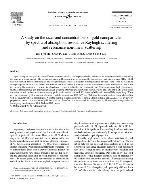
Spectrochimica Acta Part A61(2005)2861–2866A study on the sizes and concentrations of gold nanoparticlesby spectra of absorption,resonance Rayleigh scatteringand resonance non-linear scatteringYou Qiu He,Shao Pu Liu∗,Ling Kong,Zhong Fang LiuSchool of Chemistry and Chemical Engineering,Southwest China Normal University,Chongqing400715,PR ChinaReceived9April2004;received in revised form19October2004;accepted27October2004AbstractLiquid phase gold nanoparticles with different diameters and colors can be prepared using sodium citrate reduction method by controlling the amounts of sodium citrate.The mean diameters of gold nanoparticles are measured by transmission electron microscope(TEM).Gold nanoparticles with different sizes have specific absorption spectra.When the diameters of nanoparticles is between12and41nm,the maximum absorption peaks locate at520–530nm and there are red shifts gradually with the increase of diameters of gold nanoparticles.And when the size of gold nanoparticle is constant,the absorbance is proportional to the concentration of gold.Obvious resonance Rayleigh scattering (RRS)and the resonance non-linear scattering such as second-order scattering(SOS)and frequency-doubling scattering(FDS)appear at the same time as well,and the maximum scattering peaks are located at286nm(RRS),480nm(SOS)and310nm(FDS),respectively.When the concentration of gold is constant,absorbance and the intensities of RRS,SOS and FDS(I RRS,I SOS and I FDS)have linear relationships with the diameters of gold nanoparticles.When the diameter of gold nanoparticle is constant,the absorbance and I RRS,I SOS,I FDS are directly proportional to the concentrations of gold nanoparticles.Therefore,it is very useful for studying the liquid phase gold nanoparticles by investigating the absorption,RRS,SOS and FDS spectra.©2004Elsevier B.V.All rights reserved.Keywords:Gold nanoparticle;Absorption spectrum;Resonance Rayleigh scattering;Resonance non-linear scattering1.IntroductionAt present,a study on nanoparticle is becoming a hot point owing to their novel physical and chemical attributes and their special properties in electronics[1–4],optics[1,5],electro-magnetics[6].More and more interests are drawn to the par-ticular optical characters such as surface plasmon resonance (SPR)[7],plasmon absorption(PA)[8],surface enhanced Raman scattering[9]and resonance Rayleigh scattering[10] of nanoparticles.These studies are very important not only for knowing about the new optical properties but also for study-ing the characterization and detection methods of nanopar-ticles.In recent years,gold nanoparticles as biomolecular optical probes have shown vast applied prospects because ∗Corresponding author.Tel.:+862368287940;fax:+862368287940.E-mail address:liusp@(S.P.Liu).they have been used as probes for studying and determining polynucleotide[11,12]oligonucleotide sand DNA[13,14]. Therefore,it is significant for extending the characterization methods and new applications of gold nanoparticles to further study their other optical properties.Recently,applications in the characterization of gold nanoparticles have not been reported by discovering the re-lation between the sizes and concentrations as well as the absorption,resonance Rayleigh scattering,and resonance non-linear scattering such as second-order scattering and frequency-doubling scattering.Our study show that gold nanoparticles with different diameters can be prepared by the sodium citrate reduction method[15]through controlling the amounts of sodium citrate.A serious of gold nanoparti-cles with the mean diameters of12,19,24,33and41nm can be prepared.The absorption peaks gradually shift red from 520to530nm and the colors change from orange to red and1386-1425/$–see front matter©2004Elsevier B.V.All rights reserved. doi:10.1016/j.saa.2004.10.0352862Y.Q.He et al./Spectrochimica Acta Part A61(2005)2861–2866red to purple with the increase of diameters of gold nanopar-ticles.Simultaneously,the liquid phase gold nanoparticles with different diameters can also result in RRS,SOS and FDS with different intensities,however,the scattering peaks do not change according to the diameters and they are lo-cated at286nm(RRS),480nm(SOS)and310nm(FDS), respectively.When the concentration of gold nanoparticles is constant,the I RRS,I SOS and I FDS have linear relations with the diameters of gold nanoparticles.When the diameter is constant,the absorbance and above three scattering inten-sities are directly proportional to the concentration of gold nanoparticles.Therefore,the absorption and RRS,SOS and FDS spectra are important not only for the study of the optical properties but also for the development of the new spectral characterization method of gold nanoparticles.2.Experimental2.1.ReagentsHAuCl4solution,1%Au(III);sodium citrate solution,1%. Both of the reagents were of analytical reagent grades and doubly distilled water was used throughout.2.2.ApparatusA Hitachi F-2500spectrofluorophotometer(Hitachi Ltd., Tokyo,Japan),a UV–vis8500spectrophotometer(Tianmei Company,Shanghai,China),a DHT electro thermal constant temperature device with a stirrer Shandong Jiancheng Sci-ence Instrument Plant,China and a H-600transmission elec-tron microscopy(JEOL Ltd.,Japan)were used.2.3.General procedureThe gold nanoparticles with different diameters are pre-pared by sodium citrate reduction method[15].An amount of1ml of1%HAuCl4is added into suitable amount of wa-ter and the solution is heated to95◦C.Then the solution is stirred strongly at around120min,at the same time,5.0,4.0, 1.5,1.0and0.75ml of1%sodium citrate are added drop by drop into the solution to keep the reduction time about6min. After that,the solution is kept at95◦C for5min and trans-ferred into a100mlflask and diluted to the mark with water, and a series of100g ml−1of the gold nanoparticle with the colors of orange–red,red and red–purple gold nanoparticles are prepared.The diameters of gold nanoparticles prepared by control-ling different concentrations of sodium citrate were measured by transmission electron microscope(TEM),and the absorp-tion spectra of these gold nanoparticles are measured by spec-trophotometer.In addition,the RRS spectra are recorded with synchronous scanning atλex=λem(i.e. λ=0nm)and the scattering intensities of SOS and FDS(I SOS and I FDS)are measured successively atλex=1/2λem andλex=2λem un-der different incident lights by spectrofluorophotometer,then I SOS and I FDS were plotted against corresponding incident light wavelengths to make SOS and FDS spectra.3.Results and discussion3.1.Sizes of the gold nanoparticlesThe sizes of gold nanoparticles prepared by above general procedure were measured by TEM(as shown in Fig.1).It can be seen from Fig.1that nanoparticles are relatively same in size and are almost spherical.The lower the concentration of sodium citrate is,the more gold atoms will aggregate into a nanoparticle,which results in the increase of diameters.Par-ticle sizes were measured according to the statistical analysis of large number(50–100)of particles.When the amounts of sodium citrate are5.00,4.00,1.50,1.00and0.75ml,the average diameters of gold nanoparticles are measured as12, 19,24,33and41nm respectively by TEM.3.2.Absorption spectraThe absorption spectra of gold nanoparticles with differ-ent diameters are shown in Fig.2.It can be seen that when the diameters of particles are12,19,24,33and41nm,and the maximum absorption peaks(λmax)are located at520,522, 524,528and530nm,respectively.The absorption peaks shift red with the increase of diameters of gold nanoparticles.Brus [16]using spherical particle-in-a-box model has ever proved that when the diameter of spherical nanoparticle is below 10nm,there is a blue shift of absorption band caused by the further splitting of energy levels and the further increase of the energy gap with the decrease of the diameter because of the size effect of nanoparticle.Our experiments show that even the diameter of the gold nanoparticle is above10nm, during certain size range(at least below50nm),the same law is followed,namely,the absorption band shifts red with the increase of the diameters.In addition,the results also show that the colors of nanoparticles become gradually from orange–red to red and red–purple.However,the minor change of the absorption spectra( λis only10nm)does not corre-spond with color change.Therefore,the colors of the liquid phase nanoparticle are different from those of the common small molecular solution,and they are related not only to the absorption spectra,but also to many factors such as the quantum color effect,scattering,refraction and reflection etc. The colors of gold nanoparticles are affected by the above-mentioned factors all together.The relations between the di-ameters of gold nanoparticles(d)andλmax is shown in Fig.3. Fig.3shows that the maximum absorption peaks(λmax)shift red with the increase of the diameters of gold nanoparticles. When the concentration of gold is constant,the maximum absorption peaks(λmax)are plotted against the diameters(d) and theλmax is proportional to the diameters.The linear re-gression equation isλmax=515.04+0.3647d and correlation coefficients is0.9902.They have good linear relation.Y.Q.He et al./Spectrochimica Acta Part A 61(2005)2861–28662863Fig.1.Photos of transmission electron microscope (50,000×):(a)12nm;(b)19nm;(c)24nm;(d)33nm;(e)41nm.Fig.2.Absorption spectra of gold nanoparticles:(1)12nm;(2)19nm;(3)24nm;(4)33nm;(5)41nm.Fig.3.Relation between the maximum absorption wavelength and the di-ameter (d ).2864Y.Q.He et al./Spectrochimica Acta Part A 61(2005)2861–2866Fig.4.Resonance Rayleigh scattering spectra of gold nanoparticles:(1)12nm;(2)19nm;(3)24nm;(4)33nm;(5)41nm.3.3.RRS spectraFig.4shows the RRS spectra of gold nanoparticles with different diameters.It can be seen that there is a strong maxi-mum RRS peak (λRRS )at 286nm and a smaller RRS peak at 550nm.The RRS peaks do not change with different diame-ters of gold nanoparticles,but the relative intensities enhance with the increase of the diameters.3.4.SOS and FDS spectraThe SOS and FDS spectra of gold nanoparticles with dif-ferent diameters are shown in Fig.5(a)and (b).It can be seen that gold nanoparticles can result in obvious SOS and FDS,and the maximum SOS and FDS peaks (λSOS and λFDS )are respectively located at 480nm (the incident wavelength is 240nm)and 310nm (the incident wavelength is 620nm).The SOS and FDS are probably resonance non-linear scatter-ing [17–21]produced by RRS and their intensities are weaker than that of RRS.The intensity of SOS is stronger than that of FDS.When the concentration of gold is constant,thein-Fig.5.Spectra of the second-order scattering (a)and frequency-doubling scattering (b)of gold nanoparticles:(1)12nm;(2)19nm;(3)24nm;(4)33nm;(5)41nm.parison between absorption (...)and RRS spectra (—)of gold nanoparticle (d =12nm):(1)I SOS ;(2)I FDS ;(3)A ;(4)I RRS .tensities of SOS and FDS enhance with the increase of the diameters of gold nanoparticles and they are also proportional to the diameters of gold nanoparticle.3.5.Relations of I RRS ,I SOS and I FDS with the diameters of gold nanoparticlesThe intensities of RRS,SOS and FDS have linear relations with the diameters of gold nanoparticles.The linear equations and the correlation coefficients are I RRS =6.52+130.79d ,r =0.9984(RRS),I SOS =−6.87+6.55d ,r =0.9922(SOS)and I FDS =−16.70+12.96d ,r =0.9938(FDS).Therefore,the diameters of gold nanoparticles can be estimated through measuring the relative scattering intensities.3.6.Relation between RRS and absorption spectrum The diameters of gold nanoparticles are between 12and 41nm,and they are much less than the incident wavelength.Therefore,the scatterings of these gold nanoparticles are Rayleigh scatterings because the elastic scattering with in-Y.Q.He et al./Spectrochimica Acta Part A61(2005)2861–28662865 Table1Colors and characteristics of gold nanoparticles with different diametersNo.Mean diameter,D(nm)Colorλmax(nm)Number of Au atom ina gold nanoparticleλabλRRSλSOSλFDS112Orange–red520286480310 5.33×10⁴ 219Red522286480310 2.12×10⁵ 324Red524286480310 4.27×10⁵ 433Red528286480310 1.11×10⁶ 541Red–purple530286480310 2.13×10⁶cident wavelength being equal to scattering wavelength is Rayleigh scattering.And the Rayleigh scattering of the gold nanoparticle is also located near the absorption band of gold nanoparticles(as shown in Fig.6),therefore,Rayleigh scat-tering resonates with absorption light to result in resonance enhanced Rayleigh scattering,namely,RRS.Although RRS peak(286nm)is somewhat away from the absorption peak (520–530nm),but the gold nanoparticles have strong absorp-tion near300nm and Rayleigh scattering in short wave region is stronger than that in long wave region,therefore,it is nor-mal to produce RRS near300nm,and there is a smaller RRS peak(550nm)near the maximum absorption wavelength at 520–530nm.In this case,the conditions being satisfied for producing the RRS are as follows:(1)the scattering wave-length is equal to the incident wavelength;(2)the sizes of scattering particles are much smaller than the incident wave-length;(3)the scattering is located near the absorption band. Therefore,the elastic scattering is resonance Rayleigh scat-tering.In fact,it is the resonance effect that results in strong RRS and the resonance non-linear scattering such as SOS and FDS.3.7.Relations of the gold concentration with A,I RRS,I SOS and I FDS of the liquid phase gold nanoparticlesWhen the diameter of gold nanoparticle is constant (for example,d=12nm),the relations of the concen-tration of gold with A,I RRS,I FDS and I SOS were in-vestigated.Under certain conditions,A and three scat-tering intensities have linear relationships with the con-centrations of gold nanoparticles.The linear regres-sion equation and correlation coefficients(r)and linear ranges are: I RRS=12.54+967.71c,r=0.9992,linear range 0.5–6.0g ml−1; I SOS=12.01+45.2c,r=0.9921,linear range0.5–6.0g ml−1; I FDS=5.3+16.67c,r=0.9906, linear range0.5–6.0g ml−1.The detection limits for gold are170.5ng ml−1(RRS),298.2ng ml−1(SOS)and 203.9ng ml−1. A=0.022+0.52c,r=0.9903,linear range 0.5–5.0g ml−1.The colors and spectral characteristics of absorption and three resonance scatterings are shown in Table1.The above-mentioned researches show that:(1)liquid phase gold nanoparticles with different diameters and col-ors can be prepared using sodium citrate reduction method by controlling the amounts of sodium citrate;(2)the absorp-tion peaks shift red with the increase of diameters of gold nanoparticles;(3)when the concentration of liquid phase gold nanoparticle is constant,the scattering intensities of RRS, SOS,and FDS have linear relationship with the diameters of gold nanoparticles.And when the diameter of gold nanoparti-cle is constant,A,I RRS,I SOS and I FDS are directly proportional to the concentrations of gold in the liquid phase gold nanopar-ticle.Therefore,according to the colors of gold nanoparticles and the relative intensities of A,RRS,SOS and FDS,the diam-eters of gold nanoparticles can be estimated approximately. When the diameter is constant,the spectrophotometry and the RRS,SOS and FDS are simple methods for determining the concentration of gold nanoparticle. AcknowledgementsThis work has been supported by the National Natural Science Foundation of China(no.20175018). References[1]M.Brust,D.Bethell,C.J.Kiely,D.J.Schiffrin,Langmuir14(1998)5425.[2]H.Li,L.Jiang,Prog.Chem.9(1997)397.[3]G.Schmid,Chem.Rev.92(1992)1709.[4]M.Brust,D.Bethell,D.Schiffrin,J.Adv.Mater.9(1995)795.[5]C.P.Collier,R.J.Saykally,J.J.Shiang,S.E.Henrichs,J.R.Heath,Science277(1997)1978.[6]S.H.Sun,C.B.Murray,D.Weller,L.Folks,A.J.Moster,Science287(2000)1989.[7]L.A.Lyon,D.J.Pena,M.J.Natan,J.Phys.Chem.B103(1999)5826.[8]S.Link,M.A.El-sayed,J.Phys.Chem.B103(1999)4212.[9]C.K.Chen,I.I.Hemz,D.Ricard,Phys.Rev.B27(1983)1965.[10]Z.L.Jiang,Z.W.Feng,F.Li,Sci.Chin.B31(2001)185.[11]R.Elghanian,J.J.Storhoff,R.C.Mucic,R.L.Letsinger,C.A.Mirkin,Science277(1997)1078.[12]R.A.Reynolds,C.A.Mirkin,R.L.Letsinger,J.Am.Chem.Soc.122(2000)3795.[13]J.J.Storhoff, zaorides,R.C.Mucic, C.A.Mirkin,R.L.Letsinger,G.C.Schatz,J.Am.Chem.Soc.122(2000)4640.[14]T.A.Taton,C.A.Mirkin,R.L.Letsinger,Science289(2000)1757.[15]F.Y.Wang,Foreign Med.12(1991)145.2866Y.Q.He et al./Spectrochimica Acta Part A61(2005)2861–2866[16]L.Brus,J.Phys.Chem.90(1986)2555.[17]S.P.Liu,H.Q.Luo,N.B.Li,Z.F.Liu,Chin.J.Chem.21(2003)423.[18]H.Q.Luo,S.P.Liu,N.B.Li,Acta Chim.Sin.61(2003)435.[19]S.P.Liu,Z.F.Liu,Z.L.Jiang,M.Li,X.F.Long,Acta Chim.Sin.59(2001)1864.[20]N.B.Li,S.P.Liu,H.Q.Luo,Anal.Chim.Acta472(2002)89.[21]Z.L.Jiang,S.P.Liu,S.Chen,Spectra Chim.Acta58(2002)3122.。
分析化学专业英语词汇总结

专业英语词汇-----分析化学第一章绪论分析化学:analytical chemistry定性分析:qualitative analysis定量分析:quantitative analysis物理分析:physical analysis物理化学分析:physico-chemical analysis仪器分析法:instrumental analysis流动注射分析法:flow injection analysis;FIA顺序注射分析法:sequentical injection analysis;SIA化学计量学:chemometrics第二章误差的分析数据处理绝对误差:absolute error相对误差:relative error系统误差:systematic error可定误差:determinate error随机误差:accidental error不可定误差:indeterminate error准确度:accuracy精确度:precision偏差:debiation,d平均偏差:average debiation相对平均偏差:relative average debiation标准偏差(标准差):standerd deviation;S相对平均偏差:relatibe standard deviation;RSD变异系数:coefficient of variation误差传递:propagation of error有效数字:significant figure置信水平:confidence level显著性水平:level of significance合并标准偏差(组合标准差):pooled standard debiation 舍弃商:rejection quotient ;Q化学定量分析第三章滴定分析概论滴定分析法:titrametric analysis滴定:titration容量分析法:volumetric analysis化学计量点:stoichiometric point等当点:equivalent point电荷平衡:charge balance电荷平衡式:charge balance equation质量平衡:mass balance物料平衡:material balance质量平衡式:mass balance equation第四章酸碱滴定法酸碱滴定法:acid-base titrations 质子自递反应:auto protolysis reaction质子自递常数:autoprotolysis constant质子条件式:proton balance equation酸碱指示剂:acid-base indicator指示剂常数:indicator constant变色范围:colour change interval混合指示剂:mixed indicator双指示剂滴定法:double indicator titration第五章非水滴定法非水滴定法:nonaqueous titrations质子溶剂:protonic solvent酸性溶剂:acid solvent碱性溶剂:basic solvent两性溶剂:amphototeric solvent无质子溶剂:aprotic solvent均化效应:differentiatin g effect区分性溶剂:differentiating solvent离子化:ionization离解:dissociation结晶紫:crystal violet萘酚苯甲醇: α-naphthalphenol benzyl alcohol奎哪啶红:quinadinered百里酚蓝:thymol blue偶氮紫:azo violet溴酚蓝:bromophenol blue第六章配位滴定法配位滴定法:compleximetry乙二胺四乙酸:ethylenediamine tetraacetic acid,EDTA 螯合物:chelate compound金属指示剂:metal lochrome indcator第七章氧化还原滴定法氧化还原滴定法:oxidation-reduction titration碘量法:iodimetry溴量法:bromimetry ]溴量法:bromine method铈量法:cerimetry高锰酸钾法:potassium permanganate method条件电位:conditional potential溴酸钾法:potassium bromate method硫酸铈法:cerium sulphate method偏高碘酸:metaperiodic acid高碘酸盐:periodate亚硝酸钠法:sodium nitrite method重氮化反应:diazotization reaction重氮化滴定法:diazotization titration亚硝基化反应:nitrozation reaction亚硝基化滴定法:nitrozation titration外指示剂:external indicator外指示剂:outside indicator重铬酸钾法:potassium dichromate method 第八章沉淀滴定法沉淀滴定法:precipitation titration容量滴定法:volumetric precipitation method 银量法:argentometric method第九章重量分析法重量分析法:gravimetric analysis挥发法:volatilization method引湿水(湿存水):water of hydroscopicity 包埋(藏)水:occluded water吸入水:water of imbibition结晶水:water of crystallization组成水:water of composition液-液萃取法:liquid-liquid extration溶剂萃取法:solvent extration反萃取:counter extraction分配系数:partition coefficient分配比:distribution ratio离子对(离子缔合物):ion pair沉淀形式:precipitation forms称量形式:weighing forms仪器分析概述物理分析:physical analysis物理化学分析:physicochemical analysis仪器分析:instrumental analysis第十章电位法及永停滴定法电化学分析:electrochemical analysis电解法:electrolytic analysis method电重量法:electrogravimetry库仑法:coulo metry库仑滴定法:coulo metric titration电导法:conductometry电导分析法:conductometric analysis电导滴定法:conductometric titration电位法:potentiometry直接电位法:dirext potentiometry电位滴定法:potentiometric titration伏安法:voltammetry极谱法:polarography溶出法:stripping method电流滴定法:amperometric titration化学双电层:chemical double layer相界电位:phase boundary potential 金属电极电位:electrode potential化学电池:chemical cell液接界面:liquid junction boundary原电池:galvanic cell电解池:electrolytic cell负极:cathode正极:anode电池电动势:eletromotive force指示电极:indicator electrode参比电极:reference electroade标准氢电极:standard hydrogen electrode一级参比电极:primary reference electrode饱和甘汞电极:saturated calomel electrode银-氯化银电极:silver silver-chloride electrode液接界面:liquid junction boundary不对称电位:asymmetry potential表观PH值:apparent PH复合PH电极:combination PH electrode离子选择电极:ion selective electrode敏感器:sensor晶体电极:crystalline electrodes均相膜电极:homogeneous membrance electrodes非均相膜电极:heterogeneous membrance electrodes非晶体电极:non- crystalline electrodes刚性基质电极:rigid matrix electrode流流体载动电极:electrode with a mobile carrier气敏电极:gas sensing electrodes酶电极:enzyme electrodes金属氧化物半导体场效应晶体管:MOSFET离子选择场效应管:ISFET总离子强度调节缓冲剂:total ion strength adjustment buffer,TISAB永停滴定法:dead-stop titration双电流滴定法(双安培滴定法):double amperometric titration 第十一章光谱分析法概论普朗克常数:Plank constant电磁波谱:electromagnetic spectrum光谱:spectrum光谱分析法:spectroscopic analysis原子发射光谱法:atomic emission spectroscopy质量谱:mass spectrum质谱法:mass spectroscopy,MS第十二章紫外-可见分光光度法紫外-可见分光光度法:ultraviolet and visible spectrophotometry;UV-vis肩峰:shoulder peak末端吸收:end absorbtion生色团:chromophore助色团:auxochrome红移:red shift长移:bathochromic shift短移:hypsochromic shift蓝(紫)移:blue shift增色效应(浓色效应):hyperchromic effect减色效应(淡色效应):hypochromic effect强带:strong band弱带:weak band吸收带:absorption band透光率:transmitance,T吸光度:absorbance谱带宽度:band width杂散光:stray light噪声:noise暗噪声:dark noise散粒噪声:signal shot noise闪耀光栅:blazed grating全息光栅:holographic grating光二极管阵列检测器:photodiode array detector 偏最小二乘法:partial least squares method ,PLS褶合光谱法:convolution spectrometry褶合变换:convolution transform,CT离散小波变换:wavelet transform,WT多尺度细化分析:multiscale analysis供电子取代基:electron donating group吸电子取代基:electron with-drawing group第十三章荧光分析法荧光:fluorescence荧光分析法:fluorometryX-射线荧光分析法:X-ray fluorometry原子荧光分析法:atomic fluorometry分子荧光分析法:molecular fluorometry振动弛豫:vibrational relaxation内转换:internal conversion外转换:external conversion体系间跨越:intersystem crossing激发光谱:excitation spectrum荧光光谱:fluorescence spectrum斯托克斯位移:Stokes shift荧光寿命:fluorescence life time荧光效率:fluorescence efficiency荧光量子产率:fluorescence quantum yield荧光熄灭法:fluorescence quenching method散射光:scattering light瑞利光:R a yleith scattering light拉曼光:Raman scattering lightAbbe refractometer 阿贝折射仪absorbance 吸收度absorbance ratio 吸收度比值absorption 吸收absorption curve 吸收曲线absorption spectrum 吸收光谱absorptivity 吸收系数accuracy 准确度acid-dye colorimetry 酸性染料比色法acidimetry 酸量法acid-insoluble ash 酸不溶性灰分acidity 酸度activity 活度第十四章色谱法additive 添加剂additivity 加和性adjusted retention time 调整保留时间adsorbent 吸附剂adsorption 吸附affinity chromatography 亲和色谱法aliquot (一)份alkalinity 碱度alumina 氧化铝ambient temperature 室温ammonium thiocyanate 硫氰酸铵analytical quality control(AQC)分析质量控制anhydrous substance 干燥品anionic surfactant titration 阴离子表面活性剂滴定法antibiotics-microbial test 抗生素微生物检定法antioxidant 抗氧剂appendix 附录application of sample 点样area normalization method 面积归一化法argentimetry 银量法arsenic 砷arsenic stain 砷斑ascending development 上行展开ash-free filter paper 无灰滤纸(定量滤纸)assay 含量测定assay tolerance 含量限度atmospheric pressure ionization(API) 大气压离子化attenuation 衰减back extraction 反萃取back titration 回滴法bacterial endotoxins test 细菌内毒素检查法band absorption 谱带吸收baseline correction 基线校正baseline drift 基线漂移batch, lot 批batch(lot) number 批号Benttendorff method 白田道夫(检砷)法between day (day to day, inter-day) precision 日间精密度between run (inter-run) precision 批间精密度biotransformation 生物转化bioavailability test 生物利用度试验bioequivalence test 生物等效试验biopharmaceutical analysis 体内药物分析,生物药物分析blank test 空白试验boiling range 沸程British Pharmacopeia (BP) 英国药典bromate titration 溴酸盐滴定法bromimetry 溴量法bromocresol green 溴甲酚绿bromocresol purple 溴甲酚紫bromophenol blue 溴酚蓝bromothymol blue 溴麝香草酚蓝bulk drug, pharmaceutical product 原料药buret 滴定管by-product 副产物calibration curve 校正曲线calomel electrode 甘汞电极calorimetry 量热分析capacity factor 容量因子capillary zone electrophoresis (CZE) 毛细管区带电泳capillary gas chromatography 毛细管气相色谱法carrier gas 载气cation-exchange resin 阳离子交换树脂ceri(o)metry 铈量法characteristics, description 性状check valve 单向阀chemical shift 化学位移chelate compound 鳌合物chemically bonded phase 化学键合相chemical equivalent 化学当量Chinese Pharmacopeia (ChP) 中国药典Chinese material medicine 中成药Chinese materia medica 中药学Chinese materia medica preparation 中药制剂Chinese Pharmaceutical Association (CPA) 中国药学会chiral 手性的chiral stationary phase (CSP) 手性固定相chiral separation 手性分离chirality 手性chiral carbon atom 手性碳原子chromatogram 色谱图chromatography 色谱法chromatographic column 色谱柱chromatographic condition 色谱条件chromatographic data processor 色谱数据处理机chromatographic work station 色谱工作站clarity 澄清度clathrate, inclusion compound 包合物clearance 清除率clinical pharmacy 临床药学coefficient of distribution 分配系数coefficient of variation 变异系数color change interval (指示剂)变色范围color reaction 显色反应colorimetric analysis 比色分析colorimetry 比色法column capacity 柱容量column dead volume 柱死体积column efficiency 柱效column interstitial volume 柱隙体积column outlet pressure 柱出口压column temperature 柱温column pressure 柱压column volume 柱体积column overload 柱超载column switching 柱切换committee of drug evaluation 药品审评委员会comparative test 比较试验completeness of solution 溶液的澄清度compound medicines 复方药computer-aided pharmaceutical analysis 计算机辅助药物分析concentration-time curve 浓度-时间曲线confidence interval 置信区间confidence level 置信水平confidence limit 置信限congealing point 凝点congo red 刚果红(指示剂)content uniformity 装量差异controlled trial 对照试验correlation coefficient 相关系数contrast test 对照试验counter ion 反离子(平衡离子)cresol red 甲酚红(指示剂)crucible 坩埚crude drug 生药crystal violet 结晶紫(指示剂)cuvette, cell 比色池cyanide 氰化物cyclodextrin 环糊精cylinder, graduate cylinder, measuring cylinder 量筒cylinder-plate assay 管碟测定法daughter ion (质谱)子离子dead space 死体积dead-stop titration 永停滴定法dead time 死时间decolorization 脱色decomposition point 分解点deflection 偏差deflection point 拐点degassing 脱气deionized water 去离子水deliquescence 潮解depressor substances test 降压物质检查法derivative spectrophotometry 导数分光光度法derivatization 衍生化descending development 下行展开desiccant 干燥剂detection 检查detector 检测器developer, developing reagent 展开剂developing chamber 展开室deviation 偏差dextrose 右旋糖,葡萄糖diastereoisomer 非对映异构体diazotization 重氮化2,6-dichlorindophenol titration 2,6-二氯靛酚滴定法differential scanning calorimetry (DSC) 差示扫描热量法differential spectrophotometry 差示分光光度法differential thermal analysis (DTA) 差示热分析differentiating solvent 区分性溶剂diffusion 扩散digestion 消化diphastic titration 双相滴定disintegration test 崩解试验dispersion 分散度dissolubility 溶解度dissolution test 溶出度检查distilling range 馏程distribution chromatography 分配色谱distribution coefficient 分配系数dose 剂量drug control institutions 药检机构drug quality control 药品质量控制drug release 药物释放度drug standard 药品标准drying to constant weight 干燥至恒重dual wavelength spectrophotometry 双波长分光光度法duplicate test 重复试验effective constituent 有效成分effective plate number 有效板数efficiency of column 柱效electron capture detector 电子捕获检测器electron impact ionization 电子轰击离子化electrophoresis 电泳electrospray interface 电喷雾接口electromigration injection 电迁移进样elimination 消除eluate 洗脱液elution 洗脱emission spectrochemical analysis 发射光谱分析enantiomer 对映体end absorption 末端吸收end point correction 终点校正endogenous substances 内源性物质enzyme immunoassay(EIA) 酶免疫分析enzyme drug 酶类药物enzyme induction 酶诱导enzyme inhibition 酶抑制eosin sodium 曙红钠(指示剂)epimer 差向异构体equilibrium constant 平衡常数equivalence point 等当点error in volumetric analysis 容量分析误差excitation spectrum 激发光谱exclusion chromatography 排阻色谱法expiration date 失效期external standard method 外标法extract 提取物extraction gravimetry 提取重量法extraction titration 提取容量法extrapolated method 外插法,外推法factor 系数,因数,因子feature 特征Fehling’s reaction 费林反应field disorption ionization 场解吸离子化field ionization 场致离子化filter 过滤,滤光片filtration 过滤fineness of the particles 颗粒细度flame ionization detector(FID) 火焰离子化检测器flame emission spectrum 火焰发射光谱flask 烧瓶flow cell 流通池flow injection analysis 流动注射分析flow rate 流速fluorescamine 荧胺fluorescence immunoassay(FIA) 荧光免疫分析fluorescence polarization immunoassay(FPIA) 荧光偏振免疫分析fluorescent agent 荧光剂fluorescence spectrophotometry 荧光分光光度法fluorescence detection 荧光检测器fluorimetyr 荧光分析法foreign odor 异臭foreign pigment 有色杂质formulary 处方集fraction 馏分freezing test 结冻试验funnel 漏斗fused peaks, overlapped peaks 重叠峰fused silica 熔融石英gas chromatography(GC) 气相色谱法gas-liquid chromatography(GLC) 气液色谱法gas purifier 气体净化器gel filtration chromatography 凝胶过滤色谱法gel permeation chromatography 凝胶渗透色谱法general identification test 一般鉴别试验general notices (药典)凡例general requirements (药典)通则good clinical practices(GCP) 药品临床管理规范good laboratory practices(GLP) 药品实验室管理规范good manufacturing practices(GMP) 药品生产质量管理规范good supply practices(GSP) 药品供应管理规范gradient elution 梯度洗脱grating 光栅gravimetric method 重量法Gutzeit test 古蔡(检砷)法half peak width 半峰宽[halide] disk method, wafer method, pellet method 压片法head-space concentrating injector 顶空浓缩进样器heavy metal 重金属heat conductivity 热导率height equivalent to a theoretical plate 理论塔板高度height of an effective plate 有效塔板高度high-performance liquid chromatography (HPLC) 高效液相色谱法high-performance thin-layer chromatography (HPTLC) 高效薄层色谱法hydrate 水合物hydrolysis 水解hydrophilicity 亲水性hydrophobicity 疏水性hydroscopic 吸湿的hydroxyl value 羟值hyperchromic effect 浓色效应hypochromic effect 淡色效应identification 鉴别ignition to constant weight 灼烧至恒重immobile phase 固定相immunoassay 免疫测定impurity 杂质inactivation 失活index 索引indicator 指示剂indicator electrode 指示电极inhibitor 抑制剂injecting septum 进样隔膜胶垫injection valve 进样阀instrumental analysis 仪器分析insulin assay 胰岛素生物检定法integrator 积分仪intercept 截距interface 接口interference filter 干涉滤光片intermediate 中间体internal standard substance 内标物质international unit(IU) 国际单位in vitro 体外in vivo 体内iodide 碘化物iodoform reaction 碘仿反应iodometry 碘量法ion-exchange cellulose 离子交换纤维素ion pair chromatography 离子对色谱ion suppression 离子抑制ionic strength 离子强度ion-pairing agent 离子对试剂ionization 电离,离子化ionization region 离子化区irreversible indicator 不可逆指示剂irreversible potential 不可逆电位isoabsorptive point 等吸收点isocratic elution 等溶剂组成洗脱isoelectric point 等电点isoosmotic solution 等渗溶液isotherm 等温线Karl Fischer titration 卡尔·费歇尔滴定kinematic viscosity 运动黏度Kjeldahl method for nitrogen 凯氏定氮法Kober reagent 科伯试剂Kovats retention index 科瓦茨保留指数labelled amount 标示量leading peak 前延峰least square method 最小二乘法leveling effect 均化效应licensed pharmacist 执业药师limit control 限量控制limit of detection(LOD) 检测限limit of quantitation(LOQ) 定量限limit test (杂质)限度(或限量)试验limutus amebocyte lysate(LAL) 鲎试验linearity and range 线性及范围linearity scanning 线性扫描liquid chromatograph/mass spectrometer (LC/MS) 液质联用仪litmus paper 石蕊试纸loss on drying 干燥失重low pressure gradient pump 低压梯度泵luminescence 发光lyophilization 冷冻干燥main constituent 主成分make-up gas 尾吹气maltol reaction 麦牙酚试验Marquis test 马奎斯试验mass analyzer detector 质量分析检测器mass spectrometric analysis 质谱分析mass spectrum 质谱图mean deviation 平均偏差measuring flask, volumetric flask 量瓶measuring pipet(te) 刻度吸量管medicinal herb 草药melting point 熔点melting range 熔距metabolite 代谢物metastable ion 亚稳离子methyl orange 甲基橙methyl red 甲基红micellar chromatography 胶束色谱法micellar electrokinetic capillary chromatography(MECC, MEKC) 胶束电动毛细管色谱法micelle 胶束microanalysis 微量分析microcrystal 微晶microdialysis 微透析micropacked column 微型填充柱microsome 微粒体microsyringe 微量注射器migration time 迁移时间millipore filtration 微孔过滤minimum fill 最低装量mobile phase 流动相modifier 改性剂,调节剂molecular formula 分子式monitor 检测,监测monochromator 单色器monographs 正文mortar 研钵moving belt interface 传送带接口multidimensional detection 多维检测multiple linear regression 多元线性回归multivariate calibration 多元校正natural product 天然产物Nessler glasses(tube) 奈斯勒比色管Nessler’s r eagent 碱性碘化汞钾试液neutralization 中和nitrogen content 总氮量nonaqueous acid-base titration 非水酸碱滴定nonprescription drug, over the counter drugs (OTC drugs) 非处方药nonproprietary name, generic name 非专有名nonspecific impurity 一般杂质non-volatile matter 不挥发物normal phase 正相normalization 归一化法notice 凡例nujol mull method 石蜡糊法octadecylsilane chemically bonded silica 十八烷基硅烷键合硅胶octylsilane 辛(烷)基硅烷odorless 无臭official name 法定名official specifications 法定标准official test 法定试验on-column detector 柱上检测器on-column injection 柱头进样on-line degasser 在线脱气设备on the dried basis 按干燥品计opalescence 乳浊open tubular column 开管色谱柱optical activity 光学活性optical isomerism 旋光异构optical purity 光学纯度optimization function 优化函数organic volatile impurities 有机挥发性杂质orthogonal function spectrophotometry 正交函数分光光度法orthogonal test 正交试验orthophenanthroline 邻二氮菲outlier 可疑数据,逸出值overtones 倍频峰,泛频峰oxidation-reduction titration 氧化还原滴定oxygen flask combustion 氧瓶燃烧packed column 填充柱packing material 色谱柱填料palladium ion colorimetry 钯离子比色法parallel analysis 平行分析parent ion 母离子particulate matter 不溶性微粒partition coefficient 分配系数parts per million (ppm) 百万分之几pattern recognition 模式识别peak symmetry 峰不对称性peak valley 峰谷peak width at half height 半峰宽percent transmittance 透光百分率pH indicator absorbance ratio method? pH指示剂吸光度比值法pharmaceutical analysis 药物分析pharmacopeia 药典pharmacy 药学phenolphthalein 酚酞photodiode array detector(DAD) 光电二极管阵列检测器photometer 光度计pipeclay triangle 泥三角pipet(te) 吸移管,精密量取planar chromatography 平板色谱法plate storage rack 薄层板贮箱polarimeter 旋光计polarimetry 旋光测定法polarity 极性polyacrylamide gel 聚丙酰胺凝胶polydextran gel 葡聚糖凝胶polystyrene gel 聚苯乙烯凝胶polystyrene film 聚苯乙烯薄膜porous polymer beads 高分子多孔小球post-column derivatization 柱后衍生化potentiometer 电位计potentiometric titration 电位滴定法precipitation form 沉淀形式precision 精密度pre-column derivatization 柱前衍生化preparation 制剂prescription drug 处方药pretreatment 预处理primary standard 基准物质principal component analysis 主成分分析programmed temperature gas chromatography 程序升温气相色谱法prototype drug 原型药物provisions for new drug approval 新药审批办法purification 纯化purity 纯度pyrogen 热原pycnometric method 比重瓶法quality control(QC) 质量控制quality evaluation 质量评价quality standard 质量标准quantitative determination 定量测定quantitative analysis 定量分析quasi-molecular ion 准分子离子racemization 消旋化radioimmunoassay 放射免疫分析法random sampling 随机抽样rational use of drug 合理用药readily carbonizable substance 易炭化物reagent sprayer 试剂喷雾器recovery 回收率reference electrode 参比电极refractive index 折光指数related substance 有关物质relative density 相对密度relative intensity 相对强度repeatability 重复性replicate determination 平行测定reproducibility 重现性residual basic hydrolysis method 剩余碱水解法residual liquid junction potential 残余液接电位residual titration 剩余滴定residue on ignition 炽灼残渣resolution 分辨率,分离度response time 响应时间retention 保留reversed phase chromatography 反相色谱法reverse osmosis 反渗透rider peak 驼峰rinse 清洗,淋洗robustness 可靠性,稳定性routine analysis 常规分析round 修约(数字)ruggedness 耐用性safety 安全性Sakaguchi test 坂口试验salt bridge 盐桥salting out 盐析sample applicator 点样器sample application 点样sample on-line pretreatment 试样在线预处理sampling 取样saponification value 皂化值saturated calomel electrode(SCE) 饱和甘汞电极selectivity 选择性separatory funnel 分液漏斗shoulder peak 肩峰signal to noise ratio 信噪比significant difference 显著性差异significant figure 有效数字significant level 显著性水平significant testing 显著性检验silanophilic interaction 亲硅羟基作用silica gel 硅胶silver chloride electrode 氯化银电极similarity 相似性simultaneous equations method 解线性方程组法size exclusion chromatography(SEC) 空间排阻色谱法sodium dodecylsulfate, SDS 十二烷基硫酸钠sodium hexanesulfonate 己烷磺酸钠sodium taurocholate 牛璜胆酸钠sodium tetraphenylborate 四苯硼钠sodium thiosulphate 硫代硫酸钠solid-phase extraction 固相萃取solubility 溶解度solvent front 溶剂前沿solvophobic interaction 疏溶剂作用specific absorbance 吸收系数specification 规格specificity 专属性specific rotation 比旋度specific weight 比重spiked 加入标准的split injection 分流进样splitless injection 无分流进样spray reagent (平板色谱中的)显色剂spreader 铺板机stability 稳定性standard color solution 标准比色液standard deviation 标准差standardization 标定standard operating procedure(SOP) 标准操作规程standard substance 标准品stationary phase coating 固定相涂布starch indicator 淀粉指示剂statistical error 统计误差sterility test 无菌试验stirring bar 搅拌棒stock solution 储备液stoichiometric point 化学计量点storage 贮藏stray light 杂散光substituent 取代基substrate 底物sulfate 硫酸盐sulphated ash 硫酸盐灰分supercritical fluid chromatography(SFC) 超临界流体色谱法support 载体(担体)suspension 悬浊液swelling degree 膨胀度symmetry factor 对称因子syringe pump 注射泵systematic error 系统误差system model 系统模型system suitability 系统适用性tablet 片剂tailing factor 拖尾因子tailing peak 拖尾峰tailing-suppressing reagent 扫尾剂test of hypothesis 假设检验test solution(TS) 试液tetrazolium colorimetry 四氮唑比色法therapeutic drug monitoring(TDM) 治疗药物监测thermal analysis 热分析法thermal conductivity detector 热导检测器thermocouple detector 热电偶检测器thermogravimetric analysis(TGA) 热重分析法thermospray interface 热喷雾接口The United States Pharmacopoeia(USP) 美国药典The Pharmacopoeia of Japan(JP) 日本药局方thin layer chromatography(TLC) 薄层色谱法thiochrome reaction 硫色素反应three-dimensional chromatogram 三维色谱图thymol 百里酚(麝香草酚)(指示剂)thymolphthalein 百里酚酞(麝香草酚酞)(指示剂)thymolsulfonphthalein ( thymol blue) 百里酚蓝(麝香草酚蓝)(指示剂)titer, titre 滴定度time-resolved fluoroimmunoassay 时间分辨荧光免疫法titrant 滴定剂titration error 滴定误差titrimetric analysis 滴定分析法tolerance 容许限toluene distillation method 甲苯蒸馏法toluidine blue 甲苯胺蓝(指示剂)total ash 总灰分total quality control(TQC) 全面质量控制traditional drugs 传统药traditional Chinese medicine 中药transfer pipet 移液管turbidance 混浊turbidimetric assay 浊度测定法turbidimetry 比浊法turbidity 浊度ultracentrifugation 超速离心ultrasonic mixer 超生混合器ultraviolet irradiation 紫外线照射undue toxicity 异常毒性uniform design 均匀设计uniformity of dosage units 含量均匀度uniformity of volume 装量均匀性(装量差异)uniformity of weight 重量均匀性(片重差异)validity 可靠性variance 方差versus …对…,…与…的关系曲线viscosity 粘度volatile oil determination apparatus 挥发油测定器volatilization 挥发法volumetric analysis 容量分析volumetric solution(VS) 滴定液vortex mixer 涡旋混合器watch glass 表面皿wave length 波长wave number 波数weighing bottle 称量瓶weighing form 称量形式weights 砝码well-closed container 密闭容器xylene cyanol blue FF 二甲苯蓝FF(指示剂)xylenol orange 二甲酚橙(指示剂)zigzag scanning 锯齿扫描zone electrophoresis 区带电泳zwitterions 两性离子zymolysis 酶解作用簡體書目錄Chapter 1 Introduction 緒論1.1 The nature of analytical chemistry 分析化學的性質1.2 The role of analytical chemistry 分析化學的作用1.3 The classification of analytical chemistry分析化學的分類1.4 The total analytical process分析全過程Terms to understand重點內容概述Chapter 2 Errors and Data Treatment in Quantitative Analysis 定量分析中的誤差及數據處理2.1 Fundamental terms of errors誤差的基本術語2.2 Types of errors in experimental data實驗數據中的誤差類型2.2.1 Systematic errors 系統誤差2.2.2 Random errors偶然誤差2.3 Evaluation of analytical data分析數據的評價2.3.1 Tests of significance顯著性檢驗2.3.2 Rejecting data可疑值取捨2.4 Significant figures有效數字ProblemsTerms to understand重點內容概述Chapter 3 Titrimetric Analysis滴定分析法3.1 General principles基本原理3.1.1 Relevant terms of titrimetric analysis滴定分析相關術語3.1.2 The preparation of standard solution and the expression of concentration 標準溶液的配製與濃度表示方法3.1.3 The types of titrimetric reactions滴定反應類型3.2 Acid-base titration酸鹼滴定3.2.1 Acid-base equilibria 酸鹼平衡3.2.2 Titration curves滴定曲線3.2.3 Acid-base indicators酸鹼指示劑3.2.4 Applications of acid-base titration酸鹼滴定的應用3.3 Complexometric titration配位滴定3.3.1 Metal-chelate complexes金屬螯合物3.3.2 EDTA 乙二胺四乙酸3.3.3 EDTA titration curves EDTA滴定曲線3.3.4 Metal Ion indicators金屬離子指示劑3.3.5 Applications of EDTA titration techniques EDTA滴定方法的應用3.4 Oxidation-reduction titration氧化還原滴定3.4.1 Redox reactions氧化還原反應3.4.2 Rate of redox reactions氧化還原反應的速率3.4.3 Titration curves滴定曲線3.4.4 Redox indicators氧化還原指示劑3.4.5 Applications of redox titrations氧化還原滴定的應用3.5 Precipitation titration沉澱滴定3.5.1 Precipitation reactions沉澱滴定反應3.5.2 Titration curves滴定曲線3.5.3 End-point detection終點檢測ProblemsTerms to understand重點內容概述Chapter 4 Potentiometry 電位分析法4.1 Introduction簡介4.1.1 Classes and characteristics分類及性質4.1.2 Definition定義4.2 Types of potentiometric electrodes電極種類4.2.1 Reference electrodes 參比電極4.2.2 Indicator electrodes指示電極4.2.3 Electrode response and selectivity電極響應及選擇性4.3 Potentiometric methods and application電位法及應用4.3.1 Direct potentiometric measurement 直接電位法4.3.2 Potentiometric titrations電位滴定4.3.3 Applications of potentiometry 電位法應用ProblemsTerlns to understand重點內容概述Chapter 5 Chromatography色譜法5.1 An introduction to chromatographic methods色譜法概述5.2 Fundamental theory of gas chromatography氣相色譜基本原理5.2.1 Plate theory塔板理論5.2.2 Kinetic theory(rate theory) 速率理論5.2.3 The resolution Rs as a measure of peak separation 分離度5.3 Gas chromatography 氣相色譜5.3.1 Components of a gas chromatograph 氣相色譜儀的組成5.3.2 Stationary phases for gas-liquid chromatography 氣液色譜固定相5.3.3 Applications of gas-liquid chromatography 氣液色譜的應用5.3.4 Adsorption chromatography 吸附色譜5.4 High performance liquid chromatography 高效液相色譜5.4.1 Instrumentation 儀器組成5.4.2 High-performance partition chromatography 高效分配色譜5.5 Miscellaneous separation methods 其他分離方法5.5.1 High-performance ion-exchange chromatography 高效離子交換色譜5.5.2 Capillary electrophoresis 毛細管電泳5.5.3 Planar chromatography 平板色譜ProblemsTerms to understand重點內容概述Chapter 6 Atomic Absorption Spectrometry原子吸收光譜分析法6.1 Introduction 概述6.2 Principles 原理6.2.1 The process of AAS,resonance line and absorption line 原子吸收光譜法的過程,共振線及吸收線6.2.2 The number of ground atom and the temperature of flame 基態原子數與光焰溫度6.2.3 Quantitative analysis of AAS原子吸收光譜定量分析6.3 Instrumentation 儀器6.3.1 Primary radiation sources 光源6.3.2 Atomizer 原子儀器6.3.3 Optical dispersive systems 分光系統6.3.4 Detectors 檢測器6.3.5 Signal measurements 信號測量6.4 Quantitative measurements and interferences 定量測定及干擾6.4.1 Quantitative measurements 定量測定6.4.2 Interferences 干擾6.4.3 Sensitivity6.5 Applications of AAS原子吸收光譜法的應用ProblemsTerms to understand重點內容概述Chapter 7 Ultraviolet and Visible Spectrophotometry 紫外-可見分光光度法7.1 Introduction簡介7.2 Ultraviolet and visible absorption spectroscopy 紫外-可見吸收光譜7.2.1 Introduction for radiant energy 輻射能簡介7.2.2 Selective absorption of radiation and absorbance spectrum 物質對光的選擇性吸收和吸收光譜7.2.3 Absorbing species and electron transition 吸收物質與電子躍遷7.3 Law of absorption吸收定律7.3.1 Lambert-Beer's law朗伯-比爾定律7.3.2 Absorptivity吸光係數7.3.3 Apparent deviations from Beer's law對比爾定律的明顯偏離7.4 Instruments儀器7.5 General types of spectrophotometer分光光度計種類7.6 Application of UV-Vis absorption spectroscopy 紫外-可見吸收光譜的應用7.6.1 Application of absorption measurement to qualitative analysis 光吸收測定在定性分析上的應用7.6.2 Quantitative analysis by absorption measurements 光吸收測量定量分析法7.6.3 Derivative spectrophotometry 導數分光光度法ProblemsTerms to understand重點內容概述Chapter 8 Infrared Absorption Spectroscopy紅外吸收光譜8.1 Theory of infrared absorption紅外吸收基本原理8.1.1 Dipole changes during vibrations and rotations 振轉運動中的偶極距變化8.1.2 Mechanical model of stretching vibrations 伸縮振動機械模型8.1.3 Quantum treatment of vibrations 振動的量子力學處理、8.1.4 Types of molecular vibrations分子振動形式8.2 Infrared instrument components紅外儀器組成8.2.1 Wavelength selection波長選擇8.2.2 Sampling techniques 採樣技術8.2.3 Infrared spectrophotometers for qualitative analysis 定性分析用紅外分光光度計8.2.4 Other techniques其他技術8.3 The group frequencies of functional groups in organic compounds 有機化合物官能團的特徵頻率8.4 The factors affecting group frequencies 影響基團特徵吸收頻率的因素8.4.1 Adjacent groups 鄰近基團的影響。
光谱法研究药物小分子与蛋白质大分子的相互作用的英文
Spectroscopic Study of the Interaction between Small Molecules and Large Proteins1. IntroductionThe study of drug-protein interactions is of great importance in drug discovery and development. Understanding how small molecules interact with proteins at the molecular level is crucial for the design of new and more effective drugs. Spectroscopic techniques have proven to be valuable tools in the investigation of these interactions, providing det本人led information about the binding affinity, mode of binding, and structural changes that occur upon binding.2. Spectroscopic Techniques2.1. Fluorescence SpectroscopyFluorescence spectroscopy is widely used in the study of drug-protein interactions due to its high sensitivity and selectivity. By monitoring the changes in the fluorescence emission of either the drug or the protein upon binding, valuable information about the binding affinity and the binding site can be obt本人ned. Additionally, fluorescence quenching studies can provide insights into the proximity and accessibility of specific amino acid residues in the protein's binding site.2.2. UV-Visible SpectroscopyUV-Visible spectroscopy is another powerful tool for the investigation of drug-protein interactions. This technique can be used to monitor changes in the absorption spectra of either the drug or the protein upon binding, providing information about the binding affinity and the stoichiometry of the interaction. Moreover, UV-Visible spectroscopy can be used to study the conformational changes that occur in the protein upon binding to the drug.2.3. Circular Dichroism SpectroscopyCircular dichroism spectroscopy is widely used to investigate the secondary structure of proteins and to monitor conformational changes upon ligand binding. By analyzing the changes in the CD spectra of the protein in the presence of the drug, valuable information about the structural changes induced by the binding can be obt本人ned.2.4. Nuclear Magnetic Resonance SpectroscopyNMR spectroscopy is a powerful technique for the investigation of drug-protein interactions at the atomic level. By analyzing the chemical shifts and the NOE signals of the protein in thepresence of the drug, det本人led information about the binding site and the mode of binding can be obt本人ned. Additionally, NMR can provide insights into the dynamics of the protein upon binding to the drug.3. Applications3.1. Drug DiscoverySpectroscopic studies of drug-protein interactions play a crucial role in drug discovery, providing valuable information about the binding affinity, selectivity, and mode of action of potential drug candidates. By understanding how small molecules interact with their target proteins, researchers can design more potent and specific drugs with fewer side effects.3.2. Protein EngineeringSpectroscopic techniques can also be used to study the effects of mutations and modifications on the binding affinity and specificity of proteins. By analyzing the binding of small molecules to wild-type and mutant proteins, valuable insights into the structure-function relationship of proteins can be obt本人ned.3.3. Biophysical StudiesSpectroscopic studies of drug-protein interactions are also valuable for the characterization of protein-ligandplexes, providing insights into the thermodynamics and kinetics of the binding process. Additionally, these studies can be used to investigate the effects of environmental factors, such as pH, temperature, and ionic strength, on the stability and binding affinity of theplexes.4. Challenges and Future DirectionsWhile spectroscopic techniques have greatly contributed to our understanding of drug-protein interactions, there are still challenges that need to be addressed. For instance, the study of membrane proteins and protein-protein interactions using spectroscopic techniques rem本人ns challenging due to theplexity and heterogeneity of these systems. Additionally, the development of new spectroscopic methods and the integration of spectroscopy with other biophysical andputational approaches will further advance our understanding of drug-protein interactions.In conclusion, spectroscopic studies of drug-protein interactions have greatly contributed to our understanding of how small molecules interact with proteins at the molecular level. Byproviding det本人led information about the binding affinity, mode of binding, and structural changes that occur upon binding, spectroscopic techniques have be valuable tools in drug discovery, protein engineering, and biophysical studies. As technology continues to advance, spectroscopy will play an increasingly important role in the study of drug-protein interactions, leading to the development of more effective and targeted therapeutics.。
应用波谱学 英文
应用波谱学英文Applications of spectroscopySpectroscopy has a wide range of applications across various scientific disciplines. Some of the common applications of spectroscopy include:1. Chemistry: Spectroscopy is extensively used in chemistry for the identification and analysis of chemical compounds. It helps in determining the chemical composition, molecular structure, and functional groups present in a sample.2. Pharmaceuticals: Spectroscopic techniques are crucial in the drug discovery and development process. They are used for quality control, impurity analysis, and determining the stability of pharmaceutical products.3. Environmental science: Spectroscopy plays a vital role in environmental monitoring and assessment. It is used to evaluate air quality, analyze water pollutants, and identify harmful substances in soil samples.4. Biochemistry and molecular biology: Spectroscopy is employed in studying the structure, function, and dynamics of biological molecules like proteins, nucleic acids, and carbohydrates. Techniques such as UV-Visible spectroscopy, fluorescence spectroscopy, and circular dichroism spectroscopy are commonly used in this field.5. Material science: Spectroscopy helps in characterizing andstudying various materials and their properties. It is used to analyze the composition, crystal structure, and surface properties of materials such as metals, ceramics, polymers, and semiconductors.6. Astronomy: Spectroscopy is fundamental in studying the properties and composition of celestial objects. Astronomers use spectroscopic techniques to analyze the light emitted or absorbed by stars, galaxies, and other astronomical phenomena to determine their chemical composition, temperature, and motion.7. Forensics: Spectroscopic methods are employed in forensic science for the detection and analysis of trace evidence, such as drugs, explosives, and chemical residues. They are also used in analyzing questioned documents and for the identification of counterfeit or forged materials.8. Food science and agriculture: Spectroscopic techniques are used for analyzing food products, determining their quality, and detecting adulteration. They are also employed in agricultural research for monitoring plant health and analyzing soil fertility. These are just a few examples of the diverse applications of spectroscopy in various fields. Overall, spectroscopy is a powerful analytical tool that enables scientists to study and understand the properties and behavior of substances in a wide range of scientific domains.。
常用分析化学专业英语词汇
常用分析化学专业英语词汇absorbanee 吸光度absorbe nt 吸附剂absorpti on curve 吸收曲线absorpti on peak 吸收峰absorptivity 吸收系数aeeide nt error 偶然误差accuracy 准确度acid-base titrati on 酸碱滴定acidic effective coefficie nt 酸效应系数acidic effective curve酸效应曲线acidity con sta nt 酸度常数activity 活度activity coefficie nt 活度系数adsorpti on 吸附adsorpti on in dicator 吸附指示剂affin ity 亲和力aging 陈化amorphous precipitate 无定形沉淀amphiprotic solve nt 两性溶齐U amphoteric substa nee 两性物质amplificatio n reacti on 放大反应an alytical bala nee 分析天平an alytical chemistry 分析化学analytical concentration 分析浓度an alytical reage nt (AR) 分析试剂appare nt formati on eon sta nt 表观形成常数aqueous phase 水相arge ntimetry 乍艮量法ashing 灰化atomic spectrum 原子光谱autoprotolysis eon sta nt 质子自递常数auxochrome group 助色团back extracti on 反萃取band spectrum 带状光谱bandwidth 带宽blank空白color transition point 颜色转变bathochromic shift 红移block ing of in dicator 指示剂的圭封闭bromometry 溴量法buffer capacity 缓冲容量buffer solution 缓冲溶液burette 滴定管calc on carboxylic acid 钙指示齐Vcalibrated curve 校准曲线calibrati on 校准catalyzed reacti on 催化反应q cerimetry 铈量法charge bala nee 电荷平衡chelate 螯合物chelate extraction螯合物萃取chemical analysis化学分析chemical factor 卜化学因素chemically pure 化学纯chromatography 色谱法chromophoric group 发色团coefficient of variation 变异系数color reage nt 显色剂blank空白color transition point 颜色转变占八、、colorimeter 比色计colorimetry 比色法colu mn chromatography 柱色谱compleme ntary color 互补色complex络合物complexati on 络合反应complexometry complexometric titratio n 络合滴定法complex one 氨羧络合剂concen trati on con sta nt 浓度常数con diti onal extracti on con sta nt 条件萃取常数con diti onal formatio n coefficie nt 条件形成常数con diti onal pote ntial 条件电位con diti onal solubility product 条件溶度积con fide nee in terval 置信区间con fide nee level 置信水平conjugate acid-base pair 共轭酸con sta nt weight 恒量dichloro fluoresce in二氯荧光con tam in ati on 沾污黄con ti nu ous extracti on连续萃取 dichromate titrati on重铬酸钾con ti nu ous spectrum 连续光谱法coprecipitati on 共沉淀dielectric con sta nt介电常数correcti on 校正differe ntial spectrophotometrycorrelati on coefficie nt 相关系示差光度法数differe ntiati ng effect 区分效crucible 坩埚 应crystalli ne precipitate晶形沉 dispers ion 色散淀dissociati on con sta nt离解常数cumulative con sta nt "累积常数 distillati on 蒸馏curdy precipitate 凝乳状沉淀 distribution coefficient分酉己 degree of freedom 自由度系数demasking 解蔽distributi on diagram分布图 derivative spectrum导数光谱distributi on ratio分配比desicca nt; drying age nt干燥剂double beam spectrophotometerdesiccator 保干器双光束分光光度计dibasic acid 二元酸 碱对determi nate error可测误差deuterium lamp 氘灯 deviatio n偏差dual-pa n bala nee 双盘天平 dual-wavele ngthspectrophotometry 双波长分光光 度法electronic balanee 电子天平Fajans method 法杨斯法electrophoresis 电泳ferroin 邻二氮菲亚铁离子elue nt 淋洗剂filter 漏斗end point 终点filter 滤光片end point error 终点误差filter paper 滤纟纸en riehme nt 富集filtratio n 过滤eosin 曙红fluex溶剂equilibrium concen trati on 平衡fluoresce in 荧光黄浓度flusion 熔融equimolar series method 等摩尔formati on con sta nt 形成常数系列法freque ncy 频率Erele nm eyerflask锥形瓶freque ncy den sity 频率密度eriochrome black T (EBT) 铬黑T freque ncy distributi on 频率分error 误差布ethyle nediam ine tetraacetic gas chromatography (GC) 气相色acid (EDTA) 乙二胺四乙酸谱evaporati on dish 蒸发皿grati ng 光栅excha ngecapacity交换谷量gravimetric factor 重量因素exte nt of crossli nking 交联度gravimetry 重量分析extracti on con sta nt 卒取常数guara ntee reage nt (GR) 保证试齐Uextracti on rate 萃取率high performa nee liquid extracti on spectrphotometric chromatography (HPLC) 咼效液相method萃取光度法色谱histogram 直方图homoge neous precipitati on 均相沉淀hydroge n lamp 氢灯hypochromic shift 紫移ign iti on 灼烧in dicator 指示齐Uin duced reacti on 诱导反应inert solve nt 惰性溶剂in stability con sta nt 不稳定常数in strume ntal analysis仪器分析in tri nsic acidity 固有酸度in tri nsic basicity 固有碱度in tri nsic solubility 固有溶解度iodimetry 碘滴定法iodin e-t un gste nlamp碘钨灯iodometry 滴定碘法ion association extraction 离子缔合物萃取ion chromatography (IC) 离子色谱ion excha nge 离子交换ion exchange resin 离子交换树脂ion ic stre ngth 离子强度isoabsorptive point 等吸收点Karl Fisher titration 卡尔?费歇尔法Kjeldahl determ in ati on 凯氏定氮法Lambert-Beer law 朗泊-比尔定律leveli ng effect 拉平效应liga nd 配位体light source 光源line spectrum 线状光谱lin ear regressi on 线性回归liquid chromatography (LC) 液相色谱macro an alysis 常量分析masking 掩蔽mask ing in dex 掩蔽指数mass balanee 物料平衡matallochromic indicator 金属指示剂maximum absorpti on 最大吸收mean, average 平均值n eutral solve nt 中性溶剂measured value 测量值n eutralizatio n 中和measuri ng cyli nder 量筒non-aqueous titrati on 非水滴定measuri ng pipette 吸量管no rmal distributi on 正态分布median中位数occlusi on 包藏mercurimetry 水量法orga nic phase 有机相mercury lamp 水灯ossificati on of in dicator 指示mesh [筛]目剂的僵化methyl ora nge (MO) 甲基橙outlier 离群值methyl red (MR) 甲基红ove n烘箱micro an alysis 微量分析paper chromatography(PC) 纸色mixed con sta nt 混合常数谱mixed crystal 混晶parallel determ in ati平行测onmixed in dicator 混合指示齐U 疋mobile phase 流动相path le nth 光程Mohr method 莫尔法permanganate titration 高锰酸molar absorptivity 摩尔吸收系钾法数phase ratio 相比mole ratio method 摩尔比法phenolphthalein (PP) 酚酞molecular spectrum 分子光谱photocell 光电池mono acid 一元酸photoelectric colorimeter 光电mono chromatic color 单色光比色计monochromator 单色器photometric titrati on 光度滴定法photomultiplier 光电倍增管phototube 光电管pipette 移液管polar solve nt 极性溶剂polyprotic acid 多元酸populatio n 总体postprecipitati on 后沉淀precipita nt 沉淀剂precipitati on form 沉淀形precipitati on titrati on沉淀滴定法precisi on 精密度prec oncen tratio n 预富集predo minan ce-area diagram 优势区域图primary sta ndard 基准物质prism 棱镜probability 概率proto n 质子prot on con diti on 质子条件prot on atio n 质子化prot on ati on con sta nt 质子化常数purity 纯度qualitative an alysis 定性分析qua ntitative an alysis 定量分析quarteri ng 四分法random error 随机误差range全距(极差)reage nt bla nk 试剂空白Reage nt bottle试剂瓶record ingspectrophotometer 自动记录式分光光度计氧化还原指示剂氧化还原滴定仲裁分析参考水平(RM) 标准物参比溶液相对误差分辨力recovery 回收率redox in dicator redox titratio n referee an alysis reference level refere nee material 质reference soluti on relative error resolutio n rider 游码rout ine an alysis 常规分析sample样本,样品spectral an alysis 光谱分析sampling 取样spectrophotometer 分光光度计self in dicator 自身指示齐U spectrophotometry 分光光度法semimicro an alysis 半微量分析stability con sta nt 稳定常数separati on 分离sta ndard curve 标准曲线separati on factor 分离因数sta ndard deviation标准偏差side reacti on coefficie nt 畐反sta ndard pote ntial 标准电位应系数standard series method 标准系歹U sig nifica nee test 显著性检验法significant figure 有效数字sta ndard soluti on ,标准溶液simulta neous determ in ati on of sta ndardizati on 标定multipo nents 多组分同时测定starch 淀粉sin gle beam spectrophotometer stati onary phase 固定相单光束分光光度计steam bath 蒸气浴sin gle-pa n bala nee 单盘天平stepwise stability con sta nt 逐slit 狭缝级稳定常数sodium diphe ny lam ine sulfo nate stoichiometric point 化学计量二苯胺磺酸钠占八、、solubility product 溶度积structure an alysis 结构分析solve nt extracti on 溶剂萃取supersaturati on 过饱和species 型体(物种)systematic error 系统误差specific exti ncti on coefficie nt test soluti on 试液比消光系数thermodynamic constant 热力学常数volumetric flask 容量瓶thin layer chromatography (TLC)薄层色谱titra nd 被滴物titra nt 滴定剂titrati on 滴定titrati oncon sta nt 滴定常数titrati on curve 滴定曲线titrati on error 滴定误差titrati on in dex 滴定指数titrati on jump 滴定突跃q titrimetry 滴定分析trace an alysis 痕量分析tran siti on in terval 变色间隔tran smitta nce 透射比tri acid 三元酸true value 真值tun gste n lamp 钨灯ultratrace an alysis 超痕量分析UV-VIS spectrophotometry 紫外-可见分光光度法volumetry 容量分析Wash bottle 洗瓶washings 洗液water bath 水浴weighi ng bottle 称量瓶weighti ng form 称量形weights 砝码worki ng curve 工作曲线xyle nol ora nge (XO) 二甲酚橙zero level 零水平异步处理dispatch_as yn c(dispatch_get_glo bal_queue(O, 0), A{//处理耗时操作的代码块…[self testl];//通知主线程刷新dispatch_as yn c(dispatch_get_mai n_queue(),八{Volhard method 福尔哈德法。
药物分析英文词汇

药物分析英文词汇adsorbent 吸附剂adsorption 吸附affinity chromatography 亲和色谱法aliquot (一)份alkalinity 碱度alumina 氧化铝ambient temperature 室温ammonium thiocyanate 硫氰酸铵药物分析英语词汇analytical quality control(AQC)分析质量控制Abbe refractometer 阿贝折射仪anhydrous substance 干燥品 absorbance 吸收度anionic surfactant titration 阴离子表面活性剂滴定法absorbance ratio 吸收度比值absorption 吸收antibiotics-microbial test 抗生素微生物检定法absorption curve 吸收曲线absorption spectrum 吸收光谱 antioxidant 抗氧剂 absorptivity 吸收系数 appendix 附录 accuracy 准确度 application of sample 点样 acid-dye colorimetry 酸性染料比色法area normalization method 面积归一化法acidimetry 酸量法 argentimetry 银量法 acid-insoluble ash 酸不溶性灰分 arsenic 砷 acidity 酸度 arsenic stain 砷斑 activity 活度 ascending development 上行展开additive 添加剂ash-free filter paper 无灰滤纸(定量滤纸)additivity 加和性adjusted retention time 调整保留时间assay 含量测定assay tolerance 含量限度 bromate titration 溴酸盐滴定法atmospheric pressure ionization(API) 大气压离子化bromimetry 溴量法bromocresol green 溴甲酚绿 attenuation 衰减bromocresol purple 溴甲酚紫 back extraction 反萃取bromophenol blue 溴酚蓝 back titration 回滴法bromothymol blue 溴麝香草酚蓝 bacterial endotoxins test 细菌内毒素检查法bulk drug, pharmaceutical product 原料药band absorption 谱带吸收buret 滴定管 baseline correction 基线校正by-product 副产物 baseline drift 基线漂移calibration curve 校正曲线 batch, lot 批calomel electrode 甘汞电极 batch(lot) number 批号calorimetry 量热分析 Benttendorff method 白田道夫(检砷)法capacity factor 容量因子capillary zone electrophoresis (CZE) 毛细管区带电泳between day (day to day, inter-day) precision 日间精密度capillary gas chromatography 毛细管气相色谱法between run (inter-run) precision 批间精密度carrier gas 载气 biotransformation 生物转化cation-exchange resin 阳离子交换树脂bioavailability test 生物利用度试验ceri(o)metry 铈量法 bioequivalence test 生物等效试验characteristics, description 性状 biopharmaceutical analysis 体内药物分析,生物药物分析check valve 单向阀chemical shift 化学位移 blank test 空白试验chelate compound 鳌合物 boiling range 沸程chemically bonded phase 化学键合相British Pharmacopeia (BP) 英国药典chemical equivalent 化学当量 coefficient of distribution 分配系数Chinese Pharmacopeia (ChP) 中国药典coefficient of variation 变异系数color change interval (指示剂)变色范围Chinese material medicine 中成药Chinese materia medica 中药学 color reaction 显色反应 Chinese materia medica preparation 中药制剂colorimetric analysis 比色分析colorimetry 比色法 Chinese Pharmaceutical Association (CPA) 中国药学会column capacity 柱容量column dead volume 柱死体积 chiral 手性的column efficiency 柱效 chiral stationary phase (CSP) 手性固定相column interstitial volume 柱隙体积chiral separation 手性分离column outlet pressure 柱出口压 chirality 手性column temperature 柱温 chiral carbon atom 手性碳原子column pressure 柱压 chromatogram 色谱图column volume 柱体积 chromatography 色谱法column overload 柱超载 chromatographic column 色谱柱column switching 柱切换 chromatographic condition 色谱条件committee of drug evaluation 药品审评委员会chromatographic data processor 色谱数据处理机comparative test 比较试验 chromatographic work station 色谱工作站completeness of solution 溶液的澄清度clarity 澄清度compound medicines 复方药 clathrate, inclusion compound 包合物computer-aided pharmaceutical analysis 计算机辅助药物分析 clearance 清除率concentration-time curve 浓度,时间曲线clinical pharmacy 临床药学confidence interval 置信区间 deflection point 拐点confidence level 置信水平 degassing 脱气 confidence limit 置信限deionized water 去离子水 congealing point 凝点 deliquescence 潮解 congo red 刚果红(指示剂) depressor substances test 降压物质检查法content uniformity 装量差异derivative spectrophotometry 导数分光光度法controlled trial 对照试验correlation coefficient 相关系数 derivatization 衍生化 contrast test 对照试验 descending development 下行展开counter ion 反离子(平衡离子)desiccant 干燥剂 cresol red 甲酚红(指示剂)detection 检查 crucible 坩埚detector 检测器crude drug 生药developer, developing reagent 展开剂crystal violet 结晶紫(指示剂) cuvette, cell 比色池 developing chamber 展开室 cyanide 氰化物deviation 偏差 cyclodextrin 环糊精 dextrose 右旋糖,葡萄糖 cylinder, graduate cylinder, measuring cylinder 量筒diastereoisomer 非对映异构体diazotization 重氮化 cylinder-plate assay 管碟测定法2,6-dichlorindophenol titration 2,6-二氯靛酚滴定法daughter ion (质谱)子离子dead space 死体积 differential scanning calorimetry (DSC) 差示扫描热量法dead-stop titration 永停滴定法differential spectrophotometry 差示分光光度法dead time 死时间decolorization 脱色 differential thermal analysis (DTA) 差示热分析decomposition point 分解点differentiating solvent 区分性溶剂 deflection 偏差diffusion 扩散 electrophoresis 电泳digestion 消化 electrospray interface 电喷雾接口diphastic titration 双相滴定electromigration injection 电迁移进样disintegration test 崩解试验dispersion 分散度 elimination 消除 dissolubility 溶解度 eluate 洗脱液 dissolution test 溶出度检查 elution 洗脱 distilling range 馏程emission spectrochemical analysis 发射光谱分析distribution chromatography 分配色谱enantiomer 对映体 distribution coefficient 分配系数 end absorption 末端吸收 dose 剂量 end point correction 终点校正 drug control institutions 药检机构 endogenous substances 内源性物质drug quality control 药品质量控制enzyme immunoassay(EIA) 酶免疫分析drug release 药物释放度drug standard 药品标准 enzyme drug 酶类药物 drying to constant weight 干燥至恒重enzyme induction 酶诱导enzyme inhibition 酶抑制 dual wavelength spectrophotometry 双波长分光光度法eosin sodium 曙红钠(指示剂) duplicate test 重复试验 epimer 差向异构体 effective constituent 有效成分 equilibrium constant 平衡常数effective plate number 有效板数 equivalence point 等当点 efficiency of column 柱效 error in volumetric analysis 容量分析误差electron capture detector 电子捕获检测器excitation spectrum 激发光谱 electron impact ionization 电子轰击离子化exclusion chromatography 排阻色谱法expiration date 失效期 fluorescence polarization immunoassay(FPIA) external standard method 外标法荧光偏振免疫分析 extract 提取物fluorescent agent 荧光剂 extraction gravimetry 提取重量法fluorescence spectrophotometry 荧光分光光度法extraction titration 提取容量法extrapolated method 外插法,外推法fluorescence detection 荧光检测器factor 系数,因数,因子 fluorimetyr 荧光分析法 feature 特征 foreign odor 异臭Fehling’s reaction 费林反应 foreign pigment 有色杂质 field disorption ionization 场解吸离子化formulary 处方集fraction 馏分 field ionization 场致离子化freezing test 结冻试验filter 过滤,滤光片funnel 漏斗 filtration 过滤fused peaks, overlapped peaks 重叠峰fineness of the particles 颗粒细度fused silica 熔融石英 flame ionization detector(FID) 火焰离子化检测器gas chromatography(GC) 气相色谱法flame emission spectrum 火焰发射光谱gas-liquid chromatography(GLC) 气液色谱法flask 烧瓶gas purifier 气体净化器 flow cell 流通池gel filtration chromatography 凝胶过滤色谱法flow injection analysis 流动注射分析gel permeation chromatography 凝胶渗透色谱法flow rate 流速fluorescamine 荧胺 general identification test 一般鉴别试验fluorescence immunoassay(FIA) 荧光免疫分析general notices (药典)凡例general requirements (药典)通则 hydrophilicity 亲水性hydrophobicity 疏水性 good clinical practices(GCP) 药品临床管理规范hydroscopic 吸湿的hydroxyl value 羟值 good laboratory practices(GLP) 药品实验室管理规范hyperchromic effect 浓色效应 good manufacturing practices(GMP) 药品生产质量管理hypochromic effect 淡色效应规范identification 鉴别 good supply practices(GSP) 药品供应管理规范ignition to constant weight 灼烧至恒重gradient elution 梯度洗脱immobile phase 固定相 grating 光栅immunoassay 免疫测定 gravimetric method 重量法impurity 杂质 Gutzeit test 古蔡(检砷)法inactivation 失活 half peak width 半峰宽index 索引 [halide] disk method, wafer method, pellet method 压片法indicator 指示剂 head-space concentrating injector 顶空浓缩进样器indicator electrode 指示电极inhibitor 抑制剂 heavy metal 重金属injecting septum 进样隔膜胶垫 heat conductivity 热导率injection valve 进样阀 height equivalent to a theoretical plate 理论塔板高度instrumental analysis 仪器分析 height of an effective plate 有效塔板高度insulin assay 胰岛素生物检定法integrator 积分仪 high-performance liquid chromatography (HPLC) 高效液相色谱法 intercept 截距 high-performance thin-layer chromatography (HPTLC) interface 接口高效薄层色谱法interference filter 干涉滤光片 hydrate 水合物intermediate 中间体 hydrolysis 水解internal standard substance 内标物质Kjeldahl method for nitrogen 凯氏定氮法international unit(IU) 国际单位 Kober reagent 科伯试剂 in vitro 体外Kovats retention index 科瓦茨保留指数in vivo 体内labelled amount 标示量 iodide 碘化物leading peak 前延峰 iodoform reaction 碘仿反应least square method 最小二乘法 iodometry 碘量法leveling effect 均化效应 ion-exchange cellulose 离子交换纤维素licensed pharmacist 执业药师 ion pair chromatography 离子对色谱limit control 限量控制limit of detection(LOD) 检测限 ion suppression 离子抑制limit of quantitation(LOQ) 定量限ionic strength 离子强度limit test (杂质)限度(或限量)试验ion-pairing agent 离子对试剂ionization 电离,离子化 limutus amebocyte lysate(LAL) 鲎试验ionization region 离子化区linearity and range 线性及范围 irreversible indicator 不可逆指示剂linearity scanning 线性扫描 irreversible potential 不可逆电位liquid chromatograph/mass spectrometer (LC/MS) 液质联用仪isoabsorptive point 等吸收点litmus paper 石蕊试纸 isocratic elution 等溶剂组成洗脱loss on drying 干燥失重 isoelectric point 等电点low pressure gradient pump 低压梯度泵isoosmotic solution 等渗溶液isotherm 等温线 luminescence 发光 Karl Fischer titration 卡尔?费歇尔滴定lyophilization 冷冻干燥main constituent 主成分 kinematic viscosity 运动黏度make-up gas 尾吹气maltol reaction 麦牙酚试验 microsyringe 微量注射器Marquis test 马奎斯试验 migration time 迁移时间 mass analyzer detector 质量分析检测器millipore filtration 微孔过滤minimum fill 最低装量 mass spectrometric analysis 质谱分析mobile phase 流动相modifier 改性剂,调节剂 mass spectrum 质谱图molecular formula 分子式 mean deviation 平均偏差monitor 检测,监测 measuring flask, volumetric flask 量瓶monochromator 单色器 measuring pipet(te) 刻度吸量管monographs 正文 medicinal herb 草药mortar 研钵 melting point 熔点moving belt interface 传送带接口melting range 熔距multidimensional detection 多维检测metabolite 代谢物multiple linear regression 多元线性回归metastable ion 亚稳离子methyl orange 甲基橙multivariate calibration 多元校正 methyl red 甲基红natural product 天然产物 micellar chromatography 胶束色谱法Nessler glasses(tube) 奈斯勒比色管micellar electrokinetic capillary chromatography(MECC, Nessler’s reagent 碱性碘化汞钾试液MEKC) 胶束电动毛细管色谱法micelle 胶束neutralization 中和 microanalysis 微量分析nitrogen content 总氮量 microcrystal 微晶nonaqueous acid-base titration 非水酸碱滴定microdialysis 微透析micropacked column 微型填充柱 nonprescription drug, over the counter drugs (OTC drugs)非处方药 microsome 微粒体nonproprietary name, generic name 非专有名nonspecific impurity 一般杂质 orthogonal test 正交试验non-volatile matter 不挥发物 orthophenanthroline 邻二氮菲 normal phase 正相 outlier 可疑数据,逸出值 normalization 归一化法 overtones 倍频峰,泛频峰 notice 凡例 oxidation-reduction titration 氧化还原滴定nujol mull method 石蜡糊法oxygen flask combustion 氧瓶燃烧octadecylsilane chemically bonded silica 十八烷基硅烷键合硅胶packed column 填充柱 octylsilane 辛(烷)基硅烷packing material 色谱柱填料 odorless 无臭palladium ion colorimetry 钯离子比色法official name 法定名official specifications 法定标准 parallel analysis 平行分析 official test 法定试验 parent ion 母离子on-column detector 柱上检测器 particulate matter 不溶性微粒 on-column injection 柱头进样 partition coefficient 分配系数 on-line degasser 在线脱气设备 parts per million (ppm) 百万分之几on the dried basis 按干燥品计pattern recognition 模式识别 opalescence 乳浊peak symmetry 峰不对称性 open tubular column 开管色谱柱peak valley 峰谷 optical activity 光学活性peak width at half height 半峰宽 optical isomerism 旋光异构percent transmittance 透光百分率optical purity 光学纯度optimization function 优化函数 pH indicator absorbance ratio method pH指示剂吸光度比值法organic volatile impurities 有机挥发性杂质pharmaceutical analysis 药物分析orthogonal function spectrophotometry 正交函数分光光度法 pharmacopeia 药典pharmacy 药学 prescription drug 处方药phenolphthalein 酚酞 pretreatment 预处理 photodiode arraydetector(DAD) 光电二极管阵列检测器primary standard 基准物质principal component analysis 主成分分析photometer 光度计pipeclay triangle 泥三角programmed temperature gas chromatography 程序升温气相色谱法 pipet(te) 吸移管,精密量取prototype drug 原型药物 planar chromatography 平板色谱法provisions for new drug approval 新药审批办法plate storage rack 薄层板贮箱purification 纯化 polarimeter 旋光计purity 纯度 polarimetry 旋光测定法pyrogen 热原 polarity 极性pycnometric method 比重瓶法polyacrylamide gel 聚丙酰胺凝胶quality control(QC) 质量控制 polydextran gel 葡聚糖凝胶quality evaluation 质量评价 polystyrene gel 聚苯乙烯凝胶quality standard 质量标准 polystyrene film 聚苯乙烯薄膜quantitative determination 定量测定porous polymer beads 高分子多孔小球quantitative analysis 定量分析 post-column derivatization 柱后衍生化quasi-molecular ion 准分子离子 potentiometer 电位计 racemization 消旋化 potentiometric titration 电位滴定法radioimmunoassay 放射免疫分析法precipitation form 沉淀形式 random sampling 随机抽样 precision 精密度rational use of drug 合理用药 pre-column derivatization 柱前衍生化readily carbonizable substance 易炭化物preparation 制剂 reagent sprayer 试剂喷雾器recovery 回收率 safety 安全性reference electrode 参比电极 Sakaguchi test 坂口试验 refractive index 折光指数 salt bridge 盐桥 related substance 有关物质 salting out 盐析 relative density 相对密度 sample applicator 点样器 relative intensity 相对强度 sample application 点样 repeatability 重复性 sample on-line pretreatment 试样在线预处理replicate determination 平行测定sampling 取样 reproducibility 重现性saponification value 皂化值 residual basic hydrolysis method 剩余碱水解法saturated calomel electrode(SCE) 饱和甘汞电极residual liquid junction potential 残余液接电位selectivity 选择性 residual titration 剩余滴定 separatory funnel 分液漏斗 residue on ignition 炽灼残渣 shoulder peak 肩峰 resolution 分辨率,分离度signal to noise ratio 信噪比 response time 响应时间significant difference 显著性差异 retention 保留 significant figure 有效数字 reversed phase chromatography 反相色谱法significant level 显著性水平significant testing 显著性检验 reverse osmosis 反渗透silanophilic interaction 亲硅羟基作用rider peak 驼峰rinse 清洗,淋洗 silica gel 硅胶 robustness 可靠性,稳定性 silver chloride electrode 氯化银电极routine analysis 常规分析similarity 相似性 round 修约(数字)simultaneous equations method 解线性方程组法ruggedness 耐用性size exclusion chromatography(SEC) 空间排阻色谱法 standard deviation 标准差standardization 标定 sodium dodecylsulfate, SDS 十二烷基硫酸钠standard operating procedure(SOP) 标准操作规程sodium hexanesulfonate 己烷磺酸钠standard substance 标准品stationary phase coating 固定相涂布sodium taurocholate 牛璜胆酸钠sodium tetraphenylborate 四苯硼钠starch indicator 淀粉指示剂statistical error 统计误差 sodium thiosulphate 硫代硫酸钠sterility test 无菌试验 solid-phase extraction 固相萃取stirring bar 搅拌棒 solubility 溶解度stock solution 储备液 solvent front 溶剂前沿stoichiometric point 化学计量点 solvophobic interaction 疏溶剂作用storage 贮藏 specific absorbance 吸收系数stray light 杂散光 specification 规格substituent 取代基 specificity 专属性substrate 底物 specific rotation 比旋度sulfate 硫酸盐 specific weight 比重sulphated ash 硫酸盐灰分 spiked 加入标准的supercritical fluid chromatography(SFC) 超临界流体色谱法 split injection 分流进样support 载体(担体) splitless injection 无分流进样suspension 悬浊液 spray reagent (平板色谱中的)显色剂swelling degree 膨胀度 spreader 铺板机symmetry factor 对称因子 stability 稳定性syringe pump 注射泵 standard color solution 标准比色液systematic error 系统误差system model 系统模型 thymol 百里酚(麝香草酚)(指示剂)system suitability 系统适用性thymolphthalein 百里酚酞(麝香草酚酞)(指示剂)tablet 片剂tailing factor 拖尾因子 thymolsulfonphthalein ( thymol blue) 百里酚蓝(麝香草酚蓝)(指示剂) tailing peak 拖尾峰titer, titre 滴定度 tailing-suppressing reagent 扫尾剂time-resolved fluoroimmunoassay 时间分辨荧光免疫法test of hypothesis 假设检验titrant 滴定剂 test solution(TS) 试液titration error 滴定误差 tetrazolium colorimetry 四氮唑比色法titrimetric analysis 滴定分析法 therapeutic drug monitoring(TDM) 治疗药物监测tolerance 容许限toluene distillation method 甲苯蒸馏法thermal analysis 热分析法thermal conductivity detector 热导检测器toluidine blue 甲苯胺蓝(指示剂)thermocouple detector 热电偶检测器total ash 总灰分total quality control(TQC) 全面质量控制thermogravimetric analysis(TGA) 热重分析法traditional drugs 传统药 thermospray interface 热喷雾接口traditional Chinese medicine 中药The United States Pharmacopoeia(USP) 美国药典transfer pipet 移液管 The Pharmacopoeia of Japan(JP) 日本药局方turbidance 混浊turbidimetric assay 浊度测定法 thin layer chromatography(TLC) 薄层色谱法turbidimetry 比浊法turbidity 浊度 thiochrome reaction 硫色素反应ultracentrifugation 超速离心 three-dimensional chromatogram 三维色谱图ultrasonic mixer 超生混合器ultraviolet irradiation 紫外线照射 xylenol orange 二甲酚橙(指示剂) undue toxicity 异常毒性zigzag scanning 锯齿扫描 uniform design 均匀设计zone electrophoresis 区带电泳 uniformity of dosage units 含量均匀度zwitterions 两性离子 uniformity of volume 装量均匀性(装量差异)zymolysis 酶解作用uniformity of weight 重量均匀性(片重差异)validity 可靠性variance 方差versus …对…,…与…的关系曲线viscosity 粘度volatile oil determination apparatus 挥发油测定器volatilization 挥发法volumetric analysis 容量分析volumetric solution(VS) 滴定液vortex mixer 涡旋混合器watch glass 表面皿wave length 波长wave number 波数weighing bottle 称量瓶weighing form 称量形式weights 砝码well-closed container 密闭容器xylene cyanol blue FF 二甲苯蓝FF(指示剂)。
多轴差分吸收光谱法英文
多轴差分吸收光谱法英文Multi-axis differential absorption spectroscopy (MAD) is a technique used to measure the absorption of light by a sample at different angles and wavelengths. This method provides detailed information about the molecular structure and composition of the sample, making it a valuable tool in various fields such as environmental monitoring, atmospheric science, and materials analysis.In MAD, multiple light beams are directed at the sample from different angles, and the absorption of light at each angle and wavelength is measured. By analyzing the changes in absorption as a function of angle and wavelength, researchers can obtain a wealth of information about the sample, including the concentration of different molecules, their orientation, and their interactions with other substances.One of the key advantages of MAD is its ability to provide spatially resolved information about the sample. By measuring absorption at different angles, researchers can obtain a 3D map of the sample's molecular composition, allowing them to identify different components and theirspatial distribution. This makes MAD particularly usefulfor studying complex mixtures or heterogeneous samples.Another important feature of MAD is its high sensitivity. By measuring absorption at multiple angles and wavelengths, researchers can enhance the signal-to-noise ratio anddetect subtle changes in the sample's composition. This makes MAD suitable for studying trace components or low-concentration substances, which may be challenging todetect using traditional spectroscopic techniques.Furthermore, MAD can be used to study dynamic processesin real time. By continuously measuring absorption at multiple angles and wavelengths, researchers can track changes in the sample's composition as a function of time, providing valuable insights into reaction kinetics,diffusion processes, and other dynamic phenomena.In summary, multi-axis differential absorption spectroscopy is a powerful technique for studying the molecular composition and structure of samples. Its ability to provide spatially resolved, sensitive, and real-time information makes it a valuable tool for a wide range ofapplications, from environmental monitoring to materials analysis.多轴差分吸收光谱法(MAD)是一种用于测量样品在不同角度和波长下光吸收的技术。
专业英语--天然药化方面

紫外-可见(UV-VIS)吸收光谱electromagnetic spectrum 电磁谱spectrum 光谱,波谱,谱spectra(复数)spectral 光谱的spectroscopy 光谱学,光谱研究spectroscopic 分光镜的spectroscope分光器,分光镜Infrared 红外visible light 可见光Wave numbers 波数proportional to energy 能量成正比Red light 红外线low-energy end 低能量的末端violet light 紫射线high-energy end 高能量的末端nanometers (nm) 纳米deci(分) centi(厘) milli(毫) micro(微) nano(纳)ultraviolet (UV) spectrum 紫外谱conjugated diene 共轭二烯absorption maxima 最大吸收峰UV spectra 紫外光谱absorption peak 吸收峰Wavelength 波长absorbance A 吸收值Aconcentration in solution 溶液的浓度path length 厚度molar absorptivity 摩尔吸收系数literature reference 参考文献literature citation 参考文献structural information 结构信息红外光谱nmr spectroscopy:核磁共振光谱nuclear magnetic resonance spectroscopy infrared (ir) spectroscopy:红外光谱structure determination:结构鉴定organic compounds:有机化合物unknown compound:未知化合物spectroscopic methods:光谱(仪器)方法functional groups:官能团molecule:分子molecular:分子的molecular weight(M.W.) 分子量Infrared radiation:红外辐射electromagnetic spectrum: 电磁波Microwaves:微波wave number:波数micrometer(μm):微米reciprocal centimeters (cm-1):厘high-energy end:高能量端low-energy end:低能量端electromagnetic radiation:电磁波辐射vibrational energy states:振动能级状态photon:光子lowest vibrational state :最低的振动能级状态ground vibrational state:基态stretching modes:伸缩振动bending modes:弯曲振动methylene :亚甲基fingerprints:指纹,指印snowflakes:雪花superposability:相似性,重合superposable:可重合的, 可叠合的,可置于上面的hexane:己烷absorption peaks:吸收峰carbon-hydrogen stretching vibrations:碳-氢伸缩振动bending vibrations:弯曲振动physical state:物理状态solid,liquid,gas:固、液、气neat sample:纯样品sodium chloride:氯化钠disk:片,圆板,圆盘,圆盘状物thin film:薄膜carbon tetrachloride:四氯化碳chloroform:氯仿potassium bromide:溴化钾thin wafer:薄片structure determination : 结构鉴定vibrations characteristic: 振动特征functional groups:官能团fingerprint region:指纹区pattern of peaks:峰形frequencies:频率wave numbers:波数organic compounds:有机化合物质谱MASS SPECTROMETRYMass spectrometry: 质谱spectrometry n. [物]光谱测定法,度谱术spectrometric adj. [物]光谱测定的,分光光谱仪的spectrometer n. [物]分光计molecule:分子bombarded :轰击high-energy electrons :高能量电子electron-volts:电子伏特collides with :碰撞molecule : 分子electron :电子cation radical :正离子ionize : vt. 使离子化,vi. 电离ionization : n. 离子化, 电离electron impact :电子轰击molecular ion :分子离子fragment ion :碎片离子positively charged :带正电荷odd number of electrons :奇数电子,不成对电子odd : 奇数even:偶数mass :n. 质量, 块, 大多数, 大量molecular ion : 分子离子dissociating :裂解,分离,游离fragments :n. 碎片, 断片, 片段fragmental adj. 破片的, 断片的fragmentation n. 分裂, 破碎cation radical :正离子neutral fragment :中性碎片positively charged one :正离子,带正电荷fragmentations:断裂,分裂, 破碎Ionization and fragmentation:电离和裂解particle:粒子electron-impact mass spectrometer:电子轰击质谱分光仪bombarded with :轰击molecular ion: 分子离子fragment ions:碎片离子analyzer tube:分析器magnet :n. 磁体, 磁铁, 磁场magnetic : adj. 磁的,有磁性的,有吸引力的magnetically : adv. 有磁力地, 有魅力地deflects:v. (使)偏斜, (使)偏转deflect from : 使...从...偏斜, 使...从...转变方向deflected :偏离的original trajectory : 起始轨道original adj. 最初的, 原始的, 独创的, 新颖的n. 原物, 原作trajectory:n. [物](射线的) 轨道, 弹道, 轨线circular path :环形轨迹radius:n. 半径, 范围, 辐射光线, 有效航程,mass/charge ratio (m/z):质量/电荷比,质/荷比magnetic field strength :磁场强度analyzer:分析仪,分析器narrow slit :狭缝detector:检测器scan:扫描positive ions : 正离子mass spectrum:质谱图computerized data handling systems:计算机数据处理系统bar graphs :棒状图bar : n. 条, 棒, 横木, 酒吧间, 栅, 障碍物vt. 禁止, 阻挡, 妨碍, 把门关住, 除...之外graph : n. 图表, 曲线图relative intensity:相对丰度benzene:苯PROTON NUCLEAR MAGNETIC RESONANCE (1H-NMR) HOW CHEMICAL SHIFT IS MEASUREDshielding : 屏蔽proton : n. [核]质子chemical shifts:化学位移standard substance:标准物质tetramethylsilane (CH3)4Si ,TMS) :四甲基硅烷coincides with:与...一致, 与...相符frequency:频率hertz:n. 赫, 赫兹(频率单位:周/秒); (Hz)赫兹downfield:低场magnetic field strength:磁场强度60-MHz: 60 兆周nmr spectrum: 核磁共振光谱chloroform (CHCl3):氯仿signal due to the proton:氢信号downfield from:比…低场chemical shifts (δ):化学位移parts per million (ppm):百万分之几chemical shift for the proton:氢化学位移Nuclear magnetic resonance spectra:核磁共振光谱nuclear magnetic resonance:核磁共振nuclear magnetic resonance spectroscopy:核磁共振(光谱)分析parts per million (ppm):百万分之几zero point:零点field strength:场强度nmr spectrometer:核磁共振仪nuclear spin:核自旋nuclear adj. [核]核子的, 原子能的, 核的, 中心的nuclear resonance:核共振irrespective of:adj. 不顾的, 不考虑的, 无关的magnetic field strength:磁场强度signal due to the proton:氢信号carbon:碳hydrogen:氢oxygen:氧nitrogen: 氮PATTERNS OF SPIN-SPIN SPLITTING. THE ETHYL GROUPsplitting : 裂分nmr spectra:核磁共振谱structure determination :结构鉴定ethyl group:乙基nmr spectrum:核磁共振光谱ethyl bromide:溴乙烷ethyl:n. [化]乙基, 乙烷基bromide :n. [化]溴化物bromide chloride :一氯化溴electronegative atom or group : 电负性的原子或基团electronegative:adj. 负电的, 带负电bromine:溴ethyl bromide :溴乙烷triplet-quartet pattern :三重-四重峰系统triplet :n. 三重峰, 三个一组, 三份quartet: n. 四重峰, 四重奏, 四重唱methylene:亚甲基methyl:甲基coupling with :与…偶合coupling : n. 联结, 接合, 耦合vicinal coupling:邻位偶合adjacent:adj. 邻近的, 接近的Multiplet: 多重峰Singlet : 单峰Doublet :双峰Triplet :三重峰Quartet :四重峰Pentet :五重峰Hextet :六重峰Heptet :七重峰CARBON-13 NUCLEAR MAGNETIC RESONANCE.――THE SENSITIVITY PROBLEMcarbon n. [化]碳(元素符号C), (一张)复写纸carbon paper复写纸Magnetic resonance spectroscopy 核磁共振谱Nuclei n. [nucleus的复数] 核心、中心、细胞核nuclear [核]核子的, 原子能的, 核的, 中心的isotope n. [化]同位素isotopic adj. 同位素的nuclear spins :核自旋skeleton n. 骨架, 骨骼, 基干, 纲要, 万能钥匙substituent n. 取代adj. 取代的substitute n. 代用品, 代替者, 替代品v. 代替, 替换, 替代substitute A for B 用A替Bsubstituted 取代的, 代替的substituted aromatic 取代的芳香化合物substituted benzene 取代苯苯的同系物structure determination 结构鉴定isotopic form of carbon 碳的同位素nuclear spin 核自旋sensitivity 灵敏度nmr spectrometer 核磁共振仪Tune vt. 调音, 调整, 拨收, 收听n. 曲调, 调子, 和谐, 合调13C magnetic resonance 13C核磁共振background noise 背景噪音13C nmr (cmr) spectroscopy 13C核磁共振光谱routine technique 常规技术organic structure determination 有机结构鉴定nmr spectrometers 核磁共振仪sensitivity-enhancing 提高灵敏度strategy n. 策略, 军略, 计划random n. 随意, 任意adj. 任意的, 随便的, 胡乱的regardless of 不管, 不顾Scanned v. 扫描, 细看,审视,浏览n. 扫描signal-to-noise ratio 信噪比值solution to n. 解答, 解决办法, 溶解, 溶液from low field to high field 从低场到高场pulse n. 脉搏, 脉冲radiofrequency 射频higher spin state 高能级自旋态excited nuclei 被激发的核relax to their lower energy state 弛豫到低能级态Fourier Transform 傅立叶变换/转换(FT) nmr spectrometers 傅立叶变换核磁共振仪FT nmr 傅立叶变换核磁共振13C nmr 13C核磁共振SUMMARY1H Nuclear magnetic resonance spectroscopy 氢核磁共振光谱external magnetic field 外界磁场nuclear spin 核自旋proton质子flip vt. 掷, 弹, 轻击,抽打, vi. 用指轻弹, 抽打nucleus核shielded屏蔽molecule 分子chemical shifts 化学位移1H nmr spectrum 氢核磁共振波谱chemical shift nonequivalent protons 化学位移不等价质子integrated areas 积分面积splitting pattern 裂分图形adjacent 邻近的, 接近的13C Nuclear magnetic resonance spectroscopy碳核磁共振光谱signal enhancement 提高信号强度13C nmr spectra 碳核磁共振光谱carbon signals 碳信号singlets 单峰off-resonance decoupling 偏共振去偶multiplets 多重峰bonded hydrogens 键合的氢Infrared spectroscopy 红外光谱molecular structure 分子结构transitions 跃迁vibrational energy levels 振动能级electromagnetic radiation 电磁波辐射functional groups 官能团absorption 吸收frequencies 频率Ultraviolet-visible spectroscopy紫外-可见吸收光谱Transitions 跃迁electronic energy levels电子能级uv-vis spectroscopy 紫外-可见吸收光谱absorption peaks 吸收峰conjugated π-electron systems共轭π-电子系统Mass spectrometry 质谱ionized 电离,使离子化electron impact 电子轰击dissociates 裂解, 分裂fragments 碎片Positive ions正离子mass/charge ratio 质荷比deduce 推论, 推断,演绎出chromatographic 结晶,晶体reacted 起反应phenolic 酚的hydroxyl 羟基acridone 吖啶酮skeleton 骨架chelated 螯合的hydroxyl 羟基protons 质子resonances 共振aromatic 芬芳的substituted 取代的,代替的pyran ring 吡喃环carbonyl 羰基cross-peaks 相关峰Angular 角的acridone吖啶酮aromatic 芬芳的dihydro 两氢的chelated 螯合的hydroxyl signal 羟基信号correlations 相关性dihydro 二氢的Wavenumbers are directly proportional to energy,so visible light is approximately 10 times more energetic than infrared radiation. Red light is the low-energy end of the visible region, violet light the high-energy end.There are no additional absorption maxima beyond 280nm. So that portion of the spectrum has been omitted. As is typical of most UV spectra, the absorption peak is rather broad. The wavelength at which absorption is a maximum is referred to as the λmax of the sample.The absorbance A of a sample is proportional to its concentration in solution and the path length through which the beam of ultraviolet radiation passes.Prior to the introduction of nmr spectroscopy, infrared (ir) spectroscopy was the instrumental method most often applied to structure determination of organic compounds. While nmr spectroscopy is,in general, more revealing of the structure of an unknown compound,IR still retains an important place in the chemist’s inventory of spectroscopic methods be cause of its usefulness in identifying the presence of certain functional groups within a molecule.Infrared radiation comprises the portion of the electromagnetic spectrum (Figure 14.1) in which the wavelengths range from approximately 10-4 to 10-6 m.Absorption of a photon of infrared radiation excites a molecule from its lowest, or ground, vibrational state to a higher one.These vibrations include stretching and bending modes of the type illustrated for a methylene group in Figure 14.24.The peaks at 1460, 1380 and 725 cm-1 are due to various bending vibrations.It does not depend on the selective absorption of particular frequencies of electromagnetic radiation but rather examines what happens to a molecule,when it is bombarded with high-energy electrons.Electrons this energetic not only bring about the ionization of a molecule but impart a large amount of energy to the molecular ion. The molecular ion dissipates this excess energy by dissociating into smaller fragments.The sample is bombarded with 70eV electrons and the resulting positively charged ions (the molecular ion as well as fragment ions) are directed into an analyzer tube surrounded by a magnet.The protons of TMS are more shielded than those of almost all organic compounds. In a solution containing TMS. All the relevant signals appear to the left of the TMS peak.Peak positions are measured in frequency units (hertz) downfield from the TMS peak.Nuclear magnetic resonance spectra are recorded on chart paper that is calibrated in both parts per million (ppm) and hertz. And both are referred to the TMS peak as the zero point. When reporting chemical shifts in frequency units (hertz) the field strength of the instrument must be specified. A 60-MHz nmr spectrometer separates the energy of nuclear spin states only 60 percent as much as does a 100-MHz spectrometer.It tells us how many protons are vicinal to a proton responsible for a particular signal.The peaks are too weak to be detected and are lost in the background noise. This hampered the development of 13C nmr (cmr) spectroscopy as a routine techniquefor organic structure determination until a new generation of nmr spectrometers incorporating special sensitivity-enhancing features became available in the 1970s.The strategy behind sensitivity enhancement is based on the fact that background noise is random but the signals of a particular sample, even though they may be weak, always appear at the same chemical shift regardless of how many times the spectrum is scanned.In the HMBC spectrum (Fig. 1), the proton at δ5.29 showed cross-peaks with carbon signals at C-12a (δ141.5), C-12b (δ96.7), and C-4a (δ158.8). This finding clearly indicated。
核磁共振基本原理与实验操作指导说明书
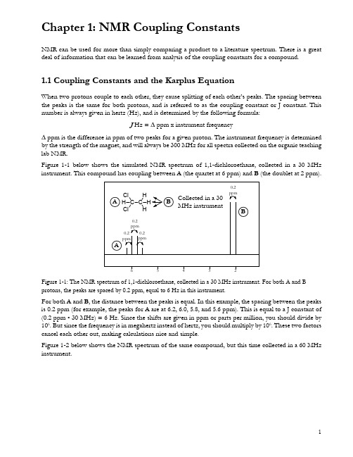
Chapter 1: NMR Coupling ConstantsNMR can be used for more than simply comparing a product to a literature spectrum. There is a great deal of information that can be learned from analysis of the coupling constants for a compound.1.1Coupling Constants and the Karplus EquationWhen two protons couple to each other, they cause splitting of each other’s peaks. The spacing between the peaks is the same for both protons, and is referred to as the coupling constant or J constant. This number is always given in hertz (Hz), and is determined by the following formula:J Hz = ∆ ppm x instrument frequency∆ ppm is the difference in ppm of two peaks for a given proton. The instrument frequency is determined by the strength of the magnet, and will always be 300 MHz for all spectra collected on the organic teaching lab NMR.Figure 1-1 below shows the simulated NMR spectrum of 1,1-dichloroethane, collected in a 30 MHz instrument. This compound has coupling between A (the quartet at 6 ppm) and B (the doublet at 2 ppm).Figure 1-1: The NMR spectrum of 1,1-dichloroethane, collected in a 30 MHz instrument. For both A and B protons, the peaks are spaced by 0.2 ppm, equal to 6 Hz in this instrument.For both A and B, the distance between the peaks is equal. In this example, the spacing between the peaks is 0.2 ppm (for example, the peaks for A are at 6.2, 6.0, 5.8, and 5.6 ppm). This is equal to a J constant of (0.2 ppm • 30 MHz) = 6 Hz. Since the shifts are given in ppm or parts per million, you should divide by 106. But since the frequency is in megahertz instead of hertz, you should multiply by 106. These two factors cancel each other out, making calculations nice and simple.Figure 1-2 below shows the NMR spectrum of the same compound, but this time collected in a 60 MHz instrument.Chapter 1: NMR Coupling ConstantsFigure 1-2: The NMR spectrum of 1,1-dichloroethane, collected in a 60 MHz instrument. For both A and B protons, the peaks are spaced by 0.1 ppm, equal to 6 Hz in this instrument.This time, the peak spacing is 0.1 ppm. This is equal to a J constant of (0.1 ppm • 60 MHz) = 6 Hz, the same as before. This shows that the J constant for any two particular protons will be the same value in hertz, no matter which instrument is used to measure it.The coupling constant provides valuable information about the structure of a compound. Some typical coupling constants are shown here.Figure 1-3: The coupling constants for some typical pairs of protons.In molecules where the rotation of bonds is constrained (for instance, in double bonds or rings), the coupling constant can provide information about stereochemistry. The Karplus equation describes how the coupling constant between two protons is affected by the dihedral angle between them. The equation follows the general format of J = A + B (cos θ) + C (cos 2θ), with the exact values of A, B and C dependent on several different factors. In general, though, a plot of this equation has the shape shown in Figure 1-4. Coupling constants will usually, but not always, fall into the shaded band on this graph.Figure 1-4: The plot of dihedral angle vs. coupling constant described by the Karplus equation.Chapter 1: NMR Coupling ConstantsThe highest coupling constants will occur between protons that have a dihedral angle of either 0° or 180°, and the lowest coupling constants will occur at 90°. This is due to orbital overlap – when the orbitals are at 90°, there is very little overlap between them, so the hydrogens cannot affect each other’s spins very much (Figure 1-5).Figure 1-5: The best orbital overlap occurs at 180° or 0°, which is why the coupling constant is higher for those angles.1.2 Calculating Coupling Constants in MestreNovaTo calculate coupling constants in MestreNova, there are several options. The easiest one is to use the Multiplet Analysis tool. To do this, go to Analysis → Multiplet Analysis → Manual (or just hit the “J” key). Drag a box around each group of equivalent protons. A purple version of the integral bar will appear below each one, along with a purple box above each one describing its splitting pattern and location in ppm. As with normal integrals, you can right-click the integral bar, select “Edit Multiplet”, and set these integrals to whatever makes sense for that particular structure. For example, in Figure 1-6, each peak is from a single proton so each integral should be about 1.00.Figure 1-6: An example NMR spectrum with multiplet analysis.HH H H HHChapter 1: NMR Coupling ConstantsOnce all peaks are labeled, you can go to Analysis → Multiplet Analysis → Report Multiplets. A text box should appear containing information about the peaks in a highly compressed format. You can then copy and paste this text into your lab report as needed. The spectrum shown above has the following multiplets listed:1H NMR (300 MHz, Chloroform-d) δ 5.14 (d, J = 11.7 Hz, 1H), 4.98 (d, J = 11.7 Hz, 1H), 4.75 (d, J = 3.2 Hz, 1H), 3.37 (d, J = 8.5 Hz, 1H), 3.30 (dd, J = 8.5, 3.3 Hz, 1H).The first set of parentheses indicates that the sample was dissolved in Chloroform-d and placed in a 300 MHz instrument. After that, there is a list of numbers. Each number or range indicates the chemical shift of each of the peaks in the spectrum, in order of descending chemical shift. Each number also has a set of parentheses after it, giving information about that peak. These parentheses contain: • A letter or letters to indicate the splitting of a peak (s=singlet, d=doublet, t=triplet, q=quartet); it is also possible to see things like dd for a doublet of doublets or b for broad. If MestreNova can’t identify a uniform splitting pattern, it will name it a multiplet (m).•The coupling constants or J-values for that peak – for example, the peak at 3.30 ppm has J-values of 8.5 and 3.3 ppm.•The integral of the peak, rounded to the nearest whole number of H.Using this information, you can determine which peaks in Figure 1-6 are coupling to each other based on which ones have matching J-values.•Peaks A and B in Figure 1-6 both have J-values of 11.7 Hz, so these two protons are coupling to each other.•Peaks C and E both have J-values of 3.2 or 3.3 Hz (similar enough, within a margin of error), so these two protons are coupling to each other.•Peaks D and E both have J-values of 8.5 Hz, so these two protons are coupling to each other. If the multiplet analysis tool is failing to determine J-values for any reason, you can always calculate them manually. To do this, you will need to get more precise values for your peak locations. Right-click anywhere in the empty space of the spectrum and select Properties, then go to Peaks and increase the decimals to 4 (Figure 1-7).Chapter 1: NMR Coupling ConstantsFigure 1-7: Changing the decimals on peak labeling.Now if you do peak-picking to label the locations of the peaks, you should see them to 4 decimal places. This will allow you to plus these into the equation to find the J-values manually. For example, in Figure 1-8, the peaks around 4.7 ppm have a J-value of (4.7550 ppm – 4.7442 ppm) • 300 MHz = 3.24 Hz. Note that this in in agreement with MestreNova’s determination of 3.2 ppm for this J-value in Figure1-6.Figure 1-8: Peaks labeled with enough precision to allow you to calculate J-values manually.Chapter 1: NMR Coupling Constants1.3 Topicity and Second-Order CouplingDuring the NMR tutorial, you learned about the concept of chemical equivalence: protons in identical chemical environments have identical chemical shifts. However, just because two protons have the same connectivity to the molecule does not mean they are chemically equivalent. This is related to the concept of topicity : the stereochemical relationship between different groups in a molecule. To find the topicity relationship of two groups to each other, you should try replacing first one group, then the other group with a placeholder atom (in the examples in Figure 1-9, a dark circle is used as the placeholder). If the two molecules produced are identical, then the groups are homotopic; if the molecules are enantiomers, then the groups are enantiotopic; and if the molecules are diastereomers, then the groups are diastereotopic. Groups that are diastereotopic are chemically inequivalent, so they will have a different chemical shift from each other in NMR, and will show coupling as if they were neighboring protons instead of on the same carbon atom.Figure 1-9: Some examples of homotopic, enantiotopic, and diastereotopic groups.If two signals are coupled to each other and have very similar (but not identical) chemical shifts, another effect will appear: second-order coupling. This means that the peaks appear to “lean” toward each other – the peaks on the outside of the coupled pair are shorter, and the peaks on the inside are taller. (Figure 1-10).Figure 1-10: As the chemical shifts of H a and H b become more and more similar, the coupling between them becomes more second-order and the peaks lean more.Chapter 1: NMR Coupling Constants This is very common for two diastereotopic protons on the same carbon atom, but it appears in other situations where two protons are almost chemically identical as well. In Figure 1-8, note the two doublets at 4.98 and 5.14 ppm. These happen to be diastereotopic protons – they are attached to the same carbon, but are chemically equivalent.Looking for pairs of leaning peaks is useful, because it allows you to identify which protons are coupled to each other in a complicated spectrum. In Figure 1-11, there are two different pairs of leaning peaks: two 1H peaks with a J = 9 Hz, and two 2H peaks with J = 15 Hz. Recognizing this makes it possible to pick apart the different components of the peaks towards the left of the spectrum: these are two overlapping doublets, not a quartet.Figure 1-11: An NMR spectrum with two different pairs of leaning peaks.The multiplet tool in MestreNova might not work immediately for analyzing overlapping multiplets like this. Instead, you should follow the instructions at /resolving-overlapped-multiplets/ to deal with them.。
吸收光谱简介 Absorption Spectrum An Introduction 英语作文论文
吸收光谱简介Absorption Spectrum AnIntroductionWhat is Absorption Spectrum?The absorption spectrum of a material can be defined as the fraction of the incident radiation which is absorbed by that material over a wide range of frequencies. The molecular and atomic composition of a material is used to determine the absorption spectrum. Fundamental radiation is generally observed at those frequencies which get mixed with the energy difference that takes place between two mechanical states of the molecules.The absorption takes place because of the transition between these two states and it is known as the absorption line. The spectrum is composed of several absorption lines. The frequencies where such absorption lines develop along with their relative intensities generally depend on the molecular structure and electronic structure of the sample. The frequencies also depend on molecular interactions. In the sample, the crystal structure is found in solids and on different environmental factors like pressure, temperature, electromagnetic fields, etc.What is Absorption Spectrum?Assingment Experts will explain Absorption Sepctrum in deatils. The absorption spectrum of a material can be defined as the fraction of theincident radiation which is absorbed by that material over a wide range of frequencies. The molecular and atomic composition of a material is used to determine the absorption spectrum. Fundamental radiation is generally observed at those frequencies which get mixed with the energy difference that takes place between two mechanical states of the molecules.The absorption takes place because of the transition between these two states and it is known as the absorption line. The spectrum is composed of several absorption lines. The frequencies where such absorption lines develop along with their relative intensities generally depend on the molecular structure and electronic structure of the sample. The frequencies also depend on molecular interactions. In the sample, the crystal structureis found in solids and on different environmental factors like pressure, temperature, electromagnetic fields, etc.The absorption lines also possess a definite shape and width which are fundamentally determined by the density of states for spectral density of the system. Absorption lines are generally classified by the feature of quantum mechanical change taking place in an atom or molecule. Rotational states sometimes get changed and give result in the development of rotational lines which are found in the region of the microwave spectrum. On the other hand, vibrational lines in correspondence to vibrational state changes in the molecule are found in the area of infrared region. The electronic lines are composed of several changes taking place in the electronic state of a molecule or atom whichare found in the ultraviolet and visible region.It can be noticed that there are various dark lines in the sun’s spectrum. These lines are developed by the atmosphere of the Sun which absorbs light at different wavelengths resulting in different light intensity at the wavelength to appear dark. The molecules and atoms present in a gas absorb certain light wavelengths. The pattern of the lines is very unique with respect to each element which provides us information about the elements which help in making the sun’s atmosphere. The absorption spectra can be observed from spatial regions in the presence of a cooler gas line between in a hotter source and the earth.The absorption spectra can also be observed from the planets with atmospheres, stars, and galaxies. In analyzing the light of the Sun, aspectrometer is used. The spectrometer is a device which separates light by colour and energy. In separating light by colour and energy, the image of the spectrum of the sun gets created. This is quite similar to the absorption spectrum. The dark lines are the areas where the light gets absorbed by different elements present in the Sun’s outer layers. The lowest energy is represented by red light and the highest energy is represented by blue light.The black gaps or lines in the spectrum of the sun are termed as absorption lines. The gas present in the sun’s outer layers develops the absorption lines by absorbing the light. There are different elements such as Helium, hydrogen, carbon, and other smaller quantities of heavy elements in the sun. When the sunlight shines, the elements the energy gets absorbed by the atoms. The atoms can only absorb the lightrelevant to the energy the atoms need. The gaps in the spectrum of the Sun get developed and help in informing the formation of the sun. The emission spectrum is quite different from the absorption spectrum.In developing an absorption spectrum, the light needs to shine through a gas but in creating and emission spectrum a gas needs to be heated up. The atoms present in the gas get absorbed the energy only for a short tenure. The atoms get energetic and jiggled up by heating the gas because of the concentration of a high level of energy. The energy is emitted or re-released as light eventually. Absorption spectrum takes place when the light passes through a dilute and cold gas and characteristic frequencies get absorbed by the atoms present in the gas. The re-emitted light cannot be emitted in a similardirection which is followed by absorbed Photon because of which dark lines in the spectrum are created in the absence of light. The absorption spectrum is the dark lines. The absorption spectrum is defined as an Electromagnetic Spectrum in which the radiation intensity at some specific wavelengths gets decreased. An absorbing substance gets manifested as bands or dark lines. Medically, the absorption spectrum is also defined as an Electromagnetic Spectrum in which radiation intensity at specific ranges of wavelength is manifested as dark lines.X-ray absorptions are highly associated with the excitation taking place in the inner shell electrons in an atom. These changes generally get combined to develop a new absorption line which is typically found in the combined energy develop mainly during the changes. The changes are mainly radiation-vibrationstransitions. The energy which is typically found in the quantum mechanical change fundamentally determines the absorption line frequency. The frequency can get shifted because of several interactions. The magnetic and electric fields can give result in a shift.The interactions with some of the neighbouring molecules can also cause shifts. Absorption lines of any gas-phase molecule can get shifted typically when the molecule is present in either solid or liquid phase and involves in interacting with neighbouring molecules strongly. The shape and width of the absorption lines are generally determined by the observation instrument. The physical environment radiation and material absorbing of that material also determine the shape and width of absorption lines. Now our experts from Instant AssingnmentHelp will tell you about the relationship between Absorption Spectrum andThe relation between Transmission and absorption spectraTransmission and absorption spectra are interconnected. Transmission and absorption spectra are found to represent similar information. Transmission spectrum can be calculated from the absorption spectrum only. Absorption spectrum can also be calculated from transmission spectra. Mathematical transformation is used in calculating either the absorption spectrum or transmission spectrum. It has been observed that a transmission spectrum has maximum intensities where thewavelengths of the absorption spectrum are quite weak because of the transmission of more light through the sample takes place. Similarly, an absorption spectrum is found to have maximum intensities at its wavelengths where the absorption rate is quite stronger.The absorption spectrum is also related to any emission spectrum. Now, it is important to understand the concept of the emission spectrum. The process by which a substance can release energy is known as emission process. The energy which is released from a substance through any emission process can be found in electromagnetic radiation from. Emission can take at any frequency of absorption which makes the absorption lines to gets determined from the emission spectrum. But it is to be remembered that the emissionspectrum will always have different intensity pattern where it becomes distinguished from that of the absorption spectrum. Hence, it can be said that the absorption spectrum and emission spectrum can never be equivalent. The emission spectrum can be used to calculate the absorption spectrum with the application of effective theoretical models and other relevant information from where quantum mechanical states of a substance can be understood.Relationship between Absorption spectrum and reflection and scattering spectraThe absorption spectrum is also related to reflection and scattering spectra. The scattering and reflection spectra of any material getinfluenced by the absorption spectrum and index of refraction of that material. Extinction coefficient quantifies the absorption spectrum and index coefficients along with extinction coefficients which are related through Kramers-Kroening relation quantitatively. Therefore, it can be said that reflection or scattering spectrum standardize absorption spectrum can give rise to absorption spectrum.Reflection or scattering spectrum assumptions or models need to be simplified so that it can lead to effect an approximation of the derivation of absorption spectra. In the domain of chemical analysis, we can find the use of absorption spectroscopy because of the quantitative nature and specificity of the absorption spectrum. The specificity enables the compounds to get distinguished from each other in a mixture whichmakes absorption spectroscopy to be highly useful in different applications. For example, the presence of any pollutant in the air can be identified by the use of infrared gas analyzers.These analyses are also used to distinguish the air pollutant from oxygen, water, nitrogen, and other constituents. The specificity is also helpful in allowing several unknown samples to get rightly identified. It can be done by comparing the measured spectrum with the findings of reference spectra. It has been found that qualitative information of any sample can also be determined even if the information is not present in a library. For example, infrared spectra have several characteristics absorption bands which help in indicating the presence of carbon-oxygen bond or Carbon hydrogen bonds.Absorption spectrum can also be related to the quantity of material present with the use of Beer-Lambert law. This relationship is established quantitatively. In determining the typical compound concentration, it needs knowledge of the absorption coefficient of the compound. The absorption coefficient can be known from several reference sources and can be measured by accessing calibre standard spectrum with an available target concentrationabsorption spectrumAbsorption spectroscopy and its applicationAbsorption spectroscopy is one of the methods with the help of which a substance can get characterized by the support of wavelengths at which the spectrum of colour gets absorbedduring the passage of light through a substance solution. It is one of the fundamentally used methods used in assessing the chromospheres concentrations in the solutions. Absorption spectroscopy can also be explained as a non-destructive technique which is widely used by biochemists and biologists to assess the characteristic parameters and cellular components of functional molecules.This quantification is highly important in the domain of systems biology. In developing metabolic pathway quantitative depiction, various variables and parameters are needed which are to be assessed experimentally. Ultraviolet-visible absorption spectroscopy is used in producing experimental data which help in modelling techniques of system biology. These techniques use kinetic parameters andconcentrations of enzymes of signalling on metabolic pathways, fluxes, and intercellular metabolic concentrations. Absorption spectroscopy also describes the usage of the technique in quantifying bio-molecules and investigating bio-molecular interactions.Absorption spectroscopy is a significant technique which is used in chemistry to study simple inorganic species. It refers to spectroscopic techniques which are used in measuring radiation absorption as a function of wavelength or frequency when the interaction between absorption radiation and sample takes place. Photons are absorbed by the samples from the field of radiation. The absorption intensity varies as a frequency function and this absorption intensity is the absorption spectrum. Absorption spectroscopy is fundamentallyperformed across an absorption spectrum or electromagnetic spectrum.In the domain of analytical chemistry, absorption spectroscopy is used to assess the presence of any specific substance in a sample. In several cases, absorption spectroscopy is also used to quantify the quantity of a substance. In the domain of analytical applications, ultraviolet-visible and infrared spectroscopy is commonly observed. In the study of atomic physics, remote sensing, molecular physics, and astronomical spectroscopy, the use of absorption spectroscopy are widely observed.There are various experimental approaches which are used to measure the absorption spectrum. The most commonly used arrangement is to guide the regeneratedradiation beam at the sample in detecting the radiation intensity passing through it. The transmitted energy can be applied in calculating the absorption. The sample arrangement source and detection technique are also very used quite significantly depending on the objective of the experiment and that of the frequency range.Advantages of absorption spectroscopyThere can be several advantages of absorption spectroscopy because it can be used as an analytical method where measurements can be accomplished without any contact between the sample and the instrument. Radiation which travels between an instrument and a sample contains some important spectral information and measurement which is done remotely. Remote spectral sensing is quite significant indifferent situations. For example, hazardous and toxic environments can be measured without risking any instrument or operator.The material of the sample needs not to be brought into direct contact with any instrument which can prevent cross-contamination at a possible rate. Remote spectral measurements have certain challenges as compared to that of the laboratory measurements. To reduce such challenges, differential optical absorption spectroscopy has become quite popular because it mainly emphasizes on the features of differential absorption and erasers broadband absorption like the extinction of aerosol extinction because of Rayleigh scattering. This technique is used in airborne, ground-based, and satellite-based measuring actions. There are certain ground-based techniques whichprofile the possibilities of retrieving stratospheric and tropospheric trace gas profiles.The absorption lines also possess a definite shape and width which are fundamentally determined by the density of states for spectral density of the system. Absorption lines are generally classified by the feature of quantum mechanical change taking place in an atom or molecule. Rotational states sometimes get changed and give result in the development of rotational lines which are found in the region of the microwave spectrum. On the other hand, vibrational lines in correspondence to vibrational state changes in the molecule are found in the area of infrared region. The electronic lines are composed of several changes taking place in the electronic state of a molecule or atom which are found in the ultraviolet and visible region.Itcan be noticed that there are various dark lines in the sun’s spectrum. These lines are developed by the atmosphere of the Sun which absorbs light at different wavelengths resulting in different light intensity at the wavelength to appear dark. The molecules and atoms present in a gas absorb certain light wavelengths. The pattern of the lines is very unique with respect to each element which provides us information about the elements which help in making the sun’s atmosphere. The absorption spectra can be observed from spatial regions in the presence of a cooler gas line between in a hotter source and the earth.The absorption spectra can also be observed from the planets with atmospheres, stars, and galaxies. In analyzing the light of the Sun, a spectrometer is used. The spectrometer is adevice which separates light by colour and energy. In separating light by colour and energy, the image of the spectrum of the sun gets created. This is quite similar to the absorption spectrum. The dark lines are the areas where the light gets absorbed by different elements present in the Sun’s outer layers. The lowest energy is represented by red light and the highest energy is represented by blue light.The black gaps or lines in the spectrum of the sun are termed as absorption lines. The gas present in the sun’s outer layers develops the absorption lines by absorbing the light. There are different elements such as Helium, hydrogen, carbon, and other smaller quantities of heavy elements in the sun. When the sunlight shines, the elements the energy gets absorbed by the atoms. The atoms can only absorb the light relevant to the energy the atoms need. The gapsin the spectrum of the Sun get developed and help in informing the formation of the sun. The emission spectrum is quite different from the absorption spectrum.In developing an absorption spectrum, the light needs to shine through a gas but in creating and emission spectrum a gas needs to be heated up. The atoms present in the gas get absorbed the energy only for a short tenure. The atoms get energetic and jiggled up by heating the gas because of the concentration of a high level of energy. The energy is emitted or re-released as light eventually. Absorption spectrum takes place when the light passes through a dilute and cold gas and characteristic frequencies get absorbed by the atoms present in the gas. The re-emitted light cannot be emitted in a similar direction which is followed by absorbed Photonbecause of which dark lines in the spectrum are created in the absence of light. The absorption spectrum is the dark lines. The absorption spectrum is defined as an Electromagnetic Spectrum in which the radiation intensity at some specific wavelengths gets decreased. An absorbing substance gets manifested as bands or dark lines. Medically, the absorption spectrum is also defined as an Electromagnetic Spectrum in which radiation intensity at specific ranges of wavelength is manifested as dark lines.X-ray absorptions are highly associated with the excitation taking place in the inner shell electrons in an atom. These changes generally get combined to develop a new absorption line which is typically found in the combined energy develop mainly during the changes. The changes are mainly radiation-vibrations transitions. The energy which is typically foundin the quantum mechanical change fundamentally determines the absorption line frequency. The frequency can get shifted because of several interactions. The magnetic and electric fields can give result in a shift.The interactions with some of the neighbouring molecules can also cause shifts. Absorption lines of any gas-phase molecule can get shifted typically when the molecule is present in either solid or liquid phase and involves in interacting with neighbouring molecules strongly. The shape and width of the absorption lines are generally determined by the observation instrument. The physical environment radiation and material absorbing of that material also determine the shape and width of absorption lines. Now our experts from Instant AssingnmentHelp will tell you about the relationship between Absorption Spectrum andThe relation between Transmission and absorption spectraTransmission and absorption spectra are interconnected. Transmission and absorption spectra are found to represent similar information. Transmission spectrum can be calculated from the absorption spectrum only. Absorption spectrum can also be calculated from transmission spectra. Mathematical transformation is used in calculating either the absorption spectrum or transmission spectrum. It has been observed that a transmission spectrum has maximum intensities where thewavelengths of the absorption spectrum are quite weak because of the transmission of more light through the sample takes place. Similarly, an absorption spectrum is found to have maximum intensities at its wavelengths where the absorption rate is quite stronger.The absorption spectrum is also related to any emission spectrum. Now, it is important to understand the concept of the emission spectrum. The process by which a substance can release energy is known as emission process. The energy which is released from a substance through any emission process can be found in electromagnetic radiation from. Emission can take at any frequency of absorption which makes the absorption lines to gets determined from the emission spectrum. But it is to be remembered that the emissionspectrum will always have different intensity pattern where it becomes distinguished from that of the absorption spectrum. Hence, it can be said that the absorption spectrum and emission spectrum can never be equivalent. The emission spectrum can be used to calculate the absorption spectrum with the application of effective theoretical models and other relevant information from where quantum mechanical states of a substance can be understood.Relationship between Absorption spectrum and reflection and scattering spectraThe absorption spectrum is also related to reflection and scattering spectra. The scattering and reflection spectra of any material getinfluenced by the absorption spectrum and index of refraction of that material. Extinction coefficient quantifies the absorption spectrum and index coefficients along with extinction coefficients which are related through Kramers-Kroening relation quantitatively. Therefore, it can be said that reflection or scattering spectrum standardize absorption spectrum can give rise to absorption spectrum.Reflection or scattering spectrum assumptions or models need to be simplified so that it can lead to effect an approximation of the derivation of absorption spectra. In the domain of chemical analysis, we can find the use of absorption spectroscopy because of the quantitative nature and specificity of the absorption spectrum. The specificity enables the compounds to get distinguished from each other in a mixture whichmakes absorption spectroscopy to be highly useful in different applications. For example, the presence of any pollutant in the air can be identified by the use of infrared gas analyzers.These analyses are also used to distinguish the air pollutant from oxygen, water, nitrogen, and other constituents. The specificity is also helpful in allowing several unknown samples to get rightly identified. It can be done by comparing the measured spectrum with the findings of reference spectra. It has been found that qualitative information of any sample can also be determined even if the information is not present in a library. For example, infrared spectra have several characteristics absorption bands which help in indicating the presence of carbon-oxygen bond or Carbon hydrogen bonds.Absorption spectrum can also be related to the quantity of material present with the use of Beer-Lambert law. This relationship is established quantitatively. In determining the typical compound concentration, it needs knowledge of the absorption coefficient of the compound. The absorption coefficient can be known from several reference sources and can be measured by accessing calibre standard spectrum with an available target concentrationabsorption spectrumAbsorption spectroscopy and its applicationAbsorption spectroscopy is one of the methods with the help of which a substance can get characterized by the support of wavelengths at which the spectrum of colour gets absorbed during the passage of light through a substancesolution. It is one of the fundamentally used methods used in assessing the chromospheres concentrations in the solutions. Absorption spectroscopy can also be explained as a non-destructive technique which is widely used by biochemists and biologists to assess the characteristic parameters and cellular components of functional molecules.This quantification is highly important in the domain of systems biology. In developing metabolic pathway quantitative depiction, various variables and parameters are needed which are to be assessed experimentally. Ultraviolet-visible absorption spectroscopy is used in producing experimental data which help in modelling techniques of system biology. These techniques use kinetic parameters and concentrations of enzymes of signalling onmetabolic pathways, fluxes, and intercellular metabolic concentrations. Absorption spectroscopy also describes the usage of the technique in quantifying bio-molecules and investigating bio-molecular interactions.Absorption spectroscopy is a significant technique which is used in chemistry to study simple inorganic species. It refers to spectroscopic techniques which are used in measuring radiation absorption as a function of wavelength or frequency when the interaction between absorption radiation and sample takes place. Photons are absorbed by the samples from the field of radiation. The absorption intensity varies as a frequency function and this absorption intensity is the absorption spectrum. Absorption spectroscopy is fundamentallyperformed across an absorption spectrum or electromagnetic spectrum.In the domain of analytical chemistry, absorption spectroscopy is used to assess the presence of any specific substance in a sample. In several cases, absorption spectroscopy is also used to quantify the quantity of a substance. In the domain of analytical applications, ultraviolet-visible and infrared spectroscopy is commonly observed. In the study of atomic physics, remote sensing, molecular physics, and astronomical spectroscopy, the use of absorption spectroscopy are widely observed.There are various experimental approaches which are used to measure the absorption spectrum. The most commonly used arrangement is to guide the regeneratedradiation beam at the sample in detecting the radiation intensity passing through it. The transmitted energy can be applied in calculating the absorption. The sample arrangement source and detection technique are also very used quite significantly depending on the objective of the experiment and that of the frequency range.Advantages of absorption spectroscopyThere can be several advantages of absorption spectroscopy because it can be used as an analytical method where measurements can be accomplished without any contact between the sample and the instrument. Radiation which travels between an instrument and a sample contains some important spectral information and measurement which is done remotely. Remote spectral sensing is quite significant indifferent situations. For example, hazardous and toxic environments can be measured without risking any instrument or operator.The material of the sample needs not to be brought into direct contact with any instrument which can prevent cross-contamination at a possible rate. Remote spectral measurements have certain challenges as compared to that of the laboratory measurements. To reduce such challenges, differential optical absorption spectroscopy has become quite popular because it mainly emphasizes on the features of differential absorption and erasers broadband absorption like the extinction of aerosol extinction because of Rayleigh scattering. This technique is used in airborne, ground-based, and satellite-based measuring actions. There are certain ground-based techniques which。
色谱中英文对照

中英文对照色谱词汇间接检测indirect detection间接荧光检测indirect fluorescence detection间接紫外检测indirect ultraviolet detection检测器detector检测器检测限detector detectability检测器灵敏度detector sensitivity检测器线性范围detector linear range阴离子交换剂anion exchanger阴离子交换色谱法anion exchange chromatography, AEC高速逆流色谱法high speed counter-current chromatography高温凝胶色谱法high temperature gel chromatography高效液相色谱-付里叶变换红外分析法high performance liquid ch…高效液相色谱法high performance liquid chromatography高效柱high performance column高压流通池技术high pressure flow cell technique高压输液泵high pressure pump高压梯度high-pressure gradient高压液相色谱法high pressure liquid chromatography阴离子交换树脂anion exchange resin荧光薄层板fluorescent thin layer plate荧光检测器fluorescence detector荧光色谱法fluorescence chromatography迎头色谱法frontal chromatography迎头色谱法frontal method硬(质)凝胶hard gel有机改进剂organic modifier有机相生物传感器Organic biosensor有效峰数effective peak number EPN有效理论塔板数number of effective theoretical plates有效塔板高度effective plate height有效淌度effective mobility淤浆填充法slurry packing method予柱pre-column在线电堆集on-line electrical stacking在柱电导率检测on-column electrical conductivity detection噪声noise噪信比noise –signal ratio增强紫外-可见吸收检测技术UV-visible absorption enhanced det…窄粒度分布narrow particle size distribution折射率检测器refractive index detector, RID真空脱气装置vacuum degasser阵列毛细管电泳capillary array electrophoresis蒸发光散射检测器evaporative light-scattering detector, ELSD整体性质检测器integral property detector正相高效液相色谱法normal phase high performance liquid chro…正相离子对色谱法normal phase ion-pair chromatography正相毛细管电色谱positive capillary electrokinetic chromatog…直接化学离子化direct chemical ionization GC-MS直接激光在柱吸收检测on-column direct laser detection纸色谱法paper chromatography置换色谱法displacement chromatography制备色谱preparative chromatography制备色谱仪preparative chromatograph制备柱preparation column智能色谱chromatography with artificial intelligence质量色谱mass chromatography质量型检测器mass detector质量型检测器mass flow rate sensitive detector中压液相色谱middle-pressure liquid chromatography重建色谱图reconstructive chromatogram重均分子量weight mean molecular weight轴向扩散longitudinal diffusion轴向吸收池absorption pool of axial direction轴向压缩柱axial compression column柱端电导率检测out-let end detection of electrical conductiv…柱负载能力column loadability柱后衍生化post-column derivatization柱老化condition (aging) of column柱流出物(column) effluent柱流失column bleeding柱内径column internal diameter柱前衍生化pro-column derivatization柱切换技术column switching technique柱清洗column cleaning柱容量column capacity柱入口压力column inlet pressure柱色谱法column chromatography柱上检测on-line detection柱渗透性column permeability柱寿命column life柱头进样column head sampling柱外效应extra-column effect柱温箱column oven柱效column efficiency柱压column pressure柱再生column regeneration柱中衍生化on-column derivatization注射泵syringe pump转化定量法trans-quantitative method紫外-可见光检测器ultraviolet visible detector, UV-Vis紫外吸收检测器ultraviolet absorption detector自动进样器automatic sampler自由溶液毛细管电泳free solution capillary electrophoresis总分离效能指标over-all resolution efficiency总交换容量total exchange capacity总渗透体积total osmotic volume纵向扩散longitudinal diffusion组合式仪器系统building block instrument最佳流速optimum flow rate最佳实际流速optimum practical flow rate最小检测量minimum detectable quantity最小检测浓度minimum detectable concentration萃取色谱法extraction chromatography脱气装置degasser外标法external standard method外梯度outside gradient网状结构reticular structure往复泵reciprocating pump往复式隔膜泵reciprocating diaphragm pump微分型检测器differential detector微孔树脂micro-reticular resin微库仑检测器micro coulometric detector微量进样针micro-syringe微量色谱法micro-chromatography微乳液电动色谱microemulsion electrokinetic chromatography 微生物传感器Microbial sensor微生物显影bioautography微填充柱micro-packed column微吸附检测器micro adsorption detector微型柱micro-column涡流扩散eddy diffusion无机离子交换剂inorganic ion exchanger无胶筛分毛细管电泳non-gel capillary electrophoresis无孔单分散填料non-porous monodisperse packing无脉动色谱泵pulse-free chromatographic pump物理钝化法physical deactivation吸附等温线adsorption isotherm吸附剂adsorbing material吸附剂活性adsorbent activity吸附平衡常数adsorption equilibrium constant吸附溶剂强度参数adsorption solvent strength parameter吸附色谱法adsorption chromatography吸附型PLOT柱adsorption type porous-layer open tubular colum…吸附柱adsorption column吸光度比值法absorbance ratio method洗脱强度eluting power显色器color-developing sprayer限制扩散理论theory of restricted diffusion线速度linear velocity线性梯度linear gradient相比率phase ratio相对保留值relative retention value相对比移值relative Rf value相对挥发度relative volatility相对灵敏度relative sensitivity相对碳(重量)响应因子relative carbon response factor相对响应值relative response相对校正因子relative correction factor相交束激光诱导的热透镜测量heat lens detection of intersect …相似相溶原则rule of similarity响应时间response time响应值response小角激光散射光度计low-angle laser light scattering photomet…小内径毛细管柱Microbore column校正保留体积corrected retention volume校正曲线法calibration curve method校正因子correction factor旋转薄层法rotating thin layer chromatography旋转小室逆流色谱rotational little-chamber counter-current c…选择性检测器selective detector循环色谱法recycling chromatography压电晶体piezoelectric crystal压电免疫传感器Piezoelectric Immunosensor压电转换器piezoelectric transducer压力保护pressure protect压力上限pressure high limit压力梯度校正因子pressure gradient correction factor压力下限pressure low limit衍生化法derivatization method衍生化试剂derivatization reagent阳离子交换剂cation exchanger阳离子交换色谱法cation exchange chromatography, CEC氧化铝色谱法alumina chromatography样品环sample loop样品预处理sample pretreatment液-液分配色谱法liquid-liquid partition chromatography液-液色谱法liquid-liquid chromatography液滴逆流色谱drop counter-current chromatography液固色谱liquid-solid chromatography液晶固定相liquid crystal stationary phase液态离子交换剂liquid ion exchanger液相传质阻力resistance of liquid mass transfer液相色谱-傅里叶变换红外光谱联用liquid chromatography-FTIR 液相色谱-质谱分析法liquid chromatography-mass spectrometry 液相色谱-质谱仪liquid chromatography-mass spectrometer液相色谱法liquid chromatography液相载荷量liquid phase loading溶剂效率solvent efficiency溶解度参数solubility parameter溶液性能检测器solution property detector溶胀swelling溶质性质检测器solute property detector容量因子capacity factor渗透极限分子量permeation limit molecular weight生物色谱biological chromatography生物特异性柱biospecific column生物自显影法bioautography升温速率temperature rate湿法柱填充wet column packing十八烷基键合硅胶octadecyl silane石墨化碳黑graphitized carbon black示差折光检测器differential refraction detector试剂显色法reagent color-developing method手动进样器manual injector手性氨基酸衍生物GC固定相chiral amino acid derivatives stat…手性拆分试剂chiral selectors手性固定相chiral stationary phase手性固定相拆分法chiral solid phase separation手性环糊精衍生物GC固定相chiral cyclodextrin der GC手性金属络合物GC固定相chirametal stationary phase in GC 手性流动相chiral mobile phase手性流动相拆分法chiral mobile phase separation手性色谱chiral chromatography手性试剂chiral reagent手性衍生化法chiral derivation method疏溶剂理论solvophobic theory疏溶剂色谱法solvophobic chromatography疏溶剂作用理论solvophobic interaction principle疏水作用色谱hydrophobic interaction chromatography树脂交换容量exchange capacity of resin数均分子量number mean molecular weight双保留机理dual reservation mechanism双活塞往复泵two-piston reciprocating pump双束差分检测器detector of dual-beam difference 双柱色谱法dual column chromatography水凝胶hydragel水系凝胶色谱柱aqua-system gel column死区域dead zone死体积dead volume塔板理论方程plate theory equation碳分子筛carbon molecular sieve特殊选择固定液selective stationary phase梯度洗脱gradient elution梯度洗脱装置gradient elution device梯度液相色谱gradient liquid chromatography体积排斥理论size exclusion theory体积排斥色谱size exclusion chromatography体积色谱法volumetric chromatography填充柱packed column填料packing material停流进样stop-flow injection通用型检测器common detector涂层毛细管coated capillary拖尾峰tailing peak拖尾因子tailing factor流动分离理论separation by flow流动相mobile phase流动相梯度eluent gradient流体动力学进样hydrostatic pressure injection流体力学体积hydrodynamic volume流型扩散dispersion due to flow profile脉冲阻尼器pulse damper酶传感器Enzyme sensor酶联免疫传感器Enzyme linked immunosensor 酶免疫分析enzyme immnunoassay内标法internal standard method内标物internal standard内梯度inside gradient逆流色谱法counter-current chromatography逆流色谱仪counter current chromatograph凝胶过滤色谱gel filtration chromatography凝胶内体积gel inner volume凝胶色谱法gel chromatography凝胶色谱仪gel chromatograph凝胶渗透色谱gel permeation chromatography凝胶外体积gel interstitial volume凝胶柱gel column浓度梯度成像检测器concentration gradient imaging detector浓度型检测器concentration detector排斥极限分子量exclusion limit molecular weight排斥体积exclusion volume排阻薄层色谱法exclusion TLC漂移drift迁移时间migration time迁移时间窗口the window of migration time前延峰leading peak前沿色谱法frontal chromatography强碱性阴离子交换剂strong-base anion exchanger强酸性阳离子交换剂strongly acidic cation exchanger切换时间switching time去离子水deionized water全多孔硅胶macro-reticular silica gel全多孔型填料macro-reticular packing material全二维色谱Comprehensive two-dim ensional gas chromatography…全硅烷化去活complete silylanization deactivation溶剂强度solvent strength激光解吸质谱法laser desorption MS, LDMS激光色谱laser chromatography激光诱导光束干涉检测detection of laser-induced light beam I…激光诱导毛细管振动测量laser-reduced capillary vibration det…激光诱导荧光检测器laser-induced fluorescence detector记忆峰memory peak记忆效应memory effect夹层槽sandwich chamber假峰ghost peak间断洗脱色谱法interrupted-elution chromatography间接光度(检测)离子色谱法indirect photometric ion chromato…间接光度(检测)色谱法indirect photometric chromatography减压液相色谱vacuum liquid chromatography键合固定相bonded stationary phase键合型离子交换剂bonded ion exchanger焦耳热joule heating胶束薄层色谱法micellar thin layer chromatography胶束液相色谱法micellar liquid chromatography交联度crosslinking degree阶梯梯度stagewise gradient进样阀injection valve进样量sample size进样器injector聚苯乙烯PSDVB聚硅氧烷高温裂解去活high-temperature pyrolysis deactivation…聚合物基质离子交换剂polymer substrate ion exchanger绝对检测器absolute detector可见光检测器visible light detector可交换离子exchangable ion空间性谱带加宽band broadening in space空穴色谱法vacancy chromatography孔结构pore structure孔径pore diameter孔径分布pore size distribution控制单元control unit快速色谱法high-speed chromatography理论塔板高度height equivalent to a theoretical plate(HETP)理论塔板数number of theoretical plates峰面积peak area峰面积测量法measurement of peak area峰面积校正calibration of peak area峰容量peak capacity固定相stationary phase固定液stationary liquid固定液的相对极性relative polarity of stationary liquid固定液极性stationary liquid polarity固相扩散solid diffusion固相荧光免疫分析solid phase fluorescence immunoassay固有粘度intrinsic viscosity光散射检测器light scattering detector硅胶silica gel硅烷化法silanization硅烷化法silanizing硅烷化载体silanized support过压液相色谱法over pressured liquid chromatography,OPLC恒流泵constant flow pump恒温操作constant temperature method恒压泵constant pressure pump红色载体red support红外检测器infrared detector红外总吸光度重建色谱图total infrared absorbance reconstruct…化合物形成色谱compound-formation chromatography化学发光检测器chemiluminescence detector化学发光检测器Chemiluminescence detector, SCD化学键合固定相bonded stationary phase化学键合相色谱bonded phase chromatography化学色谱法chemi-chromatography环糊精电动色谱cyclodextrin electrokinetic chromatography环形展开比移值circular development Rf value环形展开法circular development缓冲溶液添加剂buffer additives辉光放电检测器glow discharge detector混合床离子交换固定相mixed-bed ion exchange stationary phase 混合床柱mixed bed column活塞泵piston pump活性activation活性硅胶activated silica gel活性氧化铝activated aluminium oxide基流background current or base current基线baseline基线宽度baseline width基质substrate materials基质隔离技术matrix isolation technique电歧视效应the effect of electrical discrimination电迁移进样electrophoretic injection电渗流electroendosmotic flow电渗流标记物electroendosmotic flow marker电渗流淌度electroendosmotic mobility电泳淌度electrophoretic mobility调整保留时间adjusted retention time调整保留体积adjusted retention volume叠加内标法added internal standard method高效毛细管电泳high-performance capillary electrophoresis归一化法normalization method毛细管等电聚焦capillary isoelectric focusing毛细管等速电泳isotachophoresis毛细管电色谱capillary electrochromatography毛细管电泳capillary electrophoresis毛细管电泳电喷雾质谱联用capillary electrophoresis – electr芯片电泳microchip electrophoresis色谱法chromatography色谱峰chromatographic peak色谱峰区域宽度peak width色谱富集过样samt injection of chromatography色谱工作站chromatographic working station色谱图chromatogram色谱仪chromatograph色谱柱chromatographic column色谱柱column色谱柱切换技术switching column technique毛细管超临界流体色谱法capillary supercritical fluid chromat…毛细管电泳基质辅助激光解吸电离质谱离线检测off-line capillar…毛细管电泳离子分析capillary ion analysis毛细管电泳免疫分析immunity analysis of capillary elec tropho…毛细管胶束电动色谱micellar electrokinetic chromatography毛细管凝胶电泳capillary gel electrophoresis毛细管凝胶柱capillary gel column毛细管亲和电泳affinity capillary electrophoresis毛细管区带电泳capillary zone electrophoresis毛细管有效长度the effective length of capillary electroph or…二极管阵列检测器diode-array detector, DAD二维色谱法two-dimensional chromatography二元溶剂体系dual solvent system反冲洗back wash反吹技术back flushing technique反峰negative peak反离子counter ion反相高效液相色谱法reversed phase high performance liquid ch…反相离子对色谱reversed phase ion pair chromatography反相离子对色谱法reversed phase ion-pair chromatography反相毛细管电色谱reverse capillary electrokinetic chromatogr…反相柱reversed phase column反应色谱reaction chromatography反圆心式展开anti-circular development反转电渗流reverse electroendosmotic flow范第姆特方程式van Deemter equation仿生传感器Biomimic electrode放射性检测器radioactivity detector放射自显影autoradiography非极性固定相non-polar stationary phase非极性键合相non-polar bonded phase非水系凝胶色谱柱non-aqua-system gel column非水相色谱nonaqueous phase chromatography非吸附性载体non-adsorptive support非线性分流non-linearity split stream非线性色谱non-linear chromatography非线性吸附等温线non-linear adsorption isotherm酚醛离子交换树脂phenolic ion exchange resin分离-反应-分离展开SRS development分离数separation number分离因子separation factor分离柱separation column分配等温线distribution isotherm分配色谱partition chromatography分配系数partition coefficient分析型色谱仪analytical type chromatograph分子扩散molecular diffusion封尾endcapping峰高peak heightpH梯度动态分离dynamic separation of the pH gradientpH值梯度洗脱pH gradient elutionZata电势Zata potentialZ形池Z-form pool氨基键合相amino-bonded phase氨基酸分析仪amino acid analyzer安培检测器ampere detector白色载体white support半微柱semimicro-column半制备柱semi-preparation column包覆型离子交换剂coated ion exchanger包覆型填料coated packing material保护柱guard column保留间隙retention gap保留时间retention time保留体积retention volume保留温度retention temperature保留值定性法retention qualitative method保留值沸点规律boiling point rule of retention保留值碳数规律carbon number rule of retention保留指数retention index单活塞往复泵single piston reciprocating pump单相色谱仪single phase chromatograph单向阀one-way valve单柱离子色谱法single column ion chromatography等度洗脱isocratic elution等离子体色谱法plasma chromatography等途电泳-毛细管区带电泳耦合进样isotachophoresis injection-c…低负荷柱low load column低容量柱low capacity column低压梯度low-pressure gradient低压液相色谱low-pressure liquid chromatography电导池conductance cell电导检测法conductance detection电荷转移分光光度法charge transfer spectrophotometry电化学检测器electrochemical detector电解抑制器electrolyze suppressor保留指数定性法retention index qualitative method背景电导background conductance苯酚磺酸树脂phenol sulfonic acid resin苯乙烯styrene比保留体积specific retention volume比例阀proportional valve比渗透率specific permeability比移值Rf value便携式色谱仪portable chromatograph标准偏差standard deviation表观电泳淌度apparent electrophoretic mobility表观交换容量apparent exchange capacity表面电位检测器surface potential detector表面多孔硅胶superficially porous silica gel表面多孔填料superficially porous packing material表面多孔型离子交换剂superficially porous ion-exchanger玻璃球载体glass beads support不分流进样splitless sampling参比柱reference column场放大进样electrical field magnified injection场流分离field-flow fractionation场流分离仪field-flow fractionation场效应生物传感器Field effect transistor based Biosensor常压液相色谱法common-pressure liquid chromatography超声波脱气ultrasonic degas程序变流色谱法programmed flow (gas) chromatography程序升温进样programmed temperature sampling程序升温色谱法programmed temperature (gas) chromatography 程序升温蒸发器programmed temperature vaporizer ,PTV程序升压programmed pressure大孔树脂macro-reticular resin大孔填料macro-reticular packing material大内径毛细管柱Megaobore column。
SPECTROSCOPIC SYSTEMS AND METHODS FOR THE IDENTIFI

专利名称:SPECTROSCOPIC SYSTEMS AND METHODS FOR THE IDENTIFICATION ANDQUANTIFICATION OF PATHOGENS发明人:WOOD, Bayden Robert,HERAUD, PhilipRobert,GUAITA, David Perez,KOCHAN,Kamila Natalia申请号:EP17841196申请日:20170819公开号:EP3500679A4公开日:20200318专利内容由知识产权出版社提供摘要:A system for detecting a disease agent in a sample derived from a patient biofluid may comprise a receiver coupled to a communication network and a controller coupled to the receiver. The controller may comprise a processor and a memory. The controller may be configured to generate an infrared spectrum of the sample. The sample spectrum may comprise one or more sample spectral components, the sample spectral components comprising a sample wavenumber and a sample absorbance value.A set of reference spectral models may comprise one or more reference spectral components. The reference spectral components may comprise a reference wavenumber and a reference absorbance value. The reference spectral components may comprise one or more pathogen characteristics associated with sepsis. The one or more sample spectral components may be classified as pathogenic using the reference spectral models. Pathogen data may be generated using the classified sample spectral components.申请人:Monash University 更多信息请下载全文后查看。
SPECTROSCOPIC ANALYZING DEVICE AND SPECTROSCOPIC A

更多信息请下载全文后查看
专利内容由知识权出版社提供
专利名称:SPECTROSCOPIC ANALYZING DEVICE AND SPECTROSCOPIC ANALYZING METHOD
发明人:HASEGAWA, TAKESHI 申请号:EP07859657 申请日:20071221 公开号:EP 21124 98A4 公开日:2014 04 23
摘要:A spectrometric analyzing device is capable of analyzing a thin film with high accuracy by using light having an arbitrary wavelength, such as not only infrared light but also visible light, ultraviolet light and X-ray, and using whatever refractive index of a supporting member of the thin film. A spectrometric analyzing device comprises a light source (1), a polarizing filter (2), a detection unit (3), a regression operation unit (4) and an absorbance spectrum calculation unit (5). The light source (1) emits light at n different angles of incidence (θn) to a measurement portion. The polarizing filter (2) shields an spolarized component. The detection unit (3) detects transmitted spectra (S). The regression operation unit (4) uses the transmitted spectra (S) and a mixing ratio (R) to obtain an in-plane mode spectrum (sip) and an out-of-plane mode spectrum (sop) through a regression analysis. The absorbance spectrum calculation unit (5) calculates an in-plane mode absorbance spectrum (Aip) and an out-of-plane mode absorbance spectrum (Aop) of the thin film, based on the results from a state in which the thin film is on the supporting member and a state in which no thin film is on the supporting member.
元素分布曲线的英文

元素分布曲线的英文Element Distribution Curve.The element distribution curve is a graphical representation that illustrates the concentration or abundance of elements in a given system or material. It is a valuable tool in various fields such as chemistry, physics, geology, and environmental science to understand the distribution patterns of elements in different media.In chemistry, the element distribution curve can be used to represent the concentration of elements in a solution or solid sample. This curve is typically generated by analyzing the sample using spectroscopic techniques such as atomic absorption spectroscopy, inductively coupled plasma-mass spectrometry, or X-ray fluorescence spectroscopy. These techniques allow for the quantification of elemental concentrations in the sample, which can then be plotted as a function of the element's atomic number or mass.The shape and features of the element distribution curve can provide insights into the chemical composition and properties of the sample. For instance, peaks in the curve correspond to elements that are present in higher concentrations, while troughs indicate elements that are less abundant. The relative heights of the peaks can also indicate the relative abundances of different elements in the sample.In physics, the element distribution curve can be used to describe the distribution of elements in a material or system. For example, in materials science, the curve can reveal the elemental composition of alloys or compounds, which is crucial for understanding their physical and mechanical properties. Similarly, in plasma physics, the element distribution curve can provide information about the elemental abundances in a plasma, which is essentialfor understanding its behavior and interactions.In geology, the element distribution curve plays a pivotal role in understanding the composition and evolutionof rocks, minerals, and sediments. By analyzing the elemental abundances in these materials, geoscientists can infer their origin, formation processes, and geochemical signatures. The curve can also be used to identify trace elements and minerals that are diagnostic of particular geological environments or processes.Moreover, the element distribution curve hassignificant applications in environmental science. By examining the elemental abundances in soil, water, and air samples, researchers can assess the pollution levels, contaminant sources, and ecological risks associated with these environments. This information is crucial for developing effective strategies for environmentalprotection and remediation.Overall, the element distribution curve is a fundamental tool in various scientific fields for understanding the elemental composition and distribution patterns in systems and materials. It provides a visual representation of the concentrations of elements, which can be used to infer properties, processes, and interactions ina given system. As technological advancements continue to improve the precision and sensitivity of elemental analysis techniques, the element distribution curve will remain a valuable resource for scientific research and applications.。
Spectroscopic techniques
12Spectroscopic techniques:I Spectrophotometric techniquesA.H O F M A N N12.1Introduction12.2Ultraviolet and visible light spectroscopy12.3Fluorescence spectroscopy12.4Luminometry12.5Circular dichroism spectroscopy12.6Light scattering12.7Atomic spectroscopy12.8Suggestions for further reading12.1INTRODUCTIONSpectroscopic techniques employ light to interact with matter and thus probe certainfeatures of a sample to learn about its consistency or structure.Light is electromag-netic radiation,a phenomenon exhibiting different energies,and dependent on thatenergy,different molecular features can be probed.The basic principles of interactionof electromagnetic radiation with matter are treated in this chapter.There is noobvious logical dividing point to split the applications of electromagnetic radiationinto parts treated separately.The justification for the split presented in this text ispurely pragmatic and based on‘common practice’.The applications considered in thischapter use visible or UV light to probe consistency and conformational structure ofbiological ually,these methods are thefirst analytical procedures usedby a biochemical scientist.The applications covered in Chapter13present a higherlevel of complexity in undertaking and are employed at a later stage in biochemical orbiophysical characterisation.An understanding of the properties of electromagnetic radiation and its interactionwith matter leads to an appreciation of the variety of types of spectra and,conse-quently,different spectroscopic techniques and their applications to the solution ofbiological problems.47712.1.1Properties of electromagnetic radiationThe interaction of electromagnetic radiation with matter is a quantum phenomenon and dependent upon both the properties of the radiation and the appropriate structural parts of the samples involved.This is not surprising,since the origin of electromag-netic radiation is due to energy changes within matter itself.The transitions which occur within matter are quantum phenomena and the spectra which arise from such transitions are principally predictable.Electromagnetic radiation (Fig.12.1)is composed of an electric and a perpendicular magnetic vector,each one oscillating in plane at right angles to the direction of propagation.The wavelength l is the spatial distance between two consecutive peaks (one cycle)in the sinusoidal waveform and is measured in submultiples of metre,usually in nanometres (nm).The maximum length of the vector is called the amplitude .The frequency of the electromagnetic radiation is the number of oscillations made by the wave within the timeframe of 1s.It therefore has the units of 1s À1¼1Hz.The frequency is related to the wavelength via the speed of light c (c ¼2.998Â108m s À1in vacuo )by ¼c l À1.A historical parameter in this context is the wavenumber "v which describes the number of completed wave cycles per distance and is typically measured in 1cm À1.12.1.2Interaction with matterFigure 12.2shows the spectrum of electromagnetic radiation organised by increasing wavelength,and thus decreasing energy,from left to right.Also annotated are the types of radiation and the various interactions with matter and the resulting spectro-scopic applications,as well as the interdependent parameters of frequency and wavenumber.Electromagnetic phenomena are explained in terms of quantum mechanics.The photon is the elementary particle responsible for electromagnetic phenomena.It carries the electromagnetic radiation and has properties of a wave,as well asof Fig.12.1Light is electromagnetic radiation and can be described as a wave propagating transversally in space and time.The electric (E )and magnetic (M )field vectors are directed perpendicular to each other.For UV/Vis,circular dichroism and fluorescence spectroscopy,the electric field vector is of most importance.For electron paramagnetic and nuclear magnetic resonance,the emphasis is on the magnetic field vector.478Spectroscopic techniques:I Photometric techniquesa particle,albeit having a mass of zero.As a particle,it interacts with matter by transferring its energy E :E ¼hc l ¼h ð12:1Þwhere h is the Planck constant (h =6.63Â10À34Js)and is the frequency of the radiation as introduced above.When considering a diatomic molecule (see Fig.12.3),rotational and vibrational levels possess discrete energies that only merge into a continuum at very high energy.Each electronic state of a molecule possesses its own set of rotational and vibrational levels.Since the kind of schematics shown in Fig.12.3is rather complex,the Jablonski diagram is used instead,where electronic and vibrational states are schematically drawn as horizontal lines,and vertical lines depict possible transitions (see Fig.12.8below).In order for a transition to occur in the system,energy must be absorbed.The energy change D E needed is defined in quantum terms by the difference in absolute energies between the final and the starting state as D E ¼E final –E start ¼h .Electrons in either atoms or molecules may be distributed between several energy levels but principally reside in the lowest levels (ground state ).In order for an electron to be promoted to a higher level (excited state ),energy must be put into the system.If this energy E ¼h is derived from electromagnetic radiation,this gives rise to an absorption spectrum,and an electron is transferred from the electronic ground state (S 0)into the first electronic excited state (S 1).The molecule will also be in an excited vibrational and rotational state.Subsequent relaxation of the molecule into the vibrational ground state of the first electronic excited state will occur.The electron can then revert back to the electronic ground state.For non-fluorescent molecules,this is accompanied by the emission of heat (D H ).Microwave X-ray Vis UVFar NearIR Radio Wavelength in nm Wavenumber in cm –1Frequency in s –1Energy in J mol –1101021031041051061071081091 µm 1 cm 1 mChange inelectronic statesX-ray absorptionUV/Visabsorption IR/Raman Microwave spectroscopyESR Electron spin NMR Nuclear spin Change in rotational states of Change in molecular rotational and vibrational states 10171016101510141013101210111010109108106105104103102101110–110–2107106105104103102101110–1Fig.12.2The electromagnetic spectrum and its usage for spectroscopic methods.47912.1Introduction480Spectroscopic techniques:I Photometric techniquesNuclear displacementR e(ground)Fig.12.3Energy diagram for a diatomic molecule exhibiting rotation,vibration as well as an electronic structure.The distance between two masses m1and m2(nuclear displacement)is described as a Lennard–Jones potential curve with different equilibrium distances(R e)for each electronic state.Energetically lower states always have lower equilibrium distances.The vibrational levels(horizontal lines)are superimposed on the electronic levels.Rotational levels are superimposed on the vibrational levels and not shown for reasons of clarity.The plot of absorption probability against wavelength is called absorption spectrum .In the simpler case of single atoms (as opposed to multi-atom molecules),electronic transitions lead to the occurrence of line spectra (see Section 12.7).Because of the existence of more different kinds of energy levels,molecular spectra are usually observed as band spectra (for example Fig.12.7below)which are molecule-specific due to the unique vibration states.A commonly used classification of absorption transitions uses the spin states of electrons.Quantum mechanically,the electronic states of atoms and molecules are described by orbitals which define the different states of electrons by two parameters:a geometrical function defining the space and a probability function.The combination of both functions describes the localisation of an electron.Electrons in binding orbitals are usually paired with antiparallel spin orientation (Fig.12.8).The total spin S is calculated from the individual electron spins.The multipli-city M is obtained by M ¼2ÂS þ1.For paired electrons in one orbital this yields:S ¼spin ðelectron 1Þþspin ðelectron 2Þ¼ðþ1=2ÞþðÀ1=2Þ¼0The multiplicity is thus M ¼2Â0þ1¼1.Such a state is thus called a singlet state and denotated as ‘S ’.Usually,the ground state of a molecule is a singlet state,S 0.In case the spins of both electrons are oriented in a parallel fashion,the resulting state is characterised by a total spin of S ¼1,and a multiplicity of M =3.Such a state is called a triplet state and usually exists only as one of the excited states of a molecule,e.g.T 1.According to quantum mechanical transition rules,the multiplicity M and the total spin S must not change during a transition.Thus,the S 0!S 1transition is allowed and possesses a high transition probability.In contrast,the S 0!T 1is not allowed and has a small transition probability.Note that the transition probability is proportional to the intensity of the respective absorption bands.Most biologically relevant molecules possess more than two atoms and,therefore,the energy diagrams become more complex than the ones shown in Fig.12.3.Different orbitals combine to yield molecular orbitals that generally fall into one of five different classes (Fig.12.4):s orbitals combine to the binding s and the anti-binding s *orbitals.Some p orbitals combine to the binding p and the anti-binding p*Anti-binding Non-binding BindingEs *p *s pnFig.12.4Energy scheme for molecular orbitals (not to scale).Arrows indicate possible electronic transitions.The length of the arrows indicates the energy required to be put into the system in order to enable thetransition.Black arrows depict transitions possible with energies from the UV/Vis spectrum for some biological molecules.The transitions shown by grey arrows require higher energies (e.g.X-rays).48112.1Introduction482Spectroscopic techniques:I Photometric techniquesorbitals.Other p orbitals combine to form non-binding n orbitals.The population of binding orbitals strengthens a chemical bond,and,vice versa,the population of anti-binding orbitals weakens a chemical bond.12.1.3LasersLaser is an acronym for light amplification by stimulated emission of radiation.A detailed explanation of the theory of lasers is beyond the scope of this textbook.A simplified description starts with the use of photons of a defined energy to excite anabsorbing material.This results in elevation of an electron to a higher energy level.If, whilst the electron is in the excited state,another photon of precisely that energy arrives, then,instead of the electron being promoted to an even higher level,it can return to the original ground state.However,this transition is accompanied by the emission of two photons with the same wavelength and exactly in phase(coherent photons).Multipli-cation of this process will produce coherent light with extremely narrow spectral bandwidth.In order to produce an ample supply of suitable photons,the absorbing material is surrounded by a rapidlyflashing light of high intensity(pumping).Lasers are indispensable tools in many areas of science,including biochemistry and biophysics.Several modern spectroscopic techniques utilise laser light sources, due to their high intensity and accurately defined spectral properties.One of the probably most revolutionising applications in the life sciences,the use of lasers in DNA sequencing withfluorescence labels(see Sections5.11.5,5.11.6and12.3.3), enabled the breakthrough in whole-genome sequencing.12.2ULTRAVIOLET AND VISIBLE LIGHT SPECTROSCOPYThese regions of the electromagnetic spectrum and their associated techniques are probably the most widely used for analytical work and research into biological problems.The electronic transitions in molecules can be classified according to the partici-pating molecular orbitals(See Fig.12.4).From the four possible transitions(n!p*, p!p*,n!s*,s!s*),only two can be elicited with light from the UV/Vis spectrum for some biological molecules:n!p*and p!p*.The n!s*and s!s*transitions are energetically not within the range of UV/Vis spectroscopy and require higher energies.Molecular(sub-)structures responsible for interaction with electromagnetic radi-ation are called chromophores.In proteins,there are three types of chromophores relevant for UV/Vis spectroscopy:•peptide bonds(amide bond);•certain amino acid side chains(mainly tryptophan and tyrosine);and•certain prosthetic groups and coenzymes(e.g.porphyrine groups such as in haem).The presence of several conjugated double bonds in organic molecules results in an extended p-system of electrons which lowers the energy of the p*orbital through electron delocalisation.In many cases,such systems possess p!p*transitions in the UV/Vis range of the electromagnetic spectrum.Such molecules are very useful tools in colorimetric applications(see Table12.1).Table12.1Common colorimetric and UV absorption assaysSubstance Reagent Wavelength(nm) Amino acids(a)Ninhydrin570(proline:420)(b)Cupric salts620Cysteine residues, thiolates Ellman reagent(di-sodium-bis-(3-carboxy-4-nitrophenyl)-disulphide)412Protein(a)Folin(phosphomolybdate,phosphotungstate,cupric salt)660(b)Biuret(reacts with peptide bonds)540(c)BCA reagent(bicinchoninic acid)562(d)Coomassie Brilliant Blue595(e)Direct Tyr,Trp:278,peptide bond:190 Coenzymes Direct FAD:438,NADH:340,NADþ:260 Carotenoids Direct420,450,480 Porphyrins Direct400(Soret band) Carbohydrate(a)Phenol,H2SO4Glucose:490,xylose:480(b)Anthrone(anthrone,H2SO4)620or625 Reducing sugars Dinitrosalicylate,alkaline tartrate buffer540Pentoses(a)Bial(orcinol,ethanol,FeCl3,HCl)665(b)Cysteine,H2SO4380–415 Hexoses(a)Carbazole,ethanol,H2SO4540or440(b)Cysteine,H2SO4380–415(c)Arsenomolybdate500–570 Glucose Glucose oxidase,peroxidase,o-dianisidine,phosphate buffer420 Ketohexose(a)Resorcinol,thiourea,ethanoic acid,HCl520(b)Carbazole,ethanol,cysteine,H2SO4560(c)Diphenylamine,ethanol,ethanoic acid,HCl635Hexosamines Ehrlich(dimethylaminobenzaldehyde,ethanol,HCl)53048312.2Ultraviolet and visible light spectroscopy12.2.1Chromophores in proteinsThe electronic transitions of the peptide bond occur in the far UV.The intense peak at 190nm,and the weaker one at 210–220nm is due to the p !p *and n !p *transitions.A number of amino acids (Asp,Glu,Asn,Gln,Arg and His)have weak electronic transitions at around ually,these cannot be observed in proteins because they are masked by the more intense peptide bond absorption.The most useful range for proteins is above 230nm,where there are absorptions from aromatic side chains .While a very weak absorption maximum of phenylalanine occurs at 257nm,tyrosine and tryptophan dominate the typical protein spectrum with their absorption maxima at 274nm and 280nm,respectively (Fig.12.5).In praxi ,the presence of these two aromatic side chains gives rise to a band at $278nm.Cystine (Cys 2)possesses a weak absorption maximum of similar strength as phenylalanine at 250nm.This band can play a role in rare cases in protein optical activity or protein fluorescence.Proteins that contain prosthetic groups (e.g.haem,flavin,carotenoid)and some metal–protein complexes,may have strong absorption bands in the UV/Vis range.These bands are usually sensitive to local environment and can be used for physical studies of enzyme action.Carotenoids,for instance,are a large class of red,yellow and orange plant pigments composed of long carbon chains with many conjugated double bonds.They contain three maxima in the visible region of the electromagnetic spectrum ($420nm,450nm,480nm).Porphyrins are the prosthetic groups of haemoglobin,myoglobin,catalase and cytochromes.Electron delocalisation extends throughout the cyclic tetrapyrrole ring of porphyrins and gives rise to an intense transition at $400nm called the Soret band .The spectrum of haemoglobin is very sensitive to changes in the iron-bound ligand.These changes can be used for structure–function studies of haem proteins.Table 12.1(cont.)SubstanceReagent Wavelength (nm)DNA (a)Diphenylamine595(b)Direct 260RNA Bial (orcinol,ethanol,FeCl 3,HCl)665Sterols and steroids Liebermann–Burchardt reagent (acetic anhydride,H 2SO 4,chloroform)425,625Cholesterol Cholesterol oxidase,peroxidase,4-amino-antipyrine,phenol 500ATPase assay Coupled enzyme assay with ATPase,pyruvatekinase,lactate dehydrogenase:ATP !ADP (consumes ATP)phosphoenolpyruvate !pyruvate (consumes ADP)pyruvate !lactate (consumes NADH)NADH:340484Spectroscopic techniques:I Photometric techniquesMolecules such as FAD (flavin adenine dinucleotide),NADH and NAD þare impor-tant coenzymes of proteins involved in electron transfer reactions (RedOx reactions).They can be conveniently assayed by using their UV/Vis absorption:438nm (FAD),340nm (NADH)and 260nm (NAD þ).Chromophores in genetic materialThe absorption of UV light by nucleic acids arises from n !p *and p !p *transitions of the purine (adenine,guanine)and pyrimidine (cytosine,thymine,uracil)bases that occur between 260nm and 275nm.The absorption spectra of the bases in polymers are sensitive to pH and greatly influenced by electronic interactions between bases.12.2.2PrinciplesQuantification of light absorptionThe chance for a photon to be absorbed by matter is given by an extinction coefficient which itself is dependent on the wavelength l of the photon.If light withthe Wavelength (nm)A b s o r b a n c eFig.12.5The presence of larger aggregates in biological samples gives rise to Rayleigh scatter visible by a considerable slope in the region from 500to 350nm.The dashed line shows the correction to be applied to spectra with Rayleigh scatter which increases with l À4.Practically,linear extrapolation of the region from 500to 350nm is performed to correct for the scatter.The corrected absorbance is indicated by the double arrow.48512.2Ultraviolet and visible light spectroscopyintensity I0passes through a sample with appropriate transparency and the path length (thickness)d,the intensity I drops along the pathway in an exponential manner. The characteristic absorption parameter for the sample is the extinction coefficient a, yielding the correlation I=I0eÀa d.The ratio T=I/I0is called transmission. Biochemical samples usually comprise aqueous solutions,where the substance of interest is present at a molar concentration c.Algebraic transformation of the expo-nential correlation into an expression based on the decadic logarithm yields the law of Beer–Lambert:lg I0I¼lg1T¼eÂcÂd¼Að12:2Þwhere[d]¼1cm,[c]¼1mol dmÀ3,and[e]¼1dm3molÀ1cmÀ1.e is the molar absorption coefficient(also molar extinction coefficient)(a¼2.303ÂcÂe).A is the absorbance of the sample,which is displayed on the spectrophotometer.The Beer–Lambert law is valid for low concentrations only.Higher concentrations might lead to association of molecules and therefore cause deviations from the ideal behaviour.Absorbance and extinction coefficients are additive parameters,which complicates determination of concentrations in samples with more than one absorbing species.Note that in dispersive samples or suspensions scattering effects increase the absorbance,since the scattered light is not reaching the detector for read-out.The absorbance recorded by the spectrophotometer is thus overestimated and needs to be corrected(Fig.12.5).Deviations from the Beer–Lambert lawAccording to the Beer–Lambert law,absorbance is linearly proportional to the concen-tration of chromophores.This might not be the case any more in samples with high absorbance.Every spectrophotometer has a certain amount of stray light,which is light received at the detector but not anticipated in the spectral band isolated by the mono-chromator.In order to obtain reasonable signal-to-noise ratios,the intensity of light at the chosen wavelength(I l)should be10times higher than the intensity of the stray light(I stray).If the stray light gains in intensity,the effects measured at the detector have nothing or little to do with chromophore concentration.Secondly,molecular events might lead to devi-ations from the Beer–Lambert law.For instance,chromophores might dimerise at high concentrations and,as a result,might possess different spectroscopic parameters.Absorption or light scattering–optical densityIn some applications,for example measurement of turbidity of cell cultures(determin-ation of biomass concentration),it is not the absorption but the scattering of light(see Section12.6)that is actually measured with a spectrophotometer.Extremely turbid samples like bacterial cultures do not absorb the incoming light.Instead,the light is scattered and thus,the spectrometer will record an apparent absorbance(sometimes also called attenuance).In this case,the observed parameter is called optical density(OD).Instruments specifically designed to measure turbid samples are nephelometers or Klett meters;however,most biochemical laboratories use the general UV/Vis spectrometer for determination of optical densities of cell cultures.486Spectroscopic techniques:I Photometric techniques48712.2Ultraviolet and visible light spectroscopyFactors affecting UV/Vis absorptionBiochemical samples are usually buffered aqueous solutions,which has two major advantages.Firstly,proteins and peptides are comfortable in water as a solvent, which is also the‘native’solvent.Secondly,in the wavelength interval of UV/Vis (700–200nm)the water spectrum does not show any absorption bands and thus acts as a silent component of the sample.The absorption spectrum of a chromophore is only partly determined by its chem-ical structure.The environment also affects the observed spectrum,which mainly can be described by three parameters:•protonation/deprotonation(pH,RedOx);•solvent polarity(dielectric constant of the solvent);and•orientation effects.Vice versa,the immediate environment of chromophores can be probed by assessing their absorption,which makes chromophores ideal reporter molecules for environ-mental factors.Four effects,two each for wavelength and absorption changes,have to be considered:•a wavelength shift to higher values is called red shift or bathochromic effect;•similarly,a shift to lower wavelengths is called blue shift or hypsochromic effect;•an increase in absorption is called hyperchromicity(‘more colour’),•while a decrease in absorption is called hypochromicity(‘less colour’).Protonation/deprotonation arises either from changes in pH or oxidation/reduction reactions,which makes chromophores pH-and RedOx-sensitive reporters.As a rule of thumb,l max and e increase,i.e.the sample displays a batho-and hyperchromic shift, if a titratable group becomes charged.Furthermore,solvent polarity affects the difference between the ground and excited states.Generally,when shifting to a less polar environment one observes a batho-and hyperchromic effect.Conversely,a solvent with higher polarity elicits a hypso-and hypochromic effect.Lastly,orientation effects,such as an increase in order of nucleic acids from single-stranded to double-stranded DNA,lead to different absorption behaviour.A sample of free nucleotides exhibits a higher absorption than a sample with identical amounts of nucleotides but assembled into a single-stranded polynucleotide.Accordingly,double-stranded polynucleotides exhibit an even smaller absorption than two single-stranded polynucleotides.This phenomenon is called the hypochromicity of polynucleotides.The increased exposure(and thus stronger absorption)of the individual nucleotides in the less ordered states provides a simplified explanation for this behaviour.12.2.3InstrumentationUV/Vis spectrophotometers are usually dual-beam spectrometers where thefirst chan-nel contains the sample and the second channel holds the control(buffer)for correction.488Spectroscopic techniques:I Photometric techniquesAlternatively,one can record the control spectrumfirst and use this as internal referencefor the sample spectrum.The latter approach has become very popular as many spectro-meters in the laboratories are computer-controlled,and baseline correction can be carriedout using the software by simply subtracting the control from the sample spectrum.The light source is a tungstenfilament bulb for the visible part of the spectrum,anda deuterium bulb for the UV region.Since the emitted light consists of many differentwavelengths,a monochromator,consisting of either a prism or a rotating metal gridof high precision called grating,is placed between the light source and the sample.Wavelength selection can also be achieved by using colouredfilters as monochromatorsthat absorb all but a certain limited range of wavelengths.This limited range is calledthe bandwidth of thefilter.Filter-based wavelength selection is used in colorimetry,a method with moderate accuracy,but best suited for specific colorimetric assayswhere only certain wavelengths are of interest.If wavelengths are selected by prismsor gratings,the technique is called spectrophotometry(Fig.12.6).Example1ESTIMATION OF MOLAR EXTINCTION COEFFICIENTSIn order to determine the concentration of a solution of the peptide MAMVSEFLKQAWFIENEEQE YVQTVKSSKG GPGSAVSPYP TFNPSS in water,the molarabsorption coefficient needs to be estimated.The molar extinction coefficient e is a characteristic parameter of a molecule andvaries with the wavelength of incident light.Because of useful applications of thelaw of Beer–Lambert,the value of e needs be known for a lot of molecules beingused in biochemical experiments.Very frequently in biochemical research,the molar extinction coefficient ofproteins is estimated using incremental e i values for each absorbing protein residue(chromophore).Summation over all residues yields a reasonable estimation for theextinction coefficient.The simplest increment system is based on values of Gill andvon Hippel.1The determination of protein concentration using this formula onlyrequires an absorption value at l¼280nm.Increments e i are used to calculate a molarextinction coefficient at280nm for the entire protein or peptide by summation overall relevant residues in the protein:Residue Gill and von Hippel e i(280nm)in dm3molÀ1cmÀ1Cys2120Trp5690Tyr1280For the peptide above,one obtains e¼(1Â5690þ2Â1280)dm3molÀ1cmÀ1¼8250dm3molÀ1cmÀ11Gill,S.C.and von Hippel,P.H.(1989).Calculation of protein extinction coefficients from aminoacid sequence data.Analytical Biochemistry,182,319–326.Erratum:Analytical Biochemistry(1990),189,283.A prism splits the incoming light into its components by refraction.Refraction occurs because radiation of different wavelengths travels along different paths in medium of higher density.In order to maintain the principle of velocity conservation,light of shorter wavelength (higher speed)must travel a longer distance (i.e.blue sky effect).At a grating,the splitting of wavelengths is achieved by diffraction.Diffraction is a reflection phenom-enon that occurs at a grid surface,in this case a series of engraved fine lines.The distance between the lines has to be of the same order of magnitude as the wavelength of the diffracted radiation.By varying the distance between the lines,different wavelengths are selected.This is achieved by rotating the grating perpendicular to the optical axis.The resolution achieved by gratings is much higher than the one available by prisms.Nowadays instruments almost exclusively contain gratings as monochromators as they can be reproducibly made in high quality by photoreproduction.The bandwidth of a colorimeter is determined by the filter used as monochromator.A filter that appears red to the human eye is transmitting red light and absorbs almost any other (visual)wavelength.This filter would be used to examine blue solutions,as these would absorb red light.The filter used for a specific colorimetric assay is thus made of a colour complementary to that of the solution being tested.Theoretically,a single wavelength is selected by the monochromator in spectrophotometers,and the emergent light is a parallel beam.Here,the bandwidth is defined as twice the half-intensity bandwidth.The bandwidth is a function of the optical slit width.The narrower the slit width the more reproducible are measured absorbance values.In contrast,the sensitivity becomes less as the slit narrows,because less radiation passes through to the detector.In a dual-beam instrument,the incoming light beam is split into two parts by a half-mirror.One beam passes through the sample,the other through a control (blank,reference).This approach obviates any problems of variation in light intensity,as both reference and sample would be affected equally.The measured absorbance is the difference between the two transmitted beams of light recorded.Depending on the instrument,a second detector measures the intensity of the incoming beam,although some instruments use an arrangement where one detector measures the incoming and the transmitted intensity alternately.The latter design is better from an analytical point of view as it eliminates potential variations between the two Vis light UV lightBeam Fig.12.6Optical arrangements in a dual-beam spectrophotometer.Either a prism or a grating constitutes the monochromator of the instrument.Optical paths are shown as green lines.48912.2Ultraviolet and visible light spectroscopy。
质谱分析法中英文专业词汇
质谱分析法:mass spectrometry质谱:mass spectrum,MS棒图:bar graph选择离子检测:selected ion monitoring ,SIM直接进样:direct probe inlet ,DPI接口:interface气相色谱-质谱联用:gas chromatography-mass spectrometry,GC-MS 高效液相色谱-质谱联用:high performance liquid chromatography-mass spectrometry,HPLC-MS电子轰击离子源:electron impact source,EI离子峰:quasi-molecular ions化学离子源:chemical ionization source,CI场电离:field ionization,FI场解析:field desorptiion,FD快速原子轰击离子源:fast stom bombardment ,FAB质量分析器:mass analyzer磁质谱仪:magnetic-sector mass spectrometer四极杆质谱仪(四极质谱仪):quadrupole mass spectrometer紫外-可见分光光度法:ultraviolet and visible spectrophotometry;UV-vis 相对丰度(相对强度):relative avundance原子质量单位:amu离子丰度:ion abundance基峰:base peak质量范围:mass range分辨率:resolution灵敏度:sensitivity信噪比:S/N分子离子:molecular ion碎片离子:fragment ion同位素离子:isotopic ion亚稳离子:metastable ion亚稳峰:metastable peak母离子:paren ion子离子:daughter含奇数个电子的离子:odd electron含偶数个电子的离子:even eletron,EE 均裂:homolytic cleavage异裂(非均裂):heterolytic cleavage 半均裂:hemi-homolysis cleavage重排:rearragement分子量:MWα-裂解:α-cleavage 电磁波谱:electromagnetic spectrum光谱:spectrum光谱分析法:spectroscopic analysis原子发射光谱法:atomic emission spectroscopy肩峰:shoulder peak末端吸收:end absorbtion生色团:chromophore助色团:auxochrome红移:red shift长移:bathochromic shift短移:hypsochromic shift蓝(紫)移:blue shift增色效应(浓色效应):hyperchromic effect 减色效应(淡色效应):hypochromic effect 强带:strong band弱带:weak band吸收带:absorption band透光率:transmitance,T吸光度:absorbance谱带宽度:band width杂散光:stray light噪声:noise暗噪声:dark noise散粒噪声:signal shot noise闪耀光栅:blazed grating全息光栅:holographic graaing光二极管阵列检测器:photodiode array detector偏最小二乘法:partial least squares method ,PLS褶合光谱法:convolution spectrometry 褶合变换:convolution transform,CT离散小波变换:wavelet transform,WT 多尺度细化分析:multiscale analysis供电子取代基:electron donating group 吸电子取代基:electron with-drawing group荧光:fluorescence荧光分析法:fluorometryX-射线荧光分析法:X-ray fulorometry 原子荧光分析法:atomic fluorometry分子荧光分析法:molecular fluorometry 振动弛豫:vibrational relexation内转换:internal conversion外转换:external conversion 体系间跨越:intersystem crossing激发光谱:excitation spectrum荧光光谱:fluorescence spectrum斯托克斯位移:Stokes shift荧光寿命:fluorescence life time荧光效率:fluorescence efficiency荧光量子产率:fluorescence quantum yield荧光熄灭法:fluorescence quemching method散射光:scattering light瑞利光:Reyleith scanttering light拉曼光:Raman scattering light红外线:infrared ray,IR中红外吸收光谱:mid-infrared absorption spectrum,Mid-IR远红外光谱:Far-IR微波谱:microwave spectrum,MV红外吸收光谱法:infrared spectroscopy 红外分光光度法:infrared spectrophotometry振动形式:mode of vibration伸缩振动:stretching vibrationdouble-focusing mass spectrograph 双聚焦质谱仪trochoidal mass spectrometer 余摆线质谱仪ion-resonance mass spectrometer 离子共振质谱仪gas chromatograph-mass spectrometer 气相色谱-质谱仪quadrupole spectrometer 四极(质)谱仪Lunar Mass Spectrometer 月球质谱仪Frequency Mass Spectrometer 频率质谱仪velocitron 电子灯;质谱仪mass-synchrometer 同步质谱仪omegatron 回旋质谱仪。
仪器分析专业名词英文及名词解释

仪器分析专业名词英文及名词解释仪器分析专业名词英文及名词解释一、紫外-可见光分光光度法1、透光率(transmittance):透过样品的光强度与入射光强度之比。
2、吸收度(absorbance):透光率的负对数。
3、生色团(chromophore):含有π→π*或n→π*跃迁的基团。
4、助色团(auxochrome):含孤对电子(非键电子)的杂原子基团。
5、摩尔吸收系数(molar absorptivity):一定波长时,溶液浓度为1mol/L,光程为1cm时的吸收度。
6、比吸收系数(specific absorptivity):一定波长时,溶液浓度为1%(W/V),光程为1cm时的吸收度。
7、红移(red shift):化合物结构改变(共轭,引入助色团,溶剂改变等),使吸收峰向长波长移动的现象。
8、蓝移(blue shift):当化合物结构改变或受溶剂的影响等原因使吸收峰向短波长移动的现象。
也称短移(hypso chromic shift)。
9、增色效应(hyperchromic effect):由于化合物结构改变或其他原因使吸收强度增加的效应。
10、减色效应(hypochromic effect):由于化合物结构改变或其他原因使吸收强度减弱的效应。
11、末端吸收(end absorption):在短波长处(200nm左右)只呈现强吸收,而不成峰形的部分。
12、标准对照法:在相同条件下配制标准溶液和样品溶液,在选定的波长下分别测定吸光度,根据朗伯-比尔定律计算样品浓度的定量定性分析方法。
13、K带:共轭双键中π→π*跃迁所产生的吸收带,强吸收,ε>104。
14、R带:由n→π*引起的吸收带,弱吸收。
15、吸收带:吸收峰在紫外可见光谱中的波带位置(R、K、B和E带)。
16、B带和E带:芳香族(含芳香族)化合物的特征吸收带。
二、荧光分析法1、荧光(fluorescence):由第一激发单线态的最低振动能级回到基态任一振动能级时发射的光。
- 1、下载文档前请自行甄别文档内容的完整性,平台不提供额外的编辑、内容补充、找答案等附加服务。
- 2、"仅部分预览"的文档,不可在线预览部分如存在完整性等问题,可反馈申请退款(可完整预览的文档不适用该条件!)。
- 3、如文档侵犯您的权益,请联系客服反馈,我们会尽快为您处理(人工客服工作时间:9:00-18:30)。
a rXiv:as tr o-ph/411680v22Fe b25Submitted to Ap.J.Spectroscopic Constants,Abundances,and Opacities of the TiH Molecule A.Burrows 1,M.Dulick 2,C.W.Bauschlicher,Jr.3,P.F.Bernath 4,5,R.S.Ram 4,C.M.Sharp 1,som 6ABSTRACT Using previous measurements and quantum chemical calculations to derive the molecular properties of the TiH molecule,we obtain new values for its ro-vibrational constants,thermochemical data,spectral line lists,line strengths,and absorption opacities.Furthermore,we calculate the abundance of TiH in M and L dwarf atmospheres and conclude that it is much higher than previously thought.We find that the TiH/TiO ratio increases strongly with decreasing metallicity,and at high temperatures can exceed unity.We suggest that,particularly for subdwarf L and M dwarfs,spectral features of TiH near ∼0.52µm ,0.94µm ,and in the H band may be more easily measurable than heretofore thought.The recent possible identification in the L subdwarf 2MASS J0532of the 0.94µm feature of TiH is in keeping with this expectation.We speculate that looking for TiH in other dwarfs and subdwarfs will shed light on the distinctive titanium chemistry of the atmospheres of substellar-mass objects and the dimmest stars.Subject headings:infrared:stars —stars:fundamental parameters —stars:low mass,brown dwarfs,subdwarfs,spectroscopy,atmospheres,spectral synthesis1.INTRODUCTIONThe discovery of the new spectroscopic classes L and T(Nakajima et al.1995;Oppen-heimer et al.1995;Burgasser et al.2000abc;Kirkpatrick et al.1999)at lower effective temperatures,(T eff),(2200K→700K)than those encountered in M dwarfs has necessi-tated the calculation(and recalculation)of thermochemical and spectroscopic data for the molecules that predominate in such cool atmospheres.At temperatures(T)below2500K, a variety of molecules not dominant in traditional stellar atmospheres become important. Though a few molecules,such as TiO,VO,CO,and H2O,have been features of M dwarf studies for some time,for the study of L and T dwarfs,even for solar metallicities,the molecules CH4,FeH,CrH,CaH,and MgH take on increasingly important roles.The last four metal hydrides are particularly important in subdwarfs,for which the metallicity is significantly sub-solar.In subdwarf M dwarfs,the spectral signatures of“bimetallic”species such as TiO and VO weaken,while those of the“monometallic”hydrides,such as FeH, CaH,CrH,and MgH,increase in relative strength.This shift from metal oxides to metal hydrides in subdwarfs is an old story for M dwarfs,but recently,Burgasser et al.(2003) and Burgasser,Kirkpatrick&L´e pine(2004)have discovered two L dwarf subdwarfs,2MASS J05325346+8246465and2MASS J16262034+3925190,and this shift to hydrides is clear in them as well.In fact,Burgasser,Kirkpatrick&L´e pine(2004)have recently tentatively identified titanium hydride(TiH)in2MASS J0532at∼0.94µm,a molecule that heretofore had not been seen in“stellar”atmospheres(see also Burgasser2004).In support of L and T dwarf studies,we have an ongoing project to calculate line lists, line strengths,abundances,and opacities of metal hydrides.Burrows et al.(2002)and Dulick et al.(2003)have already published new calculations for CrH and FeH,respectively. With this paper,and in response to the recent detection of TiH in2MASS J0532,we add to this list new spectroscopic,thermochemical,and absorption opacity calculations for TiH. In§2,we summarize the spectroscopic measurements we use to help constrain the spectro-scopic constants of TiH.We continue in§3with a discussion of the computational chemistry calculations we performed in conjunction with our analysis of the laboratory work on TiH. In§5,we calculate TiH abundances with the newly-derived thermochemical data and par-tition functions andfind that the TiH abundances,while not high,are much higher than previous estimates.We also describe the updates to our general chemical abundance code that are most relevant to titanium chemistry.Section6describes how we use these lists to derive opacities at any temperature and pressure and§7summarizes our general conclusions concerning these updated TiH ro-vibrational constants,abundances,and opacities.2.SUMMARY OF SPECTROSCOPIC WORKAlthough spectra of TiH have been available for several decades(Smith&Gaydon1971), major progress on the spectral assignment has been made only recently(Steimle et al.1991; Launila&Lindgren1996;Andersson,Balfour&Lindgren2003;Andersson et al.2003).The spectrum of TiH wasfirst observed by Smith and Gaydon in1971in a shock tube and a tentative4∆−4Φassignment was proposed for a band near530nm(Smith&Gaydon1971). By comparing the TiH measurements(Smith&Gaydon1971)with the spectra ofαOri,αSco,andδVir,Yerle(1979)proposed that TiH was present in stellar photospheres.Yerle’s tentative TiH assignments in these complex stellar spectra are very dubious.Thefirst high-resolution investigation of TiH was made by Steimle et al.(1991)who observed the laser excitation spectrum of the530-nm band.After rotational analysis,this band was assigned as the4Γ5/2−X4Φ3/2sub-band of a4Γ−X4Φtransition(called the B4Γ−X4Φtransition in the present paper).The spectrum of the530-nm transition of TiH was later investigated by Launila and Lindgren(1996)by heating titanium metal in an atmosphere of about250Torr of hydrogen,and the spectra were recorded using a Fourier transform spectrometer.The assignment of the530-nm system was confirmed as a4Γ−X4Φtransition by the rotational analysis of the four sub-bands of the0-0vibrational band.The spectrum of the analogous4Γ−X4Φtransition of TiD has recently been studied at high resolution using a Fourier transform spectrometer(Andersson,Balfour&Lindgren2003). TiD was also produced by heating titanium powder and250Torr of D2in a King furnace. The rotational analysis of the4Γ7/2−X4Φ5/2and4Γ5/2−X4Φ3/2sub-bands produced the first rotational constants for TiD(Andersson,Balfour&Lindgren2003).In a more recent study,the spectra of TiH and TiD were observed in the near infrared (Andersson et al.2003)using the Fourier transform spectrometer of the National Solar Ob-servatory at the Kitt Peak.The molecules were produced in a titanium hollow cathode lamp by discharging a mixture of Ne and H2or D2gases.A new transition with complex rotational structure was observed near938nm and was assigned as a4Φ−X4Φtransition (called the A4Φ−X4Φtransition in the present paper).The complexity of this transition is due to the presence of perturbations,as well as overlapping from another unassigned transi-tion(Andersson et al.2003).The spectroscopic constants for the TiH and TiD states were obtained by rotational analysis of the0-0band of the four sub-bands.In other experimental studies of TiH,a dissociation energy of48.9±2.1kcal mole−1was obtained by Chen et al.(1991)using guided ion-beam mass spectrometry.The fundamental vibrational intervals of1385.3cm−1for TiH and1003.6cm−1for TiD were measured from matrix infrared absorption spectra(Chertihin&Andrews1994).The molecules were formed by the reaction of laser-ablated Ti atoms with H2or D2and were isolated in an argon matrixat10K.There have been several previous theoretical investigations of TiH.Ab initio calculations for TiH and otherfirst-row transition metal hydrides have been carried out by Walch and Bauschlicher(1983),Chong et al.(1986),and Anglada et al.(1990).Spectroscopic prop-erties of the ground states of thefirst row transition metal hydrides were predicted in these studies.Chong et al.(1986)and Anglada et al.(1990)have also calculated spectroscopic properties of some low-lying states.In a recent study the spectroscopic properties of the ground and some low-lying electronic states of TiH were computed by Koseki et al.(2002) by high level ab initio calculations that included spin-orbit coupling.The potential energy curves of low-lying states were calculated using both effective core potentials and all-electron approaches.PUTATIONAL CHEMISTRY METHODS AND RESULTSTo supplement the sparse spectroscopic measurements summarized in§2,we calculate the spectroscopic constants,transition dipole moments,line strengths,and ro-vibrational constants of the TiH molecule using standard ab initio quantum chemical techniques.The orbitals of the TiH molecule are optimized using a state-averaged complete-active-space self-consistent-field(SA-CASSCF)approach(Roos,Taylor&Siegbahn1980),with symmetry and equivalence restrictions imposed.Our initial choice for the active space include the Ti3d,4s,and4p orbitals and the H1s orbital.In C2v symmetry,this active space corresponds to four a1,two b1,two b2,and one a2orbital,which is denoted(4221).Test calculations showed that this active space is much too small to study both the B4Γ−X4Φand A4Φ−X4Φsystems simultaneously.In fact,no practical choice for the active space was found that allowed the study of both band systems simultaneously.Therefore,the two band systems are studied using separate calculations.For the B4Γ−X4Φsystem,the(4221)active space is sufficient.In this series of calculations,two4∆states and one state each of4Π,4Φ,and4Γare includedin the SA procedure,with equal weight given to each state.In the A4Φ−X4ΦSA-CASSCF calculations,it was necessary to increase the active space,and ourfinal choice is a(7222) active space that has two additionalσorbitals and one additionalδorbital,relative to the (4221)active space.Only the two4Φstates are included in the SA-CASSCF calculations for the A4Φ−X4Φsystem.More extensive correlation is included using the internally contracted multireference con-figuration interaction(IC-MRCI)approach(Werner&Knowles1988;Knowles&Werner1988). All configurations from the CASSCF calculations are used as references in the IC-MRCI cal-culations.The valence calculations correlate the Ti3d and4s electrons and the H electron,while in the core+valence calculations the Ti3s and3p electrons are also correlated.The Ti 3s-and3p-like orbitals are in the inactive space so they are doubly occupied in all reference configurations.The effect of higher excitations is accounted for using the multireference analogue of the Davidson correction(+Q)(Langhoff&Davidson1974).Since the IC-MRCI calculations are performed in C2v symmetry,the∆andΓstates are in the same symmetry as are theΦandΠstates.Thus,for the B4Γ−X4Φcalculation,the IC-MRCI reference space included three roots of4A1symmetry and two roots of4B1symmetry.For the A4Φ−X4Φsystem,the reference space includes four roots of4B1symmetry since there are two4Πstates below the A4Φstate.Scalar relativistic effects are accounted for using the Douglas-Kroll-Hess(DKH)ap-proach(Hess1986).For Ti we use the(21s16p9d4f3g1h)/[7s8p6d4f3g1h]quadruple zeta (QZ)3s3p basis set(Bauschlicher1999),while for H we use the correlation-consistent po-larized valence QZ set(Dunning1989).The nonrelativistic contraction coefficients are replaced with those from DKH calculations.All calculations are performed with Mol-pro2002(Werner&Knowles2002)that is modified to compute the DKH integrals[C.W. Bauschlicher,unpublished].The transition dipole moments for the forbidden4Π-4Γand4Π-4Φtransitions are close to zero.Therefore,we conclude that the states are quite pure despite the fact that the IC-MRCI calculations are performed in C2v symmetry.We summarize the computed spectroscopic con-stants along with available experimental data(Andersson et al.2003;Steimle et al.1991; Launila&Lindgren1996;Chertihin&Andrews1994)in Table1.We do not include the (2)4∆state from the B4Γ−X4Φcalculation,since,based on the weight of the reference con-figurations in IC-MRCI calculations,this state is not well described by these calculations.Wefirst compare the results for the X4Φstate as a function of treatment.For the treatments that include only valence correlation,A4Φ−X4Φand B4Γ−X4Φcalculations yield very similar results for the X4Φstate.When core correlation is included,there is a larger difference between the two sets of calculations.For example,note that theωe values(see Table1)which differ by6.4cm−1at the valence level,differ by19.7cm−1 when core correlation is included.Our computedωe values are significantly larger than the value assigned by Chertihin and Andrews(1994)to TiH in an Ar matrix.However, recently L.Andrews[personal communication]has suggested that their value is incorrect. The A4Φ−X4Φcore+valence treatment results in a small increase in the dipole moment, which is now in excellent agreement with experiment,while for the B4Γ−X4Φtreatment, the inclusion of core correlation leads to a significant decrease in size and a value that is significantly smaller than ing afinitefield approach for the X state in the B4Γ−X4Φcore+valence treatment yields a value that is in better agreement withexperiment,but is still0.29D too small.The A4Φ−X4Φcore+valence treatment yields the best agreement with experiment for the value of the Ti-H bond distance(r0)of the X state, in addition to yielding the best dipole moment.The core+valence treatment yields a better r0value for the A4Φstate,but the dis-agreement with experiment is larger than for the X state.The valence treatment yields a somewhat better term value(T0),with both the valence and core+valence values being larger than experiment.The inclusion of core correlation improves the agreement with experiment for the r0value for the B4Γstate and also improves the agreement with the experimental T0.While the change in mostωe values with the inclusion of core correlation is small,the inclusion of core correlation leads to a sizable reduction inωe for the A4Φstate.We observe a decrease in the dipole moments of the4Π,4∆,and B4Γstates when core correlation is included,as found for the X state in the B4Γ−X4Φtreatment.As for the X state,the va-lence level dipole moment for the B4Γstate agrees better with experiment.We suspect that true values for the4Πand4∆states are closer to those obtained in the valence treatment than those obtained in the core+valence treatment.While the dipole moments vary significantly when core correlation is included,the effect of including core correlation on the dipole transition moment is only up to about3percent in the Franck-Condon region for both transitions of interest.For the A4Φ−X4Φtransition, core correlation increases the moment,while for the B4Γ−X4Φtransition core correlation decreases the moment.Given the limited experimental data it is difficult to definitively determine the best values forωe,ωe x e,and the transition moment.On the basis of previous calculation on CrH (Bauschlicher et al.2001;Burrows et al.2002)and FeH(Dulick et al.2003),we feel that the calculations including the core+valence may yield superior values for all spectroscopic con-stants excluding the dipole moment,where the smaller active space used in the B4Γ−X4Φcalculations results in the valence treatment being superior for this property.For the X4Φstate,we believe the results obtained using the A4Φ−X4Φtreatment are superior because it has a larger active space and fewer states in the SA procedure.We also note that for the A4Φ−X4Φtreatment,the X state dipole moment improves when core correlation is included,which also supports using the A4Φ−X4Φtreatment for the X state.In the end, transition dipole moment functions computed at the core+valence level were used to gen-erate the Einstein A v′v′′values for the B4Γ−X4Φand A4Φ−X4Φtransitions in Table2. The transition moments and IC-MRCI+Q potentials used to compute the A values in Table 2are plotted in Figures1and2,respectively.Finally,we compare our best values to previous theoretical values.Our best results are in reasonable agreement with those reported by Anglada et al.(1990);for the X and Astates their bond distances are0.07and0.02˚A longer than our best values,respectively, and therefore in slightly worse agreement with experiment.Their X stateωe value is about 50cm−1smaller than our best value,while their A state value is about140cm−1larger than our best value.For the one bond distance for which they report their transition moments, their A−X transition moment differs from our value by about10%,while their B−X moment is only very slightly smaller than our value.The X state r e values reported by Koseki et al.(2002)are about0.1˚A longer than our r e value.Their all electronωe value is in better agreement with our value than is their value with scalar relativistic effects added. This is a bit surprising since our calculations include the scalar relativistic effects.Overall the agreement between the different theoretical approaches is relatively good.Our use of much larger basis sets and the inclusion of core-valence correlation results in bond lengths that are in better agreement with experiment,and we assume that the other spectroscopic properties are more accurate in our calculations as well.4.LINE POSITIONS AND INTENSITIESA common feature associated with the electronic structure of transition metal hydrides is that the excited electronic states are often heavily perturbed by unobserved neighboring states whereas the ground states are for the most part unperturbed.The X4Φ,A4Φ,and B4Γstates of TiH are no exception.Recent work on the analyses of high-resolution Fourier transform emission and laser excitation spectra of the A4Φ−X4Φ(Andersson et al.2003) and B4Γ−X4Φ(Steimle et al.1991;Launila&Lindgren1996)0−0bands havefinally yielded rotational assignments for the heavily perturbed A andB states.As a prerequisite to calculating opacities for temperatures up to2000K,a synthesized spectrum was generated comprised of rovibrational line positions and Einstein A-values for quantum numbers up to v′′=v′=5and J′′=J′=50.5.Because the only available experimental data for these states is for v=0,we had to rely heavily on the ab initio calculations described above to supply the missing information on the vibrational structure. In particular,thefirst4vibrational levels were calculated from the ab initio potentials and fitted with3vibrational constants as reported at the end of Table6.Note that the vibrational constants of Table1were generated in the same way but with3vibrational levels and2fitted vibrational constants(as listed in Table1).The vibrational constants in Tables1and6are therefore slightly different.The electronic coupling in the X,A,and B states is intermediate Hund’s case(a)–(b) over a wide range of J.The case(a)Hamiltonians for the4Φand4Γstates in Tables3and4, respectively,were selected for least-squaresfitting of the experimental data.In determiningthe molecular constants for the X state,the reported rotational lines in Andersson et al. (2003)were reduced to lower state combination differences,yielding the molecular constants listed in Table5.¿From thesefitted constants,the X state term energies were calculated and then used in transforming the observed rotational lines in the A−X and B−X0−0 bands into term energies for the A and B states.Molecular constants derived fromfits of the A and B term energies are also listed in Table5.Except for the X state,the centrifugal constants D0and H0were not statistically determined.Instead,these constants were heldfixed in thefits of the A and B states to the values determined by the Dunham approximations,D v≃ξ2B v and H v≃ξ5ωe−ξ3αe/3(1) whereξ=2B v/ωe and estimated values forωe andαe were supplied by our ab initio calcu-lations(Table6).The presence of an anomalously large e/f parity splitting in the A state, undoubtedly due to perturbations,required separatefits of the e-and f-parity term energies, yielding the two sets of molecular constants in Table5.Finally,the standard deviations from thefits,0.031,0.28,0.16,and1.18cm−1for the X,A(e),A(f),and B states,are comparable in quality to0.026and0.42cm−1for X and A(Andersson et al.2003)and0.020and0.573 cm−1for X and B(Launila&Lingren1996)with roughly the same number of data points and adjustable parameters.The molecular constants for v>0were generated in a straightforward manner.With the ab initio estimates,ωe,ωe x e,ωe y e,αe,γe,andǫe,for each state(Table6),the rotational constants for v>0were computed fromB v=B e−αe(v+1/2)+γe(v+1/2)2+ǫe(v+1/2)3(2) where the B e’s were determined from B0’s,the centrifugal constants D v and H v from eq.(1), and the spin-component energies T v(Ω)by starting from the T0(Ω)values and recursively adding vibrational separations of∆G(v)=ωe−2vωe x e+(3v2+1/4)ωe y e(3) for v>0in which the dependence of A,λ,andγon v was assumed to be negligible.The list of molecular constants in Table6were then used with the4Φand4ΓHamiltonians in computing the term energies from v=0to v=5for e/f rotational levels up to J=50.5 for the X,A(e),A(f),and B states.Note that all of the vibrational constants have been obtained using ab initio calculations so the location of bands with∆v=0(computed using equation(3))is relatively uncertain.In our calculations the Einstein A-value is partitioned asA=A v′v′′HLF/(2J′+1)where A v′v′′is the Einstein A for the vibronic transition v′−v′′,calculated using the ab initio electronic transition dipole moment function,and HLF is the H¨o nl–London factor. For reference,the H¨o nl-London factors(labeled as S(J))in Tables7–10for4Φ−4Φand 4Γ−4Φtransitions involving pure Hund’s case(a)and(b)coupling were derived using the method discussed in Dulick et al.(2003).Because the coupling in the X,A,and B states is intermediate Hund’s case(a)–(b),the actual HLF’s were computed by starting from TU u,where U l and U u are the eigenvectors obtained from the diagonalizations of the U†llower and upper state Hamiltonians and T is the electric-dipole transition matrix(cf.,eq.(1)TU u yield numbers proportional in Dulick et al.(2003)).Squaring the matrix elements of U†lto the HLF’s for intermediate coupling.In the absence of perturbations from neighboring states,as J becomes large,the intermediate case HLF’s asymptotically approach the pure Hund’s case(b)values.5.CHEMICAL ABUNDANCES OF TITANIUM HYDRIDEIn order to estimate the contribution TiH makes to M dwarf spectra,its abundance must be calculated.This is accomplished by minimizing the total free energy of the ensemble of species using the same methods as Sharp&Huebner(1990)and Burrows&Sharp(1999) (see Appendix A).To calculate the abundance of a particular species,its Gibbs free energy of formation from its constituent elements must be ing the new spectroscopic constants derived in§2and§3,we have calculated for TiH this free energy and its associated partition function.In the case of TiH,its dissociation into its constituent elements can be written as the equilibriumTiH⇋Ti+H.The Gibbs energy of formation is then calculated from eq.(A1)in Appendix A.Eq.(A1)is the general formula for obtaining the energy of formation of a gas-phase atomic or molecular species,be it charged or neutral.Since the publication of Burrows&Sharp(1999),we have upgraded our chemical database significantly with the inclusion of several additional titanium-bearing compounds, and have updated most of the thermochemical data for titanium-bearing species(Barin 1995).In addition to TiH,the new species are the solid condensates TiS,Mg2TiO4,Ca3Ti2O7, and Ca4Ti3O10,and the revised data are for the gas-phase species TiO,TiO2,and TiS,the liquid condensates TiN,TiO2,Ti2O3,Ti3O5,Ti4O7,MgTiO3,Mg2TiO4,and the solid con-densates TiN,TiO2,Ti2O3,Ti3O5,Ti4O7,MgTiO3,CaTiO3,and MgTi2O5.The reviseddata for TiO2,in particular,have resulted in refined abundance estimates of TiO.We have obtained much better least-squarefits to the free energy,and in nearly all cases thefitted polynomials agree to better than10cal mole−1compared with the tabulated values in Barin (1995)over thefitted temperature range,which for condensates corresponds to their stability field.For computational efficiency we sometimes extrapolate the polynomialfits of eq.(A2) for the condensates beyond the temperature range in which they are stable,but we ensure that the extrapolation is in a direction that further decreases stability.Since ionization plays a significant role at higher temperatures,a number of ionized species are included in the gas phase,including electrons as an additional“element”with a negative stoichiometric coefficient.The ions considered in our equilibrium code are e−,H+, H−,Li+,Na+,K+,Cs+,Mg+,Al+,Si+,Ti+,Fe+,H+2,H−2,OH+,CO+,NO+,N+2,N−2,and H3O+.Ions were not considered in the earlier work of Burrows and Sharp(1999).The partition function of TiH is obtained from the estimated spectroscopic constants in Tables11and12(data taken from Table1and Anglada et al.1990,or simply guessed). Table11lists15electronic states,with the quartet X4Φground state being resolved into each of the four separate spin substates.The partition function is calculated using the method of Dulick et al.(2003).The contribution to the total partition function of each electronic state is determined using asymptotic approximations from the vibrational and rotational constants in Table12,then these contributions are summed according to eq.(7)in Dulick et al.(2003),with the Boltzmann factor for the electronic energy being applied to each state, where the electronic energy of the lowest spin substate of the ground X electronic state is zero by definition.Since the separate electronic energies for the spin substates of the ground electronic state are available,the X state is treated as four separate states in the summation, so that effectively the sum is over18states.The partition function of titanium is obtained by summing over the term values below20,000cm−1,with degeneracies as listed in Moore (1949).The partition function of hydrogen is set to a value of2for all the temperatures considered.Table13lists our resulting TiH partition function in steps of200K from1200K ing the partition functions of TiH,Ti,and H in eq.(A1),∆G(T)is calculated at100K intervals over the temperature range required to compute the abundance of TiH,fitted to a polynomial,and then incorporated into our database.The thermochemistry indicates that the most important species of titanium are TiO, TiO2,TiH,Ti,and Ti+.TiO2is always subdominant and Ti and Ti+are important only at higher temperatures than obtain in M,L,or T dwarf atmospheres.At solar metallicity, just before thefirst titanium-bearing condensate appears,TiO2replaces Ti as the second most abundant titanium-bearing species,reaching an abundance of∼9%.Figure3depicts the dependence of the TiO and TiH abundances with temperature from1500K to5000Kfor a range of pressures(P)from10−2atmospheres to102atmospheres.Thisfigure also shows the metallicity dependence of the TiO and TiH abundances.For solar metallicity, TiO dominates at low pressures below∼4000K and at high pressures below∼4500K, but the TiH abundances are not small.For solar metallicity,the TiH/TiO ratio at10−2 atmospheres and2500K is10−4and at102atmospheres and2500K it is∼10−2(one percent). The predominance of TiO before condensation,then its disappearance with condensation, matches the behavior seen in the M to L dwarf transition.However,the abundance of TiO is a more strongly decreasing function of temperature than is that of TiH.For a given pressure,there is a temperature above which the TiH/TiO ratio actually goes above one.As Fig.3indicates,this transition temperature decreases with decreasing metallicity.For0.01×solar metallicity and a pressure of1atmosphere,the TiH/TiO ratio hits unity at only∼3500K and at102atmospheres it does so near2500 K.As a result,we expect that TiH will assume a more important role in low-metallicity subdwarfs than in“normal”dwarfs,with metallicities near solar.This fact is in keeping with the putative measurement of the TiH band near0.94µm(§6)in the subdwarf2MASS J0532.As expected,the abundance of gas-phase species containing titanium drops rapidly when thefirst titanium-bearing condensates appear,which occurs for solar metallicity at tempera-tures between2200K and1700K for pressures of102and10−2atmospheres,respectively(see Fig.3).At solar metallicity,when titanium-bearing condensates form,the TiH abundance is already falling with decreasing temperature;at condensation its abundance is at least an order of magnitude down from its peak abundance.As Fig.3indicates,the TiH abundance is an increasing function of pressure,but assumes a peak,sometimes broad,between the for-mation of condensates at lower temperatures and the formation of atomic Ti/Ti+at higher temperatures.The only significant previous study that included TiH is that of Hauschildt et al.(1997). These authors present numerical data in the form of fractional abundances in the gas phase for a number of titanium-bearing species,including TiO and TiH,as a function of optical depth.Unfortunately,this makes comparison with our results difficult.Nevertheless,we can still see that for their two solar-metallicity models the maximum ratio of the abundance of TiH to TiO is smaller than we obtain,sometimes by large factors.For instance,for their T eff=2700K model,at an optical depth of unity,their TiH abundance is over four orders of magnitude lower than that of TiO,and at an optical depth of100,it is still down by three orders of magnitude.For their T eff=4000K model at unit optical depth,their TiH abundance is below that of TiO by three orders of magnitude.These very low abundances are in contrast to what we obtain at comparable T/P points(cf.Fig.3).。
