电子元器件应用-Agilent Liquid Cell and Sample Plate Data Sheet
Agilent Cary 50 UV-Vis Spectrophotometers

VARIAN, INC.SpeCtRophotometeRSCary ®50 UV-Vis2Eliminate degradation of photosensitive samples. A diode array continuously exposes the sample to the entire wavelength range while the Xenon lamp only flashes when acquiring a data point and only exposes the sample to a very narrow wavelength range for each data point.3The Sipper accessory delivers liquid samples to a flow cell.The Solid Sample Holders arecompatible with a range of sample types.The Rapid Mixaccessory is ideal for stopped flow kinetics measurements.The PCB-150 water bath connects to any of the water-thermostatted cell holders.A range of cell holders are compatible with most cell types.The Fibre OpticCoupler allows you to connect a fibre optic probe to suit your application.Fixed Angle Relative Specular Reflectance The Multicell Holder A range of fibre optic4Varian’s Cary ® 50 SpectrophotometersVarian’s Cary 50 is controlled by the innovative Cary WinUV software. With its modular design, Cary WinUV delivers the specific functionality you need today and makes it easy to upgrade whenever your requirements change.Three Varian Cary WinUV packages are available for the most common UV-Vis applications—Analysis, Bio, and Pharma. These intuitive packages consist of the following software modules:• Scanning software with Maths module • Simple Reads module • Advanced Reads module• C oncentration module (with built-in protein concentration methods)• Kinetics module• Scanning Kinetics module • Instrument Validation module • GLP module for file security• Applications Development Language (ADL)• Enzyme Kinetics module 1• RNA/DNA module 1• 21 CFR 11 module 2These packages are ideal for multi-user, multi-discipline laboratories.1 Only included in Bio and Pharma software packages.2 Only included in Pharma software package.modular simplicityThe Rapid Mixaccessory allows you to automatically start an analysis in less than 1/10th of a second after the two componentsare mixed.You can collect all your kinetics data and perform enzyme kineticscalculations in the same application. The following plots are available:Lineweaver-Burk, Eadie-Hofstee, Hanes-Woolf,Eadie-Scatchard, V 0 vs [S], Dixon 1/V 0 vs [I].for Life Science5Focusing on data qualityThe light beam in the Cary 50 sample compartment is very narrowly focused and extremely intense. This design ensures that approximately 80% of the beam passes through the small aperture of a 40 μL microcell. With this much energy available, your data will be smooth and much more precise.What functionality does Varian offer for kinetics measurements?The Cary WinUV Bio Package includes the Kinetics and Enzyme Kinetics modules designed specifically for the rigorous demands of kinetics data acquisition and interpretation. These applications are the ideal tools for determining reaction rates and enzyme activity.Can I vary the data collection rate?You certainly can. With the Kinetics application, you can vary the data collection rate depending on the reaction. For example, if you have a reaction that starts off very fast and then slows, you may choose to collect data at different rates over the course of the reaction—fast at the start (up to 80 data points per second) and then slower during the later part of the reaction.To do this, you simply specify multiple data collection rates for different time segments of the assay.The Kinetics software also accommodates for long, slow reactions and is capable of collecting data for up to 8000 minutes.Need an extension?No problem. If you decide during an assay that you need to change the end time, you can extend the length of the assaywithout stopping the measurement.The RNA/DNA module offers pre-programmed parameters for routine analyses such asprotein and nucleic acid estimation.Enter substrate and inhibitor concentrations into a User Data Form for V 0 determinations.The Cary 50’s extremely fast scan speed means that you can repetitively scan your kinetics sample many times during the reaction. Shown are 30 scanscollected over 1 minute. The insert shows the kinetics curve at 410 nm.6What about concentration measurements?Supplied as standard, the concentration measurements available with the Varian Cary ® 50 make it ideal for quality control laboratories and for those who perform quantitative measurements with a requirement for the occasional wavelength scan.Up to 30 standardsYou can choose the level of precision you want in your results by making use of up to 30 standards and up to five replicates (multiple readings on the same aliquot). Built-in weight and volume correction gives you the final result without requiring additional calculations.Flexible sample handling choicesIf you deal with diverse samples, your productivity solution is Varian’s Sample Preparation System (SPS). The SPS3 autosampler features:• Fastest-ever sample to sample changes• High sample capacity to enhance laboratory productivity • Advanced rinse options to reduce carry-over• F lexible configuration with economical laboratory racks for different tube types and probes• O ptional environmental enclosure for purging/fume extractionThe SPS3 autosampler—fast and accurateThe Varian Cary 50’s excellent linearity makes it ideal for measuringsamples up to 3.5 Abs accurately and with confidence.Quantitative analysis made easyWhat about testing the performanceof a Varian UV-Vis instrument?Varian’s Cary 50 is equipped with a range of tools tomake instrument testing easy. Validation software is supplied with each package which automates the testing of the instrument hardware.Compliance and validationIf you need to validate your Varian UV-Vis system, validation documentation is available for instruments, software, and accessories. Varian, Inc. service organizations around the world support validation of our instruments in a number of ways, including training programs, support agreements, hotlines, telediagnostics, service contracts, and certification. Check with your local Varian, Inc. office for an overview of the validation documentation and services that Varian, Inc. provides.Eliminate compliance headachesOptional 21 CFR 11 software, included in the Cary Pharma software package, helps you achieve compliance with the U.S. FDA 21 CFR 11 ruling, and provides:• Multi-level access with specific privileges for each user• S ecure electronic records, full data audit trails, and three levels of electronic signing.Can I get my instrument recertified?At installation, your instrument will be checked against specifications. As part of your ongoing validation program, you may want to have your instrument recertified to ensure that it is still meeting those specifications. Varian, Inc.offers a recertification service, which involves an on-site visit from a Varian engineer who is equipped with various traceable standards and other test equipment. This means that you don’t have to purchase and maintain expensive standard materials, and if the instrument needs adjustment, the engineer will fix it for you.The complete solution…Varian, Inc. provides the answers that scientists need through a growing number of analytical solutions, including Fluorescence, FT-IR, UV-Vis-NIR, NMR, HPLC, LC/MS, and more.These products deliver the highest level of sensitivity and structural and compositional information – allowing you to do more than ever before. What’s more, with Varian’s full range of sample prep products, consumables, software solutions, and industry-leading support and training, you get the complete solution you need and want.Varian 2000 FT-IR.The instrument of choice for mid-IRanalysis where reliability, performance,and ease-of-use are essential.Varian Cary Eclipse.With xenon flash lamptechnology, a wide rangeof accessories, and feature-packed, intuitive software,this high performancefluorescencespectrophotometerprovides solutions tomeet your mostchallenging molecularspectroscopy needs.performance and reliability7VARIAN, INC.Cary ®50 UV-VisGC • LC • MS • AA • ICP • UV-Vis-NIR • FT -IR • Raman • Fluorescence • Dissolution • NMR • MRI • Consumable Products • Data SystemsVarian, Inc.North America: 800.926.3000, 925.939.2400Europe The Netherlands: 31.118.67.1000Asia Pacific Australia: 613.9560.7133Latin America Brazil: 55.11.3845.0444Other sales offices and dealers throughout the world— check our Web site.SI-0137 08/05 Printed in AustraliaS p e C t R o p h o t o m e t e R SVarian, Inc. is committed to a process of continuous improvement, driving us to exceed customer expectations in everything we do.Varian, Inc. – serving markets worldwide Biosciences PharmaceuticalsClinical Research and Forensics Food and Agriculture Chemical Analysis Environmental Fuels and Energy Material Sciences。
Agilent ICP-MS原理
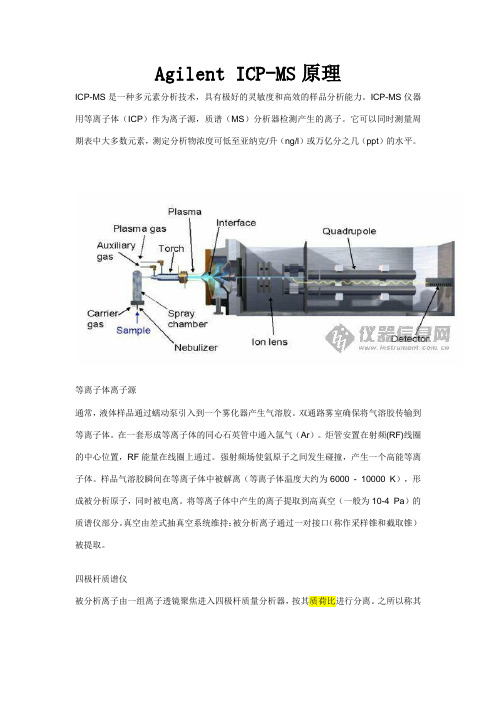
Agilent ICP-MS原理ICP-MS是一种多元素分析技术,具有极好的灵敏度和高效的样品分析能力。
ICP-MS仪器用等离子体(ICP)作为离子源,质谱(MS)分析器检测产生的离子。
它可以同时测量周期表中大多数元素,测定分析物浓度可低至亚纳克/升(ng/l)或万亿分之几(ppt)的水平。
等离子体离子源通常,液体样品通过蠕动泵引入到一个雾化器产生气溶胶。
双通路雾室确保将气溶胶传输到等离子体。
在一套形成等离子体的同心石英管中通入氩气(Ar)。
炬管安置在射频(RF)线圈的中心位置,RF能量在线圈上通过。
强射频场使氩原子之间发生碰撞,产生一个高能等离子体。
样品气溶胶瞬间在等离子体中被解离(等离子体温度大约为6000 - 10000 K),形成被分析原子,同时被电离。
将等离子体中产生的离子提取到高真空(一般为10-4 Pa)的质谱仪部分。
真空由差式抽真空系统维持:被分析离子通过一对接口(称作采样锥和截取锥)被提取。
四极杆质谱仪被分析离子由一组离子透镜聚焦进入四极杆质量分析器,按其质荷比进行分离。
之所以称其为四极杆,是因为质量分析器实际上是由四根平行的不锈钢杆组成,其上施加RF和DC电压。
RF和DC电压的结合允许分析器只能传输具有特定质荷比的离子。
检测器最后,采用电子倍增器测量离子,由一个计数器收集每个质量的计数。
质谱质谱图非常简单。
每个元素的同位素出现在其不同的质量上(比如,27Al会出现在27 am u处),其峰强度与该元素在样品溶液中同位素的初始浓度直接成正比。
1-3分钟内可以同时分析从低质量的锂到高质量数的铀范围内的大量元素。
用ICP-MS,一次分析就可以测量浓度水平从ppt级到ppm级的很宽范围的元素。
应用ICP-MS广泛用于许多工业领域,包括半导体工业、环境领域、地质领域、化学工业、核工业、临床以及各类研究实验室,是痕量元素测定的关键分析工具。
百度百科解释ICP-MS介绍ICP-MS介绍电感耦合等离子体质谱 ICP-MS所用电离源是感应耦合等离子体(ICP),它与原子发射光谱仪所用的ICP 是一样的,其主体是一个由三层石英套管组成的炬管,炬管上端绕有负载线圈,三层管从里到外分别通载气,辅助气和冷却气,负载线圈由高频电源耦合供电,产生垂直于线圈平面的磁场。
安捷伦使您美梦成真

安捷伦使您美梦成真
佚名
【期刊名称】《电子元器件应用》
【年(卷),期】2005(007)001
【摘要】安捷伦科技是由美国惠普公司战略重组分立而成的一家致力于高速增长
领域的多元化高科技跨国公司,其业务重点包括通信、电子及化学分析与生命科学。
【总页数】3页(Pi014-i016)
【正文语种】中文
【中图分类】F407.63
【相关文献】
1.《财富有情》使“梦想者”美梦成真 [J], 陈为琳
2.安捷伦科技推出J6900A三重播放分析仪支持中国AVS视频编码标准——三重
播放分析仪帮助加速AVS—IPTV网络的开发、部署和监测,使该业务更快地推向市场 [J], 无
3.SRS技术使家庭影院美梦成真 [J], 子纯
4.安捷伦科技推出J-BERT软件的最新版本,抖动容限测试速度称雄业界软件升级使多千兆位收发信机表征一键完成 [J],
5.安捷伦提供新型低价校准服务,使安捷伦校准服务系列成为业内最完善的校准服务——安捷伦为通用应用中的测试设备推出专门设计的最新服务 [J],
因版权原因,仅展示原文概要,查看原文内容请购买。
Agilent Cell Viability Workstation 应用笔记
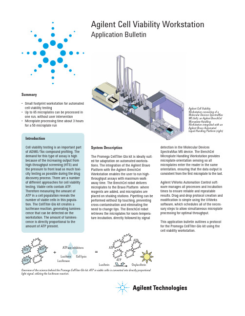
Agilent Cell Viability Workstation Application BulletinAgilent Cell ViabilityWorkstation consisting of aMolecular Devices SpectraMaxM5 (left), an Agilent BenchCelMicroplate HandlingWorkstation integrated with anAgilent Bravo AutomatedLiquid Handling Platform (right)=+LuciferinLuciferinCell lysisLuciferaseATPase inhibitorsOverview of the science behind the Promega CellTiter-Glo kit. ATP in viable cells is converted into directly proportionallight signal, utilizing the luciferase reaction.Summary•Small footprint workstation for automatedcell viability testing•Up to 65 microplates can be processed inone run, without user intervention•Microplate processing time about 3 hoursfor a 50-microplate runIntroductionCell viability testing is an important partof ADME/Tox compound profiling. Thedemand for this type of assay is highbecause of the increasing output fromhigh-throughput screening (HTS) andthe pressure to front load as much toxi-city testing as possible during the drugdiscovery process. There are a numberof different approaches for cell viabilitytesting. Viable cells contain ATP.Therefore measuring the amount ofATP in a cell population reveals thenumber of viable cells in this popula-tion. The CellTiter-Glo kit creates aluciferase reaction, generating lumines-cence that can be detected on theworkstation. The amount of lumines-cence is directly proportional to theamount of ATP present.System DescriptionThe Promega CellTiter-Glo kit is ideally suit-ed for adaptation on automated worksta-tions. The integration of the Agilent BravoPlatform with the Agilent BenchCelWorkstation enables the user to run high-throughput assays with maximum walk-away time. The BenchCel robot deliversmicroplates to the Bravo Platform wherereagents are added, and microplates areplaced on shaking stations. Pipetting can beperformed without tip touching, preventingcross-contamination and eliminating theneed to change tips. The BenchCel robotretrieves the microplates for room-tempera-ture incubation,directly followed by signaldetection in the Molecular DevicesSpectraMax M5 device. The BenchCelMicroplate Handling Workstation providesmicroplate-orientation sensing so allmicroplates enter the reader in the sameorientation, ensuring that the data output isconsistent from the first microplate to the last.Agilent VWorks Automation Control soft-ware manages all processes and incubationtimes to ensure reliable and repeatableresults. Drag-and-drop protocol creation andmodification is simple using the VWorkssoftware, which schedules all of the neces-sary steps to allow simultaneous microplateprocessing for optimal throughput.This application bulletin outlines a protocolfor the Promega CellTiter-Glo kit using thecell viability workstation.Instrument LayoutAgilent Bravo deck layout: locations 5 and 8 are configured with Orbital Shaking Stations (shaker) for enhanced throughput. A reservoir and a tipbox are placed manually at locations 2 and 6, respec-tively, before the protocol starts.Agilent BenchCel stacker layout: stacker 1 contains microplate A (can store up to 65 microplates), stackers 2 and 3 are used for incubation, and stacker 4 receives the processed microplates.MaterialsComponent List•Agilent BenchCel Workstation(R-series with 4 stackers)•Agilent Bravo Platform with gripper, 384ST disposable-tip pipette head, reservoir, 2 Orbital Shaking Stations•Molecular Devices SpectraMax M5•Agilent VWorks AutomationControl softwareLabware List•Microplate A: Greiner 96PS black,tissue-culture treated•Tipbox A: Agilent Tips 384 ST 70 µL Reagent List•Reservoir A: CellTiter-Glo reagent Protocol Workflow1.Move microplate A from BenchCelstacker 1 to Bravo deck location 7.2.Press on tips at Bravo decklocation 6.3.Aspirate 25 µL CellTiter-Glo fromreservoir A and dispense intomicroplate A.4.Move microplate A from decklocation 7 to 5 (shaker).5.Shake for 2 min.6.Move microplate A from decklocation 5 to 7.7.Move microplate A from Bravo decklocation 7 to BenchCel stacker 2.8.Incubate for 10 min.9.Move microplate A from BenchCelstacker 2 to the SpectraMax device.10.Read microplate A on theSpectraMax device.11.Move microplate A from theSpectraMax device to BenchCelstacker 4./lifesciences/automationThis item is intended for Research Use Only.Not for use in diagnostic procedures. Information,descriptions, and specifications in this publicationare subject to change without notice.Agilent Technologies shall not be liable for errorscontained herein or for incidental or consequentialdamages in connection with the furnishing,performance, or use of this material.Promega and CellTiter-Glo are registered trade-marks of Promega Corporation. Molecular Devicesand SpectraMax are registered trademarks of MDSAnalytical Technologies.© Agilent Technologies, Inc., 2009Published in the U.S.A., February 26, 2009Publication Number 5990-3555EN ConclusionsThe Agilent Cell Viability Workstation usingthe Promega CellTiter-Glo kit provides a reli-able, high-throughput solution for analyzingcell viability. The integration of microplatehandling, liquid handling, and microplatereading enables up to 65 microplates to beprocessed in one run without user interven-tion. Following the guidelines set by Promega,the typical throughput for this setup is about3 hours for 50 microplates, depending onexact protocol and liquid-handling steps.。
安捷伦液质联用培训教材(中文版)

=安捷伦 G6300 系列LC/MSD Trap现场培训教材质谱数据系统毛细管电泳液相色谱气相色谱注意包含在该文件中的信息将可能在未通知的情况下改变。
安捷伦科技有限公司不对与该材料有关的任何活动做担保。
这些活动包括但不仅限于为了某特殊目的而进行的销售和适应性。
安捷伦科技有限公司将不会对包含在材料里的与装备,表现和材料使用有关的错误或导致的损失负责。
这份文件中的任何部分都不得拷贝或复制或未经安捷伦科技公司的预先允许进行翻译。
安捷伦科技有限公司售后服务电话:800-8203278手机用户:400-8203278中文网站:/chem/cn2007年6月G6300A 系列离子阱软件概述以及开机关机操作仪器硬件概述1.1典型配置1.2仪器原理简介1.2.1离子阱的主体包含一个环电极和两个端电极,环电极和端电极都是绕Z轴旋转的双曲面,并满足r20=2Z20( r0为环形电极的最小半径,Z0为两个端电极间的最短距离)。
射频电压V rf加在环电极上,两个端电极都处于零电位。
1.2.2与四极杆分析器类似,离子在离子阱内的运动遵循马修方程,也有类似四极杆分析器的稳定图。
在稳定区内的离子,轨道振幅保持一定大小,可以长时间留在阱内,不稳定区的离子振幅很快增长,撞击到电极而消失。
离子阱的操作只有射频RF电压,没有直流DC电压,因此离子阱的操作只对应于稳定图上的X轴。
对于一定质量的离子,在一定V rf下,不同质量数的离子按照m/z由小到大在稳定图的X轴上自右向左排列。
当射频电压从小到大扫描时,排在稳定图上的离子自左向右移动,振幅逐渐加大,依次到达稳定图右边界,从离子阱中抛出,经过高能打拿极然后由电子倍增器检测。
1.3仪器硬件概述1.3.1离子源1.3.2离子源原理1.3.3仪器构造-示意图1.3.4 仪器构造-实物离子阱整体离子阱分解图1.3.5 LC-MSD Trap 的典型操作模式(以MS2为例):首先样品组分通过LC 进行分离,然后通过大气压电离源电离产生离子,离子阱在电场作用下,通过离子电荷控制(ICC )在阱中进行离子累积存储一定数量的离子,然后通过扫描隔离掉低于目标离子质量数的离子,通过在端电极上施加附加电场排除掉阱中高于目标质量数的离子,这个过程为Isolation ,接下来通过在端电极上施加特定离子的共振波形,使其与He 碰撞导致离子内能增加而使离子碎裂,此过程称之为Fragmenation 或CID ,最后在离子阱上扫描Rf 电压得到二级质谱。
安捷伦新的混合模式阴离子交换剂聚合物固相萃取小柱

Agilent SampliQ-SAX 具有如下性能:•优异的重现性•可用于酸性和中性化合物•萃取程序简单•颗粒大小可控概述固相萃取(SPE )是复杂样品分析流程中的基础,样品制备仍保持为分析过程中的重要组成部分,即使采用高专属性的检测器如L C /M S /M S 也如此,LC/MS/MS 共流出杂质的离子抑制作用可反过来影响定量分析。
进行净化萃取可以减少分析的复杂性。
延长HPLC 柱的寿命,得到更为准确的结果。
SPE 是一种液-液萃取经济实惠的代用技术,因为它只使用很少溶剂、快速、并且产生的废物少,SPE 与液-液萃取相比是一种改进了的样品制备技术,因为它具有较大的灵活性,提高了回收率和重现性,并且是一种更为有效的净化工具。
在食品安全、药物、环境和法医行业的研究人员都使用SPE 。
在Agilent Sa mpliQ-SAX 树脂是用季胺对二乙烯基苯进行改性的聚合物(图1)。
其结果使这一聚合物在很宽的疏水性(logP )范围内既对酸性化合物又对中性化合物具有保留作用。
这一萃取小柱既具有阴离子交安捷伦新的混合模式阴离子交换剂聚合物固相萃取小柱:SampliQ-SAX技术摘要换剂保留行为,又有反相固定相的保留行为,易于进行方法的开发。
这种树脂对各种溶剂有惰性,在pH 0到14之间是稳定的,并且用水可以湿润它。
图1.SAX 树脂质量控制产品的质量控制保证结果有较高的可信度,在经认证的环境中有严格的组成,严格控制和检测其颗粒特性,用球形聚合物颗粒以保证装填的均匀性和重现性,颗粒的规格和分布是用电子区域检测进行分析,颗粒的形状是用光学显微镜表征的,其比表面和孔隙度是用氮吸附进行检测,严格的尺寸控制使其具有很好的重现性和流动性,而且对每一批产品测试其色谱性能和纯度。
另外,测试每一批产品的性能,并把性能测试证书放在每个包装盒里。
操作指南使用Agilent Sa mpliQ-SAX小柱的程序很简单,图2是为方法开发建议的开始基本步骤,在此例中小柱的容积为3mL/60g。
应用 Agilent Bond Elut Plexa PCX 与 …

敏度。 LC
安捷伦以前的分析方法(由 Moorman 和 Hughes 开发,2010)使 用的是 Agilent 6410 Triple Quadrupole LC/MS 系统和其它的
• Agilent Poroshell 120 EC-C18 3 × 50 mm,2.7 µm 色谱柱 (部件号:699975-302)
×10 6-AM – 5 ߲౪܈ೝĂ๑ᆩ 5 ߲౪܈ೝĂ5 ߲ۅĂ๑ᆩ 5 ߲ۅĂ11 QCs
y = 0.060882 * x + 0.011665 2 R2 = 0.99961057
၎ܔၚᆌ
线。向不含有 6-乙酰吗啡的尿液中加入 6-乙酰吗啡标准品,配制 成浓度分别为 1.0、10、50、200 和 400 ng/mL 的校准标准溶液。 1
3. 淋洗 1:1 mL 2% 甲酸溶液
4. 淋洗 2:1 mL 甲醇
5. 在ห้องสมุดไป่ตู้空(10–15 英寸汞柱)条件下,抽干 5-10 分钟
MS 条件
ES 离子源参数 离子检测模式 毛细管电压 干燥气流速 干燥气温度 雾化气压力 鞘气流速 鞘气温度 喷嘴电压 MS 参数 扫描模式 预运行脚本
时间段 Delta EMV (+)
本方法在较小的样品进样体积(10 µL)和无预富集的条件下,具有 极高的信噪比(样品浓度 1 ng/mL 时> 190:1,该浓度为 SAMHSA 规定限量浓度的 10%)。这得益于应用了 AJST 技术的 Agilent 6460 Triple Quadrupole LC/MS 系统电喷雾离子源增强了检测灵
应用报告
法医与药物测试
摘要
由美国药物滥用和精神健康服务管理局(SAMHSA)颁布,于 2010 年 10 月起生效的新 准则,允许在政府认证的药物检测实验室使用 LC/MS/MS 法对初步药物检测结果进行 确认。由于 LC/MS/MS 法不需要衍生步骤,因此比先前使用的 GC/MS 法简便得多。 我们提出了一种满足最新 SAMHSA 准则要求的 6-乙酰吗啡的分析方法,并对其线性、 检测限(LOD)、准确度和精密度进行论证,还对该方法的基质效应、萃取回收率和总处 理效率进行了考察。这是涵盖所有 SAMHSA 监控药物类别的一系列六种简便检测方法 之一,该方法主要使用安捷伦产品进行分析,包括 Agilent Bond Elut Plexa PCX 混合模式 聚合物 SPE 吸附剂、Agilent Poroshell 120 EC-C18 2.7 µm 表面多孔 LC 色谱柱、Agilent 1200 Infinity LC 系统以及应用安捷伦喷射流技术(AJST)增强电喷雾离子源的 Agilent 6460 Triple Quadrupole LC/MS 系统。
使用Agilent7900ICP-MS对牛奶和奶粉进行常规的高通量多元素分析

前言牛奶和乳制品是人类膳食中重要的营养来源,对婴儿和儿童更是如此。
乳制品的食用范围遍布世界各地。
随着人们口味的变化和收入的增加,乳制品在许多亚洲和发展中国家/地区的普及程度日益提高。
为了满足日益增长的需求,乳制品产量增加的同时产品质量也显得尤为重要。
通过检测 Na 、K 、Mg 、Ca 等常量元素以及 Se 、P 、Mn 、Zn 等必需元素的浓度,可以提供有价值的营养信息。
另外还需要检测动物奶中的 As 、Cd 、Sn 、Hg 和 Pb 等潜在的有害元素,用于监测来自土壤、肥料、饲料或加工设备的潜在污染。
由于其卓越的灵敏度、快速多元素分析和广泛的元素覆盖,安捷伦 ICP-MS 仪器在环境和食品检测实验室中得到广泛使用。
随着最近的技术进步,Agilent 7900 ICP-MS 的分析动态范围已扩展至 10 个数量级以上,在同一次常规分析运行中可以将牛奶中的常量元素(如 Na 、使用 Agilent 7900 ICP-MS 对牛奶和奶粉进行常规的高通量多元素分析应用简报作者Courtney Tanabe美国加州大学戴维斯分校Jenny Nelson 、Craig Jones 安捷伦科技有限公司 美国食品检测与农业K 和 Ca)和痕量元素进行同时检测。
碰撞/反应池(CRC) 同样经过了改进,以确保待测元素在样品基质中多原子干扰下依然能得到准确的结果。
7900 ICP-MS 拥有市场领先的等离子体稳定性,利用选配的超高基质进样 (UMHI) 技术,样品耐受量可进一步拓展至高达 25% 的总溶解固体 (TDS)。
结合与集成样品引入系统 (ISIS 3) 的不连续采样 (DS) 功能带来的高样品通量,7900 ICP-MS 非常适合食品样品中宽范围元素的常规、高通量检测。
本研究介绍了 Agilent 7900 ICP-MS 配合可选 UHMI 和 ISIS 3,用于牛奶和乳制品中常量和痕量元素的快速分析。
Agilent 1260 Infinity II Manual Preparative Inject

Agilent 1260 Infinity IIManual PreparativeInjectorTechnical NoteIn this note we describe how to install and use the 1260 Infinity II Manual Preparative Injector.ContentsInstalling the Manual Injector2Unpacking the Manual Injector2Install the Manual Injector3Flow Connections6Install Internal Reducers8Leak Drainage9Using the Manual Injector10Warnings and Cautions10Information on Injection Seal Material11Needles11Inject a Sample12Agilent TechnologiesInstalling the Manual InjectorUnpacking the Manual InjectorDamaged PackagingUpon receipt of your manual injector, inspect the shipping containers for any signs of damage. If the containers or cushioning material are damaged, save them until the contents have been checked for completeness and the manual injector has been mechanically checked. If the shipping container or cushioning material is damaged, notify the carrier and save the shipping material for the carriers inspection.Delivery ChecklistEnsure all parts and materials have been delivered with the manual injector. The delivery checklist is shown in Table1 on page2. Please report missing or damaged parts to your local Agilent Technologies sales and service office. Table1Delivery ChecklistDescription QuantityManual Injection Valve-Prep-Kit (5067-6717) 1Start cable (0100-1677) 1Manual Injector ERI Start-Cable (5188-8056) 1Ring stand, mounting bracket (1400-3166) 1Syringe, 25 mL PTFE removable Luer lock (5190-1544) 1Holder Manual Injector (G9328-00001) 1User Documentation (G9300-64500) 1Additionally, one or more loops can be ordered as an option.23Install the Manual Injector1Loosen the setscrews that hold the injector lever onto the manual injector assembly.2Slide off the manual injector lever assembly.3Depending on where the injector is installed, either push the holder onto the manual injector valve assembly, or push the ring stand mounting bracketonto the manual injector assembly.44Tighten the screws to fix the holder to the valve assembly.5Push the manual injector lever assembly on the valve assembly.6Tighten the setscrews to fix the injector lever assembly to the valveassembly.57Insert the t-nut of the holder into the guide conduct of the mounting plate and slide the holder into its desired position, or slide the ring stand mounting bracket onto the front pole of the column organizer.8Tighten the screws to fix the holder with the manual injector onto the mounting plate.The manual injector is ready to be connected to the flow path of the system.6Flow Connections Figure 1Vent CapillariesToxic, flammable and hazardous solvents, samples and reagentsThe handling of solvents, samples and reagents can hold healthand safety risks.➔When working with these substances observe appropriate safety procedures (for example by wearing goggles, safetygloves and protective clothing) as described in the materialhandling and safety data sheet supplied by the vendor, andfollow good laboratory practice.➔The volume of substances should be reduced to the minimumrequired for the analysis.➔Do not operate the instrument in an explosive atmosphere.Prevent siphoning➔The outlets of the two vent capillaries (ports 5 and 6) and the needle port must be at the same level to prevent siphoning(see Figure 1 on page 6).9HQW FDSLOODULHV DQG QHHGOH SRUW DW WKH VDPH OHYHO71Connect capillaries.Figure 2Flow connections (G1328D):DVWH:DVWH6DPSOH ORRS 3XPS &ROXPQInstall Internal ReducersInternal reducers (IZR) are used to adapt small capillaries to a valve with larger fittings. This helps optimizing a preparative system to low flow rates.Initial installation of an IZR1Remove the secondary nut and ferrule from the IZR body.2Screw the IZR body with the liner and primary ferrule into the valve port.Fingerthighten the IZR body.3Insert the tubing into the IZR body.4Push the tubing firmly to seat it properly in the valve port fitting. At the same time use a wrench to tighten the IZR body with 1/3 of a turn.5Remove the tubing from the IZR body.6Slide the secondary nut and secondary ferrule onto the tubing.7Insert the tubing/secondary nut/secondary ferrule assembly into the IZR body and screw it fingertight.8Push firmly on the tubing to seat it properly in the liner. At the same time use a wrench to tighten the secondary nut with 1/3 of a turn.8Remove an IZR1Remove the secondary nut, ferrule, and tubing.2Remove the IZR body, liner, and primary ferrule.Reinstallation of an IZR1Reinsert the IZR body, primary ferrule, and liner into the valve port fitting, and fingertighten the IZR body.2Use a wrench to tighten the IZR body 1/8 turn.3Reinsert the secondary nut, ferrule, and tubing into the IZR body, and screw the secondary nut in fingertight.4Use a wrench to tighten the secondary nut 1/8 turn.Leak DrainageLarge amounts of pressurized solventsExplosive and intoxication hazard➔Install the preparative manual injector in the preparativecolumn organizer.For details, see installation instructions for the Agilent InfinityLab LCSeries 1260 Infinity II Column Compartment.9Using the Manual InjectorWarnings and CautionsEjection of mobile phaseWhen using sample loops larger than 100µL, mobile phase maybe ejected from the needle port as the mobile phase in thesample loop decompresses.➔Please observe appropriate safety procedures (for example,goggles, safety gloves and protective clothing) as described inthe material handling and safety data sheet supplied by thesolvent vendor, especially when toxic or hazardous solventsare used.Splashing of solvent➔When using the Needle Port Cleaner, empty the syringe slowlyto prevent solvent from splashing back at you.➔Please observe appropriate safety procedures (for example,goggles, safety gloves and protective clothing) as described inthe material handling and safety data sheet supplied by thesolvent vendor, especially when toxic or hazardous solventsare used.Potential damage to the valve➔Rinse the valve with water after using buffer solutions toprevent crystals from forming, which can cause scratches onthe rotor seal.10Information on Injection Seal MaterialThe manual injector is supplied with a PEEK injection seal. PEEK is compatible with pH 0 – 14, incompatible with some concentrated mineral acids.NeedlesNeedle can damage valve➔Always use the correct needle size.Use needles with 0.028-inch outer diameter (22gauge)×2-inch long needle, without electro-taper, and with 90° point style (square tip).1112Inject a SampleFor the manual injector different sample injection methods exist:•Complete loop filling for highest possible precision:Use at least two to three times of the loop volume (for example 40 – 60μL of sample for a 20μL sample loop).•Partial loop filling if there is only little sample available:Use a maximum of half of the loop volume (for example 10μL of sample for a 20μL sample loop).1Turn the handle to the LOAD position.Preparations •Connect the injector to the system •Make sure the system is ready for use •Flush the injection valve and loop properly •Place a waste beaker below the valve •Set the injection source to Manual Injector and create an instrument method •Fill the syringe with the sample :DVWH:DVWH1HHGOH SRUW6DPSOH ORRS3XPS &ROXPQ132Insert the syringe with needle into the needle port.3Slowly push the syringe piston to load the sample onto the loop.Loop is filled with sample.NOTEYou should feel slight resistance as the needle passes through the needle seal before it stops against the stator face.NOTE To achieve higher precision over fill the loop (complete loop fillingmethod only).*G1328-90030**G1328-90030*G1328-90030Part Number:G1328-90030 Rev. C SD-29000152 Rev. CEdition: 10/2019Printed in Germany © Agilent Technologies, Inc 2017-2019Agilent Technologies, Inc Hewlett-Packard-Strasse 876337 Waldbronn, Germany 4Leave the syringe in the needle port and turn the handle to the INJECT position.The sample is in the flow path and is flushed towards the column.5Remove the syringe with needle from the needle port. :DVWH:DVWH1HHGOH SRUW 6DPSOH ORRS3XPS&ROXPQ。
Agilent EZChrom Elite 高级报告参考

本文档中提及的硬件和/或软件以 许可权的方式提供,其使用或复 制必须遵守许可。
有限权利声明
如果在履行美国政府某项重要合 同或转包合同时要使用此软件, 将以以下方式提供并授权软件: DFAR 252.227-7014(1995 年 6 月) 定义的“商业计算机软件”; FAR2.101 (a) 定 义 的 “ 商 业 项 目”;FAR 52.227-19(1987 年 6 月)或任何同等机构规定或合同 条款定义的“受限计算机软 件”。软件的使用、复制或公开 必须遵守安捷伦科技有限公司的 标准商业许可条款的规定,美国 政府的任何非 DOD 部门和机构 所 拥 有 的 权 利 不 得 超 出 FAR 52.227-19(c)(1-2)(1987 年 6 月) 中定义的“有限权利”的范围。 美国政府用户拥有的权利不得超 出 FAR 52.227-14(1987 年 6 月) 或 DFAR 252.227-7015 (b)(2)(1995 年 11 月 ) 中 定 义 的 “ 有 限 权 利”的范围(适用于所有技术数 据)。
2 简介 .................................................................................................... 13
3 教程 #1 – 创建序列摘要报告..................................................... 15 步骤 1:(创建新报告模板) ....................................................................16 步骤 2:(创建报告页眉和页脚)...........................................................16 步骤 3:(创建页眉) ..................................................................................18 步骤 4:(创建摘要表) .............................................................................20 步骤 5:(创建数据图表).........................................................................24 步骤 6:(保存模板) ..................................................................................26 步骤 7:(创建再处理序列) ....................................................................26 步骤 8:(查看报告) ..................................................................................28 步骤 9:(向报告添加新统计)................................................................29
Agilent Bravo 自动化液体处理平台配件选择指南说明书

Accessories for the Agilent Bravo Automated Liquid Handling PlatformSelection GuideIntroductionThe Agilent Bravo Automated Liquid Handling Platform provides unparalleledversatility and precision in a compact footprint. With proven high-accuracy pipette heads and a flexible nine-position open deck, it offers the flexibility needed to scale-up applications ranging from serial dilutions to PCR and cell-based assays. A carefully designed group of accessories is at the heart of the Bravo’s flexibility, providing the range of tools needed by scientists and engineers to reduce manual intervention and increase throughput and walkway time when automating routine and customized applications. Bravo accessories make it possible to configure any of the nine available deck positions for shaking, heating, cooling, filtration, and more.The Bravo Automated Liquid Handling Platform is compatible with the following: • Agilent designed and tested accessories• Proven, industry standard accessories customized for use with the Bravo platform • User accessories that fit with the Bravo platform open architectureFigure 1. The Bravo Platform 9-position deck accommodates accessories designed to facilitate applications. The labeled deck image includes a Bravo accessories configuration designed for Next Generation Sequencing sample preparation.Example of a Bravo deck layout Components1. Available for an accessory2. Available for an accessory3. Available for an accessory4. Peltier Thermal Station with Custom Plate Nest5. Orbital Shaking Station6. Peltier Thermal Station with Custom Plate Nest7. Magnetic Bead Accessory 8. Available for an accessory 9. Thermal Station1472583691 - Agilent VWorks Automation Control Software.2 - Agilent Bravo Platform accessories may contain 3rd-party components which have been customized and optimized for use with the Bravo Platform, and which are supported by Agilent.3 - NA = Not applicable.For Research Use Only. Not for use in diagnostic procedures. This information is subject to change without notice.©Agilent Technologies, Inc., 2012Published in the USA, April 12, 20125991-0126ENApplications Enabled by Bravo AccessoriesBravo Platform accessories are carefully designed tools that deliver the flexibility and versatility you need to automate a wide range of key applications. The table below shows a selection of applications that are enabled using BravoPlatform accessories. For complete configurations tailored to yourapplication, contact Agilent.Agilent Bravo Platform ConsumablesA complete range of pipette tips is also available for use with Bravo Platform accessories. Visit /lifesciences/pipettetips for more information.Learn More/lifescience/automationFind a local Agilent customer center /chem/contactus USA and Canada 1-800-227-9770*****************************Europe************************Asia Pacific************************。
5500 ILM AFM SPM说明书

Simultaneous AFM/Fluorescence Imaging of Living Cells – Fluorescence-guided Force SpectroscopyApplication NoteJosef Madl1Sebastian Rhode1Gerhard J. Schütz1Peter Hinterdorfer1Gerald Kada21Institute of Biophysics, University of Linz, Altenbergerstr. 69, 4040 Linz, Austria2Agilent Technologies, Nano Measurements Division, Roemerstr. 18, 4020 Linz, Austria SummaryHigh resolution atomic forcemicroscopy (AFM) can be per-formed simultaneously withoptical microscopy techniques,such as fluorescence or differentialinterference contrast (DIC)microscopy. The combinedmethodologies provide comple-mentary information about thestudied sample which establishesthe basis for a better understand-ing of physiological processes andthe function of biomolecules, andallow a more detailed characteriza-tion of the structure of living cells[1]. Here we present the Agilent5500 ILM AFM/SPM, which is aneasy-to-use AFM and invertedlight microscope (ILM) combinationthat enhances live-cell imagingunder physiological conditions.The advantage of this combinedsetup is further demonstrated bystudying fluorescence-labeledmembrane receptors via forcespectroscopy using antibody-functionalized AFM tips.IntroductionAtomic force microscopy hasevolved as a method capable ofresolving biological structuresat the molecular level [2]Furthermore it provides the possi-bility to study interaction forcesbetween single receptor-ligandcomplexes. Fluorescencemicroscopy has proven to be apowerful method for the specificvisualization of labeled moleculesdown to the single molecule level[1]. This allows the determinationof their distribution observing dif-fusion or trafficking processes incells. Other optical techniques,such as bright field, phase contrast[3], and DIC microscopy revealinformation about cell morphologyand composition. By enabling amore detailed characterization ofinternal and external cellularstructures and biological process-es, the combination of AFM withoptical microscopy provides amore comprehensive examinationof the sample than either of thetechniques alone can deliver.MethodsChinese hamster ovary (CHO) cells were utilized in this study. These cells, which are widely used as expression system for a number of proteins, grow in low cost, fully defined media, and they are adherent to solid matrices. Since immobi-lized samples are necessary,cellular adhesion is an advantage in AFM imaging. In addition, CHO cells, and other mammalianautocrine cells, are more resistant to apoptosis, so they are compati-ble with lengthy experiments.The CHO cells utilized express high levels of recombinantscavenger receptor class B type 1(SRB1) [4]. The scavenger receptors were fused to a green fluorescent protein (eGFP). This fluorescent protein can be excited with 488nm light and emits light at 508 nm.Figure 1 shows a typical fluores-cence microscopy image of a living CHO cell expressing the SRB1-eGFP construct. The cells were grown in medium A (see [4] for details) on 22 mm glass slides in conventional 35 mm Petri dishes at 37 degree Celsius and 5% CO 2. The sample slides were mounted onto the Agilent 5500 ILM AFM/SPM with an Agilent AFM/SPM liquid cell sample holder. The cells wererinsed with PBS buffer (phosphate buffered saline, 50 mM NaPi, 150mM NaCl, pH 7.4) and kept in this buffer throughout the experiments.InstrumentationUsing a QuickSlide Series 5500mechanical stage, an Agilent 5500AFM/SPM was adapted to a Zeiss Axiovert 200 inverted light micro-scope. The QuickSlide stage allows convenient sample changing and permits positioning of the sample directly under the AFM cantilever.The ILM AFM/SPM was placed onto a passive anti-vibration table (Newport GmbH) without anyadditional active damping (figure 2).For the simultaneous fluores-cence/AFM imaging, the excitation of SRB1-eGFP was accomplished with a mercury arc lamp (HBO100,Zeiss) with a 488 nm laser(Sapphire, Coherent). For com-bined fluorescence/force spec-troscopy experiments a halogen lamp (HAL, Zeiss) was used.AFM/DIC images were acquired using a CCD camera (Casade 512B,Roper Scientific). In order toreduce the mechanical noise level,the cooling fan of the camera was removed and mounted in an exter-nal fan box, which was placed outside the optical table and con-nected to the camera via a long polymer (PVC) hose. A 40x oil immersion objective (NeoFluar,NA=1.3, Zeiss) was used for DIC measurements, a 100x oil immer-sion objective (Apochromat,NA=1.4, Zeiss) was utilized for fluorescence microscopy and a 60x water immersion objective (NA=1.2, Olympus) was utilized for the fluorescence guided force spec-troscopy experiments. For AFM experiments, a large (100 µm scan range), multipurpose Agilent AFM/SPM scanner was utilized.The AFM images were acquired inFigure 1. Fluorescence microscopy image of eGFP-labeled receptors on a living CHO cell.Image acquired using an Agilent 5500 ILM AFM/SPM. Image size 40 x 40 µm2.Figure 2. Agilent 5500 ILM AFM/SPM with a Zeiss Axiovert 200 inverted light microscope.The 5500 ILM AFM/SPM is also compatible with other inverted light microscope makes and models.contact mode using 10 pN/nm sili-con nitride cantilevers at scanning rates of 0.4 - 1 line/sec, so com-plete AFM images were acquired in from four to ten minutes. For force spectroscopy experiments the same type of contact-mode levers, were used after functional-ization with antibodies as described in [1] (fig. 6c).Resultsa) Simultaneous DIC and AFM imaging Contact mode AFM images were acquired on the CHO cells that were grown on glass cover slips as described above and fixed in2% glutaraldehyde for ten minutes.DIC microscopy images wererecorded simultaneously with the AFM scanning process. One frame from this sequence is shown in Figure 3a. The DIC image visualiz-es the cell morphology including the cell body, protrusions or lamel-lipodia, and also prominent intracel-lular features, such as the nucleus and several large organelles.Figure 3. Simultaneous DIC microscopy (3a) and AFM (3b and c) images. Glutaraldehyde-fixed CHO cells were imaged in contact mode AFM while acquiring a DIC microscopy image sequence. Three cells are visible in the scan area of 60 x 60 µm2. Complementary structural information results from the DIC microscopy image and the contact mode AFM deflection (3b) and topography (3c) images.a b cFigures 3b and 3c are the corre-sponding AFM deflection (error)and topography (height) images,respectively, of the same CHO cells. AFM imaging showed the sample topography at high resolu-tion. In some regions parts of the membrane associated cytoskeleton are resolved, furthermore lamel-lipodia and cell debris are easily visualized. The small globular structures on the cell membrane are artefacts resulting from cell fixation. By comparing the cells in 3 a, b and c, one can see that cells of the same type have variable shapes, which is mainly due to different states of cell maturation.b) Simultaneous Fluorescence and AFM imagingCombined AFM-optical microscopy measurements are not restricted to fixed cells, also living cells can be easily imaged and analyzed with the Agilent 5500 AFM/SPM.Figure 4a shows one frame from a fluorescence image sequence of a living CHO-SRB1-eGFP cell. This image is a snapshot from an image sequence, which was acquired while simultaneously scanning the sample with the AFM. The corre-sponding AFM images are present-ed in figures 4b and 4c (deflection and topography image, respectively).The fluorescence microscopy image gives a good impression of the cell shape because the visual-ized eGFP-labeled SRB1 receptors are primarily located in the plas-ma membrane. Some fluorescence signals also originated from the cell interior because a part of the SRB1-eGFP constructs was either endocytosed or freshly synthesized inside the living cell [4].Figure 4. Fluorescence microscopy (a) and contact mode AFM deflection (b) and topography (c) images of live CHO cells obtained with an Agilent 5500 ILM AFM/SPM. The AFM images were obtained while simultaneously acquiring a fluorescence microscopy image sequence with a CCD camera. The fluorescence images were unaffected by light emitted from the AFM laser. Image size about 30 x 30 µm2.AFM images of living CHO cells (figures 4b and 4c) illustrate different features than images of fixed CHO cells in figures 3b and 3c [2]. For example, the cell mem-brane of a living cell is much softer than the cell membrane of a fixed cell, the elastic modulus is com-paratively much smaller. In this case the AFM tip can more easily depress the plasma membrane which is why the membrane asso-ciated cytoskeleton (e.g. the network of actin fibers and microtubules)is better revealed in living cells than in fixed cells. Alternatively,soft, flexible features like cellular protrusions or lamellipodia are not easily resolved within the AFM images of the living cells because they are easily pushed away by the scanning AFM tip.The AFM cantilever can be indi-rectly recognized as a bright spot,because eGFP-protein (mostly from lamellipodia) adsorbed onto the AFM tip during previous imaging processes.c) Fluorescence-Guided Force SpectroscopyAFM force spectroscopy is anexcellent technique for the investi-gation of receptor-ligand interaction forces and the corresponding bind-ing kinetics [1]. In some cases the receptor density on the cell surface can be low. When investigating cells for example, this might be due to low expression levels for the protein of interest or – as with the cells shown here – due to clustering of the receptors in microdomains within the plasma membrane [4]. The CHO-SRB1-EGFP cells were fixed in order to prevent endocytosis and diffusion of the SRB1 clusters [4]. A mild fixation protocol was applied (2% glutaraldehyde, 1 min) so that most of the SRB1 remained in their native conformation. The fixed cells were additionally treat-ed with NaBH 4in order to reduce fixative-induced auto-fluorescence. For the force spectroscopy experi-ments SRB1 clusters were localized by acquiring a fluorescence image of the cell using a GFP filter seta b cFigure 5. Fluorescence microscopy images of CHO-SRB1-eGFP cells that were fixed with glutaraldehyde. The eGFP-labeled SRB1 receptors are visible with a GFP filter on the Agilent 5500 ILM AFM/SPM in figure 5a. In figure 5b, the GFP filter was replaced with a Cy3 filter and the auto-luminescent silicon nitride AFM tip (outlined in yellow) is visible. In figure 5c, the two previous fluorescence images were used to precisely reposition the AFM tip over a cluster of eGFP-labeled SRB1 receptors. The AFM tip was modified with anti-SRB1 antibodies for subsequent force spectroscopy experiments (fig. 6). Images size about 60 x 60 µm2.a b cused to locate the silicon nitride AFM tip. When using a GFP filter silicon nitride luminescence is cov-ered by the stronger eGFP fluores-cence. However, after blocking the eGFP signal with the Cy3 filter the weak cantilever luminescence is enough to identify its position on the cell (X-shaped form in fig. 5b).This facilitated accurate position-ing of the tip at the end of the AFM cantilever directly above areas that contained a cluster of SRB1 receptors.Anti-SRB1 antibodies were teth-ered to AFM cantilevers via flexible polymer linkers at a low density,so that on average only one ligand molecule was located within the effective AFM tip/ cell surface interaction area (fig. 6c) [1]. In figure 5c, the functionalized AFM tip was aligned on top of the pre-chosen SRB1-eGFP-cluster(emission filter 535 ±25 nm; fig. 5a). The focal plane wasadjusted on top of the cell, so that mainly clusters of eGFP-SRB1receptors, which are accessible to the functionalized AFM tip are visible. One of the SRB1-eGFP clusters (outlined by a white cir-cle) was chosen for the following force spectroscopy measurements.Auto-fluorescent areas (no SRB1-EGFP-fluorescence signals) which remained despite NaBH 4treatment were identified and excluded from the experiments by switching to a Cy3 filter set (emission 610 ±38nm), which blocked the eGFP fluorescence.When illuminated with 488 nm light, a silicon nitride AFM tip emits light at wavelengths up to 750 nm (silicon cantilevers are not auto-luminescent; see [5]).Therefore, auto-luminescence was(fig. 5a) of receptors. Consecutive force-distance cycles were record-ed and a series of specific single molecule unbinding events were detected (fig. 6a). From more than one hundred force-distance cycles,a distribution of unbinding forces was obtained and the binding probability between anti-SRB1 antibodies and the SRB1 receptors was determined. The binding prob-ability for sites that contained SRB1 receptors was calculated to be 13.6 %. At an AFM cantilever velocity of 2 µm/sec (which corre-sponds to a loading rate of approx-imately 10 nN/sec), the unbinding force distribution of the anti-SRB1antibody/SRB1 receptor complexes contained two maxima, one at 64pN, and one at 85 pN (see figure 6b). It is possible that the SRB1antibody/SRB1 receptor complexes have two separate maxima because some of the receptors may have been more accessible to the AFMFigure 6. Results from force-distance spectroscopy experiments between anti-SRB1 antibodies tethered to AFM cantilevers and SRB1 receptors expressed in CHO cells. An Agilent 5500 ILM AFM/SPM was used to identify areas of fluorescence, corresponding to eGFP-labeled SRB1 receptors on the plasma membrane of the cells. Areas containing clusters of SRB1 receptors were probed with an AFM tip that was functionalized with anti-SRB1 antibodies. Figure 6a: a force-distance curve showing a single unbinding event between an anti-SRB1 antibody and a SRB1 receptor. From this force-distance curve, the unbinding force for a single antibody-antigen interaction was calculated to be approximately 70 pN. Figure 6a inset shows a force-distance spectroscopy curve resulting from an area that did not fluoresce from eGFP-labeled SRB1 receptors. Figure 6b: a force distribution derived from more than hundred anti-SRB1 antibody-SRB1 unbinding events. From the force distribution curve, two unbinding force maximas for anti-SRB1 antibody-SRB1 complexes were identified at 64 pN and 85 pN. Figure 6c: Tethering single antibodies to an AFM cantilever tip via a flexible polymer linker.a bctip-bound antibodies, while others may have been less accessible which was due to their location in caveolae [4].When the plasma membrane was “blindly” probed without guidance from fluorescence microscopy a binding probability of only three to four percent was measured. When force-distance experiments were performed on regions that lacked eGFP-labeled SRB1 receptors a 2.5%probability was measured. In approximately 50% of the areasexamined, specific unbinding events between the anti-SRB1 antibodies and SRB1 receptors were not detect-ed, even though the areas were iden-tified as having a high density of eGFP-labeled SRB1 receptors. This could have been due to inaccessibili-ty of the SRB1 receptors arising from, for example, endocytosis immediately before the cells were fixed. The inset in figure 6a shows the results of a force-distance exper-iment in which no unbinding event was detected./find/afmFor more information on Agilent Technologies’products, applications or services, please contact your local Agilent office. The complete list is available at:/find/contactus Phone or FaxUnited States:(tel) 800 829 4444 (fax) 800 829 4433Canada:(tel) 877 894 4414 (fax) 800 746 4866China:(tel) 800 810 0189 (fax) 800 820 2816Europe:(tel) 31 20 547 2111Japan:(tel) (81) 426 56 7832 (fax) (81) 426 56 7840Korea:(tel) (080) 769 0800 (fax) (080) 769 0900Latin America:(tel) (305) 269 7500Taiwan:(tel) 0800 047 86 (fax) 0800 286 331Other Asia Pacific Countries:(tel) (65) 6375 8100 (fax) (65) 6755 0042Email:*****************Revised: 09/14/06Product specifications and descriptions in this document subject to change without notice.© Agilent Technologies, Inc. 2007Printed in USA, May, 20075989-6902ENAFM Instrumentation from Agilent TechnologiesAgilent Technologies offers high-pre-cision, modular AFM solutions for research, industry, and education.Exceptional worldwide support is provided by experienced application scientists and technical service personnel. Agilent’s leading-edge R&D laboratories ensure the continued, timely introduction and optimization of innovative,easy-to-use AFM technologies.ReferencesOriginal research paper:J. Madl, S. Rhode, H. Stangl, H. Stockinger, P. Hinterdorfer, G.J. Schütz, G.Kada (2006). A combined optical and atomic force microscope for live cell investigations. Ultramicroscopy 106, 645-651.[1] P. Hinterdorfer, G.J. Schütz, F. Kienberger, H. Schindler (2001). Detection and Characterization of Single Biomolecules at Surfaces. Rev. Molec.Biotechn. 82, 25-35.[2] F. Kienberger, C. Stroh, G. Kada, R. Moser, W. Baumgartner, V.Pastushenko, C. Rankl, U. Schmidt, Harald Müller, E. Orlova, C. LeGrimellec,D. Drenckhahn, D. Blaas, P. Hinterdorfer. (2003). Dynamic Force Microscopy Imaging Of Native Membranes. Ultramicroscopy 97, 229-237.[3] E. Defranchi, E. Bonaccurso, M. Tedesco, M. Canato, E. Pavan, R. Raiteri,C. Reggiani. (2005) Imaging and Elasticity Measurements of the Sarcolemna of Fully Differentiated Skeletal Muscle Fibers. Microsc. Res. Techn. 67, 27-35.[4] S. Rhode, A. Breuer, J. Hesse, M. Sonnleitner, T.A. Pagler, M. Doringer, G.J. Schütz, H. Stangl. (2004). Visualization of the Uptake of Individual HDL Particles in Living Cells Via the Scavenger Receptor Class B Type I. Cell Biochem. Biophys. 41, 343-356.[5] A. Gaiduk, R, Kühnemuth, M. Antonik, C.A.M. Seidel. (2005). Optical Characteristics of Atomic Force Microscopy Tips for Single-Molecule Fluorescence Applications. ChemPhysChem 6, 976-983.。
安捷伦-液质联用技术(LCMS)及其应用
含有气溶胶的分析物
[溶剂+H]++M--------->溶剂 +[M]+
溶剂在蒸发 器中蒸发
+
++ ++
+
+
+
+
+ +
+
+ +
++
++
+ +
++
+
电荷转移至 分 析物分子
蒸汽
通过电晕针放电形 成带电荷的反应剂 离子
流动相
分析物
++ + ++
分析物离子
by Li Ping
High fragmentor: 130 V
1
2
3
4
5
6
7 min
by Li Ping
108.1 218.1
245.1 311.1
156.1
100
200
300
m/z
31
312.1
可同时采集多种质谱信号: SIM/Scan
SIM 来定量所选的目 标化合物离子
对未知的化合物 SCAN
by Li Ping
40000
20000
100
200
300
m/z
1
2
3
4
5
min
by Li Ping
33
可同时采集多种质谱信号: 时间编程信号
Improve data quality and manage data file size:
安捷伦化学工作站-Agilent

132
特征峰确认
132
特征峰比率计算
132
识别过程 134
找出参考峰 134
找出内标峰 134
找出其余的校正峰
135
未识别出的峰的分类 135
7 校正 137
术语定义 138
校正表 139
校正曲线 140
未知样品 142
校正类型 143
单级校正 143
多级校正 144
校正范围
146
校正曲线拟合
146
1 安捷伦化学工作站功能 本章介绍化学工作站的主要组成部分及功能。有关化学工作站具体任务使用说 明,请参见在线帮助或软件附带的化学工作站教学软件。
2 方法 本章介绍方法的概念及如何使用这些方法。
3 数据采集 本章介绍数据采集、数据文件和工作日志等概念。
4 积分 本章介绍化学工作站中积分器算法的积分概念。介绍了积分算法、积分和手动积 分。
Agilent Technologies
15
1 安捷伦化学工作站功能 概述
概述
用于 GC、 LC 及 A/D 系统的化学工作站是集仪器控制、数据采集及数据处理于 一体的化学工作站系统,适用于: • 安捷伦 6890N 气相色谱, • 用于 LC 的安捷伦 1100 系列模块和系统, • 安捷伦 35900E 双通道模数转换接口。 该软件适用于 IBM 兼容的个人计算机,在 Microsoft® Windows XP Professional 操作环境下运行。 该软件按单机运行的基本化学工作站进行销售,共有两个版本。所有版本均包括 对一个分析仪器进行数据采集、仪器控制、数据分析 (积分、定量及报告)、自 动化和自定义。仪器被定义为按单个时间表运行,但可以同时从多个不同的检测 器采集数据。这两个版本包括: • 用于气相色谱 (GC) 系统的单机运行的化学工作站,产品号为 G2070BA, • 用于液相色谱 (LC) 系统的单机运行的化学工作站,产品号为 G2170BA, 通过购买附加的仪器数据采集和控制模块允许配置多台仪器和多种技术,可以扩 展化学工作站软件的仪器控制功能。
安捷伦液相狭缝宽度 -回复

安捷伦液相狭缝宽度-回复题目:安捷伦液相狭缝宽度:探秘其原理与应用引言:液相色谱(Liquid Chromatography,简称LC)是一种常见的色谱分离技术,广泛应用于生物分析、药物研究等领域。
而在LC中,狭缝宽度(Slit Width)是一个重要的参数。
安捷伦(Agilent)作为液相色谱仪领域的领导者,其液相狭缝宽度技术备受关注,本文将详细介绍安捷伦液相狭缝宽度的原理和应用。
第一部分:液相狭缝宽度概述液相狭缝宽度是液相色谱仪中一个关键的参数,它指的是进样、分离和检测过程中液流通过的最小路径。
狭缝宽度的大小直接影响色谱分离的分辨率和检测灵敏度。
一般来说,较小的狭缝宽度能提供更高的分离效率和检测灵敏度,但也会增加背压和分析时间。
第二部分:安捷伦液相狭缝宽度的原理安捷伦液相狭缝宽度技术采用了微机械加工和电子控制等先进工艺,其原理可以概括为以下几个步骤:1. 采用微加工技术,在轴向和径向两个方向上制备狭缝线阵列;2. 利用电子控制系统,通过调整液相流的压力和流量,在狭缝区域形成一定的流场;3. 控制狭缝宽度的变化,实现灵活的调整和优化。
第三部分:安捷伦液相狭缝宽度的优点安捷伦液相狭缝宽度技术相比传统液相色谱仪具有以下优点:1. 灵活性高:可以实现狭缝宽度的快速调整和优化,满足不同样品分析的需求;2. 高分辨率:使用微加工技术制备的狭缝线阵列,能提供更细的分析通道,提高分辨率;3. 高灵敏度:狭缝宽度小,可减少干扰物质的进样量,提高检测灵敏度;4. 符合标准:安捷伦液相狭缝宽度技术已通过各类国际标准认证,具有较高的可靠性和稳定性。
第四部分:安捷伦液相狭缝宽度的应用安捷伦液相狭缝宽度技术广泛应用于多个领域,如:1. 生物医药:用于药物代谢动力学研究、蛋白质组学分析等;2. 环境检测:用于水质分析、土壤污染研究等;3. 食品安全:用于食品添加剂分析、农药残留检测等;4. 能源化工:用于石油化工产品质量分析、新能源材料研究等。
安捷伦 Lab Advisor 用户手册
Agilent Lab Advisor 用户手册Agilent Technologies声明© 安捷伦科技有限公司, 2016-2018根据美国和国际版权法,未经 Agilent Technologies, Inc. 事先同意和书面许 可,不得以任何形式、任何方式(包括 存储为电子版、修改或翻译成外文)复 制本手册的任何部分。
手册部件号M8550-97007 Rev. B版本05/2018Germany印刷Agilent TechnologiesHewlett-Packard-Strasse 876337 Waldbronn 担保说明本文档内容按 “ 原样 ” 提供,在将来的版本中如有更改,恕不另行通知。
此外,在适用法律允许的最大范围内,Agilent 对本手册以及此处包含的任何信息不作任何明示或暗示担保,包括但不仅限于适销性和针对某一特殊用途的适用性的暗示担保。
对于因提供、使用或执行本手册或此处包含的任何信息而产生的错误,或造成的偶然或必然的损失,Agilent 不承担任何责任。
如果 Agilent 与用户签订了单独的书面协议,其中涉及本文档内容的担保条款与这些条款冲突,则以协议中的担保条款为准。
技术许可本文档中所述的硬件和 / 或软件是根据许可提供的,只能根据此类许可的条款进行使用或复制。
权力限制说明美国政府受限权利。
授予联邦政府的软件和技术数据权利仅包括通常提供给最终用户的那些权利。
Agilent 根据FAR12.211(技术数据)和 12.212(计算机软件)和 (对于国防部)DFARS252.227-7015 (技术数据 -商品)以及DFARS 227.7202-3(商业计算机软件或计算机软件文档中的权利)来提供软件和技术数据方面的此常规商业许可。
安全警告小心提示表示危险。
提醒您注意某个操作步骤、某项操作或类似问题,如果执行不当或未遵照提示操作,可能会损坏产品或丢失重要数据不要忽视小心提示,直到完全理解和符合所指出的条件。
电子元器件应用-Agilent N8973A, N8974A, N8975A NFA Series Noise Figure Analyzers Data Sheet

AgilentN8973A, N8974A, N8975ANFA Series Noise Figure Analyzers Data SheetSpecificationsSpecifications are only valid for the stated operating frequency, and apply over0°C to +55°C unless otherwise noted. The analyzer will meet its specificationsafter 2 hours of storage within the operating temperature range, 60 minutes afterthe analyzer is turned on, with Alignment running. A user calibration is requiredbefore corrected measurements can be made.FrequencyFrequency range1N8973A10 MHz to 3.0 GHzN8974A10 MHz to 6.7 GHzN8975A10 MHz to 26.5 GHzMeasurement bandwidth (nominal)N8973A, N8974A, N8975A 4 MHz, 2 MHz, 1 MHz, 400 kHz, 200 kHz, 100 kHzFrequency referenceStandard2Opt.1D53Aging±< 2 ppm4/year±< 0.1 ppm/yearTemperature stability±< 6 ppm±< 0.01 ppmSettability±< 0.5 ppm±< 0.01 ppmTuning accuracy5(start, stop, center, marker)4 MHz measurement bandwidth (default on all models of noise figure analyzer)Frequency Error10 MHz – 3.0 GHz ±< reference error + 100 kHz3.0 GHz – 26.5 GHz ±< reference error + 400 kHz< 4 MHz- measurement bandwidth (functionality not present in N8972A)Frequency Error10 MHz – 3.0 GHz < ±reference error + 20 kHz3.0 GHz – 26.5 GHz < ±reference error + 20% of measurement bandwidth1. The N8974A and N8975A models have a mechanical switch fitted. This switch allows the analyzers to change between the 10 MHz to 3.0 GHzand the 3.0 GHz to 6.7 GHz frequency range on the N8974A. On the N8975A, the switch allows the change between the 10 MHz to 3.0 GHz and the 3.0 GHz to26.5 GHz frequency range. If the current measurement frequency range crosses the 3.0 GHz point, the switch will operate. The mechanical switch has a limitednumber of cycles over which it is reliable. To maximize the reliable life of the switch, switching over the 3.0 GHz frequency point should be kept to a minimum.2. Temperature stability on the standard frequency reference is achieved 60 minutes after the analyzer is turned on.3. Option 1D5 recommended for applications requiring high frequency stability.4. Parts per million (10-6).5. Tuning accuracy is dependent on measurement bandwidth.231.Excess noise ratio 2.For measurement bandwidths below 4 MHz, and spacing between measurement points below 3 MHz,gain uncertainty may increase to a maximum of ±0.7 dB.1.Characteristic values are met or bettered by 90% of instruments with 90% confidence.2.Subject to maximum operating input power.3.Specified for a 4 MHz measurement bandwidth. Jitter in noise figure is equivalent to jitter in Y-factor to within 10% for ENR > 14 dB and F < 4 dB.At minimum smoothing, jitter can limit accuracy; the small jitter at high smoothing does not.4.For true Gaussian noise, jitter reduces with increased averaging typically by a factor of 1/√(number of averages).41.Characteristic values are met or bettered by 90% of instruments with 90% confidence.51.Characteristic values are met or bettered by 90% of instruments with 90% confidence.2. This is the total wide-band noise power. Contributing factors are: Noise source ENR, external gain, noise figure and bandwidth (including DUT). 6MeasurementSweepNumber of points 2 to 401, or fixed frequencySetting Start/stop, center/span,Frequency list of up to 401 pointsSweep trigger Continuous or singleMeasurement speed1 (nominal)8 averages 64 averagesN8973A( 10 MHz to 3.0 GHz)< 50 ms/measurement < 42 ms/measurement8 averages64 averagesN8974A( 10 MHz to 3.0 GHz)< 50 ms/measurement< 42 ms/measurementN8974A( 3.0 GHz to 6.7 GHz)< 70 ms/measurement< 50 ms/measurementN8975A( 10 MHz to 3.0 GHz)< 50 ms/measurement< 42 ms/measurementN8975A( 3.0 GHz to 26.5 GHz)< 70 ms/measurement< 50 ms/measurementModesAmplifierDownconverter in DUT With fixed or variable IF.Instrument capable of controlling an externalLO via dedicated 'LO GPIB' connectorUpconverter in DUT With fixed or variable IF.Instrument capable of controlling an externalLO via dedicated 'LO GPIB' connectorSystem downconverter Allows the use of an external downconvertingmixer as part of the measurement system.Instrument capable of controlling an externalLO via dedicated 'LO GPIB' connectorLoss compensation Table of values vs. frequency for losses betweennoise source and DUT, and between DUTand analyzerSNS Series noise source ENR tables automatic upload. Continuous uploadof T cold1. Corrected Noise Figure and Gain measured on a 3 dB pad with a repetitive sweep of 101 points from 600 MHz to 1.0 GHz with a 4 MHz measurement bandwidth.2. Corrected Noise Figure and Gain measured on a 3 dB pad with a repetitive sweep of 101 points from 4.0 GHz to 6 GHz with a 4 MHz measurement bandwidth.7DisplayType17 cm color LCD panelOutput format Graphical, table of values, or meter modeDisplay channels2Number of markers 4 per display channelLimit lines Upper and lower for each of 2 channels Display unitsNoise figure Noise figure (F dB), or as a ratio (F)Gain Gain (G dB), or as a ratio (G)Y-factor Y-factor (Y dB) or as a ratio (Y)Effective noise temperature Effective input noise temperature in Kelvin, °C, °FP hot Relative power density in dB or as a ratioP cold Relative power density in dB or as a ratioConnectivityGPIB IEEE-488 bus connectorLO GPIB IEEE-488 bus connector dedicated to localoscillator control (SCPI or custom command set) Serial RS-232, 9-pin D-Sub malePrinter25-pin parallel D-Sub female, for connectionwith IEEE 1284 cable to a PCL3 or PCL5compatible printerVGA output15-pin mini D-Sub female1Probe power (nominal)+15 Vdc, -12.6 Vdc at 150 mA max.10 MHz Ref out50 Ωnominal BNC (f), > 0 dBm10 MHz Ref in50 Ωnominal BNC (f), -15 to +10 dBmBNC noise source drive outputConnector type50 Ω-type BNC (f)Output voltage On: 28.0 V ±0.1 V at up to 60mA peakOff: < 1 VSNS noise source connector For use with Agilent Technologies’SNS Series noise sources1. 31.5 kHz horizontal, 60 Hz vertical sync rates, non-interlaced, analog RGB 640 x 480.8General specificationsData storage (nominal)Internal drive30 traces, states or ENR tablesFloppy disk30 traces, states or ENR tablesPower requirementsOn (line 1)90 to 132 V rms, 47 to 440 Hz195 to 250 V rms, 47 to 66 HzPower consumption< 300 WStandby (line 0)< 5 WDimensionsWithout handle222mm(H) x 410mm(D) x 375mm(W)With handle (max)222mm(H) x 515mm(D) x 409mm(W)Weight (typical, without options)N8973A15.5 kg (34.2 lbs.)N8974A17.5 kg (38.61 lbs.)N8975A17.5 kg (38.61 lbs.)Audible noise< 42 dBa pressure and < 5.0 bels power (ISODP7779)Temperature rangeOperating0°C to +55°CStorage-40°C to +70°CHumidity rangeOperating Up to 95% relative humidity to 40°C(non-condensing)Altitude rangeOperating to 4,600 metersCalibration interval1-year minimum recommended9Electromagnetic CompatibilityThis product conforms with the protection requirements of European Council Directive 89/336/EEC for Electromagnetic Compatibility (EMC).The conformity assessment requirements have been met using the technical construction file route to compliance, using EMC test specifications EN 55011:1991(Group 1, Class A) and EN 50082-1:1992.In order to preserve the EMC performance of the product, any cable which becomes worn or damaged must be replaced with the same type and specification.Radio-Frequency ElectromagneticField ImmunityWhen a 3 Vm-1 radio-frequency electromagnetic field is applied to the noise figure analyzer according to IEC 61000-4-3:1995, degradation of performance may be observed. When the frequency of the incident filed matches the frequency of a measured noise figure or gain, the values displayed will deviate from those expected. This phenomenon will only affect that specific frequency, and the analyzer will continue to perform to specification at all other frequency sample points.The noise figure analyzer may be unable to calibrate a chosen frequency sample point,if the frequency matches that of an incident electromagnetic field.1For further informationKey literature:Please visit the Agilent noise figure analysis web site for on-lineaccess to literature or contact your local Agilent sales office orrepresentative.Noise Figure Analyzers - NFA Series - Brochure,literature number 5980-0166ENoise Figure Analyzers - NFA Series - Configuration Guide,literature number 5980-0163EFundamentals of RF and Microwave Noise Figure Measurements,Application Note 57-1, literature number 5952-8255ENoise Figure Measurement Accuracy, Application Note 57-2,literature number 5952-3706E10 Hints for Making Successful Noise Figure Measurements;Application Note 57-3, literature number 5980-0288EKey web resources:For the latest information on our noise figure solutions, see ourweb page at:/find/nfFor the latest news on the component test industry, see ourweb page at:/find/component_testFor the latest news in the aerospace industry, see our web page at:/find/aerospace11/find/emailupdates Get the latest information on the products and applications you select./find/agilentdirectQuickly choose and use your test equipment solutions with confidence./find/openAgilent Open simplifies the process of connecting and programming test systems to help engineers design, validate and manufacture electronic products. Agilent offers open connectivity for a broad range of system-ready instruments, open industry software, PC-standard I/O and global support, which are combined to more easily integrate test system development.Agilent Email UpdatesAgilent OpenFor more information on Agilent T echnologies’products, applications or services, please contact your local Agilent office. The complete list is available at:/find/contactusNorth America Canada(877) 894-4414Latin America 305 269 7500United States (800) 829-4444Asia Pacific Australia 1 800 629 485China800 810 0189Hong Kong 800 938 693India 1 800 112 929Japan 81 426 56 7832Korea 080 769 0800Malaysia 1 800 888 848Singapore 1 800 375 8100Taiwan 0800 047 866Thailand 1 800 226 008Europe Austria 0820 87 44 11Belgium 32 (0) 2 404 93 40 Denmark 45 70 13 15 15Finland 358 (0) 10 855 2100France 0825 010 700Germany01805 24 6333* *0.14/minuteIreland 1890 924 204Italy 39 02 92 60 8484Netherlands 31 (0) 20 547 2111Spain 34 (91) 631 3300Sweden 0200-88 22 55Switzerland (French)41 (21) 8113811(Opt 2)Switzerland (German)0800 80 53 53 (Opt 1)United Kingdom 44 (0) 118 9276201Other European Countries:/find/contactusRevised: May 7, 2007Product specifications and descriptions in this document subject to change without notice.© Agilent Technologies, Inc. 2001- 2007Printed in USA, November 5, 20075980-0164ERemove all doubtOur repair and calibration services will get your equipment back to you,performing like new, when promised.You will get full value out of your Agilent equipment throughout its lifetime. Your equipment will be serviced by Agilent-trained technicians using the latest factory calibration procedures,automated repair diagnostics and genuine parts. You will always have the utmost confidence in your measurements. Agilent offers a wide range of additional expert test and measurement services for your equipment, including initial start-up assistance onsite education and training, as well as design, system integration, and project management. For more information on repair and calibration services, go to:/find/removealldoubt。
Agilent U1701A U1701B 可移动电容表数据手册说明书

Agilent U1701A/U1701B Handheld Capacitance MeterData SheetAgilent handheld capacitance meters expand Agilent’s portfolio of handheld tools into electronics assembly and passive components troubleshooting. Agilent now offers its latest handheld capacitance meter, the U1701B, in all-new orange, providing capabilities and functionalities that are equivalent to the U1701A.Features11,000 counts resolution Dual display with backlight Wide range: 0.1 pF to 199.99 mF Compare mode with 25 sets of High/Low limit settingsTolerance mode: 1%, 5%, 10% and 20% Relative modeHold and Min/Max/A verage recordingsData logging to PC with optionalIR-to-USB cableEfficient capacitor sortingWith up to 25 sets of High/Low limits that you can store and choose from in compare mode, the U1701A/U1701B lets you breeze through capacitor sorting without the need to set and reset the standard reference for different capacitors-under-test.The U1701A/U1701B also comes with other handy functions, including tolerance and relative modes, Hold, Min/Max/Average recordings, and PC data logging.Uncompromised quality and reliabilityThe handheld capacitance meter comes in a robust overmold and tested to stringentindustrial standards. Each capacitance meter is also sealed with a three-year warranty and the assurance that you can test your components with confidence.Figure 1: Automate the recording of continuous readings when you hook the U1701A/U1701B to a PCMaximum, Minimum and Average values recordingVisible and audibletolerance mode for capacitor sortingBacklight function to ease viewing in subdued lightingCompare mode with up to25 limit rangesSecondary display 11,000 counts resolutionWide measurement range of 0.1 pF to 199.99 mFData Hold function to freeze measured valuesRelative modeGuard terminal to be used with SMD tweezer for better noise immunityTake a closer lookFigure 2: U1701B front viewU1701A/U1701B Electrical Specifications Accuracy is given as ± (% of output + counts of least significant digit) at 23 °C ± 5 °C, with relative humidity less than 80% R.H.For example, 1% ±10 = 1% of reading + 10 counts of least significant digit Capacitance*Accuracy is specified to measure film capacitor or better. Use Relative mode to zero residual.General SpecificationsParameterDisplayPower supplyPower consumption Battery lifeOperating temperature Operating humidityAltitudeStorage temperature Storage humidity Temperature coefficient Low battery indicator WeightDimensions (H x W x D) Safety and EMC complianceCalibrationWarranty U1701A/U1701B4½-digit liquid crystal display (LCD) with a maximum resolution of11,000 counts and automatic polarity indication9 V Alkaline battery (ANSI/NEDA 1604A or IEC 6LR61)AC power adapter and cord available as options5.6 mA (on battery operation)~80 hours without backlight and based on new alkalineFull accuracy at 0 °C to 50 °CUp to 80% relative humidity (R.H.) for temperatures up to 31 °C,decreasing linearly to 50% R.H. at 50 °C0 to 2000 m–20 °C to 60 °C0 to 80% R.H. non-condensing0.1 x (specified accuracy)/ °C (from 0 °C to 18 °C or 28 °C to 50 °C)will appear when the voltage drops below ~ 6.0 V320 g184 mm x 87 mm x 41 mmIEC 61010-1:2001/EN 61010-1:2001 (2nd Edition) Pollution Degree 2, IEC 61326-2-1:2005/ EN 61326-2-1:2006, ICES-001:2004, AS/NZS CISPR11:2004One-year calibration cycle recommended3 years for U1701A/U1701B3 months for standard shipped accessoriesOrdering InformationQuick Start GuideCertificate of Calibration (CoC) Alligator clip leads 9 V Alkaline batteryStandard shipped accessoriesOption U1701A-SMD ordering includes (For U1701A only) :SMD tweezer and soft carrying case in addition to the standard shipped itemsStandard U1701A/U1701B ordering includes:Recommended accessoriesU1780A Power adapter andcord (according to country)U1782ASMD tweezerU5481AIR-to-USB cable U1174ASoft carrying case U1781AAlligator clip leadsU1701A U1701B/find/emailupdatesGet the latest information on the products and applications you select./find/agilentdirectQuickly choose and use your test equipment solutions with confidence.Agilent Email UpdatesAgilent DirectRemove all doubtOur repair and calibration services will get your equipment back to you, performing like new , when prom-ised. You will get full value out of your Agilent equipment through-out its lifetime. Y our equipment will be serviced by Agilent-trained technicians using the latest factory calibration procedures, automated repair diagnostics and genuine parts. You will always have the utmost confidence in your measurements.Agilent offers a wide range of ad-ditional expert test and measure-ment services for your equipment, including initial start-up assistance onsite education and training, as well as design, system integration, and project management.For more information on repair and calibration services, go to/find/removealldoubtProduct specifications and descriptions in this document subject to change without notice./find/handheldlcrFor more information on Agilent Technologies’ products, applications or services, please contact your local Agilent office. The com-plete list is available at:/find/contactus Phone or Fax Americas CanadaLatin America United States (877) 894-4414305 269 7500(800) 829-4444Asia Pacific Australia ChinaHong Kong India Japan Korea Malaysia Singapore Taiwan Thailand1 800 629 485800 810 0189800 938 6931 800 112 9290120 (421) 345080 769 08001 800 888 848180****81000800 047 8661 800 226 008Europe & Middle East Austria Belgium Denmark Finland FranceGermany Ireland Israel Italy Netherlands Spain Sweden Switzerland United KingdomOther European Countries:/find/contactus01 36027 7157132 (0) 2 404 93 4045 70 13 15 15358 (0) 10 855 21000825 010 700**0.125/minute************1890 924 204972-3-9288-504/54439 02 92 60 848431 (0) 20 547 211134 (91) 631 33000200-88 22 550800 80 53 5344 (0) 118 9276201Revised: October 6, 2008© Agilent Technologies, Inc. 2009Printed in USA, November 24, 20095990-3525EN。
- 1、下载文档前请自行甄别文档内容的完整性,平台不提供额外的编辑、内容补充、找答案等附加服务。
- 2、"仅部分预览"的文档,不可在线预览部分如存在完整性等问题,可反馈申请退款(可完整预览的文档不适用该条件!)。
- 3、如文档侵犯您的权益,请联系客服反馈,我们会尽快为您处理(人工客服工作时间:9:00-18:30)。
Features and Benefits• Modular, symmetric plate design accommodates large sample size • Magnetically suspendedplate mounting minimizes mechanical drift• Preset electrode connector allows quick, simple setup for electrical measurements and electrochemistry applications• Superior thermal stability facilitates temperature dependent experiments• Self-contained cell assembly provides easy sample loadingand exchange• Leak-proof cell design prevents damage to samples and instrumentation• Flow-through cell capability lets researchers monitor real-time changes while exchanging solutions • Inert, easy-to-clean cell helps prevent cross-contamination OverviewAgilent’s versatile liquid cell is designed to provide easy setup,a clean imaging environment,and open-cell accessibility. Eachcell is made of Tefl on® or Kel-F®, both of which make thorough cleaning easy and help prevent cross-contamination when the cellis utilized for different experiments. The unique cell design enables scientists to perform a wide variety of STM/AFM experiments in liquids and/or under electrochemical and/ or temperature control (Figure 2). The liquid cell can be used with aqueous, non-aqueous, or harsh (acid/base) solutions. A fl ow-through option allows researchers to monitor real-time changes in surface chemistry or biological processes while exchanging solutions. The unique design of Agilent’s sample plates (Figure 3) delivers superior sample stability and ease of use. Magnetic suspension provides easy loading and eliminates mechanicalAgilent Liquid Cell and Sample PlateData SheetFigure 1. Schematic drawing of liquid cell.Figure 2. Cross-section view of plate, liquidcell, and scanner’s nose cone module.drift. The standalone plate permits simple sample mounting and customization of the sample plate.A modular design allows the plate to be used with an unparalleled number of options, such as open liquid cells, fl ow through cells, salt-bridge cells (for electrochemistry), Petri dishes (live-cell imaging), and glass microscope slides. Temperature control is available with heatingup to 250°C and cooling down to-30°C. All options are available with MAC Mode®, Agilent’s patented technique for high-resolution AFM imaging in fl uid. The integrated electrode connection permits easy hookup for electrical measurements and electrochemistry applications. Additional sample plates may be added for multiple samples and rapid throughput. Temperature ControlAgilent’s temperature controller uses a patented thermal insulation and compensation design to deliver precise temperature control and excellent stability for high-resolution scanning probe microscopy (SPM). It allows for imaging during temperaturechanges and is fully compatible with all imaging modes. (See the Agilent Temperature Control data sheet for further information.)Figure 4. TMA on AU (111) inElectrolyte imaged with 5500 STMscanner in liquid.(A) Translational domain boundary(B) Rotational domain boundary(C) High resolution image(D) Structural modelFigure 3. Sample stages: (A) Hot MAC Mode, (B) cover slip withliquid cell, and (C) Petri dish.A.B.C.D.A.B.C.Figure 8. In-situ AFM study using fl ow-thru system. Serial “salt-melting” images of MMTV Chromatin and DNA.Salt content (A) 0M, (B) 0.2M, (C) 0.4M.Figure 5. AFM image of Mica atoms at 200°C(FFT filtered). 16 nm x 16 nm.Figure 6. DNA Histon Complex images inwater with MAC Mode on a 5500AFM.Figure 7. Poly-peptite image on mica in DI water with MAC Mode on a5500 AFM.A.B.C.AFM Instrumentation from Agilent TechnologiesAgilent Technologies offers high-precision, modular AFM solutions for research, industry, and education. Exceptional worldwide support is provided byexperienced application scientists and technical service personnel. Agilent’s leading-edge R&D laboratories arededicated to the timely introduction and optimization of innovative and easy-to-use AFM technologies./find/afmAmericas Canada(877) 894 4414Latin America 305 269 7500United States (800) 829 4444Asia Pacifi c Australia 1 800 629 485China 800 810 0189Hong Kong 800 938 693India 1 800 112 929Japan 0120 (421) 345Korea 080 769 0800Malaysia 1 800 888 848Singapore 1 800 375 8100T aiwan 0800 047 866Thailand1 800 226 008Europe & Middle EastAustria 43 (0) 1 360 277 1571Belgium 32 (0) 2 404 93 40Denmark 45 70 13 15 15Finland 358 (0) 10 855 2100France 0825 010 700* *0.125 €/minute Germany 49 (0) 7031 464 6333Ireland 1890 924 204Israel 972-3-9288-504/544Italy 39 02 92 60 8484Netherlands 31 (0) 20 547 2111Spain 34 (91) 631 3300Sweden 0200-88 22 55Switzerland 0800 80 53 53United Kingdom 44 (0) 118 9276201Other European Countries: /fi nd/contactusProduct specifi cations and descriptions in this document subject to change without notice.© Agilent Technologies, Inc. 2009Printed in USA, December 4, 20095989-6117EN RevASpecificationsLiquid Cell Standard cell: 33mm x 22mm block with 15mm x 3mm cell volume Flow-through cell: Standard cell with 0.9mm tubing holes Salt-bridge cell: 33mm x 23mm block with 15mm x 2.5mm cell volume Ag-AgCl reference electrode: ~95mm long with 2mm barrel diameter Sample PlateX-Y translation range: 4mm x 4mm Sample size: < 21mm x 30mm for standard sample Standard plate size: 74mm x 1.6mm Hot plate: Ambient to 250°CHot MAC Mode plate: Room temperature to 100°C 1x Peltier plate: -5°C to 40°C1x Peltier MAC Mode plate: -5°C to 40°C 3x Peltier plate: Room temperature to -30°C Petri dish plate: 74mm x 1.6mm with 36mm opening for Petri dish Glass cover slip stage: 74mm x 1.6mm with 22mm opening for 22mm slip。
