poly(lactic acid) hybrid
聚乳酸PLA、PolylacticAcid完全分解绿色高分子材料
聚乳酸PLA、PolylacticAcid完全分解绿⾊⾼分⼦材料PLA是 Poly lactic Acid 的缩写。
中⽂名称为聚乳酸PLA 的⽣命周期是从植物种植光合作⽤吸收⼆氧化碳释放出氧⽓开始,到植物收割后经过加⼯提取出葡萄糖,到发酵成为乳酸(PLA),之后再⽤聚乳酸原料经过各种⼀般塑料的加⼯⼯艺,如吸塑成型、注塑成型、挤出成型。
等制成格式各样的产品。
产品最终的处理⽅式包或堆肥(最终分解成⼆氧化碳跟⽔,也是植物⽣长的要素)以及透过传统的回收,经特殊技术分离出乳酸后再制成聚乳酸,有别于⼀般的⽯化塑料产品,PLA的循环可以是⽣⽣不息的,因此是理想的绿⾊⾼分⼦材料。
1.由100%可再⽣资源制成的纯PLA⽆毒性、可完全分解。
不同于⼀般所使⽤的塑胶以⽯油为基质,不可再⽣、密度⾼、⽆法⾃然分解。
2.PLA制品适⽤各种废弃物处理⽅式:⾃然分解、堆肥、焚化处理。
产⽣的热量较传统塑胶低,借由光合作⽤可放出吸收后的CO2⽽达成碳中和的效果,减少⼤⽓温室效应。
3.玻璃转化温度Tg 约58~60,在⽣物分解材料中较⾼,但因为是线形结构聚合物,在耐热性和材料强度上的不⾜,因此应⽤端受到限制。
4.可利⽤化合的⽅式结合其他⾼分⼦塑胶原料,以提⾼其性能达到部分产品所需要的机械特性,但是依不同添加物的成分相对会产⽣环保、回收等问题,失去了原来使⽤不具破坏环境材料的⽬的。
5.虽是可以完全分解的聚合物,但在⼀般的⼤⽓环境与储存仓库中并不会进⾏分解,仅在下列皆具备之环境下才会快速进⾏分解反应:充⾜的⽔汽(相对湿度90%以上)充⾜的氧⽓(⾮密闭环境中)适当的温度(58~70)产品应⽤范围有:热成形如冷饮杯盘,双轴延伸薄膜如糖果、花束包材、⾷品包装膜/盒、化妆品包装、及PLA淋膜纸、PLA吸塑包装产品、瓶⼦杯⼦、个⼈卫⽣⽤品、酒店⽤品⼀次性⽛刷杯⼦、化妆品包装、卡⽚⽤板材、PLA 3D打印耗材制作、PLA3D打印丝、⾐物纤维、农业⽣态覆膜,家庭装饰⽤布如沙发、窗帘、寝具,填充物如枕头、棉被、发泡物、⾼净度溶剂。
PCL-PEG-PCL三嵌段共聚物的合成与表征毕业论文

题目:PCL-PEG-PCL三嵌段共聚物的合成与表征毕业论文任务书辽宁大学本科毕业论文(设计)指导记录表指导教师评语辽宁大学毕业论文(设计)成绩评定单注:评阅人评分满分为100分,指导教师评分满分为100分,答辩成绩满分为100分;总成绩为三者的算术平均值(四舍五入)。
生物可降解材料,如聚丙交酯(PLA)、聚乙交酯(PGA)、聚己内酯(PCL)等现如今在生物医用领域具有广阔的应用前景,如手术的缝合、药物的释放体系以及人体组织工程等领域,这都是因为它们具有优异的生物可降解性能、良好的生物相容性和力学性能。
特别是聚己内酯(PCL)作为一种目前最受关注的可生物降解聚合物,它无毒,并且具有良好的生物相容性、血液相容性、药物通透性等。
但与其它的生物可降解高分子相比,PCL有其自身的缺点,例如:结晶性强、生物降解速度慢,而且还是疏水性高分子,如何控制其在体内的降解速度至关重要,仅靠调节分子量及其分布来控制会有一定的局限性。
因此通过共聚将聚乙二醇(PEG)引入PCL中有望得到新型可降解材料,构建新的降解系统,提高药物在体内的生物利用度。
本文以辛酸亚锡和聚乙二醇为共引发剂,引发ε-己内酯的开环聚合反应。
在聚乙二醇(PEG)链段分子量保持不变的情况下,合成了PCL-PEG-PCL三嵌段共聚物,采用凝胶色谱法GPC、红外光谱法FTIR以及高效液相色谱法1H-NMR等分析手段表征了共聚物的结构和性能。
关键词::PCL-PEG-PCL;两亲性聚合物;聚己内酯;聚乙二醇;开环聚合Biodegradable Polymers,such as Poly (lactic acid)(PLA).Poly(glutamicacid) (PGA).Poly(ε-caprolactone)(PCL) have been investigated worldwide as biomaterials because of their biocompatibility and biodegradability.These polymers present growing interests for temporary therapeutic applications such as sutures.sustained drug-delivery devices and scaffolds in tissue engineering.PCL is one of great interest as a biodegradable polymer,it has excellent biocompatibility、blood compatibility、drug Penetrability and nontoxic.But compare to another biodegradable copolymer PCL shows shortcoming that is crystallization、slowly biodegradable speed And as the Hydr0Phobie polymer has some limitation only adjusting molecular weight and distribution to control degradation speed.The Po1y(ethylene glycol)(PEG) were introduced into PCL by using copolymerization to got new biodegradable copolymerA serles of poly (ε-caprolactone) - poly (ethylene glycol) - poly (ε-caprolactone) (PCL-PEG-PCL) block copolymers was synthesized by ring-opening polymerization ofε-caprolactone initiated by PEG and Sn(Oct)2 as a co-initiator. The polymer-ization process was investigated by GPC, and the block copolymers were characterized by GPC、FTIR、1H-NMR and water absorption experiment.Key words:PCL-PEG-PCL isothermal crystallization; ε-caprolactone ; ethylene glycol ; ring-opening polymerization目录序言 (1)1.1 两亲性嵌段共聚物胶束 (1)1.2内酯(含交酯) (2)1.3聚己内酯(PCL) (3)1.3.1 PCL的生物相容性 (3)1.3.2 PCL良好的渗透性和形状记忆特性 (3)1.4 聚乙二醇(PEG) (4)第一章实验原料和仪器 (5)1.1实验原料 (5)1.2实验仪器设备 (6)第二章实验部分 (6)2.1实验方法 (6)2.1.1甲苯除水 (6)2.1.2 聚乙二醇除水 (6)2.1.3 聚己内酯-聚乙二醇-聚己内酯的合成 (6)2.2表征 (7)2.2.1测定相对分子质量 (7)2.2.2 基团和结构的测定 (7)2.3结果与讨论 (8)2.3.1 PCL-PEG-PCL三嵌段共聚物的FTIR分析 (8)2.3.2 PCL-PEG-PCL三嵌段共聚物的1H-NMR分析 (8)2.3.3 GPC 表征的结果 (10)第三章结果与建议 (10)参考文献 (11)致谢 (13)序言人类的健康问题得到当前医学界的高度重视,是当前大家必须面对和亟待解决的重大问题之一,长期以来人们需要一种疗效时间长、治疗效果可靠、安全的药物制剂,提高药物在体内的利用度,以达到高效方便的治疗效果。
聚乳酸介绍
聚乳酸介绍PLA聚乳酸历史聚乳酸PLA (Poly lactic acid)一种新的生物塑料材料,早在1932年Dupont的科学家Wallace Carothers在真空中将乳酸进行聚合,产生低分子量的聚合物,但是由于生产成本过高,直到1987年食品公司Cargill开始投资研发新的聚乳酸制造过程,Cargill随后于2001年与Dow合资进行商业化产量名为:Nature-Works的聚乳酸商品。
由于聚乳酸材料同时有生体相容性与生物可分解性,因此在所有的可分解性塑料中占有42%的市场。
由专利分析来看聚乳酸的用途,2005年DERWENT专利資料库中共有聚乳酸专利1740篇,其中医用专利542篇,设备方面专利517篇,包装方面专利293篇,纤维方面专利419篇。
除生物可分解的特性外,聚乳酸的主要优势包括有良好的机械特性与其材料来源,聚乳酸的材料来源为淀粉,在今日原油价格上涨,石油储存量下降的环境之下,除具有环境保护的优势,也同时有能源经济的效益。
比较聚乳酸与其他常规塑料的物性发現,聚乳酸的机械性质相當强韧,与聚苯乙烯、聚氯乙烯接近,韧度超过聚丙烯,用于包裝材料、医疗与纤维的潛力相當好,唯一影响其近一步取代塑料包裝材料的障碍是其生产成本,依照制造过程与規模不同,聚乳酸的生產成本目前为 20-28元/公斤,高于目前常规塑料的价格。
已商业化生产的生物可分解塑料,可以看出聚乳酸在整個生物可分解塑料占有举足轻重的地位,而Cargill Dow LLC每年14万吨的聚乳酸产量則为世界最高。
日本方面三井化學也開始规模化生产,预计该公司2008年聚乳酸的销售量可以超过30000吨。
依照Frost Sullivan推测,全世界的生物可分解性塑料在2002年時的市场为12万公吨,到2010年可望成达到每年50.5~70万公吨,而如果按照以上各主要公司所公布的产能扩建预计更是大幅超过此数字,如德国的Inventa Fisher计划将其设备放大至每年80000吨,而Cargill Dow LLC更预计在2009年可以将其聚乳酸产能提升至每年45万公吨,可以看出其強大的商机与市场成长潛力。
聚乳酸(聚丙交酯)

HO-R-OH
O
-(CH2)5-C-O-
+
NH2-R-NH2 催化剂
聚己内酯
e-己内酯
NH2-R-OH 引发剂
Ti(OBu)4 SnCl2·2H2O C16H30O4Sn
e-己内酯的开环聚合
直接缩聚法
乳酸
聚乳酸
早期 m.w.< 4000
(■) 有真菌; (●) 无真菌
* 真菌种类: Fusarium L023
结束语
在实际应用中,对材料种类、 形状、结构的选择,以及对材 料的保存,必须仔细考虑材料 的降解性能和降解机理
无实性
• 1995年,日本Mitishi公司Ajioka
溶液缩聚法
PLA: m.w. 30×104
• 2000年 Sung IL Moon等
本体聚合法
高分子量PLA
• 缺点
n 催化剂有毒、难除去 n 分子量难以进一步提高 n 分子量分布难以控制
CH3 n HO-CHCOOH
羟丙酸, 乳酸
聚丙交酯的合成
Polylactide (PLA)
ZnO or Sb2O3
O
CH3 C CH O
+
O CH C CH3 O
2H2O
O
CH3 C CH O
0.5n
O CH C CH3 O
Sn(Oct)2
O HO-[C-CH-O-]n-H + n/2 H2O
C+H3
聚丙交酯, 聚乳酸
扩链法
• 异氰酸酯偶联扩链法
-N=C=O TDI MDI HDI
无毒 生物可降解 生物相容性好 力学性能优良
plla聚左旋乳酸特点
plla聚左旋乳酸特点聚左旋乳酸(Poly-L-lactic acid,简称PLLA)是一种生物可降解的合成聚合物,它具有许多独特的特点和应用领域。
在本文中,我们将详细介绍PLLA的特点,并对其在医学、化妆品和材料科学等领域的应用进行扩展描述。
1. 特点:1.1 生物可降解性:PLLA是一种生物可降解的聚合物,可以在体内被酶类降解为天然氨基酸,最终由体内的代谢途径排出。
这种特性使得PLLA在医学领域具有广泛的应用前景,特别适用于医疗器械和组织工程等领域。
1.2 生物相容性:PLLA具有良好的生物相容性,与人体组织接触时不会引起明显的炎症反应或免疫排斥。
这使得PLLA成为一种理想的材料用于制备生物医学材料,如可降解缝线、植入物和骨修复材料等。
1.3 可塑性和可加工性:PLLA具有良好的可塑性和可加工性,可以通过热加工、溶剂法和挤出成型等方法制备成不同形状的材料。
这种特点使得PLLA在医疗器械制造和组织工程中具有广泛的应用前景。
1.4 高强度和刚度:PLLA具有较高的强度和刚度,可以用于制备具有良好力学性能的材料。
这使得PLLA在骨修复、组织工程和可降解缝线等领域中具有重要的应用价值。
1.5 可控降解速率:PLLA的降解速率可以通过材料的物理结构和化学组成进行调控。
通过调整PLLA的分子量、晶形结构和添加剂等方法,可以实现PLLA的降解速率的精确控制。
这种特性使得PLLA 可以根据特定应用的需求进行设计和调整。
1.6 生物活性修饰:PLLA的表面可以通过化学修饰和生物活性分子的引入进行改性。
这使得PLLA可以在材料表面引入生物活性物质,如细胞诱导因子、药物和抗菌剂等,以实现特定的生物功能。
2. 应用扩展:2.1 医学领域:PLLA作为一种生物可降解的材料,在医学领域具有广泛的应用。
它可以被制备成缝合线、骨修复材料、脂肪填充材料和支架材料等,用于创伤修复、组织工程和医疗器械制造等方面。
2.2 美容领域:PLLA在美容领域的应用也越来越受到关注。
20853910_国内外聚乳酸物理增韧改性进展_

作者简介:闫涵(1998-),男,河南工业大学在读大学生。
收稿日期:2019-07-09聚乳酸,又称聚丙交酯,是近年来研究较为广泛的生物可再生资源,被广泛应用于医学器械和生活塑料中。
一般情况下是以乳酸或丙交酯作为原料从而得到高分子量的聚合物,针对其脆性大、韧性差的缺陷,常选用改性的方法对PLA 进行增韧处理来提高材料的力学性能,同时增强其降解性能[1]。
通常改性的方法包括物理改性和化学改性等方法,本文通过对其进行不同的物理改性方法进行了综述。
1 添加增韧剂改性增韧剂,一般也叫做塑化剂,通常作为高分子材料助剂被广泛应用于工业生产上 ,其用途是添加在加工的过程中,可以使产品的塑性增强。
在聚乳酸基体中加入增塑剂,通过比较增塑前后的PLA ,可知增塑剂的加入不仅明显降低了聚乳酸的玻璃化转变温度、拉伸强度,同时还极大的增强了PLA 的韧性和耐冲击性,使PLA 按理想的结构发展,从而达到增韧改性的目的。
[2]一般将甘油(GL )、丁酸甘油酯、柠檬酸甘油酯、聚乙二醇400(PEG400)、环氧大豆油(ESO )、乙酰柠檬酸丁酯(ATBC )等有机分子作为PLA 的增塑剂。
龚新怀[3]采用茶粉作为生物质填料,利用甘油(GL )、聚乙二醇(PEG400)、环氧大豆油(ESO )、乙酰柠檬酸丁酯(ATBC )作为增塑剂来制备TD/PLA 复合材料,研究这四种增塑剂对复合材料韧性以及强度的影响。
实验表明,ATBC 和ESO 都可以有效的提高复合材料的韧性,其中ESO 的效果尤为显著。
龚新怀[4]采用竹粉作为生物质填料,利用乙酰柠檬酸丁酯(ATBC )作为增韧剂,与PLA 进行熔融共混制备复合材料来研究ATBC 对复合材料结构性能的影响,实验表明复合材料的韧性与断裂伸长率有很大的提高,表明ATBC 与PLA 之间存在着相互作用力,ATBC 的加入使得复合材料的玻璃化转变温度(T g )、冷结晶温度、熔融温度要低于PLA ,极大的改善了PLA 的力学性能。
聚乳酸的性能、合成方法及应用
聚乳酸的性能、合成方法及应用一、本文概述聚乳酸(Polylactic Acid,简称PLA)是一种由可再生植物资源(例如玉米)提取淀粉原料制成的生物降解材料,具有良好的生物相容性和生物降解性。
随着全球环保意识的日益增强和可持续发展理念的深入人心,聚乳酸作为一种环保型高分子材料,其研究和应用受到了广泛的关注。
本文将全面介绍聚乳酸的性能特点、合成方法以及在实际应用中的广泛用途,旨在为读者提供关于聚乳酸的深入理解,推动其在各个领域的应用和发展。
本文首先将对聚乳酸的基本性能进行概述,包括其物理性能、化学性能以及生物相容性和降解性等方面的特点。
接着,将详细介绍聚乳酸的合成方法,包括开环聚合和缩聚法等,并分析不同合成方法的优缺点。
在此基础上,文章还将深入探讨聚乳酸在各个领域的应用情况,如包装材料、医疗领域、汽车制造、农业等。
文章还将对聚乳酸的未来发展趋势进行展望,以期为读者提供全面的聚乳酸知识,并为其在实际应用中的创新和发展提供参考。
二、聚乳酸的性能聚乳酸(PLA)作为一种生物降解塑料,具有一系列独特的性能,使其在众多领域中具有广泛的应用前景。
聚乳酸具有良好的生物相容性和生物降解性。
由于其来源于可再生生物质,聚乳酸在自然界中能够被微生物分解为二氧化碳和水,不会对环境造成污染。
这使得聚乳酸在医疗、包装、农业等领域具有广阔的应用空间。
聚乳酸具有较高的机械性能。
通过调整合成方法和工艺条件,可以得到具有优异拉伸强度、模量和断裂伸长率的聚乳酸材料。
这些特性使得聚乳酸在制造包装材料、纤维、薄膜等方面具有显著优势。
聚乳酸还具有良好的加工性能。
它可以在熔融状态下进行热塑性加工,如挤出、注塑、吹塑等,从而制成各种形状和尺寸的制品。
同时,聚乳酸的表面光泽度高,易于印刷和染色,为其在装饰、包装等领域的应用提供了便利。
另外,聚乳酸还具有较好的阻隔性能。
它可以有效地阻止氧气、水分和其他气体的渗透,从而保护包装物品免受外界环境的影响。
PLGA
PLGA聚乳酸-羟基乙酸共聚物(poly(lactic-co-glycolic acid),PLGA)由两种单体——乳酸和羟基乙酸随机聚合而成,是一种可降解的功能高分子有机化合物,具有良好的生物相容性、无毒、良好的成囊和成膜的性能,被广泛应用于制药、医用工程材料和现代化工业领域。
在美国PLGA通过FDA认证,被正式作为药用辅料收录进美国药典。
不同的单体比例可以制备出不同类型的PLGA,例如:PLGA 75:25表示该聚合物由75%乳酸和25%羟基乙酸组成。
所有的PLGA都是非定型的,其玻璃化温度在40-60 °C之间。
纯的乳酸或羟基乙酸聚合物比较难溶,与之不同的是,PLGA展现了更为广泛的溶解性,它能够溶解于更多更普遍的溶剂当中,如:氯化溶剂类,四氢呋喃,丙酮或乙酸乙酯等。
破坏酯键会导致PLGA的降解,降解程度随单体比不同而有差异,乙交酯比例越大越易降解。
也存在特例,当两种单体比为50:50时,降解的速度会更快,差不多需要两个月。
PLGA的降解产物是乳酸和羟基乙酸,同时也是人代谢途径的副产物,所当它应用在医药和生物材料中时不会有毒副作用。
当然,乳糖缺陷者除外。
通过调整单体比,进而改变PLGA的降解时间,这种方法已广泛应用于生物医学领域中,如:皮肤移植,伤口缝合,体内植入,微纳米粒等。
市售的治疗晚期前列腺癌的Lupron Depot即是用PLGA充当药物载体。
聚乳酸-乙醇酸(PLGA);制备;降解Synthesis and Degradation of Poly(lactic-co-glycolic acid)Zhou Chao,YanYuhua. Biomaterials and Engineering Research Center,Wuhan University of Technology,Wuhan 430070[Abstract] Methods often used for synthesizing poly(lactic-co-glycolic acid) was described in this paper. The degradation mechanism of poly(lactic-co-glycolic acid) was also discussed.[Keywords] poly(lactic-co-glycolic acid);synthesis;degradation聚乳酸-乙醇酸(PLGA)有良好的生物相容性和生物降解性能且降解速度可控,在生物医学工程领域有广泛的用途。
静电纺丝制备具有纤维取向和大孔结构的纤维网
貌,随着 纺 丝 时 间 的 增 加, 纤 维 网 越 来 越 厚 ( 见 表 1),在圆孔处的结构越来越和周围趋向于一致。 纺丝 1 h 时,纤维网较薄,圆孔处和周围结构差异 较大;而当纺丝 4 h 时,纤维网的这种差异基本消 失,即随着厚度的增加纤维的密度趋于一致,同时 纤维的取向程度明显增加( 类似于平滑无孔滚筒 接收)。 这说明采用冲孔滚筒作为接收装置,只有 在纤维网较薄时接收装置的冲孔才会对纤维排列 起作用,才会出现不同的纤维密度排列,这是静电 场和接收滚筒转动两者造成的。 在静电纺丝过程 中,带电纤维的运动轨迹主要是由空间静电场的电 场力所决定的,带电纤维和接收装置之间电荷的相 互作用使纳米纤维选择性地聚集并形成与接收装 置表面相同的图形结构;同时,接收装置表面的高 速定向运动又使纤维受到定向的拉伸作用。 当纤 维网较薄时,接收装置上的电荷起主要作用,使纤 维趋向于接收装置图形的排列;但随着纤维网厚度 的增加,电荷由接收装置表面转移到纤维表面,使
2015 年第 4 期 产业用纺织品 研究报告
静电纺丝制备具有纤维取向和 大孔结构的纤维网倡
刘桂阳 周 媛 ( 江苏工程职业技术学院纺染工程学院,南通,226007 )
摘 要:静电纺丝是制备纳米纤维简便而有效的方法,然而由传统平板收集的纤维网中纤维不能形成有序的 排列,并且孔隙较小,影响其应用。 采用带有冲孔的金属滚筒作为接收装置收集静电纺聚己内酯纤 维。 试验表明:纤维收集 1 h 时,纤维网能在接收装置冲孔处形成圆孔形状,随着收集纤维的增多,圆 孔形状逐渐模糊,4 h 时基本消失。 电镜图片显示,在纺丝 1 h 时,纤维网中纤维有两种密度的排列, 圆孔处密度小( 即形成大孔) ,周围密度大;并且,纤维的取向排列也不同,在圆孔处纤维的取向度高, 而在周围取向度低。
聚乳酸纳米复合材料降解行为的红外成像方法研究
第40卷,第10期 光谱学与光谱分析Vol.40,No.1 0,pp3 1-3 22 0 2 0年1 0月 Spectroscopy and Spectral Analysis October,2020 聚乳酸纳米复合材料降解行为的红外成像方法研究周冰垚,梁玉兰,窦彤彤,杨和丽,张普敦*北京化工大学分析测试中心,北京 100029摘 要 聚乳酸是一类生物可降解聚合物,将纳米材料引入其中可显著提高其力学性质和使用性能,这种纳米复合材料在降解过程中结构和性质会发生改变。
采用红外成像结合主成分分析法对聚乳酸纳米复合材料进行了系统研究,两种方法的结合能从分子水平上反映生物聚合物的结构变化,为深入研究提供依据。
关键词 红外成像;聚乳酸;纳米复合材料;降解文献标识码:A 文章编号:1000-0593(2020)10-0031-02 收稿日期:2020-03-30,修订日期:2020-07-10 作者简介:周冰垚,1995年生,北京化工大学化学学院分析测试中心硕士研究生*通讯联系人 e-mail:zhangpd@mail.buct.edu.cn 化石资源的日渐枯竭使得“绿色”和“可再生”材料在近年来深入人心。
生物基聚合物由于具有生物相容、可再生、可降解等特性,在最近20年得到广泛重视。
聚乳酸(Poly(lacticacid),PLA)的原料来自于多糖类农产品(如淀粉、甜菜、纤维素以及农作物秸秆)的发酵,价廉易得。
根据不同使用目的还可对其分子量、结晶度、降解性等进行调控,因此是最具发展前景的一类生物基聚合物。
由于来自天然产物的乳酸均为左旋光性(L-型),基于这种原料制备的聚乳酸也都为L-型(即PLLA)。
PLA在包装工业和生物医学领域中均有非常广阔的应用前景,但是高的脆性、差的结晶性以及低的气体阻隔性限制了其应用。
由于纳米材料拥有极大的表面积,其与聚合物基体间会产生强的界面相互作用,因而有可能显著提高PLA的性质。
左旋聚乳酸结构式
左旋聚乳酸结构式左旋聚乳酸(Poly L-lactic acid,PLLA)是一种广泛应用于生物医学领域的高分子材料。
它以一种特殊的结构形态,表现出了许多独特的性质和应用前景。
本文将对左旋聚乳酸的结构式、性质和应用进行详细介绍。
首先,左旋聚乳酸的结构式可表示为[-CH(CH3)COO-]n,其中n 表示重复单元的个数。
这种结构使左旋聚乳酸具有一定的晶体性质和可控的降解性能。
晶体结构使得左旋聚乳酸在降解时更加稳定,可以持续释放药物或生长因子。
同时,由于其对环境的敏感性,可以通过改变聚合条件控制其降解速率,从而满足不同临床需求。
左旋聚乳酸具有良好的生物相容性和生物可降解性,这使得它成为医学领域中重要的材料之一。
在组织工程和再生医学方面,左旋聚乳酸可以作为支架材料用于修复骨骼、软骨和皮肤等组织缺损。
在这些应用中,左旋聚乳酸不仅能够提供支持和结构,还能够促进细胞的生长和分化,最终实现组织的功能恢复。
另外,左旋聚乳酸还广泛应用于缝合线、疏水性纤维和微球等医疗器械和药物输送系统中。
由于其良好的可塑性和可拉伸性,左旋聚乳酸纤维可以用于制备高强度的缝合线,具备出色的机械性能和生物相容性,有效地降低了手术创伤和并发症的风险。
同时,左旋聚乳酸微球可以用于携带和缓释药物,实现药物的定向治疗和长效释放。
除了医学领域,左旋聚乳酸在食品包装、纺织品和环保等领域也有广泛应用。
由于其可降解性和透明性,左旋聚乳酸可以作为生物塑料在食品包装中替代传统的塑料材料,减少对环境的污染。
此外,左旋聚乳酸纤维具有良好的吸湿性和透气性能,可以用于制备高功能的纺织品,提高穿着的舒适性和材料的透气性。
总之,左旋聚乳酸作为一种重要的生物医学材料,在医学、食品包装、纺织品和环保等领域都有广泛应用。
其特殊的结构和性质赋予了它许多独特的优势和应用前景,为人类的健康和环境保护做出了重要贡献。
未来,随着科学技术的不断进步和创新,相信左旋聚乳酸的应用领域将会得到更加广泛的拓展,为人类社会的发展做出更大的贡献。
高分子英语单词
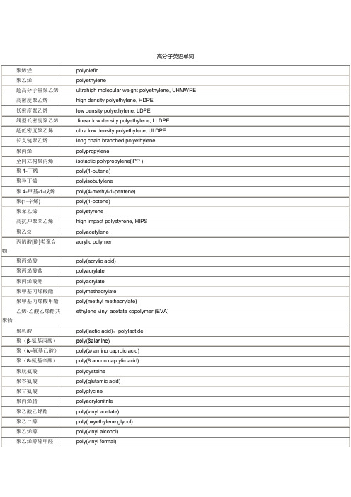
polyester fiber
聚丙烯腈纤维
acrylic fiber
聚丙烯纤维
polypropylene fiber
聚乙烯醇纤维
polyvinyl alcohol fiber
聚乙烯醇缩甲醛纤维
formalized PVA fiber
聚氯乙烯纤维
polyvinyl chloride fiber
乙烯-乙酸乙烯酯共聚物
ethylene vinyl acetate copolymer (EVA)
聚乳酸
poly(lactic acid),polylactide
聚(β-氨基丙酸)
poly(βalanine)
聚(ω-氨基己酸)
poly(ωamino caproic acid)
聚(8-氨基辛酸)
poly(8 amino caprylic acid)
聚偏二氟乙烯
poly(vinylidene fluoride)
聚三氟氯乙烯
poly(chlorotrifluoroethylene), PCTFE
聚四氟乙烯
poly(tetrafluoroethylene)
聚全氟丙烯
poly(perfluoropropene)
聚丁二烯
polybutadiene
1,4-聚丁二烯
聚乙烯醇缩甲醛
poly(vinyl formal)
聚乙烯醇缩丁醛
poly(vinyl butyral)
聚氯乙烯
poly(vinyl chloride)
聚1,2-二氯亚乙烯
poly(vinylene chloride)
聚偏二氯乙烯
poly(vinylidene chloride)
低密度聚乙烯/聚乳酸共混物性能的研究
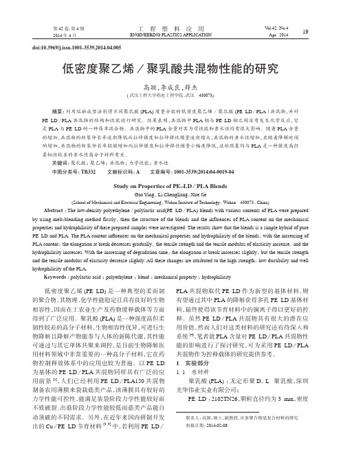
工 程 塑 料 应 用ENGINEERING PLASTICS APPLICATION第42卷,第4期2014年4月V ol.42,No.4Apr. 201419doi:10.3969/j.issn.1001-3539.2014.04.005低密度聚乙烯/聚乳酸共混物性能的研究高颖,李成良,薛杰(武汉工程大学机电工程学院,武汉 430073)摘要:利用熔融成型法制得不同聚乳酸(PLA)质量分数的低密度聚乙烯/聚乳酸(PE–LD /PLA )共混物,并对PE–LD /PLA 共混物的结构和性能进行研究。
结果表明,共混物中PLA 相与PE–LD 相之间没有发生化学反应,它是PLA 与PE–LD 的一种简单混合物。
共混物中的PLA 含量对其力学性能和亲水性均有很大影响。
随着PLA 含量的增加,共混物的断裂伸长率逐渐降低而拉伸强度和拉伸弹性模量逐渐增大,共混物的亲水性增加,且随着降解时间的增加,共混物的断裂伸长率轻微增加而拉伸强度和拉伸弹性模量小幅度降低,这些现象均与PLA 是一种强度高但柔韧性较差的亲水性高分子材料有关。
关键词:聚乳酸; 聚乙烯; 共混物; 力学性能; 亲水性中图分类号:TB332 文献标识码:A 文章编号:1001-3539(2014)04-0019-04Study on Properties of PE–LD /PLA BlendsGao Ying , Li Chengliang ,Xue Jie(School of Mechanical and Electrical Engineering , Wuhan Institute of Technology , Wuhan 430073, China)Abstract :The low-density polyethylene /polylactic acid(PE–LD /PLA) blends with various contents of PLA were prepared by using melt-blending method firstly , then the structure of the blends and the influences of PLA content on the mechanical properties and hydrophilicity of these prepared samples were investigated. The results show that the blends is a simple hybrid of pure PE–LD and PLA. The PLA content in fluences on the mechanical properties and hydrophilicity of the blends ,with the increasing of PLA content , the elongation at break decreases gradually , the tensile strength and the tensile modulus of elasticity increase ,and the hydrophilicity increases. With the increasing of degradation time ,the elongation at break increases slightly ,but the tensile strength and the tensile modulus of elasticity decrease slightly. All these changes are attributed to the high strength ,low ductibility and well hydrophilicity of the PLA.Keywords :polylactic acid ; polyethylene ; blend ; mechanical property ; hydrophilicity联系人:高颖,硕士,副教授,从事聚合物基复合材料的研究收稿日期:2014-02-06低密度聚乙烯(PE–LD)是一种典型的柔而韧的聚合物,其物理、化学性能稳定且具有良好的生物相容性,因而在工农业生产及药物缓释载体等方面得到了广泛应用。
聚乙醇酸_缝线_降解原理_概述说明以及解释
聚乙醇酸缝线降解原理概述说明以及解释1. 引言1.1 概述在当今社会,环境保护和可持续发展已经成为全球热议的话题。
传统的合成材料常常难以降解,对环境造成严重影响。
而聚乙醇酸作为一种生物可降解塑料,在解决这一问题上具有巨大潜力。
本文将详细介绍聚乙醇酸及其相关领域应用,并着重探讨了聚乙醇酸降解的原理和机制。
1.2 文章结构本文分为五个部分进行阐述:引言、聚乙醇酸、缝线、降解原理概述说明以及结论与展望。
引言部分旨在对文章整体进行概述,明确文章写作的目的和结构安排。
1.3 目的本文旨在全面介绍聚乙醇酸的定义、性质以及制备方法,并阐述缝线在各个领域中的作用、选择因素,以及缝线材料的特点和生产工艺。
同时,将着重探讨聚乙醇酸降解过程中涉及到的因素以及降解产物对环境的影响评估。
最后对整个文章进行总结,并提出相关问题和未来研究方向。
以上是关于“1. 引言”部分的详细内容说明,从概述、文章结构到目的进行了清晰的阐述,为接下来的撰写工作提供了指导和框架。
2. 聚乙醇酸:2.1 定义和性质:聚乙醇酸,全称聚乳酸乙二醇共聚物,是一种由乳酸和乙二醇单体经过聚合反应得到的高分子化合物。
它具有优异的生物降解性能和可塑性,是一种生态友好型的生物材料。
聚乙醇酸在自然环境中可以通过微生物的作用逐渐降解为无毒的水和二氧化碳。
2.2 应用领域:聚乙醇酸广泛应用于医疗、食品包装、农业、纺织品等领域。
在医疗领域,聚乙醇酸可以用于制备缝线、支架材料和组织工程等产品。
在食品包装方面,聚乙醇酸被用作可降解塑料袋、容器等替代传统塑料制品。
此外,在农业领域,聚乙醇酸也可以用于土壤增肥剂和植物保护剂的包装材料。
2.3 制备方法:聚乙醇酸的制备方法通常包括直接聚合法和环状引发聚合法两种。
直接聚合法是将乳酸和乙二醇按一定比例混合后,在适当的温度和催化剂作用下进行酯交换反应,生成聚乳酸乙二醇共聚物。
环状引发聚合法则是通过首先得到以乳酸为单体的环状化合物,然后在高温条件下进行开环聚合反应得到最终产物。
PLA简介

聚乳酸的简称(PLA)PLA是生物降解塑料聚乳酸的英文简写,全写为:polylactice acid聚乳酸也称为聚丙交酯(polylactide),属于聚酯家族。
聚乳酸是以乳酸为主要原料聚合得到的聚合物,原料来源充分而且可以再生,主要以玉米、木薯等为原料。
聚乳酸的生产过程无污染,而且产品可以生物降解,实现在自然界中的循环,因此是理想的绿色高分子材料。
聚乳酸的热稳定性好,加工温度170~230℃,有好的抗溶剂性,可用多种方式进行加工,如挤压、纺丝、双轴拉伸,注射吹塑。
由聚乳酸制成的产品除能生物降解外,生物相容性、光泽度、透明性、手感和耐热性好,还具有一定的耐菌性、阻燃性和抗紫外性,因此用途十分广泛,可用作包装材料、纤维和非织造物等,目前主要用于服装(内衣、外衣)、产业(建筑、农业、林业、造纸)和医疗卫生等领域。
PLA最大的制造商是美国NatureWorks公司,其次是中国的海正生物,他们目前的产量分别是7万吨和5千吨。
PLA有很多的应用,可以在挤出、注塑、拉膜、纺丝等多领域应用。
聚乳酸的制备1.1.合成方法总的来说,聚乳酸(PLA)的制备是以乳酸为原材料进行合成的。
目前合成方法有很多种,较为成熟的是乳酸直接缩聚法,另一种是先由乳酸合成丙交酯,再在催化剂的作用下开环聚合。
另外还有一种固相聚合法。
1)乳酸直接聚合法直接聚合法早在20世界30~40年代就已经开始研究,但是由于涉及反应中的水脱除等关键技术还不能得到很好的解决,所以其产物的分子量较低(均在4000以下),强度极低,易分解,没有实用性。
日本昭和高分子公司采用将乳酸在惰性气体中慢慢加热升温并缓慢减压,使乳酸直接脱水缩合,并使反应物在220~260℃,133Pa 下进一步缩聚,得到相对分子质量在4000以上的聚乳酸。
但是该方法反应时间长,产物在后期的高温下会老化分解,变色,且不均匀。
日本三井压化学公司采用溶液聚合法使乳酸直接聚合得到聚乳酸。
直接法的主要特点是合成的聚乳酸不含催化剂,因此缩聚反应进行到一定程度时体系会出现平衡态,需要升温加压打破反应平衡,反应条件相对苛刻。
plga纳米粒制备方法

plga纳米粒制备方法PLGA (poly(lactic-co-glycolic acid))纳米粒是一种常用于药物传递系统的纳米材料,具有优异的生物相容性和可降解性。
下面将介绍几种常用的PLGA纳米粒制备方法。
1.溶剂挥发法溶剂挥发法是制备PLGA纳米粒最常用的方法之一、首先,将PLGA和所需药物溶解在有机溶剂中,如乙酸乙酯或二氯甲烷。
然后,将有机溶液缓慢滴加到水相中,加入表面活性剂(如Tween 80或磺化聚丙烯酸钠)来稳定纳米粒。
在搅拌的同时,有机溶剂会逐渐挥发,形成PLGA纳米粒。
最后,通过离心或超滤法收集纳米粒。
2.乳化法乳化法是另一种常用的PLGA纳米粒制备方法。
首先,将PLGA和所需药物溶解在有机溶剂中,如二氯甲烷。
然后,将有机溶液滴加到水相中,并在高剪切下搅拌。
这会使PLGA形成微米级的乳液颗粒。
接下来,使用超声波或高压均质机来进一步减小颗粒大小,以获得纳米级的PLGA颗粒。
最后,通过离心或超滤法分离纳米粒。
3.沉淀法沉淀法是一种简单的PLGA纳米粒制备方法。
首先,将PLGA和所需药物在有机溶剂中溶解。
然后,将有机溶液缓慢滴加到抗溶剂(如水、乙醇或二甲亚砜)中。
抗溶剂的加入会使PLGA从有机相中析出,并形成纳米粒。
最后,通过离心或超滤法分离纳米粒。
4.反相乳液法反相乳液法是一种适用于疏水性药物的PLGA纳米粒制备方法。
首先,将药物溶解在有机溶剂中。
然后,将有机溶液滴加到水相中的表面活性剂溶液中,进行乳化。
接下来,将PLGA溶解在有机溶剂中,并缓慢加入到乳液中。
这会导致PLGA在乳液中析出,并形成纳米粒。
最后,通过离心或超滤法分离纳米粒。
这些制备方法在PLGA纳米粒制备中得到广泛应用,可以根据具体的药物性质和研究要求选择合适的方法进行制备。
乳酸羟基乙酸共聚物
乳酸羟基乙酸共聚物简介乳酸羟基乙酸共聚物(Polylactic-co-glycolic acid,简称PLGA)是一种生物可降解的高分子材料,由乳酸和羟基乙酸共聚而成。
PLGA具有良好的生物相容性、可降解性和可调控的药物释放特性,广泛应用于药物传递、组织工程、生物医学材料等领域。
特性生物相容性PLGA是一种天然来源的高分子材料,不含任何有毒或致癌成分,因此具有良好的生物相容性。
在体内降解过程中,PLGA会逐渐分解为乳酸和羟基乙酸,这两种物质在人体内均有代谢途径,不会对人体造成损害。
可降解性PLGA具有优异的可降解性能。
由于其化学结构中含有大量的酯键,PLGA可以在体内被水解为无毒且易于代谢的产物。
降解速度可以通过调整聚合度、组分比例等方式进行控制。
药物释放特性PLGA具有可调控的药物释放特性,可以根据药物的特性和需求设计不同的释放方式。
PLGA可以通过控制聚合度、组分比例、微球尺寸等参数来调节药物的释放速率和持续时间。
PLGA还可以通过添加表面活性剂或改变微球结构来实现定向或渐进式的药物释放。
应用领域药物传递PLGA在药物传递领域有广泛应用。
由于其良好的生物相容性和可降解性,以及可调控的药物释放特性,PLGA被广泛用于制备缓释微球、纳米粒子、纳米纤维等载体,用于输送各种类型的药物,包括小分子药物、蛋白质药物、核酸药物等。
这些载体可以通过靶向修饰提高药物的靶向性和生物利用度。
组织工程PLGA在组织工程领域也有重要应用。
由于其可降解性和生物相容性,PLGA可以作为支架材料用于组织修复和再生。
通过将干细胞或生长因子等生物活性物质与PLGA复合,可以促进组织的再生和修复过程。
PLGA还可以通过三维打印等技术制备复杂结构的支架,为组织工程提供更多可能性。
生物医学材料PLGA还被广泛应用于生物医学材料领域。
由于其可降解性和生物相容性,PLGA可以用于制备各种类型的植入物和修复材料,如骨修复材料、缝线、薄膜等。
聚乳酸纺丝分子量要求
聚乳酸纺丝分子量要求聚乳酸(Poly lactic acid,PLA)是一种由乳酸分子通过聚合反应得到的高分子材料。
由于其生物可降解性和可再生性,聚乳酸在医学、食品包装、纺织品等领域得到了广泛应用。
而在聚乳酸纺丝过程中,聚乳酸分子量的要求对纤维的性能和应用范围有着重要影响。
聚乳酸纺丝分子量的要求主要包括平均分子量、分子量分布以及链段长度等方面。
首先,平均分子量是指聚乳酸分子量的平均值,它决定了纤维的物理性能和加工性能。
一般来说,较高的平均分子量可以提高纤维的强度和熔点,但也会增加纺丝难度。
因此,在纺丝过程中需要根据具体的应用要求选择合适的平均分子量。
分子量分布是指聚乳酸分子量在一定范围内的分布情况。
较窄的分子量分布可以提高纤维的力学性能和热稳定性,但也会增加纺丝过程中的工艺难度。
因此,在聚乳酸纺丝中,需要根据具体的应用要求选择合适的分子量分布。
链段长度也是聚乳酸纺丝分子量要求的重要指标之一。
链段长度的长短直接影响到纤维的结晶性能和熔融流动性。
较长的链段长度可以增加纤维的结晶度和熔点,提高纤维的强度和热稳定性。
然而,较长的链段长度也会影响纺丝的流动性和加工性能。
因此,在聚乳酸纺丝中,需要根据具体的应用要求选择合适的链段长度。
为了满足聚乳酸纺丝分子量的要求,可以采取不同的方法进行调控。
一种常用的方法是通过控制反应条件和聚合体系中的催化剂浓度来调节聚乳酸的分子量。
此外,也可以通过采用不同的聚合工艺和纺丝条件来调节分子量分布和链段长度。
聚乳酸纺丝分子量的要求对纤维的性能和应用范围有着重要影响。
在选择聚乳酸纺丝材料时,需要根据具体的应用要求,选择合适的平均分子量、分子量分布和链段长度。
通过调控反应条件和聚合工艺,可以实现对聚乳酸纺丝分子量的精确控制,从而得到具有理想性能的纤维材料。
plga熔点
plga熔点
PLGA(Poly(L-lactide-co-glycolide)是由L-乳酸和乙酰化的DL-乙酸基甘酸(glycolic acid)的共聚物,是一种广泛用于生物医学领域的生物降解性材料。
PLGA兼具了乳酸和甘酸的优良特性,因此广泛应用于药物缓释、组织修复等领域。
PLGA的熔点是该材料的一个关键性质,它决定了材料的热稳定性以及在加工和制备过程中的操作难度。
PLGA的熔点是指在该材料升温后形成熔体的温度,通常指T_m,即热融点。
PLGA的熔点与材料的结构、共聚物比例以及分子量等多因素有关。
PLGA由于其材料性质的优良特性,在生物医学领域中得到广泛应用。
PLGA在制备缓释药物载体中具有很高的应用价值。
由于其降解速度可以根据需求进行调整,因此可以有效控制药物的释放速率,使药物在一定时间范围内逐渐释放,从而提高药效,并减少药物的毒副作用。
此外,由于PLGA具有良好的生物相容性和生物降解性,因此还被广泛应用于组织工程和修复中。
总的来说,PLGA的熔点对于PLGA在制备和加工过程以及在应用和使用时的性能和稳定性具有重要影响。
因此,研究PLGA的熔点和影响熔点的因素,是促进PLGA在生物医学领域中更加广泛应用的重要前提之一。
- 1、下载文档前请自行甄别文档内容的完整性,平台不提供额外的编辑、内容补充、找答案等附加服务。
- 2、"仅部分预览"的文档,不可在线预览部分如存在完整性等问题,可反馈申请退款(可完整预览的文档不适用该条件!)。
- 3、如文档侵犯您的权益,请联系客服反馈,我们会尽快为您处理(人工客服工作时间:9:00-18:30)。
Electrospun microfiber meshes of silicon-doped vaterite/poly(lactic acid)hybrid for guided bone regenerationAkiko Obata a,*,Toshiki Hotta a ,Takashi Wakita a,b ,Yoshio Ota c ,Toshihiro Kasuga aaGraduate School of Engineering,Nagoya Institute of Technology,Gokiso Cho,Showa ku,Nagoya 466-8555,Japan bYamahachi Dental Manufacturing Co.,54-1Ochigara,Nishiura Cho,Gamagori,Aichi 443-0105,Japan cYabashi Industries Co.Ltd.,188-1Akasaka Cho,Ogaki,Gifu 503-2213,Japana r t i c l e i n f o Article history:Received 29May 2009Received in revised form 23October 2009Accepted 9November 2009Available online 11November 2009Keywords:Polylactic acidCalcium carbonate Silicon Membrane Osteoblasta b s t r a c tSilicon-releasable microfiber meshes consisting of silicon-doped vaterite (SiV)particles andpoly(lactic acid)(PLA)hybrids were prepared by electrospinning.Due to their flexibility and porosity they formed ideal membranes or scaffolds for guided bone regeneration.In addition,a trace amount of silicon species has been reported to stimulate osteogenic cells to mineralize and enhance bone formation.We propose a new method of preparation of silicon-releasing microfiber meshes by electrospinning.Their structure and hydroxyapatite (HA)-forming abilities in simulated body fluid were examined.In addition,we studied their stimulatory effects on osteoblast-like cells in vitro and bone-forming ability in vivo,with a special emphasis on their ability to release silicon.The meshes consisted of a hybrid of carboxy groups in PLA and amino groups in siloxane,derived from aminopropyltriethoxysilane or calcium ions on the SiV surface.This hybrid exhibited an enhanced ability to form HA.The meshes coated with HA released 0.2–0.7mg l À1silicon species into the culture medium over 7days.Enhanced proliferation of osteo-blast-like cells was observed using the meshes and new bone formed on the meshes when implanted into the calvaria of rabbits.These meshes,therefore,provide an excellent substrate for bone regeneration and exhibit enhanced bone-forming ability under both in vitro and in vivo conditions.Ó2009Acta Materialia Inc.Published by Elsevier Ltd.All rights reserved.1.IntroductionBiodegradable polymer scaffolds have been developed for use in bone reconstruction,tissue engineering and drug delivery systems [1,2].In addition,three-dimensional scaffolds are common in bio-materials.Electrospinning is a process that can generate polymer fiber meshes with high porosity and flexibility,both essential com-ponents of tissue engineering [3–5].In general,the process of elec-trospinning is affected by both system parameters and process parameters.System parameters include polymer molecular weight (Mw)and polymer solution properties such as viscosity and con-ductivity.Process parameters,on the other hand,involve flow rate of the polymer solution,electric potential and the distance be-tween the capillary and collector,among others [3].It is possible to control the fiber diameter and the thickness of fiber mesh scaf-folds by optimizing these parameters.To use electrospun fiber meshes in bone tissue engineering applications,their various prop-erties,including material,fiber orientation,porosity and surface modification,must be considered.To optimize the mechanical and biomimetic properties of a fiber mesh it is necessary to choose the appropriate materials,since theorientation and diameter of the fibers in electrospun scaffolds can influence cellular compatibility [6–8].The fiber diameter in elec-trospun scaffolds has,for example,been reported to influence osteoblast proliferation and mineralization.Electrospun fiber meshes have been fabricated to contain DNA and proteins,which enhance cellular compatibility and bone formation upon their re-lease [8–11].The DNA and proteins are released by degradation of polymer matrices such as poly(lactic acid)(PLA),poly(lactide-co -glycolide)(PLGA)and poly(glycolide acid)(PGA).Despite their stimulatory effects on osteoblasts and bone mar-row-derived stem cells,electrospun fiber scaffolds consisting of polymer/ceramic composites have not been widely researched as biomaterials.Polymer and ceramic composites have been developed for bone reconstruction and showed good mechanical and bone-forming properties [12–14].We hypothesize that elec-trospun fiber scaffolds consisting of PLA/calcium carbonate com-showed excellent hydroxyapatite (HA)-forming ability,cellular compatibility and biocompatibility in in vitro and in vivo tests [17,18].1742-7061/$-see front matter Ó2009Acta Materialia Inc.Published by Elsevier Ltd.All rights reserved.doi:10.1016/j.actbio.2009.11.013*Corresponding author.Tel./fax:+81527357250.E-mail address:obata.akiko@nitech.ac.jp (A.Obata).Acta Biomaterialia 6(2010)1248–1257Contents lists available at ScienceDirectActa Biomaterialiajournal homepage:/locate/ac tabiomatGuided bone regeneration(GBR)is a useful method of bone reconstruction in vivo.GBR membranes requireflexibility to adapt to the shape of the bone defect and strength to maintain the space for bone formation and to connect with the soft tissues.An electro-spunfiber mesh is a good candidate for a GBR membrane due to theflexibility inherent in the electrospinning process.Release of calcium and silicon species from an electrospunfiber mesh is an additional attribute which promotes bone regeneration.Calcium species and a trace amount of silicon species have been reported to enhance bone formation by osteoblasts[19–23].Previously we developed silicon-doped vaterite(SiV)powders and their PLA com-posites and tested their cellular compatibility using mouse osteo-blast-like cells[24].Afilm of the composites was prepared by dip coating the composite solution on a coverglass and drying at room temperature.Thefilm showed release of calcium and silicon into the culture medium through degradation of the PLA and SiV matrix.Cell proliferation and mineralization on the compositefilm was enhanced compared with an undoped vaterite and PLA com-positefilm,which showed no silicon release.Our preliminary experiments demonstrated that aflexiblefiber mesh consisting of PLA composites with a large amount of SiV could be successfully prepared by the electrospinning method, while afiber mesh consisting of a PLA composite with vaterite(un-doped type)was brittle.We propose to further characterize the SiV and PLA hybrid(Si–PVH)and compare its chemical structure with the undoped vaterite and PLA hybrid(PVH).To enhance cellular adhesion the compositefiber meshes were coated with HA using simulated bodyfluid(SBF).The cellular and tissue compatibilities of both preparedfiber meshes were evaluated by in vitro tests using mouse osteoblast-like cells(MC3T3-E1cells)and in vivo tests using rabbit calvaria.2.Materials and methods2.1.Preparation of SiV and vaterite powdersSilicon-doped vaterite(SiV)powders were prepared by a car-bonation process with methanol.A sample of150g Ca(OH)2was mixed with60ml aminopropyltriethoxysilane(APTES)and 2000ml methanol with the addition of CO2gas for75min at a rate of2000ml minÀ1.The resulting slurry was dried at110°C,result-ing in the SiV powder.The content of silicon in SiV was estimated to be approximately3wt.%by X-rayfluorescence analysis (RIX3000,Rigaku,Japan)(50kV,60mA).A calibration curve(X-ray intensity vs.SiO2content)was established by measuring lime-stone containing various amounts of SiO2.Vaterite powders were also prepared by the carbonation process without APTES.SiV and vaterite powders were characterized by X-ray diffractometry (XRD)(RAD-B,Rigaku,Japan)(CuK a,40kV,20mA).The specific surface areas of SiV and vaterite were measured according to a BET theory using nitrogen gas adsorption(NOVA3000,Quanta-chrome Corp.,USA).The powders were deaerated at150°C for 1h before measurement.The structure of silicon in SiV was exam-ined by29Si magic angle spinning nuclear magnetic resonance(29Si MAS-NMR)(UNITY plus,Varian,USA),operating at pulses of6.3l s and at recycle delays of15s.The sample spinning speed was 3.0kHz.Chemical shifts were measured relative to polydime-thylsiloxane.2.2.Preparation of microfiber meshesThe Si–PVH and PVH microfiber meshes were prepared by an electrospinning method.SiV or vaterite powder was mixed with PLA(PurasorbÒ;molecular weight260kDa,PURAC,The Nether-lands)at200°C for10min with a kneader,resulting in the formation of PLA composites containing60wt.%of the powder. The composites were dissolved in chloroform at10wt.%to prepare the solution for electrospinning.In our preliminary experiments, this ratio was found to be optimal for preparing microfiber meshes of approximately10l m diameter.The PLA microfiber mesh was also prepared by an electrospinning method.The PLA was dis-solved in chloroform at9wt.%to prepare the solution for electros-pinning.The prepared solutions were loaded into a syringe pump (FP-W-100,Melquest,Japan)with a syringe needle(22gauge for Si–PVH and PVH,19gauge for PLA),which was set at 0.05ml minÀ1.The distance between the needle and an aluminum collector was150mm.Electrospinning was carried out at20kV of impressed voltage and$100l A of applied current(Power Supply HARb-40P0.75,Matsusada Precision Inc.,Japan)at room tempera-ture and approximately55%relative humidity.The SiV powders and resultant electrospunfibers were coated with amorphous osmium using plasma chemical vapor deposition (CVD)equipment and then morphologically observed byfield emission scanning electron microscopy(SEM)(JSM-6301F,JEOL, Japan)(accelerating voltage5keV).Structural analyses of Si–PVH were performed using13C cross-polarization magic angle spinning nuclear magnetic resonance(13C CP/MAS-NMR)(UNITY plus,Var-ian,USA)and Fourier transform infrared(FTIR)spectroscopy(FT-IR-460Plus,JASCO,Japan).13C CP/MAS-NMR spectra were taken with4.7l s and8.0s recycle delays.The sample spinning speed was 3.0kHz.Chemical shifts were measured relative to adamantane.The molecular weights of PLA matrix in the prepared meshes were measured by gel permeation chromatography(GPC)(Shima-dzu,Japan)using two columns(KF-604,Shodex,Japan).The meshes were dissolved in chloroform at a concentration of10mg PLA per5ml chloroform and the solutionsfiltered.The measure-ments were carried out at40°C at aflow rate of0.6ml minÀ1.Poly-styrene standards(SM-105,Shodex,Japan)were used to establish a calibration curve.2.3.Coating of thefibers with HAThe prepared microfiber mesh was immersed in10ml of an aqueous solution containing ion concentrations1.5times those of SBF(1.5SBF)at37°C for24h.The1.5SBF solution consisting of 3.75mM Ca2+,213.0mM Na+, 2.25mM Mg2+,7.5mM K+, 222.45mM ClÀ,6.3mM HCO3À,1.5mM HPO42À,0.75mM SO42À, 50mM(CH2OH)3CNH2and45.0mM HCl was prepared using re-agent grade NaCl,NaHCO3,KCl,K2PO4Á3H2O,MgCl2Á6H2O,HCl, CaCl2,NaSO4and NH2C(CH2OH)3.After immersion in1.5SBF solu-tion the mesh was washed with distilled water and then dried at room temperature.The dried samples were coated with amor-phous osmium using plasma CVD equipment and observed by SEM.2.4.Measurement of silicon release from a Si–PVH microfiber mesh before and after HA coatingThe amount of silicon released from the Si–PVH microfiber meshes before and after HA coating was evaluated by immersing them in4ml of a culture medium(alpha minimum essential med-ium,a-MEM)containing10%fetal bovine serum(FBS)and incu-bated at37°C in a humidified atmosphere of95%air,5%CO2for 5days.The sample coated with HA is denoted Si–PVH/HA mesh. The size of the sample used was10Â10mm.The culture medium was changed after either1or3days soaking,according to the pro-cess of the cell culture test.The sample was taken out of the culture medium and the mediumfiltered.The level of silicon species in the resulting medium was measured by inductively coupled plasma atomic emission spectroscopy(ICP-AES)(ICPS-500,Shimadzu,Ja-A.Obata et al./Acta Biomaterialia6(2010)1248–12571249pan).A calibration curve was established using an aqueous solu-tion of Si4+ions at concentrations of1,10and20ppm.2.5.Cell culture testsThe microfiber mesh used for the cell culture tests was steril-ized using ethylene oxide gas.Mouse osteoblast-like cells (MC3T3-E1cells)were seeded onto the prepared mesh(Si–PVH/ HA or PLA mesh)in24-well plates at a density of30,000 cells wellÀ1.A TermanoxÒplastic plate,which has often been used as a control sample,was used as a control sample and a-MEM con-taining10%FBS was used as the culture medium.The medium was changed after1day of culture and then changed every other day. The number of MC3T3-E1cells was evaluated after treatment with Cell Counting Kit-8(Dojindo,Japan).One reagent in the kit is a water soluble tetrazolium salt which is reduced by dehydrogen-ases in live cells to generate water soluble formazan.Water soluble formazan has an absorption maximum at about460nm.The num-bers of live cells in the samples were counted by measuring the absorbance of the resulting medium at450nm.Student’s t-test was used for the cell counts.Differences between the samples were determined by Student’s t-test,with p<0.05considered to be sta-tistically significant.The cells cultured on the samples for3days werefixed in2.5%glutaraldehyde for40min at4°C,dehydrated through a series of increasing concentrations of ethanol and,final-ly,dried with hexamethyldisilazane.The dried samples were coated with amorphous osmium using plasma CVD equipment and observed by SEM.2.6.Preparation of a bilayered microfiber mesh consisting of Si–PVH/ HA and PLA meshesA microfiber mesh having large spaces is not an appropriate candidate for a GBR membrane because the mesh may allow fibrous cells and soft tissues to permeate through them[25].To address this issue we prepared a bilayered microfiber mesh using Si–PVH/HA mesh and the dense PLA mesh,which was not expected to allow any cell permeation.In addition,the strength of the Si–PVH/HA mesh was improved by combination with the dense PLA mesh.Details of the preparation method have been reported by Wakita et al.[26].It was found that the combination induced no fracture or fragmentation of Si–PVH/HA and PLA.The PLA mesh was pressed at15MPa for1min to increase its density.Adherence between the Si–PVH mesh and the dense PLA mesh was accomplished by pressing at15MPa using a stainless mesh(No.40)at150°C and soaking in1.5SBF for1day.The result was a bilayered mesh consisting of Si–PVH/HA mesh and the dense PLA mesh(Fig.1A).2.7.Evaluation of in vivo bone-forming abilityThe in vivo study was carried out at the laboratory of Hamri Co. Ltd.(Japan).Fifteen male New Zealand rabbits(Kbl:NZW), 14weeks of age and between2.50and3.20kg in weight,were pur-chased from Kitayama Labes Co.Ltd.(Japan).The animals were housed in vivaria at23±3°C,50±20%humidity and a12h on/ off light cycle and ventilation was cycled approximately12timesFig.1.Schematic images of(A)preparation of the bilayered mesh and(B)protocol of the in vivo test.Adherence between the Si–PVH mesh and the dense PLA mesh was accomplished by pressing with a stainless steel mesh at150°C and soaking in1.5SBF for1day.The result was a bilayered mesh consisting of Si–PVH/HA mesh and the dense PLA mesh.An8mm diameter hole was drilled into the front midline of the animal’s calvaria and then covered with the prepared bilayered mesh.1250 A.Obata et al./Acta Biomaterialia6(2010)1248–1257per hour.The animals were maintained according to the National Institutes of Health(NIH)guide for care and use of laboratory animals.After allowing the animals to adapt to the new environment for 12days they were stratified into three groups offive animals each. After anesthetizing the animals an8mm diameter hole was drilled into the front midline of the calvaria using a bone cutter(BL-302A, Osada Electric Co.Ltd.,Japan)and then covered with the prepared bilayered meshes(Fig.1B).The bilayered mesh wasfixed by being placed between the skin and bone.The meshes were implanted with the Si–PVH/HA mesh in contact with the bone and the dense PLA mesh in contact with the skin.The implanted meshes were harvested with the surrounding tissues for histological examina-tion after4,8and12weeks.The samples were dehydrated, embedded in methyl methacrylate and then sliced,resulting in slides of approximately20l m thickness.The slides were stained with Villanueva-Goldner and observed by optical microscopy (CH-2,Olympus,Japan).Villanueva-Goldner stains mineralized tis-sues green.3.Results3.1.Materials characterizationFig.2shows SEM micrographs of the SiV and vaterite particles. The SiV and vaterite particles had diameters of$1l m and $500nm,respectively.These two types of particles consisted of aggregates of nano-sized primary particles.The sizes of the SiV particles were larger than those of the vaterite particles.The re-sults of the nitrogen gas adsorption method revealed that the spe-cific surfaces of SiV and vaterite were67.2and30.7m2gÀ1, respectively.These results suggest that the conditions for aggrega-tion of the primary particles in the suspension were influenced by the addition of APTES.Fig.3shows the XRD patterns of SiV and vaterite.A weak peak corresponding to calcite appeared at approximately29°in the pat-tern of the vaterite sample.A trace amount of calcite was found with a predominant vaterite phase,because the calcite phase is more stable than the other polymorphs of calcium carbonate,vate-rite and aragonite.In the present work the sample is denoted ‘‘vaterite”.The peaks of the SiV pattern are broader than those of the vaterite pattern.SiV was found to consist predominantly of vaterite.The structure of APTES in SiV was further characterized by29Si MAS-NMR spectroscopy.Fig.4shows the29Si MAS-NMR spectrum of SiV.A peak atÀ50toÀ80ppm can be seen.The peak may be superimposed on those corresponding to T2[NH2(CH2)3Si(OSi)2 (OH,OC2H5)]and T3[NH2(CH2)3Si(OSi)3][27].This indicates that no APTES in SiV existed as the monomer and that siloxane was formed by condensation of APTES during the preparation of SiV.Meshflexibility was felt to be a reflection of its structural prop-erties,such as the molecular weight of the PLA matrix and struc-tural changes in the interface between the matrix and the vaterite or SiV powders.Table1shows the weight average molec-ular weight(Mw),number average molecular weight(Mn)and polydispersity(Mw/Mn)of PLA and the PLA matrix in PVH and Si–PVH.Although the molecular weight of PLA decreased slightly after kneading with the SiV powders,it decreased significantly after kneading with vaterite powder.The prepared Si–PVH mesh showedflexibility without breaking,whereas the PVH mesh was brittle and was easily broken by hand.The ratios Mw/Mn for PVH and Si–PVH were higher than that for PLA.This indicates that the molecular weights of these compos-ites were disperate.Fig.5shows various peaks corresponding to the carboxy groups in the13C CP/MAS-NMR spectra of the prepared composites.The band of the carboxy peak shifted slightly and a new peak appeared in the low magneticfield area of the band, where carboxy groups form coordinate bonds with bivalent ions [28].The peaks further separated into two peaks,peak A andB. Fig.2.SEM photographs of(A)vaterite and(B)SiV.The SiV and vaterite particles have diameters of$1l m and$500nm,respectively.The SiV particles are larger than the vaterite particles.This suggests that the conditions for aggregation of the primary particles in the suspension were influenced by addingAPTES.Fig.3.XRD patterns of(A)vaterite and(B)SiV.A weak peak corresponding to calcite appears at approximately29°in the pattern of the vaterite sample,but the vaterite sample and SiV were found to consist predominantly of vaterite phase.The peaks for SiV were broader than those of vaterite.A.Obata et al./Acta Biomaterialia6(2010)1248–12571251Peak A was defined at one magnetic field in the band and peak B as one magnetic field lower in the band.The intensity of peak B in the spectrum of PVH was larger than in the spectrum of Si–PVH.This indicates that more carboxy groups formed coordinate bonds with calcium ions in PVH than Si–PVH and that the formation of coordinate bonds caused the decrease in molecu-lar weight.Fig.6shows the FTIR spectra of Si–PVH and PVH.In the spectrum of Si–PVH a new weak peak appears at around 1650cm À1,as well as the peaks corresponding to PLA and CO 32À.This peak appears to originate from the amide bond,which indi-cates that carboxy groups on PLA form chemical bonds with amino groups on APTES.Fig.7A demonstrates the greater flexibility of the Si–PVH mesh,which could be folded without fracture or fragmentation using a pair of tweezers.Microfibers approximately 10l m in diameter were observed on the Si–PVH mesh surface and were found to twine around one another.The size of the gaps between the microfibers varied between 10l m and several hundred microme-ters,as shown in Fig.7B.The SiV particles observed on the micro-fiber surface were coated with a thin film of the PLA matrix,as shown in Fig.7C.The fracture face of the microfibers showed that the particles were closely packed in the microfibers (Fig.7D).The particles were embedded in the PLA matrix and after immersion in 1.5SBF solution for 1day the surfaces of the microf-ibers were coated with leaf-shaped deposits,as shown in Fig.7E.No significant change was observed in the gap sizes between the microfibers before and after immersion (Fig.7F).After HA coating further testing demonstrated several cracks,but the coating did not peel off.The deposits were confirmed by XRD to be HA of approximately 1l m thickness (Fig.7G).On the other hand,the PVH mesh consisted of short microfibers approximately 7l m in diameter (Fig.7H).3.2.Ease of silicon releaseFig.8shows the dynamics of the release of silicon ions from both the Si–PVH mesh and Si–PVH/HA mesh into the culture med-ium over 5days.Closed bars indicate the ion amount in the culture medium and open bars indicates the total amount of ions released up to each measurement point.After 1day of immersion a concen-tration of 8.2mg l À1ionic silicon species had been released from Si–PVH,while this amount was reduced to 1.2mg l À1on day 3and 0.59mg l À1on day 5.The Si–PVH mesh was then soaked in cul-ture medium after being incubated in 1.5SBF for 1day and coated with HA.The amount of the silicon species released from it was significantly reduced,with only 0.68mg l À1released from the mesh after 1day of soaking in culture medium.The amount re-leased was even smaller on days 3and 5of soaking,at 0.39mg l À1on day 3and 0.41mg l À1on day 5.3.3.Proliferation of MC3T3-E1cells on the prepared meshesThe cellular compatibility of the Si–PVH/HA mesh compared with the PLA mesh was evaluated in culture using MC3T3-E1cells.Fig.9shows the numbers of cells after culture on Si–PVH/HA mesh,PLA mesh and Thermanox Òfor 2weeks.The number of cells after culture on the Si–PVH/HA mesh was higher than that on thePLAFig. 4.29Si MAS-NMR spectrum of SiV.The peak at À50to À80ppm may be superimposed on those corresponding to T 2[NH 2(CH 2)3Si(OSi)2(OH,OC 2H 5)]and T 3[NH 2(CH 2)3Si(OSi)3].This indicates that no APTES in SiV exists as the monomer and siloxane was formed.Table 1Weight average molecular weight (Mw),number average molecular weight (Mn)and polydispersity (Mw/Mn)of PLA,PVH and Si–PVH.Mw (kDa)Mn (kDa)Mw/Mn PLA 259168 1.5PVH 177 2.4Si–PVH185862.1Fig.5.13C CP/MAS-NMR spectra of (A)PVH and (B)Si–PVH.The peaks further separated into two peaks,A and B.The intensity of peak B in the spectrum of PVH is larger than that of Si–PVH.This indicates that more carboxy groups formed coordinate bonds with calcium ions in PVH than Si–PVH.1252 A.Obata et al./Acta Biomaterialia 6(2010)1248–1257mesh at all time points.The PVH mesh could not be used as a con-trol mesh due to its extreme brittleness.Fig.10shows SEM micrograph of cells cultured on the Si–PVH/HA mesh for 3days.The cells elongated on the fiber surfaces and extended over several fibers.Some of them entered the fiber mesh and extended within it.3.4.Bone formation in the prepared meshesAn 8mm diameter hole in bone is generally not regenerated if nothing is done to stimulate it.In our preliminary experiment a PLGA mesh was implanted in the same position as that in this study for 12weeks and no new bone was formed in/on the mesh at the center of the hole (data not shown).Bone growth into the defect from the margin was observed in all of the samples.There-fore,we tested the effects of the bilayered meshes on bone forma-tion by observing new bone formation at the center of the 8mm holes.This animal test was a fundamental study.Although this ani-mal test was not a load-bearing model,we believe that it was use-ful in evaluating the bone-forming ability of the bilayered meshes.Fig.11shows the in vivo response to the bilayered meshes and new bone formation at the center of the holes.Villanueva-Goldner stain showed that new bone formed in the mesh 4weeks after implantation.Bone began to form around the pressed points formed during layering of the two types of mesh,the Si–PVH/HA mesh and the dense PLA mesh.The bone area expanded in the Si–PVH/HA mesh after 8weeks.After 12weeks new bone forma-tion was observed over almost the entire area of implanted Si–PVH/HA mesh.There was no tissue inflammation observed histologically.4.DiscussionEarlier work showed that composite films which release silicon can stimulate MC3T3-E1cells to proliferate and mineralize [17,24].These composite films were prepared by dip coating a solution of SiV and PLA composites on a coverglass,drying at room tempera-ture and pealing the dried composite from the glass.The prepared composite film had no pores to allow cells to enter,therefore,cells proliferated only on the film surface.When applying this technique to a GBR membrane the composites should have porosity in order to induce adhesion and proliferation of cells and to provide nutri-tion to the cells.To address this issue we developed a fiber mesh using PLA and SiV formed by electrospinning.We anticipated that the electrospun fiber mesh would contain a large volume of pores and would show flexibility,both essential components of a GBR membrane.The Si–PVH mesh showed greater flexibility and a higher weight averaged molecular weight PLA than the PVH mesh,in the case of SiV due to siloxane on the vaterite surface,which resulted in the formation of a flexible mesh containing a large amount of SiV powder (60wt.%).There has been increasing interest in the use of electrospun fi-brous meshes as scaffolds in medicine.In particular,biocompatible polymers such as PLGA and poly(caprolactone)have been applied as materials for meshes,incorporating various growth factors or DNA [3].On the other hand,very few studies have been performed on fibrous meshes consisting of polymer/ceramic composites.Pre-viously,PLGA and HA composite scaffolds fabricated by electros-pinning contained only 5–10wt.%HA nanocrystals [29].We believe that a larger amount of calcium is needed to enhance the bone-forming ability of electrospun fibrous meshes and vaterite powders are effective materials for providing calcium.In the pres-ent work a fibrous mesh containing a large amount of vaterite (up to 60wt.%)was successfully prepared by kneading and subsequent electrospinning.Vaterite or SiV particles were embedded in a PLA matrix,with very thin PLA layers on the particle surfaces,as shown in Fig.7C and D.Calcium ions are supposed to be released through degradation of the thin PLA layer.The PVH mesh,however,was brittle,without flexibility,due to a drastic decrease in the PLA molecular weight after kneading with vaterite powders,as shown in Table 1.Maeda et al.reported that in vaterite and PLA composites cal-cium ions on vaterite particles formed coordinate bonds with car-boxy groups in the PLA structure [30].The polymer chains in the Si–PVH mesh,however,were not broken by the formation of coor-dinate bonds between calcium ions and carboxy groups (Fig.5)due to siloxane derived from APTES on the vaterite surface,which pre-vents the formation of coordinate bonds.This resulted in the for-mation of a flexible mesh containing a large amount of SiV powder (60wt.%).The specific surface areas of SiV and vaterite were estimated to be 67.2and 30.7m 2g À1,respectively,by nitrogen gas adsorption.The peaks corresponding to vaterite in the XRD pattern of SiV were broader than that of vaterite (Fig.3).This result indicates that the diameter of the primary particles of SiV is smaller than that of vaterite.Crystal growth of the primary particles of vateriteap-Fig.6.FTIR spectra of (A)PVH and (B)Si–PVH.A weak peak appears at around 1650cm À1in the spectrum of Si–PVH.This peak appears to originate from the amide bond between carboxy groups in PLA and amino groups in APTES.A.Obata et al./Acta Biomaterialia 6(2010)1248–12571253。