Dylight 549, Rabbit Anti-Goat IgG
GoatAnti-RabbitFcspecificIgG产品使用说明书
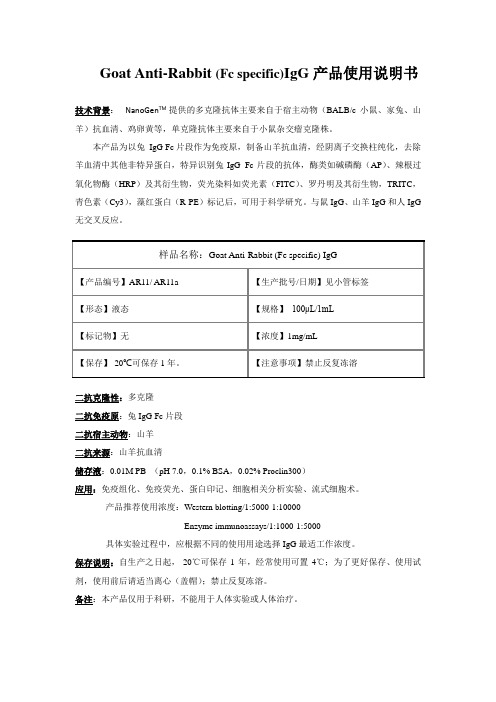
Goat Anti-Rabbit (Fc specific)IgG产品使用说明书
技术背景: NanoGen TM提供的多克隆抗体主要来自于宿主动物(BALB/c小鼠、家兔、山羊)抗血清、鸡卵黄等,单克隆抗体主要来自于小鼠杂交瘤克隆株。
本产品为以兔IgG Fc片段作为免疫原,制备山羊抗血清,经阴离子交换柱纯化,去除羊血清中其他非特异蛋白,特异识别兔IgG Fc片段的抗体,酶类如碱磷酶(AP)、辣根过氧化物酶(HRP)及其衍生物,荧光染料如荧光素(FITC)、罗丹明及其衍生物,TRITC,青色素(Cy3),藻红蛋白(R-PE)标记后,可用于科学研究。
与鼠IgG、山羊IgG和人IgG 无交叉反应。
二抗克隆性:多克隆
二抗免疫原:兔IgG Fc片段
二抗宿主动物:山羊
应用:免疫组化、免疫荧光、蛋白印记、细胞相关分析实验、流式细胞术。
产品推荐使用浓度:Western blotting/1:5000-1:10000
Enzyme immunoassays/1:1000-1:5000
具体实验过程中,应根据不同的使用用途选择IgG最适工作浓度。
保存说明:自生产之日起,-20℃可保存1年,经常使用可置4℃;为了更好保存、使用试剂,使用前后请适当离心(盖帽);禁止反复冻溶。
备注:本产品仅用于科研,不能用于人体实验或人体治疗。
DyLight594标记山羊抗兔IgG二抗

Abbkine, Inc • Redlands, CA • USA •Not for further distribution without written consent. Copyright © Abbkine, Inc. Dylight 594, Goat Anti-Rabbit IgG# A23420Product InformationProduct Name: Dylight 594 Conjugated Goat Anti-Rabbit IgG(H+L)Amax: 591nm Emax: 616nmCatalog Number: A23420 Lot Number: Refer to vial Formulation: Liquid Size:100ul/500ulStorage: Store at -20°C. Be protected from light and avoid repeated freeze / thaw cycles.Note: Contain sodium azide.Background: DyLight fluorescent dyes are a new generation of dyes with improved brightness and photostability, which are highly water soluble and remain fluorescent from pH 4 to pH 9. They are better than or comparable to the best fluorescent dyes from other companies. Abbkine's secondary antibodies are available conjugated to enzyme, biotin or fluorophore for use in a variety of antibody-based applications including WB, IHC, IF, FC and ELISA. We offer high quality secondary antibodies from goat, rabbit and donkey sources for your each application. Serum adsorbed secondary antibodies are also available and are recommended for use with immunoglobulin-rich samples.Purity: Affinity purified using solid phase Rabbit IgG (H&L) with finally > 95% purity based on SDS-PAGE.Specificity: The antibody reacts with whole molecule rabbit IgG. It also reacts with heavy chains of rabbit IgG, and light chains of all other rabbit immunoglobulins. It has no reactivity on non-immunoglobulin serum proteins, while it may cross-react with immunoglobulins from other species.Application Notes: Optimal working dilutions should be determined experimentally by the investigator. Suggested starting 1:50-1:1,000 dilutions for most applications.Storage Buffer: 0.01M Sodium Phosphate, 0.25M NaCl, 50% Glycerol pH 7.4.Stabilizer: 1% Bovine Serum Albumin (IgG and Protease Free).Preservative: 0.05% (w/v) Sodium Azide.Storage Instructions: Stable for one year at -20°C from date of shipment. For maximum recovery of product, centrifuge the original vial after thawing and prior to removing the cap. Aliquot to avoid repeated freezing and thawing.Note: The product listed herein is for research use only and is not intended for use in human or clinical diagnosis. Suggested applications of our products are not recommendations to use our products in violation of any patent or as a license. We cannot be responsible for patent infringements or other violations that may occur with the use of this product.。
Immunoway免疫组织化学试剂 Polymer HRP Goat Anti Rabbit I

Polymer HRP * Goat Anti Rabbit IgG(H+L)【产品货号】RS0010【产品名称】1、通用名称:Polymer HRP羊抗兔二抗即用型(免疫组织化学)2、英文名称:Polymer HRP * Goat Anti Rabbit IgG(H+L)【包装规格】类别规格工作液10mL/瓶、100mL/瓶【预期用途】免疫组织化学染色,配合Immunoway常规兔来源的浓缩型或即用型一抗使用。
【作用原理】数个过氧化物酶分子和数个二抗分子结合在同一多聚体上构成了一种简单而灵敏的显色系统【主要组成成份】磷酸盐缓冲液0.01mol/LGoat anti Rabbit HRP 大于0.05mg/L【储存条件及有效期】1、储存要求:2℃~8℃密封保存。
2、有效期:一年。
【样本要求】新鲜活检或外科样本组织,甲醛固定,固定时间8-24小时要求取材、脱水、石蜡包埋并制成蜡块。
配合Immunoway常规浓缩型或即用型一抗使用。
【检验方法】1、人体组织样本先经甲醛固定。
2、固定后的组织经过脱水等一系列操作制备成蜡块。
3、用切片机把组织蜡块切成切片。
4、手工操作免疫组化实验。
常规推荐步骤如下:4.1. 将需要进行实验的组织切片取出,插入玻片架上后放入60-65℃的烤箱中,烤片1-1.5h。
4.2. 将切片置于以下染色缸中梯度反应:二甲苯10分钟×2次二甲苯5分钟无水乙醇5分钟×2次95%乙醇5分钟85%乙醇5分钟PBS清洗3分钟×3次4.3. 抗原修复(方法见一抗供应商,或采用常规高压、微波、酶修复等)4.4.根据组织大小直接滴加内源性过氧化物酶阻断剂到组织上,通常为100uL, 直至完全覆盖组织。
室温10分钟,PBS清洗3分钟×3次。
此步骤通常在水浴法、酶法等温和抗原修复后加入。
或者组织本身含有较多内源性过氧化物酶中加入。
4.5. 根据组织大小滴加一抗到组织上,通常为100uL,直至完全覆盖组织。
_C57胚胎小鼠神经干细胞的分离、培养与鉴定

至细胞大部分贴壁。 弃去细胞培养液, 用 PBS 洗 3 遍。加 4% 多聚甲醛固定 30 min, 加 PBS 冲洗 5 min × 3; 0. 2% Tritox100 室 温 破 膜 10 min, PBS 洗 涤 5 min × 3; 5% 的山羊血清室温封闭 30 min, PBS 洗涤 5 min × 3; 一 抗 nestin ( 1 ∶ 50 ) 4 ℃ 孵 育 过 夜、 二抗 DyLight 594 AffiniPure Goat AntiRabbit IgG ( H + L ) ( 1 ∶ 100 ) 室 温 避 光 孵 育 l h, Hoechst33258 ( 1 μg / mL) 室温避光核复染 15 min, PBS 冲洗 2 遍, 封片, 显微镜观察。
2
2.1
结果
C57 胚胎小鼠 NSCs 的分离、 培养与纯化
分离 得 到 的 C57 胚 胎 小 鼠 的 NSCs 光 镜 下 可 见, 原代细胞接种后, 部分细胞逐渐沉降贴附于 6 孔 板表面, 胞体较小, 有光晕, 悬浮部分可以看到很亮 的圆形细胞组成的细胞团。第 2 天以后可看到细胞 大部分贴壁, 悬浮细胞较亮且较圆, 很多细胞团块成 。 辐射状贴壁生长 悬浮部分可以看到一些由十几个 细胞组成的桑葚状神经小球悬浮生长 , 如图 1A。 接 种第 3 天之后, 神经小球直径逐渐增大, 贴壁的细胞 团块颜色逐渐变暗。原代培养中可见神经球周围无 明显刺突, 存在明显细胞聚集, 如图 1B 。
NSCs ) 具有分化为 神经干细胞 ( neuralstemcell, 神经元、 星形胶质细胞和少突胶质细胞的能力 , 能自 。 我更新, 并足以提供大量脑组织细胞的细胞群 , , NSCs 现已发现 多种类型的中枢神经系统损伤后 被激活并有利于组织的损伤修复和功能恢复 。 因
异种动物ig免疫白兔实验设计
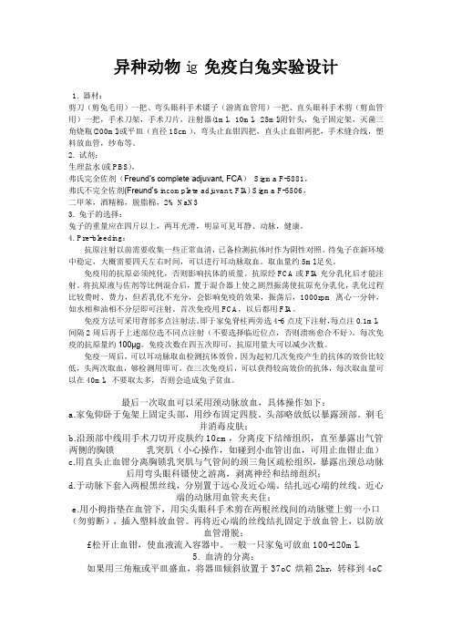
异种动物ig免疫白兔实验设计1. 器材:剪刀(剪兔毛用)一把、弯头眼科手术镊子(游离血管用)一把、直头眼科手术剪(剪血管用)一把,手术刀架,手术刀片,注射器(1ml、10ml,25ml)附针头,兔子固定架,灭菌三角烧瓶(200ml)或平皿(直径18cm)、弯头止血钳四把,直头止血钳两把,手术缝合线,塑料放血管,纱布等。
2. 试剂:生理盐水(或PBS),弗氏完全佐剂(Freund’s complete adjuvant, FCA)Sigma F-5881,弗氏不完全佐剂(Freund’s incomplete adjuvant, FIA) Sigma F-5506,二甲苯,酒精棉,脱脂棉,2% NaN33. 兔子的选择:兔子的重量应在四斤以上,两耳光滑,明显可见耳静、动脉,健康。
4. Pre-bleeding:抗原注射以前需要收集一些正常血清,已备检测抗体时作为阴性对照。
待兔子在新环境中稳定,大概需要四天左右时间,可以进行耳动脉取血。
取血量约5ml足矣。
免疫用的抗原必须纯化,否则影响抗体的质量。
抗原经FCA或FIA充分乳化后才能注射。
将抗原液与佐剂等比例混合后,置于混合器上使之剧烈振荡使抗原充分乳化,乳化过程比较费时、费力,但若乳化不充分,会影响免疫的效果,振荡后,1000rpm离心一分钟,如水相和油相不分层即可注射。
首次免疫用FCA,以后都用FIA。
免疫方法可采用背部多点注射法。
即于家兔脊柱两旁选4-6点皮下注射,每点注0.1ml,间隔2周后再于上述部位选不同点注射(不要选择临近位点,否则溃疡愈合不好)。
每次免疫的抗原量约100μg。
免疫次数在四五次即可,抗原用量大可以减少次数。
免疫一周后,可以耳动脉取血检测抗体效价。
因为起初几次免疫产生的抗体的效价比较低,头两次取血,够检测用即可。
在三次免疫后,可以获得较高效价的抗体,每次取血量可以在40ml,不要取太多,否则会造成兔子贫血。
最后一次取血可以采用颈动脉放血,具体操作如下:a.家兔仰卧于兔架上固定头部,用纱布固定四肢。
DyLight荧光二抗——高性价比的全新荧光体验

DyLight荧光二抗——高性价比的全新荧光体验美国Abbkine的DyLight™系列荧光标记是一种近年来被广泛应用的新型荧光染料,由于具有很好的光谱宽度、更强的荧光强度,更高的光学耐受性(抗淬灭性)、对pH不敏感、分子较小而渗透性更好的优势,越来越受到科研客户的喜爱。
DyLight™种类齐全,如Dylight 405是蓝色荧光,替代Alexa 405;DyLight 549是绿色荧光,可完全替代FITC/Cy2/Alexa 488;完全替代Cy3/Rhodamin/Alexa 546/Alexa 555的Dylight 549;替代Texas Red/Alexa 594的Dylight 594;Dylight 633和Dylight 649(可替代Cy5/Alexa 647)则是更强的红色荧光;另外,Dylight还有适配Odyssey系统的近红外和远红外二抗Dylight 680和DyLight 800。
下面我们来看看美国Abbkine品牌DyLight系列荧光的优异表现吧:一、DyLight荧光二抗比传统荧光标记二抗具有更高的荧光发光强度DyLight 488标记的抗体比Cy2 和FITC标记的更亮,与Alexa Fluor 488 标记的亮度相当。
DyLight 549标记的抗体比传统的罗丹明、TRITC等荧光亮,与Cy3 和Alexa Fluor 555亮度相当;而DyLight 594标记的抗体荧光亮度明显强于Alexa 594标记抗体。
相比较Texas Red 而言,不仅更亮,而且具有更好的水溶性。
二、DyLight荧光二抗比传统荧光及Alexa标记二抗具有更低的背景!三、DyLight荧光二抗比传统荧光标记二抗具有更好的光学稳定性、发光更持久、更适合拍照488594最常用的荧光颜色,激发光和发射光光谱分布合理,适用于各种荧光分析。
其中蓝色荧光的DyLight 405实验。
另外,1. 大包装二抗具有更高的性价比。
A0208 辣根过氧化物酶标记山羊抗兔IgG _H+L_
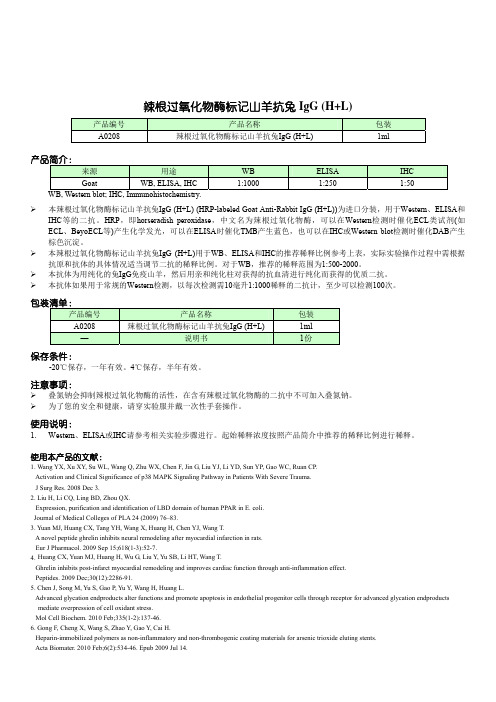
使用说明:
1. Western、ELISA或IHC请参考相关实验步骤进行。起始稀释浓度按照产品简介中推荐的稀释比例进行稀释。
使用本产品的文献:
1. Wang YX, Xu XY, Su WL, Wang Q, Zhu WX, Chen F, Jin G, Liu YJ, Li YD, Sun YP, Gao WC, Ruan CP. Activation and Clinical Significance of p38 MAPK Signaling Pathway in Patients With Severe Trauma. J Surg Res. 2008 Dec 3.
19. Huang L, Qiu B, Yuan L, Zheng L, Li Q, Zhu S. Influence of fasting, insulin and glucose on ghrelin in the dorsal vagal complex in rats. J Endocrinol. 2011 Dec;211(3):257-62. Epub 2011 Sep 19.
5. Chen J, Song M, Yu S, Gao P, Yu Y, Wang H, Huang L. Advanced glycation endproducts alter functions and promote apoptosis in endothelial progenitor cells through receptor for advanced glycation endproducts mediate overpression of cell oxidant stress. Mol Cell Biochem. 2010 Feb;335(1-2):137-46.
二抗的偶联标记

二抗的偶联标记一般来讲,耦联到二抗上的探针主要有酶(辣根过氧化酶HRP和碱性磷酸酶AP或其衍生物APAAP,PAP),荧光基团(FITC, RRX, TR, PE)和生物素。
选用哪种探针的二抗主要取决于具体的实验。
对于western blot 和ELISA,最常用的二抗是酶标二抗,而细胞或组织标记实验(细胞免疫化学,组织免疫化学,流式细胞术)中通常使用荧光基团标记的二抗,免疫组化中也可以使用辣根过氧化酶或碱性磷酸酶标记的二抗。
如果想要更大程度的放大检测信号,可以使用biotin/avidin检测系统。
下面简单介绍几种二抗探针的优缺点及其适用的实验:酶:主要有两种不同的酶耦联物,HRP和AP。
比较而言,前者更经济、快速、稳定,而后者较前者更为灵敏,通常情况下,HRP广泛应用于免疫组化,western blot 和ELISA 中,而碱性磷酸酶(AP)更适合于固相的免疫检测实验,如ELISA和western blot.1. FITC:FITC是广泛使用的荧光基团,但其最大的缺点是淬灭快,使用抗淬灭剂可以减轻。
检测其所激发的蓝色荧光,因而AMCA耦联的二抗更适用于检测大量存在的抗原。
3. 花青染料(Cy2,Cy3,Cy5):Cy2比FITC 更稳定,且在表面荧光显微镜下,Cy2的荧光FITC 更强。
Cy3和Cy5比大多数荧光基团更亮,更稳定,背景更弱。
二者常在共聚焦显微实验中作多标记。
但Cy5的缺点是不能用表面荧光显微镜观察,因此通常用具有远红外检测器的共聚焦显微镜观察。
高的背景。
在使用装有氪氢灯的激光共聚焦扫描显微镜作三标记实验时,RRX 尤其有用,可以和Cy2(或FITC)及Cy5一起使用,因为RRX 的发射波长在Cy2和Cy5中间,且和这两者的重叠很少。
5. PE :常和FITC 一起用于流式细胞术双标记实验。
生物素标记的二抗,在加入耦联有酶或荧光集团的avidin 、ExtrAvidin 或streptavidin ,后者能够结合于二抗表面的多个位点上,从而极大的放大检测信号。
热点实验:WB,IH,IF,ICC实验

4、实验委托者之需要提供样品,即可完成整个实验。
Fig 1:Lanes:Lane 1Lane 2Lane 3Lane 4Lane 5Lane 6 Rows(IOD)(IOD)(IOD)(IOD)(IOD)(IOD) Rock1242.39337.97229.78233.16151.48205.53 Rock2233.48219.11238.76191.61151.18203.66 GAPDH160.18130.47141.06156.13147.77151.07样本编号253828232624WB操作规程一.设备和试剂1.设备① 电泳电源Mini-PROTEAN 3 Elecreophoresis Cell,Mini Teans-Blot Module,and PowerPac Basic Power Supply (BIO-RAD,Catalog#165-3323)② 电泳仪及附件Mini-PROTEAN 3 Elecreophoresis Cell (BIO-RAD,Catalog#165-3301)③ 电转仪及附件Mini Teans-Blot Elecreophoresis Transfer Cell (BIO-RAD,Catalog#170- 3930) ④ NC膜PIERCE,Catalog#88018⑤ 滤纸Whatman,3MM CHRC.电泳缓冲液,1L3g Tris-Base、14.4g 甘氨酸、1g SDS,加蒸馏水至IL,pH应该在8.3左右。
也可以制成10×的储存液,在室温下长期保存。
D.电泳转移缓冲液,1L3g Tris-Base、14.4g 甘氨酸、甲醇200ml, 加蒸馏水至1L, 也可以制成10×的储存液,在湿温下长期保存。
E.5×样品缓冲液,10ml0.6ml 1mol/L的Tris-HCl(Ph6.8)、5ml 50%的甘油、2ml 10%的SDS、0.5ml 2-巯基乙醇、1ml 1%溴酚蓝,0.9ml的蒸馏水,可在4℃保存数周,或在-20℃保存数月。
WB实验简介(2015-7-9)

转膜效果:可以根据蛋白MARK的转膜情况,来简单判断你的转膜效果,一般 选择一定的转膜条件,对不同分子量的蛋白的转膜效率是不一样的。
膜的选择
几种膜的特点:
NC膜
灵敏度和cm2
SDS:导致蛋白质变性,易于蛋白结合,形成易溶于水的复 合物。
β-mercaptoethanol:作为还原剂,打开二硫键,使蛋白质的 四级或三级结构被破坏,使蛋白成为寡氨基酸链。以与二硫 苏糖醇(DTT)或三(2-甲酰乙基)膦盐酸盐(TCEP)互换使 用
溴酚蓝:作为一种指示剂,成蓝色,只是我们样品跑胶的一 种程度。
材料质地 : 干的NC膜易脆
溶剂耐受性 :
无
价格 :
价格较便宜
尼龙膜 高 较高
>400 ug/cm2 软而结实 无 便宜
PVDF膜 高 低
125-200ug/cm2 机械强度高
有 较贵
使用范围0.45um:一般蛋白 0.2um:分子量小于20kD蛋白 0.1um:分子量小于7kD蛋白低浓度
丽春红染色的实例
WB的实验基本流程
主要内容
1. 收集蛋白样品(Protein sample preparation) 2. 电泳(Electrophoresis) 3. 转膜(Transfer) 4. 封闭(Blocking) 5. 一抗孵育(Primary antibody incubation) 6. 二抗孵育(Secondary antibody inucubation) 7. 蛋白检测(Detection of proteins)
Biodlight ECL Chemiluminescent HRP Substrate
免疫荧光染色方法-use
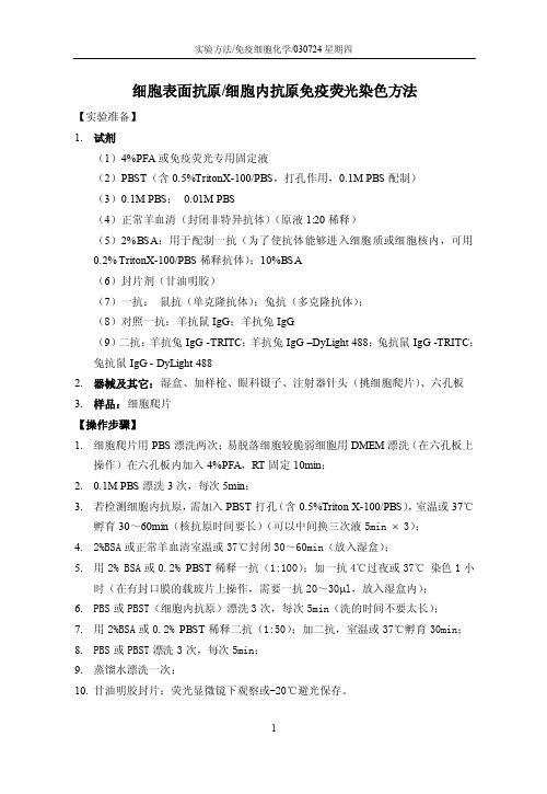
实验方法/免疫细胞化学/030724星期四细胞表面抗原/细胞内抗原免疫荧光染色方法【实验准备】1.试剂(1)4%PFA或免疫荧光专用固定液(2)PBST(含0.5%TritonX-100/PBS,打孔作用,0.1M PBS配制)(3)0.1M PBS;0.01M PBS(4)正常羊血清(封闭非特异抗体)(原液1:20稀释)(5)2%BSA:用于配制一抗(为了使抗体能够进入细胞质或细胞核内,可用0.2% TritonX-100/PBS稀释抗体);10%BSA(6)封片剂(甘油明胶)(7)一抗:鼠抗(单克隆抗体);兔抗(多克隆抗体);(8)对照一抗:羊抗鼠IgG;羊抗兔IgG.(9)二抗:羊抗兔IgG -TRITC;羊抗兔IgG –DyLight 488;兔抗鼠IgG -TRITC;兔抗鼠IgG - DyLight 4882.器械及其它:湿盒、加样枪、眼科镊子、注射器针头(挑细胞爬片)、六孔板3.样品:细胞爬片【操作步骤】1.细胞爬片用PBS漂洗两次;易脱落细胞较脆弱细胞用DMEM漂洗(在六孔板上操作)在六孔板内加入4%PFA,RT固定10min;2.0.1M PBS漂洗3次,每次5min;3.若检测细胞内抗原,需加入PBST打孔(含0.5%Triton X-100/PBS),室温或37℃孵育30~60min(核抗原时间要长)(可以中间换三次液5min ⨯ 3);4.2%BSA或正常羊血清室温或37℃封闭30~60min(放入湿盒);5.用2% BSA或0.2% PBST稀释一抗(1:100);加一抗4℃过夜或37℃染色1小时(在有封口膜的载玻片上操作,需要一抗20~30μl,放入湿盒内);6.PBS或PBST(细胞内抗原)漂洗3次,每次5min(洗的时间不要太长);7.用2%BSA或0.2% PBST稀释二抗(1:50);加二抗,室温或37℃孵育30min;8.PBS或PBST漂洗3次,每次5min;9.蒸馏水漂洗一次;10.甘油明胶封片;荧光显微镜下观察或-20℃避光保存。
羊抗兔IgG(H+L),HRP标记
羊抗兔IgG(H+L),HRP标记产品名称:Goat Anti-Rabbit IgG(H+L), Mouse/Human ads-HRP,羊抗兔IgG(H+L),HRP标记货号:4050-05规格:1.0 mL特异性:抗体识别兔子的IgG重链和轻链以及兔子IgM轻链。
来源:高效价山羊抗血清交叉作用:小鼠和人免疫球蛋白和混合血清,可能与其他物种免疫球蛋白反应。
纯化:亲和色谱法,兔IgG共价链接在琼脂糖上。
缓冲溶液成分:储存溶液50%甘油,50% PBS, pH 7.4,无添加剂(启维益成有售)。
同型对照:Goat IgG储存温度:2-8°C应用:ELISA 1-3FLISAFlow Cytometry 4-7Immunohistochemistry-Frozen Sections 8-10Immunohistochemistry-Paraffin Sections 6,11-13Immunohistochemistry-Whole Mount 14,15Immunocytochemistry 16-19Immunoblotting 20-28Separation 29参考文献:1. Maroney AC, Finn JP, Connors TJ, Durkin JT, Angeles T, Gessner G, et al. Cep-1347 (KT7515), asemisynthetic inhibitor of the mixed lineage kinase family. J Biol Chem. 2001;276:25302-8.(ELISA)2. Zhang X, He M, Cheng L, Chen Y, Zhou L, Zeng H, et al. Elevated heat shock protein 60 levelsare associated with higher risk of coronary heart disease in Chinese. Circulation.2008;118:2687-93. (ELISA)3. Jin K, Wang S, Zhang C, Xiao Y, Lu S, Huang Z. Protective antibody responses againstClostridium difficile elicited by a DNA vaccine expressing the enzymatic domain of toxin B.Hum Vaccin Immunother. 2013;9:63-73. (ELISA)4. Burger PE, Gupta R, Xiong X, Ontiveros CS, Salm SN, Moscatelli D, et al. High aldehydedehydrogenase activity: a novel functional marker of murine prostate stem/progenitor cells.Stem Cells. 2009;27:2220-8. (FC)5. Spurlock CF 3rd, Tossberg JT, Fuchs HA, Olsen NJ, Aune TM. Methotrexate increasesexpression of cell cycle checkpoint genes via JNK activation. Arthritis Rheum.2012;64:1780-9. (FC)6. Aziz M, Yang W, Matsuo S, Sharma A, Zhou M, Wang P. Upregulation of GRAIL is associatedwith impaired CD4 T cell proliferation in sepsis. J Immunol. 2014;192:2305-14. (FC, IHC-PS) 7. Boonstra MC, Tolner B, Schaafsma BE, Boogerd LS, Prevoo HA, Bhavsar G, et al. Preclinicalevaluation of a novel CEA-targeting near-infrared fluorescent tracer delineating colorectal and pancreatic tumors. Int J Cancer. 2015;137:1910-20. (FC)8. Borrego L, Maynard B, Peterson EA, George T, Iglesias L, Peters MS, et al. Deposition ofeosinophil granule proteins precedes blister formation in bullous pemphigoid. Comparison with neutrophil and mast cell granule proteins. Am J Pathol. 1996;148:897-909. (IHC-FS)9. Rønnov-Jessen L, Villadsen R, Edwards JC, Petersen OW. Differential expression of a chlorideintracellular channel gene, CLIC4, in transforming growth factor-β1-mediated conversion of fibroblasts to myofibroblasts. Am J Pathol. 2002;161:471-80. (IHC-FS)10. Xia RH, Yosef N, Ubogu EE. Clinical, electrophysiological and pathologic correlations in asevere murine experimental autoimmune neuritis model of Guillain-Barré syndrome. J Neuroimmunol. 2010;219:54-63. (IHC-FS)11. Penkowa M, Carrasco J, Giralt M, Molinero A, Hernández J, Campbell IL, et al. Altered centralnervous system cytokine-growth factor expression profiles and angiogenesis in metallothionein-I+II deficient mice. J Cereb Blood Flow Metab. 2000;20:1174-89. (IHC-PS) 12. He X, Schoeb TR, Panoskaltsis-Mortari A, Zinn KR, Kesterson RA, Zhang J, et al. Deficiency ofP-selectin or P-selectin glycoprotein ligand-1 leads to accelerated development of glomerulonephritis and increased expression of CC chemokine ligand 2 in lupus-prone mice.J Immunol. 2006;177:8748-56. (IHC-PS)13. Rossato E, Mkaddem SB, Kanamaru Y, Hurtado-Nedelec M, Hayem G, Descatoire V, et al.Reversal of arthritis by human monomeric IgA through the receptor-mediated SH2 domain-containing phosphatase 1 inhibitory pathway. Arthritis Rheumatol.2015;67:1766-77. (IHC-PS)14. Hens J, Gajda M, Scheuermann DW, Adriaensen D, Timmermans J. The longitudinal smoothmuscle layer of the pig small intestine is innervated by both myenteric and submucous neurons. Histochem Cell Biol. 2002;117:481-92. (IHC-WM)15. Monte E, Mouillesseaux K, Chen H, Kimball T, Ren S, Wang Y, et al. Systems proteomics ofcardiac chromatin identifies nucleolin as a regulator of growth and cellular plasticity in cardiomyocytes. Am J Physiol Heart Circ Physiol. 2013;305:H1624-38. (IHC-WM)16. Roberts MS, Woods AJ, Dale TC, van der Sluijs P, Norman JC. Protein kinase B/Akt acts viaglycogen synthase kinase 3 to regulate recycling of αvβ3 and α5β1 integrins. Mol Cell Biol.2004;24:1505-15. (ICC)17. Luijten MN, Basten SG, Claessens T, Vernooij M, Scott CL, Janssen R, et al. Birt-Hogg-Dubésyndrome is a novel ciliopathy. Hum Mol Genet. 2013;22:4383-97. (ICC)18. Tarafdar S, Poe JA, Smithgall TE. The accessory factor Nef links HIV-1 to Tec/Btk kinases in anSrc homology 3 domain-dependent manner. J Biol Chem. 2014;289:15718-28. (ICC)19. Carey AJ, Tan CK, Mirza S, Irving-Rodgers H, Webb RI, Lam A, et al. Infection and cellulardefense dynamics in a novel 17β-estradiol murine model of chronic human group Bstreptococcus genital tract colonization reveal a role for hemolysin in persistence and neutrophil accumulation. J Immunol. 2014;192:1718-31. (ICC)20. Chen Z, Fu L, Herrero R, Schiffman M, Burk RD. Identification of a novel humanpapillomavirus (HPV97) related to HPV18 and HPV45. Int J Cancer. 2007;121:193-8. (WB) 21. Iuchi K, Hatano Y, Yagura T. Heterocyclic organobismuth(III) induces apoptosis of humanpromyelocytic leukemic cells through activation of caspases and mitochondrial perturbation.Biochem Pharmacol. 2008;76:974-86. (WB)22. Fadnes B, Husebekk A, Svineng G, Rekdal Ø, Yanagishita M, Kolset SO, et al. Theproteoglycan repertoire of lymphoid cells. Glycoconj J. 2012;29:513-23. (WB)23. Tong J, Meyer JH, Furukawa Y, Boileau I, Chang L, Wilson AA, et al. Distribution ofmonoamine oxidase proteins in human brain: implications for brain imaging studies. J Cereb Blood Flow Metab. 2013;33:863-71. (WB)24. Wu Y, Wang X, Guo H, Zhang B, Zhang X, Shi Z, et al. Synthesis and screening of 3-MAderivatives for autophagy inhibitors. Autophagy. 2013;9:595-603. (WB)25. Au-Yeung BB, Melichar HJ, Ross JO, Cheng DA, Zikherman J, Shokat KM, et al. Quantitativeand temporal requirements revealed for Zap70 catalytic activity during T cell development.Nat Immunol. 2014;15:687-94. (WB)26. Goss JW, Kim S, Bledsoe H, Pollard TD. Characterization of the roles of Blt1p in fission yeastcytokinesis. Mol Biol Cell. 2014;25:1946-57. (WB)27. Hogan MC, Bakeberg JL, Gainullin VG, Irazabal MV, Harmon AJ, Lieske JC, et al. Identificationof biomarkers for PKD1 using urinary exosomes. J Am Soc Nephrol. 2015;26:1661-70. (WB) 28. Graf FE, Baker N, Munday JC, de Koning HP, Horn D, Mäser P. Chimerization at theAQP2-AQP3 locus is the genetic basis of melarsoprol-pentamidine cross-resistance in clinical Trypanosoma brucei gambiense isolates. Int J Parasitol Drugs Drug Resist. 2015;5:65-8. (WB) 29. Semba K, Namekata K, Guo X, Harada C, Harada T, Mitamura Y. Renin-angiotensin systemregulates neurodegeneration in a mouse model of normal tension glaucoma. Cell Death Differ. 2014;5:e1333. (Sep)。
兔抗鸡IgG荧光抗体的制备及应用
H ir 箱 B D 16 , 净 工 作 台 ( 州 净 化 a 冰 e C 一9F超 苏 设 备公 司 )T L 1 C台式高 速 离心 机 ( ,G 一6 金坛 市 医疗 仪器 厂 制造 )H , J控 温磁 力 搅 拌 器 ( 苏 医疗 仪 器 江
毒、 菌、 细 立克 次体 、 生 动物 及 真菌 以及抗 各 种微 原 生 物抗 体 的检 测 。近 年来 , 疫荧 光 技 术在 兽 医科 免 学 上 的应用 比较普 遍 , 别是 对 一些 流 行严 重 的家 特
1 m n后 弃 去 , 标本 保持 一 定 的湿度 , 后 滴加 适 0i 使 然 当稀释 的荧光 标记 的抗 体溶 液 , 其完 全覆 盖标 本 ; 使
置 于有 盖搪瓷 盒 内 , 温 3 m n后 取 出玻 片 , 保 0 i 置玻 片 架 上 , 用 0 1 o/、H . 先 . tl p 7 0o 1 4的 P S冲洗 后 , 按 顺 B 再
1 材 料 与 方 法
11 试 验 材料 .
11 1 试 验 动 物 ..
生理 盐 水 ,使 之 溶 解 ,再 加 ( H ) O 饱 和 溶 液 N 2 S 1 m , 之 成 3 %( H ) 0 溶 液 , 分 混 合 , 置 0 l使 3 N 2 S 充 静
3 m n后 3O 0 m n离心 2 m n 弃 上 清 , 除去 白 0i 0d i 0 i, 以 蛋白, 将此过 程重 复 2 3次。然后用 1 n ~ 0 d生理盐 水 溶解 沉淀 , 入 透析 袋 , 析除 盐 , 常水 中透析 过 装 透 在 夜 ( 节 常水 的 p 7 ) 再在 生理 盐水 中于 4 调 H. , 0 ℃透析
12. 。4 兔 I G 的提 取 与 浓 缩 g
常用抗体标记荧光染料的选择

1、蓝色(350-450nm处激发)CF 350、Alexa Fluor 350、AMCA等----亮蓝和紫外光激发。
CF350是类似于Alexa Fluor 350和传统荧光染料AMCA的蓝色荧光染料,CF350的荧光强度高于Alexa Fluor350、AMCA,吸附在蛋白上的荧光超过50%,水溶性更好,耐光性非常优秀亮,更容易与现有的绿色荧光基团区分。
CF 405S/ CF 405 M、Alexa Fluor 405 ----近乎完美的匹配蓝色二极管激光器。
CF 405S/ CF 405 M、Alexa Fluor 405与近来使用的荧光显微镜和流式细胞仪405nm;谱线的蓝色二极管激光器完美的匹配。
在流式细胞仪上的分析结果显示CF 405S/ CF 405 M荧光信号强度高于Alexa Fluor 405染料1.7倍。
2、绿色(488nm处激发)CF 488A、Alexa Fluor 488、FITC、FAM、DyLight 488、Cy2等----针对488nm 氩离子激光器的绿色荧光染料。
以上染料其标记的抗体蛋白适用于所有配备488nm氩离子激光器的流式细胞仪,流式细胞仪的FL1通道检测,或者可用于荧光显微镜技术。
CF 488A最低限度的带电量降低了与抗体耦联物的非特异性结合,在红色通道溢出少于Alexa Fluor 488,耐光性好、水溶性好和pH 不敏感,良好的稳定性和活性染料的标记率。
Alexa Fluor 488在较宽的PH值范围内保持稳定(PH4~10);FITC激发波长488nm,最大发射波长525nm,缺点:荧光强度易受PH值影响,PH值降低时其荧光强度减弱。
3、橙红色(543-555nm处激发)CF 543、Alexa Fluor 546、ATTO550, Cy 3, DyLight 549, Rhodamine (TRITC) 匹配543nm的橙色荧光染料;CF ™543 荧光条带明亮,耐光,水溶性好,确保了CF 543染料与抗体的耦联物保持优异的水溶性,为该波段最亮的橙色荧光染料。
DyLight549标记山羊抗兔IgG二抗

Abbkine, Inc • Redlands, CA • USA •Not for further distribution without written consent. Copyright © Abbkine, Inc. Dylight 549, Goat Anti-Rabbit IgG# A23320Product InformationProduct Name: Dylight 449 Conjugated Goat Anti-Rabbit IgG(H+L)Amax: 555nm Emax: 568nmCatalog Number: A23320 Lot Number: Refer to vial Formulation: Liquid Size:100ul/500ulStorage:Store at -20°C. Be protected from light and avoid repeated freeze / thaw cycles.Note: Contain sodium azide.Background: DyLight fluorescent dyes are a new generation of dyes with improved brightness and photostability, which are highly water soluble and remain fluorescent from pH 4 to pH 9. They are better than or comparable to the best fluorescent dyes from other companies. Abbkine's secondary antibodies are available conjugated to enzyme, biotin or fluorophore for use in a variety of antibody-based applications including WB, IHC, IF, FC and ELISA. We offer high quality secondary antibodies from goat, rabbit and donkey sources for your each application. Serum adsorbed secondary antibodies are also available and are recommended for use with immunoglobulin-rich samples.Purity: Affinity purified using solid phase Rabbit IgG (H&L) with finally > 95% purity based on SDS-PAGE.Specificity: The antibody reacts with whole molecule rabbit IgG. It also reacts with heavy chains of rabbit IgG, and light chains of all other rabbit immunoglobulins. It has no reactivity on non-immunoglobulin serum proteins, while it may cross-react with immunoglobulins from other species.Application Notes: Optimal working dilutions should be determined experimentally by the investigator. Suggested starting 1:50-1:1,000 dilutions for most applications.Storage Buffer: 0.01M Sodium Phosphate, 0.25M NaCl, 50% Glycerol pH 7.4.Stabilizer: 1% Bovine Serum Albumin (IgG and Protease Free).Preservative: 0.05% (w/v) Sodium Azide.Storage Instructions: Stable for one year at -20°C from date of shipment. For maximum recovery of product, centrifuge the original vial after thawing and prior to removing the cap. Aliquot to avoid repeated freezing and thawing.Note: The product listed herein is for research use only and is not intended for use in human or clinical diagnosis. Suggested applications of our products are not recommendations to use our products in violation of any patent or as a license. We cannot be responsible for patent infringements or other violations that may occur with the use of this product.。
二抗鼠抗兔IgG单克隆抗体
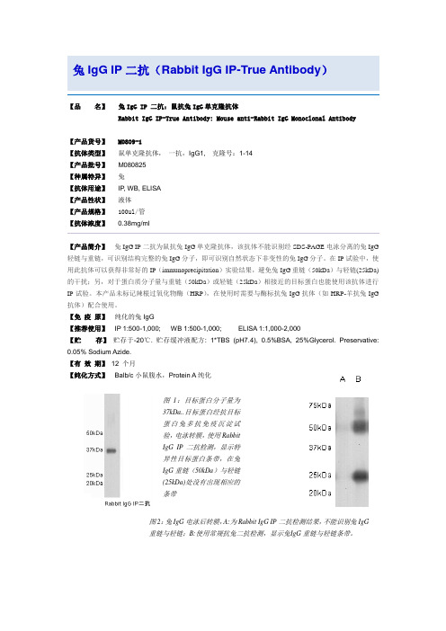
兔IgG IP二抗(Rabbit IgG IP-True Antibody)【品名】兔IgG IP 二抗:鼠抗兔IgG单克隆抗体Rabbit IgG IP-True Antibody: Mouse anti-Rabbit IgG Monoclonal Antibody【产品货号】 M0809-1【抗体类型】鼠单克隆抗体,一抗,IgG1, 克隆号:1-14【产品批号】M080825【种属特异】兔【抗体用途】IP, WB, ELISA【产品性状】液体【产品规格】100ul/管【抗体浓度】0.38mg/ml【产品简介】兔IgG IP二抗为鼠抗兔IgG单克隆抗体,该抗体不能识别经SDS-PAGE电泳分离的兔IgG 轻链与重链,可识别结构完整的兔IgG分子,即可识别自然状态下非变性的兔IgG分子。
在IP试验中,使用此抗体可以获得非常好的IP(immunoprecipitation)实验结果,避免兔IgG重链(50kDa)与轻链(25kDa)的干扰;另,对于蛋白质分子量与重链(50kDa)或轻链(25kDa)相接近的目标蛋白也能使用该抗体进行IP试验。
本产品未标记辣根过氧化物酶(HRP),在使用时需要与酶标抗兔IgG抗体(如HRP-羊抗兔IgG 抗体)配合使用。
【免疫原】纯化的兔IgG【推荐使用】IP 1:500-1,000; WB 1:500-1,000; ELISA 1:1,000-2,000【贮存】贮存于-20℃. 贮存缓冲液配方: 1*TBS (pH7.4), 0.5%BSA, 25%Glycerol. Preservative: 0.05% Sodium Azide.【有效期】12 个月【纯化方式】Balb/c小鼠腹水,Protein A纯化图1:目标蛋白分子量为37kDa..目标蛋白经抗目标蛋白兔多抗免疫沉淀试验,电泳转膜,使用RabbitIgG IP二抗检测,显示特异性目标蛋白条带,在兔IgG重链(50kDa)与轻链(25kDa)处没有出现相应的条带图2:兔IgG电泳后转膜,A:为Rabbit IgG IP二抗检测结果,不能识别兔IgG重链与轻链;B:使用常规抗兔二抗检测,显示兔I gG重链与轻链条带。
DyLight荧光二抗——高性价比的全新荧光体验

DyLight荧光二抗——高性价比的全新荧光体验美国Abbkine的DyLight™系列荧光标记是一种近年来被广泛应用的新型荧光染料,由于具有很好的光谱宽度、更强的荧光强度,更高的光学耐受性(抗淬灭性)、对pH不敏感、分子较小而渗透性更好的优势,越来越受到科研客户的喜爱。
DyLight™种类齐全,如Dylight 405是蓝色荧光,替代Alexa 405;DyLight 549是绿色荧光,可完全替代FITC/Cy2/Alexa 488;完全替代Cy3/Rhodamin/Alexa 546/Alexa 555的Dylight 549;替代Texas Red/Alexa 594的Dylight 594;Dylight 633和Dylight 649(可替代Cy5/Alexa 647)则是更强的红色荧光;另外,Dylight还有适配Odyssey系统的近红外和远红外二抗Dylight 680和DyLight 800。
下面我们来看看美国Abbkine品牌DyLight系列荧光的优异表现吧:一、DyLight荧光二抗比传统荧光标记二抗具有更高的荧光发光强度DyLight 488标记的抗体比Cy2 和FITC标记的更亮,与Alexa Fluor 488 标记的亮度相当。
DyLight 549标记的抗体比传统的罗丹明、TRITC等荧光亮,与Cy3 和Alexa Fluor 555亮度相当;而DyLight 594标记的抗体荧光亮度明显强于Alexa 594标记抗体。
相比较Texas Red 而言,不仅更亮,而且具有更好的水溶性。
二、DyLight荧光二抗比传统荧光及Alexa标记二抗具有更低的背景!三、DyLight荧光二抗比传统荧光标记二抗具有更好的光学稳定性、发光更持久、更适合拍照488594最常用的荧光颜色,激发光和发射光光谱分布合理,适用于各种荧光分析。
其中蓝色荧光的DyLight 405实验。
另外,1. 大包装二抗具有更高的性价比。
辣根过氧化物酶标记的羊抗兔IgG
辣根过氧化物酶标记的羊抗兔IgG(Goat Anti-rabbit IgG/HRP)规格:0.1ml/1ml抗体来源:Goat克隆类型:Polyclonal交叉反应:Rb产品应用:WB=1:1000-10000ELISA=1:1000-10000IHC-P=1:100-1000IHC-F=1:100-1000not yet tested in other applications.optimal dilutions/concentrations should be determined by the end user.分子量:150kDa性状:Lyophilized or Liquid浓度:2mg/1ml免疫原:Full length plasma protein亚型:IgG纯化方法:affinity purified by Protein A储存液:0.01M PBS,pH7.4with10mg/mL BSA and0.1%Gentamicin保存条件:Storage:Store at–20oC for one year.Avoid repeated freeze/thaw cycles.The lyophilized antibody is stable at room temperature for at least one month and for greater than a year when kept at-20oC.When reconstituted in sterile distilled water or diluent supplied, theantibody is stable for at least two weeks at2-4°C.产品介绍:Immunoglobulin G(IgG),is one of the most abundant proteins in serum with normal levels between 8-17mg/mL in adult blood.IgG is important for our defence against microorganisms and themolecules are produced by B lymphocytes as a part of our adaptive immune response.The IgG molecule has two separate functions;to bind to the pathogen that elicited the response and to recruit other cells and molecules to destroy the antigen.The variability of the IgG pool is generated by somatic recombination and the number of specificities in an individual at a given time point is estimated to be1011variants.Important Note:This product as supplied is intended for research use only,not for use in human,therapeutic or diagnostic applications.。
免疫荧光 双标
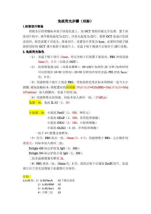
免疫荧光步骤(双标)1.冰冻切片制备将脱水后的脊髓标本放于冰冻托盘上,加OCT使组织被完全包埋,置于冰冻切片机中,调节箱体温度为-22℃,冷冻头温度为-23℃。
观察OCT冻成白色固态状时,将其放置于冷冻头,准备切片。
设置切片厚度为8µm,必要时用刷子辅助将切好的OCT薄片贴附于载玻片上。
室温下晾干载玻片后保存于-20℃冰箱。
2.免疫荧光染色(1)室温下晾干切片15min,待完全晾干后放置于湿盒内,PBS冲洗浸泡5min/次,3次(以除去OGT)。
(2)抗原修复液(1X)(双蒸水稀释),95-100℃加热约20分钟(加热时间可以控制在10-30分钟内)。
20-30分钟内冷却至室温。
PBS冲洗5min/次,3次。
(3)用滤纸吸干玻片上残余PBS,用免疫组化笔在标本周围画一适当大小圆圈,避免接触标本,将配置好的封闭液(5%驴血清=920ulPBS+50ul驴血清+30ul 10%trition)加入圆圈内,室温下封闭1h。
(4)用滤纸吸去封闭液,向标本加入相应一抗:(1%BSA)兔源一标:兔抗IL-33(1:50)小鼠源二标:小鼠抗NeuN(1:500,神经元)小鼠抗GFAP(1:300,星形胶质细胞)小鼠抗OX42(1:100,小胶质细胞)小鼠抗OLIG2(1: 25,少突胶质细胞)一抗于4℃静置过夜孵育。
(5)次日,PBS洗去一抗,10min/次,3次,用滤纸吸干PBS,之后操作均需避光,向标本加入相应二抗:Dylight 488标记驴抗兔IgG(1:300),Dylight 594标记驴抗小鼠IgG(1:300),二抗室温静置避光孵育2h。
(6)PBS清洗二抗,10min/次,3次,清洗后晾干后滴加DAPI封片,加盖玻片后于荧光显微镜下拍摄图片并保存。
计划:1.IL-33组:1)IL-33/NeuN X2个配比浓度2)IL-33/GFAP X23)IL-33/Iba-1 X24)只孵二抗X22.Sham组:1)IL-33/NeuN X12)IL-33/GFAP X13)IL-33/Iba-1 X14)只孵二抗需要购买:Triton-100,驴血清,一抗:小鼠抗RBFOX3/ NeuN、小鼠抗GFAP、小鼠抗OX42,二抗:Dylight 488标记驴抗兔IgG,BSA(牛血清白蛋白)注意:1、细胞固定和通透为达到最佳的检测效果,细胞需要经过固定和通透。
食物特异性IgG抗体检测试剂盒(条带免疫印迹法)产品技术要求siderun

食物特异性IgG抗体检测试剂盒(条带免疫印迹法)适用范围:用于体外定性检测人血清中食物特异性IgG抗体(最多可检测切达干酪,白软干酪,牛肉,鸡肉,猪肉,鸡蛋,羊肉,火鸡,酸奶,巧克力,玉米,大米,小麦,大豆,柠檬,菠菜,青豆,小米,杏仁,芝麻,腰果,花生,旱芹,茄子,青椒,欧芹,卷心菜,胡萝卜,菜花,嫩豌豆,葵花籽,黑胡桃,蓝莓,菠萝,橘子,桃,苹果,榴莲,芒果,香蕉,葡萄,柚子,草莓,哈密瓜,麦芽,黑麦,酵母,蔗糖,黄油,肉桂,芥末,蜂蜜,咖啡,燕麦,大麦,荞麦,西兰花,杂色豌豆,大蒜,洋葱,可乐豆,红茶,牛奶,螃蟹,对虾,鳕鱼,羊奶,蛤,三文鱼,沙丁鱼,带鱼,扇贝,龙虾,草鱼,牡蛎,鳟鱼,金枪鱼,红辣椒,大葱,生菜,黄瓜,马铃薯,南瓜,甘薯,香菜,小白菜,西红柿,蘑菇,西瓜,橄榄90种食物特异性IgG抗体)。
1.1包装规格6人份/盒,型号为FIGG-90;9人份/盒,型号为FIGG-64;FIGG-48;18人份/盒,型号为FIGG-36;FIGG-28;FIGG-21;FIGG-14A;FIGG-14B;FIGG-7A;FIGG-7B;FIGG-7C;FIGG-4A;FIGG-4B;其中,各型号/包装规格检测项目如下:表1 各型号/包装规格检测项目2. 主要组成成分表2 主要组成成分2.1. 外观2.1.1. 试剂盒外部包装盒应整洁,各组分应齐全完整,文字符号标识清晰,所有试剂瓶密闭,无漏液。
2.1.2. 样本缓冲液为无色或淡黄色澄清液体;浓缩洗液(10×)为无色澄清液体;酶结合物(10×)为无色或淡黄色澄清液体;底物液为淡黄色澄清液体。
2.2. 净含量液体试剂装量不小于标示值。
2.3. 膜条宽度膜条宽度不低于1.8mm。
2.4. 临界值及重复性食物特异性IgG抗体企业参考品各检测项目临界值浓度均为37.5U/mL。
阳性参考品各检测项目抗体浓度均为50U/mL,检测10次,结果的阳性率为100%,反应结果一致;阴性参考品各检测项目抗体浓度均为25U/mL,检测10次,结果的阴性率为100%,反应结果一致。
- 1、下载文档前请自行甄别文档内容的完整性,平台不提供额外的编辑、内容补充、找答案等附加服务。
- 2、"仅部分预览"的文档,不可在线预览部分如存在完整性等问题,可反馈申请退款(可完整预览的文档不适用该条件!)。
- 3、如文档侵犯您的权益,请联系客服反馈,我们会尽快为您处理(人工客服工作时间:9:00-18:30)。
Abbkine, Inc • Redlands, CA • USA •
Not for further distribution without written consent. Copyright © Abbkine, Inc.
Dylight 549, Rabbit Anti-Goat IgG # A23330
Product Information
Catalog Number: Lot Number:
Storage:light and avoid repeated freeze / thaw cycles.
Note:
Background: DyLight fluorescent dyes are a new generation of dyes with improved brightness and photostability, which are highly water soluble and remain fluorescent from pH 4 to pH 9. They are better than or comparable to the best fluorescent dyes from other companies. Abbkine's secondary antibodies are available conjugated to enzyme, biotin or fluorophore for use in a variety of antibody-based applications including WB, IHC, IF, FC and ELISA. We offer high quality secondary antibodies from goat, rabbit and donkey sources for your each application. Serum adsorbed secondary antibodies are also available and are recommended for use with immunoglobulin-rich samples.
Purity: Affinity purified using solid phase Goat IgG (H&L) with finally > 95% purity based on SDS-PAGE.
Specificity: The antibody reacts with whole molecule goat IgG. It also reacts with heavy chains of goat IgG, and light chains of all other goat immunoglobulins. It has no reactivity on non-immunoglobulin serum proteins, while it may cross-react with immunoglobulins from other species.
Application Notes: Optimal working dilutions should be determined experimentally by the investigator. Suggested starting 1:50-1:1,000 dilutions for most applications.
Storage Buffer: 0.01M Sodium Phosphate, 0.25M NaCl, 50% Glycerol pH 7.4.
Stabilizer: 1% Bovine Serum Albumin (IgG and Protease Free).
Preservative: 0.05% (w/v) Sodium Azide.
Storage Instructions: Stable for one year at -20°C from date of shipment. For maximum recovery of product, centrifuge the original vial after thawing and prior to removing the cap. Aliquot to avoid repeated freezing and thawing.
Note: The product listed herein is for research use only and is not intended for use in human or clinical diagnosis. Suggested applications of our products are not recommendations to use our products in violation of any patent or as a license. We cannot be responsible for patent infringements or other violations that may occur with the use of this product.。
