Alterations in Wnt pathway activity in mouse serum and kidneys during lupus development
西罗莫司治疗淋巴管畸形的研究进展

·综 述·西罗莫司治疗淋巴管畸形的研究进展苟艺凡*1 周倩2 李川松Δ3(1. 川北医学院临床医学系,四川 南充 637007;2. 四川大学华西口腔医院头颈肿瘤外科,四川 成都610041;3. 四川省医学科学院四川省人民医院,四川 成都 610031)摘要 头颈部淋巴管畸形可能造成毁容,诱发功能性合并症,甚至危及生命。
手术及药物治疗淋巴管畸形有一定效果,但复杂淋巴管畸形仍然是临床治疗难点。
西罗莫司是哺乳动物雷帕霉素靶蛋白的抑制剂,可减少内皮细胞血管内皮生长因子的生成,调节血管生成、细胞增殖、迁移和粘附,在淋巴管成熟和稳定中发挥关键作用。
近年临床研究提示该药物在治疗复杂淋巴管畸形治疗中有较好的应用前景,本文对其治疗机制和临床应用等研究进展进行综述。
关键词:脉管畸形;淋巴管畸形;西罗莫司;治疗Advances in the treatment of lymphatic malformation by SirolimusGou Yi-fan*1, Zhou Qian 2, Li Chuan-song Δ3(1. Department of Clinical Medicine, North Sichuan Medical College, Nanchong 637007, Sichuan, China; 2. Department of Head and Neck Oncology, National Clinical Research Center for Oral Diseases, West China Hospital of Stomatology, Sichuan University, Chengdu 610041, China; 3. Medical Scientific Academy in Sichuan People’sHospital, Chengdu 610031, China)Abstract Lymphatic malformation in oral and maxillofacial may cause malformation, induce functionalcomplications, and even endanger life. There are many related treatment methods, but the complicated lymphatic malformation of oral and maxillofacial is still a difficult point in clinical treatment. Sirolimus is an inhibitor of mammalian target of rapamycin, which can reduce the production of vascular endothelial growth factor (VEGF) in endothelial cells, regulate angiogenesis, cell proliferation, migration and adhesion, and play a critical role in lymphatic maturation and stability. Recent clinical studies indicated that this drug has a good application prospect in the treatment of complex lymphatic malformations. This paper reviews the research progress of its therapeutic mechanism and clinical application.Key words: Vascular malformations; Lymphatic malformations; Sirolimus; Therapy*作者简介:苟艺凡,女,本科在读,Email :*****************;△通讯作者:李川松,男,副主任药师,主要研究抗血管新生药物的研发和临床应用,Email :****************。
乳腺叶状肿瘤上皮组织中 E-cadherin 的表达及临床意义
乳腺叶状肿瘤上皮组织中 E-cadherin 的表达及临床意义瞿伟;彭亚婷;周国凤【摘要】目的:观察乳腺叶状肿瘤上皮组织中E-cadherin的表达,探讨其与临床病理学特征的关系。
方法采用免疫组化SP法检测E-cadherin在60例乳腺叶状肿瘤及10例纤维腺瘤上皮成分中的表达。
结果 E-cadherin蛋白在纤维腺瘤上皮成分中的表达比在乳腺叶状肿瘤中的表达更高( P=0.032);E-cadherin在乳腺叶状肿瘤上皮成分中的表达与间质细胞密度有关(P=0.00),其表达随着叶状肿瘤间质细胞密度的增加而降低;而与患者年龄、肿瘤边界、肿瘤大小、组织学分级、细胞生长过度、核分裂象、细胞异型性及Ki-67表达无关(P均>0.05)。
结论 E-cadherin可能抑制乳腺叶状肿瘤的间质生长,是鉴别乳腺叶状肿瘤与纤维腺瘤的一个重要生物学标记。
【期刊名称】《山东医药》【年(卷),期】2015(000)004【总页数】3页(P57-59)【关键词】乳腺叶状肿瘤;纤维腺瘤;上皮;E-cadherin蛋白【作者】瞿伟;彭亚婷;周国凤【作者单位】南昌市第三医院,南昌330006;南昌市第三医院,南昌330006;南昌市第三医院,南昌330006【正文语种】中文【中图分类】R737.9乳腺叶状肿瘤(PTs)是少见的纤维上皮来源的肿瘤,占乳腺肿瘤的0.3%~1%,有复发和转移的潜能。
根据间质细胞异型性、间质细胞密度、核分裂象、间质过度生长及肿瘤边界几个特征,PTs可分为良性、交界性及恶性。
虽有报道p53、c-kit、EGFR、CD10等标志物与以上病理学特征之间的关系,然而仍难以准确区分纤维腺瘤与良性PTs,良性PTs与交界性PTs,交界性PTs与恶性PTs[1]。
本研究通过免疫组化SP法检测E-cadherin蛋白在PTs及纤维腺瘤上皮组织中的表达,检测Ki-67在PTs间质成分中的表达,从而探讨E-cadherin的表达与PTs病理学特征及Ki-67表达之间的相关性,分析E-cadherin在鉴别PTs和纤维腺瘤的意义。
Wnt信号通路与阿尔茨海默病的关系
Wnt信号通路与阿尔茨海默病的关系作者:赵良友严妍房城安柏松姜波贾博宇吴娟周忠光来源:《今日健康》2014年第08期【摘要】阿尔茨海默病(Alzheimer's disease ,AD)典型的临床表现为进行性认知功能障碍和精神行为异常,是老年期痴呆的最常见类型。
患者脑部边缘系统及相关区域的新皮质选择性神经元缺失,其特征性组织病理改变为神经炎性斑或老年斑(主要为淀粉样肽)的形成和异常细胞骨架蛋白的聚集(神经纤维缠结,NTFs)。
【关键词】阿尔茨海默病 Wnt信号通路 tau蛋白【中图分类号】 R9 【文献标识码】 A 【文章编号】 1671-5160(2014)08-0033-011 Wnt信号通路1.1 Wnt信号通路组成Wnt基因是一种原癌基因,Wnt蛋白是一个富含半胱氨酸残基的分泌信号糖蛋白家族,是一种由Wnt基因编码的分泌性蛋白生长因子。
目前认为wnt信号通路主要由以下几种蛋白构成:胞外因子wnt家族分泌蛋白、β-连环蛋白(β-catenin)、特异性跨膜受体卷曲蛋白、散乱蛋白、结肠腺瘤性息肉病基因蛋白(APC)、糖原合成酶激酶3β(GSK-3β)、轴蛋白(Axin)或传导蛋白(Conductin)及转录因子家族的T细胞因子/淋巴细胞增强因子(TCF/LEF)等。
Wnt信号通路控制着体内多种程序,Wnt信号通路的失调会引起多种疾病。
1.2 wnt信号通路的分类目前认为wnt信号通路至少有4条:①经典的wnt/β-catenin信号通路;②wnt/polarity通路;③wnt/Ca2+通路。
④调节纺锤体定向和不对称细胞分裂的通路。
1.2.1 经典Wnt信号通路(Wnt/β-catenin信号通路)经典Wnt信号通路的主要机制为:当Wnt蛋白存在时,与细胞膜上7次跨膜的G蛋白偶联受体卷曲蛋白及共同受体低密度脂蛋白受体相关蛋白5/6结合,激活胞质中散乱蛋白,从而抑制GSK-3β的活性,使未磷酸化的β-catenin在细胞质中积聚并转移到细胞核中,与TCF/LEF 结合启动靶基因的转录,产生细胞的增殖、分化、迁移、极性化和凋亡等多种细胞效应。
WNT信号通路的研究进展
摘
要 : n ( n ls —y e MM TV n e rto iefmi mb r ) 号 通 路 与 细 胞 的 发 W t wi g e st p i tg ain st a l me es 信 y
育分化 密切 相 关 , 正 常和 肿 瘤 细 胞 生 长 都 至 关 重要 , 对 因其 启 动 蛋 白 为 W n 蛋 白而得 名 。 t
泛表 达 于心血管 系 统及 其 它 器 官 。F d蛋 白具 有 高 z 度保 守 的 富 含 半 胱 氨 酸 的 配 体 结 合 区 ( R , C D) 而
C D是 F d与 wn 相 结 合 的 部 位 , R 区 域 氨 基 R z t C D 酸 残 基 的 突 变 会 导 致 F d与 Wn 相 互 作 用 的 消 z t 失 ] 。来 自 Wa g等口 的研 究 表 明 缺 乏 F d n z 4基 因的小 鼠导致 小脑 的逐 渐退 化和 听觉 及食 道 的 功能
来, 并且极 难 溶 于 水 , 因此 也 限 制 了 对 其 性 质 的 研 究 。故 2 0年 来对 其 功 能 的研 究 资 料 大 都 通 过 上 清 培养 液 、 逆转 录病 毒和 与转 染 Wn 基 因的 细胞 共 培 t
养 3种方 法 间 接 获 得_ 。最 近 wie t _ 9 ] l r 等 ] 过 质 l 通
通 路 即 Wn——ae i 路 , Wn 信 号 中研 究 最 t ctnn通 B 是 t
清 楚 的一 条通 路 , 整个 进化 过程 中高度 保守 。 在
2 1 经 典 的 W n 信 号 通 路 . t
经 典 的 wn 信 号通路 参 与 了调 控 细 胞命 运 、 t 细 胞 增殖 及凋 亡 等 过 程 。分 泌 的 wn 配 体 与 细 胞 表 t 面受体 F d家 族或 L P / R 6受体 结 合形 成 复合 z R SL P 物 , 体复合 物 引起 Bctnn在 细 胞 内 的积 累 。当 受 —ae i Wn 信号通 路 活化 时 , n 与 受体 F d结 合 激 活 细 t w t z 胞 内的 D h蛋 白 , s 磷酸化 的 D h蛋 白将 信 号 传 至 细 s
上皮间质转化相关的生物标志在胃癌中的研究进展_庄铭锴
Abstract
摘要 转移是胃癌的主要死因 . 研究胃癌的转移机 制, 能为未来发展治疗胃癌的新手段提供必要 的理论基础. 上皮间质转化是上皮细胞转化成 间充质样细胞并伴随明显基因和表型改变的 一种多阶段过程, 这种转化亦可在肿瘤细胞中 发生, 其会赋予肿瘤细胞侵袭和转移能力, 使 其更易形成远处转移灶 , 故上皮间质转化是 肿瘤发生转移的重要机制之一. 本文综述了近 年来上皮间质转化的相关生物标志物 , 包括 细胞表面蛋白(E-cadherin、N-cadherin)、细 胞骨架蛋白(-catenin、Vimentin)、转录因子 (Snail、Twist、ZEB1、ZEB2)等在胃癌中的 研究进展.
3205
■研发前沿
、、、P120-ctn相互作用组成E-cad/ctn复合 体, 该复合体可与细胞骨架肌动蛋白相连, 形成 完整的上皮细胞间的黏附连接(adheren junction, A J). A J 中任一组成成分的表达或功能异常时 , 都会影响该结构的稳定性 , 使上皮细胞间的黏 附减弱 . 研究发现在上皮型肿瘤的恶性进展过 程中 , E-cad 的表达会出现下调 , 甚至是完全丢 失, 这会导致肿瘤细胞间的黏附减弱, 使其从良 性、非侵袭性向恶性、侵袭性表型转化[5]. E-cad在胃癌中的表达也有类似的改变, 且 发现至少有三种机制参与了这一调控过程: (1) 基因的突变, Becker等[6]首次在不同类型的胃癌 样本(肠型、弥漫型、混合型)中发现了E-cad的 基因突变, 提出了E-cad的突变可能是导致弥漫 型胃癌发生的分子机制之一; 随后Guilford等[7] 在家族性弥散型胃癌中发现E-c a d 基因的胚系 突变 , 且证实了该突变是造成此病发生的主要 原因; (2)启动子甲基化, Machado等[8]通过基因 分析发现了弥散型胃癌中存在着 E-c a d 启动子 区域甲基化程度的升高; Chan等[9]亦发现, 在胃 癌经典发展过程“Correa's cascade”中(即正常 黏膜-慢性活动性胃炎-慢性萎缩性胃炎-肠上皮 化生-不典型增生-原位癌), E-cad的表达是逐步 下降的, 而且E-cad 基因的甲基化频率也是逐步 升高的 , 更重要的是研究还发现幽门螺旋杆菌 感染也参与了E-cad 基因的甲基化过程, 这些都 提示了在胃癌中, 基因甲基化是调节E-cad活性 的重要机制. 而“两次打击”学说(基因突变、 启动子甲基化分别为第一、二次打击 ) 也是目 前解释遗传性弥散型胃癌发生机制的主要学 说; (3)转录抑制, Rosivatz等[10]在人胃癌临床样 本的研究中发现, E-cad的表达下调与转录因子 Snail、Twist、SIP1的表达上调是密切相关的, 而Wang等[11]在E-cad表达阴性的SV40病毒转化 的永生化胃上皮细胞株 G e s-1 和人胃癌细胞株 MGC-803 、BGC-823、SGC-7901中亦发现了 Snail、Twist、Slug等转录因子的高表达, 这些 转录因子能与E-cad 基因启动子序列上的E-box 元件相结合, 抑制E-cad 基因的转录; (4)其他, 如 microRNAs(miR)也是调控胃癌中E-cad表达的机 制之一[12], 目前已发现miR-200B能以转录抑制因 子ZEB2为靶点来调控胃癌中E-cad的表达[13], 另 外在肠型胃癌中亦发现了 miR-101 的表达是下 调的 , 且这与 E-cad 的功能异常是相关的 [14]. 除 了调节细胞间的黏附外, E-cad还可通过影响细 胞内的信号传导来影响细胞的生物学行为 . 如
幽门螺杆菌导致上皮间质转化的研究进展

DOI:10.19368/ki.2096-1782.2023.19.190幽门螺杆菌导致上皮间质转化的研究进展宁月1,2,王蒙1,2,邵利华1,2,焦婷红3,李海龙1,2,41.甘肃中医药大学第一临床医学院,甘肃兰州730000;2.甘肃省中医方药挖掘与创新转化重点实验室,甘肃兰州730000;3.甘肃中医药大学附属医院临教科,甘肃兰州730000;4.甘肃省中药新产品创制工程实验室,甘肃兰州730000[摘要]众所周知,幽门螺杆菌(helicobacter pylori, Hp)可以导致胃黏膜病变,产生肠化生,最终形成胃癌。
Hp导致的上皮间质转化(epithelial-mesenchymal transition, EMT)在学术界已经受到广泛关注。
首先,Hp对EMT促进作用通常是由细胞毒素-相关基因A(cytotoxin-associated gene A, CagA)造成的。
作为Hp首要的毒力因子,CagA可以促使Hp通过自噬、炎症、癌症干细胞(cancer stem cells, CSC)等促进EMT形成;其次,EMT 的发生也涉及受CagA影响的一些受体及因子,如CD44、程序性细胞死亡因子4(programmed cell death 4, PDCD4)、肿瘤坏死因子α(tumor necrosis factor alpha, TNF-α)。
此外,Hp促进EMT还包括某些信号通路发生信号传导改变,目前已知的受调控的通路有NF-κB、TGF-β/Smad、PI3K/AKT通路等。
本文尝试对Hp导致EMT的机制做一综述,试图阐明其机制,为Hp防控和相关疾病的治疗提供思路和线索。
[关键词]幽门螺杆菌;上皮间质转化;胃癌;肿瘤侵袭和转移[中图分类号]R735.2 [文献标识码]A [文章编号]2096-1782(2023)10(a)-0190-09Progress in the Study of Epithelial-mesenchymal Transition Caused by He⁃licobacter PyloriNING Yue1,2, WANG Meng1,2, SHAO Lihua1,2, JIAO Tinghong3, LI Hailong1,2,41.The First Clinical Medical College of Gansu University of Chinese Medicine, Lanzhou, Gansu Province, 730000 China;2.Key Laboratory of Excavation and Innovative Transformation of Traditional Chinese Medicine, Lanzhou, Gansu Province, 730000 China;3.Clinical Training Department, Affiliated Hospital of Gansu University of Chinese Medicine, Lanzhou, Gansu Province, 730000 China;4.Gansu Provincial Engineering Laboratory for the Creation and Manufacture of New Chinese Medicinal Products, Lanzhou, Gansu Province, 730000 China[Abstract] It is well known that Helicobacter pylori (Hp) can lead to gastric mucosal lesions, intestinal metaplasia and ultimately gastric cancer. Epithelial-mesenchymal transition (EMT) caused by Hp has received extensive attention in academia. Firstly, the EMT-promoting effect of Hp is usually caused by cytotoxin-associated gene A (CagA). As the primary virulence factor of Hp, CagA can prompt Hp to promote EMT formation through autophagy, inflammation, and cancer stem cells (CSC). Secondly, the occurrence of EMT also involves some receptors and factors affected by CagA, such as CD44, programmed cell death 4 (PDCD4), and tumor necrosis factor alpha (TNF-α). In addition, the promo⁃tion of EMT by Hp also includes signaling alterations in certain signaling pathways, and the known regulated pathways include NF-κB, TGF-β/Smad, and PI3K/AKT pathways. This paper attempts to give an overview of the mechanism of Hp leading to EMT, trying to elucidate the mechanism and provide ideas and clues for the prevention and control of Hp and the treatment of related diseases.[Key words] Helicobacter pylori; Epithelial-mesenchymal transition; Gastric cancer; Tumor invasion and metastasis上皮间质转化(epithelial-mesenchymal transi⁃tion, EMT)是上皮细胞转化为能动细胞的过程。
Wnt信号通路的新功能
Wnt/Snail信号通路的新功能日期:2012-05-30 来源:CancerRes标签:葡萄糖信号通路摘要: Wnt信号通路在胚胎发育中发挥重要作用,而它的失调可导致多种恶性肿瘤。
Wnt 信号通路可介导Snail依赖的上皮-间叶组织转变(EMT)。
而EMT可导致肿瘤的侵袭和转移。
5月25日Cancer Research 杂志在线发表了Su Yeon Lee等人的研究论文揭示了Wnt/Snail肿瘤发生和发展中的新功能。
研究证实,Wnt通过抑制细胞色素氧化酶C(COX)的三个亚基:COXVIc, COXVIIa, 和COXVIIc的表达,抑制线粒体呼吸及COX活天隆科技NP968自动核酸提取仪,产品试用进行中!佛山泰尔健生物细胞培养器材诚征代理Wnt信号通路在胚胎发育中发挥重要作用,而它的失调可导致多种恶性肿瘤。
Wnt信号通路可介导Snail依赖的上皮-间叶组织转变(EMT)。
而EMT可导致肿瘤的侵袭和转移。
5月25日Cancer Research杂志在线发表了Su Yeon Lee等人的研究论文揭示了Wnt/Snail 肿瘤发生和发展中的新功能。
研究证实,Wnt通过抑制细胞色素氧化酶C(COX)的三个亚基:COXVIc, COXVIIa, 和COXVIIc的表达,抑制线粒体呼吸及COX活力。
此外,Wnt还可通过增加葡萄糖的消耗和乳酸的生成,并且诱导丙酮酸羧化酶来调节糖代谢向糖酵解的方向倾斜。
Wnt诱导线粒体抑制和糖酵解作用增加是通过经典的β-catenin/T细胞因子4 (TCF4)/Snail途径实现的。
这些研究发现,揭示了Wnt/Snail信号途径在肿瘤生长和进展过程中调节线粒体呼吸和糖代谢的新功能。
他们还进一步证实短发卡RNA(shrna)介导的EMT诱导因子E-cadherin 下调,可抑制线粒体呼吸并通过激活Snail上调糖酵解作用。
这提示,EMT可有助于Wnt/Snail对线粒体呼吸和糖代谢的调节。
Wnt信号通路在强直性脊柱炎发病过程中的作用

Wnt信号通路在强直性脊柱炎发病过程中的作用刘剑雯【摘要】Ankylosing spondylitis( AS )is a complex, insidious and potentially disabling form of seronegative spondyloarthritis. Its pathological process can come down to enthesitis, bone erosion and syndesmophyte formation, syndesmophyte formation cause joint fusion stiffness and ultimately lead to disability. The process and mechanism of syndesmophyte formation remains unclear. The currently increasing studies indicate that the classic Wnt and bone morphogenetic protein signaling pathways play an important synergy in regulating the osteoblast function and bone formation.%强直性脊柱炎(AS)是一种病因复杂、发病隐匿且具有潜在致残性的血清阴性脊柱关节病.其关节病理变化过程可以归结为附着点炎、骨侵蚀、骨赘形成三个阶段,骨赘形成致关节融合强直最终导致残疾.AS的骨赘形成过程及机制尚不清楚.经典Wnt和骨形态发生蛋白信号通路在调节成骨细胞功能及骨形成中发挥重要的协同作用.【期刊名称】《医学综述》【年(卷),期】2011(017)015【总页数】3页(P2277-2279)【关键词】强直性脊柱炎;附着点炎;骨赘形成;Wnt;DKK-1【作者】刘剑雯【作者单位】福建医科大学附属第一医院风湿血液科,福州,350004【正文语种】中文【中图分类】R59强直性脊柱炎(ankylosing spondylitis,AS)是以骶髂关节和脊柱慢性炎症为主的全身性疾病。
白灵芝提取物对大鼠肝星状细胞增殖、活化及Wntβ-catenin信号通路的影响

白灵芝提取物对大鼠肝星状细胞增殖、活化及Wnt/β-catenin信号通路的影响肖毅力1,张宝月1,张亚红2,宋正己21昆明理工大学医学院,昆明650500;2云南省第一人民医院摘要:目的观察白灵芝提取物(GLE)对大鼠肝星状细胞增殖、活化的影响,并探讨其是否与Wnt/β-catenin信号通路有关。
方法将大鼠肝星状细胞HSC-T6分为GLE组和对照组,GLE组用稀释不同倍数的GLE干预,对照组加等量培养液,分别于干预24、48、72h用CCK8法测定细胞增殖OD值,计算细胞增殖抑制率,筛选GLE作用时间及作用浓度;将HSC-T6细胞分为干预组和对照组,干预组加入筛选浓度的GLE,对照组加入等量培养液,于筛选的作用时间时,采用RT-PCR法检测HSC活化标志蛋白α平滑肌肌动蛋白(α-SMA)、转化生长因子β1(TGF-β1)mRNA,Western blotting法检测Wnt/β-catenin信号通路关键蛋白Frizzled-4、β-catenin。
结果随着GLE组浓度增加及干预时间延长,HSC-T6细胞增殖的OD值逐渐降低,并以48h、稀释200倍作为GLE后续实验作用时间及浓度。
与对照组比较,干扰组HSC-T6细胞α-SMA、TGF-β1mRNA及Frizzled-4、β-catenin蛋白表达量降低(P均<0.05)。
结论GLE可能通过抑制Wnt/β-catenin信号通路的激活抑制HSC-T6的增殖与活化,具有发展为防治肝纤维化临床用药的潜力。
关键词:白灵芝提取物;肝星状细胞;细胞增殖;细胞活化;Wnt/β-catenin信号通路;抗肝纤维化doi:10.3969/j.issn.1002-266X.2021.05.010中图分类号:R965文献标志码:A文章编号:1002-266X(2021)05-0040-04Effects of white Lingzhi extracts on proliferation,activation and Wnt/β-catenin signal pathway of rat hepatic stellate cellsXIAO Yili1,ZHANG Baoyue,ZHANG Yahong,SONG Zhengyi1Kunming University of Science and Technology,Kunming650500,ChinaAbstract:Objective To observe the effects of white Lingzhi(ganoderma lucidum)extracts(GLE)on the prolifera⁃tion and activation of rat hepatic stellate cells,and to explore whether it is related to the Wnt/β-catenin signaling pathway.Methods The rat hepatic stellate cells HSC-T6were divided into the GLE group and control group.The cells in the GLE group were treated with GLE diluted in different multiples,and the control group with the same amount of culture medium.The OD values were measured by CCK-8at24,48,and72h after the intervention,and we calculated cell proliferation in⁃hibition rate,and screened GLE action time and action concentration.We divided HSC-T6cells into the intervention group and control group.The cells in the intervention group were added with the selected concentrations of GLE,and the control group with the same amount of culture medium.During the action time,RT-PCR was used to detect the HSC activa⁃tion marker proteinα-SMA and transforming growth factorβ1(TGF-β1)mRNA,and Western blotting was used to detect the key proteins Frizzled-4andβ-catenin in the Wnt/β-catenin signaling pathway.Results With the increased concen⁃trations and the extension of the intervention time,the OD values of HSC-T6cells gradually decreased in the GLE group;48h and200times dilution were used as the time and concentration of the follow-up GLE pared with the control group,the expression levels ofα-SMA,TGF-β1mRNA,Frizzled-4andβ-catenin proteins in HSC-T6cells of the interference group decreased(all P<0.05).Conclusion GLE may inhibit the proliferation and activation of HSC-T6 cells by inhibiting the activation of the Wnt/β-catenin signaling pathway,and has the potential to develop into a clinical drug for the prevention and treatment of liver fibrosis.Key words:white Lingzhi(ganoderma lucidum)extracts;hepatic stellate cells;cell proliferation;cell activation;Wnt/β-catenin signaling pathway;anti-liver fibrosis基金项目:云南省科技厅—昆明医科大学应用基础研究联合专项(2019FE001-121)。
WNT信号通路在KRAS基因突变型结直肠癌中的作用

2006ꎬ193(6) :792
[15] JY AꎬSOHN YꎬLEE SHꎬet al. Use of convalescent plasma
[18] PEIRIS JSꎬCHU CMꎬCHENG VCꎬet al. Clinical progres ̄
distress syndrome in Korea[ J] . J Korean Med Sciꎬ2020ꎬ35
靶点对于 KRAS 基因突变患者的治疗意义重大ꎮ
WNT 信号通路的异常表达参与多种肿瘤的发
生发展ꎬ 促 进 肿 瘤 细 胞 的 增 殖ꎬ 抑 制 其 凋 亡 [9 ̄10] ꎮ
RNA 反 转 录 为 cDNAꎬ 并 进 行 PCR 循 环 扩 增ꎮ β ̄
TACTG ̄3’ ꎬ下游引物序列:5’  ̄CCATCCCTFCCTGTT
was 37. 8% . The mRNA expressions of β ̄catenin and Cyclin D1 were significantly increased in samples with KRAS muta ̄
tions compared with those with wild type ( P < 0. 05) . Compared with negative control group and blank groupꎬthe prolifera ̄
殖下降( P < 0. 05) 、凋亡升高( P < 0. 05) ꎮ 结论:与野生型相比ꎬβ ̄catenin 和 Cyclin D1 两种蛋白在 KRAS 突变型结直
肠癌样本中的表达水平更高ꎬ且抑制 β ̄catenin 表达后降低了 KRAS 突变型结直肠癌细胞的增殖ꎬ促进了细胞的凋亡ꎮ
Wnt信号通路诱导肿瘤细胞上皮间质转化的研究进展
·905·
Wnt / β-catenin 信号 通 路 诱 导 EMT 发 生 的 重 要 性。Prasad 等[11]又进一步通过对 98 例临床浸润性乳腺导管癌样本中 Wnt / β-catenin 信号通路的 表 达 方 式 以 及 关 键 组 分 E 钙 粘 素、Slug 和 GSK3β 之间关系的分析发现,Slug 作为介导 EMT 发生的重要分子,可以通过激活 Wnt / β-catenin 信号通路降 低 E 钙粘素,首次提供了临床证据支持在浸润性乳腺导管癌 的 EMT 中,Wnt / β-catenin 信号通路的表达上调。Zhao 等[12] 研究发现,使用缺氧诱导因子-1α( HIF-1α) 可以诱导前列腺 癌细胞( LNCaP) 发生 EMT。细胞的间质标志物呈现高表达, 上皮标记物则低表达; 而转染 β-catenin 的短发卡 RNA( shRNA) 后,细胞上皮标记物 E 钙粘素表达增加,间质标记物 N钙粘素( N-cadherin) ,波形蛋白( vimentin) 以及基质金属蛋白 酶-2( MMP-2) 的表达则明显下降,通过 β-catenin 的 shRNA 作用,LNCaP 细胞的 EMT 发生了逆转,证明了 Wnt / β-catenin 信号通路作为一个必要的内源性信号,可能直接控制 HIF1α 诱导 EMT 的发生。Stemmer 等[13]研究发现,Wnt / β-catenin 信号激活可以使 Slug、Snail、Twist 等表达增加,降低 E 钙 粘素并形成 EMT。而在结肠癌细胞系的 EMT 过程中,Snail 过表达可以增加 Wnt 信号靶基因的表达,Snail 通过其 N 端 与 β-catenin 相互作用,进一步激活 Wnt 信号下游靶基因的 表达,从而形成 Wnt 信号激活的正反馈。
Wnt信号通路在足细胞中的作用和调节机制
·综述·Wnt信号通路在足细胞中的作用和调节机制吴影懿邢昌赢张波【摘要】Wnt信号通路在细胞的分化、增殖和凋亡等生理过程中以及在细胞癌变、肿瘤侵袭等病理过程中均发挥了重要的调控作用。
最近研究显示,Wnt信号通路在肾脏疾病发展和上皮细胞特化中起重要作用,在损伤等不同的疾病条件下该通路被激活。
基因修饰动物模型研究表明,在足细胞中Wnt信号通路持续激活通常与蛋白尿和肾小球硬化的进展有关。
本文就Wnt信号在足细胞中的作用和调节机制作一综述。
【关键词】足细胞;细胞转分化;Wnt/β-catenin信号通路The role and regulation of Wnt signaling in podocytes Wu Yingyi, Xing Changying, Zhang Bo.Department of Nephrology, the First Affiliated Hospital of Nanjing Medical University, Nanjing 210029,ChinaCorresponding author: Zhang Bo, Email: zhangbo2003@【Abstract】 Wnt signaling plays a role in a variety of physiological processes that involve celldifferentiation, proliferation, apoptosis and in the pathological process of carcinogenesis and tumorinvasion. Recent studies have shown that Wnt signaling pathway promotes kidney disease in specializedepithelial cells and the pathway is activated in different disease conditions of the injury. Geneticallymodified animal models showed that the podocytes sustained activation of Wnt signaling pathway usuallyassociated with proteinuria and glomerular sclerosis progress. Here, we discuss the role and regulationmechanism of Wnt/β-catenin signaling in podocytes.【Key words】 Podocytes; Cell transdifferentiation; Wnt/β-catenin signalingWnt信号是一种跨膜信号,参与细胞的分化和运动,是近年来研究的热点,肾脏的发育、形态以及功能的维持均有赖于Wnt信号通路正常“开启”和及时“关闭”。
Wnt信号通路调控间充质干细胞成骨分化的研究进展

Wnt信号通路调控间充质干细胞成骨分化的研究进展陈小静;高艳虹【摘要】Wnt通路作为调控细胞生长、发育和分化的重要信号途径一直是医学研究的热点.近年来的研究表明,Wnt信号通路在调控间充质干细胞(MSCs)成骨分化过程中发挥重要作用,其机制已成为骨组织工程研究的热点,也为骨质疏松症等疾病的治疗提供了新思路.该文对Wnt信号通路调控MSCs成骨分化的研究进展进行综述.%Wnt signaling pathway has been the focus of medical research as it plays a significant role in regulating the growth, development and differentiation of cells. Recent studies have revealed that Wnt signaling pathway may play an important role in regulating the osteogenic differentiation of mesenchymal stem cells ( MSCs) , the mechanism of which has been the hotspot of bone tissue engineering and provides a new way for the treatment of diseases such as osteoporosis. The research progress of Wnt signaling pathway in regulating osteogenic differentiation of MSCs is reviewed in this paper.【期刊名称】《上海交通大学学报(医学版)》【年(卷),期】2013(033)001【总页数】5页(P99-103)【关键词】间充质干细胞;Wnt信号通路;成骨分化;骨形成【作者】陈小静;高艳虹【作者单位】上海交通大学医学院附属新华医院老年医学科,上海200092;上海交通大学医学院附属新华医院老年医学科,上海200092【正文语种】中文【中图分类】Q23间充质干细胞(mesenchymal stem cells, MSCs)是近年来发现的一类具有多向分化潜能的成体干细胞,主要存在于骨髓,体外分离培养后在不同的诱导条件下MSCs具有向成骨细胞、成软骨细胞、脂肪细胞、成肌细胞和神经细胞等多种细胞系分化的能力[1]。
Wnt信号通路

wnt信号通路的生物学活性wnt信号通路(The Wnt signaling pathways)是复杂的生物信号转导结构网的一条。
其主要分为经典wnt信号途径和非经典wnt信号途径。
Wnt信号通路参与众多重要的生理病理过程,Wnt 通路调节造血干细胞及造血微环境,Wnt 通路参与控制神经前体细胞的增殖分化,在正常干/祖细胞池的保持方面有重要作用,且Wnt 信号通路与肿瘤的发生息息相关。
通过对wnt通路的研究,了解其对机体的影响,进一步针对其特征设计靶向药物是未来的研究重点。
关键词:wnt通路干细胞肿瘤生物活性WNT 名称来自于Wingless 和Int-1。
当缺失Wingless基因时,果蝇将无法长出翅膀,故命名为Wingless。
而Int-1 最早是作为老鼠乳腺癌的抑癌基因,当老鼠乳腺癌病毒占据Int-1 的结合位点时就会导致癌症的发生。
随着研究的不断深入,发现Wingless 和Int-1其实编码着同一种蛋白,故统一命名为WNT Wnt 信号途径是一类在生物体进化过程中高度保守的信号转导途径,调节控制着众多生命活动过程。
动物体早期发育中,Wnt 信号决定背腹轴的形成、胚层建立、体节分化、组织或器官形成等一系列重要事件;并直接控制着增殖、分化、极化、凋亡与抗凋亡等细胞的命运。
同时,Wnt 信号途径也与肿瘤发生密切相关。
在目前已知的癌症中,有十几种高发性癌变源于 Wnt 信号转导途径的失调。
根据 Wnt 蛋白转导信号的方式,人们又将 Wnt 信号转导途径分为经典 Wnt 信号途径(Canonical Wnt signal pathway)和非经典的 Wnt 信号途径(Noncanonical Wnt signal pathway)5-7。
2.1经典 Wnt 信号转导的分子机制经典 Wnt 信号途径也称为Wnt/β-catenin 信号途径。
在不同物种中 Wnt/β-catenin信号转导的分子机制具有极高的保守性。
b-catenin第675位氨基酸的磷酸化__解释说明以及概述
b-catenin第675位氨基酸的磷酸化解释说明以及概述1. 引言1.1 概述在细胞生物学和信号转导领域,b-catenin是一个重要的蛋白质,它在细胞间粘附和Wnt信号通路中起着关键作用。
磷酸化是一种常见的细胞信号传递方式,在调控b-catenin的功能和稳定性方面也不可忽视。
本文将重点讨论b-catenin 第675位氨基酸的磷酸化事件,并探讨其对细胞功能的影响。
1.2 文章结构本文分为引言、正文、结论和参考文献四个部分。
下面将简要介绍每个部分的内容。
1.3 目的本文旨在系统地阐述b-catenin第675位氨基酸的磷酸化现象,并深入解释该磷酸化事件的意义和作用。
通过对已有研究结果进行概述和解读,我们希望能揭示b-catenin第675位氨基酸磷酸化对细胞信号转导、细胞增殖、干细胞分化等方面的重要性,并探究可能存在的应用前景和展望。
以上是“1. 引言”部分的内容,概述了文章主要讨论的内容、结构和目的。
2. 正文:2.1 b-catenin的功能和调控机制:b-catenin是一种细胞间连接蛋白,它在细胞黏附、信号转导以及胚胎发育中发挥重要作用。
作为Wnt信号通路的核心成员,b-catenin在该通路中起着关键的调节功能。
在没有Wnt信号时,b-catenin通常会被细胞质内蛋白复合体(APC、Axin和GSK-3β)引起其磷酸化并标记为降解。
而当受到Wnt信号激活后,b-catenin逃脱蛋白复合物的调控,并进入细胞核,在那里与转录因子联合并激活目标基因的表达。
2.2 磷酸化在细胞信号转导中的作用:磷酸化是一种广泛存在于生物体内的翻译后修饰, 在细胞信号转导中起着重要作用。
通过添加一个磷酸基团到特定氨基酸上,可以改变该蛋白质分子的构象和活性。
对于b-catenin来说,磷酸化状态直接影响了其稳定性和亚细胞定位,从而调节了其在Wnt信号通路中的活性。
2.3 b-catenin第675位氨基酸的重要性:b-catenin在其C端拥有许多潜在的磷酸化位点,其中第675位氨基酸就是一个关键的位置。
敲减Wingless_Wnt1基因表达对赤拟谷盗发育中的影响_英文_
ISSN 1007-7626CN 11-3870/Q中国生物化学与分子生物学报http ://cjbmb.bjmu.edu.cnChinese Journal of Biochemistry and Molecular Biology2012年8月28(8):733 738Received :March 26,2012;Accepted :June 6,2012Supported by National Natural Science Foundation of China (No.31040018and No.31172146),Shanxi Scholarship Council of China (2010-2012)and International Cooperation Projects of Shanxi Province*Corresponding author Tel :86-25-85891763;E-mail :libin@njnu.edu.cn ;Tel :0086-351-7018268,E-mail :lzy@sxu.edu.cn收稿日期:2012-03-26;接受日期:2012-06-06国家自然科学基金((No.31040018;No.31172146)和山西省国际合作项目资助*联系人Tel :86-25-85891763;E-mail :libin@njnu.edu.cn ;Tel :0086-351-7018268,E-mail :lzy@sxu.edu.cnKnocking-down of Wingless /Wnt1Influences the Development of Tribolium castaneumPENG Ya-Nan 1),LI Cheng-Jun 2),Li Bin 2)*,LI Zhuo-Yu 1)*(1)Key Laboratory of Chemical Biology and Molecular Engineering of Ministry of Education ,Institute of Biotechnology ,Shanxi University ,Taiyuan030006,China ;2)Jiangsu Key Laboratory for Biodiversity and Biotechnology ,College of Life Sciences ,Nanjing Normal University ,Nanjing210046,China )Abstract Known as highly conserved during evolution ,the Wnt signaling pathway plays a vital role in regulating animal embryonic axis ,embryonic differentiation ,and deciding cell polarity and maintaining adult dynamic equilibrium.Mutations or deregulations of its components might cause the occurrence of carcinoma.We studied the role of Wingless /Wnt1during larva-adult development of the red flour beetle ,Tribolium castaneum ,with dsRNA-mediated Wingless (Wg )/Wnt 1gene knocked down.The treated late larvae metamorphosed into pupae with drastically increased wing interval and decreased wing width (P <0.01).The pursuant pupa-adult eclosion was also severely affected and most of pupae died during this period.The qPCR result showed that the mRNA level of Cadherin-like and Smoothened (Smo )geneswere up-regulated greatly ,and that of armadillo-2was slightly higher ,after Wingless /Wnt 1gene was knocked down.We drew the conclusion that Wnt-1signaling pathway is closely related to the proper wingdevelopment and adult metamorphosis of Tribolium .In addition ,the elevated expression of Cadherin-like and Armadillo-2may be accountable for the reduced wing width and enlarged wing interval caused by wggene silencing ,because those alterations can either enhance cell adhesion or change cell morphology.Importantly ,the up-regulation of smo gene indicates that Hedgehog signaling pathway may be affected by the RNAi of Wg and involved in the abnormal or lethal phenotypes observed in our experiment.Key words Tribolium castaneum ;Wg RNAi ;Wingless /Wnt1protein ;Wnt signaling pathway ;eclosion敲减Wingless /Wnt1基因表达对赤拟谷盗发育中的影响彭亚男1),李承军2),李斌2)*,李卓玉1)*(1)化学生物学与分子工程教育部重点实验室山西大学生物技术研究所,太原030006;2)南京师范大学生命科学学院,南京210046)摘要Wnt 信号通路是进化中高度保守的一条信号转导途径,在调控动物的胚胎轴向正常发育、胚胎分化、决定细胞极性、维持成体动态平衡等方面发挥重要作用.该信号通路的异常激活还与肿瘤的发生密切相关.本实验将体外人工合成的Wingless (Wg )/Wnt 1基因dsRNA 显微注射入赤拟谷盗晚期幼虫体内,研究Wingless /Wnt1蛋白在赤拟谷盗发育过程中发挥的作用.实验结果显示,注射Wingless (wg )/Wnt 1基因dsRNA 后,赤拟谷盗发育形成的蛹,翅膀宽度减小,翅间距明显增大,且羽化过程也受到严重影响.此外,qPCR 结果表明,赤拟谷盗Wingless (Wg )/Wnt 1基因被沉默后,Cadherin-like 和Smoothened (Smo )基因的表达显著上调,A rmadillo -2基因略上调.这些结果揭示,Chinese Journal of Biochemistry and Molecular Biology Vol.28Wnt-1信号通路和赤拟谷盗翅膀发育以及成虫羽化过程密切相关.蛹翅宽减小,翅间距增大,可能是由于调控细胞粘连及细胞形态的Cadherin-like和Armadillo-2基因的上调所引起.更重要的是,Smo基因的上调,表明了Wnt信号通路和Hedgehog信号通路在赤拟谷盗发育过程中有交互作用.关键词赤拟谷盗;Wg RNAi;Wingless/Wnt1蛋白;Wnt信号通路;羽化中图分类号Q966Wnt gene was first reported by Nusse and his colleagues in1982,and named as int-1.When this gene was abnormally activated,it would induce tumor[1].The subsequent name‘wnt’was derived from a combination of int-1and wingless(a developmental patterning gene in Drosophila),because these two genes were shown to be homologous[2].Later,the family of wnt genes was confirmed to exist in many species from nematodes to vertebrates:nineteen in human[3],seven in Drosophila,seven in Apis,six in Anopheles,and nine in Tribolium castaneum[4,5].Wnt genes encode a large family of secreted,cysteine-rich proteins that play a key role in animal development,as intercellular signaling molecules.Most Wnt proteins are comprised of350to380amino acids,with more than100conserved residues scattering across the entire sequence.The initiation of these proteins is a sequence of hydrophobic signal followed by a site that can be recognized by signal peptides.Each protein has one or more sites for N-linked glycosylation and up to 24conservative cysteine residues that form disulfide bonds[6].Wnt ligands(Wnts)bind to various transmembrane receptors by autocrine or paracrine and drive the Wnt signaling pathway,thereby triggering intercellular cascades to regulate transcription in target genes.Wnt signaling pathway activated by Wnt proteins is involved in embryonic development,as well as cell proliferation and differentiation in adult development.It is required in the processes of regulating the establishment of head-to-tail axis,the differentiation of neural crest,the correct formation of brain and heart,kidney morphogenesis and sex determination[7].The disruption of this precise system will induce developmental disabilities.A total of nine Wnt genes have been reported in Tribolium genome,including Wnt A,Wnt8,orthologs of the vertebrate Wnt5-7and Wnt9-11genes,in addition to Wg or Wnt1[5].The expression patterns of these nine Wnt genes in embryogenesis have been discussed at length in existing studies,especially their segment polarity function of the canonical Wg/Wnt1 gene.However,less is known about the role of Wnt signaling pathway in postembroyonic developmental process of Tribolium.An investigation had been carried out about the role of Wg/Wnt1protein during the larval-adult development of Tribolium.1Materials and Methods1.1Beetle strain and maintenanceThe Georgia-1(GA-1)strain of Tribolium castaneum used in this study was reared at30ħand all experiments were performed at room temperature 25ħ[8].1.2Analysis of Wg gene transcript levels in different developmental stages by RT-PCREarly and late eggs,larvae,pupae and adults were collected for RNA preparation,followed byRT-PCR.The first day of the embryonic period was taken for early egg stage,the initial1-7days of larval stage for early larval stage,and the starting1-3days of pupa/adult stage for early pupa/adult stage.Sequence of designed primers was Wg-F1:5'-GTGCCAATA ATGCGATTCAC-3',Wg-R1:5'-TTCCTTTGTAGT GCGTTTCG-3'.Amplification conditions of RT-PCR were:94ħ5min,94ħ30s,60ħ30s,72ħ30s,35cycles,25ħ10min.The housekeeping gene Rps3(Tribolium ribosomal protein3)was used as an internal control.Amplification conditions of RT-PCR were:94ħ5min,94ħ30s,60ħ30s,72ħ30s,28cycles,25ħ10min.1.3RNA interferenceDouble-stranded RNA was produced from a714 bp fragment of the T.castaneum Wingless gene (fragment positions493 1206).Template for dsRNA synthesis was amplified by using gene-specific primers that have T7promoter sequences at the end.The sequences of primers were as follows:Wg-F2:5'-TAATACGACTCACTATAGGGATAG ATACGTGCAAC TGCGA-3',Wg-R2:5'-TAATACGACTCACTATAGG GCTCGAATACGACGACTTCCT-3'.Amplification conditions of RT-PCR were:94ħ5min,94ħ30s,60ħ30s,72ħ1min,35cycles,25ħ10min.Double-stranded RNA was synthesized and purified using the Transcript Aid TM T7High Yield Transcription kit.dsRNA was diluted to1μg/μL with1ˑinjection buffer(5mmol/L KCl,0.1mmol/L K3PO4,pH6.8)prior to injection.For injection,T.castaneum late larvae were affixed to microscope slides with tweezers at their posterior abdomen.Approximately0.2μL of dsRNA solution was injected into each pupa through a micromanipulator set-up,at a ventrolateral position between abdominal segments three and four.Untreated late larvae of the same number were selected as437No.8PENG Ya-Nan et al:Knocking-down of Wingless/Wnt1Influences the Development of Tribolium castaneum controls.Beetles of group IB were injected with0.2μL of injection buffer(1ʒ10).Larva RNAi wasperformed with WPI Nanoliter microscopic injectionsystem.After,these beetles were reared in the sameconditions mentioned above.Their development statuswas observed every24hours.Five days after injection,three insects from each treatment were collected forRT-PCR to confirm Wg gene was silenced.Anotherreaction with a pair of primers for rps3gene was usedas the internal loading control.Sequences of primersfor Rps3g ene was:Rps3-F5'-TCAAATTGATCGGAGGTTTG-3',Rps3-R5'-GTCCCACGGCAACATAATCT-3',and Wg-F1and Wg-F2were used for Wggene.1.4Detection of mRNA level of other relatedgenes by qPCRTc-Armadillo-2,NCBI mRNA accession numberXM_966892,is located on LG9;smo,XM_966834,is on LG8;cadherin-like,XM_966295.Primers ofqPCR were designed and synthesized by TakaraBiotechnology CO.(Dalian,China).The detailedinformation of primer sequences was described in Table1.Three cDNA templates were prepared with threepupae from each group.Technical triplicates of eachreaction mix were prepared in20μL final volumecontaining:10μL of2ˑSYBR Green PCR Mix,0.4μL of Rox,3μL of water-diluted cDNA(1︰20),which corresponds to1μg of total RNA,0.8μL ofeach primer(10pmol).Finally,5μL DEPC H2Owas introduced to raise the system to20μL.Thefollowing cycling protocol was used:40cycles of30sat95ħ,5s at95ħ,34s at60ħ.To verify theapplicant’s consistency,the product was tested in amelting point analysis conducted directly afteramplification.Relative expression level of each genewas determined versus a constitutively expressed generps3.The results were displayed by meansʃstandarderrors with three independent experiments.Table1Details of primers used for the three evaluatedgenesGene Sequences(5'→3')Product length(bp)Armadillo-2For:caatcacggtagtcagccttttc81Rev:tgtgtgccaatctccagtccSmo For:atcggttactgcgtcctggt95Rev:aaggcggggtatttgttggCadherin-like For:gacttcaaagatgctcagtcgaaa118Rev:taacaactaaaacggcaaccacac2Results2.1Knockdown of Wg gene in Tribolium late larvaeWe first examined expression pattern of Wg gene by reverse transcription-PCR(RT-PCR).The results indicated that wg gene showed a high expression from late larval stage to late pupa stage,and the expression was declined in adult stage(Fig.1A).It is likely that Wg gene plays a crucial role in larva-pupa-adult transitions.To study the role of Wg/Wnt1protein during these transitions,we injected Wg-dsRNA into late larvae.Five days later,we verified the mRNA level of Wg gene by using RT-PCR.The results showed that Wg gene of beetles was silenced as expected (Fig.1B).Fig.1Analysis of wg gene expression by reversetranscription-PCR(A)The mRNA level of Wg ineight developmental stages.1:early egg;2:late egg;3:early larva;4:late larva;5:early pupa;6:late pupa;7:early adult;8:late adult.(B)Knocking-down of Wgtranscript by RNAi.Control:no injecting;IB:injectinginjection buffer;RNAi:injecting Wg-dsRNA.PCRanalysis of Rps3with the same cDNA template served asinternal control2.2Wnt1protein is required for wing develop-ment of Tribolium pupaeBecause a fraction of late larvae died as a result of injury caused by dsRNA injections,we observed phenotypes of the remaining bettles:n(group RNAi)=27;n(group IB)=25;n(group Control)=30.All of the remaining larvae survived on the pupa stage,but the wing interval of pupae from group RNAi was bigger than that of control beetles(Fig.2A).Furthermore,body size,the length,width and interval of each pupa wing were measured using OLYMPUS microscope.The experiments showed that the wing width of Wg RNAi pupae decreased,and the wing interval expanded distinctly (Fig.2B).However,the body size and wing length did not appear to be affected in comparison to the two control groups(Fig.2B).2.3Wnt1protein contributes to pupa-adult moltingAlthough the wg RNAi larvae had metamorphosed into pupae,three defective phenotypes were revealed at the ensuing adult eclosion(Fig.3).Ultimately,these abnormal insects died during the eclosion process.The proportion of three cases in each group was calculated:n(group RNAi)=27;n(group IB)=25;n(group Control)=30.And the result was indicated in Table 2.Most insects in group Control and IB showed normal phenotype.However,the majority of insects in group537Chinese Journal of Biochemistry and Molecular Biology Vol.28Fig.2RNAi results of wg on Tribolium pupae(A)Phenotype deficiency of pupae by RNAi.(a)Pupae in group of control;(b)Pupae in group of IB;(c)and(d)RNAi treated pupae.Arrow indicates the wing interval.These indexes were measured under OLYMPUS microscope.The photo was magnified by2.5times.(B)The statistical results of body size,wing length,wing width and wing interval of Tribolium pupae in group of control,IB and RNAi.Data processing was made usingsoftware SPSS.Star markers(**)indicates a highly significant difference between control and treated insects(P<0.01)Fig.3RNAi effects of wg on Tribolium adult eclosion(A)and(E):normal adult;(B)and(F):deficiency adult stopped at the prophase of emergence;(C)and(G):deficiency adult emerged incompletely;(D)and(H):defective adult exhibiting abnormal phenotypes(dorsal is on top row;ventral is on bottom row)637No.8PENG Ya-Nan et al :Knocking-down of Wingless /Wnt1Influences the Development of Tribolium castaneum Table 2Statistical results of three kinds of emergence phenotypes in each groupGroup Normal eclosion Stop at beginning eclosion Incomplete eclosion Finish eclosion yet with phenotype defects Control 94%0%0%6%IB 97%0%0%3%RNAi7%41%22%30%The percentage in theTable 2is the ratio of Tribolium with certain emergence phenotype in corresponding groupRNAi did not finish the adult eclosion completely.Some of the pupae stopped at the beginning eclosion ;some went further ;and others metamorphosed into pharate adults with phenotype defects.2.4Knock-down of Wg has effects on a few genesat transcript levelIn order to study the molecular mechanism of Wnt1/Wingless protein in regulating wing development and pupa-adult molting of Tribolium ,the isolated total RNA of pupae from each group was screened to test whether the observed effects of Wg gene knock-down were caused by those factors :Armadillo -2,Smo a ndCadherin-like ,which have been demonstrated to play some roles in the wing development of Drosophila [9-11].The result of qPCR indicated that the expression of Cadherin-like and Smo were up-regulated markedly ,the level of Armadillo-2with a slightincrease.Fig.4The mRNA level of Cadherin-like ,Smo ,andArmadillo-2affected by the RNAi of Wg The Y-axis denoted mRNA relative expression levels(normalized by Tcrps 3).Mean ʃSE of mRNA relative expression levels of three genes mentioned above were showed.Star markers (*)indicated a significant difference between control and treated insects (P <0.05).Double star markers (**)indicated a highly significant difference between control and treated insects (P <0.01)3DiscussionStudies have suggested that Wnts can signal through three different pathways :the canonical β-catenin pathway ,the non-canonical planar cell polarity (PCP )and Wnt /Ca 2+pathways.The β-catenin pathway is required for growth and cell fate specification [12],whereas the two non-canonical pathways are implicated in cell polarity and cellmovement [13].The classic Wingles s /Wnt signaling pathway has been demonstrated to play a key role in the wing development of Drosophila .The functional mechanism is also clear.The pathway is involved in the growth ,specification ,and morphogenesis of the wing pouch of Drosophila [11],one region of the wing disc that will be transformed to the adult wing blade during pupa development [14].Further studies suggested that Wingless promoted the wing disc pouch growth mainly through the inhibition of apoptosis [15].Meanwhile ,the regulation of wingless for vestigial ,one of its targets which defines the wing primordium and is required for its growth ,contributes partly to the increase of thewing pouch size [16,17].Wingless /Wnt signaling also participates in the cell specification of the wing disc.The depletion of wingless gene during the development of Drosophila early larvae will lead to the result of losing normal wing structures.Meanwhile ,wingless can induce the expression of genes along dorsal-ventral boundary during late larval development ,including senseless regulating cell fate specification at the wing margin [18].The latest studies have revealed the role of wingless in controlling the cell shape of late larval wing disc [11].In addition ,Wingless /Wnt signaling is implicated in cell adhesion by directing the graded expression of Shotgun ,which encodes E-cadherin in Drosophila .For cells along the proximodistal axis of the developing wing epithelium ,those further away from the source of Wg signaling in the wing imaginal discs display lower levels of DE-Cadherin expression when compared with those receiving high threshold of Wg signaling [19].The model insect Tribolium used in our experiment is one of the well-known holometabolous pests for stored products ,mainly in tropical or warmer regions around the world [20].It belongs to Tenebrionidae ,Coleopteran ,and the genome sequence of the Tribolium is now available [21].Although Tribolium belongs to holometabolous taxa as well as Drosophila ,its developmental pattern differs from that of Drosophila .In Tribolium ,primordia of anterior segments appears first ,then primordia of more posterior segments are generated from a posterior growth zone.However ,all segments in Drosophila occur nearly simultaneously [22].Our data therefore complement the Drosophila on functional genetic737Chinese Journal of Biochemistry and Molecular Biology Vol.28analysis of basic biological questions.In this study,the results showed that the disruption of canonical Wingless/Wnt signaling pathway in Tribolium also resulted in abnormal wing phenotype,along with severely affected emergence process,indicating that Wnt1protein is necessary for the wing development of Tribolium.This result also shows that Tribolium is an excellent model for studying genetic regulation.Furthermore,the qPCR result shows that Cadherin-like,Armadillo-2and Smo genes of T.castaneum pupae with abnormal wings were upregulated.Because E-cadherin and Armadillo-2play some roles in wing cell adhesion and shape[23],we believe that Cadherin-like and Armadillo-2may influence the wing growth of Tribolium by strengthening cell adhesion or changing cell shapes.In addition,the abnormal activation of Smo implies that smo gene is possibly related to the effects of wing development and adult emergence.This finding points to a conclusion that Wnt1protein may play an additional role independent of Wnt signaling pathway in postembryonic development of Tribolium.Importantly,it may mediate the cross-talk of Wnt signaling pathway and Hedgehog pathway.Further studies need to be completed in the future.参考文献(References)[1]Nusse R,Varmus H E.Many tumors induced by the mouse mammary tumor virus contain a provirus integrated in the sameregion of the host genome[J].Cell,1982,31(1):99-109[2]Rijsewijk F,Schuermann M,Wagenaar E,et al.The Drosophila homolog of the mouse mammary oncogene int-1is identical to thesegment polarity gene wingless[J].Cell,1987,50(4):649-657[3]Garriock R J,Warkman A S,Meadows S M,et al.Census of vertebrate Wnt genes:isolation and developmental expression ofXenopus Wnt2,Wnt3,Wnt9a,Wnt9b,Wnt10a,and Wnt16[J].Dev Dyn,2007,236(5):1249-1258[4]Murat S,Hopfen C,McGregor A P.The function and evolution of Wnt genes in arthropods[J].Arthropod Struct Dev,2010,39(6):446-452[5]Bolognesi R,Beermann A,Farzana L,et al.Tribolium Wnts:evidence for a larger repertoire in insects with overlappingexpression patterns that suggest multiple redundant functions inembryogenesis[J].Dev Genes Evol,2008,218(3-4):193-202[6]Cho SJ,Valles Y,Giani V C,et al.Evolutionary dynamics of the Wnt gene family:a lophotrochozoan perspective[J].MolBiol Evol,2010,27(7):1645-1658[7]Agholme F,Aspenberg P.Wnt signaling and orthopedics,anoverview[J].Acta Orthop,2011,82(2):125-130[8]Haliscak J P,Beeman R W.Status of Malathion Resistance in5 Genera of Beetles Infesting Farm-Stored Corn,Wheat,and Oatsin the United-States[J].J Econ Entomol,1983,76(6):717-722[9]Terriente-Félix A,López-Varea A,de Celis J F.Identification of genes affecting wing patterning through a loss-of-functionmutagenesis screen and characterization of med15function duringwing development[J].Genetics,2010,185(2):671-684[10]Blair S S,Ralston A.Smoothened-mediated Hedgehog signaling is required for the maintenance of the anterior-posterior lineagerestriction in the developing wing of Drosophila[J].Development,1997,124(20):4053-4063[11]Logan C Y,Nusse R.The Wnt signaling pathway in development and disease[J].Annu Rev Cell Dev Biol,2004,20:781-810[12]Kohn A D,Moon R T.Wnt and calcium signaling:beta-catenin-independent pathways[J].Cell Calcium,2005,38(3-4):439-446[13]Widmann T J,Dahmann C.Wingless signaling and the control of cell shape in Drosophila wing imaginal discs[J].Dev Biol,2009,334(1):161-173[14]Gonsalves F C,DasGupta R.Function of the wingless signaling pathway in Drosophila[J].Methods Mol Biol,2008,469:115-125[15]Johnston L A,Sanders A L.Wingless promotes cell survival but constrains growth during Drosophila wing development[J].NatCell Biol,2003,5(9):827-833[16]Kim J,Sebring A,Esch JJ,et al.Integration of positional signals and regulation of wing formation and identity byDrosophila vestigial gene[J].Nature,1996,382(6587):133-138[17]Zecca M,Struhl G.Recruitment of cells into the Drosophila wing primordium by a feed-forward circuit of vestigial auto regulation[J].Development,2007,134(16):3001-3010[18]Jafar-Nejad H,Tien A C,Acar M,et al.Senseless and Daughterless confer neuronal identity to epithelial cells in theDrosophila wing margin[J].Development,2006,133(9):1683-1692[19]Jaiswal M,Agrawal N,Sinha P.Fat and Wingless signaling oppositely regulate epithelial cell-cell adhesion and distal wingdevelopment in Drosophila[J].Development,2006,133(5):925-935[20]Brown S J,Shippy T D,Miller S,et al.The red flour beetle,Tribolium castaneum(Coleoptera):a model for studies ofdevelopment and pest biology[J].Cold Spring Harb Protoc,2009,2009(8):pdb.emo126[21]Tribolium Genome Sequencing Consortium,Richards S,Gibbs R A,et al.The genome of the model beetle and pest Triboliumcastaneum[J].Nature,2008,452(7190):949-955[22]Ober K A,Jockusch E L.The roles of wingless and decapentaplegic in axis and appendage development in the redflour beetle,Tribolium castaneum[J].Dev Biol,2006,294(2):391-405[23]Menzel N,Melzer J,Waschke J,et al.The Drosophila p21-activated kinase Mbt modulates E-cadherin-mediated celladhesion by phosphorylation of Armadillo[J].Biochem J,2008,416(2):231-241837。
Wnt_β-Catenin Signaling
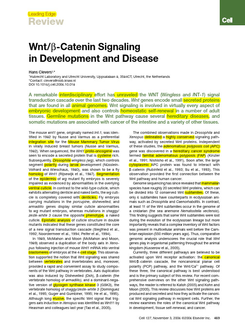
Leading EdgeReviewCell 127, November 3, 2006 ©2006 Elsevier Inc. 469The mouse wnt1 gene, originally named Int-1, was iden-tified in 1982 by Nusse and Varmus as a preferential integration site for the Mouse Mammary Tumor Virus in virally induced breast tumors (Nusse and Varmus, 1982). When sequenced, the Wnt1 proto-oncogene was seen to encode a secreted protein that is cysteine rich. Subsequently, Drosophila wingless (wg), which controls segment polarity during larval development (Nüsslein-Volhard and Wieschaus, 1980), was shown to be a fly homolog of Wnt1 (Rijsewijk et al., 1987). Segmentation of the epidermis of wg mutant fly embryos is severely impaired as evidenced by abnormalities in the overlying ventral cuticle. In contrast to the wild-type cuticle, which exhibits alternating denticle and naked belts, the wg cuti-cle is completely covered with denticles. Fly embryos carrying mutations in the porcupine , dishevelled , and armadillo genes display similar cuticle abnormalities to wg mutant embryos, whereas mutations in shaggy /zeste-white 3 cause the opposite phenotype, a naked cuticle. Epistatic analysis of cuticle structure in double mutants indicated that these genes constituted the core of a new signal transduction cascade (Siegfried et al., 1992; Noordermeer et al., 1994; Peifer et al., 1994).In 1989, McMahon and Moon (McMahon and Moon, 1989) observed a duplication of the body axis in Xeno-pus following injection of mouse Wnt1 mRNA into ventral blastomeres of embryos at the 4-cell stage. This observa-tion supported the notion that Wnt signaling was shared between vertebrates and invertebrates and, moreover, provided a rapid and convenient assay to study compo-nents of the Wnt pathway in vertebrates. Axis duplication was also induced by Dishevelled (Dsh), β-catenin (the vertebrate homolog of armadillo), and a dominant-nega-tive version of glycogen synthase kinase 3 (GSK3), the vertebrate homolog of shaggy /zeste-white 3 (Dominguez et al., 1995; Guger and Gumbiner, 1995; He et al., 1995). Although long elusive, the specific Wnt signal that trig-gers axis induction in Xenopus was identified as Wnt11 by Heasman and colleagues last year (Tao et al., 2005).The combined observations made in Drosophila and Xenopus delineated a highly conserved signaling path-way, activated by secreted Wnt proteins. Independent of these studies, the adenomatous polyposis coli (APC) gene was discovered in a hereditary cancer syndrome termed familial adenomatous polyposis (FAP) (Kinzler et al., 1991; Nishisho et al., 1991). Soon after, the large cytoplasmic APC protein was found to interact with β-catenin (Rubinfeld et al., 1993; Su et al., 1993). This observation provided the first connection between the Wnt pathway and human cancer.Genome sequencing has since revealed that mammalian species have roughly 20 secreted Wnt proteins, which can be divided into 12 conserved Wnt subfamilies. Of these, only 6 subfamilies have counterparts in ecdysozoan ani-mals such as Drosophila and Caenorhabditis . In contrast, at least 11 of the Wnt subfamilies occur in the genome of a cnidarian (the sea anemone Nematostella vectensis ). This finding suggests that some Wnt subfamilies were lost during the evolution of the ecdysozoan lineage but more importantly reveals that a complex inventory of Wnt factors was present in multicellular animals well before the Cam-brian explosion (550 million years ago). Thus, comparative genomic analysis underscores the crucial role that Wnt genes play in organismal patterning throughout the animal kingdom (Kusserow et al., 2005).Currently, three different pathways are believed to be activated upon Wnt receptor activation: the canonical Wnt/β-catenin cascade, the noncanonical planar cell polarity (PCP) pathway, and the Wnt/Ca 2+ pathway. Of these three, the canonical pathway is best understood and is the primary subject of this review. For recent com-prehensive overviews on the other Wnt signaling path-ways, the reader is referred to Katoh (2005) and Kohn and Moon (2005). This review discusses how Wnt proteins are produced and secreted and how they activate the canoni-cal Wnt signaling pathway in recipient cells. Further, the review examines the roles of the canonical Wnt pathway in development, tissue self-renewal, and cancer.Wnt/β-Catenin Signaling in Development and DiseaseHans Clevers 1,*1Hubrecht Laboratory and Utrecht University, Uppsalalaan 8, 3584CT, Utrecht, the Netherlands *Contact: clevers@niob.knaw.nl DOI 10.1016/j.cell.2006.10.018A remarkable interdisciplinary effort has unraveled the WNT (W ingless and I NT -1) signal transduction cascade over the last two decades. Wnt genes encode small secreted proteins that are found in all animal genomes. Wnt signaling is involved in virtually every aspect of embryonic development and also controls homeostatic self-renewal in a number of adult tissues. Germline mutations in the Wnt pathway cause several hereditary diseases, and somatic mutations are associated with cancer of the intestine and a variety of other tissues.470 Cell 127, November 3, 2006 ©2006 Elsevier Inc.Wnt Protein SecretionWnt proteins are characterized by a high number of con-served cysteine residues. Although Wnt proteins carry an N-terminal signal peptide and are secreted, they are relatively insoluble. This insolubility has been attributed to a particular protein modification, cysteine palmitoyla-tion, which is essential for Wnt function (Willert et al., 2003). Hofmann (2000) reported that a Drosophila gene required in the Wnt-secreting cell, termed porcupine , displays homology to acyl-transferases, enzymes that acylate a variety of substrates in the endoplasmic reticu-lum. Thus, porcupine and its worm homolog mom-1 are believed to encode the enzyme that is responsible for Wnt palmitoylation (Zhai et al., 2004).Recently, Banziger et al. (2006) and Bartscherer et al. (2006) uncovered in Drosophila another conserved gene that is essential for Wnt secretion, named wntless (wls) and evenness interrupted (evi), respectively. The gene encodes a seven-pass transmembrane protein that is conserved from worms (mom-3) to man (hWLS ). In the absence of Wls /evi , Wnts are retained inside the cell that produces them. The Wntless protein resides pri-marily in the Golgi apparatus, where it colocalizes and physically interacts with Wnts. A genetic screen in C. elegans revealed that the retromer, a multiprotein com-plex involved in intracellular trafficking and conserved from yeast to man, is also essential for Wnt secretion and for the generation of a Wnt gradient (Coudreuse et al., 2006). An attractive hypothesis is that the retromer complex is involved in recycling a Wnt cargo receptor (such as Wntless) between the default secretory path-way and a compartment dedicated to Wnt secretion (see Figure 1).Wnt is thought to act as a morphogen (that is, a long-range signal whose activity is concentration depend-ent) (reviewed in Logan and Nusse, 2004). However, it is unclear how these long-range gradients are generated. It is conceivable that the palmitoyl moiety constrains movement away from membranes or lipid particles. Thus, Wnts may be tethered to intercellular transport vesicles or lipoprotein particles (Panakova et al., 2005). Alternatively, Wnts may be transported by cytonemes, which are long, thin filopodial processes. Additionally, studies in Drosophila suggest a role for extracellular heparan sulfate proteoglycans (HSPG) in the transport or stabilization of Wnt proteins. For instance, flies car-rying mutations in Dally, a GPI-anchored HSPG, or in genes encoding enzymes that modify HSPGs resemble wingless mutants (reviewed in Lin, 2004).Receptors, Agonists, and Antagonists for WntWnts bind Frizzled (Fz) proteins, which are seven-pass transmembrane receptors with an extracellular N-ter-minal cysteine-rich domain (CRD) (Bhanot et al., 1996). The Wnt-Fz interaction appears promiscuous, in that a single Wnt can bind multiple Frizzled proteins (e.g., Bhanot et al., 1996) and vice versa. In binding Wnt, Fzs cooperate with a single-pass transmembrane molecule of the LRP family known as Arrow in Drosophila (Wehrli et al., 2000) and LRP5 and -6 in vertebrates (Pinson et al., 2000; Tamai et al., 2000). The transport of Arrow/LRP5/6 to the cell surface is dependent on a chaper-one called Boca in Drosophila and Mesd in mice (Culi and Mann, 2003; Hsieh et al., 2003). And consistent with a role of the Boca/Mesd chaperone in the transport of Arrow/LRP5/6 transport, mutations in Boca and Mesd resemble loss of Arrow /LRP5/6. Although it has not been formally demonstrated that Wnt molecules form trimeric complexes with LRP5/6 and Frizzled, surface expression of both receptors is required to initiate the Wnt signal.Derailed, a transmembrane tyrosine kinase receptor from the RYK subfamily, is an unusual Wnt receptor. Dro-sophila Wnt5 controls axon guidance in the central nerv-ous system. Embryos lacking Dwnt-5 resemble those lacking Derailed, that is, they generate aberrant neuronal projections across the midline (Yoshikawa et al., 2003). Derailed binds DWnt-5 through its extracellular WIF (Wnt inhibitory factor) domain. Signaling events downstream of this alternative Wnt receptor remain unclear. Some-what unexpectedly, the Derailed kinase domain may be dispensable for signaling. Lu et al. (2004) propose that, unlike the Drosophila Ryk homolog Derailed, mammalianRyk functions as a coreceptor along with Fz. MammalianFigure 1. Wnt SecretionTo be secreted, Wnt proteins in the endoplasmic reticulum (ER) need to be palmitoylated by the action of Porcupine. Wnt proteins also re-quire Wntless (Wls/Evi) in order to be routed to the outside of the cell. Loading onto lipoprotein particles may occur in a dedicated endo/ex-ocytic compartment. The retromer complex may shuttle Wls between the Golgi and the endo/exocytic compartment.Ryk binds Dishevelled to activate the canonical Wnt/β-catenin signaling pathway. Another tyrosine kinase receptor, Ror2, harbors a Wnt binding CRD motif. Wnt5a can engage Ror2 to inhibit the canonical Wnt signaling pathway, although paradoxically Wnt5a can also activate the canonical pathway by directly engaging Fz4 (Mikels and Nusse, 2006) and Fz5 (He et al., 1997).At least two types of proteins that are unrelated to Wnt factors activate the Frizzled/LRP receptors. One of these factors is the cysteine-knot protein Norrin, which is mutated in Norrie disease, a developmental disorder characterized by vascular abnormalities in the eye and blindness. Norrin binds with high affinity to Frizzled-4 and activates the canonical signaling pathway in an LRP5/6-dependent fashion (Xu et al., 2004). Other fac-tors that activate the canonical Wnt signaling pathway are R-spondins, which are thrombospondin domain-containing proteins. In Xenopus, R-spondin-2 is a Wnt agonist that synergizes with Wnts to activate β-catenin (Kazanskaya et al., 2004). Human R-spondin-1 has been found to strongly promote the proliferation of intestinal crypt cells, a process which involves the stabilization of β-catenin (Kim et al., 2005). Indeed, studies in cultured cells demonstrate that R-spondins can physically inter-act with the extracellular domains of LRP6 and Fzd8 and activate Wnt reporter genes (Nam et al., 2006).The secreted Dickkopf (Dkk) proteins inhibit Wnt signaling by direct binding to LRP5/6 (Glinka et al., 1998). Through this interaction, Dkk1 crosslinks LRP6 to another class of transmembrane molecules, the Kre-mens (Mao et al., 2002), thus promoting the internaliza-tion and inactivation of LRP6. An unrelated secreted Wntinhibitor, Wise, also acts by binding to LRP (Itasaki et al., 2003), as does the WISE family member SOST (Li et al., 2005; Semenov et al., 2005).Soluble Frizzled-Related Proteins (SFRPs) resemble the ligand-binding CRD domain of the Frizzled family of Wnt receptors (Hoang et al., 1996). WIF proteins are secreted molecules with similarity to the extracellular portion of the Derailed/RYK class of transmembrane Wnt receptors (Hsieh et al., 1999). SFRPs and WIFs are believed to function as extracellular Wnt inhibitors (reviewed in Logan and Nusse, 2004) but, depending on context, may also promote signaling by Wnt stabilization or by facilitating Wnt secretion or transport. Canonical Wnt SignalingOnce bound by their cognate ligands, the Fz/LRP coreceptor complex activates the canonical signal-ing pathway (Figure 2). Fz can physically interact with Dsh, a cytoplasmic protein that functions upstream of β-catenin and the kinase GSK-3. Wnt signaling controls phosphorylation of Dsh (reviewed in Wallingford and Habas, 2005). However, it remains unclear whether the binding of Wnt to Fz regulates a direct Fz-Dsh interac-tion, nor is it known how Dsh phosphorylation is con-trolled or how phosphorylated Dsh functions in Wnt signal transduction.Recent studies have indicated that the coreceptor LRP5/6 interacts with Axin through five phosphorylated PPP(S/T)P repeats in the cytoplasmic tail of LRP (David-son et al., 2005; Zeng et al., 2005). Wnts are thought to induce the phosphorylation of the cytoplasmic tail of LRP, thus regulating the docking of Axin. GSK3 phos-phorylates the PPP(S/T)P motif, whereas caseine kinase I-γ(CK1γ) phosphorylates multiple motifs close to the GSK3 sites. CK1γ is unique within the CK1 family in that it is anchored in the membrane through C-terminal palmi-toylation. Both kinases are essential for signal initia-tion. It remains presently debated whether Wnt controls GSK3-mediated phosphorylation of LRP5/6 (Zeng et al., 2005) or whether CK1γis the kinase regulated by Wnt (Davidson et al., 2005). When bound to their respective membrane receptors, Dsh and Axin may cooperatively mediate downstream activation events by heterodimeri-zation through their respective DIX (Dishevelled-Axin) domains.The Cytoplasmic Destruction ComplexThe central player in the canonical Wnt cascade is β-catenin, a cytoplasmic protein whose stability is regu-lated by the destruction complex. The tumor suppressor protein Axin acts as the scaffold of this complex as it directly interacts with all other components—β-catenin,the tumor suppressor protein APC, and the two kinase Figure 2. Canonical Wnt Signaling(Left panel) When Wnt receptor complexes are not bound by ligand, the serine/threonine kinases, CK1 and GSK3α/β, phosphorylate β-catenin. Phosphorylated β-catenin is recognized by the F box/WD repeat protein β-TrCP, a component of a dedicated E3 ubiquitin ligase complex. Following ubiquitination, β-catenin is targeted for rapid de-struction by the proteasome. In the nucleus, the binding of Groucho to TCF (T cell factor) inhibits the transcription of Wnt target genes. (Right panel) Once bound by Wnt, the Frizzled(Fz)/LRP coreceptor complex activates the canonical signaling pathway. Fz interacts with Dsh, a cy-toplasmic protein that functions upstream of β-catenin and the kinase GSK3β. Wnt signaling controls phosphorylation of Dishevelled (Dsh). Wnts are thought to induce the phosphorylation of LRP by GSK3βand casein kinase I-γ (CK1γ), thus regulating the docking of Axin. The recruitment of Axin away from the destruction complex leads to the stabilization of β-catenin. In the nucleus, β-catenin displaces Groucho from Tcf/Lef to promote the transcription of Wnt target genes.Cell 127, November 3, 2006 ©2006 Elsevier Inc. 471472 Cell 127, November 3, 2006 ©2006 Elsevier Inc.families (CK1α, -δ, -ε and GSK3α and -β [reviewed in Price, 2006]). When WNT receptor complexes are not engaged, CK1 and GSK3α/β sequentially phosphor-ylate β-catenin at a series of highly conserved Ser/Thr residues near its N terminus (Figure 2). Phosphorylated β-catenin is then recognized by the F box/WD repeat protein β-TrCP, a component of a dedicated E3 ubiq-uitin ligase complex. As a consequence, β-catenin is ubiquitinated and targeted for rapid destruction by the proteasome (Aberle et al., 1997). Note that the CK1 and GSK3 kinases perform paradoxical roles in the Wnt pathway. At the level of the LRP coreceptor they act as agonists, whereas in the destruction complex they act as antagonistsAlthough genetic observations imply an essential role for APC in the destruction complex, there is no consen-sus on its specific molecular activity. APC has a series of 15 and 20 amino acid repeats with which it interacts with β-catenin. Three Axin-binding motifs are interspersed between these β-catenin-binding motifs. Increasing the expression of Axin in cancer cells that lack APC restores the activity of the destruction complex, implying that APC is only essential when Axin levels are limiting. Quantita-tively, Axin indeed appears to be the limiting factor (Lee et al., 2003) and may be the key scaffolding molecule that promotes the rapid assembly and disassembly of the destruction complex.Given that CK1, Dsh, β-TrCP, and GSK3 participate in other signaling pathways, low levels of Axin may insu-late the Wnt pathway from changes in the abundance or activity of these signaling components. It has been proposed that APC is required for efficient shuttling and loading/unloading of β-catenin onto the cytoplasmic destruction complex. Both APC and Axin can them-selves be phosphorylated by their associated kinases, which changes their affinity for other components of the destruction complex. Our understanding of the rel-evance of these phosphorylation events in the regulation of Wnt signaling remains incomplete. For a comprehen-sive discussion of the kinases in the Wnt pathway, the reader is referred to a recent review (Price, 2006)β-catenin plays a second role in simple epithelia, that is, as a component of adherens junctions. It is an essen-tial binding partner for the cytoplasmic tail of various cadherins, such as E-cadherin (Peifer et al., 1992). Unlike the signaling pool of β-catenin, the pool that is bound to the adherens junction is highly stable. It is currently unclear whether the adhesive and signaling properties of β-catenin are interconnected. In a likely scenario, newly synthesized β-catenin first saturates the pool that is part of the adhesion junction, which never becomes available for signaling. “Excess,” free cytoplasmic β-catenin protein is then efficiently degraded by the APC complex. It is only this second, highly unstable pool that is subject to regulation by Wnt signals. In support of this model, these two functions of β-catenin are separately performed by two different β-catenin homologs in C. elegans (Korswagen et al., 2000).Upon receptor activation by WNT ligands, the intrinsic kinase activity of the APC complex for β-catenin is inhib-ited. It is unclear how this occurs, but it likely involves the Wnt-induced recruitment of Axin to the phosphorylated tail of LRP and/or to Fz-bound Dsh. As a consequence, stable, nonphosphorylated β-catenin accumulates and translocates into the nucleus, where it binds to the N terminus of LEF/TCF (lymphoid enhancer factor/T cell factor) transcription factors (Behrens et al., 1996; Mole-naar et al., 1996; van de Wetering et al., 1997).It has been suggested that protein phosphatases may regulate β-catenin stability as antagonists of the serine kinases (reviewed in Price, 2006). For example, heterotrimeric PP2A is required for the elevation of β-catenin levels that is dependent on Wnt. Moroever, PP2A can bind Axin and APC, suggesting that it might function to dephosphorylate GSK3 substrates. If and how PP2A activity is regulated by Wnt signals remains to be resolved.Crystallographic studies are starting to provide insights into the structure of the destruction complex. The central region of β-catenin (to which most partners bind) was the first component of the pathway to be crys-tallized. It consists of 12 armadillo repeats, which adopt a superhelical shape with a basic groove running along its length. Subsequently, structural interactions of Axin, APC, E-cadherin, and TCF with β-catenin have been vis-ualized (Choi et al., 2006, and references therein). APC, E-cadherin, and TCF bind the central part of the basic groove in a mutually exclusive fashion. Despite very lim-ited conservation of primary sequence in the respective interaction domains, the modes of binding are structur-ally very similar. Axin utilizes a helix that occupies the groove formed by the third and fourth armadillo repeats of β-catenin. Axin binding precludes thesimultaneousFigure 3. Transactivation of Wnt Target GenesThe β-catenin/T cf complex interacts with a variety of chromatin-remod-eling complexes to activate transcription of Wnt target genes. The re-cruitment of β-catenin to T cf target genes affects local chromatin in sev-eral ways. Bcl9 acts as a bridge between Pygopus and the N terminus of β-catenin. Evidence suggests that this trimeric complex is involved in nuclear import/retention of β-catenin (T ownsley et al., 2004), but it may also be involved in the ability of β-catenin to activate transcrip-tion (Hoffmans et al., 2005). The C terminus of β-catenin also binds to coactivators such as the histone acetylase CBP , Hyrax, and Brg-1 (a component of the SWI/SNF chromatin-remodeling complex).interaction with other β-catenin partners in this region. Based on this observation, it is suggested that a key function of APC is to remove phosphorylated β-catenin from the active site of the complex (Xing et al., 2003). In a further study, the structure of Axin bound to APC (Spink et al., 2000) was solved. These studies form step-ping stones to a better understanding of the dynamics of the destruction complex. Unfortunately, biochemical studies of the destruction complex in its different activa-tion states are sorely lacking.Nuclear EventsUpon stabilization by Wnt signals, β-catenin enters the nucleus to reprogram the responding cell (Figure 3). There is no consensus on the mechanism by which β-catenin travels between the cytoplasm and the nucleus. In many cases, cells that undergo Wnt signaling may actually display an overall rise in β-catenin protein without a clear nuclear preference. β-catenin’s nuclear import is independent of the Nuclear Localization Sig-nal/importin machinery. β-catenin itself is a close rela-tive of importin/karyopherins and directly interacts with nuclear pore components. Two proteins, Tcf and Pygo-pus are proposed to anchor β-catenin in the nucleus, although β-catenin can still localize to the nucleus in the absence of either of the two (reviewed in Staedeli et al., 2006). β-catenin can also be actively transported back to the cytoplasm, by either an intrinsic export signal or as cargo of Axin (Cong and Varmus, 2004) or APC (Rosin-Arbesfeld et al., 2000) that shuttle between cyto-plasm and nucleus.Whereas the fly and worm genomes both encode a single Tcf protein, the vertebrate genome harbors four Tcf/Lef genes. Tcf factors bind their cognate motif in an unusual fashion, i.e., in the minor groove of the DNA helix, while inducing a dramatic bend of over 90°. Tcf target sites are highly conserved between the four ver-tebrate Tcf/Lef proteins and Drosophila Tcf. These sites resemble AGATCAAAGG (van de Wetering et al., 1997). Wnt/TCF reporter plasmids such as pTOPflash (Korinek et al., 1997), widely used to measure Wnt pathway acti-vation, consist of concatamers of 3–10 of these binding motifs cloned upstream of a minimal promoter. The four vertebrate TCF/LEF differ dramatically in their embry-onic and adult expression domains, yet they are highly similar biochemically, explaining the extensive redun-dancy unveiled in double knockout experiments (as in Galceran et al., 1999).In the absence of Wnt signals, Tcf acts as a tran-scriptional repressor by forming a complex with Grou-cho/Grg/TLE proteins (Cavallo et al., 1998; Roose et al., 1998). The interaction of β-catenin with the N terminus of Tcf (Behrens et al., 1996; Molenaar et al., 1996; van de Wetering et al., 1997) transiently converts it into an activator, translating the Wnt signal into the transient transcription of Tcf target genes. To accomplish this, β-catenin physically displaces Groucho from Tcf/Lef (Daniels and Weis, 2005). The recruitment of β-catenin to Tcf target genes affects local chromatin in several ways. Its C terminus is a potent transcriptional activa-tor in transient reporter gene assays (van de Wetering et al., 1997). It binds coactivators such as the histone acetylase CBP and Brg-1, a component of the SWI/SNF chromatin remodeling complex (reviewed in Staedeli et al., 2006). A recent study implies that the human and fly homologs of yeast Cdc37 (Parafibromin and Hyrax, respectively) also interact with the C-terminal transacti-vation domain of β-catenin to activate target gene tran-scription (Mosimann et al., 2006). Cdc37 is a component of the PAF complex. In yeast the PAF complex directly interacts with RNA polymerase II to regulate transcrip-tion initiation and elongation.Two dedicated, nuclear partners of the TCF/β-cat-enin complex, Legless/Bcl9 and Pygopus, were recently found in genetic screens in Drosophila(Kramps et al., 2002; Parker et al., 2002; Thompson et al., 2002). Muta-tions in these genes result in phenotypes similar to wingless, and overexpression of both genes promotes TCF/β-catenin activity in mammalian cells (Thompson et al., 2002). Bcl9 bridges Pygopus to the N terminus of β-catenin. The formation of this trimeric complex has been implicated in nuclear import/retention of β-catenin (Townsley et al., 2004) but may also directly contribute to the ability of β-catenin to transactivate transcription (Hoffmans et al., 2005). Although most if not all Wnt sig-naling events in Drosophila appear to be dependent on Bcl9 and Pygopus, it is currently unclear if this holds true in vertebrate development.Tcf itself can be regulated by phosphorylation. The MAP kinase-related protein kinase NLK/Nemo (Ishitani et al., 1999) phosphorylates Tcf, thereby decreasing the DNA-binding affinity of the β-catenin/Tcf complex and inhibiting transcriptional regulation of Wnt target genes. In C. elegans, LIT-1/NLK-dependent phosphorylation results in PAR-5/14-3-3- and CRM-1-dependent nuclear export of POP-1/Tcf (Meneghini et al., 1999; Lo et al., 2004). And lastly, a recent study utilizing chromatin immunoprecipitations suggests that APC, independent of its role in the cytoplasmic destruction complex, acts on chromatin to facilitate CtBP-mediated repression of Wnt target genes in normal, but not in colorectal cancer cells (Sierra et al., 2006).Wnt Target GenesLoss of components of the Wnt pathway can produce dramatic phenotypes that affect a wide variety of organs and tissues. A popular view equates Wnt signaling with maintenance or activation of stem cells (Reya and Cle-vers, 2005). It should be realized, however, that Wnt signals ultimately activate transcriptional programs and that there is no intrinsic restriction in the type of bio-logical event that may be controlled by these programs. Thus, Wnt signals may promote cell proliferation and tissue expansion but also control fate determination or terminal differentiation of postmitotic cells. Sometimes, these disparate events, proliferation and terminal differ-entiation, can be activated by Wnt in different cell typesCell 127, November 3, 2006 ©2006 Elsevier Inc. 473474 Cell 127, November 3, 2006 ©2006 Elsevier Inc.within the same structure, such as the hair follicle or the intestinal crypt (Reya and Clevers, 2005).Numerous Tcf target genes have been identified in diverse biological systems. These studies tend to focus on target genes involved in cancer, as exemplified by the wide interest in the Wnt target genes cMyc and Cyclin D1. For a comprehensive, updated overview of Tcf tar-get genes, the reader is referred to the Wnt homepage (/~rnusse/wntwindow.html). The Wnt pathway has distinct transcriptional outputs, which are determined by the developmental identity of the responding cell, rather than by the nature of the signal. In other words, the majority of Wnt target genes appear to be cell type specific. It is not clear whether “universal” Wnt/Tcf target genes exist. The best cur-rent candidates in vertebrates are Axin2/conductin (Jho et al., 2002) and SP5 (Weidinger et al., 2005). As noted (Logan and Nusse, 2004), Wnt signaling is autoregulated at many levels. The expression of a variety of positive and negative regulators of the pathway, such as Friz-zleds, LRP and HSPG, Axin2, and TCF/Lef are all con-trolled by the β-catenin/TCF complex.Wnt Signaling in Self-Renewing Tissues in Adult MammalsWnt signaling not only features in many developmental processes; in some self-renewing tissues in mammals it remains essential throughout life. It is this aspect of Wnt signaling that is intricately connected to the development of disease. The examples discussed below illustrate how the Wnt pathway is involved in adult tissue self-renewal. Mutations in the Wnt pathway tip the homoeostatic bal-ance in these tissues to cause pathological conditions such as disturbances in skeletal bone mass or cancer.GutThe absorptive epithelium of the small intestine is ordered into numerous villi and crypts. It constitutes the most rapidly self-renewing tissue in adult mammals. In the mouse, the epithelium turns over entirely within 3–5 days. A massive rate of cell production by a “transit-amplify-ing” crypt compartment is compensated by apoptosis at the tip of the villus. The proliferating crypt precursors and differentiated villus cells form a contiguous sheet of cells that is in perpetual upward motion. Stem cells reside near the bottom of the crypt and escape this flow. These slowly cycling cells produce the transit-amplify-ing progenitor cells that are capable of differentiating toward all epithelial lineages. The stem cells self-renew throughout life and regenerate the epithelium after injury (Figure 4). Committed progenitors arrest their cell cycle and differentiate when they reach the crypt-villus junc-tion (reviewed in Reya and Clevers, 2005).Current evidence indicates that the Wnt cascade is the dominant force in controlling cell fate along the crypt-vil-lus axis. In neonatal mice lacking Tcf4, the differentiatedvillus epithelium appears unaffected, but the crypt pro-Figure 4. Self-Renewing Tissues in the Adult Mammal(Left) In the intestinal epithelium, proliferating crypt precursors and differentiated villus cells form a contiguous sheet of cells that is in per-petual upward motion. Stem cells, which produce the transit-amplifying progenitor cells, reside near the bottom of the crypt and escape this flow. Evidence suggests that Wnt signaling is required for the establishment of the progenitor compartment in the intestinal epithelium. Wnt proteins also promote the terminal differentiation of Paneth cells at the base of the crypt. A small adenoma carrying a mutation in APC grows inside the right-hand villus. As a consequence of the loss of APC, β-catenin protein accumulates to high levels in the adenoma. (In-set) A section of a mouse small intestine displaying crypts, villi, and a growing adenoma. Staining for β-catenin (brown) reveals its pres-ence in adhesion junctions as well as a modest accumulation at the bottom of the crypts due to local Wnt signaling. Photo by Daniel Pinto. (Right) Wnt signaling in hair follicles activates bulge stem cells, promotes entry into the hair lineage, and recruits the cells to the transit-amplify-ing matrix compartment.。
西红花苷基于Wntβ-catenin信号通路改善阿尔茨海默病大鼠的空间记忆
林玲等西红花苷基于WnU'—atemn信号通路改善阿尔茨海默病大鼠的空间记忆第1期-153-西红花苷基于WnU'—atenin信号通路改善阿尔茨海默病大鼠的空间记忆林玲1刘国良2杨丽娜1高丽1(河南医学高等专科学校1生理学教研室,河南郑州451191;2预防医学教研室)〔摘要〕目的观察西红花苷对阿尔茨海默病(AD)大鼠海马突触小泡蛋白(SYP)和Wnt/'catemn信号通路的影响。
方法SD大鼠随机分为正常对照组、AD模型组、西红花苷组。
侧脑室注射'淀粉样蛋白(A'25~35)建立AD大鼠模型,西红花苷组从次日开始腹腔注射西红花苷(40mgky),连续注射14d$Mor n s水迷宫实验检测各组学习记忆能力,免疫组化、免疫荧光和Western印迹检测海马SYP的表达‘Western印迹检测WnUL—atenin信号通路主要蛋白GSK3'、'-catenin、p-'-/</-蛋白的表达。
结果在Morns水迷宫实验中,相比于正常对照组,AD模型组学习记忆能力明显减退,相比于AD模型组,西红花苷组学习记忆能力明显增强。
免疫组化、免疫荧光和Western印迹实验中,AD模型组海马SYP的表达显著少于正常对照组"P<0.05),西红花苷组SYP的表达显著高于AD模型组(P<0.05)$在Western印迹中,与正常对照组比较,AD模型组大鼠'-atenin的表达明显减少(P<0.05),GSK3'、pCcatenin的表达明显增加(P<0.05);与AD模型组比较,西红花苷组'-atenin的表达明显增加(P<0.05),GSK3'、pF-atenin的表达明显减少(P<0.05)$结论西红花苷可通过激活WnUL-c/emn信号通路,增加海马SYP的表达,改善AD大鼠空间记忆能力。
Wnt3a重组蛋白溶剂对脑梗死模型大鼠干预效果及作用机制
剂组 Wnt3a、β ̄catenin 蛋白相对表达量升高ꎬ差异有统计学意义( P<0 05) ꎮ 与模型组相比ꎬWnt3a nt3a 溶剂组 SOD、GSH 水平降低ꎬMDA 水
1 2 3 氧化应激、炎症指标检测 抽取大鼠腹部静
10 minꎬ分离上层血清ꎮ 采用硫代巴比妥酸检测丙
二醛( MDA) 水平ꎻ采用黄嘌呤氧化酶反应检测超氧
化物歧化酶( SOD) 水平ꎻ采用比色法检测谷胱甘肽
( GSH) 水平ꎮ 采用酶联免疫吸附试验检测肿瘤坏
显色ꎬ定量分析蛋白表达情况ꎮ 以 GAPDH 为内参ꎮ
1 3 统计学处理 采用 SPSS20 0 软件行方差分
析、t 检验ꎮ
组和模型组给予等体积的生理盐水进行干预ꎮ 三组
大鼠均连续干预 1 wꎮ
2 结 果
脉血 3 mlꎬ 以 3 000 r / min 离 心 速 度ꎬ 离 心 处 理
白相对表达量降低ꎬ差异有统计学意义( P<0 05) ꎮ 结论 Wnt3a 重组蛋白溶剂对脑梗死模型大鼠干预效果显著ꎬ能抑制其氧
化应激、炎症反应ꎬ减少脑梗死体积ꎬ提高神经功能ꎬ其作用机制可能与 Wnt3a / β ̄catenin 信号通路有关ꎮ
〔 关键词〕 Wnt 信号通路ꎻWnt3a 蛋白ꎻ脑梗死
用相关研究较少ꎮ 本文旨在研究 Wnt3a 重组蛋白
袁小冬等 Wnt3a 重组蛋白溶剂对脑梗死模型大鼠干预效果及作用机制 第 5 期
1093
溶剂对脑梗死模型大鼠的干预效果及作用机制ꎬ为
1 2 5 脑组织病理学观察及脑梗死体积统计 处
Wnt通路对胰岛素样生长因子-1抑制小鼠毛细胞凋亡的作用
Wnt通路对胰岛素样生长因子-1抑制小鼠毛细胞凋亡的作用宫亮;李里;陈冬【期刊名称】《中国生化药物杂志》【年(卷),期】2012(33)6【摘要】目的探讨胰岛素样生长因子-1(IGF-1)抑制小鼠毛细胞凋亡作用的调控机制.方法体外培养新生小鼠耳蜗基底膜.在分别含有正常对照培养波(A组)、0.5 mmol/L庆大霉素(GM)(B组)、0.5 mmol/L GM+1 ng/mL IGF (C组)、0.5 mmol/L GM+1 ng/mL IGF+5μg/mL wif(D组)、0.5 mmol/L GM+1 ng/mL IGF+ 10 μg/mLwif(E组)、0.5 mmol/L GM+1 ng/mL IGF+ 15μg/mL wif(F组)的成分中培养24 h后收集细胞及固定.应用TUNEL法观察各组耳蜗毛细胞的凋亡情况.应用Western blot及Real-time PCR法观察Caspase-3的表达情况.结果 B 组耳蜗毛细胞经刺激后,细胞凋亡数量及Caspase-3 mRNA和蛋白表达较A组明显上升(P<0.01),C组耳蜗毛细胞经IGF刺激后,细胞凋亡数量及Caspase-3mRNA和蛋白表达较B组明显下降(P<0.01),应用Wnt阻断剂(wif)后D、E及F 组细胞凋亡数量及Caspase-3 mRNA和蛋白表达较C组升高,其中F组升高显著(P<0.01).结论 IGF可能通过激活Wnt通路抑制毛细胞凋亡.%Purpose To investigate the gene regulation mechanism of Insulin-like Growth Factor-1 (IGF-1) antiapoptosis activity on rat hair cells. Methods Basilar membrane of cochlea of newly born mice were cultured in vitro in six group: control culture solution(group A) ,0. 5 mmol/L GM(gentami-cin)( group B) ,0.5 mmol/L GM +1 ng/mL IGF-1 (group C),0. 5 mmol/L GM +1 ng/mL IGF-1 +5 μg/mL wif( group D) ,0.5 mmol/L GM + 1 ng/mL IGF-1 + 10 μg/mLwif( group E) ,0. 5 mmol/L GM + 1 ng/mL IGF-1 +15 μg/mL wif ( group F). After cultured, cochlear preparation were made by used TUNEL method, the apoptosis of cochlear hair cells was observed with laser scanning confocal microscope. Caspase-3 protein and m RNA was examed by western blot and real-time PCR. Results There was no apoptosis cell labeled by TUNEL in basilar membrane in group A. Apoptosis cells labeled by TUNEL in basal,in group B, was higher than that in group A(P<0.05). Apoptosis cells in group C was lower than that in group B (P < 0. 05). Apoptosis cells in group D, E, F were higher than that in group C, espe-cialy in group F. The expression of Caspase-3 showed the same result. Conclusion Wntpathway plays an important role in the process of apoptosis in cochlear hair cells by IGF-1.【总页数】3页(P766-768)【作者】宫亮;李里;陈冬【作者单位】辽宁医学院附属第一医院耳鼻咽喉科,辽宁锦州121000;辽宁医学院儿科,辽宁锦州121000;辽宁医学院附属第一医院耳鼻咽喉科,辽宁锦州121000【正文语种】中文【中图分类】R961【相关文献】1.胰岛素样生长因子-1抑制庆大霉素引起的小鼠耳蜗毛细胞凋亡 [J], 马志云;宫亮;王雪峰2.环磷腺苷效应元件结合蛋白磷酸化在胰岛素样生长因子-1抑制耳蜗毛细胞凋亡中的作用 [J], 宫亮;陈冬;李里3.激活Wnt信号通路阻滞沉默ZEB2对结直肠癌细胞增殖抑制和凋亡促进作用 [J], 杨洲;刚海菊;覃先蓬;贾贵清;王波;杨春4.隐丹参酮通过G蛋白偶联受体55调节Wnt/β-catenin信号通路抑制舌鳞状细胞癌细胞增殖、凋亡及其作用机制研究 [J], 方松城; 林钊宇; 叶志佳; 刘志国5.胰岛素样生长因子1抑制小鼠耳蜗毛细胞凋亡 [J], 陈冬;李永新;韩德民因版权原因,仅展示原文概要,查看原文内容请购买。
- 1、下载文档前请自行甄别文档内容的完整性,平台不提供额外的编辑、内容补充、找答案等附加服务。
- 2、"仅部分预览"的文档,不可在线预览部分如存在完整性等问题,可反馈申请退款(可完整预览的文档不适用该条件!)。
- 3、如文档侵犯您的权益,请联系客服反馈,我们会尽快为您处理(人工客服工作时间:9:00-18:30)。
ARTHRITIS&RHEUMATISMVol.63,No.2,February2011,pp513–522DOI10.1002/art.30116©2011,American College of RheumatologyAlterations in Wnt Pathway Activity in Mouse Serum and Kidneys During Lupus DevelopmentAnders Aune Tveita and Ole Petter RekvigObjective.The canonical Wnt/-catenin pathway was recently identified as a factor in the pathogenesis of several renal diseases.The aim of this study was to evaluate Wnt signaling activity during disease develop-ment in a murine model of lupus nephritis.Methods.Wnt activity and Dkk-1expression were serially assayed in the serum and kidneys of(NZB؋NZW)F1mice during progression of lupus nephritis. The effects of serum obtained from mice with lupus and serum-equivalent concentrations of Dkk-1on mesangial cells were assessed in vitro.Results.Gene expression analyses revealed in-creased canonical Wnt pathway activity in kidneys during development of lupus nephritis,paralleled by an increase in renal and serum levels of the Wnt inhibitor Dkk-1.Sera obtained from proteinuric-stage(NZB؋NZW)F1mice showed strong Wnt-inhibitory effects in vitro.Dkk-1concentrations comparable to those ob-served in lupus-prone mice induced apoptosis in tubular and mesangial cells in vitro,whereas no such effect was seen for the range of concentrations observed in young prediseased mice and control BALB/c mice.Conclusion.These data demonstrate that renal Wnt signaling activity is increased in lupus and is accompanied by an increase in renal and serum levels of Dkk-1.The Wnt pathway is involved in the turnover of extracellular matrix constituents and represents a po-tential mediator of the morphologic changes that occur within the glomerulus during the development of ne-phritis.Furthermore,increased levels of Dkk-1serve as a potential proapoptotic stimulus in vitro and possibly in vivo and could be an important element in the initiation and progression of systemic and end-organ disease manifestations in systemic lupus erythemato-sus.The Wnt signaling pathway is an intracellular signaling cascade that was initially linked to embryogen-esis and cancer development(1).Understanding of the roles of Wnt signaling in postembryonic mammals is currently increasing,and during the last few years, interest in this signaling network has increased due to the discovery of its involvement in several important physiologic and pathophysiologic conditions.The classic, so-called canonical Wnt pathway is activated through binding of Wnt agonist proteins to members of the Frizzled family of receptors.This interaction initiates an intracytoplasmic signal transduction cascade that pre-vents glycogen synthase kinase3(GSK3)–mediated degradation of-catenin,followed by translocation of -catenin into the nucleus,where it associates with the T cell factor/lymphocyte enhancement factor(TCF/LEF) family of transcription factors.These transcription fac-tors activate a variety of target genes involved in cellular growth and differentiation.The canonical Wnt/-catenin pathway is tightly regulated by several secreted antagonists,including the secreted Frizzled-related pro-teins,Wnt inhibitory factor,and the Dickkopf protein family.Of these,Dkk-1is a specific inhibitor of canon-ical Wnt/-catenin signaling and has been studied in the context of regulation of the Wnt pathway(2).Systemic lupus erythematosus(SLE)is an auto-immune disease characterized by the development of autoreactivity against nuclear antigens,including double-stranded DNA(dsDNA).In(NZBϫNZW)F1 (NZB/NZW)mice,a lupus-like disease develops spon-taneously and is complicated by severe,progressive nephritis.The production of circulating anti-dsDNA autoantibodies is accompanied by the deposition of immune complexes within extracellular matrices,partic-Supported by the Northern Norway Regional Health Author-ity Medical Research Program(grants SFP-100-04and SFP-101-04). Dr.Rekvig received Milieu support from the University of Tromsø.Anders Aune Tveita,MD,PhD,Ole Petter Rekvig,MD, PhD:Institute of Medical Biology,University of Tromsø,Tromsø, Norway.Address correspondence to Anders Aune Tveita,MD,PhD, Molecular Pathology Research Group,Institute of Medical Biology, University of Tromsø,N-9037Tromsø,Norway.E-mail:anders.aune. tveita@uit.no.Submitted for publication May8,2010;accepted in revised form October21,2010.513ularly the glomerular basement membrane.This process is commonly accompanied by the apparent expansion of extracellular matrices.The initiating mechanisms re-main unknown but are thought to involve signaling pathways triggered by the deposited immune complexes.In the current study,we analyzed whether the Wnt signaling pathway may be involved in progression of lupus nephritis,based on emerging reports indicative of a role of this pathway in renal extracellular matrix homeostasis.Increased renal Wnt signaling was recently observed in various models of renal fibrosis,including unilateral ureteral obstruction(3,4).In this model,acti-vation of the Wnt/-catenin pathway caused increased interstitial expression of extracellular matrix constitu-ents,including type I procollagen and fibronectin(4). Moreover,inhibition of Wnt signaling by overexpression of the secreted Wnt antagonist Dkk-1attenuated inter-stitial collagen matrix accumulation and the severity of renal fibrosis(4).These data point to Wnt signaling as a potential intervention target in fibrotic and possibly inflammatory renal disease processes(5).In addition, increased Wnt signaling has been shown to induce the expression of matrix metalloproteinases(MMPs)(6,7), which may be of importance in extracellular matrix remodeling and the loss of membrane integrity that occur in lupus nephritis(8).Less is known about the roles of Wnt signaling in glomerular cells and diseases affecting the glomerular compartment.A recent study revealed that adriamycin-induced nephropathy caused Wnt/-catenin signaling in podocytes and demonstrated a crucial role of-catenin expression in the consequent development of protein-uria(9).Increased-catenin expression was also dem-onstrated in biopsy specimens obtained from patients with diabetic nephropathy and focal segmental glomer-ulosclerosis(9).Although the spectrum of effects of renal Wnt signaling remains incompletely characterized, these data are suggestive of a role of Wnt/-catenin signaling in several glomerulopathies(9).Based on these results and as part of the ongoing effort to characterize the molecular changes underlying the development and progression of lupus nephritis,we evaluated renal Wnt signaling in NZB/NZW mice at different stages of disease progression.Various disturbances in the clearance of apopto-tic cells have been reported and are thought to play a role in the development of SLE(10,11),but key ele-ments in the pathogenesis of the disease are still un-known.Conceivably,surpassing the capacity for apopto-tic cell clearance could trigger a local inflammatory reaction via the exposure of endogenous danger signals (12).Under such circumstances,apoptotic cell antigens are taken up by antigen-presenting cells(e.g.,dendritic cells)that have been activated by proinflammatory signals.This allows for immunogenic presentation of self antigens,which might launch and maintain an auto-immune response(for review,see ref.13).In this respect,an increased load of apoptotic cells has been proposed to inhibit clearance mechanisms(14).An emerging aspect of Wnt signaling seems to be its role in the protection of cells against apoptosis upon exposure to various types of cellular stress,at least in vitro(15,16). The aim of the current study was to evaluate whether alterations in Wnt pathway activity could influence the apoptotic cell load during the development of lupus in NZB/NZW mice.MATERIALS AND METHODSAnimals,tissue samples,and cell culture.Female NZB/NZW mice and BALB/c mice were purchased from Harlan.The treatment and care of the mice were performed in accordance with the guidelines of the Norwegian Ethical and Welfare Board for Research Animals,and the study was approved by the Institutional Review Board.Serum samples from NZB/NZW mice and BALB/c mice were collected every second week and stored atϪ20°C. Proteinuria in NZB/NZW mice was monitored weekly by semiquantitative dipstick measurements(Bayer).At the age of 10weeks and20weeks or upon development of overt nephritis (proteinuriaϾ3gm/liter),mice were killed by CO2suffocation. The kidneys were extirpated,cut,and preserved in RNAlater (Qiagen)for further analysis of gene expression or were snap-frozen in liquid nitrogen.Mouse mesangial cells were obtained from the Amer-ican Type Culture Collection and cultured in Dulbecco’s modified Eagle’s medium/Nutrient Mixture F-12Ham(Sigma-Aldrich)with20%fetal bovine serum,2m M L-glutamine, 50m M penicillin,and50m M streptomycin.Clonetics human renal proximal tubular epithelial cells(Lonza)were main-tained in serum-free renal epithelial cell culture medium with supplements provided by the manufacturer.All cells used were from passage4.Recombinant human and mouse Dkk-1were obtained from R&D Systems.RNA extraction and complementary DNA(cDNA) synthesis.Total RNA was isolated from RNAlater-preserved kidneys(5mice in each group)using EZ1RNA Tissue Mini Kit(Qiagen).The concentration of extracted RNA was deter-mined spectrophotometrically with a NanoDrop spectrophoto-meter.Samples(2g RNA in100-l reaction mixture)were reverse transcribed into cDNA with random primers,using a High Capacity cDNA Reverse Transcription Kit(Applied Biosystems).Real-time polymerase chain reaction(PCR).Quanti-tative PCR was performed using an ABI Prism7900HT Sequence Detection System(Applied Biosystems).Prede-signed FAM-labeled primer/probe sets(Applied Biosystems) specific for the analyzed genes(Axin2,accession no. Mm00443610_m1;Actb,accession no.Mm00607939_s1;Dkk1, accession no.Mm00438422_m1;Myc,accession no. Mm00487803_m1;Mycn,accession no.Mm00627179_m1;514TVEITA AND REKVIGPpard,accession no.Mm00803184_m1;Lef1,accession no. Mm01310389_m1)were used,and TATA box binding protein (accession no.Mm00498583_m1)was used as an endogenous control.FAM-labeled primer/probe sets for additional TCF/ LEF-responsive genes were obtained from Integrated DNA Technologies,and the sequences are shown in the legend of Table1.The relative expression levels were calculated usingthe⌬⌬Ct method.Normalization was accomplished by com-paring the relative expression for each sample with that for one of the4-week-old control BALB/c mice.For cell culture experiments,samples were normalized against untreated con-trols.Immunofluorescence microscopy.Four-micrometer–thick OCT compound–embedded cryostat sections of kidney were blocked with10%calf serum and1%bovine serum albumin(BSA)in phosphate buffered saline(PBS).The sections were subsequently incubated with a rabbit anti-human Dkk-1polyclonal antibody,rabbit anti-human phosphorylated glycogen synthase kinase3(GSK3;Santa Cruz Biotechnol-ogy),or a rabbit anti-human-catenin polyclonal antibody (Abcam)followed by incubation with an Alexa546–conjugatedanti-rabbit F(abЈ)2secondary antibody(Invitrogen).Normalrabbit IgG was used as a negative control.For immunostaining using a mouse anti-human acti-vated-catenin monoclonal antibody(Millipore),ultrathin zinc-fixated cryosections were incubated with0.1mg/ml anti-mouse F(abЈ)2overnight at4°C to reduce background staining by in vivo–bound antibodies.Sections were then processed as described above.All sections were run in parallel,with the omission of the primary antibody to evaluate background staining for each section.Anti-dsDNA and anti–Dkk-1enzyme-linked immu-nosorbent assay(ELISA).Serum antibodies against calf thy-mus dsDNA were detected and titrated by standard indirect ELISA,as previously described(17).A murine monoclonal anti-dsDNA antibody was included as an intraassay control for the cutoff level(18).Detection of serum Dkk-1was performed using the mouse Dkk-1DuoSet ELISA kit(R&D Systems) according to the supplied protocol.Serum samples were diluted1:6in PBS with1%BSA.Protein isolation and Western blot analysis.Protein extracts were prepared from snap-frozen kidney tissue by homogenization in Tris buffer(0.5moles/liter Tris HCl,pH 7.5,150mmoles/liter NaCl).The samples were then centri-fuged for5minutes,and the supernatant was collected. Nuclear and nucleus-depleted lysates were prepared from 20-mg pieces of snap-frozen lyophilized kidney tissue,using the Pierce Nuclear and Cytoplasmic Extraction Reagent Kit. Total protein was measured,and the protein content of the samples was normalized using the Pierce BCA Protein Assay.Sodiumdodecylsulfate–polyacrylamidegelelectrophor-esis and Western blotting were performed according tostan-Figure1.Wnt/-catenin pathway activity in(NZBϫNZW)F1(NZB/NZW;B/W)mouse kidneys.Real-time polymerase chain reaction analysis was performed on RNA isolated from NZB/NZW and BALB/c mice of different ages(nϭ5per age group).Gene expression data are shown for -catenin(A)and the T cell factor/lymphocyte enhancement factor–regulated genes Axin2(B),Ppard(C),and Lef1(D).These data demonstrate an increase in gene expression during progression toward proteinuria.Gene expression in BALB/c mice of corresponding ages remained stable or decreased with increasing age.Expression levels were normalized against that of TATA box binding protein and are expressed as fold change values relative to8-week-old mice,using the⌬⌬C t method.Values are the meanϮSEM.ءϭPϽ0.05;ءءϭPϽ0.01;ءءءϭPϽ0.001.Wnt PATHWAY AND Dkk-1IN SYSTEMIC LUPUS ERYTHEMATOSUS515dard procedures.Membranes were blocked with5%(weight/ volume)BSA and probed with the relevant primary antibody overnight at4°C.Membranes were washed and incubated with a horseradish peroxidase–conjugated secondary antibody,and binding was assayed by chemiluminescence detection.The determination of molecular weight was performed using MagicMark XP molecular weight markers(Invitrogen).Flow cytometry.Cell samples were analyzed on a FACSCalibur cytometer(BD Biosciences).Apoptosis was detected using an annexin V–fluorescein isothiocyanate and propidium iodide staining kit(BD Biosciences)according to the protocol supplied by the manufacturer.Data were analyzed using CellQuest software(BD Biosciences).Statistical analysis.Results are presented as the meanϮparisons between groups were performed by one-way analysis of variance followed by Bonferroni’s correction for multiple comparisons,using GraphPad Prism version5.0.P values less than0.05were considered significant.RESULTSIncreased expression of messenger RNA(mRNA) for-catenin and TCF/LEF-responsive genes in kidneys from preproteinuric-and proteinuric-stage NZB/NZW mice.Real-time PCR analysis of mRNA isolated from kidney homogenates revealed an increase in the expres-sion of-catenin,occurring atϳ20weeks and further increasing during the progression toward proteinuria (Figure1A).This increase in gene expression paralleled the appearance of significant titers of anti-dsDNA auto-antibodies(see below).In contrast,-catenin was stably expressed at a lower level in age-matched BALB/c mice (Figure1A).In order to determine whether the increase in-catenin expression was a reflection of increased Wnt activity,the mRNA expression of several known TCF/LEF-responsive genes,including Axin2,Ppard, Myc,Lef1,and RhoU(19,20),was assayed.The results revealed a consistent increase in the expression of these genes in NZB/NZW mice(Figures1B–D and Table1) that paralleled the increase in-catenin mRNA expres-sion,suggesting that canonical Wnt signaling is in-creased within the kidneys during the development of lupus nephritis.In contrast,a reduction in Wnt pathway activity with increasing age was seen in BALB/c mice (data not shown).Immunofluorescence studies of kidney biopsy specimens revealed an increase in total-catenin stain-ing throughout the tubular and glomerular compart-ments,paralleling the increase in-catenin mRNA expression(Figure2B).Glomerular staining was partic-ularly strong in proteinuric-stage mice.These findings suggest that the observed increase in Wnt/-catenin activ-ity occurs diffusely throughout the kidney.Immunostaining of cryosections gave no detectable signals for active -catenin in BALB/c mice or10-week-old NZB/NZW mice,whereas diffuse intracellular staining was seen in 20-week-old NZB/NZW mice(Figure2C).Similarly, immunoblotting against the phosphorylated,inactive form of GSK3was increased in20-week-old NZB/NZW mice (Figure3C),with corresponding speckled intracytoplasmic immunostaining in cryosections from these mice(Figure 2D).Immunoblotting of nuclear protein extracts revealed increased expression of the active form of-catenin in 20-week-old NZB/NZW mice(Figure2A),further sup-porting activation of Wnt signaling in these mice.Table1.Expression of selected TCF/LEF-responsive genes*Mice Myc RhoU Foxn1Dvl2JunBALB/c 1.42Ϯ0.36 1.15Ϯ0.290.85Ϯ0.55 1.02Ϯ0.150.46Ϯ0.04(NZBϫNZW)F110weeks old 1.02Ϯ0.25 1.02Ϯ0.28 1.01Ϯ0.11 1.00Ϯ0.04 1.00Ϯ0.0820weeks old 1.48Ϯ0.26† 1.39Ϯ0.28† 1.65Ϯ0.45 1.31Ϯ0.14†0.83Ϯ0.18Proteinuric8.5Ϯ3.52† 5.19Ϯ1.21†18.38Ϯ10.3† 1.77Ϯ0.48† 2.81Ϯ1.65†*Values are the meanϮSEM fold change relative to the average expression level in10-week-old(NZBϫNZW)F1mice(nϭ5)after normalization for the expression level of TATA box binding protein withinthe same samples.The following forward primer and probe sequences were used:for Tbp,forwardAAGAAAGGGAGAATCATGGACC,probe CCTGAGCATAAGGTGGAAGGCTGTT,reverseGAGTAAGTCCTGTGCCGTAAG;for Myc,forward GCTGTTTGAAGGCTGGATTTC,probe CG-TAGTCGAGGTCATAGTTCCTGTTGGT,reverse GATGAAATAGGGCTGTACGGAG;for RhoU,forward CCCACCGAGTACATCCCTAC,probe ATGAGTTTGACAAGCTGAGGCCCC,reverseTGTCTGTGTTGGTGTAGCAG;for Foxn1,forward AAGAACAGTAAGACCGGAAGC,probe TTCT-GTTCGCCATAACCTGTCCCTC,reverse TCTCCACCTTCTCAAAGCAC;for Dvl2,forward TTTCAA-GAGCGTTTTGCAGC,probe TCCGATGACAATGCCCGCCTAC,reverse ACACAAGCCAGGAGA-CAAC;for Jun,forward TTGTTACAGAAGCAGGGACG,probe AGGCTAACCCCGCGTGAAGT,reverse GTCGTAGAAGGTCGTTTCCATC.TCF/LEFϭT cell factor/lymphocyte enhancement factor.†PϽ0.05versus BALB/c mice.516TVEITA AND REKVIGIncreased renal expression of Dkk-1during de-velopment of lupus nephritis.Dkk-1is a secreted in-hibitor of the canonical Wnt pathway and is known to be induced by canonical Wnt signaling (21),probably serving as a negative feedback mechanism.In line with our findings of increased expression of TCF/LEF-responsive genes,mRNA expression of Dkk-1in kidney homogenates increased beginning at 20weeks of age (Figure 3A).Immunofluorescence staining similarly revealed a strong increase in Dkk-1expression in both tubular and glomerular cells from NZB/NZW mice,in both the preproteinuric (20weeks old)and proteinuric groups compared to younger NZB/NZW mice,in which Dkk-1immunostaining was weaker,at levels comparable to those in BALB/c mice (Figure 3B).Western blotting revealed similar increases in Dkk-1expression in 20-week-old and proteinuric mice (Figure 3C).Wnt-inhibitory effects of lupus serum.The ob-served increase in renal Wnt/-catenin signaling could be caused by either intrinsic or circulating Wnt agonists.In order to evaluate the net influence of serum factors on canonical Wnt pathway activity,mesangial cells were exposed to serum from NZB/NZW mice andBALB/cFigure 2.Increased abundance of active (Act.)-catenin in NZB/NZW (B/W)mouse kidneys.A,Western blot analysis of nuclear protein extracts demonstrated increased amounts of the active (nonphosphorylated)form of -catenin in 20-week-old and proteinuric-stage NZB/NZW mice compared to 10-week-old mice.-actin was used as a loading control.B–D,Immunofluorescence staining using an anti–-catenin antibody (B )and an antibody specific for the active,nonphosphorylated form of -catenin (C )demonstrated increased amounts of -actin immunostaining in 20-week-old and proteinuric-stage NZB/NZW mice compared to 10-week-old mice.Scattered,intracytoplasmic immunostaining against phosphor-ylated,inactive glycogen synthase kinase 3(pGSK3)was detectable in proteinuric-stage NZB/NZW mice but not in 10-week-old mice (D ).Images were obtained using identical exposure settings.Tot.ϭtotal.Original magnification ϫ200.Wnt PATHWAY AND Dkk-1IN SYSTEMIC LUPUS ERYTHEMATOSUS 517mice of different ages.Messenger RNA expression for the TCF/LEF-responsive genes Axin2and Ppard was as-sayed,revealing a dramatic decrease in the expression of these genes upon exposure to sera from 20-week-old and proteinuric-stage NZB/NZW mice compared to sera from 10-week-old mice (Figures 4A and B).This suggests that NZB/NZW mouse sera contain increased levels of Ն1Wnt inhibitors,and that the regulation of renal Wnt activity in lupus nephritis is not controlled by circulating factors.Based on our findings of increased expression of Dkk-1in the kidneys of NZB/NZW mice,Dkk-1levels in sera were determined by ELISA.Serum samples obtained from normal BALB/c mice showed stable concentrations in all age groups (data not shown).In NZB/NZW mice,Dkk-1was present at levels comparable to those in control BALB/c mice at age 10weeks (Figure 4C).In 20-week-old and proteinuric-stage NZB/NZW mice,there was a 5-fold increase in the serum level of Dkk-1compared to that in the younger mice (Figure 4C).Serial measurements of anti-dsDNA titers and serum Dkk-1revealed a significant increase in the concentration of Dkk-1at ϳ18weeks,with an apparent inverse correlation between the pattern of fluctuation in serum Dkk-1and the anti-dsDNA titer (Figure 4D).To evaluate whether these fluctuations in serum Dkk-1concentrations could be explained byautoantibodiesFigure 3.Dkk-1expression in mouse kidneys.A,Gene expression for Dkk-1in renal homogenates from NZB/NZW (B/W)mice and BALB/c mice (n ϭ5per age group)showed increased expression in 20-week-old and proteinuric-stage NZB/NZW mice.Expression levels were normalized against TATA box binding protein and expressed as the fold change values,using the ⌬⌬C t method.B,Immunofluorescence staining of kidney cryosections demonstrated a similar increase in Dkk-1immunostaining in 20-week-old and proteinuric-stage NZB/NZW mice as compared to that in prediseased 10-week-old mice.Immunostaining was increased within both the tubular and glomerular compartments.Images were obtained using identical exposure settings.Original magnification ϫ200.C,Western blotting against Dkk-1and the phosphorylated,inactivated form of GSK3(pGSK3)in renal protein homogenates demonstrated increased levels of Dkk-1and pGSK3in 20-week-old and proteinuric-stage NZB/NZW mice compared to those in 10-week-old NZB/NZW mice and BALB/c mice.-actin was used as a loading control.ءءϭP Ͻ0.01;ءءءϭP Ͻ0.001.518TVEITA AND REKVIGagainst Dkk-1,immunoblotting of sera against Dkk-1was performed under denaturing conditions,to reveal Dkk-1masked by autoantibodies.The results of these studies were consistent with the ELISA results,suggest-ing that the variations in serum Dkk-1concentrations are not explained by the presence of autoantibodies against this protein (data not shown).Proapoptotic effects of Dkk-1.Incubation of cul-tured primary mesangial cells with increasing concentra-tions of recombinant Dkk-1led to induction of apoptosis at concentrations comparable to those seen in serum from mice with lupus (12,000–20,000pg/ml),as evidenced by increased mRNA expression of the apoptosis-related genes casp3(Figure 5A)and Bax (data not shown).In contrast,expression of the apoptosis-inhibiting factor Bcl2remained relatively stable,as reflected by an in-creased Bax:Bcl2ratio at Dkk-1concentrations Ͼ10,000pg/ml (Figure 5B),which is considered to be a sign of induction of apoptosis in mesangial cells (22,23).Cell surface annexin V staining of mouse mes-angial and human proximal tubular cells after 24hours of incubation with Dkk-1showed a dramatically in-creased number of apoptotic cells in the Dkk-1con-centration range observed in sera from mice with lupus,with only a modest increase in apoptosis at concentrations comparable to those in BALB/c mouse sera (Figure 5C).These data indicate a strong pro-apoptotic effect of the increased circulating levels of Dkk-1observed in NZB/NZW mice during lupus devel-opment.Consistent with previous reports suggestive of proapoptotic effects of sera from patients with SLE (24),incubation of murine mesangial cells with sera from NZB/NZW mice caused an increased Bax:Bcl2ratio using sera from 20-week-old and nephritic-stage mice compared to younger mice (Figure 5D).Similar results were obtained using human proximal tubular cells (data notshown).Figure 4.Serum Dkk-1concentration (Conc.)and in vitro Wnt-inhibitory effects of serum obtained from mice with lupus.A and B,The expression of axin2(A )and Ppard (B )was determined in primary mouse mesangial cells following 6-hour incubation in the presence of serum (10%by volume)from NZB/NZW mice of various ages.Expression was decreased in cells exposed to serum from 20-week-old and proteinuric-stage NZB/NZW mice (n ϭ5per group)compared to that in cells exposed to serum from 10-week-old NZB/NZW mice.Expression levels were normalized against TATA box binding protein and expressed as the fold change,using the ⌬⌬C t method.C,Serum concentrations of Dkk-1,as determined by enzyme-linked immunosorbent assay,were significantly increased in 20-week-old and proteinuric-stage NZB/NZW mice compared to the concentrations in control BALB/c mice and 10-week-old NZB/NZW mice (n ϭ5per group).D,Serum titers of anti–double-stranded DNA (anti-dsDNA)autoantibodies and serum concentrations of Dkk-1were determined by serial sampling of NZB/NZW mice at different ages.Values are the mean ϮSEM.ءءϭP Ͻ0.01;ءءءϭP Ͻ0.001.O.D.ϭoptical density.Wnt PATHWAY AND Dkk-1IN SYSTEMIC LUPUS ERYTHEMATOSUS 519DISCUSSIONOur data show evidence of increased renal ex-pression of -catenin and several TCF/LEF-responsive genes during the preproteinuric phase of the develop-ment of lupus nephritis in NZB/NZW mice,reflecting increased renal Wnt/-catenin signaling during disease progression.This suggests a role of canonical Wnt signaling in the development of nephritis and,based on recent results for other glomerulopathies (9),provides a potential mechanism to explain the extracellular mem-brane expansion that accompanies progression toward proteinuria and renal failure.Previous work by our group revealed an increase in the glomerular expression of MMP-2and MMP-9during the development of lupus nephritis (25),which was accompanied by qualitative changes in the composition of the type IV collagen matrix of the glomerular basement membrane.Interest-ingly,secretion of both MMP-2and MMP-9is induced by canonical Wnt signaling in T cells in vitro (26).Little is known about the effects of increased Wnt pathway activity on glomerular extracellular matrix synthesis,and this issue will need to be addressed by in vivo studies.Such studies are further warranted by recent data impli-cating increased -catenin expression as a mediator of podocyte dysfunction and proteinuria in mice (9).Increased serum and renal levels of the Wntinhibitor Dkk-1paralleled the observed increase in re-nal canonical Wnt pathway activity.Dkk1is a Wnt-responsive gene in what is presumed to be a negative regulatory feedback mechanism of Wnt signaling (21).Although it is seemingly counterintuitive,increased lev-els of Dkk-1within the kidney could thus be seen asaFigure 5.In vitro induction of apoptosis by recombinant Dkk-1.A,The addition of recombinant Dkk-1to cultured primary mouse mesangial cells (n ϭ5per group)revealed increased mRNA expression levels of the proapoptotic caspase 3gene at high concentrations (Conc.)of Dkk-1(Ͼ10,000pg/ml)following 6-hour incubation.B,The mRNA expression ratio for Bax and Bcl-2was significantly increased at Dkk-1concentrations Ͼ10,000pg/ml after 6-hour incubation.C,Apoptosis was assayed using flow cytometry after fluorescence-based annexin V/propidium iodide staining,revealing an increased percentage of annexin V positivity in both murine mesangial cells and human proximal tubular epithelial cells after 24-hour incubation with Dkk-1in the concentration range seen in NZB/NZW mice (10,000–20,000pg/ml).All experiments were performed in triplicate.D,The mRNA expression ratio for Bax and Bcl-2following 6-hour incubation of mesangial cells (n ϭ5per group)with serum (20%by volume)from NZB/NZW mice (10weeks old,20weeks old,and proteinuric-stage)revealed an increased ratio at increasing ages.Values are the mean ϮSEM.ءϭP Ͻ0.05;ءءϭP Ͻ0.01;ءءءϭP Ͻ0.001.520TVEITA AND REKVIG。
