An Approach for generating meshes similar to reference mesh
重庆大学生物有机化学_第五章 酶模型

O O LO
O
O
L
O
O
OO O
O
O L
L
OO
O
O
12-冠-4
• The abbreviated nomenclature used is simple;the first number represents the total number of atoms in the ring and the second the total number of heteroatoms.
采用合理的酶模型,可能在忽略不考虑的因素的 情况下,评估每种催化因子的相对重要性。
• That is, with the tools of synthetic chemistry, it becomes possible to construct a “miniature enzyme微型酶” which lacks a macromolecular peptide backbone, but contains reactive chemical groups correctly presented in the geometry dictated by an enzyme active site. It is often referred to as the biomimetic chemical approach to biological systems.
为此,需要知道许多有关于特殊酶行为的机理,包括:活性中 心的结果和酶-底物复合物,酶的特异性及其与底物的结合能 力,酶催化各步的反应动力学和反应过程的中间体。
• Enzymes are complicated molecules and only a few mechanism have been definitively established. This is one of the reasons why model systems are necessary. Among the functional groups found to polypeptide chain those generaily involved in catalytic processes are the imidazole ring, aliphatic and aromatic hydroxyl groups, carboxyl groups, sulphydryl groups, and amino groups.
融合基因分析方法在肿瘤研究中的应用与发展
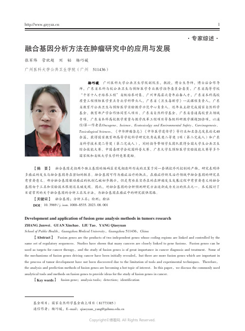
【摘 要】 融合基因是指两个独立基因的编码区首尾相连所形成的且置于同一套调控序列控制的产物,研究表明许多癌症的发生与融合基因存在密切的联系。
融合基因可作为癌症治疗的靶点,在癌症诊断及治疗领域中融合基因的研究具有重要意义。
部分融合基因驱动癌症的机制已被初步揭示,但是有些真实存在的在肿瘤发生发展过程中有重要意义的融合基因由于工具和实验技术限制还未被发现。
因此,对融合基因的分析预测研究方法逐渐成为关注的热点之一。
本文探讨了目前常用的关于融合基因的分析工具及方法,为融合基因在癌症中的研究提供思路。
【关键词】 融合基因;分析工具;检测;验证DOI:10. 3969 / j. issn. 1000-8535. 2023. 08. 001Development and application of fusion gene analysis methods in tumors researchZHANG Junwei,GUAN XinchaoSchool of Public Health,Guangzhou Medical University【Abstract】 Fusion genes are the products of two independent genes whose coding regions are linked and controlled by the same set of regulatory sequencesused as targets for cancer therapythe mechanisms of fusion genes driving cancer have been initially revealedthe process of tumor development have not been discovered due to the limitation of tools and experimental techniquesthe analysis and prediction methods of fusion genes are becoming a hot topic of interestanalytical tools and methods on fusion genes to provide ideas for the study of fusion genes in cancer【Key words】 fusion gene现代研究普遍认为,肿瘤的发生与遗传物质的改变密切相关。
英文翻译
A Facial Aging Simulation Method Using flaccidity deformation criteriaAlexandre Cruz Berg Lutheran University of Brazil.Dept Computer ScienceRua Miguel Tostes, 101. 92420-280 Canoas, RS, Brazil berg@ulbra.tche.br Francisco José Perales LopezUniversitat les Illes Balears.Dept Mathmatics InformaticsCtra Valldemossa, km 7,5E-07071 Palma MallorcaSpainpaco.perales@uib.esManuel GonzálezUniversitat les Illes Balears.Dept Mathmatics InformaticsCtra Valldemossa, km 7,5E-07071 Palma MallorcaSpainmanuel.gonzales@uib.esAbstractDue to the fact that the aging human face encompasses skull bones, facial muscles, and tissues, we render it using the effects of flaccidity through the observation of family groups categorized by sex, race and age. Considering that patterns of aging are consistent, facial ptosis becomes manifest toward the end of the fourth decade. In order to simulate facial aging according to these patterns, we used surfaces with control points so that it was possible to represent the effect of aging through flaccidity. The main use of these surfaces is to simulate flaccidity and aging consequently.1.IntroductionThe synthesis of realistic virtual views remains one of the central research topics in computer graphics. The range of applications encompasses many fields, including: visual interfaces for communications, integrated environments of virtual reality, as well as visual effects commonly used in film production.The ultimate goal of the research on realistic rendering is to display a scene on a screen so that it appears as if the object exists behind the screen. This description, however, is somewhat ambiguous and doesn't provide a quality measure for synthesized images. Certain areas, such as plastic surgery, need this quality evaluation on synthesized faces to make sure how the patient look like and more often how the patient will look like in the future. Instead, in computer graphics and computer vision communities, considerable effort has been put forthto synthesize the virtual view of real or imaginary scenes so that they look like the real scenes.Much work that plastic surgeons put in this fieldis to retard aging process but aging is an inevitable process. Age changes cause major variations in the appearance of human faces [1]. Some aspects of aging are uncontrollable and are based on hereditary factors; others are somewhat controllable, resulting from many social factors including lifestyle, among others [2].1.1.Related WorkMany works about aging human faces have been done. We can list some related work in the simulation of facial skin deformation [3].One approach is based on geometric models, physically based models and biomechanical models using either a particle system or a continuous system.Many geometrical models have been developed, such as parametric model [4] and geometric operators [5]. The finite element method is also employed for more accurate calculation of skin deformation, especially for potential medical applications such as plastic surgery [6]. Overall, those works simulate wrinkles but none of them have used flaccidity as causing creases and aging consequently.In this work is presented this effort in aging virtual human faces, by addressing the synthesis of new facial images of subjects for a given target age.We present a scheme that uses aging function to perform this synthesis thru flaccidity. This scheme enforces perceptually realistic images by preserving the identity of the subject. The main difference between our model and the previous ones is that we simulate increase of fat and muscular mass diminish causing flaccidity as one responsible element for the sprouting of lines and aging human face.In the next section will plan to present the methodology. Also in section 3, we introduce the measurements procedure, defining structural alterations of the face. In section 4, we present a visual facial model. We describe age simulation thrua deformation approach in section 5. In the last section we conclude the main results and future work.2.MethodologyA methodology to model the aging of human face allows us to recover the face aging process. This methodology consists of: 1) defining the variations of certain face regions, where the aging process is perceptible; 2) measuring the variations of those regions for a period of time in a group of people and finally 3) making up a model through the measurements based on personal features.That could be used as a standard to a whole group in order to design aging curves to the facial regions defined.¦njjjpVM2.1Mathematical Background and AnalysisHuman society values beauty and youth. It is well known that the aging process is influenced by several parameters such: feeding, weight, stress level, race, religious factors, genetics, etc. Finding a standard set of characteristics that could possibly emulate and represent the aging process is a difficult proposition.This standard set was obtained through a mathematical analysis of some face measurements in a specific group of people, whose photographs in different ages were available [7]. To each person in the group, there were, at least, four digitized photographs. The oldest of them was taken as a standard to the most recent one. Hence, some face alterations were attained through the passing of time for the same person.The diversity of the generated data has led to the designing of a mathematical model, which enabled the acquiring of a behavior pattern to all persons of the same group, as the form of a curve defined over the domain [0,1] in general, in order to define over any interval [0,Į] for an individual face. The unknown points Įi are found using the blossoming principle [8] to form the control polygon of that face.The first step consisted in the selection of the group to be studied. Proposing the assessment of the face aging characteristics it will be necessary to have a photographic follow-up along time for a group of people, in which their face alterations were measurable.The database used in this work consisted of files of patients who were submitted to plastic surgery at Medical Center Praia do Guaíba, located in Porto Alegre, Brazil.3.MeasurementsAccording to anatomic principles [9] the vectors of aging can be described aswhich alter the position and appearance of key anatomic structures of the face as can be shown in figure 1 which compares a Caucasian mother age 66 (left side) with her Caucasian daughters, ages 37 (right above) and 33 (right below) respectively.Figure 1 - Observation of family groupsTherefore, basic anatomic and surgical principles must be applied when planning rejuvenative facial surgery and treating specific problems concomitantwith the aging process.4.Visual Facial ModelThe fact that human face has an especially irregular format and interior components (bones, muscles and fabrics) to possess a complex structure and deformations of different face characteristics of person to person, becomes the modeling of the face a difficult task. The modeling carried through in the present work was based on the model, where the mesh of polygons corresponds to an elastic mesh, simulating the dermis of the face. The deformations in this mesh, necessary to simulate the aging curves, are obtained through the displacement of the vertexes, considering x(t) as a planar curve, which is located within the (u,v ) unit square. So, we can cover the square with a regular grid of points b i,j =[i/m,j/n]T ; i=0,...,m; j=0,...,n. leading to every point (u,v ) asfrom the linear precision property of Bernstein polynomials. Using comparisons with parents we can distort the grid of b i,j into a grid b'i,j , the point (u,v )will be mapped to a point (u',v') asIn order to construct our 3D mesh we introduce the patch byAs the displacements of the vertexes conform to the certain measures gotten through curves of aging and no type of movement in the face is carried through, the parameters of this modeling had been based on the conformation parameter.4.1Textures mappingIn most cases the result gotten in the modeling of the face becomes a little artificial. Using textures mapping can solve this problem. This technique allows an extraordinary increase in the realism of the shaped images and consists of applying on the shaped object, existing textures of the real images of the object.In this case, to do the mapping of an extracted texture of a real image, it is necessary that the textureaccurately correspond to the model 3D of that is made use [9].The detected feature points are used for automatic texture mapping. The main idea of texture mapping is that we get an image by combining two orthogonal pictures in a proper way and then give correct texture coordinates of every point on a head.To give a proper coordinate on a combined image for every point on a head, we first project an individualized 3D head onto three planes, the front (x, y), the left (y, z) and the right (y, z) planes. With the information of feature lines, which are used for image merging, we decide on which plane a 3D-head point on is projected.The projected points on one of three planes arethen transferred to one of feature points spaces suchas the front and the side in 2D. Then they are transferred to the image space and finally to the combined image space.The result of the texture mapping (figure 2) is excellent when it is desired to simulate some alteration of the face that does not involve a type of expression, as neutral. The picture pose must be the same that the 3D scanned data.¦¦¦ mi nj lk n j m i lk k j i w B v B u B b w v u 000,,)()()(')',','(¦¦ m i nj n jmij i v B u B b v u 00,)()(),(¦¦ m i nj n j m i j i v B u B b v u 00,)()(')','(¦¦¦ mi nj lk n j m i lk k j i w B v B u B b w v u 000,,)()()(')',','(Figure 2 - Image shaped with texturemapping5.Age SimulationThis method involves the deformation of a face starting with control segments that define the edges of the faces, as¦¦¦ mi nj lk n j m i lk k j i w B v B u B b w v u 000,,)()()(')',','(Those segments are defined in the original face and their positions are changed to a target face. From those new positions the new position of each vertex in the face is determined.The definition of edges in the face is a fundamental step, since in that phase the applied aging curves are selected. Hence, the face is divided in influencing regions according to their principal edges and characteristics.Considering the face morphology and the modeling of the face aging developed [10], the face was divided in six basic regions (figure 3).The frontal region (1) is limited by the eyelids and the forehead control lines. The distance between these limits enlarges with forward aging.The orbitary region (2) is one of the most important aging parameters because a great number of wrinkles appears and the palpebral pouch increases [11]. In nasal region (3) is observed an enlargement of its contour.The orolabial region (4) is defined by 2 horizontal control segments bounding the upper and lower lips and other 2 segments that define the nasogenian fold. Figure 3 - Regions considering the agingparametersThe lips become thinner and the nasogenian fold deeper and larger. The mental region (5) have 8 control segments that define the low limit of the face and descend with aging. In ear curve (6) is observed an enlargement of its size. The choice of feature lines was based in the characteristic age points in figure 6.The target face is obtained from the aging curves applied to the source face, i.e., with the new control segment position, each vertex of the new image has its position defined by the corresponding vertex in the target face. This final face corresponds to the face in the new age, which was obtained through the application of the numerical modeling of the frontal face aging.The definition of the straight-line segment will control the aging process, leading to a series of tests until the visual result was adequate to the results obtained from the aging curves. The extremes of the segments are interpolated according to the previously defined curves, obtained by piecewise bilinear interpolation [12].Horizontal and vertical orienting auxiliary lines were defined to characterize the extreme points of the control segments (figure 4). Some points, that delimit the control segments, are marked from the intersection of the auxiliary lines with the contour of the face, eyebrow, superior part of the head and the eyes. Others are directly defined without the use of auxiliary lines, such as: eyelid hollow, eyebrow edges, subnasion, mouth, nasolabial wrinkle andnose sides.Figure 4 - Points of the control segmentsOnce the control segments characterize the target image, the following step of the aging process can be undertaken, corresponding to the transformations of the original points to the new positions in the target image. The transformations applied to the segments are given by the aging curves, presented in section 4.In the present work the target segments are calculated by polynomial interpolations, based on parametric curves [12].5.1Deformation approachThe common goal of deformation models is to regulate deformations of a geometric model by providing smoothness constraints. In our age simulation approach, a mesh-independent deformation model is proposed. First, connected piece-wise 3D parametric volumes are generated automatically from a given face mesh according to facial feature points.These volumes cover most regions of a face that can be deformed. Then, by moving the control pointsof each volume, face mesh is deformed. By using non-parallel volumes [13], irregular 3D manifolds are formed. As a result, smaller number of deformvolumes are necessary and the number of freedom incontrol points are reduced. Moreover, based on facialfeature points, this model is mesh independent,which means that it can be easily adopted to deformany face model.After this mesh is constructed, for each vertex on the mesh, it needs to be determined which particularparametric volume it belongs to and what valueparameters are. Then, moving control points ofparametric volumes in 3D will cause smooth facialdeformations, generating facial aging throughflaccidity, automatically through the use of the agingparameters. This deformation is written in matricesas , where V is the nodal displacements offace mesh, B is the mapping matrix composed ofBernstein polynomials, and E is the displacementvector of parametric volume control nodes.BE V Given a quadrilateral mesh of points m i,j ,, we define acontinuous aged surface via a parametricinterpolation of the discretely sampled similaritiespoints. The aged position is defined via abicubic polynomial interpolation of the form with d m,n chosen to satisfy the known normal and continuity conditions at the sample points x i,j .>@>M N j i ,...,1,...,1),(u @@>@>1,,1,),,( j j v i i u v u x ¦3,,),(n m n m n m v u d v u x An interactive tool is programmed to manipulate control points E to achieve aged expressions making possible to simulate aging through age ranges. Basic aged expression units are orbicularis oculi, cheek, eyebrow, eyelid, region of chin, and neck [14]. In general, for each segment, there is an associated transformation, whose behavior can be observed by curves. The only segments that do not suffer any transformation are the contour of the eyes and the superior side of the head.5.2Deformation approachThe developed program also performs shape transformations according to the created aging curves, not including any quantification over the alterations made in texture and skin and hair color. Firstly, in the input model the subjects are required to perform different ages, as previouslymentioned, the first frame needs to be approximately frontal view and with no expression.Secondly, in the facial model initialization, from the first frame, facial features points are extracted manually. The 3D fitting algorithm [15] is then applied to warp the generic model for the person whose face is used. The warping process and from facial feature points and their norms, parametric volumes are automatically generated.Finally, aging field works to relieve the drifting problem in template matching algorithm, templates from the previous frame and templates from the initial frame are applied in order to combine the aging sequence. Our experiments show that this approach is very effective. Despite interest has been put in presenting a friendly user interface, we have to keep in mind that the software system is research oriented. In this kind of applications an important point is the flexibility to add and remove test facilities. 6.Results The presented results in the following figuresrefer to the emulations made on the frontalphotographs, principal focus of this paper, with theobjective to apply the developed program to otherpersons outside the analyzed group. The comparisonswith other photographs of the tested persons dependon their quality and on the position in which theywere taken. An assessment was made of the new positions, of the control segments. It consisted in: after aging a face, from the first age to the second one, through the use of polynomial interpolation of the control segments in the models in the young age, the new positions are then compared with the ones in the model of a relative of older age (figure 5). The processed faces were qualitatively compared with theperson’s photograph at the same age. Figure 5 - Synthetic young age model,region-marked model and aged modelAlso the eyelid hollow, very subtle falling of the eyebrow, thinning of the lips with the enlarging of the nasion and the superior part of the lip, enlargingof the front and changing in the nasolabial wrinkle.7.ConclusionsModelling biological phenomena is a great deal of work, especially when the biggest part of the information about the subject involves only qualitative data. Thus, this research developed had has a challenge in the designing of a model to represent the face aging from qualitative data.Due to its multi-disciplinary character, the developed methodology to model and emulate the face aging involved the study of several other related fields, such as medicine, computing, statistics and mathematics.The possibilities opened by the presented method and some further research on this field can lead to new proposals of enhancing the current techniques of plastic face surgery. It is possible to suggest the ideal age to perform face lifting. Once the most affected aging regions are known and how this process occurs over time. Also missing persons can be recognized based on old photographs using this technique. AcknowledgementsThe project TIN2004-07926 of Spanish Government have subsidized this work.8. References[1] Burt, D. M. et al., Perc. age in adult Caucasianmale faces, in Proc. R. Soc., 259, pp 137-143,1995.[2] Berg, A C. Aging of Orbicularis Muscle inVirtual Human Faces. IEEE 7th InternationalConference on Information Visualization, London, UK, 2003a.[3] Beier , T., S. Neely, Feature-based imagemetamorphosis, In Computer Graphics (Proc.SIGGRAPH), pp. 35-42, 1992.[4] Parke, F. I. P arametrized Models for FacialAnimation, IEEE Computer & Graphics Applications, Nov. 1982.[5] Waters, K.; A Muscle Model for Animating ThreeDimensional Facial Expression. Proc SIGGRAPH'87,Computer Graphics, Vol. 21, Nº4, United States, 1987. [6] Koch, R.M. et alia.. Simulation Facial SurgeryUsing Finite Element Models, Proceedings of SIGGRAPH'96, Computer Graphics, 1996.[7] Kurihara, Tsuneya; Kiyoshi Arai, ATransformation Method for Modeling and Animation of the Human Face from Photographs, Computer Animatio n, Springer-Verlag Tokyo, pp.45-58, 1991.[8] Kent, J., W. Carlson , R. Parent, ShapeTransformation for Polygon Objects, In Computer Graphics (Proc. SIGGRAPH), pp. 47-54, 1992. [9] Sorensen, P., Morphing Magic, in ComputerGraphics World, January 1992.[10]Pitanguy, I., Quintaes, G. de A., Cavalcanti, M.A., Leite, L. A. de S., Anatomia doEnvelhecimento da Face, in Revista Brasileira deCirurgia, Vol 67, 1977.[11]Pitanguy, I., F. R. Leta, D. Pamplona, H. I.Weber, Defining and measuring ageing parameters, in Applied Mathematics and Computation , 1996.[12]Fisher, J.; Lowther, J.; Ching-Kuang S. Curveand Surface Interpolation and Approximation: Knowledge Unit and Software Tool. ITiCSE’04,Leeds, UK June 28–30, 2004.[13]Lerios, A. et al., Feature-Based VolumeMetamorphosis, in SIGGRAPH 95 - Proceedings,pp 449-456, ACM Press, N.Y, 1995.[14]Berg, A C. Facial Aging in a VirtualEnvironment. Memória de Investigación, UIB, Spain, 2003b.[15]Hall, V., Morphing in 2-D and 3-D, in Dr.Dobb's Journal, July 1993.。
力学生物的英文

力学生物的英文Biomechanics is a fascinating field that combines the principles of physics and engineering with the study of living organisms. It explores the mechanical properties and functions of biological systems, providing insights into how living beings move, interact with their environment, and adapt to various challenges.At its core, biomechanics examines the forces and stresses that act on the body, and how the body responds to these forces. This includes understanding the mechanics of muscle contraction, bone and joint structure, and the dynamics of movement. By applying the laws of mechanics, biomechanists can analyze the efficiency and effectiveness of biological processes, from the microscopic level of cells and tissues to the macroscopic level of entire organisms.One of the primary areas of biomechanics is the study of human movement. Researchers in this field investigate the biomechanics of walking, running, jumping, and other physical activities, with the goal of improving athletic performance, preventing injuries, and enhancing the quality of life for individuals with physical disabilities or impairments. By understanding the biomechanical principles that govern human movement, scientists can develop better trainingmethods, design more effective prosthetic devices, and optimize the design of sports equipment.Beyond human movement, biomechanics also plays a crucial role in understanding the structure and function of other living organisms. For example, biomechanists study the locomotion of animals, such as the swimming of fish or the flight of birds, to gain insights into the evolutionary adaptations that have enabled these creatures to thrive in their respective environments. Similarly, biomechanics is essential for understanding the mechanical properties of plant tissues, which are essential for their growth, survival, and reproduction.In the field of medicine, biomechanics has numerous applications. Orthopedic surgeons use biomechanical principles to design and develop more effective prosthetic limbs, joint replacements, and other medical devices. Biomechanics also contributes to the understanding of injury mechanisms, allowing for the development of better protective equipment and rehabilitation strategies. Additionally, biomechanics plays a role in the design of medical imaging techniques, such as magnetic resonance imaging (MRI) and computed tomography (CT) scans, which provide valuable information about the structure and function of the human body.The applications of biomechanics extend beyond the realm of human health and performance. In the field of engineering, biomechanics isincreasingly being used to inspire the design of innovative technologies. Researchers are studying the remarkable abilities of various organisms, such as the adhesive properties of gecko feet or the efficient flight patterns of birds, to develop biomimetic solutions for a wide range of engineering challenges. These bioinspired designs have the potential to revolutionize fields like robotics, materials science, and energy production.As the field of biomechanics continues to evolve, it is becoming increasingly interdisciplinary, drawing on expertise from fields such as biology, physics, computer science, and mathematics. This collaborative approach allows for the development of more comprehensive and sophisticated models of biological systems, leading to groundbreaking discoveries and advancements in our understanding of the natural world.In conclusion, biomechanics is a dynamic and rapidly advancing field that has far-reaching implications for our understanding of living organisms and our ability to engineer innovative solutions to complex problems. By unraveling the mechanical principles that govern biological systems, biomechanists are paving the way for new breakthroughs in fields as diverse as medicine, sports science, and engineering. As we continue to explore the fascinating world of biomechanics, we can expect to witness even more remarkablediscoveries and applications that will shape the future of our understanding of the natural world.。
分离转移性癌细胞的方法与组合物,及其在检测癌转移性上的应用[发明专利]
![分离转移性癌细胞的方法与组合物,及其在检测癌转移性上的应用[发明专利]](https://img.taocdn.com/s3/m/d98cbf17f705cc1754270940.png)
专利名称:分离转移性癌细胞的方法与组合物,及其在检测癌转移性上的应用
专利类型:发明专利
发明人:W·T·陈
申请号:CN01817353.5
申请日:20010828
公开号:CN1484709A
公开日:
20040324
专利内容由知识产权出版社提供
摘要:本发明是关于检测、分离具有转移性的癌细胞的新方法与新组合物。
本发明还涉及检测此类癌细胞转移性的方法,以及鉴定抗转移活性药物的筛选方法。
本发明还提供了通过调节转移性癌细胞表面所表达丝氨酸膜内在蛋白酶[SIMP,包括seprase,二肽基肽酶IV(DPPIV)]的活性来抑制癌细胞转移的方法及组合物。
申请人:纽约州立大学研究基金会
地址:美国纽约州
国籍:US
代理机构:上海专利商标事务所
代理人:余颖
更多信息请下载全文后查看。
unit3 词汇练习以及paraphrasing 讲解练习

Paraphrasing
What is a paraphrase?
A statement that involves putting a passage from source material into your own words
the paraphrase has approximately the same number of words the paraphrase must be attributed to the original source
It is important to note that new technological and facilitated computer developments have _____________________ learning with tools such as electronic and online dictionaries, online translators and thesaurus features available in Microsoft word. Slight variations of meaning caused by the abstract nature of an item or by the specificity of its use confounded the choice between various _______________ denotations ________________ of the same lexical item.
Paraphrasing, similar to summarizing, is restating others’ ideas in your own words without altering the meaning or providing interpretation. But it differs from the latter in that a paraphrase will generally be about the same length as the original source material, as well as a paraphrase can have quoted words or terms that that you cannot or do not want to change. Likewise, a paraphrase should also be attributed to with an in-text citation.
基于改进的RRT^()-connect算法机械臂路径规划

随着时代的飞速发展,高度自主化的机器人在人类社会中的地位与作用越来越大。
而机械臂作为机器人的一个最主要操作部件,其运动规划问题,例如准确抓取物体,在运动中躲避障碍物等,是现在研究的热点,对其运动规划的不断深入研究是非常必要的。
机械臂的运动规划主要在高维空间中进行。
RRT (Rapidly-exploring Random Tree)算法[1]基于随机采样的规划方式,无需对构型空间的障碍物进行精确描述,同时不需要预处理,因此在高维空间被广为使用。
近些年人们对于RRT算法的研究很多,2000年Kuffner等提出RRT-connect算法[2],通过在起点与终点同时生成两棵随机树,加快了算法的收敛速度,但存在搜索路径步长较长的情况。
2002年Bruce等提出了ERRT(Extend RRT)算法[3]。
2006年Ferguson等提出DRRT (Dynamic RRT)算法[4]。
2011年Karaman和Frazzoli提出改进的RRT*算法[5],在继承传统RRT算法概率完备性的基础上,同时具备了渐进最优性,保证路径较优,但是会增加搜索时间。
2012年Islam等提出快速收敛的RRT*-smart算法[6],利用智能采样和路径优化来迫近最优解,但是路径采样点较少,使得路径棱角较大,不利于实际运用。
2013年Jordan等通过将RRT*算法进行双向搜索,提出B-RRT*算法[7],加快了搜索速度。
同年Salzman等提出在下界树LBT-RRT中连续插值的渐进优化算法[8]。
2015年Qureshi等提出在B-RRT*算法中插入智能函数提高搜索速度的IB-RRT*算法[9]。
同年Klemm等结合RRT*的渐进最优和RRT-connect的双向搜基于改进的RRT*-connect算法机械臂路径规划刘建宇,范平清上海工程技术大学机械与汽车工程学院,上海201620摘要:基于双向渐进最优的RRT*-connect算法,对高维的机械臂运动规划进行分析,从而使规划过程中的搜索路径更短,效率更高。
simmons 法

simmons 法(最新版)目录1.Simmons 法的定义和背景2.Simmons 法的具体操作步骤3.Simmons 法的应用领域和优势4.Simmons 法的局限性和改进方向正文1.Simmons 法的定义和背景Simmons 法,又称为 Simmons 合成方法,是一种有机合成中常用的反应方法。
该方法由美国化学家 S.W.Simmons 于 20 世纪 50 年代首次提出,主要用于合成具有特定结构的有机化合物。
Simmons 法以高效、高选择性以及优秀的立体控制而著称,被广泛应用于有机合成领域。
2.Simmons 法的具体操作步骤Simmons 法的基本原理是利用强还原性试剂(如硼氢化钠、铝锂氢化物等)与某些有机化合物发生反应,生成新的有机化合物。
具体的操作步骤如下:(1)选择合适的底物:底物通常是含有活泼氢原子的有机化合物,如酮、酯、酰胺等。
(2)选择合适的还原剂:根据底物的结构选择适当的还原剂,如硼氢化钠、铝锂氢化物等。
(3)反应条件:通常在低温(如 0-5℃)和高压(如 4-6 大气压)下进行反应,以提高反应的选择性和产率。
(4)反应产物:反应生成的新有机化合物,通常具有特定的结构和性质。
3.Simmons 法的应用领域和优势Simmons 法在有机合成领域具有广泛的应用,尤其在天然产物合成、药物合成等方面有着重要意义。
其优势主要体现在以下几点:(1)高选择性:Simmons 法能够以较高的选择性生成目标产物,避免了不必要的副反应。
(2)优秀的立体控制:通过对反应条件的优化,可以实现对产物立体化学的精确控制。
(3)反应条件温和:Simmons 法在低温、高压条件下进行反应,对设备和操作的要求相对较低。
4.Simmons 法的局限性和改进方向尽管 Simmons 法具有诸多优点,但仍存在一定的局限性,如反应的效率较低、部分还原剂具有剧毒等。
生物数学相关书籍介绍
综合消息 John Maynard Smith-进化生物学的思想者…………………………………………1 数学生物学为什么如此困难?………………………………………………………2 中比合作项目简介…………………………………………………………………….3 新书推介 数学生态学模型与研究方法………………………………………………………….4 数学生物学进展……………………………………………………………………….5 时滞微分方程引论……………………………………………………………….7 Essential Mathematical Biology………………………………………………………..7 学派介绍 西安交通大学应用数学系生物数学研究…………………………………………….9 会员介绍 唐 云………………………………………………...………………………………12 崔景安………………………………………………...………………………………12 王稳地…………………………………………………...……………………………13 任洪善……………………………………………………...…………………………14 马万彪………………………………………………………………………………...15 靳 祯…………………………………………………...……………………………16 宋新宇……………………………………………………...…………………………17 学位论文 脉冲微分方程在种群动力学中的应用……………………………………………...18 艾滋病的动力学模型研究………………………………...…………………………19 一阶脉冲微分方程边值问题的研究……………………...…………………………22 两种群相互作用的传染病模型的研究…………………...…………………………24 非自治时滞神经网络动力学行为研究…………………...…………………………26 传染病模型的全局性态与分支结构……………….………………………………..28 会议信息 第三届国际生物数学会议……………………………...……………………………29 第六届数学机械化国际研讨会…………………………...…………………………32 分支理论及其应用国际研讨会…………………………...…………………………33 动力系统与第八届泛函微分方程学术会议………………………...………………34 学会介绍 四川省生物数学专业委员会………………………………………………………...34 学报目录 《生物数学学报》2004 年第二期目录………………………………………………36
拟牛顿算法范文

拟牛顿算法范文拟牛顿算法(quasi-Newton algorithm),也被称为拟牛顿方法,是一类优化算法,用于求解无约束最优化问题。
它通过使用近似的海森矩阵(Hessian matrix)来迭代地逼近最优解,并可以在一定程度上替代传统的牛顿法。
牛顿法是一种基于二阶导数信息的优化方法,它对目标函数进行二次近似,并以此更新方向和步长。
然而,牛顿法需要计算和存储目标函数的海森矩阵,它的计算复杂度为O(n^2),其中n是目标函数的维度。
当目标函数的维度很高时,计算和存储海森矩阵将变得非常耗时和困难。
为了解决这个问题,拟牛顿算法采用了一种近似的方法来估计海森矩阵。
它基于牛顿法的思想,但使用更简单的Hessian矩阵估计技术。
拟牛顿方法可以通过迭代更新当前点的近似Hessian矩阵,从而逐渐接近最优解。
最著名的拟牛顿算法之一是Broyden-Fletcher-Goldfarb-Shanno (BFGS)算法,它是由四位数学家分别独立提出的。
BFGS算法使用拟牛顿方式更新近似的Hessian矩阵,以此来求解目标函数的最小值。
BFGS 算法在求解大型优化问题和非线性最小二乘问题时表现出色,因为它避免了显式计算和存储原始Hessian矩阵,并使用矩阵乘法来近似它。
另一个常用的拟牛顿方法是L-BFGS(Limited-memory BFGS)算法,它是BFGS算法的一种改进。
L-BFGS算法在迭代过程中,只需要存储有限数量的历史信息,从而降低了内存使用量,并且适用于大型问题。
L-BFGS 算法被广泛应用于机器学习领域的训练模型和优化问题中。
要使用拟牛顿算法求解无约束最优化问题,通常需要考虑以下几个关键步骤:1.选择初始点:需要根据具体问题选择一个合适的初始点作为起点。
2. 选择近似Hessian矩阵:需要选择一种拟牛顿方法,并确定如何估计和更新近似Hessian矩阵。
3. 计算方向和步长:使用近似Hessian矩阵来计算方向,并使用线或其他方法确定步长。
A method for separating the granulosa cells

A method for separating the granulosa cells,the basal lamina and the theca of the preovulatory ovarian follicle of thedomestic fowl(Gallus domesticus)A.B.Gilbert,A.J.Evans,M.M.Perry and M.H.DavidsonA.R.C.Poultry Research Centre,King's Buildings,West Mains Road,Edinburgh EH93JS,U.K.The preovulatory follicle of the domestic hen is almost certainly a steroid-producing structure(see Shahabi,Norton&Nalbandov,1975).However,the details of steroidogenesis are unknown because studies are complicated by the presence of steroidogenic cells of two types,the granulosa and the thecal interstitial cells(Text-fig.1).Furthermore,it is difficult to obtain preparations free from large amounts of yolk which interfere with the analytical techniques.A simple method has been developed for separating granulosa cells from the surrounding thecal covering and to obtain them essentially free of yolk.This method also provides a procedure for isolating the basal lamina.The hens used were from various commercially-available strains and were housed in cages or pens: the husbandry practice appeared not to affect the results obtained.Preovulatory follicles were carefully excised from the ovaries of birds under general anaesthesia or killed by an overdose of sodium pentobarbitone(Nembutal:Abbott Laboratories).Best results were obtained with follicles weighing between8and15g.The stalk of the excised follicle was held with forceps so that the clear,avascular stigma was uppermost(Gilbert,1971).A cut,about2cm long, was made with a scalpel approximately along the line of the stigma,though the exact position is not critical.This step must be carried out quickly,with one sweeping stroke,and it should be completed before much escape of yolk occurs.Scissors may be used instead of a scalpel but they were found to be less satisfactory because there was a tendency for a point to penetrate too deeply into the yolk mass. Immediately after it had been cut the follicle was inverted over a suitable dish containing an aqueous medium and the follicular contents(yolk,perivitelline layer,granulosa and basal lamina)(Gilbert, 1971)were allowed to fall into the medium.The choice of medium depended on the purpose for which the granulosa cells were being harvested.If the operation has been done correctly,the entire theca(Text-fig.1,PI.1,Fig.lb)remains held in the forceps,without contamination by yolk or granulosa material,and it can be used for studies of thecal activity.The yolk,covered by the perivitelline layer,the granulosa layer and the basal lamina (Text-fig.1,PL1,Fig.3),settles as an almost undisturbed sphere on the floor of the vessel.The split occurs at a position different from that occurring during ovulation when the granulosa cells and the basal lamina remain with the theca(PI.1,).Removal of the granulosa layer,together with its associated basal lamina and perivitelline layer, from the yolk was carried out under a low-power lens or dissecting microscope with a black back¬ground.The cut ends of the composite membrane surrounding the yolk were located,grasped with fine dissecting forceps and gently pulled away from the yolk thereby everting the membrane over the surface of the yolk:this was made easier by using the yolk as a mass to pull against.Care was taken to protect the yolk from undue disturbance because yolk material dispersed throughout the medium prevented clear observation and caused unnecessary contamination of the preparation.It proved possible to remove the composite membrane in one piece with a very small amount of adherent yolk: when it was necessary to remove larger masses of yolk,the membrane was washed with a stream of medium from a Pasteur pipette.The method provides large sheets of living granulosa cells sandwiched between the basal lamina and the perivitelline layer(PI.1,Figs.2and3);staining with Trypan Blue and Nigrosin revealed very few(<5%)dead cells.ranulosa cells(GOBasal lamina(BUOolemmaYolk¡vitelline layer(PL)ThecaText-fig.1.Simplified diagram of a section through the mid-line of a preovulatory ovarian follicle of the domestic hen.The relative proportions of the layers are not to scale.After the incision has been made the contents of the follicle listed on the right-hand side are allowed to fall under gravity into the dish containing the aqueous medium(see text).The preparation was checked for cleanliness by examining conventionally fixed and sectioned material in the electron microscope.On its outer aspect the basal lamina was completely free of thecal cells,whereas the perivitelline layer was contaminated to a variable extent by oocyte debris consisting of vesicular material,yolk granules and remnants of the oolemma(PL1,Fig.3).The granulosa cells,arranged in a single layer but separated from each other by gaps,were covered with numerous microvilli which,at the cell apices,extended into the meshwork of dense,rod-shaped elements of the perivitelline layer.The basal surfaces of the cells were comparatively smooth and were closely associated with the basal lamina.Details of the structure will be described in a later publication.Isolated granulosa cells were obtained by separating the basal lamina from the perivitelline layer. This is best done under a dissecting microscope with magnifications of about50-100:very fine watch-maker's forceps were used to tease the basal lamina from the granulosa cells,which tended to remain attached to the perivitelline layer.Once separation had started,it was possible to pull the basal lamina away in large sheets,exposing the granulosa cells.These were then washed from the perivitel¬line layer by gentle agitation or a stream of medium from a Pasteur pipette.The granulosa cells were then collected by gentle centrifugation.This technique is being used in several pieces of research,including an investigation of the physical and biochemical properties of the basal lamina and an examination of the detailed structure of the granulosa cells with the electron microscope.Tissue-culture work is being developed for study of yolk-transport mechanisms and steroidogenesis in granulosa cells and thecal interstitial cells.EXPLANATION OF PLATE IFig.1.Conventional histological section of the hen theca stained with Mallory.(a)A normal discharged postovulatory follicle with intact granulosa(GC).(b)Preparation after removal of the granulosa,basal lamina and perivitelline layer,as described in the text.Scale bar=100pm.Fig.2.Preparation of hen granulosa cells with the basal lamina and perivitelline membrane.Most of the cells are apparently alive,though a few dead cells(DC)are visible.Trypan blue,500.Fig.3.Electron micrograph of a section at right angles to the composite membrane fixed after stripping from the oocyte.A granulosa cell is seen sandwiched between the basal lamina(BL)and the perivitelline layer(PL).Scale bar=5pm.PLATE1ReferencesGilbert, A.B.(1971)The ovary.In Physiology and Biochemistry of the Domestic Fowl,Vol.Ill,pp.1163-1208.Eds D.J.Bell&B.M.Freeman.Academic Press,London.Shahabi,N.A.,Norton,H.W.&Nalbandov,A.V.(1975)Steroid levels in follicles and the plasma of hens during the ovulatory cycle.Endocrinology96, 962-968.Received13December 1976。
High-order small-time local controllability
This work was partially supported through NSF grants DMS-8500941 and DMS-8603156
1
way to such a solution are many difficult problems, which are of high mathematical interest in their own right. One of these problems is (small-time) local controllability (or reachability), which essentially asks for the set of points which may be reached from a given initial point by solutions of system (1). On a purely formal level any solution to this problem may be thought of as a nonlinear open mapping theorem (for mappings from a function space into a finite dimensional space), thus explaining the deepness and intricacy of this problem. Many different notions of controllability have been developped, for a survey see section 2.3 of [23]. Here we only will use accessibility and small-time local controllability (STLC) (about an equilibrium point). The attainable set AU (t) at time t > 0 is the set of all points which can be reached in time t by solutions of (1). We say (1) is accessible if AU (t) has n-dimensional interior for all t > 0, and we say (1) is small-time locally controllable if AU (t) contains the equilibrium (i.e. the reference solution) x ≡ 0 in its interior for all t > 0. (Here a regrettable confusion of terminology has occurred, as intuitively one would like to call this property small-time local reachability from zero, while small-time local controllability should refer to that the equilibrium can be reached from a neighbourhood in small time. However, in our setting these two notions are symmetric under time reversal, and we shall stick to the now established definition as given above.) We recall the Kalman-Rank-Condition which is both a necessary and sufficient condition for both local controllability and accessibility of the linear system (2): Theorem 1.1 If and only if rank{B, AB, . . . , An−1 B } = n then 0 ∈intAU (t) for all t > 0. In the nonlinear case, however, a system may be accessible without being locally controllable; for example consider the system Example 1.1 x ˙ 1 (t) = u(t) x ˙ 2 (t) = x2 1 x(0) = 0 |u(·)| ≤ 1. (3)
E 质谱检测有机磷农药---综述
ReviewMass Spectrometric Analyses of Organophosphate Insecticide Oxon Protein AdductsCharles M. Thompson, John M. Prins, and Kathleen M. GeorgeCenter for Structural and Functional Neuroscience, Department of Biomedical and Pharmaceutical Sciences, University of Montana, Missoula, Montana, USAThompson et al.Mass spectrometry of organophosphate–protein adductsThompson et al.Mass spectrometry of organophosphate–protein adductstethers and attenuated reactivity as a phospho-nate compared with phosphate likely select for a protein population different from that of the OP oxon structures. Thus, differences in reactivity and cell permeability properties of FP-biotin probes must be considered when assessing possible OP–protein biomarkers.In one study, Kidd et al. (2001) investi-gated the rate differences in which the serine hydrolases react with FP-biotin probes after treatment with oleoyl trifluoro m ethyl ketone (OTFMK), a reversible inhibitor of ser-ine hydrolases. Serine hydrolase targets were identified directly in complex proteomes by comparing the rates of binding of the ser-ine hydrolases to the FP-biotin probes in the control and OTFMK-treated samples (Kidd et al. 2001). An in vitro assay exposing puri-fied tubulin to an OP–biotin probe demon-strated not only that OPs bind this previously unidentified target, but also that they bind tyrosine residues in tubulin. The OP-reactive tyrosine residues reside either near the GTP binding site or within loops that interact later-ally with proto fi laments, indicating that this binding, if it occurs in vivo, may lead to non-cholinergic toxic outcomes (Grigoryan et al. 2008). Lockridge and colleagues (Ding et al. 2008; Schopfer et al. 2005a, 2005b) also used FP-biotin combined with MS to iden-tify OP-modified proteins in human plasma. The plasma was treated with FP-biotin; the proteins were separated into low- and high-abundance portions and digested with trypsin; and the FP-biotinylated peptides were isolated by binding to avidin beads. Proteins identi-fied in these studies included albumin, ES1 carboxylesterase, propionyl and methyl c rotonyl coenzyme A carboxylase-α, and pyruvate car-boxylase. The investigators were able to readily identify FP-biotin–labeled albumin because of its high concentration in human plasma (Ding et al. 2008; Schopfer et al. 2005a, 2005b). The results demon s trated that albumin is an OP scavenger and undergoes a covalent reac-tion with OP on five tyrosines and two serines (Aardema and MacGregor 2002; Peeples et al. 2005; Schopfer et al. 2005a, 2005b; Wieseler et al. 2006). Although the FP-biotin probe was unable to identify any additional novel OP protein targets, the results from this study sug-gest that OP–albumin adducts could be used to monitor OP exposure (Ding et al. 2008).FP-biotin has a relatively large structure compared with OPs, so it can be argued that this probe may react differently with various proteins in biological systems and possibly fail to detect important OP protein targets. This could prove to be problematic with enzymes such as AChE and BChE that contain OP binding sites located deep within the molecule. Schopfer et al. (2005a, 2005b) investigated the rates of reactivity and binding affinity of FP-biotin with AChE and BChE. The results from their study demonstrate that, despite its large biotingroup, FP-biotin reacts with both AChE andBChE at rates comparable to reaction rates ofother OPs. Therefore, the authors concludedthat FP-biotin is a potent OP and can serveas a useful probe for identifying OP proteintargets. Taken together, the studies describedhere demon s trate that molecular probes suchas biotinylated OPs can be powerful tools forproteomic studies. Although FP-biotin does notprecisely simulate OP structure, the ability torapidly isolate and identify labeled proteins incrude cell and tissue extracts, as well as provideinsight into post t ranslational events that regu-late protein function, outweighs some of thepotential limitations.ConclusionsThe mechanisms by which OP insecticideoxons covalently adduct AChE, BChE, andother serine hydrolases have been relativelywell understood, but many of the post-inhibition processes have not been well char-acterized. Advances in protein MS have clearlyaided investigators with a new and power-ful tool to interpret not only the adductionevent but also the molecular steps that occurafter adduction. As a result, it is now some-what routine to investigate not only OP–ChEadduct formation but also each step in theprocess that leads to injurious modificationsof the protein. With these important stepsaccomplished, investigators can now broadenthe scope of their inquiry into how proteinsother than cholinesterases are modified by OPcompounds and, hence, how the connectedcellular level events may be compromised.R efeRencesAardema MJ, MacGregor JT. 2002. Toxicology and genetictoxicology in the new era of “toxicogenomics”: impact of“-omics” technologies. Mutat Res 499:13–25.Adam GC, Cravatt BF, Sorensen EJ. 2001. Profiling the specificreactivity of the proteome with non-directed activity-basedprobes. Chem Biol 8:81–95.Adam GC, Sorensen EJ, Cravatt BF. 2002. Trifunctional chemi-cal probes for the consolidated detection and identifica-tion of enzyme activities from complex proteomes. MolCell Proteomics 1:828–835.Adamczyk M, Gebler JC, Wu J. 2001. Selective analysis of phos-phopeptides within a protein mixture by chemical modi-fi c ation, reversible biotinylation and mass spectrometry.Rapid Commun Mass Spectrom 15:1481–1488.Albuquerque EX, Aracava Y, Cintra WM, Brossi A,Schonenberger B, Deshpande SS. 1988. Structure-activityrelationship of reversible cholinesterase inhibitors: activa-tion, channel blockade and stereospecificity of the nicotinicacetylcholine receptor-ion channel complex. Braz J MedBiol Res 21:1173–1196.Arima H, Sobue K, So M, Morishima T, Ando H, Katsuya H. 2003.Transient and reversible parkinsonism after acute organo-phosphate poisoning. J Toxicol Clin Toxicol 41:67–70.Ashani Y, Gentry MK, Doctor BP. 1990. Differences in conforma-tional stabilty between native and phosphorylated acetyl-cholinesterase as evidenced by a monoclonal antibody.Biochemistry 29:2456–2463.Ashani Y, Leader H, Rothschild N, Dosoretz C. 1998. Combinedeffect of organophosphorus hydrolase and oxime on thereactivation rate of diethylphosphoryl-acetylcholinesteraseconjugates. Biochem Pharmacol 55:159–168.Ashani Y, Radic Z, Tsigelny I, Vellom DC, Pickering NA,Quinn DM, et al. 1995. Amino acid residues controllingreactivation of organophosphonyl conjugates of acetyl-cholinesterase by mono- and bisquaternary oximes. J BiolChem 270:6370–6380.Ballantyne B, Marrs T. 1992. Clinical and Experimental Toxicologyof Organophosphates and Carbamates. Boston:ButterworthHeinemann.Barak R, Ordentlich A, Barak D, Fischer M, Benschop HP,De Jong LPA, et al. 1997. Direct determination of the chemi-cal composition of acetylcholinesterase phos p honylationproducts utilizing electrospray-ionization mass spectrom-etry. FEBS Lett 407:347–352.Barr DB, Allen R, Olsson AO, Bravo R, Caltabiano LM,Montesano A, et al. 2005. Concentrations of selectivemetabolites of organophosphorus pesticides in the UnitedStates population. Environ Res 99:314–326.Bennett KL, Stensballe A, Podtelejnikov AV, Moniatte M,Jensen ON. 2002. Phosphopeptide detection and sequencingby matrix-assisted laser desorption/ionization quadru p oletime-of-flight tandem mass spectrometry. J Mass Spectrom37:179–190.Berkman CE, Quinn DA, Thompson CM. 1993a. Interaction ofacetylcholinesterase with the enantiomers of malaoxonand isomalathion. Chem Res Toxicol 6:724–730.Berkman CE, Ryu S, Quinn DA, Thompson CM. 1993b. Kineticsof the postinhibitory reactions of acetylcholinesterase poi-soned by chiral isomalathion: a surprising nonreactivationinduced by the RP stereoisomers. Chem Res Toxicol 6:28–32.Berkman CE, Thompson CM, Perrin SR. 1993c. Synthesis, abso-lute configuration, and analysis of malathion, malaoxon, andisomalathion enantiomers. Chem Res Toxicol 6:718–723.Bomser J, Casida JE. 2000. Activation of extracellular signal-regulated kinases (ERK 44/42) by chlorpyrifos oxon inChinese hamster ovary cells. J Biochem Mol Toxicol14:346–353.Bomser JA, Casida JE. 2001. Diethylphosphorylation of rat car-diac M2 muscarinic receptor by chlorpyrifos oxon in vitro.Toxicol Lett 119:21–26.Bomser JA, Quistad GB, Casida JE. 2002. Chlorpyrifos oxonpotentiates diacylglycerol-induced extracellular signal-regulated kinase (ERK 44/42) activation, possibly bydiacylglycerol lipase inhibition. Toxicol Appl Pharmacol178:29–36.Bronstein AC, Spyker DA, Cantilena LR Jr, Green JL, Rumack BH,Heard SE. 2008. 2007 Annual report of the AmericanAssociation of Poison Control Centers’ National PoisonData System (NPDS): 25th annual report. Clin Toxicol (Phila)46:927–1057.Broomfield CA, Millard CB, Lockridge O, Caviston TL. 1995.Enzymes of the Cholinesterase Family. New York:PlenumPress.Casida JE, Quistad GB. 2005. Serine hydrolase targets of organo-phosphorus toxicants. Chem Biol Interact 157–158:277–283.Centers for Disease Control and Prevention. 2005. ThirdNational Report on Human Exposure to EnvironmentalChemicals. Available: /exposurereport/pdf/thirdreport.pdf [accessed 17 November 2009].Claudio L, Kwa WC, Russell AL, Wallinga D. 2000. Testing methodsfor developmental neurotoxicity of environmental chemicals.Toxicol Appl Pharmacol 164:1–14.Corrigan FM, MacDonald S, Brown A, Armstrong K,Armstrong EM. 1994. Neurasthenic fatigue, chemicalsensitivity and GABAa receptor toxins. Med Hypotheses43:195–200.Costa LG. 1998. Neurotoxicity testing: a discussion of in vitroalternatives. Environ Health Perspect 106(suppl 2):505–510.Costa LG. 2006. Current issues in organophosphate toxicology.Clin Chim Acta 366:1–13.Costa LG, Cole TB, Vitalone A, Furlong CE. 2005. Measurementof paraoxonase (PON1) status as a potential biomarker ofsusceptibility to organophosphate toxicity. Clin Chim Acta352:37–47.Costa LG, Richter RJ, Li WF, Cole T, Guizzetti M, Furlong CE.2003. Paraoxonase (PON 1) as a biomarker of susceptibilityfor organophosphate toxicity. Biomarkers 8:1–12.Dahlgren JG, Takhar HS, Ruffalo CA, Zwass M. 2004. Healtheffects of diazinon on a family. J Toxicol Clin Toxicol42:579–591.Ding SJ, Carr J, Carlson JE, Tong L, Xue W, Li Y, et al. 2008. Fivetyrosines and two serines in human albumin are labeled bythe organophosphorus agent FP-biotin. Chem Res Toxicol21:1787–1794.Doorn JA, Gage DA, Schall M, Talley TT, Thompson CM,Richardson RJ. 2000. Inhibition of acetylcholinesteraseThompson et al.by (1S,3S)-isomalathion proceeds with loss of thiomethyl: kinetic and mass spectral evidence for an unexpected pri-mary leaving group. Chem Res Toxicol 13:1313–1320. Doorn JA, Schall M, Gage DA, Talley TT, Thompson CM, Richardson RJ. 2001a. Identification of butyrylcholinest-erase adducts after inhibition with isomalathion using mass spectrometry: difference in mechanism between (1R)- and (1S)-stereoisomers. Toxicol Appl Pharmacol 176:73–80. Doorn JA, Talley TT, Thompson CM, Richardson RJ. 2001b.Probing the active sites of butyrylcholinesterase and choles-terol esterase with isomalathion: conserved stereo s elective inactivation of serine hydrolases structurally related to acetyl c holinesterase. Chem Res Toxicol 14:807–813. Ehrich M, Intropido L, Costa LG. 1994. Interaction of organo-phosphorus compounds with muscarinic receptors in SH-SY5Y human neuroblastoma cells. J Toxicol Environ Health 43:51–63.Eldefrawi ME, Eldefrawi AT. 1983. Neurotransmitter receptors as targets for pesticides. J Environ Sci Health B 18:65–88. Elhanany E, Ordentlich A, Dgany O, Kaplan D, Segall Y, Barak R, et al. 2001. Resolving pathways of interaction of covalent inhibitors with the active site of acetylcholinesterases: MALDI-TOF/MS analysis of various nerve agent phosphyl adducts. Chem Res Toxicol 14:912–918.Ellman GL, Courtney KD, Andres V Jr, Feather-Stone RM. 1961.A new and rapid colorimetric determination of acetyl-cholinesterase activity. Biochem Pharmacol 7:88–95. Eskenazi B, Marks AR, Bradman A, Harley K, Barr DB, Johnson C, et al. 2007. Organophosphate pesticide exposure and neurodevelopment in young Mexican-American children.Environ Health Perspect 115:792–798.Eto M. 1974. Organophosphorus Pesticides; Organic and Biological Chemistry. Cleveland, OH:CRC Press.Fenske RA, Kissel JC, Lu C, Kalman DA, Simcox NJ, Allen EH, et al. 2000. Biologically based pesticide dose estimates for children in an agricultural community. Environ Health Perspect 108:515–520.Fest C, Schmidt KJ. 1973. The Chemistry of Organophosphorus Pesticides; Reactivity, Synthesis, Mode of Action, Toxicology. New York:Springer Verlag.Fukuto TR. 1990. Mechanism of action of organophosphorus and carbamate insecticides. Environ Health Perspect 87:245–254. Godovac-Zimmermann J, Soskic V, Poznanovic S, Brianza F.1999. Functional proteomics of signal transduction by membrane receptors. Electrophoresis 20:952–961. Goldman LR, Koduru S. 2000. Chemicals in the environment and developmental toxicity to children: a public health and policy perspective. Environ Health Perspect 108(suppl 3):443–448. Gordon C. 1993. Acute and delayed effects of diisopropyl fl uoro-phosphate on body temperature, heart rate, and motor activity in the awake, unrestrained rat. J Toxic Environ Health 39:247–260.Grigoryan H, Schopfer LM, Thompson CM, Terry AV, Masson P, Lockridge O. 2008. Mass spectrometry identifies covalent binding of soman, sarin, chlorpyrifos oxon, diisopropyl fluoro p hosphate, and FP-biotin to tyrosines on tubulin: a potential mechanism of long term toxicity by organophos-phorus agents. Chem Biol Interact 175:180–186.Haas R, Jackson B, Reinhold B, Foster J, Rosenberry T. 1996.Glycoinositol phospholipid anchor and protein C-terminus of bovine erythrocyte acetylcholinesterase: analysis by mass spectrometry and by protein and DNA sequencing.Biochem J 314:817–825.Haas R, Rosenberry T. 1995. Protein denaturation by addition and removal of acetonitrile: application to tryptic digestion of acetylcholinesterase. Anal Biochem 224:425–427. Harel M, Su CT, Frolow F, Ashani Y, Silman I, Sussman JL.1991. Refined crystal structures of “aged” and “non-aged”organophosphoryl conjugates of gamma-chymotrypsin.J Mol Biol 221:909–918.Huff RA, Corcoran JJ, Anderson JK, Abou-Donia MB. 1994.Chlorpyrifos oxon binds directly to muscarinic receptors and inhibits cAMP accumulation in rat striatum. J Pharmacol Exp Ther 269:329–335.Jacobson SW, Jacobson JL. 2006. New evidence of effects of organophosphate pesticides on neurodevelopment in children: commentary on the article by Kofman et al. on page 88. Pediatr Res 60:22–23.Jayawardane P, Dawson AH, Weerasinghe V, Karalliedde L, Buckley NA, Senanayake N. 2008. The spectrum of interme-diate syndrome following acute organophosphate poisoning:a prospective cohort study from Sri Lanka. PLoS Med 5:e147;doi:10.1371/journal.pmed.0050147 [Online 15 July 2008]. Jennings LL, Malecki M, Komives EA, Taylor P. 2003. Directanalysis of the kinetic profiles of organophosphate-acetyl-cholinesterase adducts by MALDI-TOF mass spectrometry.Biochemistry 42:11083–11091.Jiang X, Wang Y. 2004. Beta-elimination coupled with tandemmass spectrometry for the identification of in vivo andin vitro phosphorylation sites in maize dehydrin DHN1protein. Biochemistry 43:15567–15576.Johal MS, Sharma ML, Ravneet. 2007. Impact of low dose oforganophosphate, monocrotophos on the epithelial cellsof gills of Cyprinus carpio communis Linn.—SEM study.J Environ Biol 28:663–667.Katz L, Marquis J. 1992. Organophosphate-induced alteration inmuscarinic receptor binding and phosphoinositide hydrolysisin the human SK-N-SH cell line. Neurotoxicology 13:365–378.Kidd D, Liu Y, Cravatt BF. 2001. Profiling serine hydrolase activitiesin complex proteomes. Biochemistry 40:4005–4015.Kofman O, Berger A, Massarwa A, Friedman A, Jaffar AA. 2006.Motor inhibition and learning impairments in school-agedchildren following exposure to organophosphate pesticidesin infancy. Pediatr Res 60:88–92.Langenberg JP, De Jong LP, Otto MF, Benschop HP. 1988.Spontaneous and oxime-induced reactivation of acetyl-cholinesterase inhibited by phosphoramidates. ArchToxicol 62:305–310.Lanks KW, Seleznick MJ. 1981. Spontaneously reactivation ofacetylcholinesterase inhibited by diisopropylfluorophos-phate. Biochim Biophys Acta 660:91–95.Larsen MR, Sørensen GL, Fey SJ, Larsen PM, Roepstorff P.2001. Phospho-proteomics: evaluation of the use ofenzymatic de-phosphorylation and differential massspectrometric peptide mass mapping for site specificphosphorylation assignment in proteins separated by gelelectrophoresis. Proteomics 1:223–238.Li Y, Li M, Xing G, Hu Z, Wang Q, Dong C, et al. 2000a.Stimulation of the mitogen-activated protein kinasecascade and tyrosine phosphorylation of the epidermalgrowth factor receptor by hepatopoietin. J Biol Chem275:37443–37447.Li Y, Liu L, Kang J, Sheng JG, Barger SW, Mrak RE, et al. 2000b.Neuronal-glial interactions mediated by inter l eukin-1enhance neuronal acetylcholinesterase activity and mRNAexpression. J Neurosci 20:149–155.Liu Y, Patricelli MP, Cravatt BF. 1999. Activity-based proteinprofiling: the serine hydrolases. Proc Natl Acad Sci USA96:14694–14699.Lockridge O, Masson P. 2000. Pesticides and susceptible popu-lations: people with butyrylcholinesterase genetic variantsmay be at risk. Neurotoxicology 21:113–126.Lotti M, Becker CE, Aminoff MJ. 1984. Organophosphate poly-neuropathy: pathogenesis and prevention. Neurology34:658–662.Menzies GW, MacConnell CB. 1998. Integrated Pest Managementfor Raspberries: A Guide for Sampling and Decision-Makingfor Key Raspberry Pests in Northwest Washington. Available:/ag/comhort/nooksack/ipmweb/Pesticideselection.html [accessed 17 November 2009].Murray A, Rathbone A, Ray D. 2005. Novel protein targets fororganophosphorus pesticides in rat brain. Environ ToxicolPharmacol 19:451–454.National Center for Biotechnology Information. 2009. PubMed.Available: /pubmed/ [accessed17 November 2009].Niven AS, Roop SA. 2004. Inhalational exposure to nerveagents. Respir Care Clin N Am 10:59–74.Nomura DK, Durkin KA, Chiang KP, Quistad GB, Cravatt BF,Casida JE. 2006. Serine hydrolase KIAA1363: toxicologicaland structural features with emphasis on organophos-phate interactions. Chem Res Toxicol 19:1142–1150.Noort D, Fidder A, van der Schans MJ, Hulst AG. 2006.Verification of exposure to organophosphates: genericmass spectrometric method for detection of human butyryl-cholinesterase adducts. Anal Chem 78:6640–6644.Pala I, Vig PJ, Desaiah D, Srinivasan A. 1991. In vitro effectsof organophosphorus compounds on calmodulin activity.J Appl Toxicol 11:391–395.Peeples ES, Schopfer LM, Duysen EG, Spaulding R, Voelker T,Thompson CM, et al. 2005. Albumin, a new biomarker oforganophosphorus toxicant exposure, identified by massspectrometry. Toxicol Sci 83:303–312.Qian WJ, Goshe MB, Camp DG II, Yu LR, Tang K, Smith RD.2003. Phosphoprotein isotope-coded solid-phase tagapproach for enrichment and quantitative analysis ofphosphopeptides from complex mixtures. Anal Chem75:5441–5450.Quistad GB, Casida JE. 2000. Sensitivity of blood-clotting factorsand digestive enzymes to inhibition by organophosphoruspesticides. J Biochem Mol Toxicol 14:51–56.Quistad GB, Sparks SE, Casida JE. 2001. Fatty acid amidehydrolase inhibition by neurotoxic organophosphoruspesticides. Toxicol Appl Pharmacol 173:48–55.Quistad GB, Zhang N, Sparks SE, Casida JE. 2000. Phospho-acetyl c holinesterase: toxicity of phosphorus oxychlorideto mammals and insects that can be attributed to selectivephosphorylation of acetylcholinesterase by phosphoro-dichloridic acid. Chem Res Toxicol 13:652–657.Richards P, Johnson M, Ray D, Walker C. 1999. Novel proteintargets for organophosphorus compounds. Chem BiolInteract 119–120:503–511.Roberts WL, Santikarn S, Reinhold VN, Rosenberry TL.1988. Structural characterization of the glycoinositolphospho l ipid membrane anchor of human erythrocyteacetyl c holinesterase by fast atom bombardment massspectrometry. J Biol Chem 263:18776–18784.Rowsey PJ, Gordon CJ. 1999. Tumor necrosis factor is involvedin chlorpyrifos-induced changes in core temperature inthe female rat. Toxicol Lett 109:51–59.Schopfer LM, Champion MM, Tamblyn N, Thompson CM,Lockridge O. 2005a. Characteristic mass spectral frag-ments of the organophosphorus agent FP-biotin andFP-biotinylated peptides from trypsin and bovine albumin(Tyr410). Anal Biochem 345:122–132.Schopfer LM, Voelker T, Bartels CF, Thompson CM, Lockridge O.2005b. Reaction kinetics of biotinylated organophosphorustoxicant, FP-biotin, with human acetyl c holinesterase andhuman butyrylcholinesterase. Chem Res Toxicol 18:747–754.Schuh RA, Lein PJ, Beckles RA, Jett DA. 2002. Noncholinesterasemechanisms of chlorpyrifos neurotoxicity: altered phos-phorylation of Ca2+/cAMP response element binding proteinin cultured neurons. Toxicol Appl Pharmacol 182:176–185.Sickmann A, Meyer HE. 2001. Phosphoamino acid analysis.Proteomics 1:200–206.Slotkin TA. 2004. Cholinergic systems in brain developmentand disruption by neurotoxicants: nicotine, environmentaltobacco smoke, organophosphates. Toxicol Appl Pharmacol198:132–151.Slotkin TA, Ryde IT, Levin ED, Seidler FJ. 2008. Developmentalneurotoxicity of low dose diazinon exposure of neonatalrats: effects on serotonin systems in adolescence andadulthood. Brain Res Bull 75:640–647.Solomon C, Poole J, Palmer KT, Peveler R, Coggon D. 2007.Acute symptoms following work with pesticides. OccupMed (Lond) 57:505–511.Spaulding RS, George KM, Thompson CM. 2006. Analysis andsequencing of the active-site peptide from native andorganophosphate-inactivated acetylcholinesterase byelectrospray ionization, quadrupole/time-of-flight (QTOF)mass spectrometry. J Chromatogr B Analyt TechnolBiomed Life Sci 830:105–113.Stephens R, Spurgeon A, Calvert IA, Beach J, Levy LS, Berry H,et al. 1995. Neuropsychological effects of long-term exposureto organophosphates in sheep dip. Lancet 345:1135–1139.Sudakin DL, Power LE. 2007. Organophosphate exposures inthe United States: a longitudinal analysis of incidentsreported to poison centers. J Toxicol Environ Health A70:141–147.Sultatos LG. 1994. Mammalian toxicology of organophosphoruspesticides. J Toxicol Environ Health 43:271–289.Swiss Institute of Bioinformatics. 2007. PeptideCutter.Available: /tools/peptidecutter/[accessed 17 November 2009].Thaler F, Valsasina B, Baldi R, Xie J, Stewart A, Isacchi A, et al.2003. A new approach to phosphoserine and phospho-threonine analysis in peptides and proteins: chemicalmodifi c ation, enrichment via solid-phase reversible binding,and analysis by mass spectrometry. Anal Bioanal Chem376:366–373.Thompson CM, Fukuto TR. 1982. Mechanism of cholinest-erase inhibition by methamidophos. J Agric Food Chem30:282–284.Timofeeva OA, Roegge CS, Seidler FJ, Slotkin TA, Levin ED.2008. Persistent cognitive alterations in rats after earlypostnatal exposure to low doses of the organophosphatepesticide, diazinon. Neurotoxicol Teratol 30:38–45.Tseng HC, Ovaa H, Wei NJ, Ploegh H, Tsai LH. 2005. Phospho-proteomic analysis with a solid-phase capture-release-tagapproach. Chem Biol 12:769–777.U.S. EPA. 2006. Organophosphorus Cumulative RiskAssessment (2006 Update). EPA-HQ-OPP-2006-0618-0002.Mass spectrometry of organophosphate–protein adductsWashington, DC:U.S. Environmental Protection Agency.Available: /search/Regs/home.html#documentDetail?R=09000064801ac312 [accessed17 November 2009].van den Beukel I, van Kleef RG, Oortgiesen M. 1998. Differential effects of physostigmine and organophosphates on nico-tinic receptors in neuronal cells of different species.Neurotoxicology 19:777–787.Ward TR, Ferris DJ, Tilson HA, Mundy WR. 1993. Correlation of the anticholinesterase activity of a series of organo-phosphates with their ability to compete with agonist binding to muscarinic receptors. Toxicol Appl Pharmacol 122:300–307.Ward TR, Mundy WR. 1996. Organophosphorus compounds preferentially affect second messenger systems coupled to M2/M4 receptors in rat frontal cortex. Brain Res Bull 39:49–55.Whyatt RM, Camann D, Perera FP, Rauh VA, Tang D, Kinney PL,et al. 2005. Biomarkers in assessing residential insecticideexposures during pregnancy and effects on fetal growth.Toxicol Appl Pharmacol 206:246–254.Whyatt RM, Rauh V, Barr DB, Camann DE, Andrews HF,Garfinkel R, et al. 2004. Prenatal insecticide exposures andbirth weight and length among an urban minority cohort.Environ Health Perspect 112:1125–1132.Wieseler S, Schopfer L, Lockridge O. 2006. Markers of organo-phosphate exposure in human serum. J Mol Neurosci30:93–94.Wilson BW, Henderson JD. 1992. Blood esterase determina-tions as markers of exposure. Rev Environ Contam Toxicol128:55–69.Wilson BW, Hooper MJ, Hansen ME, Nieberg PS. 1992.Reactivation of organophosphorus inhibited AChE withoximes. In: Organophosphates: Chemistry, Fate, and Effects(Chambers JE, Levi PE, eds). San Diego, CA:AcademicPress, 107–137.Wilson BW, Padilla S, Henderson JD, Brimijoin S, Dass PD,Elliot G, et al. 1996. Factors in standardizing automated cho-linesterase assays. J Toxicol Environ Health 48:187–195.Yanagisawa N, Morita H, Nakajima T. 2006. Sarin experiencesin Japan: acute toxicity and long-term effects. J NeurolSci 249:76–85.Young JG, Eskenazi B, Gladstone EA, Bradman A, Pedersen L,Johnson C, et al. 2005. Association between in utero organo-phosphate pesticide exposure and abnormal reflexes inneonates. Neurotoxicology 26:199–209.Zaim M, Jambulingam P. 2007. Global Insecticide Use forVector-Borne Disease Control. Geneva:World HealthOrganization. Available: http://whqlibdoc.who.int/HQ/2007/WHO_CDS_NTD_WHOPES_GCDPP_2007.2_eng.pdf[accessed 17 November 2009].Zwiener RJ, Ginsburg CM. 1988. Organophosphate and car-bamate poisoning in infants and children. Pediatrics81:121–126.。
急性移植物抗宿主病的预防研究新进展

• 40 •Int .1Blood T r a n s fn s ll e m a l o l. J an u a ry^〇21. V()l. .1.1. No. 1•专题论坛•急性移植物抗宿主病的预防研究新进展潘婧李建勇繆扣荣南京医科大学第一附属医院江苏省人民医院血液科210029通信作者:缪■扣荣,Em ail:kourongmiao@【摘要】异基因造血干细胞移植(allo-H SC T)是治愈血液系统恶性疾病的重要方法,但是急性移植物抗宿主病(aG V H D)严重影响移植成功率,是患者发生移植相关死亡的重要原因之一。
目前关于最佳的a G V H D预防方案尚未达成共识。
随着对aG V H D发生机制研究的深入,以及相关临床试验的开展,为allo-H S C T后a G V H D的预防提供更多的新药及联合用药选择。
笔者拟就alio-HSCT后发生aG V H D的危险因素、生物标志物及预防方案的最新研究进展进行阐述,旨在为临床更有效地预防aG V H D,提高allo-H S C T成功率提供参考。
【关键词】造血干细胞移植;移植物抗宿主病;危险因素;生物标志物;环磷酰胺基金项目:国家自然科学基金(81470329)D0I:10. 3760/cm a. j. cn511693-20200316-00063Advances in acute graft versus host disease preventionPan J in g i L i Jianyong 9Miao KourongDepartment o f H em atology ^First A ffiliated Hospital with N a n jin g Medical University ^JiangsuProvince Hospital ^Narijing210029» Jiangsu Province *ChinaCorresponding a u th o r:Miao Kourong »E m a i l:kuurongmiao@\63. com【Abstract】Allogeneic hematopoietic stem cell transplantation (allo-H S C T) offers curativetreatm ent for hematological malignant diseases. H owever, acute graft versus host disease (aG H V D)has seriously affected the success rate of allo-HSCT and becomes the significant cause for thetransplantation-related death. Consensus on the optim al aG VH D prophylaxis has not beenestablished. W ith the further understanding of aGVHD pathogenesis and conducting of related clinicaltrials* new drugs and drugs combinations offer more options for the prevention of aG VHD after allo-H SCT. T his article sum m aries recent advances in risk factors»biom arkers and prevention strategy foraG V H D, in order to provide references for preventing aG VHD effectively and improving the successrate of allo-H SCT in clinical practica.【Key words】H ematopoietic stem cell transplantation; Graft vs host disease;Risk factors;Biom arkers; CyclophosphamideFund program:National N atural Science Foundation of China (81470329)D O I:10. 3760/cm a. j. cn511693-20200316-00063急性移植物抗宿主病(acute graft versus host disease,aG V H D)是异基因造血干细胞移植(allogeneic hematopoietic stem cell,allo-H SC T)后 常见并发症之~■,通常于移植后100 d内发病,患者 常见临床症状包括典型皮疹、腹痛伴腹泻,以及血 清胆红素水平升高等。
自我延伸过程对木聚糖酶-CBM融合基因的影响
o f p o l y m e r a s e a n d t w o c o m p l e m e n t a r y D N A f r a g m e n t s( m u t u a l p i r m e r s a n d mu t u a l t e m p l a t e s ) , a n d t h e s e l f —e x t e n s i o n P C R p r o c e s s w a s c l a i r i f e d t h e o r e t i c a l l y .T h u s ,a d d i n g 3 c y c l e s o f s e f—e l x t e n s i o n p r o g r a m( 9 4℃ f o r 1 mi n , 5 2 o C f o r 1 m i n , 7 2℃ f or 1 m i n )
a n d Ce l l ul o s e- - bi n di ng Mo d ul e
YAN P e n g—f e i ,MA S h a n—s h a h ,Z HAO L i ,HU Yu ,L I U L i a n g—we l ‘
( 1 .C o l l e g e o f L i f e S c i e n c e , H e n a n A g r i c u l t u r a l U n i v e r s i t y , Z h e n g z h o u 4 5 0 0 0 2 , C h i n a ; 2 .C o l l e g e o f M e c h a n i c s a n d E l e c t r o n i c s ,H e n a n A g i r c u l t u r l a U n i v e s r i t y , Z h e n g z h o u 4 5 0 0 0 2 ,C h i n a ) A b s t r a c t : T h i s a r t i c l e e x p l o r e d t h e f u s i o n p r o c e s s o f x y l a n a s e a n d C e l l u l o s e —b i n d i n g Mo d u l e( C B M) , nd a a m p l i i f e d t h e c o n r ・
医学遗传学19遗传疾病的治疗engv

Viruses have evolved a way of encapsulating and delivering their genes to human cells in a pathogenic manner.
Scientists have tried to take advantage of this capability and manipulate the virus genome to remove disease-causing genes and insert therapeutic genes.
In most gene therapy studies, a "normal" gene is inserted into the genome to replace an "abnormal," disease-causing gene.
A carrier molecule called a vector must be used to deliver the therapeutic gene to the patient's target cells. Currently, the most common vector is a virus that has been genetically altered to carry normal human DNA.
Another nonviral approach involves the creation of an artificial lipid sphere with an aqueous core. This liposome, which carries the therapeutic DNA, is capable of passing the DNA through the target cell's membrane.
基于PCR技术扩增序列改造纤维素分解菌的教学设计
16生物学通报2021年第56卷第2期基于P C R技术扩增序列改造纤维素分解菌的教学设计李真沈璟[对外经济贸易大学附属中学(北京市第九十四中学)北京100102]摘要以纤维素产量高、应用潜力大的真实情境为背景,通过递进式的问题为引导,驱动 学生利用P C R技术设计引物,将枯草芽孢杆菌的信号肽序列和真菌的纤维素酶基因序列拼接并扩增,理论上解决产物分泌问题并增加酶的种类,利用酵母表达系统,实现从纤维素到葡萄糖再到酒精的发酵过程,联系生产实践体会社会责任。
关键词纤维素P C R信号肽酵母表达系统拓展课程中国图书分类号:G633.91 文献标识码:C高中生物学教学要求在真实情境中解决问题,培养学生的科学思维和科学探究能力,这要求教师 在课程中向学生提供丰富的素材,包括实验素材和 科研素材,以及生产实践中的具体应用,使学生既能运用知识解决问题,又能体会生物学科的价值。
《普通高中生物学课程标准(2017年版)》,更强调学生的获得,课堂的活动要有互动性、趣味性,凸显 学生的主体地位,有效提升课堂质量。
1教材及学情分析本节是拓展课程,内容涉及选修1专题3课题 3及专题5课题2,具体包括:纤维素酶分解纤维素的原理、纤维素分解菌的初步筛选、P C R的原理及 应用。
本节课以课题1微生物的实验室培养、课题2 微生物的分离与计数、P C R为技术基础,以必修1的酶的作用和特点及必修2的D N A复制为理解的 前提,又与选修3现代生物科技专题紧密联系。
学生具备酶的作用及特点的基础知识,了解纤 维素酶分解纤维素和P C R的原理,但知识不够系统化,尚不能前后联系;通过亲身的实验操作,初步掌握P C R技术,但对于如何设计P C R引物获得目的基因仍有一定困难,尤其是如何设计引物、改造D N A片段;观察到了日常生活中的现象,例如,落叶的分解、植食性动物对植物的消化,但不能将其与所学知识相联系,并应用于生活、生产中。
2教学目标1) 通过制备临时装片,在显微镜下观察并比较破壁机、土壤中初步分离鉴定的纤维素分解菌、纤维素酶的破壁效果,从定性的角度明确纤维素酶的破壁效果最好,形成初步的实验设想:利用生物技术对纤维素分解菌进行改造。
有关于太空生活的作文英语
Living in space is a fascinating concept that has captured the imagination of many. Here are some key aspects to consider when writing an essay on this topic:1.Introduction to Space Living:Begin by introducing the idea of living in space, mentioning the reasons why humans are interested in space exploration and the potential for establishing habitats beyond Earth.2.Current Space Habitats:Discuss the International Space Station ISS as the most prominent example of a space habitat.Describe the living conditions,including the microgravity environment,the limited space,and the need for a closedloop life support system.3.Challenges of Space Living:Address the various challenges astronauts face,such as bone density loss,muscle atrophy,and the psychological effects of isolation and confinement.Explain how these issues are being studied and mitigated.4.Life Support Systems:Elaborate on the necessity of life support systems in space. Discuss the importance of recycling air,water,and waste,and the technologies used to maintain a habitable environment.5.Food and Nutrition:Describe the types of food available to astronauts and the importance of a balanced diet in space.Mention the use of hydroponics and other methods for growing food in space.6.Physical Exercise:Since living in microgravity can have detrimental effects on the human body,discuss the importance of regular exercise.Explain the types of equipment used on the ISS and the exercise routines astronauts follow.munication and Social Interaction:Highlight the importance of communication with family and friends on Earth,as well as the social dynamics among crew members. Discuss the psychological support systems in place to help astronauts cope with the challenges of space living.8.Future of Space Living:Speculate on the future of space living,including potential colonization of other planets like Mars.Discuss the technological advancements and innovations that will be necessary for longterm space habitation.9.Ethical Considerations:Address the ethical implications of space colonization,such as the potential impact on Earths environment and the rights and responsibilities of spacefaring nations.10.Conclusion:Summarize the main points of the essay and reiterate the significance of space living for human exploration and survival.End with a note of optimism for the future of space exploration and the potential benefits it could bring to humanity. Remember to use a variety of sources to support your arguments and to cite any specific data or research findings.Writing about space living can be both informative and inspiring,so try to convey the excitement and possibilities that come with this unique way of life.。
The FASEB Journal Research Communication
The FASEB Journal•Research CommunicationA new microrheometric approach reveals individual andcooperative roles for TGF-1and IL-1infibroblast-mediated stiffening of collagen gels Lester Y.Leung,*David Tian,*Clifford P.Brangwynne,†David A.Weitz,†and Daniel J.Tschumperlin*,1*Program in Molecular and Integrative Physiological Sciences,Department of Environmental Health, Harvard School of Public Health,Boston,Massachusetts,USA;and†Division of Engineering andApplied Sciences and Department of Physics,Harvard University,Cambridge,Massachusetts,USAABSTRACT The stiffness of the extracellular matrix can profoundly influence cell and tissue behaviors. Thus there is an emerging emphasis on understanding how matrix mechanical environments are established, regulated,and modified.Here we develop a microrheo-metric assay to measure the mechanical properties of a model extracellular matrix(type I collagen gel)and use it to explore cytokine-induced,cell-mediated changes in matrix mechanical properties.The microrheometric assay uses micron-scale ferrimagnetic beads embedded within collagen gels duringfibrillogenesis.The beads are magnetized,then subjected to a twistingfield,with the aggregate rotation of the beads measured by a magnetometer.The degree of bead rotation reflects the stiffness of the surrounding matrix.We show that the microscale assay provides stiffness measures for colla-gen gels comparable to those obtained with standard macroscale rheometry.To demonstrate the utility of the assay for biological discovery,we measure stiffness changes infibroblast-populated collagen gels exposed to three concentrations of six cytokines over2to14 days.Among the cytokines tested,transforming growth factor-1and interleukin-1enhanced matrix stiffness, and together exerted cooperative effects on cellular modulation of matrix mechanics.The microrheometry approach developed here should accelerate the discov-ery of biological pathways orchestrating cellular modu-lation of matrix mechanics.—Leung,L.Y.,Tian,D., Brangwynne,C.P.,Weitz,D.A.,Tschumperlin,D.J.A new microrheometric approach reveals individual and cooperative roles for TGF-1and IL-1infibroblast-mediated stiffening of collagen gels.FASEB J.21, 2064–2073(2007)Key Words:compliance⅐extracellular matrix⅐remodeling ⅐myofibroblast⅐fibrosisThe extracellular matrix(ECM)provides struc-tural scaffolding for tissue organization and endows tissues with much of their mechanical strength and integrity(1).The ECM also provides a rich signaling environment,contributing biochemical and mechani-cal cues important to morphogenesis,wound healing, and disease processes(1–3).Thus the mechanical properties of a tissue’s ECM are optimized for proper organ function,and these same properties strongly influence the functional behaviors of resident cells (4–7).When pathophysiological changes in tissue me-chanics develop(2,3,5,6),therapeutic approaches require a mechanistic understanding of the signals and cellular processes that regulate matrix mechanics(2). Although sophisticated tools are available to study the global and local changes in matrix architecture medi-ated by cell-matrix interactions(2,8–10),fewer options exist to quickly and efficiently measure cell-mediated changes in matrix mechanical properties.The objec-tives of this study are to validate a new method for measuring the mechanics of a simplified matrix(type I collagen gels)undergoing cell-mediated remodeling and to demonstrate the assay’s unique screening capa-bilities by measuring changes in matrix stiffness driven by a panel of cytokines implicated infibrotic disorders. Previous investigations of cell-mediated matrix re-modeling have relied extensively onfibroblasts,the primary matrix regulatory cell,embedded within3-di-mensional scaffolds of purified reconstituted extracel-lular matrix proteins.Such models have underscored the distinct ways cells interact with3-dimensional ma-trices as opposed to traditional2-dimensional tissue culture surfaces(11).The3-dimensional models have been instrumental in the discovery and mechanistic understanding of cell-mediated matrix compaction, and the influence of an array of soluble stimuli on this process(reviewed by Grinnell,ref.10).Moreover,these 3-dimensional models have demonstrated important roles for matrix dimensionality(12)and mechanics (13)in regulating gene expression,particularly expres-sion of matrix constituents,emphasizing the potential for cells to sense and change the mechanics of their environment.However,measuring changes in gene1Correspondence:HSPH-Environmental Health,665 Huntington Ave.,Boston,MA02115,USA.E-mail: dtschump@.doi:10.1096/fj.06-7510com20640892-6638/07/0021-2064©FASEBand protein expression,even those strongly implicated in matrix biology,is likely to be only a rough proxy for measuring cell-mediated changes in matrix mechanical properties.To directly assess the mechanical properties of extra-cellular matrix preparations,traditional engineering methods such as rotational shear(14,15),confined compression(16),and uniaxial tension(17,18)testing have been used.While these methods allow compre-hensive testing of the matrix,they are challenging to adapt for cell-matrix constructs,especially for the incu-bation times needed to observe cell-mediated matrix remodeling(e.g.,matrix compaction,fibril reorganiza-tion,collagen synthesis;refs.2,9,10,19–21).At the macroscale,these challenges have been overcome us-ing cell-matrix constructs cultured in tubular geome-tries and mechanically characterized under defined mechanical and biological conditions(19,22,23).The resource costs associated with these experiments have so far largely precluded exploration of the effects of soluble mediators,small molecule inhibitors,and ge-netic modifications outside the knownfibro-prolifera-tive molecular pathways.A complementary approach that could reduce re-source costs and enhance discovery efforts is to use micron-scale particles embedded within the matrix as mechanical probes.The approach is similar to micro-rheometric methods for tracking spontaneous(24–27) or forced(28–31)motions of micron-scale particles within or on the surface of polymer,protein,and cellular preparations.Velegol and Lanni(29)first explored the tracking of forced bead oscillations as a method to mechanically characterize collagen gels. Though successful,the method relied on laser trapping and oscillating individual beads dispersed throughout a gel and calculating local mechanical properties,a cum-bersome task given the regional heterogeneity of bead behavior within the collagen gels.To overcome this limitation,we adapted a previously developed method (28,32)to simultaneously measure the collective be-havior of a population of beads embedded within a type I collagen matrix;the method provides a single robust readout of matrix mechanics suitable for screening responses to a variety of interventions.Here we validate this approach in collagen gels,extend it to matrix constructs undergoing cell-mediated remodeling,and use the assay to screen the effects of selected cytokines on cell-mediated changes in matrix mechanical prop-erties.We also measure changes in expression of four matrix remodeling-associated genes under identical treatment conditions for comparison with changes in matrix mechanical properties.MATERIALS AND METHODSMaterialsAdult human lungfibroblasts(LL-24)were obtained from the American Type Culture Collection(ATCC,Rockville,MD,USA).Recombinant human transforming growth factor-1 (TGF-1),platelet-derived growth factor-BB(PDGF-BB),and interleukin(IL)-1,IL-4,and IL-13were obtained from R&D Systems(Minneapolis,MN,USA).Human endothelin-1 (Et-1)was obtained from Calbiochem(San Diego,CA,USA). Porous polyethylene sheets were obtained from Small Parts, Inc.(Miami Lakes,FL,USA).Collagen type I solution(Vitro-gen100)was from Cohesion Tech(Palo Alto,CA,USA).Cell cultureLL-24adult humanfibroblasts were grown in a monolayer culture at37°C and5%CO2in F12K plus15%fetal bovine serum(FBS),as recommended by ATCC.The medium was supplemented with100U/ml penicillin and100g/ml streptomycin.Cells between passages9and11were used for all experiments.Adult lungfibroblasts were used in this study based on their involvement in pulmonaryfibrosis and other matrix disorders of the adult lung(3,33).Collagen gel preparationsVitrogen100was mixed with10ϫphosphate-buffered saline (PBS)and0.1M NaOH in a ratio of8:1:1to prepare isotonic solutions at pH of7.4Ϯ0.2and afinal collagen concentra-tion of2.0mg/ml.Ferrimagnetic beads(4.5Ϯ0.45m diam-eter)in several batches were produced and characterized at the Harvard School of Public Health.Unless otherwise indi-cated,beads were used at afinal mass concentration of12.5g per100l offinal collagen solution.Beads were equili-brated in1ϫPBS before addition to collagen mixtures.The method used here for embedding beads within collagen gels is equivalent to a previously described method for passively adsorbing a collagen coating onto ferrimagnetic beads(34). The protocol described above thus results in passive adsorp-tion of collagen onto the surface of the beads duringfibril-logenesis and integration of the beads into the matrix of the collagen gel.To create cell-matrix constructs,the neutralized dilute collagen and ferrimagnetic beads were mixed with LL-24fibroblasts suspended in F12K medium supplemented with 5%FBS to achieve the desiredfinal cell concentration.To prepare constructs that maintain their radial dimension dur-ing extended culture periods,200l of the combined solu-tion was aliquoted into porous polyethylene tubes punched and hollowed from porous polyethylene sheets(average pore size of80–120m,dimensions of10mm external diameter, 6mm internal diameter,and length of6mm).The bottoms and outer sides of the polyethylene cylinders were covered in Parafilm to prevent collagen leakage duringfibrillogenesis and each cylinder was placed in a well of a24-well plate.The constructs were incubated for45min at37°C in room air to facilitatefibrillogenesis.Whenfibrillogenesis was complete, the Parafilm was removed from each cylinder and500l of F12K plus5%FBS was added to each well;this serum concentration was chosen to maximize cellular responsive-ness to subsequent cytokine stimulation(35).Constructs were stimulated with TGF-1,PDGF-BB,Et-1, IL-1,IL-4,and IL-13at concentrations of2,20,and50 ng/ml,covering the typical range of biological activity for these cytokines(35–47).Cytokine stimulation was initiated when the cells were fed1day afterfibrillogenesis.Cells were then fed every2days with F12K plus5%FBS and the indicated cytokines,alone or in combination.Time course experiments were continued for up to14days,which allowed sufficient time for slow-developing cellular processes such as matrix neosynthesis(2,9,10,19–21)to occur and their effects on matrix mechanics to be included in the analysis.2065CELLULAR MODULATION OF MATRIX STIFFNESSMicrorheometry of collagen gelsMagnetic bead twisting analysis of collagen gels was per-formed using a described device (28,32,48).The device exposes matrix constructs to a brief magnetic pulse,which uniformly magnetizes the beads embedded within the matrix (Fig.1A ).The device then exposes constructs to either a constant twisting field (10,20,or 40Gauss)for 60s or a sinusoidally varying twisting field applied at one of two frequencies (0.05and 0.2Hz)for 10cycles.The twisting field reorients the beads,and the ensemble average rotation of the beads is calculated by measuring the change in the remanent magnetic field of the beads in the direction orthogonal to the twisting field.The remanent field strength can decay slowly due to random bead motions,so this slow decay is accounted for by measuring the decrease in magnetic field strength in time-matched control experiments.Changes in the corrected remanent field strength are the sole result of bead rotation in the twisting field (Fig.1B ).The extent of bead rotation in response to a twisting field reflects the properties of the material in which the beads are embedded;the stiffer the material,the smaller the rotation.For constant twist experiments,the material is characterized by an “apparent stiffness,”which is computed from the applied torque and the resulting ensemble angular rotation of the beads.The applied torque is a product of the magni-tude of the twisting field applied,and the magnetic content of the beads determined from bead rotations in a fluid of known viscosity (32,49).The measurement of cumulative bead rotation after 60s in a constant twisting field is analogous to a shear-creep test.The apparent stiffness calculated in this manner is closely related to the material shear modulus (G)for beads that are uniformly and fully embedded within a linear isotropic material (50).For oscillatory twist experiments,the twisting field H z (t )applied to the beads is a sinusoidal function with amplitude H a and frequency f:H z (t )ϭH a sin (2f t ).The time-varying angle of bead rotation ⌽(t )produced by the twisting field is computed by comparing the measured bead field B(t )to a reference (before twist)field B 0:⌽(t )ϭcos Ϫ1[B(t )/B 0].The specific torque present at the bead surface T(t )is then:T(t )ϭc H z (t)cos [⌽(t )],where c is a bead calibration constant incorporating the magnetic content of the beads (49).The complex modulus G for the material at any frequency is defined as:G(f)ϭT(f)/⌽(f).This can be represented asG(f)ϭG’(f)ϩi G“(f),where i is sqrt(-1),G’is the storage modulus,and G”is the loss modulus (48).Macrorheometry of collagen gelsThe bulk elastic modulus of collagen gels was measured using a rheometer (CVOR,Bohlin Instruments,East Brunswick,NJ,USA)with a 4°,40mm cone and plate geometry.The rheometer is equipped with a heating unit,and samples were maintained at 37°C during testing.The elastic modulus,G ’(),and loss modulus,G “(),were measured as a function of frequency over the range of ϭ0.15–5Hz in a stress-controlled mode.The maximum applied stress was 0.1Pa.This led to a predominantly in-phase (elastic)strain with a maximum of ϳ0.09%.We verified that the frequency-depen-dent modulus did not depend on the magnitude of the applied stress until it was increased to larger than ϳ1Pa (with a corresponding maximum strain of ϳ1%),and thus our measurements were in the linear regime.Fluorescent staining of fibroblastsConstructs were fixed with 3%paraformaldehyde,permeabil-ized with 1%triton X-100,and stained with Alexa Fluor 488phalloidin and DAPI (Molecular Probes,Carlsbad,CA,USA).Filamentous actin cytoskeletal structures and nuclei were imaged using an inverted fluorescence microscope (Nikon)connected to a cooled CCD camera (Roper Scientific,Du-luth,GA,USA).Reverse transcription and real-time polymerase chain reaction (RT-PCR)Cell-matrix constructs were freed from porous polyethylene rings and minced with forceps,then digested in collagenase solution (3mg/ml,Invitrogen)at 37°C for 10min.The crude digests from 8–10constructs per condition were pooled,the fibroblasts collected by centrifugation,and total RNA was purified with a commercial kit (Rneasy,Qiagen,Valencia,CA,USA).Equal amounts of RNA (1g)were reverse transcribed with Ready-to-Go RT-PCR beads (Amersham,Arlington Heights,IL,USA).PCR primers (Invitrogen)targeting alpha-smooth muscle actin (␣-SMA),collagen ␣I(I),collagen ␣I(III),connective tissue growth factor (CTGF),andglycer-Figure 1.Validation of bead twisting for mechan-ical characterization of collagen type I gel.A)Schematic of magnetic twisting approach show-ing a matrix-embedded bead exposed to a mag-netizing field,resulting in a remanent magnetic field associated with the bead.Exposure to a transverse magnetic field causes twisting of the bead,reorienting it toward the twisting field.The reorientation of the bead is detected by a magnetometer that measures the change in the remanent bead field transverse to the twisting field.B)The ensemble average angular rotation of beads (calculated from the change in bead remanent field)induced by varying strength twisting fields.C)The remanent magnetic field associated with beads during static twist and control experiments.D)Oscillatory bead twist-ing allows measurement of the matrix complex modulus at 0.05and 0.2Hz.The complex modulus is decomposed into storage (G’,filledsquares)and loss moduli (G“,open squares).Macroscale rheometric characterization of collagen gel properties over 0.15–5Hz showing similar results for G’(solid line)and G”(dashed line).2066Vol.21July 2007LEUNG ET AL.The FASEB Journalaldehyde phosphate dehydrogenase(GAPDH)were designed using Primer Express software(Applied Biosystems,Foster City,CA,USA)with similar melting point temperatures, primer lengths,and amplicon lengths in order to obtain similar PCR efficiency.Real-time PCR reactions used SYBR Green Master Mix(Bio-Rad,Hercules,CA,USA)to detect and quantify PCR products.The fold change in the expres-sion of each gene in response to each treatment was calcu-lated relative to untreated controls using the delta-delta Ct method with normalization to GAPDH(51).Statistical analysisResults from step twist experiments are presented as percent changes in apparent stiffness normalized to internal controls for each experiment.Results from cytokine stimulation dose-and time-response experiments were analyzed by Student’s t test,comparing the cytokine-induced change in apparent stiffness at each time and dose to untreated time-matched controls.The modulation of TGF-1effects by other cyto-kines was analyzed by1-way ANOVA with a Bonferroni correction applied for multiple comparisons.P valuesϽ0.05 were considered significant.RESULTSMagnetic bead twisting for mechanical characterization of collagen gelsTo confirm that the beads embedded during the cast-ing of collagen gels werefirmly coupled to the matrix, we compared the rotation of beads during and after a step twist experiment.As shown in Fig.1C,the rema-nent magneticfield associated with the beads,mea-sured transverse to the twistingfield,declines as the beads rotate toward alignment with the applied twisting field.Upon removal of the twistingfield,the detected remanentfield recovers to the level of no-twist controls (Fig.1C),indicating that the beads rotate quickly back to their starting orientation.If the beads were not coupled to the matrix,some residual bead rotation would remain.These results demonstrate strong cou-pling of the beads to the collagen gel and a no-slip interaction at the bead-matrix interface.Using the step twist microrheometry method,the apparent stiffness measured in a20G twistingfield averaged38.7Ϯ5.3Pa in collagen gels ranging in volume from40to100l(2to5mm in thickness),with bead concentrations ranging from20to100mg/well (data not shown).Despite the relatively high mass concentration of beads in the constructs,the volume fraction of beads in the constructs was low(ϳ0.005%). The apparent stiffness of the collagen gels measured here is within the range reported for the shear storage modulus of similar concentration collagen prepara-tions(52).Moreover,the robustness of apparent stiff-ness measures reported above,which were obtained from gels over a range of different gel volumes and bead densities,alleviated concerns that the assay might be influenced by mechanical interactions between par-ticles or between particles and the rigid walls of the wells.Because biological materials frequently exhibit rate-dependent(viscoelastic)mechanical behavior,we also characterized the viscoelastic properties of the collagen gels using the same microrheometry system.Collagen preparations with embedded beads were exposed to an oscillatory twistingfield at one of two frequencies(0.05 and0.2Hz)for10cycles,and the resulting complex modulus was decomposed into storage(G’)and loss (G“)moduli(48).While the measured loss modulus exhibited greater variability than did the storage mod-ulus,both were independent of frequency within the limited range measured(Fig.1D).The storage modu-lus averaged121Ϯ18Pa,substantially higher than the apparent stiffness measured in static twist experiments. This difference likely reflects the fact that afinite viscous loss modulus(G”)corresponds to a shear modulus that decreases slowly in time in the static twist measurement,and thus produces less apparent stiff-ness.However,when we exposed the same collagen preparations to static and oscillatory twist measurement techniques,the difference in measures of stiffness was much smaller,indicating that variability in collagen preparations also plays a role(data not shown).To validate the microscale magnetic bead twisting approach,we compared it to a traditional macrorheo-metric method.Similar collagen preparations(1.5mg/ ml)were cast in a cone and plate rheometer and exposed to oscillatory shear in a stress-controlled mode over a wide frequency spectrum(53).Due to differ-ences in the two different mechanical characterization methods,we were only able to directly compare the mechanical properties at a single frequency.Neverthe-less,the magnitudes of the storage and loss moduli and their relative ratios were similar in the bead twisting and macrorheometry approaches(Fig.1D),and both yielded results within the range of reported values for collagen preparations of similar concentration(52,54). The highly sensitive macrorheometric technique was able to detect the subtle frequency-dependent increase in G’and G“(Fig.1D),but as with the bead twisting approach,the macrorheometric technique appeared to display increasing variability in G”measurements with declining frequencies.Measuring cell-mediated changes in matrix mechanicsTo studyfibroblast-collagen constructs in a relatively constant geometry for extended culture periods,we carried outfibrillogenesis inside short hollow tubes machined from porous polyethylene(Fig.2A).The intercalation offibrils with the porous microstructure of the polyethylene prevented radial compaction of the constructs during the2wk of culture we studied.The constructs were left free to compact in the axial direc-tions;this geometry facilitated microscopic visualiza-tion of the cells(and beads)during and after extended culture(Fig.2B).Over7days in culture,the cell-matrix constructs underwent cell-dependent changes in matrix storage modulus,as measured by the microscale bead twisting2067CELLULAR MODULATION OF MATRIX STIFFNESSassay in oscillatory mode (0.2Hz frequency).In the absence of cells,the measured stiffness of the matrices underwent a gradual decline over 7days (Fig.2C ).In the presence of human lung fibroblasts,the stiffness measured 1day after seeding varied inversely with cell density.Over time,the cells increased the gel stiffness relative to no-cell controls,and attenuated or com-pletely reversed the cell-free decline in stiffness (Fig.2C ).The strongest response was observed at the highest cell density (100,000cells/ml).Although this cell den-sity is at the low end of those used in other matrix remodeling assays (13,20–23,38,55),higher cell densities sometimes led to detachment of gels from the porous polyethylene ing a rough approxi-mation for average cell volume of 1000m 3,the startingvolume fraction of cells in the constructs was ϳ0.01%at the highest cell density used,highlighting the sparse-ness of the cells in the constructs.Keeping cell (and bead)densities low minimized potential bead-cell in-teractions and ensured that the mechanical measure-ments reflected matrix,not cellular,mechanical prop-erties.Cytokine-induced changes in matrix mechanics We used the assay in step twisting mode to screen the effects of cytokines and growth factors on cell-mediated changes in matrix stiffness.While collagen gel mechan-ical properties were consistent within each preparation,they exhibited variability between preparations.There-fore,all experiments to measure cell-mediated changes in matrix stiffness were performed with suitable inter-nal controls (cell-matrix constructs without exogenous soluble mediators)to normalize changes in matrix stiffness across experiments and conditions.We com-pared the effect of six different cytokines and growth factors implicated in fibrotic processes in the lung:TGF-1,PDGF-BB,Et-1,IL-1,IL-4,and IL-13(3,33,36,46,47,56–59).Each was administered to cell-matrix constructs 1and 3days after fibrillogenesis,and the apparent stiffness of cell-matrix constructs was mea-sured at 4days (Fig.3A ).TGF-1,the most widely studied profibrotic mediator,increased cell-matrix con-struct stiffness in a concentration-dependent fashion,reaching statistical significance at 50ng/ml (P ϭ0.048).IL-1appeared to be the second most potent factor tested,but its effectiveness peaked at the intermediate concentration of 20ng/ml;this effect did not reach statistical significance in this preliminary experiment.Contrary to expectations,PDGF-BB did not signifi-cantly enhance the stiffness of the cell-matrix con-structs at any of the three concentrations.Et-1,IL-4,and IL-13also exhibited statistically negligible effects on matrix stiffness at all three concentrations tested.To confirm and extend these results,we measured time-dependent changes in cell-matrix construct stiff-ness at 2,4,and 8days for all cytokines and at 14days for a subset,using the 20ng/ml concentration for each soluble factor (Fig.3B ).This more extensive time course study,which used separate collagen prepara-tions for each measurement group,produced consis-tent trends across and within treatments.As in the previous experiment,TGF-1and IL-1exhibited the most potent enhancing effects,with statistically signifi-cant positive contributions to matrix stiffness for TGF-1at days 2(P ϭ0.043),8(P ϭ0.004),and 14(P ϭ0.006),and IL-1at days 4(P ϭ0.020)and 8(P ϭ0.017).Whereas PDGF-BB increased matrix stiff-ness at day 2(P ϭ0.0005),it decreased stiffness at days 4(P ϭ0.011),8(P ϭ0.002),and 14(P ϭ0.035).The remaining cytokines exhibited more modest and vari-able effects on matrix stiffness,with statistically signifi-cant increases in matrix stiffness for Et-1at day 2(P ϭ0.0007),IL-4at day 8(P ϭ0.026),and IL-13at day 2(P ϭ0.012).Figure 2.Measuring matrix mechanics in fibroblast-seeded collagen gels.A)Schematic of the bead-cell matrix constructs cast in a porous polyethylene cylinder to resist radial compac-tion of the matrix.B)Fibroblasts in collagen gel construct treated with TGF-1(20ng/ml),fixed and stained at day 8with Alexa 488phalloidin (green)and DAPI (blue).C)Time-and cell density-dependent changes in storage modulus (G’)of collagen matrix measured by microrheometry.Data are mean Ϯse ,n ϭ3–4.2068Vol.21July 2007LEUNG ET AL.The FASEB JournalGiven the strong effect of TGF-1on cell-matrix construct stiffness and the frequency with which inflam-matory and fibrotic processes develop together,we wondered whether any of the mediators,which by themselves had only modest effects,would augment or modulate TGF-1’s stiffening effects.At 8days,Et-1and IL-13did not significantly modify the stiffening re-sponse of cell-matrix constructs to TGF-1(Fig.4A ).In contrast,IL-1did augment the TGF-1-induced stiff-ening response (P ϭ0.029,Fig.4A ).In separate exper-iments,the combination of TGF-1and IL-1exhib-ited cooperative to synergistic effects on matrix stiffness at days 8and 14(Fig.4B ).To begin the process of delineating how IL-1and TGF-1could produce an augmented stiffening re-sponse,we tested for cooperative effects of these cyto-kines on the expression of four signature genes impli-cated in matrix remodeling processes:␣-SMA,collagen types I and III,and CTGF.We treated cell-matrix constructs with 20ng/ml IL-1,TGF-1,or both cyto-kines at 1and 3days after fibrillogenesis,then at 4days collected and pooled mRNA from 8–10constructs from each cytokine treatment group (and atime-Figure 4.Augmentation of the TGF-1stiffening response by IL-1.A)As measured at day 8,the stiffening response to TGF-1alone was unaffected by simultaneous treatment with Et-1and IL-13but was significantly enhanced by IL-1(*P ϭ0.029).B)In separate experiments,at day 8the combi-nation of TGF-1and IL-1induced a cooperative effect on matrix stiffness that was not statistically different from each alone,whereas at 14days the combination of TGF-1and IL-1induced greater stiffening of the matrix than either alone (*P ϭ0.028,#P ϭ0.003).A,B)All cytokines at 20ng/mL Data are mean Ϯse ,n ϭ4–6for each group.C)Change in expression of matrix remodeling signature genes from 4day constructs stimulated at 24and 72h by TGF-1,IL-1,or both.Cells from 8–10constructs per group were pooled,their RNA extracted and gene expression was analyzed by quantitative real-time RT-PCR.Fold change in expression relative to unstimulated time-matched controls was calculated and the log2transform isplotted.Figure 3.Screening cytokine and growth factor modulation of cell-matrix construct stiffness.A)Concentration-dependent effects of cytokines on apparent stiffness of matrix constructs measured by microrheometry 4days after fibrillogenesis.B)Time-dependent changes in matrix apparent stiffness in-duced by cytokine and growth factor treatment at 20ng/ml.In both panels data are mean Ϯse ,n ϭ4–6for each group,*P Ͻ0.05relative to no cytokine treatment.2069CELLULAR MODULATION OF MATRIX STIFFNESS。
可解释人工智能 生成式模型

可解释人工智能生成式模型可解释人工智能(Explainable Artificial Intelligence, XAI)是指一种人工智能系统或模型,其决策和推理过程可以被清晰地解释和理解的能力。
在当今的人工智能领域,许多模型都是黑盒模型,即其内部运行机制难以理解,也无法解释其决策的逻辑。
这对于一些敏感或重要的应用场景来说,可能会带来一定的风险和不确定性。
可解释人工智能成为了人工智能领域一个备受关注的研究方向,其目的是提高人工智能系统的透明度和可解释性,从而增强用户对其信任和接受度。
在生成式模型的领域中,可解释人工智能同样具有重要意义。
生成式模型是一类用于进行数据生成、生成样本或模拟数据分布的模型,常见的生成式模型包括生成对抗网络(GAN)、变分自编码器(VAE)等。
这些模型在图像生成、文本生成、音频生成等领域具有广泛的应用。
生成式模型由于其复杂的结构和参数设置,往往也是难以解释的黑盒模型。
研究人员开始关注如何使生成式模型具备可解释性,以便在实际应用中更好地理解其生成过程和结果。
一方面,为了使生成式模型具备可解释性,研究者们提出了各种解释方法和技术。
针对GAN模型,有研究提出了生成模型的可解释性技术,包括对抗可解释性方法和可解释性优化方法。
对抗可解释性方法通过训练特定的解释器网络来解释生成模型的决策过程,从而生成可解释性的结果。
而可解释性优化方法则利用可解释性指标来调整生成模型的训练目标,使其生成的结果更具可理解性。
这些方法使得生成式模型在生成结果时可以更好地保留输入数据的特征和结构,从而提高了其可解释性。
随着深度学习技术的不断发展,也涌现了一些基于生成式模型的可解释人工智能新思路。
一些研究者通过在生成式模型中引入更多的可解释性约束和先验知识,来指导模型的生成过程。
这样一来,生成式模型的生成结果就更容易被解释和理解。
一些研究者还探索了结合生成式模型的生成结果和可解释性模型的解释结果,以获得更全面的解释性信息。
- 1、下载文档前请自行甄别文档内容的完整性,平台不提供额外的编辑、内容补充、找答案等附加服务。
- 2、"仅部分预览"的文档,不可在线预览部分如存在完整性等问题,可反馈申请退款(可完整预览的文档不适用该条件!)。
- 3、如文档侵犯您的权益,请联系客服反馈,我们会尽快为您处理(人工客服工作时间:9:00-18:30)。
ABSTRACT
This paper proposes an algorithm for generating a mesh that is similar to a given reference mesh in particular features. In the design process, similar kinds of meshes are generated for the same shape or similar shapes repeatedly. Once a mesh having the desired features is obtained, our algorithm can generate the succeeding meshes efficiently by imitating a selected set of features of the reference mesh. The algorithm (1) captures the significant features (element size, element orientation, and anisotropy) of the reference mesh in the form of a tensor field, (2) transforms the tensor field to modify the features, and (3) generates a refined mesh according to the modified tensor field. The results show that the generated mesh globally preserves specified features of the reference mesh. A mesh having the desired qualities will be easily obtained with just a few local mesh modifications. Keywords: mesh generation, tensor field, quadrilateral mesh, and mesh copy different precisions. Another common situation is that, for a single shape, meshes with different characteristics are generated depending on the analysis type such as crash, vibration, or static stress. Moreover, when a shape is slightly modified, the desired mesh will normally have similar features to the previous one, but just copying and pasting of the mesh does not make a fit to the modified shape. For these situations, it is not efficient to generate a suitable mesh each time only from the shape data. However, if the important features of the previous mesh can be imitated, the result mesh is very close to the desired mesh. In the case, only an additional few local modifications such as edge swapping will make a final quality mesh. This paper proposes an algorithm to generate a mesh that is similar to the previously generated reference mesh in particular features. Once a good mesh has been obtained, the succeeding meshes can be generated by imitating a selected set of features of this reference mesh. The goal of this work is to reduce the total meshing time in the design process. To attain this goal, it is important to give intelligence to meshers as well as to pursue improved meshing algorithms. We believe that the algorithm is one step in this direction. The algorithm mainly consists of the following three parts:
1) Capture features of a reference mesh in the form of a tensor field. 2) Transform the tensor field to modify particular mesh features while preserving the other features. 3) Generate a mesh from the modified tensor field. This paper deals with quad-dominant meshes rather than triangular meshes. This is because the quad-dominant mesh shows very different features when the rotations of the quadrilateral elements are changed. Adaptive meshing and remeshing are categories of approaches that accept a mesh as an input and modify it according to certain conditions as our approach does. Adaptive meshing as, for example, seen in references [5][6][7] dynamically updates the mesh in the solver process by the feedback of the analysis results. Remeshing is a more general category to updating a mesh, and is used in shape modeling and computer graphics as well as for FE meshing. In a sense, our approach is categorized as a kind of remeshing. However, in our understanding, most approaches categorized as remeshing do not pursue the similarity of mesh features. On the other hand, our approach is specifically intended to preserve similar features to obtain a high quality mesh for FEA. There is an approach [8] to copy or morph a quadrilateral mesh from a shape to another similar shape. It uses a sophisticated smoothing technique to generate a highquality quadrilateral mesh. However, the use of the approach is limited to a case that topological structures and the number of nodes are not changed. Therefore, it cannot be applied to a problem to produce a mesh of different resolution. On the other hand, our approach can deals with a mesh copy with different topological structures and different number of nodes, although it does not reproduce an exact copy. The remainder of the paper is organized as follows. In Section 2, we describe metrics that can be used to determine mesh similarity. In Section 3, we then describe the algorithm to generate a similar mesh from a reference mesh. After describing the results in Section 4, we offer some conclusions in Section 5.