Gomisin G_62956-48-3_DataSheet_MedChemExpress
MetaPolyzyme DNA free 产品说明书

MAC4LDFpis Rev 04/221Product InformationMetaPolyzyme, DNA freeSuitable for Microbiome researchMAC4LDFSynonym: Multilytic Enzyme Mix Storage Temperature –20 °CProduct DescriptionMetagenomics investigates all DNA that has been isolated directly from given single samples, such as environmental samples or biological organisms.1,2Metagenomics allows for the investigation of microbes that exist in extreme environments, and which have been historically difficult to isolate, culture, andstudy.3 Metagenomics has revealed the existence of novel microbial species.4 Applications ofmetagenomics work include public health dataanalysis,5,6 discovery of novel proteins, enzymes and natural products,7,8 environmental studies,9,10 and agricultural investigations.11,12Microbes are difficult to disrupt because the cell walls may form capsules or resistant spores. DNA can be extracted by using lysing enzymes such as lyticase, chitinase, zymolase, and gluculase to induce partial spheroplast formation. Spheroplasts are subsequently lysed to release DNA.MetaPolyzyme products (Cat. Nos. MAC4L, MAC4LDF) are based on a multi-lytic enzyme mixture, originally developed by Scott Tighe, for use in microbiome and DNA extraction efficiency studies, and formulated for effective lysis of microbiome samples from extreme environments. MetaPolyzyme was originally evaluated and developed in consultation and collaboration with the Association of Biomolecular Resource Facilities (ABRF) Metagenomics and Microbiome ResearchGroup (MMRG; formerly the Metagenomics Research Group, MGRG).13-16Studies of microbial communities have beenenhanced by the use of culture-independent analytical techniques such as 16S rRNA gene sequencing and metagenomics. DNA contamination during sample preparation is a major problem of sequence-based approaches. Extraction reagents free of DNA contaminants are thus essential. MetaPolyzyme, DNA free was developed to address the need for DNA-free reagents, to minimize microbial DNA contamination from reagents. This productundergoes strict quality control testing to ensure the absence of detectable levels of contaminatingmicrobial DNA using 35 cycles PCR amplification of 16S and 18S rDNA using universal primer sets.Precautions and DisclaimerFor R&D use only. Not for drug, household, or other uses. Please consult the Safety Data Sheet for information regarding hazards and safe handling practices.ReagentThe enzymes in MetaPolyzyme, DNA free are:• Mutanolysin • Achromopeptidase • Lyticase • Chitinase • Lysostaphin •LysozymeAll the enzymes are individually tested for theabsence of contaminating DNA using 16S and 18S PCR amplification.Mutanolysin (from Streptomyces globisporus )Mutanolysin is a muralytic enzyme (muramidase) that cleaves the β-N -acetylmuramyl-(1→4)-N -acetylglucosamine linkage of the bacterial cell wall peptidoglycan-polysaccharide, particularly the β(1→4) bond in MurNAc-GlcNAc.17 Mutanolysin particularly acts on many Gram-positive bacteria, where the enzyme’s carboxy -terminal moietiesparticipate in the recognition and binding of unique cell wall structures.MAC4LDFpis Rev 04/22AchromopeptidaseAchromopeptidase (known also as β-lytic protease 18) has potent bacteriolytic activity on many Gram-positive aerobic bacteria 19 with high lytic activity, against bacterial strains with the A1α chemotype (such as Aerococcus viridans ), and the A3αchemotype (such as Staphylococcus epidermidis ) for cell wall peptidoglycan structures. The enzyme has been reported to have particular recognition for Gly-X sites in peptide sequences, and for Gly-Gly and ᴅ-Ala-X sites in peptidoglycans.20Lyticase (from Arthrobacter luteus )Lyticase is useful in digestion of linear glucosepolymers with β(1→3) linkages, of yeast glycan coats and for spheroplast formation, and of the cell wall of active yeast cells.Chitinase (from Streptomyces griseus )Chitinase degrades chitin by enzymatic hydrolysis to N-acetyl-D-glucosamine. Degradation occurs via two consecutive enzyme reactions: •Chitodextrinase-chitinase, apoly(1,4-β-[2-acetamido-2-deoxy-D-glucoside])-glycanohydrolase, removes chitobiose units from chitin.•N-acetylglucosaminidase-chitobiase cleaves the disaccharide to its monomer subunits, N-acetyl-D-glucosamine (NAGA).Lysostaphin (from Staphylococcus staphylolyticus )Lysostaphin is a lytic enzyme with activity against Staphylococcus species, including S. aureus . Lysostaphin has hexosaminidase, amidase, and endopeptidase activities. It cleaves polyglycine crosslinks in the cellular wall of Staphylococcus species, which leads to cell lysis.21,22Lysozyme (from chicken egg white)Lysozyme hydrolyzes β(1→4) linkages betweenN -acetylmuraminic acid and N -acetyl-D-glucosamine residues in peptidoglycan, and betweenN -acetyl-D-glucosamine residues in chitodextrin. Lysozyme lyses the peptidoglycan cell wall of Gram-positive bacteria.23Storage/StabilityThis product ships at cooler temperature conditions. Long-term storage at –20 °C is recommended. Reconstituted solutions of MetaPolyzyme, DNA free may be stored at –20 °C, but long-term solution stability has not been examined.Preparation InstructionsBecause of the great diversity of samples formetagenomics studies, it will be necessary for each researcher to work out particular conditions for optimal sample preparation and treatment. It is recommended to reconstitute MetaPolyzyme, DNA free in sterile PBS buffer, pH 7.5 (no EDTA, calcium or magnesium present in solution). The following is a sample procedure, to be scaled appropriately:1. Add 100 µL of sterile PBS (pH 7.5) to 1 vial ofMetaPolyzyme, DNA free.1.1. Resuspend by gentle agitation or pipetting. 1.2. Set solution aside at 2-8 °C until Step 7. 2. Thoroughly suspend sample in sterile PBS(pH 7.5). 3. Add 200 µL of sample in PBS to a 2 mLpolypropylene microcentrifuge tube. 4. Optional pellet wash:4.1. To sample tube, add 1 mL of PBS (pH 7.5). 4.2. Vortex, centrifuge and remove supernatant. 4.3. Repeat pellet wash two more times ifneeded. 5. Resuspend pelleted sample in 150 µL of PBS(pH 7.5). Vortex thoroughly.6. Optional: if solution will sit for more than 4 hours,sodium azide may be added to 0.02%. 7. Add 20 µL (*) of MetaPolyzyme, DNA free tosample solution. 8. Incubate at 35 °C for 2-24 hours.9. Continue with standard DNA extraction protocol. (*) The optimal volume and concentration of MetaPolyzyme, DNA free may vary in different experiments.References1. Gilbert, J.A., and Dupont, C.L., Ann. Rev. MarineSci., 3, 347-371 (2011). 2. Kang, H.S., and Brady, S.F., J. Am. Chem. Soc.,136(52), 18111-18119 (2014). 3. Ufarté, L. et al., Biotechnol. Adv., 33(8),1845-1854 (2015). 4. Davison, M. et al., Photosynth. Res., 126(1),135-146 (2015). 5. Afshinnekoo, E. et al., Cell Syst., 1(1), 72-87(2015).The life science business of Merck operatesas MilliporeSigma in the U.S. and Canada.Merck and Sigma-Aldrich are trademarks of Merck KGaA, Darmstadt, Germany or its affiliates.All other trademarks are the property of their respective owners. Detailed information on trademarks is available via publicly accessible resources.© 2022 Merck KGaA, Darmstadt, Germany and/or its affiliates. All Rights Reserved.MAC4LDFpis Rev 04/22 DK,DT,GCY,TJ,RBG,SBC,MAM36.The MetaSUB International Consortium,Microbiome, 4, 24 (2016). [Erratum inMicrobiome, 4, 45 (2016).]7.Trinidade, M. et al., Front. Microbiol., 6, 890(2015).8.Coughlan, L.M. et al., Front. Microbiol., 6, 672(2015).9.Palomo, A. et al., ISME J., 10(11), 2569-2581(2016).10.Pold, G. et al., Appl. Environ. Microbiol., 82(22),6518-6530 (2016).11.Mitra, N. et al., J. Gen. Virol., 97(8), 1771-1784(2016).12.Theuns, S. et al., Infect. Genet. Evol., 43,135-145 (2016).13.Baldwin, D.A. et al., "Life at the Extreme", ABRFMetagenomics Research Group Poster 2015,presented at the ABRF 2015 Conference, St.Louis, MO, USA, March 28-31, 2015.14.Baldwin, D.A. et al., "Implementing NewStandards in Metagenomics and the ExtremeMicrobiome Project", ABRF MetagenomicsResearch Group Poster 2016, presented at theABRF 2016 Conference, Fort Lauderdale, FL, USA, February 20-23, 2016.15.McIntyre, A. et al., "Life at the Extreme: TheABRF Metagenomics Research Group", ABRFMetagenomics Research Group Poster 2017,presented at the ABRF 2017 Conference, SanDiego, CA, March 25-28, 2017.16.Tighe, S. et al., J. Biomol. Tech., 28(1), 31-39(2017).17.Gründling, A., and Schneewind, O., J. Bacteriol.,188(7), 2463-2472 (2006).18.Li, S.L. et al., J. Bacteriol., 172(11), 6506-6511(1990).19.Ezaki, T., and Suzuki, S., J. Clin. Microbiol.,16(5), 844-846 (1982). 20.Li, S. et al., J. Biochem., 124(2), 332-339(1998).21.Browder, H.P. et al., Biochem. Biophys. Res.Commun., 19, 383-389 (1965).22.Robinson, J.M. et al., J. Bacteriol., 137(3),1158-1164 (1979).23.Vocaldo, D.J. et al., Nature, 412(6849), 835-838(2001).NoticeWe provide information and advice to our customers on application technologies and regulatory matters to the best of our knowledge and ability, but without obligation or liability. Existing laws and regulations are to be observed in all cases by our customers. This also applies in respect to any rights of third parties. Our information and advice do not relieve our customers of their own responsibility for checking the suitability of our products for the envisaged purpose. The information in this document is subject to change without notice and should not be construed as a commitment by the manufacturing or selling entity, or an affiliate. We assume no responsibility for any errors that may appear in this document.Technical AssistanceVisit the tech service page at/techservice.Standard WarrantyThe applicable warranty for the products listed in this publication may be found at /terms. Contact InformationFor the location of the office nearest you, go to /offices.。
镁原卟啉酶联免疫分析试剂盒使用说明书
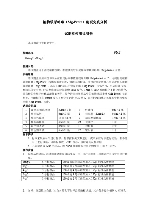
植物植物镁原卟啉镁原卟啉镁原卟啉((Mg-Proto )酶联免疫酶联免疫分析分析分析试剂试剂盒使用说明书盒使用说明书盒使用说明书本试剂盒仅供研究使用。
检测范围检测范围:: 96T0.4 ng/L -20 ng/L使用目的使用目的::本试剂盒用于测定植物组织,细胞及其它相关样本中镁原卟啉(Mg-Proto )含量。
实验原理本试剂盒应用双抗体夹心法测定标本中植物镁原卟啉(Mg-Proto )水平。
用纯化的植物镁原卟啉(Mg-Proto )抗体包被微孔板,制成固相抗体,往包被单抗的微孔中依次加入植物镁原卟啉(Mg-Proto ),再与HRP 标记的镁原卟啉(Mg-Proto )抗体结合,形成抗体-抗原-酶标抗体复合物,经过彻底洗涤后加底物TMB 显色。
TMB 在HRP 酶的催化下转化成蓝色,并在酸的作用下转化成最终的黄色。
颜色的深浅和样品中的植物镁原卟啉(Mg-Proto )呈正相关。
用酶标仪在450nm 波长下测定吸光度(OD 值),通过标准曲线计算样品中植物镁原卟啉(Mg-Proto )浓度。
试剂盒组成 1 30倍浓缩洗涤液 20ml ×1瓶 7 终止液6ml ×1瓶 2 酶标试剂 6ml ×1瓶 8 标准品(32ng/L ) 0.5ml ×1瓶 3 酶标包被板 12孔×8条 9 标准品稀释液 1.5ml ×1瓶 4 样品稀释液 6ml ×1瓶 10 说明书 1份 5 显色剂A 液 6ml ×1瓶 11 封板膜 2张 6显色剂B 液6ml ×1/瓶12密封袋1个标本标本要求要求1.标本采集后尽早进行提取,提取按相关文献进行,提取后应尽快进行实验。
若不能马上进行试验,可将标本放于-20℃保存,但应避免反复冻融2.不能检测含NaN3的样品,因NaN3抑制辣根过氧化物酶的(HRP )活性。
操作步骤1. 标准品的稀释:本试剂盒提供原倍标准品一支,用户可按照下列图表在小试管中进行稀释。
AMM氨测定试剂盒(谷氨酸脱氢酶法)产品说明书

AMM氨测定试剂盒(谷氨酸脱氢酶法)产品说明书AMM氨测定试剂盒(谷氨酸脱氢酶法)一、产品概述AMM氨测定试剂盒是一种基于谷氨酸脱氢酶法的高精度、高灵敏度氨测定试剂盒,用于测量生物体内氨浓度。
本试剂盒可广泛应用于医学、生物科学研究以及临床实验室等领域。
其准确度和可靠性得到了专业机构的认可。
二、试剂组成1. 测定液:包含谷氨酸脱氢酶、NADH(二磷酸腺苷二核苷酸)、缓冲液等。
2. 校准液:含有已知浓度的氨溶液,用于校准仪器。
3. 样本提取液:用于提取样本中的氨,使其能够反应产生信号。
三、试剂储存和稳定性1. 试剂的储存温度应为2-8摄氏度,避免阳光直接照射。
2. 开封后的试剂应储存在2-8摄氏度,并尽快使用。
3. 严禁冷冻试剂,以免造成试剂失效。
4. 正确存放条件下,试剂的稳定期为12个月。
四、仪器要求本试剂盒适用于多种型号的分光光度计,要求光敏器件工作稳定、精密度高,峰值吸光度范围在340-380nm之间。
五、操作步骤1. 准备工作a. 取出试剂,放置于室温下30分钟,使其回温到室温。
b. 将待检样本从冰箱取出后,回温至室温。
c. 开机并调整光源和光敏器件,确保仪器正常工作。
2. 样本制备a. 取适量样本加入样本提取液中,按照规定的比例稀释,混匀待用。
3. 样本测定a. 取适量测定液加入试管或微孔板中,作为反应底物。
b. 加入适量样本提取液,搅拌均匀。
c. 读取反应开始后一段时间内的吸光度值。
d. 将读数输入分光光度计或计算机,根据标准曲线计算样品中氨的浓度。
4. 结果判定a. 根据标准曲线上的吸光度值和已知浓度的校准液所对应的吸光度值,计算样品中氨的浓度。
b. 根据实验需求,判断测量结果的可靠性和准确性。
六、注意事项1. 操作前请阅读使用说明书。
2. 所有操作都必须在规定的温度下进行,以保证试剂和样本的稳定性。
3. 使用量杯、试管和微孔板等器具时,应保证其清洁干燥,以免发生干扰现象。
4. 实验过程中应避免直接接触皮肤和吸入试剂,如有意外接触,请立即清洗。
胰岛素样生长因子结合蛋白 3 定量测定试剂盒(酶联免疫法) 说明书
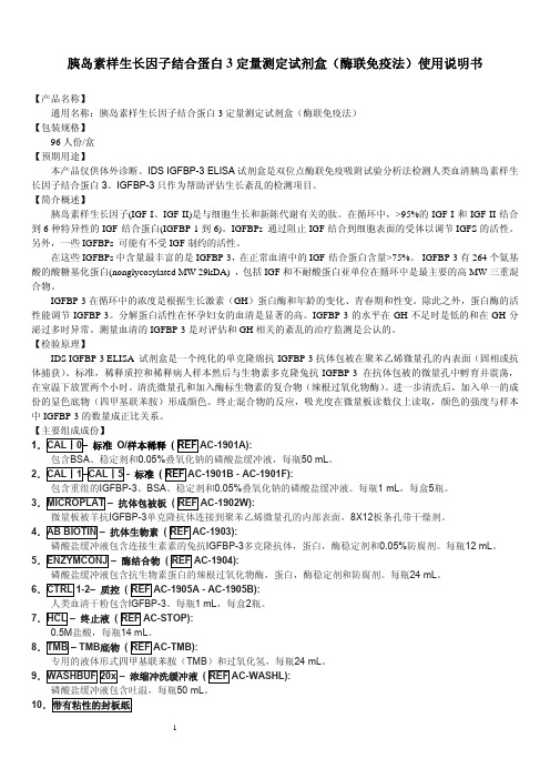
选择减少数据的方法可能被使用,但是操作人员必须选择适合数据的曲线和给出可接受的结果。推荐曲线要平 整光滑。
IDS结果计算使用MultiCalc (PerkinElmer)数据约简软件,绘制吸光度VS Log浓度的适合曲线图。
3
分析报告的范围是200 – 10000 ng/mL。任何结果低于最低的标准200 ng/mL时,应作用一个推断值并且以“低于
结果U/L
1259 4499
典型的标准曲线:
【检验方法的局限性】
1. 样本包含的分析物浓度超过最高的标准液浓度时,样本要用Calibrator 0稀释后重做。
2. 在任何疾病诊断程序的结果必须连同病人的临床症状和医生取得的信息一同进行解释。
3. IGFBP-3有内生血清蛋白酶时会下降,蛋白分解在怀孕期间会显著升高。这个试剂盒不适用检测怀孕妇女
磷酸盐缓冲液包含抗生物素蛋白的辣根过氧化物酶,蛋白,酶稳定剂和防腐剂。每瓶24 mL。 6.CTRL 1-2– 质控 ( REF AC-1905A - AC-1905B):
人类血清干粉包含IGFBP-3。每瓶1 mL,每盒2瓶。 7.HCL – 终止液 ( REF AC-STOP):
0.5M盐酸,每瓶14 mL。 8.TMB – TMB底物 ( REF AC-TMB):
2
9. 带有粘性的封板纸盖好微量板,并在18-25°C下孵育2小时30分钟。 10.重复第7个步骤。 18-25°C下孵育2小时30分钟。 注意:TMB 底物容易受到污染。仅能从瓶子中移出分析所需的用量,不使用的TMB 底物丢掉,不能再倒回瓶 子中。 12.用多道加样器在所有微量孔加入100 μL的终止液HCL。 13.在加入终止液的30分钟内使用微孔板读数仪在405 nm(参考波长为650 nm)下测量所有孔的吸光度。 标准 IGFBP-3 试剂盒的标准是在很多数量单位的基础上较准并且是重组的人类IGFBP-3糖基化蛋白预分析的。标准 已经进行预稀释1/101以致病人的样本可以直接读取能从标准曲线上读取结果而不需要进行较正。 质量控制 质控规则使用的质控样本要求几种水平的分析物,以确保每天的结果在可接受的范围内。提供两套质控。为了 能正确评估IDS分析推荐所有实验室包括每次适当分析部分的内部混合血浆,除试剂提供的质控外。质控应作为未知 物进行检测。质量控制图应按照测试的结果进行绘画。 【参考值(参考范围)】 以下的范围是由IDS IGFBP-3 ELISA 试剂进行测定并且仅提供参考。每个实验室必须建立自己地区人群的范围 值。
《中国药典》(2020年版)复方磺胺恶唑片中的甲氧苄啶含量测

《中国药典》(2020年版)复方磺胺噁唑片中的甲氧苄啶含量测甲氧苄啶(Trimethoprim, TMP),又称为甲氧苄氨嘧啶、甲氧苄嘧啶,是一种抗菌增效药,与磺胺类药物联合使用时,能使磺胺类药物抗菌谱扩大、抗菌活性大大增强。
由于其独特的作用,甲氧苄啶在养殖业病害防治中被广泛应用。
迪信泰检测平台采用高效液相色谱(HPLC)和液相色谱-三重四极杆质谱(LC-MS/MS)法,可高效、精准的检测甲氧苄啶的含量变化。
此外,我们还提供其他抗生素检测服务,以满足您的不同需求。
HPLC和LC-MS测定甲氧苄啶样本要求:1. 请确保样本量大于0.2g或者0.2mL。
周期:2~3周项目结束后迪信泰检测平台将会提供详细中英文双语技术报告,报告包括:1. 实验步骤(中英文)2. 相关质谱参数(中英文)3. 质谱图片4. 原始数据5. 甲氧苄啶含量信息应用范围:本方法采用高效液相色谱法测定复方磺胺甲恶唑片中磺胺甲恶唑和甲氧苄啶的含量。
本方法适用于复方磺胺甲恶唑片。
方法原理:供试品加甲醇稀释,最终用流动相定量稀释后,进入高效液相色谱仪进行色谱分离,用紫外吸收检测器,于波长240nm处检测磺胺甲恶唑和甲氧苄啶的吸收值,计算出其含量。
试剂: 1. 0.1mol/L盐酸2. 乙腈3. 三乙胺4. 氢氧化钠试液5. 冰醋酸仪器设备: 1. 仪器1.1 高效液相色谱仪1.2 色谱柱十八烷基硅烷键合硅胶为填充剂,理论塔板数按磺胺甲恶唑峰计算不低于4000。
磺胺甲恶唑和甲氧苄啶的分离度应符合要求。
1.3 紫外吸收检测器2. 色谱条件2.1 流动相:水乙腈三乙胺=799 200 1(用氢氧化钠试液或冰醋酸调节pH 值至5.9。
)2.2 检测波长:240nm2.3 柱温:室温试样制备:1. 称取供试品取本品10片,精密称定,研细,精密称取适量(约相当于磺胺甲恶唑44mg)置100mL量瓶中。
2. 对照品溶液的制备精密称取磺胺甲恶唑对照品和甲氧苄啶对照品适量,用0.1mol/L盐酸溶液溶解并定量稀释制成每1mL中约含有磺胺甲恶唑0.44mg的和甲氧苄啶89µg的溶液。
AM966_DataSheet_MedChemExpress
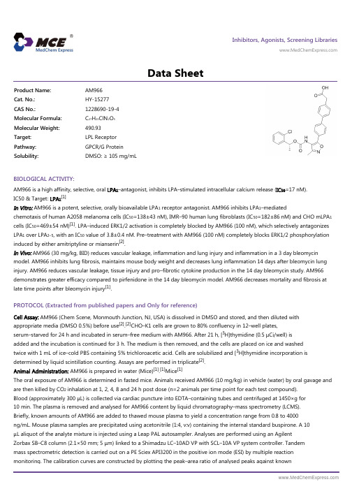
Inhibitors, Agonists, Screening Libraries Data SheetBIOLOGICAL ACTIVITY:AM966 is a high affinity, selective, oral LPA 1–antagonist, inhibits LPA–stimulated intracellular calcium release (IC 50=17 nM).IC50 & Target: LPA 1[1]In Vitro: AM966 is a potent, selective, orally bioavailable LPA 1 receptor antagonist. AM966 inhibits LPA 1–mediatedchemotaxis of human A2058 melanoma cells (IC 50=138±43 nM), IMR–90 human lung fibroblasts (IC 50=182±86 nM) and CHO mLPA 1cells (IC 50=469±54 nM)[1]. LPA–induced ERK1/2 activation is completely blocked by AM966 (100 nM), which selectively antagonizes LPA 1 over LPA 2–5, with an IC 50 value of 3.8±0.4 nM. Pre–treatment with AM966 (100 nM) completely blocks ERK1/2 phosphorylation induced by either amitriptyline or mianserin [2].In Vivo: AM966 (30 mg/kg, BID) reduces vascular leakage, inflammation and lung injury and inflammation in a 3 day bleomycin model. AM966 inhibits lung fibrosis, maintains mouse body weight and decreases lung inflammation 14 days after bleomycin lung injury. AM966 reduces vascular leakage, tissue injury and pro–fibrotic cytokine production in the 14 day bleomycin study. AM966demonstrates greater efficacy compared to pirfenidone in the 14 day bleomycin model. AM966 decreases mortality and fibrosis at late time points after bleomycin injury [1].PROTOCOL (Extracted from published papers and Only for reference)Cell Assay: AM966 (Chem Scene, Monmouth Junction, NJ, USA) is dissolved in DMSO and stored, and then diluted withappropriate media (DMSO 0.5%) before use [2].[2]CHO–K1 cells are grown to 80% confluency in 12–well plates,serum–starved for 24 h and incubated in serum–free medium with AM966. After 21 h, [3H]thymidine (0.5 μCi/well) isadded and the incubation is continued for 3 h. The medium is then removed, and the cells are placed on ice and washed twice with 1 mL of ice–cold PBS containing 5% trichloroacetic acid. Cells are solubilized and [3H]thymidine incorporation isdetermined by liquid scintillation counting. Assays are performed in triplicate [2].Animal Administration: AM966 is prepared in water (Mice)[1].[1]Mice [1]The oral exposure of AM966 is determined in fasted mice. Animals received AM966 (10 mg/kg) in vehicle (water) by oral gavage and are then killed by CO 2 inhalation at 1, 2, 4, 8 and 24 h post dose (n=2 animals per time point for each test compound).Blood (approximately 300 μL) is collected via cardiac puncture into EDTA–containing tubes and centrifuged at 1450×g for 10 min. The plasma is removed and analysed for AM966 content by liquid chromatography–mass spectrometry (LCMS).Briefly, known amounts of AM966 are added to thawed mouse plasma to yield a concentration range from 0.8 to 4000ng/mL. Mouse plasma samples are precipitated using acetonitrile (1:4, v:v) containing the internal standard buspirone. A 10μL aliquot of the analyte mixture is injected using a Leap PAL autosampler. Analyses are performed using an AgilentZorbax SB–C8 column (2.1×50 mm; 5 μm) linked to a Shimadzu LC–10AD VP with SCL–10A VP system controller. Tandem mass spectrometric detection is carried out on a PE Sciex API3200 in the positive ion mode (ESI) by multiple reactionmonitoring. The calibration curves are constructed by plotting the peak–area ratio of analysed peaks against knownProduct Name:AM966Cat. No.:HY-15277CAS No.:1228690-19-4Molecular Formula:C 27H 23ClN 2O 5Molecular Weight:490.93Target:LPL Receptor Pathway:GPCR/G Protein Solubility:DMSO: ≥ 105 mg/mLconcentrations. The lower limit of quantitation is 0.8 ng/mL. The data are subjected to linear regression analysis with 1/x2weighting.References:[1]. Swaney, JS, et al. A novel, orally active LPA1 receptor antagonist inhibits lung fibrosis in the mouse bleomycin model. Br J Pharmacol. 2010 Aug; 160(7):1699–713.[2]. Olianas MC, et al. Antidepressants activate the lysophosphatidic acid receptor LPA(1) to induce insulin–like growth factor–I receptor transactivation, stimulation of ERK1/2 signaling and cell proliferation in CHO–K1 fibroblasts. Biochem Pharmacol. 2015 JuCaution: Product has not been fully validated for medical applications. For research use only.Tel: 609-228-6898 Fax: 609-228-5909 E-mail: tech@Address: 1 Deer Park Dr, Suite Q, Monmouth Junction, NJ 08852, USA。
MG Chemicals Fast Cure Thermal Glue 8329TFF商品说明书
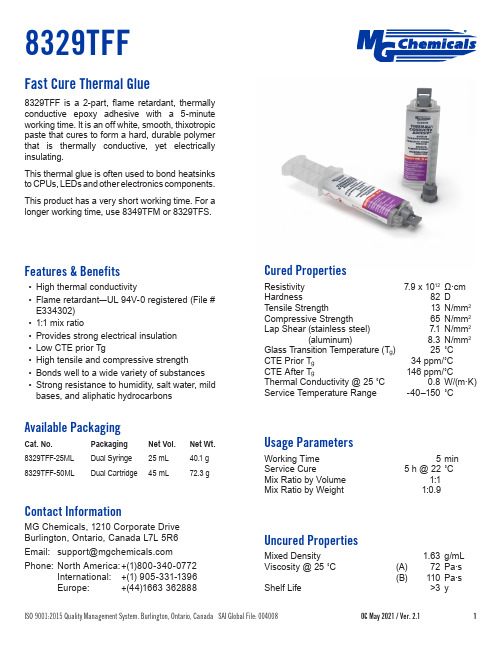
Features & Benefits• High thermal conductivity• Flame retardant —UL 94V-0 registered (File # E334302)• 1:1 mix ratio• Provides strong electrical insulation • Low CTE prior Tg• High tensile and compressive strength• Bonds well to a wide variety of substances • Strong resistance to humidity, salt water, mild bases, and aliphatic hydrocarbonsAvailable PackagingCat. No. Packaging Net Wt.8329TFF-25ML Dual Syringe 25 mL 40.1 g 8329TFF-50MLDual Cartridge45 mL72.3 gContact InformationMG Chemicals, 1210 Corporate DriveBurlington, Ontario, Canada L7L 5R6Phone: North America: +(1)800-340-0772 International: +(1) 905-331-1396 Europe: +(44)1663 362888Fast Cure Thermal Glue8329TFF is a 2-part, flame retardant, thermally conductive epoxy adhesive with a 5-minute working time. It is an off white, smooth, thixotropic paste that cures to form a hard, durable polymer that is thermally conductive, yet electrically insulating.This thermal glue is often used to bond heatsinks to CPUs, LEDs and other electronics components.This product has a very short working time. For a longer working time, use 8349TFM or 8329TFS.Cured PropertiesResistivity 7.9 x 10¹² Ω·cm Hardness 82 D Tensile Strength 13 N/mm 2Compressive Strength 65 N/mm 2Lap Shear (stainless steel)(aluminum) 7.1 N/mm 28.3 N/mm 2Glass Transition Temperature (T g ) 25 ˚C CTE Prior T g 34 ppm/˚C CTE After T g 146 ppm/˚C Thermal Conductivity @ 25 ˚C 0.8 W/(m·K)Service Temperature Range -40–150 ˚CUsage ParametersWorking Time 5 min Service Cure5 h @ 22 ˚C Mix Ratio by Volume 1:1Mix Ratio by Weight1:0.9Uncured PropertiesMixed Density1.63 g/mL Viscosity @ 25 ˚C (A) 72 Pa·s (B) 110 Pa·s Shelf Life>3 yApplication InstructionsRead the product SDS and Application Guide for more detailed instructions before using this product (downloadable at ). Recommended PreparationClean the substrate with Isopropyl Alcohol, MG #824, so the surface is free of oils, dust, and other residues. Syringe or Cartridge1.Twist and remove the cap from the syringe orcartridge. Do not discard cap.2. Dispense a small amount to ensure even flow ofboth parts. A manual or pneumatic dispensing gun is required for a 50 mL cartridge.3. (Optional) Attach a static mixer.a. Dispense and discard 3 to 5 mL of the product toensure a homogeneous mixture.b. After use, dispose of static mixer.4.Without a static mixer, dispense material on amixing surface or container, and thoroughly mix parts A and B together.5. To stop the flow, pull back on the plunger.6. Clean nozzle to prevent contamination and materialbuildup.7.Re-place the cap on the cartridge or syringe orcartridge.Dispensing AccessoriesConsult the table below for accessory selection. See the Dispensing Accessories Application Guide for usage instructions. 8MT-50-FT should only be used with a pneumatic dispenser.Cat. No. Dispensing Gun Static Mixer8329TFF-25ML N/A N/A8329TFF-50ML8DG-50-1-18MT-50, 8MT-50-FT Cure InstructionsAllow to cure at room temperature for 4 hours, or cure the adhesive in an oven at one of these time/ temperature options:Temperature65 °C80 °CTime15 minutes10 minutes Storage and HandlingStore between 16 and 27 ˚C in a dry area, away from sunlight (see SDS). To maximize shelf life, recap product firmly when not in use.DisclaimerThis information is believed to be accurate. It is intended for professional end-users who have the skills required to evaluate and use the data properly. M.G. Chemicals Ltd. does not guarantee the accuracy of the data and assumes no liability in connection with damages incurred while using it.。
叶酸ELISA试剂盒产品手册说明书
Product ManualFolic Acid ELISA KitCatalog NumberMET-5068 96 assaysMET-5068-5 5 x 96 assays FOR RESEARCH USE ONLYNot for use in diagnostic proceduresIntroductionFolic acid is a B vitamin also known as Vitamin B9. Since humans don’t synthesize folic acid, it is required from the diet and is therefore considered to be an essential vitamin. In cells, folic acid is required for amino acid metabolism as well as to carry one-carbon groups used for methylation reactions and synthesis of nucleic acids (such as thymine and purine bases). Therefore, deficiency in folic acid disrupts DNA synthesis and cell division, affecting mostly hematopoietic cells and abnormal tissue growths because of their higher rate of cell division.Folic acid is used to supplement folic acid deficiency. This deficiency can cause anemia. Symptoms of anemia can include fatigue, heart palpitations, difficulty breathing, open sores observed on the tongue, and color changes of the hair or skin. Deficiency can occur in children after only a month of consuming a folic acid deficient diet. In adults, normal total body folic acid levels are between10,000–30,000 micrograms (µg) with plasma levels of greater than 7 nM (3 ng/mL) (Table 1). Women also take supplemental folic acid during pregnancy to prevent fetal neural tube defects (NTDs). Insufficient levels of dietary folic acid in early pregnancy are thought to be the cause of over half of babies born with neural tube defects. As a result, over 50 countries add folic acid to certain foods as a way to decrease NTD incidents in the population.Concentration (ng/mL) Concentration (nM) Adults 3-20 7-45.3Children 5-21 11.3-47.6Infants 14-51 31.7-115.5Table 1. Reference ranges for folic acid in human plasma.The Folic Acid ELISA Kit is a competitive enzyme immunoassay developed for rapid detection and quantitation of folic acid in serum, cell or tissue samples. The quantity of folic acid in unknown samples is determined by comparing its absorbance with that of a known folic acid standard curve. The kit has detection sensitivity limit of 24 pg/mL folic acid. Each Folic Acid ELISA Kit provides sufficient reagents to perform up to 96 assays, including standard curve and unknown samples. Assay PrincipleThe Folic Acid ELISA kit is a competitive ELISA for the quantitative measurement of folic acid. The unknown folic acid samples or folic acid standards are first added to a Folic Acid Conjugate preabsorbed microplate. After a brief incubation, an Anti-Folic Acid antibody is added, followed by an HRP conjugated secondary antibody. The folic acid content in unknown samples is determined by comparison with a predetermined folic acid standard curve.Related Products1.MET-5054: L-Amino Acid Assay Kit2.MET-5056: Branched Chain Amino Acid Assay Kit3.MET-5151: S-Adenosylhomocysteine (SAH) ELISA Kit4.MET-5152: S-Adenosylmethionine (SAM) ELISA Kit5.STA-674: Glutamate Assay KitKit ComponentsBox 1 (shipped at room temperature)1.96-well Protein Binding Plate (Part No. 231001): One strip well 96-well plate.2.Anti-Folic Acid Antibody (500X) (Part No. 50681C): One 10 µL vial.3.Secondary Antibody, HRP Conjugate (Part No. 231009): One 20 µL vial.4.Assay Diluent (Part No. 310804):One 50 mL bottle.5.10X Wash Buffer (Part No. 310806): One 100 mL bottle.6.Substrate Solution (Part No. 310807): One 12 mL amber bottle.7.Stop Solution (Part. No. 310808): One 12 mL bottle.8.Folic Acid Standard (Part No. 50682C): One 100 µL amber vial of 10 µg/mL Folic Acid inwater.Box 2 (shipped on blue ice packs)1.100X Folic Acid Conjugate (Part No. 50683C): One 100 µL amber vial.Materials Not Supplied1.Folic acid samples such as serum, plasma, or folic acid extracted from cells or tissues2.Tissue Homogenizer3.1X PBS4.10 µL to 1000 µL adjustable single channel micropipettes with disposable tips5.50 µL to 300 µL adjustable multichannel micropipette with disposable tips6.Multichannel micropipette reservoir7.Microplate reader capable of reading at 450 nm (620 nm as optional reference wave length) StorageUpon receipt, aliquot and store 100X Folic Acid Conjugate at -20ºC and avoid multiple freeze/thaw cycles. Store all other components at 4ºC. The 100X Folic Acid Conjugate and Folic Acid Standard are light sensitive and must be stored accordingly.Preparation of Reagents•Folic Acid Conjugate Coated Plate: Dilute the proper amount of 100X Folic Acid Conjugate 1:100 into 1X PBS. Add 100 μL of the diluted 1X Folic Acid Conjugate to each well and incubate at 37ºC for two hours or overnight at 4ºC. Remove the coating solution and wash twice with 200 μL of 1X PBS. Blot plate on paper towels to remove excess fluid. Add 200 μL of Assay Diluent to each well and block for 1 hr at room temperature. Transfer the plate to 4ºC and remove the Assay Diluent immediately before use.Note: The Folic Acid Conjugate-coated wells are not stable and should be used within 24 hrs after coating. Only coat the number of wells to be used immediately.•1X Wash Buffer: Dilute the 10X Wash Buffer to 1X with deionized water. Stir to homogeneity. •Anti-Folic Acid Antibody and Secondary Antibody: Immediately before use dilute the Anti-Folic Acid Antibody 1:500 and Secondary Antibody 1:1000 with Assay Diluent. Do not store diluted solutions.Preparation of Standard Curvee the provided stock Folic Acid Standard 10 µg/mL solution to prepare a series of the remainingstandards according to Table 1 below.Standard Tubes 10 µg/mL Folic AcidStandard (µL)Assay Diluent(µL)Folic Acid(ng/mL)Folic Acid(nM)1 10 990 100 2272 100 of Tube #1 300 25 56.753 100 of Tube #2 300 6.25 14.194 100 of Tube #3 300 1.56 3.555 100 of Tube #4 300 0.391 0.8876 100 of Tube #5 300 0.098 0.2227 100 of Tube #6 300 0.024 0.0558 0 300 0 0 Table 1. Preparation of Folic Acid Standards.Preparation of Samples•Serum: Avoid hemolyzed and lipemic blood samples. Collect blood in a tube with no anticoagulant. Allow the blood to clot at room temperature for 30 minutes. Centrifuge at 2500 x g for 20 minutes. Remove the yellow serum supernatant without disturbing the white buffy layer.Aliquot samples for testing and store at -80ºC. Perform dilutions in Assay Diluent or PBS containing 0.1% BSA as necessary.•Plasma: Avoid hemolyzed and lipemic blood samples. Collect blood with heparin or citrate and centrifuge at 2000 x g and 4ºC for 10 minutes. Remove the plasma layer and store on ice. Avoid disturbing the white buffy layer. Aliquot samples for testing and store at -80ºC. Perform dilutions in Assay Diluent or PBS containing 0.1% BSA as necessary.Note: This assay is not compatible with rabbit serum or plasma due to high levels of rabbit IgG that will cross react with the secondary antibody.•Cells or tissues: Homogenize 50-200 mg of the cell pellet or tissue in 0.5-2 mL of ice-cold PBS using a mortar and pestle or by dounce homogenization. Incubate the homogenate at 4°C for 20 minutes. Transfer the homogenate to a centrifuge tube and centrifuge at 12000 x g for 20 minutes.Recover the supernatant and transfer to a fresh tube. Store resuspended sample at -20°C or colder until ready to test by ELISA. Perform dilutions in Assay Diluent or PBS containing 0.1% BSA as necessary.Assay Protocol1.Prepare and mix all reagents thoroughly before use. Each folic acid sample including unknownand standard should be assayed in duplicate.2.Add 50 µL of unknown sample or Folic Acid standards to the wells of the Folic Acid Conjugatecoated plate. Incubate at room temperature for 10 minutes on an orbital shaker.3.Add 50 µL of the diluted Anti-Folic Acid antibody to each well, incubate at room temperature for 1hour on an orbital shaker.4.Wash microwell strips 3 times with 250 µL 1X Wash Buffer per well with thorough aspirationbetween each wash. After the last wash, empty wells and tap microwell strips on absorbent pad or paper towel to remove excess 1X Wash Buffer.5.Add 100 µL of the diluted Secondary Antibody-HRP Enzyme Conjugate to all wells.6.Incubate at room temperature for 1 hour on an orbital shaker.7.Wash microwell strips 3 times according to step 4 above. Proceed immediately to the next step.8.Warm Substrate Solution to room temperature. Add 100 L of Substrate Solution to each well,including the blank wells. Incubate at room temperature on an orbital shaker. Actual incubation time may vary from 2-30 minutes.Note: Watch plate carefully; if color changes rapidly, the reaction may need to be stopped sooner to prevent saturation.9.Stop the enzyme reaction by adding 100 µL of Stop Solution into each well, including the blankwells. Results should be read immediately (color will fade over time).10.Read absorbance of each microwell on a spectrophotometer using 450 nm as the primary wavelength.Example of ResultsThe following figures demonstrate typical Folic Acid ELISA results. One should use the data below for reference only. This data should not be used to interpret actual results.Figure 1: Folic Acid ELISA Standard Curve.Figure 2: Folic Acid Levels in Human, Mouse or Rat Serum compared to Negative Control (Assay Diluent). Serum samples were diluted 1:5 in Assay Diluent and tested according to the Assay Protocol.References1.Bibbins-Domingo, K; Grossman, DC.; Curry, SJ.; Davidson, KW.; Epling, John W.; García, FAR.;Kemper, AR.; Krist, AH.; Kurth, AE.; Landefeld, CS; Mangione, CM.; Phillips, William R.;Phipps, MG.; Pignone, MP.; Silverstein, M; Tseng, C-W (2017). JAMA. 317: 183.2.Marino, BS.; Fine, KS (2009). Blueprints Pediatrics. Lippincott Williams & Wilkins. p. 131.3.Bailey, LB. (2009). Folate in Health and Disease, Second Edition. CRC Press. p. 198.4.Obeid, R; Herrmann, W (2012). Curr. Drug Metab. 13: 1184–1195.5.Wilson RD, Wilson RD, Audibert F, Brock JA, Carroll J, Cartier L, Gagnon A, Johnson JA,Langlois S, Murphy-Kaulbeck L, Okun N, Pastuck M, Deb-Rinker P, Dodds L, Leon JA, Lowel HL, Luo W, MacFarlane A, McMillan R, Moore A, Mundle W, O'Connor D, Ray J, Van den Hof M (2015). J Obstet Gynaecol Can. 37: 534–52.6.Figueiredo JC1, Grau MV, Haile RW, Sandler RS, Summers RW, Bresalier RS, Burke CA,McKeown-Eyssen GE, Baron JA. J. Natl. Cancer Inst.101: 432–5.Recent Product Citations1.McCarthy, G.A. et al. (2023). A Novel 3DNA® Nanocarrier effectively delivers payloads topancreatic tumors. Transl Oncol. 32:101662. doi: 10.1016/j.tranon.2023.101662.2.Shinagawa, A. et al. (2022). Short-Term Combined Intake of Vitamin B2 and Vitamin E DecreasesPlasma Homocysteine Concentrations in Female Track Athletes. Dietetics. 1(3):216-226. doi:10.3390/dietetics1030019.3.Siervo, M. et al. (2020). Nitrate-Rich Beetroot Juice Reduces Blood Pressure in Tanzanian Adultswith Elevated Blood Pressure: A Double-Blind Randomized Controlled Feasibility Trial. J Nutr.doi: 10.1093/jn/nxaa170.4.Simanjuntak, Y. et al. (2020). Preventive effects of folic acid on Zika virus-associated poorpregnancy outcomes in immunocompromised mice. PLoS Pathog. 16(5):e1008521. doi:10.1371/journal.ppat.1008521.5.Zhu, J. et al. (2018). Effect of maternal folic acid supplementation on prostatitis risk in the ratoffspring. Int Urol Nephrol. 50(11):1963-1973. doi: 10.1007/s11255-018-1969-8.WarrantyThese products are warranted to perform as described in their labeling and in Cell Biolabs literature when used in accordance with their instructions. THERE ARE NO WARRANTIES THAT EXTEND BEYOND THIS EXPRESSED WARRANTY AND CELL BIOLABS DISCLAIMS ANY IMPLIED WARRANTY OF MERCHANTABILITY OR WARRANTY OF FITNESS FOR PARTICULAR PURPOSE. CELL BIOLABS’sole obligation and purchaser’s exclusive remedy for breach of this warranty shall be, at the option of CELL BIOLABS, to repair or replace the products.In no event shall CELL BIOLABS be liable for any proximate, incidental or consequential damages in connection with the products.Contact InformationCell Biolabs, Inc.7758 Arjons DriveSan Diego, CA 92126Worldwide: +1 858-271-6500USA Toll-Free: 1-888-CBL-0505E-mail: ********************©2017-2023: Cell Biolabs, Inc. - All rights reserved. No part of these works may be reproduced in any form without permissions in writing.。
普洛麦格(北京)生物技术有限公司CTM692产品说明书

2022版 CTM692原英文技术手册TM692中 文 说 明 书适用产品目录号:GA1330和GA1332FcγRI ADCP BioassayEffector Cells, Propagation Model普洛麦格(北京)生物技术有限公司Promega (Beijing) Biotech Co., Ltd 地址:北京市东城区北三环东路36号环球贸易中心B 座907-909电话:************网址: 技术支持电话:400 810 8133技术支持邮箱:*************************CTM 6922022制作1所有技术文献的英文原版均可在/ protocols 获得。
请访问该网址以确定您使用的说明书是否为最新版本。
如果您在使用该试剂盒时有任何问题,请与Promega 北京技术服务部联系。
电子邮箱:*************************1. 描述 ....................................................................................................................................................................................22. 产品组分和储存条件............................................................................................................................................................43. 开始实验前 ..........................................................................................................................................................................54. 制备Fc γRI ADCP 效应细胞 ................................................................................................................................................6 4. A. 细胞解冻和初始细胞培养 .......................................................................................................................................... 6 4. B. Fc γRI ADCP 效应细胞的细胞维持和增殖 ..................................................................................................................7 4. C. Fc γRI ADCP 效应细胞冷冻和储存 .............................................................................................................................75. 检测方案 .............................................................................................................................................................................8 5. A. Bio-Glo™试剂、检测缓冲液和测试及对照样品的制备...............................................................................................9 5. B. 孔板布局设计 ...........................................................................................................................................................10 5. C. 检测前一天制备和铺板贴壁靶细胞 ...........................................................................................................................10 5. D. 检测当天制备悬浮靶细胞 .........................................................................................................................................11 5. E. 制备抗体系列稀释液 ................................................................................................................................................12 5. F. 制备Fc γRI 效应细胞 ................................................................................................................................................12 5. G. 铺板悬浮靶细胞、抗体和Fc γRI 效应细胞 ................................................................................................................13 5. H. 将抗体和Fc γRI 效应细胞加至预先铺板的贴壁靶细胞中 ...........................................................................................13 5. I. 加入Bio-Glo™试剂 ..................................................................................................................................................14 5. J. 数据分析...................................................................................................................................................................146. 疑难解答 ...........................................................................................................................................................................157. 参考文献 ...........................................................................................................................................................................168. 代表性检测结果.. (17)FcγRI ADCP Bioassay Effector Cells, Propagation Model普洛麦格(北京)生物技术有限公司Promega (Beijing) Biotech Co., Ltd 地址:北京市东城区北三环东路36号环球贸易中心B座907-909电话:************网址:技术支持电话:400 810 8133技术支持邮箱:*************************CTM 6922022制作21. 描述抗体依赖性细胞介导的吞噬作用(ADCP)是治疗性抗体识别和介导消除病毒性感染细胞或病变(如肿瘤)细胞的重要作用机制。
骨钙素N端中分子片段检测试剂盒(酶联免疫法)说明书
14.洗板机要经常保养,以免试验中由于机械问题导致 试验失败或交叉污染。 15.所有试剂和实验室设备应按传染性物品处理和丢弃。
注意事项:由于不同的实验方法和试剂在识别位点、特
异性及干扰因素等诸方面不尽相同,因此对于某一特定
样本,其测定结果也存在一定差异;所以实验室在向临
床医生提供检测结果的同时也必须包括相应的实验方
如果离失效期不足 8 周,则以有效期为准。 复溶后校准品和质控品应在–18°C 以下保存不超
过 3 个月,且只能冻融两次。当抗体试剂混合后,剩余 混合物应存放在 2-8°C 且不超过 1 个月,或在–18°C 以下冰冻保存。
浓缩洗液在稀释后室温放置可稳定 2 天,在 2~8 ℃保存可稳定 8 周,如果离失效期不足 8 周,则以有效 期为准。
软件 2. 试剂准备
1) 试剂盒内所有组分和样本平衡至室温(18-22 ℃);酶标板铝箔袋每次使用前也需平衡至室 温,以免在酶标板上凝集水珠,从而影响实验 结果。在取出所需的板条后,余下的须尽快放 回铝箔袋,封口后置于 2-8℃保存。
2) 校准品 0 用 5mL 蒸馏水复溶,校准品 1-5 和质 控品各用 0.5mL 蒸馏水复溶。复溶 15 分钟。
骨钙素n端中分子片段检测试剂盒酶联免疫法说明书酶联免疫试剂盒说明书酶免试剂盒说明书小片段dna回收试剂盒酶联免疫试剂盒降钙素原检测试剂盒碱性磷酸酶试剂盒碱性磷酸酶染色试剂盒胃蛋白酶原试剂盒胃蛋白酶检测试剂盒
骨钙素N端中分子片段检测试剂盒(酶联免疫法)说明书
【产品名称】 通用名称:骨钙素 N 端中分子片段检测试剂盒(酶 联免疫法) 英文名称:N-MID Osteocalcin ELISA
良好实验室管理规范(GLP)要求在每轮实验中使 用质控品以检测实验操作质量。质控品应以待测样本对
谷胱甘肽还原酶(GR)活性测定试剂盒说明书

货号:MS1111 规格:100管/96样谷胱甘肽还原酶(GR)活性测定试剂盒说明书微量法注意:正式测定之前选择 2-3个预期差异大的样本做预测定。
测定意义:GR是广泛存在于真核和原核生物中的一种黄素蛋白氧化还原酶,是谷胱甘肽氧化还原循环的关键酶之一(通常昆虫中GR被TrxR取代)。
GR催化NADPH还原GSSG生成GSH,有助于维持体内GSH/GSSG比值。
GR在氧化胁迫反应中对活性氧清除起关键作用,此外GR还参与抗坏血酸-谷胱甘肽循环途径。
测定原理:GR能催化NADPH还原GSSG再生GSH,同时NADPH脱氢生成NADP+;NADPH在340nm有特征吸收峰,相反NADP+在该波长无吸收峰;通过测定340nm吸光度下降速率来测定NADPH脱氢速率,从而计算GR活性。
自备仪器和用品:低温离心机、水浴锅、移液器、紫外分光光度计/酶标仪、微量石英比色皿/96孔板、和蒸馏水试剂组成和配置:试剂一:液体×1 瓶,4℃保存。
试剂二:粉剂×1 瓶,4℃保存。
临用前加入 2mL 蒸馏水,混匀。
试剂三:液体×1 支,4℃保存。
粗酶液提取:1. 组织:按照组织质量(g):试剂一体积(mL)为1:5~10的比例(建议称取约0.1g组织,加入1mL试剂一)进行冰浴匀浆。
8000g,4℃离心15min,取上清,置冰上待测。
2. 细菌、真菌:按照细胞数量(104个):试剂一体积(mL)为500~1000:1的比例(建议500万细胞加入1mL试剂一),冰浴超声波破碎细胞(功率300w,超声3秒,间隔7秒,总时间3min);然后8000g,4℃,离心15min,取上清置于冰上待测。
3. 血清等液体:直接测定。
操作步骤:1. 分光光度计/酶标仪预热30min,调节波长到340nm,蒸馏水调零。
2. 试剂一置于25℃(普通物质)或者37℃(哺乳动物)中预热30min。
3. 空白管:取微量石英比色皿或96孔板,加入170μL试剂一,20μL试剂二,10μL试剂三,于340nm 测定10s和190s吸光度,记为A空1和A空2,△A空白管= A空1﹣A空2。
格锐思可溶性糖含量试剂盒说明书 (货号:G0501W 微板法 96 样)

可溶性糖含量试剂盒说明书(货号:G0501W微板法96样)一、产品简介:糖类物质是构成植物体的重要组成成分之一,也是新陈代谢的主要原料和贮存物质。
糖类在浓硫酸作用下经脱水反应生成糠醛或羟甲基糖醛,生成的糠醛或羟甲基糖醛与蒽酮脱水缩合,形成糠醛的衍生物,呈蓝绿色物质,其在可见光区620nm波长处有最大吸收,且其光吸收值在一定范围内与糖的含量成正比关系。
该方法用于可溶性单糖、寡糖和多糖的含量测定,具有灵敏度高﹑简便快捷﹑适用于微量样品的测定等优点。
该方法的特点是几乎可以测定所有的糖类(包括单糖:戊糖、己糖、糖原、多缩葡萄糖等),所以用该方法测出的糖类含量是溶液中全部可溶性糖类总含量。
二、试剂盒组分与配制:试剂名称规格保存要求备注试剂一粉剂×1瓶4℃避光保存试剂二液体5mL×1瓶4℃保存标准品粉剂×1支4℃保存若重新做标曲,则用到该试剂工作液配制:吸取4mL试剂二加入到试剂一的试剂瓶中,混匀并充分溶解,即得工作液。
(如难溶解,可60℃水浴溶解;剩余试剂4℃保存一周)三、所需的仪器和用品:酶标仪、96孔板、水浴锅、可调式移液器、乙醇、浓硫酸(不允许快递)、研钵。
四、可溶性糖含量的测定:建议正式实验前选取2个样本做预测定,了解本批样品情况,熟悉实验流程,避免实验样本和试剂浪费!建议:选取样本做几个梯度的稀释,选取适合本次实验的稀释倍数D。
1组织样本:称取0.1g样本(若是干样,如烘干烟叶等可取0.05g;若是水分充足的样本可取0.2g),先加入0.8mL的80%乙醇(自备:取80mL乙醇溶于20mL蒸馏水中),冰浴匀浆,倒入有盖离心管中,再用80%乙醇冲洗研钵并转移至同一EP管中,使EP管中粗提液终体积定容为1.5mL(若用自动研磨机可直接加入1.5mL的80%乙醇研磨);置50℃水浴20min(封口膜缠紧,防止液体散失,且间隔2min振荡混匀一次),冷却后(若有损失,可加80%乙醇补齐至1.5mL),12000rpm,室温离心10min,取上清液备用。
线粒体复合体Ⅱ试剂盒说明书

货号:MS3301 规格:100管/96样线粒体复合体Ⅱ试剂盒说明书微量法正式测定前务必取2-3个预期差异较大的样本做预测定测定意义:线粒体复合体Ⅱ又称琥珀酸-辅酶Q还原酶,广泛存在于动物、植物、微生物和培养细胞,后者进一步还原氧的线粒体中,催化琥珀酸氧化生成延胡索酸,同时辅基FAD还原为FADH2化型辅酶Q生成还原型辅酶Q,是呼吸电子传递链的支路。
测定原理:复合体Ⅱ的催化产物还原型辅酶Q可进一步还原2,6-二氯吲哚酚,2,6-二氯吲哚酚在605nm有特征吸收峰,通过检测2,6-二氯吲哚酚的减少速率来计算该酶活性。
自备实验用品及仪器:可见分光光度计/酶标仪、台式离心机、水浴锅、可调式移液器、微量石英比色皿/96孔板、研钵、冰和蒸馏水。
试剂组成和配制:试剂一:液体100mL×1瓶,-20℃保存;试剂二:液体20mL×1瓶,-20℃保存;试剂三:液体1.5mL×1支,-20℃保存;试剂四:液体25mL×1瓶,4℃保存;试剂五:粉剂×1支,-20℃保存;试剂六:液体2.5mL×1瓶,4℃保存;样本的前处理:组织、细菌或细胞中胞浆蛋白与线粒体蛋白的分离:1、准确称取0.1g组织或收集500万细胞,加入1mL试剂一和10uL 试剂三,用冰浴匀浆器或研钵匀浆。
2、将匀浆600g,4℃离心5min。
3、弃沉淀,将上清液移至另一离心管中,11000g,4℃离心10min。
4、上清液即为除去线粒体的胞浆蛋白,可用于测定从线粒体泄漏的复合体Ⅱ(此步可选做)。
5、步骤④中的沉淀即为线粒体,加入200uL试剂二和2uL 试剂三,超声波破碎(冰浴,功率20%或200W,超声3s,间隔10秒,重复30次),用于复合体Ⅱ酶活性测定。
测定步骤:1、分光光度计或酶标仪预热30min以上,调节波长至605nm,蒸馏水调零。
2、样本测定(1)工作液的配制:临用前把试剂五转移到试剂四中混合溶解,置于37℃(哺乳动物)或25℃(其它物种)孵育5min;用不完的试剂分装后-20℃保存,禁止反复冻融。
人巨噬细胞集落刺激因子(M-CSF)酶联免疫分析试剂盒使用说明书

人巨噬细胞集落刺激因子(M-CSF)酶联免疫分析试剂盒使用说明书本试剂盒仅供研究使用检测范围:15.6pg/ml-1000pg/ml特异性:本试剂盒可同时检测天然或重组的人M-CSF,且与其他相关蛋白无交叉反应。
有效期:6个月预期应用:ELISA法定量测定人血清、血浆、细胞培养上清或其它相关生物液体中M-CSF 含量。
说明1.试剂盒保存:-20℃(较长时间不用时);2-8℃(频繁使用时)。
2.浓洗涤液低温保存会有盐析出,稀释时可在水浴中加温助溶。
3.中、英文说明书可能会有不一致之处,请以英文说明书为准。
4.刚开启的酶联板孔中可能会含有少许水样物质,此为正常现象,不会对实验结果造成任何影响。
概述巨噬细胞集落刺激因子(macrophage colony stimulating factor,M-CSF)又称为集落刺激因子-1(CSF-1),是造血系统重要的细胞因子。
M-CSF主要调节单核-巨噬细胞系细胞的生长、增殖和分化。
多种细胞均可产生M-CSF,包括成纤维细胞、子宫内膜中分泌型上皮细胞、骨髓基质细胞、脑星状细胞、成骨细胞;LPS等激活的巨噬细胞、B细胞、T细胞和内皮细胞等;此外,多种肿瘤细胞如原粒细胞性白血病、淋巴母细胞性白血病、肺腺癌细胞、乳腺癌和卵巢癌等。
人和小鼠天然M-CSF为糖蛋白,由二硫键连接的同源双体,分子量40~90kDa。
人M-CSF前体长度554~256个氨基酸不等,均有32个氨基酸的信号肽和23个氨基酸的穿膜部分。
膜结合型M-CSF表达在单层培养的成纤维细胞,可刺激表达M-CSF受体的巨噬细胞的粘附和增殖。
成熟M-CSF分子靠近N端150氨基酸在与M-CSF受体结合中起关键作用,人和小鼠M-CSF分子这个区域结构高度保守,其同源性达80%。
人和小鼠M-CSF基因定位于5对染色体,与GM-CSF、IL-3、IL-4、IL-5、IL-13和酸性FGF基因密切连锁。
M-CSF受体为高亲和力,表达于循环的单核细胞和组织巨噬细胞以及胎盘滋养层细胞。
氧化型谷胱甘肽(GSSG)含量检测试剂盒说明书__ 微量法UPLC-MS-4489

氧化型谷胱甘肽(GSSG)含量检测试剂盒说明书微量法货号:UPLC-MS-4489规格:100T/96S产品组成:使用前请认真核对试剂体积与瓶内体积是否一致,有疑问请及时联系工作人员。
试剂名称规格保存条件试剂一液体100mL×1瓶4℃保存试剂二液体130μL×1支4℃保存试剂三液体20mL×1瓶4℃保存试剂四液体 2.5mL×1瓶4℃保存试剂五粉剂×1瓶4℃保存试剂六液体12.5μL×1瓶4℃保存标准品粉剂10mg×1支4℃保存溶液的配制:1、试剂二:有毒易挥发试剂,涉及该试剂的步骤建议在通风橱内操作。
2、试剂五:临用前加入2.5mL蒸馏水,溶解后-20℃分装保存;3、试剂六:液体置于试剂瓶内EP管中。
临用前根据样本量将试剂六、蒸馏水按1:20(V:V)的比例配制备用,现用现配;4、标准品:用1mL蒸馏水溶解,浓度为10mg/mL,4℃保存。
产品说明:氧化型谷胱甘肽(GSSG)是谷胱甘肽(GSH)的氧化形式,又称为二硫代谷胱甘肽,是两分子的谷胱甘肽氧化而成。
GSSG会被谷胱甘肽还原酶还原成GSH,因此机体中大多数是以还原型形式存在。
测定细胞内GSH 和GSSG含量以及GSH/GSSG比值,能够很好地反映细胞所处的氧化还原状态。
本试剂盒利用谷胱甘肽能和5,5’-二硫代-双-(2-硝基苯甲酸)(5,5’-dithiobis-2-nitrobenoic acid,DTNB)反应产生2-硝基-5-巯基苯甲酸,2-硝基-5-巯基苯甲酸在波长412nm处具有最大光吸收的特点,通过2-乙烯吡啶抑制样本中原有的还原型谷胱甘肽,然后利用谷胱甘肽还原酶将GSSG还原为GSH,借此测定氧化型谷胱甘肽的含量。
技术指标:最低检出限:3.211μg/mL线性范围:3.9-125μg/mL注意:实验之前建议选择2-3个预期差异大的样本做预实验。
如果样本吸光值不在测量范围内建议稀释或者增加样本量进行检测。
MUT056399-SDS-MedChemExpress

Inhibitors, Agonists, Screening LibrariesSafety Data Sheet Revision Date:Sep.-30-2018Print Date:Sep.-30-20181. PRODUCT AND COMPANY IDENTIFICATION1.1 Product identifierProduct name :MUT056399Catalog No. :HY-18169CAS No. :1269055-85-71.2 Relevant identified uses of the substance or mixture and uses advised againstIdentified uses :Laboratory chemicals, manufacture of substances.1.3 Details of the supplier of the safety data sheetCompany:MedChemExpress USATel:609-228-6898Fax:609-228-5909E-mail:sales@1.4 Emergency telephone numberEmergency Phone #:609-228-68982. HAZARDS IDENTIFICATION2.1 Classification of the substance or mixtureNot a hazardous substance or mixture.2.2 GHS Label elements, including precautionary statementsNot a hazardous substance or mixture.2.3 Other hazardsNone.3. COMPOSITION/INFORMATION ON INGREDIENTS3.1 SubstancesSynonyms:NoneFormula:C15H13F2NO3Molecular Weight:293.27CAS No. :1269055-85-74. FIRST AID MEASURES4.1 Description of first aid measuresEye contactRemove any contact lenses, locate eye-wash station, and flush eyes immediately with large amounts of water. Separate eyelids with fingers to ensure adequate flushing. Promptly call a physician.Skin contactRinse skin thoroughly with large amounts of water. Remove contaminated clothing and shoes and call a physician.InhalationImmediately relocate self or casualty to fresh air. If breathing is difficult, give cardiopulmonary resuscitation (CPR). Avoid mouth-to-mouth resuscitation.IngestionWash out mouth with water; Do NOT induce vomiting; call a physician.4.2 Most important symptoms and effects, both acute and delayedThe most important known symptoms and effects are described in the labelling (see section 2.2).4.3 Indication of any immediate medical attention and special treatment neededTreat symptomatically.5. FIRE FIGHTING MEASURES5.1 Extinguishing mediaSuitable extinguishing mediaUse water spray, dry chemical, foam, and carbon dioxide fire extinguisher.5.2 Special hazards arising from the substance or mixtureDuring combustion, may emit irritant fumes.5.3 Advice for firefightersWear self-contained breathing apparatus and protective clothing.6. ACCIDENTAL RELEASE MEASURES6.1 Personal precautions, protective equipment and emergency proceduresUse full personal protective equipment. Avoid breathing vapors, mist, dust or gas. Ensure adequate ventilation. Evacuate personnel to safe areas.Refer to protective measures listed in sections 8.6.2 Environmental precautionsTry to prevent further leakage or spillage. Keep the product away from drains or water courses.6.3 Methods and materials for containment and cleaning upAbsorb solutions with finely-powdered liquid-binding material (diatomite, universal binders); Decontaminate surfaces and equipment by scrubbing with alcohol; Dispose of contaminated material according to Section 13.7. HANDLING AND STORAGE7.1 Precautions for safe handlingAvoid inhalation, contact with eyes and skin. Avoid dust and aerosol formation. Use only in areas with appropriate exhaust ventilation.7.2 Conditions for safe storage, including any incompatibilitiesKeep container tightly sealed in cool, well-ventilated area. Keep away from direct sunlight and sources of ignition.Recommended storage temperature:Powder-20°C 3 years4°C 2 yearsIn solvent-80°C 6 months-20°C 1 monthShipping at room temperature if less than 2 weeks.7.3 Specific end use(s)No data available.8. EXPOSURE CONTROLS/PERSONAL PROTECTION8.1 Control parametersComponents with workplace control parametersThis product contains no substances with occupational exposure limit values.8.2 Exposure controlsEngineering controlsEnsure adequate ventilation. Provide accessible safety shower and eye wash station.Personal protective equipmentEye protection Safety goggles with side-shields.Hand protection Protective gloves.Skin and body protection Impervious clothing.Respiratory protection Suitable respirator.Environmental exposure controls Keep the product away from drains, water courses or the soil. Cleanspillages in a safe way as soon as possible.9. PHYSICAL AND CHEMICAL PROPERTIES9.1 Information on basic physical and chemical propertiesAppearance White to off-white (Solid)Odor No data availableOdor threshold No data availablepH No data availableMelting/freezing point No data availableBoiling point/range No data availableFlash point No data availableEvaporation rate No data availableFlammability (solid, gas)No data availableUpper/lower flammability or explosive limits No data availableVapor pressure No data availableVapor density No data availableRelative density No data availableWater Solubility No data availablePartition coefficient No data availableAuto-ignition temperature No data availableDecomposition temperature No data availableViscosity No data availableExplosive properties No data availableOxidizing properties No data available9.2 Other safety informationNo data available.10. STABILITY AND REACTIVITY10.1 ReactivityNo data available.10.2 Chemical stabilityStable under recommended storage conditions.10.3 Possibility of hazardous reactionsNo data available.10.4 Conditions to avoidNo data available.10.5 Incompatible materialsStrong acids/alkalis, strong oxidising/reducing agents.10.6 Hazardous decomposition productsUnder fire conditions, may decompose and emit toxic fumes.Other decomposition products - no data available.11.TOXICOLOGICAL INFORMATION11.1 Information on toxicological effectsAcute toxicityClassified based on available data. For more details, see section 2Skin corrosion/irritationClassified based on available data. For more details, see section 2Serious eye damage/irritationClassified based on available data. For more details, see section 2Respiratory or skin sensitizationClassified based on available data. For more details, see section 2Germ cell mutagenicityClassified based on available data. For more details, see section 2CarcinogenicityIARC: No component of this product present at a level equal to or greater than 0.1% is identified as probable, possible or confirmed human carcinogen by IARC.ACGIH: No component of this product present at a level equal to or greater than 0.1% is identified as a potential or confirmed carcinogen by ACGIH.NTP: No component of this product present at a level equal to or greater than 0.1% is identified as a anticipated or confirmed carcinogen by NTP.OSHA: No component of this product present at a level equal to or greater than 0.1% is identified as a potential or confirmed carcinogen by OSHA.Reproductive toxicityClassified based on available data. For more details, see section 2Specific target organ toxicity - single exposureClassified based on available data. For more details, see section 2Specific target organ toxicity - repeated exposureClassified based on available data. For more details, see section 2Aspiration hazardClassified based on available data. For more details, see section 212. ECOLOGICAL INFORMATION12.1 ToxicityNo data available.12.2 Persistence and degradabilityNo data available.12.3 Bioaccumlative potentialNo data available.12.4 Mobility in soilNo data available.12.5 Results of PBT and vPvB assessmentPBT/vPvB assessment unavailable as chemical safety assessment not required or not conducted.12.6 Other adverse effectsNo data available.13. DISPOSAL CONSIDERATIONS13.1 Waste treatment methodsProductDispose substance in accordance with prevailing country, federal, state and local regulations.Contaminated packagingConduct recycling or disposal in accordance with prevailing country, federal, state and local regulations.14. TRANSPORT INFORMATIONDOT (US)This substance is considered to be non-hazardous for transport.IMDGThis substance is considered to be non-hazardous for transport.IATAThis substance is considered to be non-hazardous for transport.15. REGULATORY INFORMATIONSARA 302 Components:No chemicals in this material are subject to the reporting requirements of SARA Title III, Section 302.SARA 313 Components:This material does not contain any chemical components with known CAS numbers that exceed the threshold (De Minimis) reporting levels established by SARA Title III, Section 313.SARA 311/312 Hazards:No SARA Hazards.Massachusetts Right To Know Components:No components are subject to the Massachusetts Right to Know Act.Pennsylvania Right To Know Components:No components are subject to the Pennsylvania Right to Know Act.New Jersey Right To Know Components:No components are subject to the New Jersey Right to Know Act.California Prop. 65 Components:This product does not contain any chemicals known to State of California to cause cancer, birth defects, or anyother reproductive harm.16. OTHER INFORMATIONCopyright 2018 MedChemExpress. The above information is correct to the best of our present knowledge but does not purport to be all inclusive and should be used only as a guide. The product is for research use only and for experienced personnel. It must only be handled by suitably qualified experienced scientists in appropriately equipped and authorized facilities. The burden of safe use of this material rests entirely with the user. MedChemExpress disclaims all liability for any damage resulting from handling or from contact with this product.Caution: Product has not been fully validated for medical applications. For research use only.Tel: 609-228-6898 Fax: 609-228-5909 E-mail: tech@Address: 1 Deer Park Dr, Suite Q, Monmouth Junction, NJ 08852, USA。
线粒体呼吸链复合体Ⅳ活性检测试剂盒说明书

线粒体呼吸链复合体可见分光光度法货号:BC0940规格:25T/24S、50T/48S产品组成:使用前请认真核对试剂体积与瓶内体积是否一致,有疑问请及时联系索莱宝工作人员。
试剂名称/规格25T50T保存条件提取液液体40 mL×1瓶液体80 mL×1瓶4℃保存试剂一液体21 mL×1瓶液体21 mL×2瓶4℃保存试剂二粉剂×1支粉剂×2支-20℃保存试剂三粉剂×1支粉剂×2支4℃保存溶液的配制:工作液的配制:临用前取试剂一、试剂二、试剂三,将试剂二和试剂三依次转移到试剂一中产品说明:线粒体复合体Ⅳ又称细胞色素C氧化酶,也是线粒体呼吸电子传递链主路和支路的共有成分,负责催化还原型细胞色素C的氧化,并最终把电子传递给氧生成水。
还原型细胞色素C在550nm有特征光吸收,线粒体复合体Ⅳ催化还原型细胞色素C生成氧化型细胞色素C,因此550nm光吸收下降速率能够反映线粒体复合体Ⅳ酶活性。
注意:实验之前建议选择2-3个预期差异大的样本做预实验。
如果样本吸光值不在测量范围内建议稀释或者增加样本量进行检测。
需自备的仪器和用品:可见分光光度计、台式离心机、水浴锅、可调式移液器、1mL玻璃比色皿、研钵/匀浆器、冰和蒸馏水。
操作步骤:一、样本处理(可适当调整待测样本量,具体比例可以参考文献)1.称取约0.1g组织或收集500万细胞,加入1.0 mL提取液,用冰浴匀浆器或研钵匀浆。
4 ℃ 600g离心10min。
2.将上清液移至另一离心管中,4 ℃ 11000g离心15min。
3.上清液即胞浆提取物,可用于测定从线粒体泄漏的复合体Ⅳ(此步可选做,可以判断线粒体提取效果)。
4.在沉淀中加入400μL提取液,超声波破碎(功率20%,超声5秒,间隔10秒,重复15次),用于复合体Ⅳ酶活性测定,并且用于蛋白含量测定。
二、测定步骤1、可见分光光度计预热30min以上,调节波长至550nm,蒸馏水调零。
alp活性检测
WAKO原装产品现货特价:碱性磷酸酶(ALP)的活性检测试剂盒/offer_sale/detail/2039842.html摘要碱性磷酸酶的活性检测试剂盒。
常用于检测培养的成骨细胞、实验动物血清和骨组织中的碱性磷酸酶活性。
碱性磷酸酶(ALP)广泛分布于肝脏、骨、小肠之中。
特别在骨代谢研究领域方面被用作为骨形成指标之一。
本品是用p–硝基苯磷酸作为底物的碱性磷酸酶检测试剂盒,利用微孔板,可进行多种样品的检测。
【检测原理】样品在含有p-硝基苯磷酸的碳酸盐缓冲液(pH9.8)中反应,在样品中的碱性磷酸酶作用下,p-硝基苯磷酸分解为p-硝基酚和磷酸。
生成的p-硝基酚为碱性,呈黄色。
根据测量其405nm的吸光值,计算出样品中碱性磷酸酶的活性。
■LabAssay TM ALP [p-硝基苯磷酸基质法]【性能】检测范围:>0.06 mmol/L标准曲线范围:0~0.5 mmol/L再现性:C.V.<10%【试剂盒组成】・底物---------------------20片(溶解时:p-硝基苯磷酸二钠6.7mmol/L)・底物溶解液-------100ml×1瓶(2.0mmol/L氯化镁含有0.1mol/L碳酸盐缓冲液pH9.8)・反应终止液-------100ml×1瓶(0.2mol/L氢氧化钠溶液)・标准液---------------10ml×1瓶(0.5mmol/L p-硝基酚溶液)【试剂的配制】1)底物缓冲液:1片底物溶解于5mL底物溶解液中。
2)反应终止液:直接使用。
3)系列稀释标准液:将标准液用蒸馏水依次稀释为0.5、0.25、0.125、0.0625 mmol/L等2倍率稀释液系列。
【标准操作法】向96孔微孔板内添加100μl底物缓冲液和20μl样品,在微孔板震荡器上充分振荡1分钟后,37℃孵育15分钟。
添加80μl反应终止液,在微孔板震荡器上充分振荡1分钟后,用酶标仪测量405nm处的吸光值。
