Thiol-Capping of CdTe Nanocrystals, An Alternative to Organometallic Synthetic Routes
治疗腱鞘巨细胞瘤新药-培西达替尼

胞ꎮ 已知 CSF ̄1R 具有 2 种配体: 集落 刺 激 因 子
( Colony ̄stimulating factor ̄1ꎬ CSF ̄1 ) 和 白 细 胞 介
素 ̄34 ( Interleukin 34ꎬ IL ̄34 ) [8] ꎮ CSF ̄1R 由原 癌
瘤ꎬCSF ̄1R 的过度表达会促进滑膜中细胞的增殖
和积累ꎮ 而培西达替尼可选择性地抑制 CSF ̄1R、
c ̄KIT 原癌基因受体酪氨酸激酶以及 Fms 样酪氨
酸激酶 ̄3 基 因 的 内 部 串 联 重 复 ( Internal tandem
duplication mutations in Fms ̄like tyrosine kinase ̄3ꎬ
DOI:10. 14053 / j. cnki. ppcr. 202008022
集落刺激因子 ̄1 受体( Colony ̄stimulating fac ̄
子 受 体 ( Macrophage colony ̄stimulating factor re ̄
ceptorꎬM ̄CSFR) ꎬ是一种跨膜受体酪氨酸激酶ꎬ广
药物 [7] ꎮ 本文主要对培西达替尼的作用机制、用
法用量、用药剂量调整、用药注意事项、药动学、临
床研究及不良反应等进行叙述ꎮ
生恶变和转移 [2] ꎮ TGCT 常见于手部ꎬ只有 3% ~
1 作用机制
会出现骨侵蚀 [3] ꎮ 有报道ꎬTGCT 的全球发病率约
tor ̄1 receptorꎬCSF ̄1R) 又称巨噬细胞集落刺激因
0 引言
不适合手术改善的 TGCT 患者的全身治疗ꎮ 培西
腱鞘巨细胞瘤( Tenosynovial giant cell tumorꎬ
载抗肿瘤药物的壳聚糖纳米粒氨基化靶向修饰物的研究进展
载抗肿瘤药物的壳聚糖纳米粒氨基化靶向修饰物的研究进展目的:为寻找或开发更优的载抗肿瘤药物的壳聚糖纳米粒靶向修饰物提供参考。
方法:以“壳聚糖”“纳米粒”“氨基”“靶向修饰”“抗肿瘤”“Chitosan”“Nanoparticles”“Amino”“Targeting modification”“Antitumor”等为关键词,组合查询2005年1月-2018年3月在中国知网、万方、维普、PubMed、Web of Science、Elsevier、SpringerLink等数据库中的相关文献,对载抗肿瘤药物的壳聚糖纳米粒氨基化靶向修饰物从大分子和小分子配体两方面进行论述。
结果与结论:共检索到相关文献300篇,其中有效文献36篇。
对壳聚糖纳米粒表面的氨基进行修饰,可以获得具有主动寻靶作用的壳聚糖纳米粒,现有的靶向修饰配体有小分子的叶酸、生物素、乳糖酸、甘草酸等,大分子的透明质酸、鱼精蛋白、转铁蛋白、缬氨酸-精氨酸-甘氨酸-天冬氨酸-谷氨酸环肽、CD59特异性配体肽、促黄体生成素释放激素及MUC1等。
未来研究重点应利用壳聚糖纳米粒氨基這一表面特性,寻找或开发出更优的壳聚糖纳米粒的靶向修饰物,进一步提高壳聚糖纳米粒对肿瘤细胞的靶向效率。
关键词壳聚糖;纳米粒;氨基;靶向修饰;抗肿瘤主动靶向纳米给药系统,即对纳米粒表面进行靶向特异性修饰,使其靶向于细胞表面特异性表达或过表达的某种受体而实现靶向,该靶向系统可将药物定向递送于病变部位,达到降低毒副作用、增强疗效的目的[1-2]。
壳聚糖是目前自然界中发现的唯一一个碱性多糖[3],含量仅次于维生素。
壳聚糖纳米粒作为一种新型的给药系统,能够保护药物的稳定性,延长药物在体内的循环时间,有效提高药物利用度,并且具有良好的生物相容性和生物可降解性,因此,在药剂学中备受青睐[4]。
此外,壳聚糖分子结构中的氨基使其具有许多特殊功能,可进行多功能基化学反应和立体结构修饰,如将具有主动寻靶作用的单抗或配体通过化学修饰与之相结合,可获得具有定位传输功能的主动靶向制剂[5]。
纳米氧化铜对生物体的毒害作用及机理
抗生物活性,喷于聚丙烯塑料制品或者纺织品上具 有防菌作用 [11],可制成抗菌涂料,应用于油轮等船 只的水草污染防治。它也可用于生物医药,如防毒 面具和伤口敷料等。因 CuO NPs 的物理催化性能, 还可在工业生产中用作催化剂,如对高氯酸铵的热
收稿日期:2014-12-22; 修回日期:2015-02-03 基金项目:国家自然科学基金重点国际(地区)合作研 究项目(41120134004) *通信作者:E-mail: yltang@
CuO NPs 还影响植物体内多种酶的活性,如 CuO NPs 处理水稻 (Oryza sativa L.) 幼苗,会使其
根系活力下降,根内丙二醛 (malondialdehyde, MDA) 含量和过氧化氢 (hydrogen peroxide, H2O2) 含量显著 升高,超氧化物歧化酶 (superoxide dismutase, SOD) 和过氧化物酶 (peroxidase, POD) 活性明显增强 。 [21]
纳米材料是指粒径为 1~100 nm 的超细粒子材 料 [1]。氧化铜纳米材料有氧化铜纳米颗粒 、 [2-3] 氧 化铜纳米棒 [4-5]、氧化铜纳米片 [6-8] 和氧化铜纳米梭 [9] 等。与普通氧化铜相比,CuO NPs 在磁性、光吸收、 化学活性、热阻、催化剂和熔点等方面表现出独特 的物理化学性能,具有表面效应、小尺寸效应、体 积效应及宏观量子隧道效应等,由此,CuO NPs 已 成功用于电子设备、光学电子等领域,如传感器、 超导材料和热导材料等 [10]。另外,因其具有抗菌和
3 Shenzhen Key Laboratory of Genetic Engineering Microbes, Shenzhen 518060, China)
纳米二氧化钛在生物医学中的应用进展
纳米二氧化钛在生物医学中的应用进展李智;葛少华【摘要】纳米二氧化钛( nano⁃sized dioxide titanium,nano⁃TiO2)具有较大的比表面积、优良的光催化性能,是目前世界上使用最多的纳米材料之一。
纳米技术的快速发展使得纳米二氧化钛在生物医学领域也得到了广泛的关注和应用,该文综述了纳米TiO2在肿瘤治疗、种植体表面改性、抗菌方面的应用及其可能的不良反应。
%TiO2 nanomaterial is one of the most widely used nanomaterials in the world, which has bigger specific surface area and outstanding performance in photocatalysis. Recently the application of nano⁃sized dioxide titanium in the field of biomedicine has raised much attention because of the advanced development of nanotechnology. This review summarizes the application and the possible adverse reactions ofnano⁃sized dioxide titanium in tumor therapy, implant surface modification and antibacterial aspects.【期刊名称】《口腔医学》【年(卷),期】2017(037)001【总页数】4页(P85-88)【关键词】纳米二氧化钛;光催化;肿瘤治疗;抗菌作用;种植体表面改性【作者】李智;葛少华【作者单位】山东省口腔组织再生重点实验室,山东大学口腔医学院牙周科,山东济南 250000;山东省口腔组织再生重点实验室,山东大学口腔医学院牙周科,山东济南 250000【正文语种】中文【中图分类】R783.1二氧化钛(TiO2)是自然界中天然存在的一种半导体物质,分为金红石、锐钛矿、板钛矿和二氧化钛B几种晶型,具有化学性能稳定、价廉易得、催化活性高、生物相容性好等特点。
三磷酸腺苷生物发光法在卵巢癌细胞株药物敏感试验中的应用(精)

三磷酸腺苷生物发光法在卵巢癌细胞株药物敏感试验中的应用楼江燕彭芝兰刘珊玲王和1983年,Moyer等提出内源性三磷酸腺苷(ATP)的数量可以反映细胞的活性度。
随后Kangas等提出三磷酸腺苷生物发光 (ATP-CVA) 法这一新的药物敏感(药敏)试验方法。
ATP-CVA的原理为,内源性ATP是活体细胞最基本的能量来源,细胞死亡时,由于有ATP酶的存在,ATP迅速水解。
细胞内的ATP与荧光素-荧光素酶复合物作用产生可测定光,测定所产生的光,从而可获得ATP的数量。
通过测定已加入化疗药物的癌细胞中的ATP与对照组进行比较,可评价药物的敏感性。
本研究采用ATP-VCA法对卵巢癌细胞株SKOV3进行药敏检测,以探讨ATP-CVA法用于卵巢癌药敏试验的可行性。
一、材料与方法1. 研究对象:卵巢癌细胞株SKOV3购于美国Type CultureCollection(ATCC)公司。
2.试剂及仪器:试剂及仪器包括RPMI-1640培养基、胰岛素、胰酶、ATP 标准品,胎牛血清、荧光素、荧光素酶。
药物有顺氯胺铂 (cDDP)和拓扑特肯(topotecan)。
药物浓度的配制按其血浆峰浓度值(PPC)进行计算。
另外,还备有二氧化碳培养箱、超纯水器、F2105液闪计数仪、超净工作台及倒置生物显微镜等。
3.实验方法:(1)ATP标准品的检测:用培养基将ATP标准品配制成系列浓度:10-5、10-6、10-7、10-8、10-9mol/ml,加入等量的4%三氯醋酸溶液混匀,取100 μl进行测定,每种浓度做3个平行管。
(2)ATP生物荧光检测法:将样品100 μl加入闪烁瓶中,加200 μl中和液,调整pH为7.8,加入300 μl含荧光素-荧光素酶的反应混合液,轻轻摇匀,再加入4 ml反应缓冲液,于缓冲液闪计数仪中检测1 min,计算其累计计数值。
(3)卵巢癌细胞株SKOV3的培养及检测:将冻存的细胞株解冻复苏后,置于二氧化碳培养箱,于37℃、5%二氧化碳、95%湿度条件下进行培养。
生物素修饰纳米银探针的制备及在蛋白芯片可视化检测中的应用_李慧

Vol.31高等学校化学学报No.112010年11月CHEMICAL JOURNAL OF CHINESE UNIVERSITIES 2184 2189生物素修饰纳米银探针的制备及在蛋白芯片可视化检测中的应用李慧1,钟文英1,许丹科2(1.中国药科大学基础部分析化学教研室,南京210009;2.南京大学化学化工学院,南京210093)摘要采用寡核苷酸为连接分子成功制备了生物素修饰的纳米银探针,并建立了纳米银催化同种金属离子的特异性还原显色反应.实验采用蛋白质芯片为分析工具,以微量人IgG 为蛋白分析模式研究了纳米银探针/氢醌/硝酸银体系的显色分析性能.实验结果表明,上述检测体系可对160fg 100pg 含量范围内的微量蛋白显示可视化结果,蛋白点的灰度值与其浓度具有良好的相关性,最小蛋白检测量可达160fg.同时还开展了与商品化链亲和素纳米金/银增强试剂显色方法的对比实验,结果表明,本法制备的探针对蛋白的检出限降低了约40倍,且具有存储稳定、反应快速等优点.关键词生物素修饰纳米银;氢醌/硝酸银;微量蛋白检测;蛋白芯片中图分类号O657.39文献标识码A 文章编号0251-0790(2010)11-2184-06收稿日期:2010-03-12.基金项目:国家“九七三”计划项目(批准号:2006CB910803)和蛋白质组学国家重点实验室开放课题资助.联系人简介:钟文英,女,博士,副教授,主要从事量子点的合成及分析应用和蛋白芯片检测新技术研究.E-mail :wyzhong@cpu.edu.cn许丹科,男,博士,教授,主要从事生物阵列传感器件及蛋白质芯片领域的研究.E-mail :xudanke@nju.edu.cn 随着纳米材料科学的发展,研究具有检测生物分子独特功能的纳米试剂已成为纳米材料研究的前沿.其中纳米金已经被广泛应用于免疫分析[1]、免疫层析[2]及显色反应等领域.此外,纳米金也被进一步用于催化银增强显色反应研究[3 7].近年来,具有优良光学特性的纳米银正被越来越多地应用于蛋白质[8]和DNA [9 11]等生物分子的检测分析[12,13].纳米银具有摩尔消光系数高、表面增强拉曼散色效应强和催化活性好等独特的物理化学性能[8],但其良好的催化显色特性并未见应用于微量蛋白的检测.文献[14]报道,在金属纳米颗粒上催化还原同种金属离子具有较好的灵敏度,如在纳米金上采用金增强试剂比采用银增强试剂检测蛋白质的灵敏度更高.基于此,本文开展了纳米银表面催化还原银离子的蛋白显色检测方法的研究.通过以蛋白芯片分析为模式进一步研究了纳米银探针高灵敏、快速检测微量蛋白的可能性.微量蛋白通过微阵列点样仪固定于醛基修饰的载玻片表面,依次加入的生物素标记的羊抗人IgG 、亲和素以及生物素修饰纳米银探针/氢醌/硝酸银检测试剂可与微量蛋白发生特异性的显色反应.实验对可能影响可视化检测效果的探针和氢醌/硝酸银浓度以及相关反应条件进行了优化,并将所建立的方法与商品化的纳米金/银增强显色试剂的分析结果进行了系统比较,对此方法的优势进行了探讨.1实验部分1.1材料、试剂与仪器醛基修饰的载玻片(上海百傲科技有限公司),生物素标记的羊抗人IgG (Bio-gahIgG ,美国KPL 公司),亲和素标记的胶体金(SA-AuNPs )和银增强试剂A ,B (美国Sigma 公司),寡核苷酸PA [5'SH-(CH )6-AAAAAAAAAAAAAAA3'-Biotin ](上海生工生物工程公司);牛血清白蛋白(BSA )、人IgG 及1ˑPBS (137mmol /L NaCl +2.7mmol /L KCl +10mmol /L +Na 2HPO 4·12H 2O +2mmol /L KH 2PO 4)均购自南京布克生物有限公司,氢醌(分析纯,南京化学试剂有限公司),硝酸银(分析纯,上海申博化工有限公司),硼氢化钠(分析纯,天津市化学试剂研究所),吐温-20(天津市科密欧化学试剂开发中心).微阵列点样仪(博奥生物技术有限公司),Scanmaker i900型扫描仪,LuxScan3.0芯片图像分析软件(北京博奥生物技术有限公司),JEM-2100型透射电子显微镜(日本JEOL 公司).1.2实验步骤1.2.1银纳米粒子的制备参照文献[15]的方法,在冰浴条件下,将40mL 2mmol /L 的硝酸银溶液逐滴加入至80mL 3mmol /L 硼氢化钠溶液中,不断搅拌至反应完全,继续搅拌至室温,得到纳米银溶液,常温保存.1.2.2生物素修饰银纳米粒子探针的制备参照文献[16]的方法,将1mL 纳米银溶液与10μmol /L 寡核苷酸PA 混合放置18h ,加入122μL 1ˑPBS ,静置6h 后,加入2mol /L NaCl 22μL ,然后每隔2h 加入2mol /L NaCl 21μL 至NaCl 的终浓度为0.1mol /L.放置48h 后,取600μL 液体离心3次(15min /次,转速为15000r /min ),沉淀用200μL 0.1mol /L PBS (0.1mol /L NaCl +0.1ˑPBS )重悬,制得Bio-AgNPs 探针.1.2.3蛋白芯片的制备以牛血清白蛋白作为阴性对照蛋白,生物素标记的羊抗人IgG 为阳性对照蛋白,6个不同浓度的人IgG (抗原)作为样品蛋白,采用微阵列点样仪在醛基修饰的载玻片上点制18个4ˑ4蛋白点的阵列(即18个反应池,每个反应池里是一个4ˑ4的小阵列),点样量约为10nL /点;于37ħ放置2h ,4ħ放置过夜;使用前进行封闭(25μL 10mg /mL BSA ,封闭1h )和清洗处理[1ˑPBST (1ˑPBS +0.05%Tween )清洗2次,5min /次,氮气吹干].1.2.4蛋白芯片的分析检测在制备好的蛋白芯片上加入5μg /mL 生物素标记的羊抗人IgG 25μL ,反应1h ,以1ˑPBST 清洗3次,加入25μL 10μg /mL SA 反应45min ,用1ˑPBST 清洗3次,加入V (Bio-AgNPs )ʒV (0.1mol /L PBS )=1ʒ20的25μL 检测探针反应30min ,用1ˑPBST 清洗3次,0.1mol /L PBN (0.1ˑPBS +0.1mol /L NaNO 3)清洗1次除去氯离子,干燥后加显色剂.常温下,氢醌/硝酸银反应5min ,银增强试剂反应10min.采用LuxScan3.0芯片图像分析软件采集扫描数据(文中所有的图像均用此软件处理),数据处理用灰度值进行比较.2结果与讨论与经典的酶联免疫分析反应[17]相比,基于纳米材料的催化显色反应具有试剂相对稳定及易于合成等优点.纳米金的银增强显色反应已被广泛应用于组织化学[18,19]与蛋白芯片[3 6]的图像分析中,但将其用于蛋白质检测时检出限未见明显降低.为了进一步降低检出限,本文自行设计合成了银纳米探针及显色试剂,建立了蛋白芯片上检测微量蛋白的方法,其分析流程如图1所示.Fig.1Schematic illustration of Bio-AgNPs-conjugated antibody recognition and signal amplificationwith hydroquinone /Ag +2.1纳米银探针的修饰及TEM 分析文献[9 11]报道的纳米银采用5'端修饰巯基的寡核苷酸作为探针,通过与寡核苷酸的杂交以检测目标寡核苷酸片段.本法以寡核苷酸PA 为连接分子,在寡核苷酸的5'端与3'端分别修饰巯基与生,5'端的巯基与纳米银形成稳定的配位结合,3'端的生物素特异性结合链亲和素.同时,在纳5812No.11李慧等:生物素修饰纳米银探针的制备及在蛋白芯片可视化检测中的应用Fig.2TEM image of Bio-AgNPs米银催化氢醌/硝酸银时还要利用寡核甘酸磷酸基团的负电性及其具有一定长度的特性.纳米银寡核苷酸在0.1mol /L PBS (pH =7)缓冲液中能稳定存在.图2为生物素修饰纳米银的TEM 图.从图2可见,所制备的生物素修饰的银纳米粒子的平均粒径为(17.5ʃ3.3)nm ,粒径分布为14.220.8nm.2.2氢醌和硝酸银显色剂浓度的选择氢醌和硝酸银反应的原理如下[20]:2AgNO 3+C 6H 4(OH )→ 2CO (CHCH )2CO +2HNO 3+2Ag ↓氢醌是常用显影剂,其浓度为1.82mmol /L.氢醌、银离子及缓冲液作为显色剂常被用于组织免疫化学[18,19,21]和蛋白芯片[3 6]研究,可被纳米金标记物特异性催化而显色.此显色剂的缺点在于,在缓冲溶液的作用下,氢醌和银离子的自身氧化还原反应会导致样品点周围的背景升高,因此选择氢醌和银离子在水溶液(pH =7)的环境下反应以减少背景信号.为考察此反应中硝酸银的合适浓度,将1mg /mL BSA 和不同浓度的链亲和素固定在醛基修饰的载玻片上,将氢醌与不同浓度的硝酸银(182,18.2和1.82mmol /L )等体积混合后加到蛋白芯片反应池中反应.扫描检测结果显示,浓度为18.2mmol /L 的硝酸银与等体积氢醌混合后得到的信噪比最大,因此选择硝酸银浓度为18.2mmol /L ,氢醌浓度为1.82mmol /L.2.3蛋白芯片反应条件的优化2.3.1链亲和素(SA )浓度与生物素修饰纳米银(Bio-AgNPs )浓度的优化固定1mg /mL BSA 和不同浓度的生物素标记的羊抗人IgG ,固定抗体的浓度分别为32,160和800ng /mL 以及4,20,100和500μg /mL ,依次加入10μg /mL SA 和Bio-AgNPs 探针[V (Bio-AgNPs )ʒV (0.1mol /L PBS )=1ʒ20],其余反应条件参照芯片分析过程.扫描结果显示,固定抗体浓度低于800ng /mL 的抗体点没有信号,随着抗体浓度的增加,其灰度值随之增加(芯片扫描图见图3).Fig.3Schematic illustration (A )and actual images (B )of selecting concentration of SA and Bio-AgNPs10μg /mL SA ,V (Bio-AgNPs )ʒV (PBS )=1ʒ20.a .1mg /mL BSA ;b .32ng /mL Bio-gah-IgG ;c .160ng /mL Bio-gah-IgG ;d .800ng /mLBio-gah-IgG ;e .4μg /mL Bio-gah-IgG ;f .20μg /mL Bio-gah-IgG ;g .100μg /mL Bio-gah-IgG ;h .500μg /mL Bio-gah-IgG.选择灰度值中等强度的抗体固定量(20μg /mL )来优化SA 和Bio-AgNPs 的浓度,图4(图4的偏差来源于2次测量结果的平均值)说明了当抗体固定浓度为20μg /mL 时,不同SA 浓度(100,10和1μg /mL )与不同稀释倍数的Bio-AgNPs [V (Bio-AgNPs )ʒV (0.1mol /L PBS )分别为1ʒ10,1ʒ20和1ʒ40]的芯片信号变化(样品点信号值/背景值)关系.当SA 浓度为100μg /mL 时,1:10的探针产生的背景高,信噪比低,1ʒ20和1ʒ40的探针产生的信噪比高,但1ʒ20探针的偏差大;当SA 浓度为10μg /mL 时,1ʒ10,1ʒ20和1ʒ40的探针浓度产生的信噪比差别不大,其中1ʒ20信号产生的偏差最小;当SA 浓度为1μg /mL 时,信噪比降低.综合上述结果,选择10μg /mL SA 和1ʒ20(体积比)Bio-AgNPs 作为以6812高等学校化学学报Vol.31下实验的分析条件.Fig.4Selecting concentration of SA andBio-AgNPs a .100μg /mL SA ;b .10μg /mL SA ;c .1μg /mL SA.V (Bio-AgNPs )ʒV (PBS ):Ⅰ.1ʒ10;Ⅱ.1ʒ20;Ⅲ.1ʒ40.Fig.5Selecting concentration of Bio-goat anti human IgGa .Signal intensity ;b .backgroud intensity.ρ(Bio-Goat anti Human IgG )/(ng ·mL -1):Ⅰ.50000;Ⅱ.5000;Ⅲ.500;Ⅳ.50;Ⅴ.5.2.3.2生物素标记的抗体浓度的筛选按蛋白芯片制备方法,选择人IgG 固定量为2μg /mL ,加入不同浓度的生物素标记的羊抗人IgG (50和5μg /mL 以及500,50和5ng /mL ,空白),其余反应条件参照芯片分析过程.样品点信号强度与生物素标记的羊抗人IgG 浓度的关系如图5所示(图5的偏差来源于6次测量结果的平均值).扫描结果显示,当生物素标记的羊抗人IgG 浓度为50μg /mL 时,背景较高,图像不清晰;当生物素标记的羊抗人IgG 浓度为5μg /mL 以及500和50ng /mL 时,信号强度高,背景低,且5μg /mL 时信号最强,信号的偏差最小;当浓度为5ng /mL 时,信号明显降低.综合以上结果,选择5μg /mL 作为生物素标记的羊抗人IgG 的最佳浓度.2.4纳米银/氢醌/硝酸银试剂在蛋白芯片上的检测性能将1mg /mL BSA 和浓度为16,80和400ng /mL 以及2,10和50μg /mL 的人IgG ,50μg /mL Bio-Fig.6Detection of IgG using Bio-AgNPs a .Linear relationship between IgG concentration and relative grayscale using hydroquinone /Ag +as color reagent ;b .linear re-lationship between IgG concentration and relative Grayscale using silver enhancer as color reagent.gahIgG 制备成蛋白芯片,依照蛋白芯片分析方法检测、扫描,结果如图6所示.氢醌/硝酸银作为显色剂时,人IgG 浓度在16ng /mL 10μg /mL 之间,IgG 浓度与相对灰度值的对数(样品点信号值/阳性点)具有较好的相关性(如图6曲线a 所示,图6偏差均来源于6次测量结果的平均值),检出限为16ng /mL (样品点信号平均值/阳性点信号平均值约等于背景信号平均值/阳性点信号平均值+3SD ,则此样品点浓度为最低检测量),与文献[22]报道的利用芯片技术检测IgG 的方法相比,本文建立的显色法检测IgG 具有方法简单、直接且所需样品少等特点,检测浓度可低至16ng /mL ;银增强试剂作为显色剂时,IgG 浓度在80ng /mL 50μg /mL 之间具有较好的相关性(如图6曲线b 所示).实验结果表明,氢醌/硝酸银的显色效果要优于银增强试剂,主要表现在两个方面:(1)在常温下(25ħ),前者的显色时间更短,只需5min ,而后者需要10min ,且前者产生的信号比后者强(IgG 浓度在16ng /mL 10μg /mL 范围内);(2)前者的背景比后者低.这是由于两者反应的原理稍有不同所致,以氢醌/硝酸银为显色剂仅仅是利用纳米粒子的催化特性,当有银纳米粒子存在时,纳米粒子起催化作用可加快氢醌和硝酸银的反应,使银离子在纳米粒子表面迅速被还原而呈现显著的黑色;而没有纳米银粒子存在时,氢醌和硝酸银反应缓慢,被还原的银也较少,信号点周围的背景较低.当采用银,它被纳米粒子催化显色的同时,在缓冲溶液中其自身也较快地发生氧化还原反7812No.11李慧等:生物素修饰纳米银探针的制备及在蛋白芯片可视化检测中的应用应,使信号点周围产生黑色沉淀,即背景高.2.5与商品化链亲和素纳米金试剂的比较链亲和素修饰纳米金与银增强试剂的显色结果显示,当人IgG 浓度在625ng /mL 20μg /mL之间Fig.7Detection of IgG using SA-AuNPs a .Linear relationship between IgG concentration and relative grayscale using hydroquinone /Ag +as color reagent ;b .linear re-lationship between IgG concentration and relative grayscale using silver enhancer as color reagent.时,人IgG 浓度与相对灰度值的对数(样品点信号值/阳性点)具有相关性(如图7曲线b 所示,图7偏差均来源于6次测量结果的平均值);纳米金与氢醌/硝酸银的显色结果显示,当人IgG 浓度在625ng /mL 20μg /mL 之间时与相对灰度值的对数具有相关性(如图7曲线a 所示).实验结果表明,显色剂对纳米金探针的显色能力较弱.其主要的原因有两方面:(1)两种探针修饰结构不同,导致两种纳米粒子的催化作用不同.纳米银是由具有一定长度的寡核苷酸修饰的,显色剂可以和纳米粒子的整个表面接触,因而纳米粒子可以充分发挥其催化作用;而纳米金是利用生物大分子链亲和素修饰的,部分表面被占据,无法起到催化作用,因而催化能力较低;(2)寡核苷酸上有很多带负电的磷酸基团,能静电吸附Ag +,使Ag+快速、大量地聚集在纳米银粒子表面被催化还原而迅速显色;且在反应的过程中,1分子链亲和素可以和3分子纳米银探针反应,增加了探针的结合量.所以此种修饰的银纳米粒子的催化特性更优良.为了对纳米金和纳米银探针及其两种显色剂进行比较分析,上述实验可以分为以下4组:(1)生物素修饰纳米银催化氢醌/硝酸银显色(结果如图6曲线a 所示);(2)生物素修饰纳米银催化银增强显色(结果如图6曲线b 所示),(3)链亲和素纳米金催化氢醌/硝酸银显色(结果如图7曲线a 所示);(4)链亲和素纳米金催化银增强显色(结果如图7曲线b ).综合图6和图7的结果可以发现,实验(1)和(2)的检出限低于实验(3)和(4),说明本实验中修饰状态下的纳米银催化特性强于纳米金.此外,实验(1)的最低检测量及图像背景均低于实验(2),且显色时间也比实验(2)短,说明氢醌/硝酸银作为显色剂的效果强于商品化的银增强试剂.综上所述,本实验基于探针修饰方法的创新及显色剂的优化,设计合成了生物素修饰纳米银探针及氢醌/硝酸银显色试剂,并成功地实现了对蛋白芯片上微量蛋白的灵敏、可视化检测.与商品化的链亲和素纳米金检测探针及银增强显色试剂相比,本文报道的检测试剂具有更高的检测灵敏度(检测灵敏度提高了约40倍),显色速度更快,且探针十分稳定,常温保存即可.参考文献[1]Deng X.Y.,Gao D.J.,Tian Y.,Chen Y.H.,Yu A.M.,Zhang H.Q.,Wang X.H.,Chen Y..Chem.Res.Chinese Universities[J ],2010,26(1):23—26[2]Liu B.H.,Tsao Z.J.,Wang J.J.,Yu F.Y..Anal.Chem.[J ],2008,80:7029—7035[3]Liang R.Q.,Tan C.Y.,Ruan K.C..J.Immunol.Methods [J ],2004,285:157—163[4]Duan L.L.,Wang Y.F.,Li S.S.C.,Wan Z.X.,Zhai J.X..BMC Infectious Diseases [J ],2005,5:53[5]Guo H.S.,Zhang J.N.,Yang D.,Xiao P.F.,He N.Y..Colloid.Surf.B [J ],2005,40:195—198[6]Gupta S.,Huda S.,Kilpatrick P.K.,Velev O.D..Anal.Chem.[J ],2007,79:3810—3820[7]Taton T.A.,MirkinC.A.,Letsinger R.T..Science [J ],2000,289:1757—1760[8]Wei H.,Chen C.G.,Han B.Y.,Wang E.K..Anal.Chem.[J ],2008,80:7051—7055[9]Thompson D.G.,Enright A.,Faulds K.,Smith W.E.,Graham D..Anal.Chem.[J ],2008,80:2805—2810[10]Lee J.S.,Lyttonjean A.K.R.,Hurst S.J.,Mirkin C.A..Nano Lett.[J ],2007,7(7):2112—2115[11]Tokareava I.,Hutter E..J.Am.Chem.Soc.[J ],2004,126:15784—15789[12]Sengupta A.,Thai C.K.,Sastry M.S.R.,Matthaei J.F.,Schwartz D.T.,Davis E.J.,Baneyx F..Langmuir [J ],2008,24:2000—20088812高等学校化学学报Vol.31[13]KaleleA S.A.,Kundu A.A.,Gosavi S.W.,Deobagkar D.N.,Deobagkar D.D.,Kulkarni S.K..Small [J ],2006,2(3):335—338[14]Kim D.,Daniel W.L.,Mirkin C.A..Anal.Chem.[J ],2009,81:9183—9187[15]Munro C.H.,Smith W.E.,Garner M.,Clarkson J.,White P.C..Langmuir [J ],1995,11:3712—372[16]Li H.,Sun Z.Y.,Zhong W.Y.,Xu D.K.,Chen H.Y..Anal.Chem.[J ],2010,82:5477—5483[17]Joos T.O.,Schrenk M.,Hopfl P.,Kroger K.,Chowdhury U.,Stoll D.,Schorner D.,Durr M.,Herick K.,Rupp S.,Sohn K.,Hammerle H..Electrophoresis [J ],2000,21:2641—2650[18]Holgate C.S.,Jackson P.,Cowen P.N.,Bird C.C..J.Histochem.Cytochem.[J ],1983,31:938—944[19]Lackie P.M..Histochem.Cell Biol.[J ],1996,106:9—17[20]Xie F.,Baker M .S.,Goldys E.M..J.Phys.Chem.B [J ],2006,110:23085—23091[21]Chou L.Y.T.,Fischer H.C.,Perrault S.D.,Chan W.C.W..Anal.Chem.[J ],2009,81:4560—4565[22]Li T.,Guo L.P.,Wang Z.X..Biosens.Bioelectro.[J ],2008,23:1125—1130Preparation of Biotinylated Silver Nanoparticles and Its Application ofVisual Detection Method for Protein ChipLI Hui 1,ZHONG Wen-Ying 1*,XU Dan-Ke 2*(1.Department of Analytical Chemistry ,Department of Basic Sciences ,China Pharmaceutical University ,Nanjing 210009,China ;2.School of Chemistry and Chemical Engineering ,Nanjing University ,Nanjing 210093,China )Abstract Biotinylated silver nanoparticles (Bio-AgNPs )were successfully prepared using oligonucelotide as coupling molecules.The resulted bio-AgNPs could be used for visual detection for protein arrays by a catalyti-cal reaction with hydroquinone /AgNO 3.To probe the feasibility of visual detection ,human IgG was used as a model protein sample to be immobilized on the glass slides and bio-AgNPs were employed to couple with the protein via stripavaidin labeled anti-human IgG.The results show that the linear relationship of protein concen-tration is between 160fg and 100pg and the limit of detection is 160fg (S /N =3).Compared with the method using SA-labeled gold nanoparticle or silver enhancement ,the sensitivity of this method is increased about 40fold.The presented method shows its advantages including high sensitivity ,stability and rapidity.KeywordsBiotinylated silver nanoparticle (Bio-AgNP );Hydroquinone /AgNO 3;Micro-protein detection ;Protein chip(Ed.:A ,G )9812No.11李慧等:生物素修饰纳米银探针的制备及在蛋白芯片可视化检测中的应用。
新型可见光光催化剂C3N4 的制备技术及其光催化研究进展
新型可见光光催化剂C3N4的制备技术及其光催化研究进展摘要:作为理论预测的超硬新材料,氮化碳可能具有良好的力学、电学、光学性能和广泛的应用前景,其合成和性能的研究引起了各国研究人员的广泛关注,已合成了具有独特性的氮化碳。
目前主要采用化学气相沉积法、物理气相沉积法、高温高压法、脉冲放电与高速冲击法、溶剂热合成和机械合金化法等。
本文对氮化碳的制备方法以及研究现状进行了比较详细的介绍。
关键词:C3N4,制备方法,光催化,研究进展Abstract:Carbon nitride materials predicted by theoretic calculating may have excellent properties in mechanics,electricity,photics and abroad applications.The research on syntheses and properties of carbon nitride materials is interesting for scholars form different countries.Carbon nitride materials with particular properties has been synthesized. Its structure and character were reviewed,and the synthetic methods,including CVD, PVD, high pressure and temperature, impulsive discharge and high speed impact, solvothermal method, mechanochemical reaction et al.,were completely introduced.The perspectives of the investigations of the C3N4 were discussed by looking at the new progresses of the corresponding application studies. Keywords: C3N4,preparation method,superhard material,study development1.引言近些年,人们在合成新型超硬材料方面取得了明显的进展。
尖顶羊肚菌胞外多糖提取物抗氧化作用的研究
尖顶羊肚菌胞外多糖提取物抗氧化作用的研究潘志福;兰瑛;张松【摘要】Inhibitions of the exopolysaccharide extract from Morchella conica on superoxide anion, hydroxyl radical and ABTS free radical were tested for evaluating the in -vitro -antioxidant activity. And through gastric perfusion to aging model mice, effects of the extract on superoxide dismutase activity, content of malondialdehyde and lipofuscin in brains of mice were studied. The results showed that the exopolysaccharide extract inhibited superoxide anion by 43.06% at 2 mg/mL, scavenged hydroxyl radical by 72. 4% at 1 mg/mL and ABTS free radical by 77.43% at 0.1mg/mL. And at 50 mg/(kg · d) of the extract, SOD activities of nude and female mice were increased by 20.70% and 21.13% respectively, while contents of LF and MDA were decreased 35. 14% and 46. 93%, 15. 10%and 15. 61%. It suggests that the exopolysaccharide extract from M. conica has obviously antioxidative and antiaging effects, and had gender differences on the antiaging effects, expressing a superior effect on female mice.%用尖顶羊肚菌胞外多糖提取物检测了对超氧阴离子、羟基自由基及ABTS自由基的抑制作用,用其灌胃衰老模型小鼠,观察其对小鼠大脑超氧化物歧化酶活性(SOD)、脂褐素(LF)和丙二醛(MDA)含量的影响.结果表明:2 mg/mL多糖提取物对超氧阴离子的抑制率为43.06%,1 mg/mL多糖提取物对羟基自由基的抑制率为72.40%,0.1 mg/mL多糖提取物对ABTS自由基的抑制率为77.43%;50 mg/(kg·d)的尖顶羊肚菌胞外多糖提取物能使雄性、雌性小鼠脑SOD活性分别提高20.70%、21.13%,LF含量分别降低35.14%、46.93%,MDA含量分别减少15.10%、15.61%.表明尖顶羊肚菌胞外多糖提取物具有良好的抗氧化和延缓衰老作用,对雌性小鼠效果略优于对雄性小鼠.【期刊名称】《华南师范大学学报(自然科学版)》【年(卷),期】2011(000)002【总页数】5页(P124-128)【关键词】尖顶羊肚菌;胞外多糖;抗氧化【作者】潘志福;兰瑛;张松【作者单位】华南师范大学生命科学学院,广东广州,510631;华南师范大学生命科学学院,广东广州,510631;深圳市国想生物科技有限公司,广东深圳,518114;华南师范大学生命科学学院,广东广州,510631【正文语种】中文【中图分类】S646尖顶羊肚菌是一种珍稀的食药用真菌,研究发现,它有抗氧化、抗肿瘤、免疫调节、抗疲劳、降血脂及抗菌等作用,具有重要的开发和利用价值[1-4].多糖是其主要的活性物质,然而关于尖顶羊肚菌胞外多糖提取物抗氧化和延缓衰老的研究尚不多见.本研究通过液体培养获得尖顶羊肚菌胞外多糖提取物,检测其对超氧阴离子自由基、羟基自由基及ABTS自由基的抑制作用,研究其体外抗氧化活性,并将尖顶羊肚菌胞外多糖提取物灌胃衰老模型小鼠,观察其对小鼠超氧化物歧化酶(SOD)、脂褐素(LF)、丙二醛(MDA)等的影响来研究其抗氧化作用,为认识其作用机制和开发健康食品提供实验依据.1.1 材料1.1.1 供试菌种尖顶羊肚菌(Morchella conica),由华南师范大学生命科学学院提供.1.1.2 实验动物昆明种小鼠,SPF级,体质量(20±2)g由中山医科大学动物实验中心提供,饲养环境温度24~26 ℃,许可证号:SCXK(粤)2004-0011.1.1.3 主要试剂 2,2联氮-二(3-乙基-苯并噻唑-6-磺酸)二铵盐(ABTS)、抗超氧阴离子试剂盒、SOD试剂盒、MDA试剂盒、LF试剂盒均购自南京建成生物工程研究所;D-半乳糖购自上海伯奥生物科技有限公司;维生素E购自广州星群(药业)股份公司.1.2 方法1.2.1 尖顶羊肚菌胞外多糖的提取将尖顶羊肚菌接种到液体培养基(葡萄糖50 g、NH4NO3 2 g、KH2PO4 1 g、MgSO4 1 g、维生素B1 0.1 g,蒸馏水定容至1 L,pH调至6.5),26 ℃、180 r/min培养10 d,收集培养液,浓缩,Sevag法去蛋白,加95%乙醇沉淀,将沉淀物真空冷冻干燥,获得尖顶羊肚菌胞外多糖提取物[5](简称多糖提取物,下同). 其主要成分为:总糖含量59.40%;蛋白质含量7.88%.1.2.2 多糖提取物对超氧阴离子自由基的抑制作用模拟机体中黄嘌呤与黄嘌呤氧化酶反应系统,产生超氧阴离子自由基,测定多糖提取物对超氧阴离子自由基的抑制率,测定的具体方法参照抗超氧阴离子试剂盒说明书.1.2.3 多糖提取物对羟基自由基的抑制作用参照柳红[6]的方法,利用邻二氮菲-金属铁离子-H2O2体系测定提取物对羟基自由基的抑制率.1.2.4 多糖提取物对ABTS自由基抑制作用按照文献[7-8]的方法并稍加改进,测定多糖提取物对ABTS自由基的抑制率.1.2.5 小鼠生理生化实验将小鼠随机分为空白对照组、造模组、阳性对照组、多糖提取物低剂量组、中剂量组、高剂量组共6个组,每组10 只,雌雄各半,自由进食和饮水,造模组、阳性对照组、低剂量组、中剂量组、高剂量组每天腹腔注射剂量为120 mg/(kg·d)的D-半乳糖溶液0.2 mL,空白对照组注射等量的生理盐水,连续注射42天.低、中、高剂量组分别灌胃尖顶羊肚菌多糖提取物为50、200、800 mg/(kg·d),阳性对照组灌胃量为800 mg/(kg·d)的维生素E,空白对照组和造模组分别灌胃生理盐水,各组小鼠的灌胃量均为0.2 mL,连续灌胃42天,实验结束后,断颈法取脑,在超声波细胞粉碎仪制成大脑匀浆,离心后,取上清液,用黄嘌呤氧化酶法测定小鼠的SOD活性,硫代巴比妥酸法测定MDA含量,荧光比色法测定LF含量,测定的具体方法均参照试剂盒说明书. 数据采用DPS 3.01 软件进行统计,采用LSD(least significant difference)法(最小显著性差异法)进行方差分析.1.2.6 肝脏切片组织的观察取出小鼠肝脏,体积分数10%的福尔马林溶液保存,按常规石蜡切片制作方法,梯度乙醇脱水处理、石蜡包埋、二甲苯脱蜡,伊红-苏木素染色,在光学显微镜下观察肝脏切片的组织形态.2.1 多糖提取物对超氧阴离子自由基的抑制作用多糖对超氧阴离子自由基的清除能力以抑制率表示,抑制率越高,说明多糖抗氧化作用越强.图1可见,低质量浓度时,维生素C(Vc)对超氧阴离子自由基的抑制率较高,随质量浓度的增大,多糖提取物和Vc二者对超氧阴离子自由基抑制效果接近.达到2 mg/mL时,多糖提取物的抑制率为43.06%,这表明多糖提取物对超氧阴离子自由基具有一定的抑制作用,且抑制效率与多糖提取物质量浓度呈正相关.2.2 多糖提取物对羟基自由基的抑制作用图2显示,多糖提取物和Vc均具有抑制羟基自由基的能力,它们的抑制能力均与其质量浓度呈正相关.抑制率为50%对应的提取物质量浓度为0.81 mg/mL;当质量浓度为1 mg/mL时,提取物对羟基自由基的抑制率为72.4%.这表明多糖提取物对羟基自由基具有一定的抑制能力.2.3 多糖提取物对ABTS自由基的抑制作用图3显示多糖提取物对ABTS自由基具有较好的抑制作用,随着多糖提取物质量浓度的增加,对ABTS自由基的抑制能力逐渐加强,当抑制率为50%时,分别对应的多糖提取物质量浓度为0.026 mg/mL、Vc 质量浓度为0.027 mg/mL.当质量浓度为 0.1 mg/mL时,提取物对ABTS自由基的抑制率为77.43%.说明多糖提取物对ABTS自由基有较强的抑制能力.2.4 多糖提取物对小鼠脑SOD活性、LF和MDA的影响从表1可知,多糖提取物在50 mg/(kg·d)的剂量下使雄性小鼠脑SOD活性提高20.70%(P<0.01),脑LF含量降低35.14%(P<0.01),脑MDA含量降低15.10%;其对雌性小鼠脑SOD活性提高21.13%(P<0.05),脑LF含量降低46.93%(P<0.01),脑MDA含量降低15.61%(P<0.05),小鼠SOD活性比阳性对照组高, LF含量比阳性对照组低.200 mg/(kg·d)多糖提取物能使雄性小鼠脑LF含量降低24.32%(P<0.01),MDA 含量降低18.68%;其使雌性小鼠脑LF含量、MDA含量分别降低45.91%(P<0.01)、19.41%(P<0.05),但是对雄性和雌性小鼠脑SOD活性均与模型组没有显著差异.800 mg/(kg·d)的多糖提取物能使雄性小鼠脑LF含量降低29.73%(P<0.01),MDA含量降低19.13%(P<0.05),脑SOD活性则比模型组降低13.20%,但没有显著差异(P>0.05);其能使雌性小鼠脑LF含量、MDA含量分别降低32.65%(P<0.01)、23.07%(P<0.05),使脑SOD活性提高了18.02%(P<0.05). 图4表明,通过与模型组比较,多糖提取物对小鼠的LF影响最大,本实验所设置的3个剂量组中,50 mg/(kg·d)(低剂量组)使LF含量增加优于阳性对照组,且具有性别的差异,对雌性小鼠的LF作用略优于对雄性小鼠的作用.Y轴表示各组与模型组比较,SOD活性增加的百分比以及LF、MDA减少的百分比.2.5 小鼠肝脏切片的组织形态模型组雄性小鼠肝组织切片(图5a)中细胞核凝聚,细胞质皱缩,细胞呈现干枯状,表现出衰老状态;50 mg/(kg·d)(低剂量组)多糖提取物灌胃的雄性小鼠肝组织切片(图5b)边界清晰,细胞形态较为正常.衰老是生物界的普遍现象,一般与体内自由基含量有关.细胞组织受到自由基攻击后,生物大分子发生变形、断裂,破坏细胞结构和功能,从而导致机体衰老[9]. 羟基自由基及超氧阴离子等自由基引发的体内脂质过氧化是机体衰老的重要原因[10].本研究发现,尖顶羊肚菌胞外多糖提取物对羟基自由基、超氧阴离子自由基、ABTS自由基具有抑制作用,并呈一定的量效关系.这表明尖顶羊肚菌胞外多糖提取物具有清除自由基,阻断自由基反应链,降低自由基对机体组织、细胞结构及生物大分子的损害作用.机体存在多种抗氧化物酶如超氧化物歧化酶、谷胱甘肽过氧化物酶、过氧化氢酶等具有相互协调作用来清除自由基[9].超氧化物歧化酶能有效清除自由基,发挥抗氧化作用,MDA是自由基攻击机体组织导致脂质过氧化的主要产物,而LF是机体老化的代谢产物,能反映其氧化损伤的程度.食药用菌多糖能降低过氧化脂质含量、提高超氧化物歧化酶和谷胱甘肽过氧化物酶活力[11].牛肝菌胞外多糖能有效清除超氧阴离子自由基和抑制羟基自由基产生,减少小鼠肝组织MDA含量[12].栎金钱菌活性提取物通过提高衰老模型小鼠血清SOD活性、减少肝MDA含量和LF含量,具有良好的抗氧化和延缓衰老效果[13].本研究发现,50 mg/kg·d尖顶羊肚菌胞外多糖提取物能提高雌性小鼠SOD活性21.13%,降低LF含量46.93%和MDA含量15.61%.表明尖顶羊肚菌胞外多糖提取物可提高SOD活性,抑制了体内羟基自由基、超氧阴离子等自由基,保护机体组织免受自由基损伤,减少了丙二醛、脂褐素等衰老产物,从而延缓机体衰老.食药用菌活性提取物抗氧化与延缓衰老的效果有性别差异.栎金钱菌活性提取物对雄性小鼠的抗衰老效果比雌性好[13].本研究发现尖顶羊肚菌胞外多糖提取物对雌性小鼠的延缓衰老效果要略优于对雄性小鼠.这可能与尖顶羊肚菌胞外多糖提取物调节雌性激素及类似物有关,因为雌性激素可以促进抗衰老基因的表达,减少体内自由基产生,某些雌性激素类似物还具有延寿的效果[14].尖顶羊肚菌胞外多糖提取物是一种无毒具有开发潜力的天然抗氧化剂资源,它能有效提高脑SOD活性,降低脑LF和MDA含量,抑制羟基自由基、超氧阴离子自由基和ABTS自由基,具有良好的抗氧化效果.Key words: Morchella conica; exopolysaccharide; superoxide radical; hydroxyl anion radical; ABTS free radical; antioxidant; antiaging【相关文献】[1] 陈彦,潘见,周丽伟,等.羊肚菌胞外多糖抗肿瘤作用的研究[J].食品科学,2008,29(9):553-556. CHEN Yan,PAN Jian,ZHOU Liwei,et al.Antitumor activity of extracellular polysaccharides from Morchella conica Pers [J].Science of Food,2008,29(9):553-556.[2] 张利平,陈彦,王子尧,等.羊肚菌胞外多糖免疫活性研究[J].中国食用菌,2009,28 (3):47-49. ZHANG LiPing,CHEN Yan,WANG Ziyao,et al.Immunological activities of polysaccharide from Morchella conica (L.) Pers[J].Edible Fungi of China,2009,28 (3):47-49.[3] 明建,曾凯芳,赵国华,等.羊肚菌水溶性多糖PMEP-1降血脂作用研究[J].食品科学,2009,30(17):285-288.MING Jian,ZENG Kaifang,ZHAO Guohua,et al.Hypolipidemic activity of water soluble polysaccharide PMEP-1 from Morchella esculenta (L.) Pers[J].Science of Food,2009,30(17):285-288.[4] TURKOGLU A,KIVRAK I,MERCAN N,et al.Antioxidant and antimicrobial activities of Morchella conica Pers.[J].African Journal of Biotechnology,2006,5 (11):1146-1150.[5] LEE J H,KIM J H,ZHU I H,et al.Optimization of conditions for production of pullulan and high molecular weight pullulan by Aureobasidium pullulans[J].BiotechnologyLetters,2001,23:817-820.[6] 柳红,张静.不同南瓜多糖体外清除羟基自由基作用的研究[J].武汉植物学研究,2007,25(4):356-359.LIU Hong,ZHANG Jing.Study on the scavenging effect of different pumpk in polysaccharide on hydroxide free radicals in vitro[J].Journal of Wuhan Botanical Research,2007,25(4):356-359.[7] LI G,MIN B S,ZHENG C,et al.Neuroprotective and free radical scavenging activities of phenolic compounds from Hovenla dulcis [J].Archives of PharmacalResearch,2005,28(7):804-809.[8] LEE I K,YUN B S.Hispidin analogs from the mushroom Inonotus xeranticus and their free radical scavenging activity[J].Bioorganic & Medicinal ChemistryLetters,2006,16(9):2376-2379.[9] 王玉华,叶加,李长龄,等.冬虫夏草提取物延缓衰老实验研究[J],中国中药杂志,2004,29(8):773-776.WANG Yuhua,YE Jia,LI Changling,et al.An experimental study on antiaging action of Cordyceps extract[J].China Journal of Chinese Materia Medica,2004,29(8):773-776. [10] 单斌,张卫国.苦瓜多糖抗氧化活性的研究[J].安徽农业科学,2009,37(1):182-183. SHAN Bin,ZHANG Weiguo.Study on the antioxidant activity of polysaccharide from Momordica Charantia L.[J].Journal of Anhu Agri,Sci,2009,37(1):182-183.[11] 兰瑛,潘志福,张松,等.食药用菌多糖延缓衰老评价体系的研究[J].中药材,2010,33(5):834-837.LAN Ying,PAN Zhifu,ZHANG Song,et al.An study on antiaging action of polysaccharides from edible-medicinal fungi[J].Chinese Medicinal plant,2010,33(5):834-837.[12] 阚国仕,矫丽曼,杨玉红,等.牛肝菌胞外多糖体外抗氧化能力的研究[J],食品与发酵工业,2009,35(2):57-60.KAN Guoshi,JIAO Liman,YANG Yuhong,et al.Study on the antioxidative activities of extracellular polysaccharide of Boletus edudis Bull[J].Food and Fermentation Industries,2009,35(2):57-60.[13] 刘金庆,张松,梅晓灯,等.栎金钱菌活性提取物抗衰老研究[J].菌物学报,2007,26(4):570-574.LIU Jinqing,ZHANG Song,MEI Xiaodeng,et al.Antiaging effect of the bioactive extract from Collybia dryophila[J].Mycosystema,2007,26(4):570-574.[14] VINA J,BORRAS C.Women live longer than men:Understanding molecular mechanisms offers[J].Antioxid Redox Signal,2010,13(3):269-278.。
薯蓣皂苷靶向泛素-蛋白酶体的抗肿瘤活性研究

doi:10.3971/j.issn.1000-8578.2023.23.0326薯蓣皂苷靶向泛素-蛋白酶体的抗肿瘤活性研究李欣茹,王嘉琦,柯细松,周香莲Dioscin Inhibits Ubiquitin-Proteasome System in Cancer Cells LI Xinru, WANG Jiaqi , KE Xisong, ZHOU XianglianCenter for Chemical Biology, Institute of Interdisciplinary Integrative Medicine Research, Shanghai University of Traditional Chinese Medicine, Shanghai 201203, ChinaCorrespondingAuthor:KEXisong,E-mail:*******************.cn;ZHOUXianglian,E-mail:************************.cn柯细松 博士,研究员,博士生导师,上海市高层次海外人才,上海中医药大学学术荣誉体系讲席教授,中药化学生物学研究中心主任,上海中医药大学上海高水平地方高校战略创新团队带头人。
长期致力于创新抗肿瘤靶向药物发现和基于靶标的药物作用机制研究,尤其是针对“不可成药”肿瘤治疗靶标的药物发现。
以通讯作者在Nat Chem Biol 、Cell Reports 、Proc Natl Acad Sci U S A 和Cancer Res 等知名学术期刊发表系列研究论文,并被Trends Biochemical Sci 和Trends Pharmacol Sci 等国际著名期刊邀请发表多篇综述论文。
担任国际药学领域知名期刊Pharmacological Research 编委,以及Nature Chemical Biology 、Cell Reports 、Nature Comm 、Cell Chem Biol 和Trends Pharmacol Sci 等20余种学术期刊审稿人。
硫氧还蛋白的结构及在生物抗氧化中的功能
文章编号 : 1000-1336(2011)03-0429-05硫氧还蛋白的结构及在生物抗氧化中的功能马宇光 杨 帆 杨卫军浙江大学生命科学学院细胞与发育生物学研究所,杭州 310058摘要:硫氧还蛋白(thioredoxin, Trx)是广泛存在于原核与真核生物体内的氧化还原调节蛋白。
Trx 通过对目标蛋白质进行还原, 从而调节机体的氧化还原平衡。
Trx 与硫氧还蛋白还原酶(thioredoxin reductase, TrxR)及NADPH 共同组成硫氧还蛋白系统参与众 多生理过程。
细胞中的活性氧是导致生物氧化胁迫的一个主要方面。
Trx 可以通过对细胞内被氧化的二硫键的还原来修复机体 的氧化损伤,并通过这种方式防止机体衰老。
同时,Trx 系统可以与其它氧化还原系统如谷胱甘肽(GSH)系统协调配合,并消除 体内过多的活性氧。
关键词:硫氧还蛋白;氧化胁迫;活性氧 中图分类号:Q71硫氧还蛋白(thioredoxin , Trx)是一类广泛存在于 原核和真核生物体内的小分子蛋白质,具有维持生 物体内氧化还原平衡和调控生物信号传导等多种功 能。
Tr x 与硫氧还蛋白还原酶(thioredo xin reductase, T rx R )[1]和NADPH 构成了T rx 硫氧还蛋白系统[2],该系 统具有修复被氧化蛋白质、消除生物体内氧自由基 的抗氧化作用并对肿瘤的生长起着促进作用。
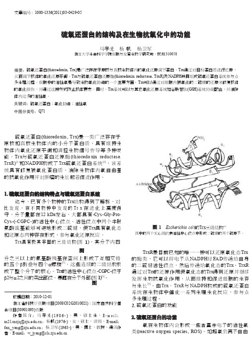
1. 硫氧还蛋白的结构特点与硫氧还蛋白系统迄今,已有多个物种的Trx 结构得到了解析,对 比发现,在不同物种中发现的T r x 在进化上高度保 守,分子量都在12 kDa 左右,大都具有-Cys-Gly-Pro- Cys-(-CGPC-)的活性中心位点。
活性位点中两个半胱 氨酸巯基能够可逆地形成二硫键,使Trx 具有氧化态 和还原态两种存在形式,参与氧化还原反应。
Trx 具有极其牢固的三级结构(图 1),其分子内四图 1 Escherichia coli 的T rx 三级结构[4]图中的两个实心圆代表活性中心位点中形成二硫键的两个硫原子。
反相微乳液法制备CdTe@SiO2荧光微球及其荧光性能研究

反相微乳液法制备CdTe@SiO2荧光微球及其荧光性能研究宁振动;张纪梅;魏君;成耀宇【摘要】利用巯基丙酸(MPA)为稳定剂,水相合成高质量的CdTe纳米晶,然后通过反相微乳液方法制备得到了具有明显核壳结构并单分散的CdTe@SiO2荧光复合纳米粒子;利用透射电子显微镜(TEM)、荧光分光光度计以及紫外可见光分光光度计对制备的纳米粒子进行表征;研究了未包裹的CdTe量子点与核壳型CdTe@SiO2纳米复合粒子分别对pH及离子强度的耐受性.研究发现:由于SiO2壳层的存在使得CdTe@SiO2能够在很高的离子强度及很广泛的pH范围下仍具有较强的荧光.由于这些优点,使其在生物标记、细胞成像等生物领域具有广泛应用.%High-quality CdTe nanocrystals capped by mercaptopropionic acid (MPA) were prepared in aqueous solution, then fluorescent, monodispersed, well-separated, core/shell CdTe@SiO2 particles were prepared via reverse microemulsion method. Obtained samples were characterized by means of transmission electron microscope (TEM), UV-vis and PL emission. The influences of ionic strength and pH on the PL emission of uncoated CdTe and CdTe/SiO2 composite nanoparticles were investigated thoroughly. The results show that due to the presence of SiO2 shell, CdTe@ SiO2 still remain a strong fluorescence at high ionic strength and a very broad pH range. Because of these advantages, it has broad applications in biological fields, such as biomarkers, cell imaging.【期刊名称】《天津工业大学学报》【年(卷),期】2012(031)001【总页数】4页(P49-52)【关键词】反相微乳液;CdTeSiO2荧光微球;荧光性能;核壳型;离子强度;pH【作者】宁振动;张纪梅;魏君;成耀宇【作者单位】天津工业大学环境与化学工程学院,天津300160;天津工业大学环境与化学工程学院,天津300160;天津工业大学环境与化学工程学院,天津300160;天津工业大学理学院,天津300160【正文语种】中文【中图分类】O482.3由于量子点(quantum dots,QDs)具有发光性质的尺寸依赖性,近来引起了人们对其在生物标记、细胞成像、传感材料方面的广泛兴趣[1-3].特别是与传统的有机荧光染料分子相比,量子点具有许多特殊的光学性能.比如,激发光可选范围宽,可以用同一波长的光激发不同尺寸量子点,荧光发射波长可单纯地通过改变粒子尺寸进行调节,具有狭窄对称的荧光发射峰,光稳定性强不易发生荧光漂白等[4].水相合成方法制备的量子点存在着稳定性差,光学性质强烈依赖于其表面状态以及受应用环境的影响较大的缺点[5].如果将其用于生物领域还需解决重金属元素的毒性问题.目前的解决办法主要有利用无毒或者危害小的元素代替重金属元素[6],或者采用惰性物质,比如二氧化硅(SiO2)对量子点表面进行包覆[7-11].Liz-Marzán 及其合作者利用传统的Stöber法成功制备得到了具有核壳结构的CdS@SiO2纳米复合粒子,并且发现SiO2壳层能够有效的阻止CdS的光氧化[7].但是,利用Stöber法包裹量子点存在诸多缺点,例如包裹前需要对量子点进行复杂的预处理,导致量子点的发光效率急剧降低,另外还有发光蓝移现象的发生 [8].除了传统的Stöber法,反相微乳液法(油包水)也被用来制备SiO2包裹量子点纳米复合粒子[9-11].利用反相微乳液方法可以制备得到尺寸均一的、大小可调的球形纳米复合粒子.量子点表面包覆SiO2的目的主要有:①阻止量子点的光氧化;②解决量子点的生物毒性问题;③使得量子点能够更容易功能化.本文采用反相微乳液法合成核/壳型CdTe@SiO2荧光纳米复合粒子,并且对比研究CdTe量子点与CdTe@SiO2荧光纳米复合粒子在不同pH及不同离子强度下的性质.1 实验部分1.1 试剂和仪器氯化镉(CdCl2·2.5H2O)、碲粉(Te)、硼氢化钠(NaBH4)等,购于国药集团化学试剂有限公司;氨水(w=25%),环己烷,正己醇等购于天津科密欧化学试剂公司;正硅酸乙酯(TEOS),购于 TCI公司;TritonX-100、巯基丙酸(MPA),购于 Sigma-Aldrich公司,均为分析纯(AR级);实验中用水均为超纯水,Aquapro超纯水设备生产.制备得到的CdTe@SiO2的纳米复合粒子的尺寸和形态通过Hitachi H-7650型透射电镜(TEM)进行表征,操作电压80 kV;荧光发射光谱和紫外可见光吸收光谱分别利用天津港东F-380荧光分光光度计、美国热电Helios γ紫外可见光分光光度计进行表征.1.2 水溶性CdTe量子点的制备MPA稳定的CdTe量子点的制备采用常见的水相合成法[12].具体步骤为:首先制备NaHTe溶液,将80 mg硼氢化钠溶解在2 mL去离子水中,加入127.5 mg碲粉,低温反应8 h后,黑色的碲粉消失,并产生白色晶体;澄清的NaHTe溶液用来制备CdTe量子点.另外需要注意反应体系要留一小孔与大气相通以便反应产生的氢气可以排走;然后按照Cd2+、HTe-、MPA的摩尔比为1∶0.5∶2.4,分别将0.002 mol的CdCl2·2.5H2O、0.0048 mol的MPA加入到125 mL超纯水中,磁力搅拌下利用1 mol/L的NaOH调节溶液pH至9.1,然后通氮气30 min后,加入新制备的NaHTe溶液.最后,在没有氮气保护下,将前驱体溶液回流一段时间,即得到具有高量子效率的CdTe水溶胶.在不同的回流时间下取一定量样品利用荧光分光光度计和紫外可见光分光光度计进行表征.1.3 核壳型荧光纳米粒子的制备CdTe@SiO2的制备采用反相微乳液方法[9].详细步骤如下:将7.5 mL环己烷、1.77 mL TritonX-100以及1.8 mL正己醇混合,磁力搅拌至光学透明.然后加入250 μL 25%的氨水和500 μL上述回流24 h的CdTe QDs水溶液,搅拌30 min 形成油包水的微乳液.随后在剧烈搅拌下加入150 μL TEOS,密封避光反应24 h.反应结束后,加入20 mL丙酮破乳,在10000 r/min下离心分离,弃去上清液,将得到的沉淀分别用异丙醇、乙醇和水清洗离心,最后将得到的样品分散在超纯水中进行性能表征.2 结果与讨论2.1 光学性质新制备的CdTe前驱体溶液是没有荧光的,但是回流几分钟后,便出现了较强的绿色荧光.随着回流时间的增加,微粒的紫外吸收光谱和荧光发射光谱发生红移,如图1所示.由于纳米晶存在量子尺寸效应,通过改变纳米晶的粒径,可以调节其荧光的颜色.通过延长回流时间,可以制备得到不同粒径的纳米晶.随着粒径的改变,可以分别得到绿、黄、橙及红色荧光.吸收光谱与荧光光谱的红移说明粒径在随着回流时间的增加而增加[13].图2为CdTe@SiO2荧光复合纳米粒子的荧光发射光谱.从图2中可以看到,所制的纳米复合物仍具有较强的荧光.但是与包覆前相比却发生了蓝移(641 nm蓝移至632 nm).这一现象已有相关报道[8],分析认为发射峰蓝移的原因在于:随着硅层的包被,可能导致量子点表面结构的改变从而导致其发光行为的改变;另外,CdTe量子点在包被硅层的过程中,由于巯基丙酸大量地从量子点表面离去,使得CdTe量子点进一步发生光氧化,从而导致发射峰的蓝移.如图3所示为CdTe@SiO2复合纳米粒子的TEM照片,用反相微乳液法制备的荧光纳米复合粒子具有明显的核壳结构,粒子呈球形,平均粒径为(68±4.4)nm,尺寸分布均一,并且每个SiO2壳基本上只包覆1个CdTe量子点.另外还可以通过改变反应体系中TEOS、水及Triton X-100等的量来调节SiO2壳的厚度[10].2.2 pH及离子强度的影响在生物应用中,缓冲溶液的离子强度和pH值都是非常重要的参数,但是高离子强度及强酸性都会对CdTe量子点的发光行为造成影响.所以有必要研究离子强度和pH对CdTe量子点及CdTe@SiO2复合粒子的影响,结果如图4、图5所示.由图4可知,在不同的离子强度的溶液中,CdTe@SiO2比单独的CdTe要稳定,当NaCl浓度达到200 mmol/L时,CdTe@SiO2复合纳米粒子的荧光几乎没有变化;而对于CdTe量子点其荧光则下降的很明显,这是由于加入电解质后会使CdTe量子点聚集,粒子间发生荧光共振能量转移,使得发光变弱 [14].对于CdTe@SiO2由于SiO2壳的保护作用,使其能够在很大的离子强度范围内荧光基本上不受影响.又由于荧光共振能量转移发生的作用距离一般是小于10 nm,而SiO2壳层的厚度大于10 nm,所以一般情况下即便CdTe@SiO2复合纳米粒子产生聚集,荧光共振能量转移也不会对其发光性质造成较大影响[15].对于pH的影响,CdTe量子点在pH<5时便产生沉淀,pH<2时开始分解完全猝灭.这是由于在H+的作用下,表面配体MPA与Cd的相互作用逐渐减弱,使得量子点表面的缺陷增多导致发光减弱,当pH<2时,量子点开始分解最终导致其基本上完全猝灭.而对于CdTe@SiO2纳米复合物,由于SiO2壳层的存在,使其发光在pH=2.0左右时仍能保持原来的27%,而此时未包覆的量子点的发光基本上已完全猝灭.这说明SiO2壳可以很好的保护CdTe量子点的荧光发射,提高其稳定性.对于CdTe 量子点pH值刚开始下降时发光性质有所加强,这是因为不但巯基可以和Cd2+配位,羰基氧也存在和Cd2+的次级配位,pH值降低使CdTe表面MPA中羧基质子化使得羰基氧和Cd2+的次级配位作用加强,更好地钝化量子点的表面,提高发光效率[12].另外,CdTe量子点的发光不单纯由纳米晶表面的MPA的羧基的质子化决定,降低pH值也会使溶液中多余的Cd2+与MPA形成的复合物与纳米晶表面作用提高其发光效率和稳定性[5].但是如果pH太低,则会破坏形成的可以钝化量子点表面的物质及量子点本身的结构,从而使其发光猝灭.同样,对于CdTe@SiO2纳米复合物,由于SiO2壳层并非致密结构[16],故H+及其他小分子可以扩散到壳层里面与CdTe作用,也会在pH刚下降的时候使其发光增强.由于壳层的存在,使得其可以耐受比较低的pH.3 结论(1)通过反相微乳液方法成功制备得到核壳型、尺寸均一、分散性好的CdTe@SiO2荧光复合纳米粒子.(2)与未包裹SiO2壳的CdTe量子点相比,该复合纳米粒子具有很好的光学稳定性,如耐高离子强度以及对pH稳定的范围比较广.参考文献:【相关文献】[1]BRUCHEZ Jr M,MORONNE M,GIN P,et al.Semiconductor nanocrystals as fluorescent biological labels[J].Science,1998,281:2013-2016.[2]CHAN W C W,NIE SHUMING.Quantum dot bioconjugates for ultrasensitive nonisotopic detection[J].Science,1998,281:2016-2018.[3]MEDINTZ I L,UYEDA H T,GOLDMAN E R,et al.Quantum dot bioconjugates for imaging,labelling and sensing[J].Nat Mater,2005,4:435-446.[4]RESCH-GENGER U,GRABOLLE M,CAVALIERE-JARICOT S,et al.Quantum dots versus organic dyes as fluorescent labels[J].Nat Methods,2008,5(9):763-775.[5]GAO M,KIRSTEIN S,MOHWALD H,et al.Strongly photoluminescent CdTe nanocrystals by proper surface modification[J].J Phys Chem B,1998,102:8360-8363. [6]ERWIN S C,ZU L,HAFTEL M I,et al.Doping semiconductor nanocrystals[J].Nature,2005,436:91-94.[7]CORREA-DUARTE M A,GIERSIG M,LIZ-MARZAN.Stabilization of CdS semiconductor nanoparticles against pho todegradation by a silica coating procedure[J].Chem Phys Lett,1998,286:497-501.[8]ROGACH A L,NAGESHA D,OSTRANDER J W,et al."Raisin Bun"-type composite spheres of silica and semiconductor nanocrystals[J].Chem Mater,2000,12:2676-2685. [9]YANG Y,GAO M.Preparation of fluorescent SiO2particles with single CdTe nanocrystal cores by the reverse microemulsion method[J].Adv Mater,2005,17:2354-2357.[10]YANG Y,JING L,YU X,et al.Coating aqueous quantum dots with silica via reverse microemulsion method:Toward size-controllable and robust fluorescentnanoparticles[J].Chem Mater,2007,19:4123-4128.[11]JING L,YAN C,QIAO R,et al.Highly fluorescent CdTe@SiO2 particles prepared via reverse microemulsion method[J].Chem Mater,2010,22:420-427.[12]ZHANG H,ZHOU Z,YANG B,et al.The influence of carboxyl groups on the photoluminescence of mercaptocarboxylic acid-stabilized CdTe nanoparticles[J].J Phys Chem B,2003,107:8-13.[13]ROGACH A L,FRANZL T,KLAR T A,et al.Aqueous synthesis of thiol-capped CdTe nanocrystals:State-of-the-art[J].J Phys Chem C,2007,111:14628-14637.[14]MAYILO S,HILHORST J,SUSHA A S,et al.Energy transfer in solution-based clusters of CdTe nanocrystals electrostatically bound by calcium ions[J].J Phys Chem C,2008,112:14589-14594.[15]CLAPP A R,MEDINTZ I L,MATTOUSSI H.Forster resonance energy transfer investigations using quantum-dot fluorophores[J].Chem Phys Chem,2006,7:47-57. [16]FINNIE K S,BARTLETT J R,BARBE C J,et al.Formation of silica nanoparticles in microemulsions[J].Langmuir,2007,23:3017-3024.。
不同酶消化法提取猪原代肝细胞的效果比较
532024.4·试验研究0 引言猪圆环病毒(PCV )是Circoviridae 科Circovirus 属的一种无囊膜的单链环状DNA 病毒。
在已知的4个血清型中,PCV2为猪易感的致病性病毒[1]。
PCV2感染会诱导宿主免疫抑制引起猪圆环病毒病(PCVD ),包括断奶仔猪多系统衰竭综合征、新生仔猪先天性脑震颤、皮炎与肾病综合征、猪呼吸道病综合征、母猪繁殖障碍等,给全世界养猪业带来较大的经济损失,是世界各国的兽医与养猪业者公认的造成重大影响的猪传染病[2]。
PCV2的感染在猪生长发育的不同阶段有不同的组织嗜性。
但无论是胎儿阶段还是出生后,肝细胞都是PCV2感染和复制的靶细胞。
因此,PCV2也被视为一种能够诱导猪肝炎的病毒[3]。
且PCV2诱导的肝细胞凋亡在PCV2引发的相关病变和疾病的发病机制中具有关键性作用[4]。
因此,方便、快捷地获取大量有活性的猪肝细胞对于研究PCVD 的致病机制具有重大意义。
目前获取肝细胞常用的方法主要包括机械分离细胞法、非酶分离细胞法、离体酶消化法和酶灌流法等[5]。
因此,本试验采用简便、经济、无需特殊设备、仅需部分肝组织的离体酶消化法,比较不同酶消化分离猪原代肝细胞的效果,为一般实验室提取分离大量有活性的猪肝细胞提供参考。
1 材料与方法1.1 材料1.1.1 主要试剂新鲜猪肝组织,Hank's 平衡盐溶液(HBSS ),磷酸盐缓冲液(无菌PBS ),4%多聚甲醛(PFA ),收稿日期:2024-01-27基金项目:国家自然科学基金项目:复杂器官与组织在脾脏内的功能性再生(32230056)作者简介:周徐倩(1999-),女,汉族,浙江温州人,硕士在读,研究方向:组织工程与再生医学。
*通信作者简介:董磊(1978-),男,汉族,安徽阜阳人,博士,教授,研究方向:组织工程与再生医学、生物材料。
周徐倩,董磊.不同酶消化法提取猪原代肝细胞的效果比较[J].现代畜牧科技,2024,107(4):53-55. doi :10.19369/ki.2095-9737.2024.04.014. ZHOU Xuqian ,DONG Lei .Comparison of the Effect of Different Enzyme Digestion Methods on Extraction of Porcine Primary Hepatocytes[J].Modern Animal Husbandry Science & Technology ,2024,107(4):53-55.不同酶消化法提取猪原代肝细胞的效果比较周徐倩,董磊*(南京大学,江苏 南京 210023)摘要:猪肝细胞是猪圆环病毒的靶细胞,简单快速地提取猪原代肝细胞对于研究猪圆环病毒病的致病机制具有重要意义。
天冬酰胺合成酶通过促进β-catenin核转位驱动胆管癌转移
天冬酰胺合成酶通过促进β-catenin 核转位驱动胆管癌转移*褚珍珍1,2, 周栩萱1,2, 刘力豪1, 张鲍欢3△, 姚楠1,2△(1暨南大学基础医学院病理生理学系,广东 广州 510632;2国家中医药管理局病理生理科研实验室,广东 广州510632;3暨南大学基础医学院形态学实验教学中心,广东 广州 510632)[摘要] 目的:检测天冬酰胺合成酶(ASNS )在胆管癌(CCA )中的表达情况,探讨ASNS 在CCA 转移中的作用及其机制。
方法:通过公共数据库分析各肿瘤组织中ASNS 的mRNA 表达;收集CCA 患者病理组织(n =27),构建硫代乙酰胺诱导的大鼠自发CCA 模型和左中位胆管结扎联合二乙基亚硝胺诱导的小鼠自发CCA 模型,通过免疫组化、Western blot 和免疫荧光法检测ASNS 蛋白表达。
采用CCK8、划痕和Transwell 实验检测ASNS 对人CCA 细胞HuCCT1和HCCC -9810增殖、迁移和侵袭的影响。
构建ASNS 稳定敲减的CCA 细胞株HuCCT1shNC 、HuCCT1shASNS 、HCCC -9810shNC 和HCCC -9810shASNS ,通过肝原位种植和尾静脉注射研究ASNS 对CCA 细胞肝内生长和肺转移的影响。
利用公共数据库富集与ASNS 相关的信号通路,并用免疫荧光和Western blot 验证相关分子机制。
结果:无论在人或动物CCA 组织中,ASNS 表达水平均高于癌旁组织(P <0.01)。
ASNS 以酶活性非依赖性方式促进CCA 细胞HuCCT1和HCCC -9810的增殖、迁移与侵袭。
生物信息学分析显示,β-catenin 在ASNS 高表达的CCA 组织中富集,ASNS 通过促进β-catenin 核转位,启动CCA 细胞上皮-间充质转化(EMT )。
β-catenin 抑制剂XAV -939可显著抑制CCA 细胞的侵袭与迁移。
铁死亡诱导剂在结直肠癌中的研究进展

- 180 -end-expiratory pressure alone minimizes atelectasis formation in nonabdominal surgery:a randomized controlled trial[J].Anesthesiology,2018,128(6):1117-1124.[39] KIM N,LEE S H,CHOI K W,et al.Effects of positive end-expiratory pressure on pulmonary oxygenation and biventricular function during one-lung ventilation:a randomized crossover study[J].J Clin Med,2019,8(5):740.[40] KATZ J A,LAVERNE R G,FAIRLEY H B,et al.Pulmonaryoxygen exchange during endobronchial anesthesia:effect of tidal volume and PEEP[J].Anesthesiology,1982,56(3):164-171.[41] SENT ÜRK N M,DILEK A,CAMCI E,et al.Effects ofpositive end-expiratory pressure on ventilatory and oxygenation parameters during pressure-controlled one-lung ventilation[J]. J Cardiothorac Vasc Anesth,2005,19(1):71-75.[42] KANG W S,KIM S H,CHUNG J parison of pulmonarygas exchange according to intraoperative ventilation modes for mitral valve repair surgery via thoracotomy with one-lung ventilation:a randomized controlled trial[J].J Cardiothorac Vasc Anesth,2014,28(4):908-913.(收稿日期:2023-03-03) (本文编辑:田婧)*基金项目:安溪县科技计划项目(2022S002)①福建省安溪县医院 福建 安溪 362400通信作者:许永鹏铁死亡诱导剂在结直肠癌中的研究进展*陈伟鸿① 苏小苹① 苏宇超① 黄栋钦① 许永鹏① 【摘要】 结直肠癌(colorectal cancer,CRC)是全球第三大常见癌症,传统治疗方案对CRC 晚期患者的疗效不佳,因此,发现新的治疗策略可能有助于改善CRC 患者的治疗和预后。
肝靶向甘草次酸修饰的壳聚糖纳米粒子的合成和表征
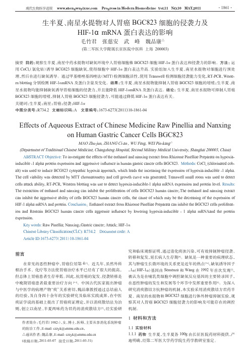
Vol .28高等学校化学学报No .62007年6月 CHE M I CAL JOURNAL OF CH I N ESE UN I V ERSI TI ES 1098~1100[研究快报]肝靶向甘草次酸修饰的壳聚糖纳米粒子的合成和表征查瑞涛1,2,贺晓婷1,杜 田1,袁 直1(1.南开大学高分子化学研究所,功能高分子材料教育部重点实验室,天津300071;2.天津科技大学材料科学与化学工程学院,天津300457)关键词 甘草次酸;壳聚糖;肝靶向中图分类号 O625 文献标识码 A 文章编号 025120790(2007)0621098203收稿日期:2006212204.基金项目:教育部天津大学2南开大学科技合作基金和天津市自然科学基金(批准号:07JCZ DJC00700)资助.联系人简介:袁 直(1961年出生),女,博士,教授,博士生导师,从事生物医用材料研究.E 2mail:zhiy@nankai .edu .cn肝靶向给药系统[1](Hepatic targeted drug delivery syste m ,HT DDS )是药剂学研究领域中的热点之一,HT DDS 可将药物有效地输送至肝脏的病变部位,减少用药剂量和给药次数,并减少对其它脏器的伤害,提高治疗效果,因此,HT DDS 对肝脏疾病的治疗具有积极的推动作用.甘草为豆科植物,它的药理作用的有效成分主要是甘草酸(Glycyrrhizic acid,G L )和甘草次酸(Gly 2cyrrhetinic acid,G A ).甘草次酸在肝脏中高度蓄积[2~4],甘草酸在体内水解为甘草次酸,脂质体经甘草酸表面修饰后具有良好的趋肝性和肝细胞靶向性[5].壳聚糖(CTS )来源广泛,廉价易得且具有很好的生物相容性和可降解性,作为一种新型药用辅料在缓控释给药系统中,特别是在微球中的应用已引起了人们浓厚的兴趣[6].本文合成了修饰甘草次酸的壳聚糖(G A 2CTS ),采用离子交联法制备了G A 2CTS 纳米粒子.该材料可能具有肝细胞主动靶向作用,为进一步的肝靶向药物控释的研究奠定了基础.1 实验部分1.1 试剂与仪器 甘草次酸(HP LC 纯度≥98%),西安富捷公司;水溶性壳聚糖(分子量10000,脱乙酰度>95%),浙江玉环海洋生物公司;其它化学试剂均为分析纯.M illi pore 微孔滤膜(450nm ,低蛋白吸附).Varian UN I TY Plus 2300MHz 核磁共振谱仪(室温,015mol/L DCl/D 2O,T MS 内标);N icolet5DX FTI R 光谱仪(漫反射法,在397~4000c m -1范围内扫描);E M 400ST 透射电镜仪(TE M )(Phili p s公司);光子相关谱仪(PCS 美国B r ookhaven ).1.2 甘草次酸修饰壳聚糖的合成 将1g 甘草次酸(G A )的50mL DMF (N ,N 2二甲基甲酰胺)溶液和质量分数为1%的壳聚糖(CTS )水溶液混合搅拌均匀,向混合液中缓慢滴加EDC ・HCl [12(32二甲氨基丙基)232乙基碳二亚胺盐酸盐]的水溶液,反应24h 后,倾入到800mL 丙酮中,静置24h .将沉淀过滤,依次用丙酮、乙醇、乙醚洗涤,室温真空干燥.合成路线如Sche me 1所示.取代度(每100个壳聚Sche m e 1 Syn thesis route of the GA 2CTS糖糖环单元修饰甘草次酸的个数)通过紫外光谱确定.1.3 G A 2CTS 纳米粒子的制备 分别配制修饰甘草次酸的壳聚糖(015mg/mL )和离子交联剂TPP (三聚磷酸钠,012mg/mL )的水溶液.取2mL TPP 溶液,在超声条件下滴加到2mL G A 2CTS 溶液中.1.4 G A 2CTS 纳米粒子对牛血清白蛋白(BS A )的包封 将不同浓度的BS A 加入到5mL 的G A 2CTS 溶液中,使BS A 的最后质量浓度分别为012,015,110,210mg /mL,并使G A 2CTS 的最后质量浓度为210mg/mL,再加入2mL 016mg/mL TPP 溶液,在室温磁力搅拌条件下,自发生成包封有BS A 的纳米粒子.将样品在15℃下以17000r/m in 转速离心30m in,从水溶液中分离出纳米粒子,冷冻干燥.上层清液用紫外分光光度仪测定λ=278n m 处的吸收,计算游离的BS A 质量,平行测定4次.按下式计算样品的包封率(EE )和载BS A 率(LC ):EE =[(W 总-W 游)/W 总]×100%,LC =[(W 总-W 游)/W 纳米粒]×100%,式中,W 总表示(BS A )药物总量,W 游表示游离(BS A )药物质量;W 纳米粒表示纳米粒质量.1.5 包封牛血清白蛋白(BS A )的G A 2CTS 纳米粒子的体外释放 将包封有BS A 的G A 2CTS 纳米粒子置于含有10mL 缓冲液的试管中,在转速80r/m in 的摇床中于37℃恒温处理,适当间隙.样品在4℃下以17000r/m in 的速度离心30m in,取出2mL 上层清液,用新鲜生理盐水补充.用考马斯亮蓝法测量释放出的BS A 的质量,绘制纳米粒子的释放曲线,取4次测定的平均值.用未包封BS A 的纳米粒子作校正曲线.1.6 G A 2CTS 纳米粒子的稳定性 将G A 2CTS 纳米粒子溶液过M illi pore 450n m 微孔滤膜;取2mL 滤液加入到光学试剂瓶中,在室温下保存,每间隔一段时间观察.2 结果与讨论2.1 G A 2CTS 的合成与表征 壳聚糖在3363c m -1处出现ν(O —H )和ν(N —H )伸缩振动吸收峰,在2877c m -1处出现ν(CH 2)伸缩振动吸收峰.在1590和1310c m -1处分别出现δ(N —H )弯曲振动和ν(C —N )伸缩振动吸收峰.在1400c m -1附近分别出现δ(CH 2)弯曲振动吸收峰.在1155c m -1处出现糖苷键ν(C —O —C )伸缩振动吸收峰,在1095和1050c m -1附近出现多糖ν(C —OH )伸缩振动吸收峰.G A 2CTS 的红外光图谱与壳聚糖相比,在1657和1540c m -1处出现了较明显的酰胺峰,1590c m -1处的F i g .1 1H N M R spectra of CTS(a )and GA 2CTS(b )NH 2峰减弱,说明在氨基上进行了酰化反应.壳聚糖的1H NMR 谱(图1谱线a )(400MHz,DCl/D 2O ),δ:417(H1),219(H2),314~319(H3,H4,H5,H6),119(NHCOCH 3).在G A 2CTS 的1H NMR 谱(图1谱线b )中,δ215和217处出现了新的吸收峰,分别是G A 基团的CH 2和与G A 基团相联的H2.其它的基团归属,δ:417(H1),219(H2),316~411(H3,H4,H5,H6),119(NHCOCH 3).核磁共振谱说明壳聚糖NH 2上已经修饰了G A 基团.2.2 纳米粒子的表征 用动态光散射研究了G A 2CTS 2TPP 纳米粒子的粒径分布,结果表明,过膜的纳米粒子平均直径为(12012±214)n m ,分布较窄.由透射电镜照片(略)可见,G A 2CTS 2TPP 和BS A 2G A 2CTS 2TPP 纳米粒子都具有较好的球形外观.2.3 G A 2CTS 纳米粒子BS A 的包封 由图2可见,BS A 的初始浓度越高,制备的纳米粒子对BS A 的包封率越低.当BS A 的质量浓度从012mg/mL 增加到215mg/mL 时,G A 2CTS 2TPP 纳米粒子对BS A 的包封率从81%下降到52%,但BS A 的负载量从26%增加到47%.随着BS A 浓度的增大,单个BS A 分子与TPP 发生离子相互作用的几率降低,同时与壳聚糖的吸附作用的相对几率也降低,所以BS A 包封率降低.但随着BS A 浓度的增大,与TPP 和G A 2CTS 发生离子作用与吸附作用的BS A 总量在增多,所以BS A 的负载量增大.BS A 的包封也受到G A 基团取代度的影响(图3).当G A 基团取代度从1%增加到5%时,G A 2CTS 2TPP 纳米粒子对BS A 的包封率从45%增加到75%.这主要是由于随着G A 基团的疏水作用和空间位阻作用的加大,G A 2CTS 主链上氨基与TPP 的相互作用减弱,BS A 的氨基可以和TPP 发生更多的相互作用,BS A 的包封率升高.9901 No .6 查瑞涛等:肝靶向甘草次酸修饰的壳聚糖纳米粒子的合成和表征F i g .2 I nfluence of BSA i n iti a l concen tra ti on onencapsul a ti on eff i c iency F i g .3 I nfluence of GA substituen t on encapsul a ti on eff i c i ency2.4 G A 2CTS 纳米粒子的稳定性 将过膜前后的G A 2CTS 纳米水溶液于室温下放置,观测其稳定性 F i g .4 S i ze d istr i buti on s of GA 2CTS 2TPP nanoparti 2cles before(A)and after f iltra ti on(B)(见图4).发现过膜后的纳米粒子可以稳定放置14个月而未发生明显变化,光散射研究结果表明,纳米粒子平均粒径仅增大约510n m.没有过膜的纳米粒子水溶液在3d 后有轻度混浊,光散射研究结果表明,此时体系内有约5μm 的较大粒径粒子存在.没有过膜的纳米水溶液在14d 后有沉淀生成.由于没有过膜的G A 2CTS 纳米水溶液存在较大颗粒的灰尘或比较大的交联粒子,溶液在放置后,其它纳米粒子以它们为核团聚而形成沉淀.参 考 文 献[1] Kumar V.,Banker G .S ..Targeted O riented D rug Delivery System s[A ].Banker G .S .,Rhodes C .T .Eds .;Modern Phar maceutics,3ed .,Vol .72[M ],Ne w York:Marcel Dekker,1996:611—680[2] W ang Z .,N ishi oka M.,Kur osaki Y .,et al ..B i ol .Phar m.Bull .[J ],1995,18(9):1238—1241[3] Y ANG Shan 2Mai (杨山麦),ZHOU Fang 2Cheng (周方成),G U Yun 2Ti (顾云娣).Chinese Journal of Hepat ol ogy (中华肝脏病杂志)[J ],1999,7(S1):27—29[4] NegishiM.,Irie A.,Nagataa N.,et al ..B i ochi m ica et B i ophysica Acta (BBA )B i ome mbranes[J ],1991,1066(1):77—82[5] Sayoko O.,H ideki T .,H ir oshi K ..B i ol .Phar m.Bull .[J ],1994,17(7):940—943[6] ZHENG J ian 2Hua (郑建华),L I U Chao 2W u (刘朝武),BAO De 2Cai (包德才).Chem.J.Chinese Universities (高等学校化学学报)[J ],2006,27(6):1182—1185Syn thesis and Character i za ti on of Ch itos an Nanoparti cles M od i f i edby Glycyrrheti n i c Ac i d a s a L i ver Targeti n g D rug Carr i erZ HA Rui 2Tao 1,2,HE Xiao 2Ting 1,DU Tian 1,Y UAN Zhi 13(1.Key L aboratory of Functional Polym erM aterials,M inistry of Education,Institute of Polym er Che m istry,N ankai U niversity,T ianjin 300071,China;2.College of M aterial Science and Che m ical Engineering,T ianjin U niversity of Science &Technology,Tianjin 300457,China )Abstract Chit osan derivative with glycyrrhetinic acid (G A ),which was accu mulated s pecifically in liver,was synthesized .By i on 2cr osslinking of TPP in G A 2CTS s oluti on,G A 2CTS 2TPP comp lex nanoparticles were obtained .The physicochem ical p r operties of these nanoparticles were investigated by using TE M and DLS .The experi m ent in vitro BS A entrapped was studied .G A 2CTS 2TPP nanoparticles are well dis persed and stable in aqueous s oluti on in 14months .Keywords Glycyrrhetinic acid;Chit osan;L iver targeting (Ed .:H,J,Z )0011高等学校化学学报 Vol .28 。
细胞蛇的研究进展
2007年,英国牛津大学的刘骥陇等在研究果蝇U 小体和P 小体(U 小体和P 小体是真核生物细胞质中的无膜细胞器)的功能关系时,用4种针对Cup (P 小体中的一种蛋白质)的抗体,对雌性果蝇的卵巢组织进行免疫组织化学染色,染色结果除了预期标记上的P 小体外,还标记出了长条形的丝状结构[1]。
这种结构的形状和数量与纤毛很相似,导致当时以为在果蝇中找到了有纤毛的新细胞类型。
但后来的一系列实验表明,该结构与纤毛没有关系,于是将其命名为“细胞蛇”。
最初是抗Cup 抗体不纯产生假象,意外发现的细胞蛇,而采用亲和层析纯化后的抗Cup 抗体无法再DOI:10.16605/ki.1007-7847.2020.10.0258细胞蛇的研究进展收稿日期:2020-10-22;修回日期:2020-11-19;网络首发日期:2021-07-27基金项目:宁夏自然科学基金项目(2020AAC03179);国家自然科学基金资助项目(31560329)作者简介:李欣玲(1999—),女,广西贵港人,学生;*通信作者:俞晓丽(1984—),女,宁夏银川人,博士,副教授,主要从事干细胞与生殖生物学研究,E-mail:********************。
李欣玲,张樱馨,李进兰,潘文鑫,王彦凤,杨丽蓉,王通,俞晓丽*(宁夏医科大学生育力保持教育部重点实验室临床医学院基础医学院,中国宁夏银川750000)摘要:细胞蛇是近年来细胞生物学研究的热门方向之一,由于其在细胞的增殖、代谢和发育上具有一定的生物学功能,因此,对一些疾病如癌症等的临床诊断或治疗具有一定的指导意义。
细胞蛇是由三磷酸胞苷合成酶(cytidine triphosphate synthetase,CTPS)聚合而成的无膜细胞器,其形成过程及功能在不同类型的细胞中不尽相同。
例如:细胞蛇能促进癌细胞增殖,并使患者病情恶化;过表达的细胞蛇可抑制神经干细胞增殖,影响大脑皮层发育;在卵泡细胞中,细胞蛇相当于CTPS 的存储库,在卵子发生过程起到促进细胞增殖和代谢的作用。
生半夏、南星水提物对人胃癌BGC823细胞的侵袭力及HIF-1αmRNA蛋白表..
现代生物医学进展 Progress in Modern Biomedicine Vol.11NO.10MAY.2011生半夏、南星水提物对人胃癌BGC823细胞的侵袭力及HIF-1αmRNA 蛋白表达的影响毛竹君张慈安武峰魏品康△(第二军医大学附属长征医院中医科上海200003)摘要目的:观察生半夏、南星中药水提物对缺氧环境中人胃癌细胞株BGC823细胞HIF-1α蛋白表达和侵袭力的影响。
方法:运用CoCl 2(氯化钴)诱导BCG823细胞缺氧,使得细胞中HIF-1α蛋白表达升高.实验组加入生半夏、南星水提物对细胞进行预处理,然后在进行缺氧诱导。
通过甲基噻唑基四唑法(MTT )检测细胞活性,使用Transwell 检测细胞侵袭能力变化,RT-PCR 、Weste-rn blotting 分别检测HIF-1αmRNA 及蛋白含量及变化。
结果:生半夏、南星水提物能抑制人胃癌BGC823细胞的增殖;生半夏、南星水提物均能抑制缺氧诱导胃癌细胞的侵袭力,并且能降低HIF-1αmRNA 及蛋白表达。
结论:生半夏、南星水提物可抑制人胃癌BGC823细胞的增殖,抑制人胃癌BGC823细胞侵袭力,可能通过降低HIF-1α蛋白表达有关。
关键词:生半夏;南星;胃癌;侵袭;HIF-1α中图分类号:R734.2文献标识码:A 文章编号:1673-6273(2011)10-1861-04Effects of Aqueous Extract of Chinese Medicine Raw Pinellia and Nanxingon Human Gastric Cancer Cells BGC823MAO Zhu-jun,ZHANG Ci-an ,WU Feng,WEI Pin-kang △(Department of Traditional Chinese Medicine,Changzheng Hospital,Second Military Medical University,Shanghai 200003,China)ABSTRACT Objective:To investigate the effects of the enthanol and nanxing extract from Rhizome Pinelliae Preparata on hypoxia-inducible -1alpha protein expression and aggressive influence in human gastric cancer cells BGC823.Methods:CoCl 2(chlorinated cob-alt)was used to induce BCG823cytopathic hypoxia approach,which leads the increasing the expression of hypoxia-inducible -1alpha.The cell viability was detected by MTT chromatometry and cell growth curve was generated;Transwell small room was used to detect cells attack ability,RT-PCR,Western blotting was use to detect hypoxia-inducible-1alpha mRNA expression and protein level.Results:The extraction of enthanol and nanxing can inhibit the proliferation of cells BGC823human cancer;.The enthanol and nanxing extract can inhibit the aggressive ability of cells BGC823human cancer cells,the cause of which may be the decreasing of the expression of HIF-1alpha mRNA and protein.Conclusion :Enthanol extract from Rhizome Pinelliae Preparata can inhibit the BGC823cells proliferat-ion and Restrain BGC823human cancer cells aggressie influence by lowering hypoxia-inducible -1alpha mRNAand the protein expression.Key words:Raw Pinellia;Nanxing ;Gastric cancer;Attack;HIF-1αChinese Library Classification(CLC):R734.2Document code:A Article ID:1673-6273(2011)10-1861-04作者简介:毛竹君(1982-),女,博士,医师,主要从事消化系统肿瘤的防治工作,E-mail:**************.cn ,△通讯作者:魏品康,E-mail:**************.cn (收稿日期:2011-03-07接受日期:2011-03-31)前言在常见的恶性肿瘤中,胃癌位居第4[1]。
- 1、下载文档前请自行甄别文档内容的完整性,平台不提供额外的编辑、内容补充、找答案等附加服务。
- 2、"仅部分预览"的文档,不可在线预览部分如存在完整性等问题,可反馈申请退款(可完整预览的文档不适用该条件!)。
- 3、如文档侵犯您的权益,请联系客服反馈,我们会尽快为您处理(人工客服工作时间:9:00-18:30)。
Thiol-Capping of CdTe Nanocrystals:An Alternative to Organometallic Synthetic Routes Nikolai Gaponik,*Dmitri V.Talapin,Andrey L.Rogach,*Kathrin Hoppe,Elena V.Shevchenko,Andreas Kornowski,Alexander Eychmu1ller,and Horst WellerInstitute of Physical Chemistry,Uni V ersity of Hamburg,Bundesstr.45,20146Hamburg,GermanyRecei V ed:January24,2002;In Final Form:March28,2002New approaches to synthesize photostable thiol-capped CdTe nanocrystals are reported.Post-preparative size-selective precipitation and selective photochemical etching have been developed as methods providing anincrease of photoluminescence quantum efficiency of the nanocrystals of up to40%.Some advantages ofthiol-capping in comparison to conventional organometallic syntheses of quantum dots are discussed.1.IntroductionSemiconductor nanocrystals,or colloidal quantum dots(QDs), show unique size-dependent optical properties1and are currently of great interest for applications in optoelectronic2-4and photovoltaic5,6devices,optical amplifier media for telecom-munication networks,7,8and for biolabeling.9-12The achieve-ment of desired particle sizes over the largest possible range, narrow size distribution,good crystallinity,photostability, desired surface properties,and high luminescence quantum yields are the parameters that are considered to be characteristics of a“high quality”of the chemically prepared semiconductor QDs.In the framework of the colloidal chemistry approach,high quality nanocrystals of different II-VI and III-V semiconductor materials can now be obtained.The two existent general strategies of nanocrystal preparations are an organometallic synthesis based on the high-temperature thermolysis of the precursors13-19or on a dehalosilylation reaction,20,21and the synthesis in an aqueous medium using polyphosphates22or thiols23-27as stabilizing agents.CdTe nanocrystals are the substance providing very high photoluminescence quantum efficiencies:the value of65%at room temperature has been reported for organometallically prepared CdTe QDs.17,28How-ever,potential applications of these nanocrystals are hindered due to an instability of their photoluminescence in air.17,28On the other hand,since the appearance of our first report on the aqueous synthesis of mercaptoethanol-and thioglycerol-capped CdTe nanocrystals in1996,24sufficient progress has been made in the preparation and the design of the surface properties of thiol-capped CdTe nanocrystals whose luminescence is very stable and covers almost the whole visible spectral range(500-730nm)depending on the particle size,as well as in their characterization29,30and their use for numerous applications. Worth mentioning is the utilization of luminescent thiol-capped CdTe QDs in light-emitting devices(LEDs),4,31,32photonic33-36 and core-shell structures,35,37,38and as biological labels.39 Recent investigations have shown the attractivity of this material for light energy conversion.40In this article we report on the aqueous synthesis(Section3.1),the structure(Section3.2), optical properties,and processability of thiol-capped CdTe nanocrystals,with an emphasis on the procedures leading to highly luminescent(room-temperature quantum efficiencies up to40%)particles(Sections3.3and3.4).We also discuss the advantages and disadvantages of different thiols used as capping agents of CdTe QDs which are relevant for the further design of surface properties of the nanocrystals,and stress some advantages of the aqueous synthesis in comparison to the organometallic routes,e.g.,conventional TOPO-TOP(trioctyl-phosphine oxide-trioctylphosphine)approach(Section3.5). Summarizing the experimental data available today,thiol-capped CdTe nanocrystals synthesized in aqueous solution represent a kind of stable highly luminescent core-shell QD system with a naturally sulfur-capped surface(CdS shell)created by mer-capto-groups covalently attached to the surface cadmium atoms.2.Experimental SectionAll chemicals used were of analytical grade or of the highest purity available.Al2Te3(lumps)used as a source of H2Te was purchased from CERAC Inc.,li-Q water(Millipore) was used as a solvent.UV-vis absorption spectra were recorded with a Cary50 spectrophotometer(Varian).Photoluminescence(PL)measure-ments were performed at room temperature using a FluoroMax-2 spectrofluorimeter(Instruments SA).The room-temperature PL quantum efficiency(QE)of CdTe nanocrystals was estimated following the procedure of ref41by comparison with Rhodamine 6G(laser grade,Lambda Physik)in ethanol(Uvasol)assuming its PL QE as95%.42High-resolution transmission electron microscopy(HRTEM)and energy-dispersive X-ray analysis (EDX)were performed on a Philips CM-300microscope operating at300kV.TEM samples were prepared by dropping diluted solutions of transferred in toluene(by surface exchange of capping agent with1-dodecanethiol)CdTe nanocrystals onto 400-mesh carbon-coated copper grids with the excessive solvent immediately evaporated.Powder X-ray diffraction(XRD) measurements were carried out with a Philips X′Pert diffrac-tometer(Cu K R-radiation,variable entrance slit,Bragg-Brentano geometry,secondary monochromator).Samples for this study were prepared by placing finely dispersed powders of CdTe nanocrystals on standard Si supports.To investigate the photostability of CdTe QDs,dilute colloidal solutions were irradiated for different time intervals with light of a450W xenon lamp cut around400nm by a band-pass filter having a bandwidth of∼40nm and a peak transmission of∼40%.*Corresponding authors.Fax:+49-40-428383452.E-mail addresses: gaponik@chemie.uni-hamburg.de.rogach@chemie.uni-hamburg.de.7177J.Phys.Chem.B2002,106,7177-718510.1021/jp025541k CCC:$22.00©2002American Chemical SocietyPublished on Web06/21/20023.Results and Discussion3.1.Synthesis of Thiol-Stabilized CdTe Nanocrystals.In a typical synthesis (Figure 1)0.985g (2.35mmol)of Cd(ClO 4)2‚6H 2O is dissolved in 125mL of water,and 5.7mmol of the thiol stabilizer (Table 1shows mercapto-compounds typically used as capping agents)are added under stirring,followed by adjusting the pH to the appropriate values (depending on the stabilizer nature,see Table 1)by dropwise addition of 1M solution of NaOH.Occasionally,the solution can remain slightly turbid at this stage because of the incomplete solubility of Cd thiolate complexes,but this does not influence the further synthesis.The solution is placed in a three-necked flask fitted with a septum and valves and is deaerated by N 2bubbling for ∼30min.Under stirring,H 2Te gas (generated by the reaction of 0.2g (0.46mmol)of Al 2Te 3lumps with 15-20mL of 0.5M H 2SO 4under N 2atmosphere)is passed through the solution together with a slow nitrogen flow for ∼20min.CdTe precursors are formed at this stage which is accompanied by a change of the solution color,depending on the thiols used,to yellow (1-thioglycerol,2-mercaptoethanol),orange (thioglycolic acid),or dark-red (2-(dimethylamino)ethanethiol,2-mercapto-ethylamine).They show an absorption spectrum being unstruc-tured in the visible spectral region with a tail extending to 650-700nm,and no luminescence.The precursors are converted toFigure 1.Schematic presentation of the synthesis of thiol-capped CdTe QDs.First stage:formation of CdTe precursors by introducing H 2Te gas.Second stage:formation and growth of CdTe nanocrystals promoted by reflux.TABLE 1:Overview of the Conditions Used for the Aqueous Synthesis of CdTe Nanocrystals,Their Properties,and Additional Comments on Their UsestabilizerpH used for the synthesis stability a of CdTe QDs surface charge b of CdTe QDs typical PL QE of as-prepared CdTe QDsadditional comments2-mercaptoethanol 11.2-11.8stable slightly negative in alkaline <1%“Magic”clusters of the (supposed)formula Cd 54Te 32(SCH 2CH 2OH)328-are formed 291-thioglycerol11.2-11.8stableslightly negative in alkaline 3%Have been used for synthesis of highly luminescent CdHgTe QDs 27and for electrophoretic deposition of closely packed films 43mixture (1:1)of1-thioglycerol and2,3-dimercapto-1-propanol 11.2-11.8moderate slightly negative in alkaline 6%High affinity to different surfaces(gold,glass,latex,polymers,etc.)35,37thioglycolic acid (TGA)11.2-11.8stablenegative10%Have been used for fabrication of LEDs and graded films by layer-by-layer assembly 4,44,45and for electrophoretic deposition into pores of artificial opals.36Can potentially be used for bio-conjugation and for the synthesis of QDs conjugates.2-mercaptoethylamine (MA) 5.6-5.9moderate positive 10%Have been used for fabrication oflatex core -semiconductor shell structures.37Can potentially be used for bio-conjugation and for the synthesis of QDs conjugates.L -cysteine 11.2-11.8moderatenegative or positive depending on the pH 10%Have been used for synthesis of conjugates with bovine serum albumin.392-(dimethylamino)ethanethiol5.0-6.0moderatepositive30%Under investigation.a“Stable”means here that colloidal solutions of CdTe QDs are stable for months and even years being stored under air in the dark at room temperature.“Moderate”means that colloidal solutions coagulate occasionally during the storage;however,they are generally stable for months as well.Independent of the stabilizer nature,CdTe QDs are generally stable (no oxidation,no or only very minor changes of the optical properties)for years in powder form and in closely packed or nanocrystal/polymer films being kept in the dark under air.b The charge of nanocrystals was evaluated from -potential measurements with employment of oppositely charged latex beads.The experimental conditions were chosen so that the -potential of the beads changed its sign after adsorption of a monolayer of nanoparticles.7178J.Phys.Chem.B,Vol.106,No.29,2002Gaponik etal.CdTe nanocrystals by refluxing the reaction mixture at 100°C under open-air conditions with condenser attached (Figure 1).A clearly resolved absorption maximum of the first electronic transition of CdTe QDs appears at ∼420nm in 5-10min after beginning of reflux corresponding to the smallest CdTe nanoc-rystals (<2nm size)which shifts to longer wavelengths as the particles grow in the course of heating.Green (luminescence maximum at ∼510nm)band-edge emission appears in 10-15min after beginning of reflux when the CdTe nanocrystals reach the size of ∼2nm.The size of the CdTe QDs growing further is controlled by the duration of reflux and can easily be monitored by absorption and PL spectra.The duration of the heat treatment necessary to reach a certain particle size depends on the nature of the stabilizer.Thus,it takes 2-3days to grow ∼5nm large CdTe nanocrystals (luminescence maximum at ∼650nm)in the presence of thioglycolic acid or mercaptoet-hylamine and up to 12days when thioglycerol is used as a stabilizer.The same tendency was observed for the growth of CdSe nanocrystals stabilized with thio acids or thioalcohols.25To grow larger CdTe nanocrystals with the emission spreading into the near-IR (PL maximum up to 730nm)additional injections of precursors are applied.Note,that the synthesis of thiol-capped CdTe nanocrystals can also be done in a nonaqueous (dimethylformamide,DMF)solution by reaction of cadmium lactate with H 2Te gas in the presence of thioglycerol as a stabilizer.43Because of the higher boiling point of DMF (130°C)the particle growth proceeds much faster than in aqueous solution:it takes only 2h to produce CdTe nanocrystals emitting at 650nm.CdTe nanoc-rystals synthesized in DMF might be of special interest for forming composites with polymers soluble in organics.Figure 2shows typical absorption and room-temperature PL spectra of a size series of CdTe nanocrystals.The spectra were measured on as-prepared CdTe colloidal solutions which were taken from the refluxing reaction mixture at different intervals of time and diluted with water to provide the optical densitiesappropriate for PL measurements (about 0.1at the excitation wavelength).All samples show a well-resolved absorption maximum of the first electronic transition indicating a suf-ficiently narrow size distribution of the CdTe QDs,which shifts to the longer wavelengths with increasing size of the nano-crystals as a consequence of the quantum confinement.The room-temperature PL excitation spectra (Figure 3)also display electronic transitions at higher energies.PLE technique allows detection of the luminescence emitted by particles with selected size.The difference between PLE and absorption spectra is a result of the inhomogeneous broadening which affects the UV -vis spectra to a greater degree than PLE ones.The PL bands (Figure 2)are located close to the absorption thresholds (so-called band-edge or ”excitonic”photoluminescence)and are sufficiently narrow (full width at half-maximum,fwhm,as low as 35nm being increased up to 55-60nm for size fractions of large CdTe nanocrystals).The position of the PL maximum of the smallest (∼2nm)luminescing CdTe QDs is located at 510nm (green emission),whereas the largest (∼6nm)CdTe nanocrystals obtained emit in the near-IR with a PL maximum at 730nm.The whole spectral range between these two wavelengths is covered by the intermediate sizes of CdTe QDs.The PL QE of as-synthesized CdTe nanocrystals depends on the nature of the stabilizing agent (Table 1)and lies typically between 3and 10%,although values of 30-35%for 2-(dim-ethylamino)ethanethiol-stabilized nanocrystals were attained.In most cases it can be sufficiently improved by post-preparative treatments of the nanocrystals as will be shown below.Table 1provides an overview of different thiols used by us as capping agents for CdTe nanocrystals.As already mentioned above,the nature of the thiol influences the particle growth and the PL QE of as-synthesized CdTe nanocrystals.Besides that,each type of stabilizer has its own advantages and disadvantages allowing the use of CdTe nanocrystals capped by varying thiols for different purposes.This is also briefly summarized in Table 1.CdTe QDs stabilized by thioglycolic acid (TGA)or by mercaptoethylamine (MA)show efficient luminescence as prepared and possess either negative or positive surface charge because of the surface carboxylic or amino groups,respectively.Figure 2.Absorption and PL spectra (λex)400nm)of CdTe nanocrystals taken as prepared.The stabilizers used were 2-mercap-toethanol,2-(dimethylamino)ethanethiol,or thioglycolic acid.The smallest nanoparticles stabilized by 2-mercaptoethanol possess only a weak broad emission,associated with surface traps (not shown in the figure).Nanocrystals with emission maxima above 670nm were obtained by additional injections of Cd and Te precursors.Figure 3.PL excitation spectra of TGA-capped CdTe nanocrystals showing well-resolved maxima of high-energy electronic transitions.The emission wavelengths are indicated by arrows.Thiol-Capping of CdTe Nanocrystals J.Phys.Chem.B,Vol.106,No.29,20027179The possibility to manipulate these nanocrystals,e.g.,by the layer-by-layer technique4,44,45or by electrophoresis,36,43,45and to use the free functional groups of the capping molecules for conjugation39makes them especially attractive for fabrication of functional materials.In the following sections,we mainly discuss the TGA-and MA-capped CdTe nanocrystals and describe ways of further improvement of their photolumines-cence efficiency.3.2.Structural Characterization of Thiol-Capped CdTe Nanocrystals.Figure4shows XRD patterns obtained from powdered precipitated fractions of CdTe nanocrystals synthe-sized at different conditions.The nanocrystals belong to the cubic(zinc blende)structure which is also the dominant crystal phase of bulk CdTe.However,the positions of the XRD reflexes of CdTe QDs synthesized under prolonged refluxing in the presence of thioglycolic acid or thioglycerol(Figure4)are intermediate between the values of the cubic CdTe and the cubic CdS phases.As discussed in ref43,prolonged refluxing of the aqueous colloidal solutions of CdTe nanocrystals in the presence of an excess of thiols in basic media leads to partial hydrolysis of the thiols and to the incorporation of the sulfur from the thiol molecules into the growing nanoparticles.Mixed CdTe(S)QDs, most probably with some gradient of sulfur distribution from inside the nanocrystals to the surface,are formed under these conditions as evidenced from the temporal evolution of the XRD patterns during refluxing(Figure4a).Indeed,the smaller nanocrystals synthesized at moderate conditions(short refluxing time)show only slight deviations of the XRD reflexes from CdTe zinc blende diffraction patterns.The use of DMF instead of water as a solvent allows the prevention of the hydrolysis of thiols and,thus,diminishes the incorporation of sulfur into largely grown nanocrystals.43The aqueous synthesis performed at comparatively low pH values(5.6-5.9)in the presence of 2-mercaptoethylamine as a stabilizer leads to the formation of CdTe nanocrystals whose XRD patterns show only slight traces of a CdS phase(Figure4b).HRTEM investigations of CdTe nanocrystals grown at different conditions confirmed the XRD data.The longer the time of refluxing in the basic medium,the larger was the sulfur content in the mixed CdTe(S)nanocrystals as indicated by the lattice plane distances in the HRTEM images being intermediate between cubic CdTe and cubic CdS phases.43In contrast,MA-capped CdTe nanocrystals synthesized at slightly acidic pH showed the lattice plane distances of the cubic CdTe phase.As is evident from Figure5the thiol-capped CdTe nanoparticles are crystalline,sufficiently monodisperse and well separated, with a mean size of∼4.5nm which is in good accordance with the size calculated from the XRD data by the Scherrer equation. Thus,both XRD and TEM investigations show that crystalline (cubic zinc blende structure)particles of either pure CdTe or mixed CdTe(S)phase are formed in the aqueous synthesis depending on the reaction conditions(pH value,duration of reflux,nature of stabilizer).Worth mentioning is the temperature-dependent EXAFS study carried out on2-mercaptoethanol capped CdTe nanocrystals and reported in detail in ref29.The investigation allowed the separation of the structural and dynamic properties of the CdTe core and the Cd-SR shell.The formula[Cd54Te32(SCH2-CH2OH)52]8-was suggested for these extremely small clusters consisting of a tetrahedral CdTe zinc blende core partially coated by a Cd-SR surface layer.3.3.Photostability of Thiol-Capped CdTe Nanocrystals. As indicated in Table1,colloidal solutions of CdTe QDs are stable and do not change their optical properties for months and even years(depending on the nature of the stabilizer)being kept under air in the dark at room temperature.Light sensitivity (photochemical stability)of aqueous CdTe colloids was found to be strongly dependent on the presence of oxygen and free stabilizer molecules in solution and can be greatly improved by keeping them under inert atmosphere.We have compared the room-temperature photostability of aqueous colloids of TGA-capped and MA-capped CdTe nanocrystals with the photo-stability of organometallically synthesized CdTe QDs and TOPO-capped CdSe nanocrystals.The latter QDs were reported to be stable enough for,e.g.,applications in light-emitting devices.3,46The optical densities of irradiated solutions of TGA-capped CdTe QDs in water and TOPO-capped CdSe QDs in toluene were0.15at400nm.The absorption and the PL spectra of CdTe and CdSe nanocrystals were checked after different intervals of irradiation time.The long-term photostability of deaerated solutions of the both materials was found to be comparable and very high.The photodegradation of TGA-capped CdTe QDs in oxygen-saturated solutions proceeded approximately20times faster than under airless conditions. However,taking into account the rigid conditions of illumination their photostability even in this case was very high in compari-son with organometallically synthesized CdTe QDs17and was comparable with the stability of TOPO-capped CdSe nano-crystals(Figure6).The mechanism of nanocrystal photodegradation seems to be different in the cases of TGA-capped CdTe and TOPO-capped CdSe nanocrystals.Both in airless and in oxygen-Figure4.Temporal evolution of powder X-ray diffractograms during the synthesis of thioglycerol-capped CdTe nanocrystals(a).X-ray diffractograms of CdTe nanocrystals obtained in aqueous solutions in the presence of TGA and MA(b).Figure5.TEM overview and HRTEM(inset)images of TGA-capped CdTe nanocrystals.The particles were transferred from water to toluene in the presence of1-dodecanethiol in order to achieve their better separation on the TEM grids.7180J.Phys.Chem.B,Vol.106,No.29,2002Gaponik etal.saturated solutions the illumination of CdTe nanocrystals leads first to a sufficient improvement of their PL QE,followed by a gradual quenching of PL intensity and finally to coagulation of the colloid.The nanocrystals in the precipitate normally still luminesce.In the case of TOPO-capped CdSe nanocrystals the PL QE slowly goes down during the illumination until the luminescence disappeared completely,while no coagulation is observed (Figure 6).The photostability of MA-capped CdTe nanocrystals in aerated aqueous solutions was found to be lower compared to that of the TGA-capped CdTe QDs and no improvement of the PL QE was observed at initial stages of irradiation.The difference between the behavior of TGA-and MA-stabilized CdTe QDs originates most probably from their structural difference.The TGA-capped nanocrystals are essentially in the mixed CdTe(S)phase with sulfur-enriched pre-surface layers,whereas MA-capped particles are almost pure CdTe.The irradiation promotes oxidation of unsaturated Te atoms which were recently identified as hole traps by optically detected magnetic resonance 47and are known to be highly susceptible to oxidation.48This treatment leads to an improvement of the CdS shell around the CdTe core via substitution of oxidized surface Te sites by sulfur from the stabilizer molecules.The formation of such a shell enhances the PL efficiency of TGA-capped CdTe QDs at the initial stages of illumination and greatly inhibits the following photodegradation of the nanocrystals.In fact,an enhancement of the PL efficiency at initial stages of irradiation was also observed for CdSe/ZnS core -shell nano-crystals.49In the case of MA-stabilized CdTe QDs where sulfur atoms are hardly present in the crystalline structure,a complete outer CdS shell cannot be formed at the expense of thiol molecules of the stabilizing shell resulting in their faster photodegradation.The absence of a stabilizing CdS shell in the case of organometallically synthesized CdTe nanocrystals whose surface is capped by TOP and amines 17causes a very quick loss of the photoluminescence in air as well.In addition to the above-mentioned processes,a photocatalytic oxidation of thiol molecules on the surface of nanocrystals can also take place as was shown for thiol-capped CdSe nanocrys-tals.50The thiol ligands on the surface of the QDs convert into disulfides under irradiation leading to precipitation of the nanocrystals if no new thiol ligands are available in solution.This model correlates well with the behavior of the TGA-cappedCdTe nanocrystals allowing the explanation of their higher photostability in the presence of the excess of free thiol stabilizer in solution.51The photostability of all kinds of thiol-capped CdTe nano-crystals in the solid state (powders,closely packed QD films,nanocrystal/polymer composites)is comparable,very high,and independent of the nature of the stabilizer.Thus,a month of storage under daylight and air did not cause recognizable changes of the optical properties of spin-coated or casted closely packed films of both TGA-and MA-stabilized nanocrystals.The layer-by-layer formed films of TGA-capped CdTe QDs with poly(diallyldimethylammonium chloride)as a polyelectrolyte also remained highly luminescent under the same conditions,but a slight bleaching was observed within weeks.3.4.Post-Preparative Improvement of the Photolumines-cence Efficiency of Thiol-Capped CdTe Nanocrystals.Taking into account the growing demand on strongly luminescing semiconductor nanocrystals for light-emitting devices 2-4,46and tagging applications,9-12several strategies of improvement of the PL efficiency of thiol-capped CdTe nanocrystals have been developed.The earliest attempt was the study of the pH dependence on the luminescence efficiency of TGA-capped CdTe QDs.51It was found that the PL QE can be enhanced up to 5times (from 3-4%being characteristic for the TGA-capped CdTe QDs synthesized by using a NaHTe solution as the tellurium source to 18-20%)by decreasing the pH of the colloidal solution to 4.5-5.0in the presence of excessive thioglycolic acid.At these conditions,a shell of thiolate complexes is probably formed at the surface of the CdTe nanocrystals acting as a wide-bandgap material analogous to,for example,a CdS 52or ZnS shell 16,53on CdSe cores and eliminating the surface traps which are centers of nonradiative recombination.Further studies have shown the possibility of a sufficiently larger improvement of the PL efficiency (up to 40%at room temperature)of thiol-capped CdTe nanocrystals.Importantly,simple methods of the post-preparative size-selective precipita-tion and selective photochemical etching have been found to work well,yielding highly luminescent CdTe nanocrystal fractions.Both methods are discussed below in more detail.3.4.1.Size-Selecti V e Precipitation.The post-preparative size-selective precipitation procedure 13,54can successfully be applied for isolation of thiol-capped CdTe nanocrystals from their crude solutions and for narrowing down their size distribution.The method is based on the gradual precipitation of the nanocrystals induced by addition of a nonsolvent (the largest particles precipitate first)allowing the separation of the initial colloidal solution into several fractions of nanocrystals with narrowed size distributions.In the case of thiol-capped CdTe nanocrystals,the procedure is carried out under air and is typically as follows.A portion of as-prepared colloidal solution containing CdTe nanocrystals of a certain mean size is concentrated ∼5times using a rotary evaporator.Then,2-propanol is added dropwise under stirring until the solution becomes slightly turbid.The turbid dispersion is left stirring for further 15min,and the precipitate containing the first fraction of CdTe nanocrystals is isolated from the supernatant by centrifugation.Another portion of 2-propanol is added dropwise to the supernatant to obtain the second precipitated fraction of CdTe nanocrystals and so on.This procedure can be repeated several times to obtain up to 10-12size-selected fractions of CdTe nanocrystals from the initial solution.The precipitated nanocrystal fractions can be redissolved in water or dried and kept during a long period of time as water-soluble powders.Figure 6.Evolution of the relative quantum efficiency of photo-luminescence of oxygen-saturated solutions of TGA-capped CdTe and TOPO-capped CdSe QDs under irradiation with 400nm light of a 450W xenon lamp.Thiol-Capping of CdTe Nanocrystals J.Phys.Chem.B,Vol.106,No.29,20027181Importantly,no changes in the optical properties of the thiol-capped CdTe nanocrystals are induced by their size-selective precipitation.Thus,a comparison of the initial crude solution of TGA-capped CdTe QDs with a solution prepared by mixing all size-selectively isolated fractions in the same proportionsas they were presented in the crude solution shows nearly quantitative agreement between their UV -vis and PL spectra (Figure 7).This is probably a result of the strong capping of CdTe nanocrystals with thiol molecules during the post-preparative treatment.In contrast,in the case of organometal-lically synthesized CdSe or InAs nanocrystals a systematic decrease of the PL QE of size-selected fractions in comparison with their initial crude solutions was observed due to the partial removal of the more labile TOPO-stabilizing shells under conditions when the concentration of the excessive TOP in the solution was low.55Figure 8shows absorption and PL spectra of the size-selected fractions obtained from two different portions of TGA-capped CdTe nanocrystals.The portions were taken from the same crude solution of the grown nanoparticles after different refluxing times,and thus contained CdTe nanocrystals of different mean sizes.The particle size distribution in each size-selected fraction is,as expected,narrower than in the initial crude solution leading to a better pronounced electronic transition in the absorption spectra and to narrower PL bands (fwhm 38-52nm,in initial solutions ∼46-56nm).There is a clear dependence of the efficiency of the band-edge PL on the “fraction number”of CdTe nanocrystals (Figure 8).The increase of the PL intensity is nonmonotonic:the maximum of the PL efficiency is always achieved for a fraction lying in the middle of the size-selected series,independent ofFigure 7.Absorption and PL spectra of an as-prepared crude solution of CdTe nanocrystals (solid line)and of a solution obtained by re-mixing all size-selected fractions isolated from this crude solution (dashedline).Figure 8.Absorption (a,c)and PL (b,d)spectra of size-selected fractions isolated from two portions of a crude solution of CdTe nanocrystals taken after refluxing for 2.5h (a,b,)and 30h (c,d),respectively.The absorption and PL spectra of as-prepared crude solutions (bold lines)are added for comparison.PL intensities are normalized to identical absorbance at the excitation wavelength (400nm).7182J.Phys.Chem.B,Vol.106,No.29,2002Gaponik et al.。