心电图查询手册--中英文
心脏病常用临床检查中英文对照

心脏病常用临床检查中英文对照第一篇:心脏病常用临床检查中英文对照心脏病常用临床检查1、troponin T and I(TnT/I)肌钙蛋白 ['trɔpənin]2、complete blood count(CBC)全血细胞计数(血常规)[kəmˈpli:t] [blʌd] [kaunt]3、blood routine(BRT)血常规 [blʌd] [ru:ˈti:n]4、red blood cell count(RBC)红细胞计数[red] [blʌd] [sel] [kaunt]5、white blood cell count(WBC)白细胞计数[wait] [blʌd] [sel] [kaunt]6、platelet(PLT)血小板 [ˈpleitlit]7、hemoglobin(HGB)血红蛋白 [ˌhi:məu’gləubin]8、hematocrit(HCT)红细胞压积/比容[‘hemətəukrit]9、blood urea nitrogen(BUN)血尿素氮 [blʌd] [‘juəriə] [ˈnaitr ədʒən]10、liver function 肝功能 [ˈlivə] [ˈfʌŋkʃən]11、kidney function 肾功能 [ˈkidni] [ˈfʌŋkʃən]12、activated partial thromboplastin time(APTT)部分活性凝血时间 [ˈæktiveitit] [ˈpɑ:ʃəl][ˌθrɔmbə'plæstin] [taim]13、prothrombin time(PT)凝血酶原时间[prəu’θrɔmbin] [taim]14、international normalized ratio(INR)国际标准比值[ˌintəˈnæʃənəl] [ˈnɔ:məlaizd] [ˈreiʃiəu]15、high density lipoprotein(HDL)高密度脂蛋白 [hai] [ˈdensiti] [ˌlipəuˈprəuti:n]16、low density lipoprotein e(LDL)低密度脂蛋白[ləu] [ˌlipəuˈprəuti:n]17、arterial blood gas(ABG)动脉血气 [ɑ:ˈtiəriəl] [blʌd][gæs]18、brain natriuretic peptide(BNP)脑钠肽 [brein] [ˌneitrijuəˈretik] [ˈpeptaid]19、combined drug sensitive test 联合药物敏感试验[kəmˈbaind] [drʌg] [ˈsensitiv] [test]20、blood culture 血培养 [blʌd] [ˈkʌltʃə]21、cardiac enzymes 心肌酶 [ˈkɑ:diæk] [ˈenzaim]22、creatine kinase-MB(CK-MB)肌酸激酶同工酶[ˈkri:ətin] [ˈkaineiz]23、ultrasound 超声 [ˈʌltrəsaund]24、echocardiography心脏彩超 [ekəukɑ:di'ɔgrəfi]25、transesophageal echocardiography(TEE)食道超声心动图[t’rænzi:sɔfədʒi:əl] [ekəukɑ:di'ɔgrəfi]26、ejection fraction(EF)射血分数 [iˈdʒekʃən] [ˈfrækʃən]27、vascular ultrasound血管超声[ˈvæskjulə] [ˈʌltrəsaund]28、abdominal ultrasound 腹部超声[æb'dɔminəl] [ˈʌltrəsaund]29、holter monitoring(Holter)动态心电图[ˈhəutə] [ˈmɔnitə]30、stress test 负荷试验 [stres] [test]31、X-rayX线检查 [ˈeksˈrei]32、chest x-ray(CXR)胸部X线检查 [tʃest] [ˈeksˈrei]33、magnetic resonance imaging(MRI)核磁共振影像[mægˈnetik] [ˈrezənəns] [ˈimidʒiŋ]34、angiogram 血管造影 [ˈæŋgiəugræm]35、coronary angiography冠状血管造影术 ['kɔrənəri] [,æŋgiˈɔgrəfi]36、cardiac catheterization心导管检查[ˈkɑ:diæk] [kæθitə’raizeiʃen]37、computed tomography(CT)计算机断层扫描术[kəmˈpju:tit] [təˈmɔgrəfi]38、head CT 脑部CT[hed]39、cardiac CT scan心脏CT扫描 [ˈkɑ:diæk] [skæn]40、increase CT scan 增强CT扫描 [ˈinkri:s] [ˈkɑ:diæk]41、urinalysisUA 尿常规第二篇:常用临床申报资料翻译中英文对照FDA(FOOD AND DRUG ADMINISTRATION):(美国)食品药品管理局IND(INVESTIGATIONAL NEW DRUG):临床研究申请(指申报阶段,相对于NDA而言);研究中的新药(指新药开发阶段,相对于新药而言,即临床前研究结束)NDA(NEW DRUG APPLICATION):新药申请ANDA(ABBREVIATED NEW DRUG APPLICATION):简化新药申请 EP诉(EXPORT APPLICATION):出口药申请(申请出口不被批准在美国销售的药品)TREATMENT IND:研究中的新药用于治疗ABBREVIATED(NEW)DRUG:简化申请的新药 DMF(DRUG MASTER FILE):药物主文件(持有者为谨慎起见而准备的保密资料,可以包括一个或多个人用药物在制备、加工、包装和贮存过程中所涉及的设备、生产过程或物品。
心电图诊断中英对照(精选.)
心电图诊断中英互译正常心电图 Normal ECG(Electrocardiogram)窦性心动过缓 Sinus Bradycardia 窦性静止 Sinus Arrest窦性心动过速 Sinus Tachycardia 窦性心律不齐 Sinus Arrhythmia房性早搏Atrial Premature Complex房性早搏伴心室内差异传导Atrial Premature Complex with Aberrant Intraventricular Conduction房性早搏未下传Non-conducted(Blocked) Atrial Premature Complex(阵发性)心房颤动(Paroxysmal)Atrial Fibrillation(阵发性)心房扑动(Paroxysmal)Atrial Flutter双心房肥大Bi-atrial Enlargement双心室肥大 Biventricular HypertrophyST-T 改变 ST-T Abnormalities ST段/T波改变ST Segment/T Wave Abnormalities 交界性早搏Junctional Premature Complex室性早搏二联律Bigeminal Ventricular Premature Complex三联律 Trigeminal多源性室性早搏连发(成对)Multifocal Ventricular Premature Complex‘s Couplet 短阵房性心动过速Short Run of Atrial Tachycardia阵发性室上性心动过速 Paroxysmal Supraventricular Tachycardia非阵发性 non-paroxysmal 逸搏Escape Beat 逸搏心律Escape Rhythm一度房室阻滞 First Degree A-V Block高度(三度)房室阻滞High(Third) Degree A-V Block二度I型(II型)房室阻滞Second Degree A-V Block ,Mobitz Type I (II)二度I型(II型)窦房阻滞 Second Degree SinoAtrial Block , Type I (II)完全性右束支阻滞Complete Right Bundle Branch Block完全性左束支阻滞 Complete Left Bundle Branch Block左前(后)分支阻滞 Left Anterior(Posterior) Fascicular Block心室预激Ventricular Pre-excitation房性(交界性、室性)逸搏心律 Atrial (Junctional、Ventricular) Escape Rhythm 提示高钾血症Suggestion of Hyperkalemia提示低钾血症Suggestion of Hypokalemia急性广泛前壁心肌梗死 Acute Extensive Anterior Myocardial Infarction急性(陈旧性)前间壁心肌梗死Acute(Old) Anteroseptal Myocardial Infarction 急性前侧壁心肌梗死 Acute Anterolateral Myocardial Infarction前壁anterior下壁inferior后壁posterior后侧壁posterolateral电轴左偏 Left Axis Deviation电轴右偏 Right Axis Deviation心肌缺血Myocardial Ischaemia起搏器Pacmaker最新文件仅供参考已改成word文本。
心电图英文-2

2012年6月门诊单词—心电图(2)Acute anterior myocardial infarction 急性前壁心肌梗死[əˈkju:t] [æn'tiəriə] [ˌmaiə'kɑ:diəl] [in'fɑ:kʃən]Acute inferior myocardial infarction 急性下壁心肌梗死[əˈkju:t] [in'fiəriə] [ˌmaiə'k ɑ:diəl] [in'fɑ:kʃən]Acute posterior myocardial infarction 急性后壁心肌梗死[əˈkju:t] [pɔs'tiəriə] [ˌmai ə'kɑ:diəl] [in'fɑ:kʃən]ST elevation ST段抬高[ˌeliˈveiʃən]Acute ST elevation myocardial infarction (STEMI) 急性ST段抬高型心梗[əˈkju:t] [ˌeli'veiʃən] [ˌmaiə'kɑ:diəl] [in'fɑ:kʃən]Acute Non-ST elevation myocardial infarction (NSTEMI) 急性非ST段抬高型心梗[əˈkju:t] [nɔn] [ˌeli'veiʃən] [ˌmaiə'kɑ:diəl] [in'fɑ:kʃən]ST-T changing ST-T改变['tʃeindʒiŋ]Escape beat逸搏[is'keip] [bi:t]Escape rhythm逸搏节律[is'keip] ['riðəm]Atrial escape beat 房性逸搏 ['eitriəl] [is'keip] [bi:t]Junctional escape beat交界性逸搏['dʒʌŋkʃənl] [is'keip] [bi:t]Ventricular escape beat室性逸搏[ven'trikjulə] [is'keip] [bi:t]Ventricular Tachycardia (VT)室性心动过速[ven'trikjulə] [ˌtæki'kɑ:diə] Paroxysmal ventricular tachycardia 阵发性室性心动过速[ˌpærək'sizməl][ven'trikjul ə] [ˌtæki'kɑ:diə]Paroxysmal Supra Ventricular Tachycardia(PSVT)阵发性室上性心动过速[ˌpærək'sizməl] ['su:prə] [ven'trikjulə] [ˌtæki'kɑ:diə]Low voltage 低电压[ləu][ˈvəultidʒ]Left axis deviation 电轴左偏[left] [ˈæksis] [ˌdi:viˈeiʃən]Right axis deviation 电轴右偏[rait] [ˈæksis] [ˌdi:viˈeiʃən]Pacemaker signal 起搏脉冲信号 ['peisˌmeikə] ['signəl]Borderline ECG 边缘心电图(介于正常与不正常之间) ['bɔ:dəˌlain]Last ECG 临终心电图[lɑ:st]Wolf-Parkinson-White (WPW) WPW (预激)综合症[wulf] ['pɑ:kinsən] [wait] Early repolarization (ER)过早复极[ˈə:li] ['ri:pəulərai'zeiʃən]Premature atrial contraction (PAC)房性期前收缩[ˌpreməˈtʃuə]['eitriəl][kənˈtrækʃ(ə)n]Premature ventricular contraction (PVC) 室性期前收缩[ˌpreməˈtʃuə] [ven'trikjulə] [k ənˈtrækʃ(ə)n]Junctional premature contraction交界性期前收缩['dʒʌŋkʃənl][ˌpreməˈtʃuə][kənˈtrækʃ(ə)n]Bigeminy 二联律[baiˈdʒemini]Trigeminy 三联律[trai'dʒimini]high degree AV block高度房室传导阻滞[hai] [diˈgri:] [ei] [vi:] [blɔk]AV dissociation房室分离[ei] [vi:] [diˌsəusi'eiʃən]。
常见心脏病X线诊断(中英文对照)

(一)二尖瓣狭窄
(mitral stenosis)
1 病理 瓣膜表面粗糙硬化、瓣缘赘生
物形成,瓣叶间粘连
二尖瓣狭窄
2 临床表现 症状:劳累后心慌、气短,端坐呼 吸、肝大、下肢浮肿 体征:心尖区舒张中晚期隆隆样杂 音,P2亢进
二尖瓣狭窄
3 X线表现
(1)心脏增大:呈二尖瓣型 (2)左房大、左心耳(left auricle)突出 (3)右室大 (4)主动脉结小 (5)肺瘀血 (6)间质性肺水肿常见 (7)可有肺动脉高压
(二)二尖瓣关闭不全
(mitral insufficiency)
1 病理 瓣叶增厚、收缩,瓣膜表面粗
糙硬化、有赘生物,腱索缩短、粘 连
二尖瓣关闭不全
2 临床表现 症状:劳累后心慌、气短、咯血、 端坐呼吸、肝大、下肢浮肿 体征:心尖区收缩期吹风样杂音, 向腋下传导
二尖瓣关闭不全
3 X线表现 心脏增大,二尖瓣型 左房、右室、左室大,左心耳突出 主动脉球正常或缩小 肺瘀血
心包炎
3 病程 急性:心包积液(pericardial
effusion)
慢性:缩窄性心包炎
(constrictive pericarditis)
(一)心包积液
1 病理 心包腔内过多液体,心
脏舒张受限
心包积液
2 临床表现 乏力、发热等;可有心包填
塞(呼吸困难、面色苍白、发绀 和端坐呼吸);心音遥远
主动脉结缩小:体循环血流量减 少
法鲁氏四联症
(Fallot’s Tetralogy)
为肺血减少、右向左分流紫绀性 先天性心脏病。由肺动脉狭窄(漏斗 部、肺动脉瓣和肺动脉干及分支)、 室间隔缺损、主动脉骑跨和右心室肥 厚组成,前两者为主要组成部分
常用心电图波形的中英文译名

常用心电图波形的中英文译名心电图是一种常见的医学检查方法,用于评估心脏的电活动情况。
根据国际标准,心电图波形的中英文译名如下所示:1. 正常窦性心律 (Normal sinus rhythm)正常窦性心律是指心脏的起搏点在窦房结,并以正常的频率控制心脏收缩。
在心电图上表现为规则的P波、QRS波群和T波。
2. 房性早搏 (Atrial premature contraction)房性早搏是指心脏起搏点在窦房结之外的房性部位,提前激动引发心脏早期收缩。
在心电图上表现为P波形态异常,提早出现且形态与窦性P波不同。
3. 室性早搏 (Ventricular premature contraction)室性早搏是指心脏起搏点在心室,而不是在窦房结。
在心电图上表现为QRS波群提前出现,它的形态与窦性QRS波群有所不同。
4. 房扑 (Atrial flutter)房扑是一种心律失常,心脏收缩速率较快,房室传导比例不一致。
在心电图上表现为“锯齿”状的F波,代替了正常的P波。
5. 心房颤动 (Atrial fibrillation)心房颤动是一种最常见的心律失常,心脏收缩不规则而快速。
在心电图上表现为无规律的快速振颤波形,代替了正常的P波。
6. 完全性心房传导阻滞 (Complete atrioventricular block)完全性心房传导阻滞是指窦房结激动无法完全传导到心室。
在心电图上表现为心室率较慢,P波与QRS波群无关联,效应周期不规则。
7. 完全性束支传导阻滞 (Complete bundle branch block)完全性束支传导阻滞是指心室束支完全或部分阻滞,导致心室激动传导延迟。
在心电图上表现为QRS波群宽大畸形,时间延长。
8. ST段抬高 (ST segment elevation)ST段抬高在心电图上是一种异常的波形表现,可能是心肌缺血或心肌梗塞的指示之一。
9. ST段压低 (ST segment depression)ST段压低也是一种异常的波形表现,可能与心肌缺血有关。
(完整版)心脏超声中英文对照词汇
右心室
Right ve ntricle, RV
右心室收缩时间间期
Right ven tricle systolic time in tervals
右心室收缩前间期
Right ventricle pre-ejection period, RVPEP
右心室射血时间
Right ventricle ejection time ,RVET
伪像
Artifacts
伪影处理技术
Pseudocolor process ing tech nique
Atria myxoma, MYX
心室内附壁血栓
In trave ntricular mural thrombus
心绞痛
Angina pectoris
心脏声学造影
Cardial acoustic con trast
心脏肿瘤
Cardial tumor
心脏移植
Heart tran spla ntati on
平行扫描
Parallel sca nning
永存动脉干
Persiste nt arterious
电子相控阵扇型扫面
Phased array sector sca n
皮肤黏膜淋巴结综合征
Mucocuta neous lymph node syn drome, MCLS
节制束
Moderator band
一次谐波共振
First harmonic response [hd?'m?n?k]
二尖瓣
Mitral valve, MV ['ma?tr?l]
二尖瓣口
Mitral valve orifice, MVO ['a?f?s]
心电图双语教学

The normal ECG(3)
In the extremity leads the shape of the QRS complex varies with the electrical position of the heart: 1. When the heart is electrically horizontal, leads I and aVL show a qR pattern. 2. When the heart is electrically vertical, leads II,III and aVF show a qR pattern. The normal T wave generally follows the direction of the main deflection of the QRS complex in any lead. In the chest leads the T wave may normally be negative in leads V1 and V2. In most adults the T wave becomes positive by lead V2 and remains positive in the left chest leads. In the extremity leads the T wave is always positive in lead II and negative in aVR. When the heart is electrically horizontal, the QRS complex and T wave are positive in leads I and aVL. When the heart is electrically vertical, the QRS complex and T wave are positive in leads II, III, and aVF.
心脏CT解剖中英文对照标注
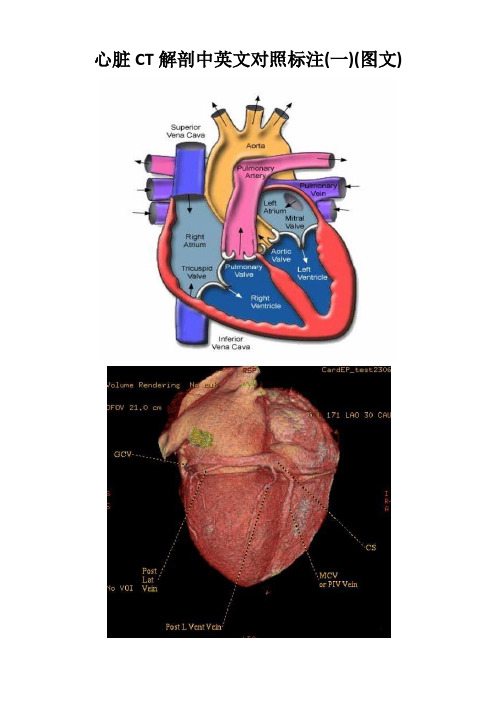
心脏CT解剖中英文对照标注(一)(图文)常用英文名称及缩写LA - Left Atrium 左心房RA - Right Atrium 右心房LV - Left Ventricle 左心室RV - Right Ventricle 右心室Mitral Valve 二尖瓣A. Aorta-Ascending Aorta 升主动脉D. Aorta-Descending Aorta 降主动脉SVC –Superior Vena Cava 上腔静脉IVC –Inferior Vena Cava 下腔静脉PA - Pulmonary Artery 肺动脉PV - Pulmonary Vein 肺静脉LMA - Left Main Artery 冠状动脉左主干LAD - Left Anterior Descending Artery 左前降支LCX - Left Circumflex Artery 左回旋支LMB - Left Obtuse Marginal Branch 左边缘支(钝缘支)RCA - Right Coronary Artery 右冠状动脉PDA - Posterior Descending Artery 后降支Conus Branch 右动脉圆锥支LAA –Left Atrial Appendage 左心耳RAA –Right Atrial Appendage 右心耳CS - Coronary Sinus 冠状窦MCV –Middle Cardiac Vein 心中静脉GCV –Great Cardiac Vein 心大静脉PIVV –Posterior Intraventricular Vein 后室间静脉(心中静脉)PLVV –Posterior Left Ventricular Vein 左室后静脉PLV –Posterior Lateral Vein 左室后侧静脉(边缘静脉)。
心脏超声中英文对照词汇
---------------------------------------------------------------最新资料推荐------------------------------------------------------心脏超声中英文对照词汇一次谐波共振 First harmonic response [hɑː’mɒnɪk] 二尖瓣Mitral valve, MV [‘maɪtrəl] 二尖瓣口 Mitral valve orifice, MVO [‘ɑrɪfɪs] 二尖瓣后瓣 Posterior mitral valve, PMV [pɒ’st ɪərɪə] 二尖瓣血流 Mistral inflow 二尖瓣前瓣 Anterior mitral valve, AMV [n’tɪərɪə] 二尖瓣裂 Mitral valve cleft, MVCLF [kleft] 二次谐波共振 Second harmonic response 二次谐波多普勒组织成像 Second Doppler tissue imaging, H-DTI 二次谐波成像Second harmonic imaging 人工瓣膜血栓 Prosthetic heart valve thrombus 三心房心 Cor triatriatum 三尖瓣 Tricuspid valve, TV 三尖瓣关闭不全Tricuspid valve insufficiency 三尖瓣闭锁Tricuspid atresia 三尖瓣前瓣 Anterior tricuspid valve, ATV 三尖瓣狭窄Tricuspid valve stenosis 三尖瓣疾病Tricuspid disease 三尖瓣隔瓣 Septal tricuspid valve, STV 三维超声心动图Three-dimensional echocardiography 大动脉转位Transposition of the great arteries 川崎病 Kawasakis disease 中场 Middle filed 分辨率 resolution 反射 Reflection 心内血栓 Intracardiac thrombus 心内膜垫缺损 endocardial cushion defect 心包疾病 Pericardial effusion 心包积液 Pericardial effusion 心外膜冠状动脉 Epicardial coronary artery 心尖多孔瑞士奶酪样室间隔缺损 Digital multiple swiss cheese septal1 / 5defect 心肌内冠状动脉 Intramyocardial coronary artery 心肌对比超声心动图 Myocardial contrast echocardiography, MCE 心肌梗塞 Myocardial infarction 心肌梗塞并发症 Complications of myocardial infarction 心房内血栓 Atria thrombus 心房黏液瘤Atria myxoma, MYX 心室内附壁血栓Intraventricular mural thrombus 心绞痛Angina pectoris 心脏声学造影Cardial acoustic contrast 心脏肿瘤Cardial tumor 心脏移植Heart transplantation 主动脉二叶瓣 Bicuspid aortic valve 主动脉瓣口Aortic valve orifice, AVO 主动脉瓣狭窄Aortic valves stenosis 主肺动脉 Main pulmonary artery, MPA 主瓣 Main lobe 功率谱 Power spectrum 右心房 Right atrium, RA 右心室 Right ventricle, RV 右心室收缩时间间期 Right ventricle systolic time intervals 右心室收缩前间期 Right ventricle pre-ejection period, RVPEP 右心室射血时间Right ventricle ejection time ,RVET 右冠状动脉起源于肺动脉 Anomalous origin of right coronary artery from pulmonary artery 右室双出口Double-outlet right ventricle 右心室双腔心 Double chambered right ventricle 右心室流出道 right ventricle outflow 对比造影谐波成像 Contrast agent harmonic imaging, CAHI 对比超声心动图学 Contrast echocardiography, CE 对数压缩 Logarithmic compensation 尼奎斯特频率极限 Nyquist frequency limit 左心耳 Left atrium apendge, LAA 左心房 Left atrium 左心左心室长---------------------------------------------------------------最新资料推荐------------------------------------------------------ 轴切面 Left ventricle, LV 左心室发育不全综合征 Hypoplastic left heart syndrome 左心室收缩末期内径 Left ventricle end systolic dimension, LVEDD 左心室流出道梗阻 Left ventricle outflow obstruction 左心室舒张末期内径 Left ventricle end diastolic dimension, LVSDD 左冠状动脉起源于肺动脉 Anomalous origin of left coronary artery from pulmonary artery 平行扫描 Parallel scanning 永存动脉干 Persistent arterious 电子相控阵扇型扫面 Phased array sector scan 皮肤黏膜淋巴结综合征Mucocutaneous lymph node syndrome, MCLS 节制束 Moderator band 伪像Artifacts 伪影处理技术Pseudo-color processing technique 先天性肺动脉口狭窄Congenital pulmonary artery fistula 先天性冠状动脉瘘 Congenital coronary artery fistula 共振Resonant 共振频率Resonant frenquency 压力半降时间Pressure half-time, PHT 回声失落 Echo drop-out 回声增强效应Effect of echo enhancement 团注 Bolus 多平面经食道超声心动图Multiplane transesophageal echocardiography 多点选通式多普勒Multigate Doppler 多普勒方程 Doppler equation 多普勒组织 M 型模式 Doppler tissue m-mode, DT-M-MODE 多普勒组织加速度图Doppler tissue acceleration, DAT 多普勒组织成像Doppler tissue imaging, DTI 多普勒组织脉冲频谱 Doppler tissue pulsed wave mode, DT-DTE 多普勒组织能量图 Doppler tissue energy, DTE3 / 5多普勒组织速度图 Doppler tissue velocity, DTV 多普勒效应Doppler effect 多普勒超声心动图 Doppler echocardiography 多普勒频移Doppler shift 导航装置Homing deveces 导管超声Catheter ultrasound 机械扇型扫描 Mechanical sector scan 纤维瘤Fibroma 自由扫查Free-hand scanning 自动边缘检测Automatic border detection 自然组织谐波成像 Native tissue harmonic imaging 色彩倒错 Color aliasing 血栓 Thrombus 血流彩色成像 Color flow mapping 血管肉瘤 Angiosarocama 血管腔内超声成像Intravascular ultrasound imaging 负荷超声心动图Stress echocardiography 体元模型 Voxel model 声束形成 Bean forming 声阻抗Acoustic impedance 声学定量Acoustic quantification 声学速度Acoustic velocity 声强Acoustic intensity 层流 Laminar flow 希阿利网 Chiari netok 快速富里叶变换Fast fourier transform 折射Refraction 时间分辨率Temporal resolution 时间增益补偿 Time gain compensation 时域法 Time domain method 纵向分辨率 Longitudinal resolution 纵波 Longitudinal wave 肛管超声 Anal endosonography 近场 Near filed 进入曲线 Wash in curves 远场 Far filed 连续式多普勒Continuous wave Doppler, CW Doppler 连续注射Continuous injection 乳头肌Papillary muscle, PM 单脉冲删除Single pulse concellation 取样容积 Sample volume 图像分辨率 Image resolution 实时频谱分析 Real-time spectral analysis 房间隔缺---------------------------------------------------------------最新资料推荐------------------------------------------------------ 损 Atrial septal defect, ASD 房间隔脂肪瘤样肥厚 Lipomatous hypertrophy of the atrial septum 房间隔瘤Atrial septal aneurysm 欧氏瓣Eustachian 空壳Hollow core 空间分辨率Spatial resolution 组织多普勒成像技术 Tissue Doppler imagine 组织多普勒超声心动图 Tissue Doppler echocardiography 经心腔内超声心动图 Intracardiac echocardiography 经阴道彩色多普勒超声 Trans-vaginal color Doppler ultrasound 经食道超声心动图Transesophageal echocardiography 限制性室间隔缺损Restrictive ventricular septal defect 非致密性心室心肌二维超声心动图Non-compaction of ventricular myocardium two dimensional echocardiography 肺动脉 Pulmonary artery 肺动脉狭窄肺动脉高压肺动脉瓣肺静脉异位引流顶端侧方扫描式顶端旋转扫描式临床基础冠心病冠状动脉内超声显像冠状动脉异常冠状动脉血流储备冠状动脉起源异常冠状静脉窦厚度分辨率室上嵴室间隔缺损室间隔膜部瘤界面相干对比造影成像相干图像形成技术类脂背向散射积分背景噪音脉冲反相谐波成像脉冲式多普勒脉冲重复频率衍射重叠房室瓣扇形扫描振幅捆扎型纤维蛋白分子旁瓣效应浦肯野纤维瘤消除曲线涡流特定定点造影剂留间隔器缺血性预适应胸骨旁短轴切面能量对比成像脂肪瘤5 / 5。
心电图诊断中英对照
心电图诊断中英互译正常心电图 Normal ECG(Electrocardiogram)窦性心动过缓 Sinus Bradycardia 窦性静止 Sinus Arrest窦性心动过速 Sinus Tachycardia 窦性心律不齐 Sinus Arrhythmia房性早搏Atrial Premature Complex房性早搏伴心室内差异传导Atrial Premature Complex with Aberrant Intraventricular Conduction房性早搏未下传Non-conducted(Blocked) Atrial Premature Complex(阵发性)心房颤动(Paroxysmal) Atrial Fibrillation(阵发性)心房扑动(Paroxysmal) Atrial Flutter双心房肥大Bi-atrial Enlargement 双心室肥大 Biventricular HypertrophyST-T 改变 ST-T Abnormalities ST段/T波改变 ST Segment/T Wave Abnormalities交界性早搏 Junctional Premature Complex室性早搏二联律Bigeminal Ventricular Premature Complex 三联律 Trigeminal多源性室性早搏连发(成对)Multifocal Ventricular Premature Complex‘s Couplet短阵房性心动过速 Short Run of Atrial Tachycardia阵发性室上性心动过速 Paroxysmal Supraventricular Tachycardia非阵发性 non-paroxysmal 逸搏Escape Beat 逸搏心律Escape Rhythm一度房室阻滞 First Degree A-V Block高度(三度)房室阻滞 High(Third) Degree A-V Block二度I型(II型)房室阻滞 Second Degree A-V Block ,Mobitz Type I (II)二度I型(II型)窦房阻滞 Second Degree SinoAtrial Block , Type I (II)完全性右束支阻滞 Complete Right Bundle Branch Block完全性左束支阻滞 Complete Left Bundle Branch Block左前(后)分支阻滞 Left Anterior(Posterior) Fascicular Block心室预激 Ventricular Pre-excitation房性(交界性、室性)逸搏心律 Atrial (Junctional、Ventricular) Escape Rhythm提示高钾血症 Suggestion of Hyperkalemia提示低钾血症 Suggestion of Hypokalemia急性广泛前壁心肌梗死 Acute Extensive Anterior Myocardial Infarction急性(陈旧性)前间壁心肌梗死 Acute(Old) Anteroseptal Myocardial Infarction急性前侧壁心肌梗死 Acute Anterolateral Myocardial Infarction前壁anterior 下壁inferior 后壁posterior 后侧壁posterolateral电轴左偏 Left Axis Deviation电轴右偏 Right Axis Deviation心肌缺血 Myocardial Ischaemia起搏器 Pacmaker感谢下载!欢迎您的下载,资料仅供参考。
心脏基本病变X线诊断(中英文对照)
(2)肺动脉圆锥隆起 左前斜位 心前下缘向前膨隆,心膈面延长,
推移左室向后平移
见于:二尖瓣狭窄、肺心病和 肺动脉狭窄等
心尖在左、右心室增大时的位置
3 左心房增大(Enlargement of Left Atrium)
• 代表左室大一类心脏病 • X线心尖主要左下大,心腰相对凹陷 • 见于高心病等
3 普大型心(general enlarged heart):
代表心包病或多个房室大一类 心脏病
X线心脏向双侧增大,心缘各弓 弧消失
见于心包炎或心肌病
Hale Waihona Puke 4 靴型心(wooden-shoe heart):
代表右室大,有肺动脉狭窄一类 心脏病
X线心脏主要向左大,心尖上翘, 心腰器质性下陷
见于法鲁氏四联症等
(四)主动脉形态和密度的改变
1 形态改变:迂曲、延长(tortuosity、elongation) 2 密度改变:增粗、钙化(dilatation、calcification)
(五)心包钙化(Pericardial Calcification)
肺血多少的判断标准
主要以右下肺动脉干直径为标准: 正常成人男性10~15mm
女性 9~14mm 一般肺动脉和伴行支气管直径之比 为1:1
3 肺动脉高压(Pulmonary Arterial Hypertension)
收缩压>4kPa(30mmHg),平 均压>2.7kPa(20mmHg)
1 肺动脉段突出 2 肺门截断征 3 中心肺动脉搏动强 4 右室大
后前位 1 左心耳(left auricle)突出 2 心底部双重密度,心右缘双重 轮廓影
【中英双语 _ 心电图教学】高度房室传导阻滞1例

病例简介Brief introduction患者女性,73岁,因“严重气促”入院。
患者6年前曾有下壁心肌梗死病史。
入院时心电图如图1所示。
A 73-year-old woman was admitted because of severe shortness of breath. Six years ago she had experienced an inferior myocardial infarction. Her ECG on admission is shown in Figure 1.图1:12导联心电图提示60次/分的窦律和约35次/分的室率。
QRS波在宽度、电轴和形态上不尽相同。
为便于解释AV传导类型,我们将这些QRS波进行编号。
请注意QRS波4和5的R-R间期较其他R-R间期短。
QRS波5前的PR间期为160ms。
这提示QRS波5为窦律下传伴左束支阻滞(LBBB)。
QRS波3为介于左束支阻滞和逸搏心律间的融合波。
Figure 1:Twelve-lead ECG showing sinus rhythm at a rate of 60/min and ventricular rate of approximately 35/min. The QRS complexes are not always the same with regard to width, axis, and configuration. They are numbered to facilitate explanation of the type of AV conduction. Note the shortening in R-R interval between QRS 4 and QRS 5 compared to the other R-R intervals. QRS 5 is preceded by a PR interval of 160 ms. This indicates that QRS 5 is a conducted beat with left bundle branch block (LBBB). QRS 3 is a fusion complex between LBBB and the escape rhythm.提问Questions1、该患者是完全性房室(AV)阻滞吗?或偶有AV传导?1、Does the patient have complete AV block, or is AV conduction sometimes present?2、AV阻滞的部位在哪?2、Where is the site of AV block?3、逸搏心律的起源位置在哪?3、What is the site of origin of the escape rhythm?4、为什么逸搏心律的QRS波较第5个QRS波窄?4、Why is the QRS of the escape rhythm narrower than QRS 5?5、下一步你会如何处理?5、What will be your management strategy?讨 论 Discussions1. 心电图示窦性心律,6个QRS波分别在图中标注为1-6。
心脏电生理检查 英语
心脏电生理检查英语When it comes to heart health, one of the most important diagnostic tools is the cardiac electrophysiology study. It's a way for doctors to get a detailed look at how your heart's electrical system is functioning. Basically, they study the electrical signals that control your heartbeat.The process itself is pretty straightforward. You'll be asked to lie down on a special table, and the doctor will insert thin, flexible wires called catheters into your veins. These catheters go straight to your heart, where they record the electrical activity. It's not painful, but you might feel a bit uncomfortable.The results of this test can be really useful. They can help doctors diagnose problems like irregular heartbeats or heart blockages. Plus, they can also figure out if certain treatments, like medication or surgery, are likely to work for you.One cool thing about cardiac electrophysiology studies is that they're often done in real-time. That means the doctor can see your heart's electrical activity as it happens, which gives them a more accurate picture of what's going on. It's kind of like having a live video feed of your heart's inner workings.Overall, cardiac electrophysiology studies are a pretty essential tool for understanding and managing heart conditions. They provide doctors with crucial information that they can use to develop the best possible treatment plans for their patients.。
【中英双语心电图教学】单个室早后的两种不同P波
【中英双语心电图教学】单个室早后的两种不同P波English audio presented by Dr.Ethan(杜先锋)素材来源:JOSEPHSON AND WELLENS ECG LESSONSHeart Rhythm 2018, 15 (12) :1891-1892Let's enjoy the English learningin the EP world!病例简介Brief introduction患者男性,33岁,因“心悸3月”至门诊就诊,并否认存在快速心律。
除心悸外,该患者无其他主诉,且运动耐量正常。
体格检查未见异常,心脏超声也显示正常。
12导联心电图(如图1)示室早二联律。
室早在运动时消失。
A 33-year-old man came to the outpatient clinic because of palpitations. He noticed their occurrence during the last 3 months. He denied to have felt rapid heart rhythms.Apart from palpitations he had no other complaints and normal exercise tolerance.Physical examination did not reveal abnormalities. An echocardiogram was normal. A ventricular bigeminal rhythm was present on the 12-lead electrocardiogram (Figure 1). The ectopic ventricular rhythm disappeared during exercise.图1:12导联心电图示:窦性心律伴成对室性早搏二联律。
医学常用心电图术语中英文翻译

医学常用心电图术语中英文翻译Medical Terminology Translation of Commonly Used Electrocardiogram Terms in Chinese and EnglishIntroductionIn the field of medicine, accurate communication is vital to ensure effective diagnosis and treatment. Electrocardiography, commonly known as ECG or EKG, is a crucial tool in diagnosing and monitoring various cardiac conditions. Understanding the terminology used in ECG reports is essential for healthcare professionals to interpret the findings correctly. This article aims to provide a comprehensive translation of commonly used ECG terms from Chinese to English, ensuring clear communication and enhancing medical practice.1. Basics of Electrocardiogram1.1 心电图(xīn diàn tú) - Electrocardiogram (ECG)1.2 心率(xīn lǜ) - Heart rate1.3 心律(xīn lǚ) - Cardiac rhythm1.4 导联(dǎo lián) - Lead1.5 间期(jiàn qī) - Interval1.6 幅度 (fú dù) - Amplitude2. P Waves2.1 P波(P bō) - P wave2.2 P波增宽(P bō zēng kuān) - P wave widening2.3 P波高尖(P bō gāo jiān) - P wave tall and pointed2.4 P波倒置(P bōdǎo zhì) - Inverted P wave3. QRS Complex3.1 QRS波群(QRS bō qún) - QRS complex3.2 Q波(Q bō) - Q wave3.3 R波(R bō) - R wave3.4 S波(S bō) - S wave3.5 QRS时限延长 (QRS shí xiàn yán cháng) - Prolonged QRS duration3.6 QRS时限缩短(QRS shí xiàn suō duǎn) - Shortened QRS duration4. ST Segment4.1 ST段 (ST duàn) - ST segment4.2 ST段抬高(ST duàn tái gāo) - ST segment elevation4.3 ST段压低(ST duàn yā dī) - ST segment depression4.4 J点抬高(J diǎn tái gāo) - J-point elevation5. T Wave5.1 T波(T bō) - T wave5.2 T波高大(T bō gāo dà) - Tall T wave5.3 T波低平(T bō dī píng) - Flat T wave5.4 T波倒置(T bō dǎo zhì) - Inverted T wave6. U Wave6.1 U波(U bō) - U wave6.2 U波增高(U bō zēng gāo) - Increased U wave6.3 U波降低(U bō jiàng dī) - Decreased U wave7. QT Interval7.1 QT间期 (QT jiàn qī) - QT interval7.2 QT间期延长(QT jiàn qī yán cháng) - Prolonged QT interval7.3 QT间期缩短(QT jiàn qī suō duǎn) - Shortened QT intervalConclusionHaving a solid foundation in medical terminology is crucial for effective communication in the field of healthcare. This article provided a comprehensive translation of commonly used terms in electrocardiography, from Chinese to English, ensuring accurate interpretation and diagnosis. By understanding these terms, healthcare professionals can communicate effectively and deliver optimal patient care.。
ECG-中英文版

1.What is an ECG? 2.How does it come about? 3. How to recording an ECG? 4. How to explain an ECG?
What is an ECG?
ECG = Electrocardiogram
Tracing of heart’s electrical activity
What Produce recording Interpretation
Let’s Practice
First Degree Heart Block
PR interval >200ms
Supraventricular (室上性心动过速) Tachycardia
Retrograde P waves
Narrow complex, regular; retrograde退化 P waves, rate <220
First Degree Heart Block, Mobitz Type I (Wenckebach)
PR progressively lengthens until QRS drops
QRS = ventricular depolarisation
What do the components represent?
What Produce recording Interpretation
T = repolarisation(复极化) of the ventricles
How to recording an ECG
追踪心脏的电活动
What Produce recording Interpretation
What is an ECG?
超声心动图常用中英对照表

. 超声心动图术语英文对照表:A 面积Abdominal Aorta (AA) 腹主动脉AccT 血流加速时间ALS 主动脉瓣叶开放Angiography 血管显像Ann 瓣环Annotation 注释ASD 心房间隔缺损Automatic gain control 自动增益控制AV 主动脉瓣膜AV- A 连续性方程计算的主动脉瓣膜面积AV Cusp 主动脉瓣膜尖端开放AV Cusp 主动脉瓣膜尖端开放AV Di am) 主动脉瓣膜直径AVA 主动脉瓣膜面积Axill 腋下动脉Axillary Vein 腋静脉Ao 主动脉Ao Arch Diam 主动脉弓直径Ao Asc 升主动脉直径Ao Desc Diam 降主动脉直径Ao Diam 主动脉根部直径Ao Isthmus 主动脉峡部Ao st junct 主动脉ST 接合AR 主动脉返流Asc 上升BBA 基底动脉Basil V 基底静脉Brac V 臂静脉Brightness 辉度、亮度BSA 体表面积Buffer 阻尼器CCalcification (CAL) 钙化Calibration 定标、校正Catheter-based US probe 导管超声探头CCA 颈总动脉Ceph V V 头静脉CFM processing board 彩色多普勒处理功能板Character 字符CI 心脏指数Clear 消除CO 心脏输出量Color capture 彩色捕获Color cut 彩色消除Color doppler energy 彩色多普勒能量图Color doppler flow imaging 彩色多普勒血流显像Color Doppler Flow Imaging (CDFI) 彩色多普勒血流显像.Color doppler level 彩色多普勒强度Color edge 彩色边界Color enhance 彩色增强Color flow angiography 彩色血流造影Color lock 彩色锁定Color persistence 彩色余辉Color polarity 彩色极性Color power angio 彩色能量图Color scale display 彩阶显示Color steering 彩色转向Color velocity imaging 彩色速度显像Color video monitor 彩色视频监视器Color wall filter 彩色壁滤波Com Femoral 股总动脉Common Jugular Artery 颈总动脉Confocusing 全场连续聚焦Contrast resolution 对比分辨力Convex (CVX) 凸形、凸阵Convex array 凸阵Cornea 角膜Cross sectional Area (CSA) 切面面积DD 直径Dec 减速度DecT 减速时间Demodulator 解调器、检波器Depth gain compensation 深度增益补偿Desc 递减Detail resolution 细节分辨力Digital image 数字成像Doppler flow-direction resolution 多普勒流向分辨力Doppler flow-velocity distributive resolution 多普勒流速分布分辨力Doppler minimum flow-velocity resolution 多普勒最低流速分辨力Doppler sample volume 多普勒取样容积Dorsal Pedal Artery 足背动脉Duodenum (Du) 十二指肠Dur 持续时间Dynamic focusing 动态聚焦Dynamic frequency scanning 动态频率扫描Dynamic imaging 动态影像Dynamic range 动态范围EECA 颈外动脉Echography sonography 声像图法Ed 心脏舒张EdV 舒张末期容量EF 射血分数Effusion (Eff) 积液Electric focusing 电子聚焦Embolism 栓塞Endoluminal sonography 腔内超声显像EPSS E 点到室间隔分离Erase eliminate 消除EsV 收缩末期容量ET 射血时间.External Iliac Artery 髂外动脉External Jugular Vein 颈外静脉FFast time constant 快速时间常数电路Femoral Artery 股动脉Femoral Vein 股静脉Fibrosis (Fib) 纤维化Focal distance 焦距Focus 聚焦Foreign Boby (FB)异物frame correlation 帧相关frame rate 帧率frame resolution 帧分辨力Freeze (FRZ) 冻结Freeze 冻结Frequency Spectrum 频谱FS 短轴缩短率FV 血流容量FVI 血流速度积分GGain 增益Gray scale display 灰阶显示Great Saphenous Vein 大隐静脉HHead circumference (HC) 头围Hematoma (HMA) 血肿HR 心率IICA 颈内动脉Image uniformity 图像均匀性Image-line resolution 图像线分辨力Imaging data 成像数据Inferior Vena Cava (IVC) 下腔静脉Internal Jugular Vein 颈内静脉Interventional ultrasound 介入性超声Intracardiac ultrasonic imaging 心内超声显像Intracavitary probe 腔内探头Intraluminal ultrasonic imaging 管腔内超声显像Intraoperative porbe 术中探头Intraoperative ultrasonic monitoring 术中超声监视Intravascular ultrasonic imaging 血管内超声显像Intravascular ultrasound 血管内超声Invert 倒置、反转IVC 下腔静脉IVRT 等容舒张期IVS 室间隔IVSd 、IVSs 室间隔(收缩期,舒张期)厚度LL 长度LA 左心房LA Diam 左心房直径LA Major 左心房长度LA Minor 左心房宽度.LA/Ao Ratio 左心房直径和主动脉根部直径比率LAA 左心房面积LAD 左心房直径Large Intestine 大肠Lateral Ventricle (LV) 侧脑室LV 左心室LVA 左心室面积LVI D 左心室内径LVIDd 舒张期左心室容积LVIDs 收缩期左心室容积LVL 左心室长度LVLd 舒张期左心室内径LVLs 收缩期左心室内径LVM 左心室心肌重量LVOT Diam 左心室流出道直径LVPW 左心室后壁LVPWd 左室后壁舒张期厚度LVPWs 左室后壁收缩期厚度MMagnification Magnify Zoom 放大Mass( M) 包块MCA 大脑中动脉Mcub V 中央静脉Mean Velocity (Mean Vel) 平均速度Menu selection 菜单选择metastasis (Met) 转移灶Minimum flow-velocity of color doppler 彩色多普勒最低流速分辨力Motion discrimination 运动辨别力MPA 主肺动脉MPA 主肺动脉MR 二尖瓣返流ultipurpose scanner 多用途探头Multistage focusing 多段聚焦MVA By PHT 二尖瓣口面积根据压力降半时间MVcf 纤维圆周缩短平均速度MVO 二尖瓣口NNecrosis (Nec) 坏死Node (N) 结节OOT 流出道PP 乳头肌PA 肺动脉Pancreas (P;Pa) 胰腺PAP 肺动脉压力PDA 动脉导管末闭PEd 心包渗出舒张期Penetration depth 穿透深度PEP 射血前期Peripheral Vessel (PV)外周血管PFO 卵圆孔未闭PG 压力阶差Phased annular array probe 环阵相控探头PHT 压力降半时间.PISA 最近等速线表面面积Popliteal Artery 腘动脉Popliteal Vein 腘静脉Post process 后处理Pre process 前处理Preset 预设置Prostate (Pro) 前列腺Ps 心脏收缩Pulmonic Diam 肺动脉瓣膜直径PV 肺动脉瓣PV Ann Diam 肺动脉瓣环面直径PV-A 连续性方程计算的肺动脉瓣口面积PVein 肺静脉PW 后壁QQp 肺循环血流量Qs 体循环血流量Quadrate Lobe (QL) 方叶RRA 右心房RAA 右心房面积Rad 半径RAD 右心房直径Real-time imaging 实时成像Record 记录Rejection reject suppression 抑制Rendering play back 回放Reset 重调、复原Reversed Flow (RF) 返流Right Ventricle (RV) 右心室RPA 右肺动脉RPA 右肺动脉RV 右心室RVA 右心室面积RVAW 右心室前壁RVD 右心室直径RVID 右心室内径RVL 右心室长度RVOT 右心室流出道SScan mode 扫描方式Scanner (SCNR) 扫描器、探头Sector Angle (Sec Ang) 扇扫角度Sector scanning 扇扫Sediment (Sed) 沉积物Segment focusing 分段聚焦Sensitivity time control 灵敏度时间控制Sensor 传感器Septum Pellucidum (SP) 透明隔;透明隔腔Sequential focusing 连续聚焦Shift 变换Short Saphenous Vein 小隐静脉SI 搏动指数Sliging focusing 滑动聚焦Sonogram echogram 声像图Spatial resolution 空间分辨力Spatial resolution of color doppler 彩色多普勒空间分辨力ST 缩短% STIVS 心室缩短百分比SUBC 锁骨下动脉Subclavian Vein (SCV) 锁骨下静脉Sup Femoral 股浅动脉SV 每搏量SVI 每搏量指数TT 时间TA 三尖瓣环TAML 三尖瓣环面中部到侧部Tar get_r(TAR) 靶团Temporal resolution 瞬时分辨力Thoracic cavity 胸腔Thoracic Circumference (Th C) 胸围Three dimensional display 三维显示3D image reconstruction 三维图像重建Thrombus (Th) 血栓Time gain compensation 时间增益补偿Time resolution of color doppler 彩色多普勒时间分辨力Tissue specific imaging 组织特性成像TR 三尖瓣返流Trans AVA(d)、Trans AVA(s) 横向主动脉瓣膜面积Transcranial doppler 经颅多普勒Transcranial Doppler ( TCD) 经颅多普勒Transducer 换能器Transesophagel echocardiography probe(TEE)经食管超声心动图探头Transesophagel probe 食管探头Trigger 触发器Tumor (T) 肿瘤TV 三尖瓣膜TVA 三尖瓣口面积UUltrasonic imaging 超声成像Ultrasound catheter 超声导管Ultrasound guided percutaneous Ultrasound guided probe 穿刺探头US guided percutaneous alcohol injection 超声引导经皮穿刺注射乙醇US guided percutaneous aspiration 超声引导经皮抽吸VVel 速度VERT 椎动脉VET 瓣膜射血时间Vmax 最大速度Vmean 平均速度VSD 室间隔缺损VTI 速度时间积分WWall (W) 壁Wide-band probe 宽频带探头Write 写入ZZero adjustment 零位调整Zone focusing 区域聚焦如有侵权请联系告知删除,感谢你们的配合!。
- 1、下载文档前请自行甄别文档内容的完整性,平台不提供额外的编辑、内容补充、找答案等附加服务。
- 2、"仅部分预览"的文档,不可在线预览部分如存在完整性等问题,可反馈申请退款(可完整预览的文档不适用该条件!)。
- 3、如文档侵犯您的权益,请联系客服反馈,我们会尽快为您处理(人工客服工作时间:9:00-18:30)。
SHARCHIP Method ——S ——standardization H ——heart rate A ——axis R ——rhythm C ——conduction H ——hypertrophyI ——ischemic&infarction P ——previous ECGSHARCHIP方法—— S ——标准化 H ——心率 A ——心电轴 R ——节律 C ——传导 H ——肥大I ——缺血&梗死 P ——比较之前ECG***********************************************************************************************① standardizationVertically :10mm=1mV Horizontal : 25mm=1s① 标准化纵向:10mm=1mV 走纸:25mm=1s② heart rate=300 / large time units =1500/ small time unitsTachycardia (>90 bpm) Bradycardia (<50 bpm) ② 心率=300/大格数=1500/小格数心动过速(>90 bpm) 心动过缓(<50 bpm) ③axisL-axis deviation: <- 30°I is mostly positive & III is mostly negative R-axis deviation: > 100°I is mostly negative& III is mostly positive③电轴左偏:<- 30°I III :口对口,向左走右偏:> 100°I III :尖对尖,向右偏 ④rhythm (4Q ) Pacemaker?normal P : sinus rhythm ab. P, narrow QRS :atrial r. no P, narrow QRS :junctional r. no P, wide QRS :ventricular r.Normal /Tachycardia/Bradycardia? Active or passive(escape) Any Additional ? ④节律(4问) 起搏点?正常心率/心动过速/心动过缓? 有无逸搏? 附加节律?Common arrhythmiasatrial flutter:1 zigzag F waves replace P wave2 A/V (F/QRS) is proportionaatrial fibrillation:1 Irregular f waves2 RR interval absolutely irregular PSVT:(…)ventricular flutter:1 regular big wave (200-250bpm) ventricular fibrillation:1 irregular small waves (200-500bpm) VT:(…)Premature atrial contraction(PAC):1 premature P’wave,2 normal QRS,3 imcomplete compensatory pause Premature junctional contraction:1 antidromic P’wave2 normal QRS3 complete compensatory pause. Premature Ventricular Cont. (PVC):1 no P wave2 wide QRS (T is converse with QRS)3 complete compensatory pause 常见心律异常房扑:1锯齿状F波代替P波2固定房室比房颤:1 颤动f波代替P波2心室律绝对不齐阵发性室上速:(略)室扑:大震幅波动(200-250bpm)室颤:大小不等的低小波(200-500bpm) 室速:(略)房性早搏:异位P’窄QRS不完全代偿间歇交界性早搏:逆P’窄QRS完全代偿间歇室性早搏:无P宽大畸形QRS(T波反向)完全代偿间歇⑤conductionA VBPP Intervals RR Intervals1°SAB Invariant(prolonged PR Intervals) 2°SABType I invariant Wenckebach ph.Type II invariant RR pause=2RR3°SAB(Complete) invariant Escape rhythmLBBB:left side leads: incisure on wide R right side leads: wide S wave,reverse ST-T RBBB:right side leads: remarkable rsR`left side leads: wide S wave,reverse ST-TLAFB:Left-deviation axisrS inⅡ、Ⅲ、aVF(SⅡ< SⅢ)qR inⅠ、aVL(RⅠ< R aVL)narrow QRSLPFB:right-deviation axisrS inⅠ、aVLqR inⅡ、Ⅲ、aVF(RⅡ< RⅢ) narrow QRSWPW Pre-excitation syndrome short PRwide QRSslurring of the initial part of the QRS (Δ) Secondary ST-T changes 完左:左侧导联:宽R带切迹(型),失q 右侧导联: rS /QS型(宽S),ST-T与QRS主波反向完右:右侧电联:rsR’ ( 型)左侧导联: rS /QS型(宽S),ST-T与QRS主波反向左前分支阻滞:电轴左偏(-45~ -90°)下壁导联rS型,S III>S II左侧导联呈qR型,RⅠ< R aVLQRS不增宽左后分支阻滞:电轴右偏(>110°)左侧导联呈rS型下壁导联qR型,RⅡ< RⅢQRS不增宽W-P-W预激综合征:短PR间期QRS增宽起始部粗钝(附加Δ波)继发ST-T改变⑥HypertrophyLA Hypertrophy:duration of P wave > 3 mm (m-shape) RA Hypertrophy:amplitude of P wave > 2 mmLV Hypertrophy:left-deviation axis counterclockwise rotationRV Hypertrophy:right-deviation axisclockwise rotation ⑥房室肥大左房大:M型宽P波右房大:高尖P波左室大:电轴左偏逆钟转位RV5或RV6 >2.5mVRV5+SV1 > 4.0mV(男)/3.5 mV (女)右室大:电轴右偏顺钟转位RV1+SV5>1.05mV (重者>1.2mV)⑦ischemic & infarctionV1-V3:anteroseptal wallV3-V5:anterior wallV5, V6, aVL:lateral wallI, aVL:high lateral wallV1-V5: extensive anterior wall V8, V9:posterior wallII, III, aVF:inferior wall V3R-V6R:right ventricular ⑧previous (comparison) ⑦心肌缺血V1-V3:前间壁V3-V5:前壁V5, V6, aVL:侧壁I, aVL:高侧壁V1-V5:广泛前壁V8, V9:正后壁II, III, aVF:下壁V3R-V6R:右室⑧比较既往心电图Normal range horizontal VerticalP wave <3 mm <2.5 mm in E-leads<2.0 mm in C-leadsPR intervals 3-5 mmQRS complex <3 mm ≥5 mm in E-leads≥8 mm in C-leadsQ wave <1 mm <1/4RR wave <1.5 mmS waveST segment-0.5~ 1.0 mmT wave≥ 1/10 QRSQT intervals QTc: 8-11 mmQTc=QT/√RRU wavePE:an S1Q3T3 patternsinus tachycardiaRBBBI :prominent S waveIII :a Q wave and inverted T waveV1 - V3 :inverseT waveHypokalaemia:ST segment depression,low amplitude T waves,and prominent U wavesHyperkalaemia:small or absent P wavesAFVFwide QRSshortened or absent STwide, tall and tented T wavesPUMC kyanite。
