Circulatorysystem-43页PPT资料
合集下载
(医学各论课件)6、Circulatory system
atrial natriuretic polypeptide or cardionatrin b. ventricular M: thick, long, branches • LCT: rich in capillaries *atrioventricular fibrous annulus: DCT
contain fibroblast, CF, EF, SM • subendocardial layer: LCT, with BV, N
and conducting S- Purkinje fiber
---myocardium: • cardiac M:
a. atrial muscle: /atrial granules: 0.3-0.4um in D, contain
so the boundaries between three tunica are not very clear c. contains more CT, less smooth M, SM are arranged in bundles d. vein valve:
/infolding of tunica intima /semilunar-liked /prevent back flow of blood
Chapter 9
Circulatory system
---Closed tubular system ---Blood circulatory or cardiovascular S ---Lymph vascular S
The cardiovascular S • heart • artery • vein • capillary
endothelial cell: • processes –microvilli-like, finger-liked • plasmalemmal vesicle
循环系统CirculatorySystemPPT课件
动脉圆锥:右心室腔向左上方延伸的部分逐渐变细,形似倒置的漏斗, 称动脉圆锥。
26
右半心的血流方向:
肺动脉 肺动脉口 上腔静脉口 下腔静脉口 右心房 右Biblioteka 室口 右心室 冠状窦口27
28
(三)左心房left atrium 位于右心房的左后方。左心耳 四个入口:两对肺静脉口。 一个出口:左房室口。
(四)左心室left ventricle 位于右心室的左后方。 一个入口:左房室口。口周围有二尖瓣(前瓣、后瓣)。 一个出口:主动脉口。口周围有主动脉瓣。主动脉窦(左、右、后窦)
25
(二)右心室right ventricle 位于右心房的左前下方。
一个入口:右房室口。口周围有三尖瓣环,其上附有三尖瓣tricuspid valve(借腱索连于乳头肌)。
三尖瓣复合体:三尖瓣环、三尖瓣、腱索、乳头肌共同构成,防止血液 逆流。
一个出口:肺动脉口。周围有肺动脉瓣,心室舒张时关闭,阻止血液逆 流入心室。
肺动脉
肺循环
主动脉
左心室 右心房
组织毛 细血管
上、下腔静脉 冠状窦
体循环
血液循环示意图 10
血管吻合及侧支循环
概念:血管吻合vascular anastomosis是指动脉与动脉、静脉与静脉 或动脉与静脉之间藉吻合支相互连接。
血管吻合的方式
动脉间吻合 静脉间吻合 动、静脉间吻合 侧支吻合
侧支循环collecteral circulation
11
12
13
淋巴系统 lymphatic system
组成:淋巴系统由淋巴管道、淋巴组织和淋巴器官组成。 淋巴管道:为静脉的辅助管道,流动着无色透明的淋巴液。 淋巴组织 淋巴器官
组织胚胎学课件:循环系统
2.中膜:以平滑肌为主,10-40层,肌性动脉 3.外膜:外弹性膜明显,神经纤维较多。
(二)大动脉---弹性动脉
☺内膜:由内皮和内皮下层构成,内皮 下层较厚。内皮下层比中动脉的厚无内 弹性膜。 ☺中膜:含少数平滑肌,弹性成分发达, 40-70层弹性膜,大量弹性纤维,故又称 弹性动脉。
☺外膜:疏松结缔组织,较薄,有营养血管和 神经分布。
组织学各论
器官和系统
细胞 组织 器官 系统
☺腔性器官 ☺实质性器官
动物组织胚胎学
第八章 循环系统
(Circulatory system)
循环系统概述
☺心血管系统 ☺淋巴管系统
•心 脏 •动 脉 •毛细血管 •静 脉 •毛细淋巴管 •淋巴管 •淋巴导管
一、心脏
(一)心壁的结构 1、心内膜 (1)内皮 (2)内皮下层 (3)内膜下层 2、心肌膜(层) 3、心外膜
基膜
内皮
模式图
电镜图
周细胞
分布于内皮细 胞与基膜间, 扁平多突。
功能: 机械支 持、收缩、增 殖功能
(二)毛细血管类型
连续毛细血管 有孔毛细血管
血窦
1.连续毛细血管
内皮连续 基膜完整 吞饮小泡较多
分布于结缔组织、肌组织、肺和中枢神经系统等处
吞饮小泡
连 续 毛 细 血 管 电 镜 像
紧密连接
2.有孔毛细血管
☺内皮有孔 ☺基膜完整 ☺分布于胃肠粘膜 ☺某些内分泌腺和 肾血管球
பைடு நூலகம் 内皮孔
有
孔
毛
细
血
管
电
镜
像
3.窦状毛细血管
内皮 内皮孔
腔大,内皮细胞间隙 大 基膜不连续或缺如 分布于肝、脾、骨髓, 一些内分泌腺
循环系统 (Circulatory system) PPT精品课件

容积扫描,无时间间隔,空间、密度、时间分辨率 高。
可用最大密度投影、表面阴影重建、容积重建、仿 真内窥镜技术对数据重建取得满意图象。
必需增强
(四)CT血管造影术(CTA)
CT增强扫描时,在受检的靶血管内造影剂充盈的高 峰期进行连续容积扫描,运用计算机后处理功能, 重建受检血管成立体图象的技术。
2. X线摄影 后前位(正位)(远达片) 右前斜45º(第一斜位) 左前斜60º(第二斜位) 左侧位
CT检查
(1)电子束CT检查(EBCT) 扫描速度快(50ms)
(2)常规CT检查 (3)多层螺旋CT(MSCT) (4) CT血管造影术(CTA)
CT检查要求
CT扫描速度要快,选用MSCT和EBCT,普通CT 不行。
应用: 1. 胸、腹主动脉的各种动脉瘤,先天或后天性
狭窄; 2.下腔静脉狭窄或梗阻; 3.肺动脉血栓栓塞。
CT在心血管病的应用
显示心脏、大血管钙化。包括心脏瓣膜、心 室、血管壁和腔内血栓、心包及冠状动脉钙 化。
先天性心脏病诊断。 心脏肿瘤或血栓。 冠心病、陈旧心肌梗死及室壁瘤附壁血栓。 肥厚型心肌病。 心包疾患:积液、肿瘤、心包增厚、缩窄。
MRA在血管疾病中的应用
利用快速MRI技术和特定MRI成像序列在连续 层面上获得血流的高信号,信号强弱取决于血液 流速,呈白色高信号
1)清晰显示血管畸形; 2)血管腔、血管壁的情况; 3)是夹层动脉瘤的最理想的检查方法,可显 示撕裂的内膜片、真假腔;
4)肺血管病变。
(四) 心血管造影检查 (angiocardiography)
循环系统 (Circulatory system)
引言
心脏和大血管是循环系统的重要组成部分 体循环(大循环)
Circulatory System分析课件
How does the Heart work? STEP THREE
The valves close to stop blood
flowing backwards. The ventricles contract forcing
the blood to leave the heart.
At the same time, the atria are relaxing and once again filling with blood.
The ARTERY
Arteries carry blood away from the heart.
the elastic fibres allow the artery to stretch under pressure
thick muscle and elastic fibres
the thick muscle can contract to push the blood along.
Capillaries link Arteries with Veins they exchange materials between the blood and other body cells.
the wall of a capillary is only one cell thick
The exchange of materials between the blood and the body can only occur through capillaries.
The cycle then repeats itself.
blood from the heart gets around the body through blood vessels There are 3 types of blood vessels
《循环系统》PPT课件
眼静脉 海绵窦
眼上静脉
内眦静脉
面静脉
颈外静脉
为颈部最大的浅静脉。由下颌后静脉 的后支与耳后静脉及枕静脉汇合面成 ,沿胸锁乳突肌表面下降,至该肌下 端后缘处,穿过深筋膜注入锁骨下静 脉。
颈外静脉位置 表浅,是静脉 穿刺的重要部
位。
颈外静脉
上肢的浅静脉是静脉输 液常用的血管。其中, 肘正中静脉还是静脉采
血液流动方向
静脉
心房
心室
动脉
动脉瓣 房室瓣
心脏的传导系统
心肌 普通心肌细胞: 细胞 收缩功能
特殊心肌细胞: 心脏传导系统包括 窦房结、结间束、房室结、 房室束支(His束)、浦肯 野纤维。
功能:产生和传导冲动, 控制心的节律性活动。
ppt课件
13
心的传导系统
位于上腔静脉与右心耳 窦房结 之间心外膜深面
毛细血管 静脉
新生儿心脏重量约20~25克,占体重的 0.8%
1-2岁: 60克,新生儿的2倍, 5 岁: 4倍 9 岁: 6倍 青春后期: 12~14倍
不同年龄的心率
(1)心脏的位置
心脏的位置
• 位于胸腔纵隔内,外围裹 以心包
• 正中线:2/3位于左侧, 1/3位于右侧
• 两侧:纵隔胸膜、胸膜腔静脉 ,它借其各级属支收集腹、盆 及下肢的静脉血。
下腔静脉是人体最大的静脉, 由左、右髂总静脉在第5腰椎体 右前方汇合而成,沿腹主动脉 右侧上行,经肝的腔静脉沟, 穿膈的腔静脉孔至胸腔,注入 右心房。
大隐静脉
为人体最长的浅静脉。
起自足背静脉弓的内侧,经 内踝前方,沿小腿、膝关节 和大腿内侧上行,穿大腿深 筋膜注入股静脉 。
结间束心正常节律运动的起搏点
房室结 位于冠状窦口与右房室口之 间的心内膜深面
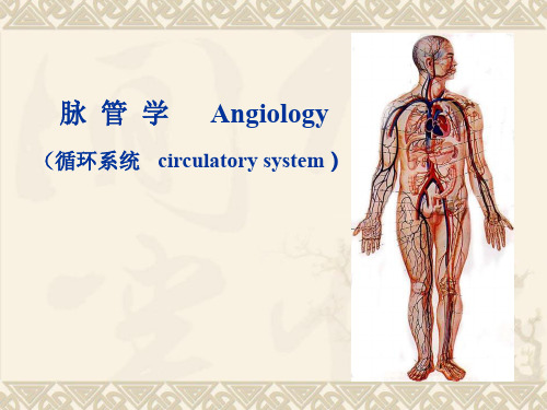
脉管学--循环系统
(一)组成 外层:Fibrous Pericardium 纤维心包 内层:Serous Pericardium 浆膜心包
脏层 心包腔 壁层 Pericardial Cavity
(二)心包窦
横窦 斜窦 前下窦
8、心的体表投影
左上点 左下点 右上点 右下点
主动脉口 二尖瓣前尖
腱索 肉柱 乳头肌
左房室口
二尖瓣复合体 Mitral Complex :二尖瓣环、二尖瓣、 腱索和乳头肌在功能和结构上密切关连,形成二尖瓣复 合体。
一入口 左房室口:左房室瓣(二尖瓣)
LV
一出口 主动脉口:主动脉瓣
心室收缩时心室各瓣膜的活动状态?
4、心的构造
(1)心纤维支架(心纤维骨骼)
• Pulmonary circulation
right ventricle→pulmonary a.→capillaries of lung→pulmonary v.→ left atrium
(二)血管吻合及其功能意义
体内血管除经动 脉、毛细血管、静脉 相连通外,动脉与动 脉之间,静脉与静脉 之间,甚至动脉与静 脉之间,可借血管吻 合支或交通支形成血 管吻合,以保证局部 血液供应或缩短循环 途径,达到调节血流 量的作用。
(三)心 Heart
1、位置 位于中纵隔内,2/3居于正中线左侧,1/3居
于右侧。
2、外形
一尖 : 心尖(LV) 一底 : 心底(LA>RA) 二面 :胸肋面(RV>LV)
膈面(LV>RV) 三缘 :下缘(RV>LV)、右缘(RA)、左缘(LV>LA) 四沟 :冠状沟、前室间沟、 后室间沟、后房间沟
各条沟代表的意义?
胸肋面 心尖切迹
Crux 房室交点 前室间支/沟 后室间支/沟
Circulatory System Artery-精品医学课件
artery and superificial palmar branch of radial artery
lie just beneath palmar aponeurosis
deep palmar arch 掌深弓 formed by terminal part of radial
artery and deep palmar branch of ulnar artery
Subclavian Artery
Axillary artery
• descends through central part of axilla and continue into brachial artery at lower border of teres major.
Brachial artery
Arteries of systemic circulation
left ventricle ascending aorta → R/L coronary artery
brachiocephalic trunk
aortic arch → left common carotid artery
left subclavian artery
lie against metacarpal bones, about 2cm proximal to superficial palmar arch
Palmar arches
Hale Waihona Puke ➢ Thoracic aorta 胸主动脉
• It extends from T4 to T12 where it passes through aortic hiatus and becomes abdominal aorta
circulatorysystem循环系统英文资料实用PPT

Lymphatic System
The lymphatic system is a network of vessels that transport lymphatic fluid throughout the body, carrying away waste and helping to fight infections. It is also involved in immune function.
PVD can lead to problems with the circulation of blood to the arms, legs, and feet. Symptoms include pain or cramping in the legs, numbness, and skin changes.
removing carbon dioxide and other waste products.
Blood Vessels
01
Types
Blood vessels come in three types: arteries, veins, and capillaries.
02 03
Function
They transport blood throughout the body, delivering essential nutrients and gases to the cells and removing waste products.
Structure
Arteries and veins are thick-walled and elastic, while capillaries are tiny, thin-walled vessels that allow for the exchange of nutrients, gases, and waste products between the blood and the surrounding tissue.
Circulatorysystem共43页文档
分布: muscle tissue,
connective tissue, nervous tissue and exocrine glands
Fenestrated capillary
结构:
1. 有大的窗孔, Closed by a diaphragm: endocrine glands or not Closed by a diaphragm: kidney (glomerulus)
动脉内的感受器
颈动脉体: 位于颈总动脉外侧壁; 对低氧、高二氧化碳和低PH质敏感的化学感受器。 主动脉体 颈动脉窦:位于颈总动脉和颈内动脉的起始膨大部分; 对血压敏感的压力感受器。
Vein Return the blood to the heart
与同级别动脉的比较
1. 官腔不规则, 因为壁薄; 官腔大 2. 结构: 内膜: 比动脉薄,没有内弹性膜 中膜: 比动脉薄,平滑肌少 外膜: 比动脉厚, 结缔组织,一些纵向分布的平滑肌细胞,
Myocardium
Epicardium
endothelium
subendothelium
Endocardium
Endocardium
Subendocardial layer
Connective tissue Myocardium
Smooth muscle
Smooth muscle
Purkinje fibers
Media of large artery
Media of large artery
营养血管
位于大血管外膜和中膜外部的动脉、静脉、 毛细血管,为大血管提供营养。
神经支配
血管壁的平滑肌由无髓鞘的交感神经支配 (血 管运动神经)。
昆虫生理学第五章循环系统ppt课件
从后向前
心脏
大动脉
血腔(头部)
围脏窦、腹血窦: 附肢
从前向后
背血窦
心门
四、昆虫血液循环的功能
1、控制血压:100~60 mm hg 2、调节体温: 3、运输:
第四节 血液的功能
一、血液的组成
又称血淋巴 hemolymph,包括血细胞和血浆。 1、血细胞
来源:胚胎期: 中胚层 特点:不输送氧;
包围着各器官。 2、血浆
具有活跃的分裂和繁殖能力,并能够转化为浆细胞等 其他血细胞。主要功能是分裂补充血细胞。 浆血细胞:
一类形态多样的吞噬细胞,主要功能是吞噬异 物, 也参与成瘤和包被作用,是重要的防卫血细胞。 粒细胞:
是一类小型的颗粒细胞,由中性黏多糖和黏蛋白 组成。主要是起到储存盒代谢功能,并参与防卫。
为了规范事业单位聘用关系,建立和 完善适 应社会 主义市 场经济 体制的 事业单 位工作 人员聘 用制度 ,保障 用人单 位和职 工的合 法权益
为了规范事业单位聘用关系,建立和 完善适 应社会 主义市 场经济 体制的 事业单 位工作 人员聘 用制度 ,保障 用人单 位和职 工的合 法权益
昆虫开放式循环系统的特点是血压低,血量大,并随 着取食和生理状态的不同,其血液的组成变化很大。其主 要功能是运输养料、激素和代谢废物,维持正常生理所需 的血压、渗透压和离子平衡,参与中间代谢,清除解离的 组织碎片,修补伤口,对侵染物产生免疫反应,以及飞行 时调节体温等。昆虫的循环系统没有运输氧的功能,氧气 由气管系统直接输入各种组织器官内,所以昆虫大量失血 后,不会危及生命安全,但可能破坏正常的生理代谢。
珠细胞: 主要由中性黏多糖和黏蛋白组成。它由粒细胞发
育而来,有储存盒分泌的作用,没有吞噬功能。 类绛色细胞:
相关主题
- 1、下载文档前请自行甄别文档内容的完整性,平台不提供额外的编辑、内容补充、找答案等附加服务。
- 2、"仅部分预览"的文档,不可在线预览部分如存在完整性等问题,可反馈申请退款(可完整预览的文档不适用该条件!)。
- 3、如文档侵犯您的权益,请联系客服反馈,我们会尽快为您处理(人工客服工作时间:9:00-18:30)。
Capillary network
Ultrastructure
of Capillary
Endothelium Pericyte
内皮细胞 (Endothelial cell): 1 -3个组成,多边形。
基膜(Basal lamina): 只有基板
周细胞(Pericyte): 有肌球蛋白、肌动蛋 白和原肌球蛋白
循环系统的组成
血液循环系统
循
Blood vascular system
环
系
统
淋巴循环系统
心脏
动脉: large artery, mediumsized artery, small artery, arteriole 毛细血管: continuous capillary, fenestrated capillary, sinusoid capillary
具有收缩和修复的功 Basal 能
lamina
Pericyte (A,B: SEM; C: TEM)
Classification of Capillary
连续性的 Continuous
有孔的 Fenestrated
血窦 Sinusoid
Continuous capillary
结构:
1. 紧密连接,完整基膜 2. 大量60-70nm的吞饮小泡 3. 无窗孔
capillary Arteriole and venule
Cardiac muscle fibers
Myocardium
Myocardium
Epicardium Small artery Small vein
Epicardium
cardiac muscle
Nerve fibers adipose
分布: muscle tissue,
connective tissue, nervous tissue and exocrine glands
Fenestrated capillary
结构:
1. 有大的窗孔, Closed by a diaphragm: endocrine glands or not Closed by a diaphragm: kidney (glomerulus)
心肌膜: 厚,心肌 心外膜:疏松结缔组织,内有冠状 valve
动脉和静脉,神经、脂肪组织;外 被覆间皮。(心包膜)
cardiac ventricle
Wall of the heart
Endothelium
Subendothelium Endocardium
Subendocardial layer (Purkinje cells)
内膜 Tunica intima
中膜Tunica media
外弹性膜External elastic lamina
结缔组织 Connective tissue
外膜Tunica adventitia
Tunica Intima
内皮: 薄, 吞饮小泡(运送物质), W-P 小体 (储存凝血因
子Ⅷ 相关抗体), 多种酶(参与多种生物活性物质合成 降解)
内皮下层: 胶原纤维,弹性纤维,平滑肌和内弹性膜。
internal elastic lamina
Tunica Media and Tunica adventitia
中膜:胶原纤维,弹性纤维,平滑肌(不同血管弹性膜不同) 外膜: 结缔组织 (纤维和成纤维细胞) 外弹性膜(有或无)
Media of medium artery
动脉内的感受器
颈动脉体: 位于颈总动脉外侧壁; 对低氧、高二氧化碳和低PH质敏感的化学感受器。 主动脉体 颈动脉窦:位于颈总动脉和颈内动脉的起始膨大部分; 对血压敏感的压力感受器。
Vein Return the blood to the heart
与同级别动脉的比较
1. 官腔不规则, 因为壁薄; 官腔大 2. 结构: 内膜: 比动脉薄,没有内弹性膜 中膜: 比动脉薄,平滑肌少 外膜: 比动脉厚, 结缔组织,一些纵向分布的平滑肌细胞,
静脉: large vein, medium-sized vein, small vein and veinule
Blood vascular system
Structural features of皮下层Subendothelium 内弹性膜Internal elastic lamina
? ?
? ?
Speak out the names please !
? ?
Speak out the name please !
?
Speak out the name please !
?
Speak out the names please !
?
?
Speak out the names please !
Connective tissue
mesothelium
Fibrous skeleton and valve
cardiac atrium Fibrous skeleton
心骨骼: 致密结缔组织,连 接心房肌和心室肌。
valve
cardiac ventricle
心脏瓣膜: 致密结缔组织的 中轴,外被覆内皮。
blood vascular system
Lymph circulates in only one direction---to the
heart
Learn it by yourself !
Speak out the names please !
? ?
Speak out the names please !
Myocardium
Epicardium
endothelium
subendothelium
Endocardium
Endocardium
Subendocardial layer
Connective tissue Myocardium
Smooth muscle
Smooth muscle
Purkinje fibers
Heart
• 肌性器官 • 节律性收缩泵血 • 分泌心房利钠肽
Wall of the heart
心内膜:
内皮 内皮下层:疏松结缔组织,内有胶
原纤维,弹性纤维和平滑肌细胞
心内膜下层:疏松结缔组织,内有血
管、神经、心脏传导系统的分支。
cardiac atrium Fibrous skeleton
Vasa vasorum
Connective tissue
Vein
4. Venous valve
Valve
Endothelium
Smoothe muscle in media
Adventitia
Collagenous fiber
Capillary
• 最小的血管,但分布最广泛 • 相互吻合成网 • 网的丰富程度与代谢率相关
Conducting system of the heart
组成:
两个节点:窦房结和房室结,内有起搏细胞 (冲动产生细胞). 房室束和位于室间隔内的左右分支,内有 Purkinje 细胞 (冲动传导细 胞)。
功能:
产生节律性刺激并 扩散到全部心肌。
控制心脏跳动
Purkinje fiber
主动脉和它大的分支
内膜: 比中动脉厚,有那弹性膜(与中膜的弹性膜 相似);内皮下层有胶原纤维和平滑肌。 中膜: 弹性纤维和大量聚集的波浪状、有窗孔的弹 性膜;平滑肌、网状纤维位于弹性膜间。 功能:稳定血流
Aldehyde-fuchsin staining
H.E staining
Elastic membrane
? ?
谢谢你的阅读
知识就是财富 丰富你的人生
Purkinje 细胞 (束细胞)
位于心室心内膜下层直到 心尖,以闰盘与工作心肌 细胞建立连接。
Lymphatic vascular system
Endothelium-lined thin-walled channels, with valves
Collect fluid from the tissue spaces (lymph) and return it to the blood---Supplement of the
Media of large artery
Media of large artery
营养血管
位于大血管外膜和中膜外部的动脉、静脉、 毛细血管,为大血管提供营养。
神经支配
血管壁的平滑肌由无髓鞘的交感神经支配 (血 管运动神经)。
Artery 1. large artery (elastic artery)
2. 吞饮小泡 3. 连续的基膜 4. 有细胞连接
分布: kidney, intestine and
some endocrine glands
Sinusoid Capillary
结构:
• 官腔较大, 血流缓慢。 • 内皮细胞不连续,间隙较大。 • 内皮细胞窗孔大小不等,无隔膜。 • 有巨噬细胞。 • 基膜不完整或缺如。
Vein
3. Large vein
Intima
MMedia
Vasa vasorum
endothelium Smooth muscle Connective tissue
adventitia
Longitudinally arranged smooth muscle cells
Collagen fiber
Small artery
Lymphatic vessel
arteriole
Small vein
