Regulated genes in mesenchymal stem cells and gastriccancer
绿色荧光蛋白转基因大鼠骨髓间充质干细胞培养及鉴定

绿色荧光蛋白转基因大鼠骨髓间充质干细胞培养及鉴定刘慧娟;胡若愚;戴王娟;刘元果;闵波;蒋犁【摘要】目的体外分离、培养和鉴定增强型绿色荧光蛋白(EGFP)转基因大鼠骨髓间充质干细胞(BMSCs).方法取EGFP转基因大鼠胫、股骨骨髓,全骨髓贴壁法分离培养、纯化BMSCs;荧光显微镜观察细胞形态;流式细胞仪分析细胞表型;CCK-8法绘制细胞生长曲线并与野生型BMSCs增殖情况相比较;分别向成脂、成骨、成软骨诱导分化鉴定;将细胞经鼠尾静脉移植入大鼠体内,观察其在体内定植情况.结果获得稳定表达EGFP的BMSCs,以长梭形为主,融合后呈漩涡状排列;生长曲线示EGFP-BMSCs增殖能力旺盛,与野生型BMSCs相比差异无统计学意义(t=-0.023,P=0.982);细胞CD29、CD90、CD34、CD49d、CD45表达率分别为99.4%、96.4%、0.171%、0.049%、0.038%;成脂、成骨、成软骨诱导后,分别给予油红O、茜素红、甲苯胺蓝染色,结果均呈阳性;在肺组织内可检测到稳定的绿色荧光.结论成功获得高纯度、稳定表达EGFP的BMSCs,干细胞特性不受EGFP 影响.细胞定植后,示踪效果良好,可用于后续实验研究.【期刊名称】《中国医学科学院学报》【年(卷),期】2016(038)001【总页数】7页(P9-15)【关键词】骨髓间充质干细胞;绿色荧光蛋白;诱导分化【作者】刘慧娟;胡若愚;戴王娟;刘元果;闵波;蒋犁【作者单位】东南大学附属中大医院儿科,南京210009;东南大学附属中大医院心胸外科,南京210009;东南大学附属中大医院儿科,南京210009;东南大学附属中大医院心胸外科,南京210009;东南大学附属中大医院心胸外科,南京210009;东南大学附属中大医院儿科,南京210009【正文语种】中文【中图分类】R394.2间充质干细胞(mesenchymal stem cells,MSCs)是来源于中胚层的一类多能干细胞,主要存在于结缔组织和器官间质中[1- 3]。
细胞培养用青霉素-链霉素产品说明书

细胞培养用青霉素-链霉素产品简介:细胞培养用青霉素-链霉素(Penicillin-Streptomycin for Cell Culture)为粉剂,是最常用的细胞培养用抗生素(即通常所谓的双抗)。
在细胞培养液中推荐的青霉素的工作浓度为100U/ml ,链霉素的工作浓度为0.1mg/ml 。
一个包装的细胞培养用青霉素-链霉素可以配制80L 细胞培养液。
保存条件:室温保存。
4ºC 保存可以使用更长时间。
注意事项:开瓶后需防止受潮。
本产品仅限于专业人员的科学研究用,不得用于临床诊断或治疗,不得用于食品或药品,不得存放于普通住宅内。
为了您的安全和健康,请穿实验服并戴一次性手套操作。
使用说明:细胞培养用青霉素-链霉素可以参考如下两种方法之一使用:1. 配制细胞培养液时加入细胞培养用青霉素-链霉素,然后再过滤除菌:配制细胞培养液时按照青霉素的工作浓度为100U/ml ,链霉素的工作浓度为0.1mg/ml 进行配制,配制完成后过滤除菌即可使用。
2. 配制青霉素-链霉素溶液(100X)母液,然后再添加到细胞培养液中:按照青霉素的含量为10KU/ml ,链霉素的含量为10mg/ml ,配制青霉素-链霉素溶液(100X)母液。
过滤除菌后即可按照100倍稀释加入到细胞培养液中使用。
配制的母液可以-20ºC 冻存。
使用本产品的文献:1. Wang PH, Gu ZH, Wan DH, Zhang MY , Weng SP, Yu XQ, He JG. The shrimp NF-κB pathway is activated by white spot syndrome virus (WSSV) 449 to facilitate the expression of WSSV069 (ie1), WSSV303 and WSSV371.PLoS One. 2011;6(9):e24773.2. Xia M, Zhu Y . Signaling pathways of ATP-induced PGE2 release inspinal cord astrocytes are EGFR transactivation-dependent. Glia. 2011 Apr;59(4):664-74. 3. He YH, Song Y , Liao XL, Wang L, Li G, A lima, Li Y , Sun CH. Thecalcium-sensing receptor affects fat accumulation via effects on antilipolytic pathways in adipose tissue of rats fed low-calcium diets. J N utr. 2011 Nov;141(11):1938-46. 4. Liu XY , Wei W, Wang CL, Yue H, Ma D, Zhu C, Ma GH, Du YGApoferritin-camouflaged Pt nanoparticles: surface effects on cellular uptake and cytotoxicity. J. Mater. Chem., 2011,21, 7105-7110. 5. Wang MN, Liu XY , Cao CB, Shi C Synthesis of band-gap tunable Cu –In –S ternary nanocrystals in aqueous solution RSC Adv. 2012 Feb; 7:2666-70. 6. Fan H, Yang L, Fu F, Xu H, Meng Q, Zhu H, Teng L, Yang M, Zhang L,Zhang Z, Liu K Cardio protective effects of salvianolic Acid a on myocardial ischemia-reperfusion injury in vivoand in vitro. Evid Based Complement Alternat Med. 2012;2012:508938. 7. Zhang J, Tang L, Shen L, Zhou S, Duan Z, Xiao L, Cao Y , Mu X, Zha L,Wang H High level of WA VE1 expression is associated with tumor aggressiveness and unfavorableprognosis of epithelial ovarian cancer. Gynecol Oncol. 2012 Oct;127(1):223-30.8. Zhang Y , Zhang Y , Chen M, Yan J, Ye Z, Zhou Y , Tan W, Lang M.Surface properties of amino-functionalized poly(ε-caprolactone) membranes and the improvement of humanmesenchymal stem cell behavior. J Colloid Interface Sci. 2012 Feb 15;368(1):64-9. 9. Jiang M, Gan L, Zhu C, Dong Y , Liu J, Gan Y . Cationic core-shellliponanoparticles for ocular gene delivery. Biomaterials. 2012 Oct; 33(30):7621-30. 10. Chai YY , Wang F, Li YL, Liu K, Xu H. Antioxidant Activities ofStilbenoids from Rheum emodi Wall. Evid Based Complement Alternat Med. 2012;2012:603678. 11. Guo S, Sun X, Cheng J, Xu H, Dan J, Shen J, Zhou Q, Zhang Y , Meng L,Cao W, Tian Y . Apoptosis of THP-1 macrophages induced by protoporphyrin IX-mediated sonodynamic therapy. Int J Nanomedicine. 2013;8:2239-46. 12. Wang S, Luo Y , Zeng S, Luo C, Yang L, Liang Z, Wang Y .Dodecanol-poly(D,L-lactic acid)-b-poly (ethylene glycol)-folate (Dol- PLA-PEG-FA) nanoparticles: evaluation of cell cytotoxicity and selecting capability in vitro. Colloids Surf B Biointerfaces. 2013 Feb 1;102:130-5.碧云天生物技术/Beyotime Biotechnology 订货热线: 400-1683301或800-8283301 订货e-mail :****************** 技术咨询: ***************** 网址: 碧云天网站 微信公众号13.Zhou S, Tang L, Wang H, Dai J, Zhang J, Shen L, Ng SW, Berkowitz RS.Overexpression of c-Abl predicts unfavorable outcome in epithelial ovarian cancer. Gynecol Oncol. 2013 Oct;131(1):69-76.14.Xia M, Zhu Y. FOXO3a Involvement in the Release of TNF-α Stimulatedby ATP in Spinal Cord Astrocytes. J Mol Neurosci. 2013 Nov;51(3):792-804.15.Zhou DH, Wang X, Yang M, Shi X, Huang W, Feng Q. Combination ofLow Concentration of (-)-Epigallocatechin Gallate (EGCG) and Curcumin Strongly Suppresses the Growth of Non-Small Cell Lung Cancer in Vitro and in Vivo through Causing Cell Cycle Arrest. Int J Mol Sci. 2013 Jun 5;14(6):12023-36.16.Jiang XY, Lu DB, Jiang YZ, Zhou LN, Cheng LQ, Chen B.PGC-1αprevents apoptosis in adipose-derived stem cells by reducing reactive oxygen species production in adiabetic microenvironment. Diabetes Res Clin Pract. 2013 Jun;100(3):368-75.17.Zhu Q, Guo T, Xia D, Li X, Zhu C, Li H, Ouyang D, Zhang J, Gan Y.Pluronic F127-modified liposome-containing tacrolimus-cyclodextrin inclusion complexes: improved solubility, cellular uptake and intestinal penetration.J Pharm Pharmacol. 2013 Aug;65(8):1107-17.18.Hu HJ, Lin XL, Liu MH, Fan XJ, Zou WW. Curcumin mediatesreversion of HGF-induced epithelial-mesenchymal transition via inhibition of c-Metexpression in DU145 cells. Oncol Lett. 2016 Feb;11(2):1499-1505.19.Sun C, Feng SB, Cao ZW, Bei JJ, Chen Q, Zhao WB, Xu XJ, Zhou Z, YuZP, Hu HY. Up-Regulated Expression of Matrix Metalloproteinases in Endothelial Cells Mediates PlateletMicrovesicle-Induced Angiogenesis.CELL PHYSIOL BIOCHEM . 2017;41(6):2319-2332.20.Wang J, Zeng H, Li H, Chen T, Wang L, Zhang K, Chen J, Wang R, Li Q,Wang S. MicroRNA-101 Inhibits Growth, Proliferation and Migration and Induces Apoptosis of BreastCancer Cells by Targeting Sex-Determining Region Y-Box 2. CELL PHYSIOL BIOCHEM .2017;43(2):717-732.21.Jiang W, Huang W, Chen Y, Zou M, Peng D, Chen D. HIV-1Transactivator Protein Induces ZO-1 and Neprilysin Dysfunction in Brain Endothelial Cellsvia the Ras Signaling Pathway. Oxid Med Cell Longev . 2017;2017:3160360.22.Li W, Yang Y, Ba Z, Li S, Chen H, Hou X, Ma L, He P, Jiang L, Li L, HeR, Zhang L, Feng D. MicroRNA-93 Regulates Hypoxia-Induced Autophagy by Targeting ULK1. Oxid Med Cell Longev .2017;2017:2709053.23.Hu SY, Zhang Y, Zhu PJ, Zhou H, Chen YD. Liraglutide directly protectscardiomyocytes against reperfusion injury possibly via modulation of intracellular calcium homeostasis. J Geriatr Cardiol . 2017 Jan;14(1):57-66.24.Li W, Wang Z, Zha L, Kong D, Liao G, Li H. HMGA2 regulatesepithelial-mesenchymal transition and the acquisition of tumor stem cellproperties through TWIST1 in gastric cancer. Oncol Rep . 2017 Jan;37(1):185-192.25.Tang J, Dong Q. Knockdown of TREM-1 suppresses IL-1β-inducedchondrocyte injury via inhibiting the NF-κB pathway. BIOCHEM BIOPH RES CO . 2017 Jan 22;482(4):1240-1245.26.Yang T, Cheng J, Yang Y, Qi W, Zhao Y, Long H, Xie R, Zhu B. S100BMediates Stemness of Ovarian Cancer Stem-Like Cells Through Inhibiting p53. Stem Cells . 2017 Feb;35(2):325-336.27.Sun K, Liu F, Wang J, Guo Z, Ji Z, Yao M. The effect of mechanicalstretch stress on the differentiation and apoptosis of human growthplate chondrocytes. IN VITRO CELL DEV-AN . 2017 Feb;53(2):141-148. 28.Peng L, Wang R, Shang J, Xiong Y, Fu Z. Peroxiredoxin 2 is associatedwith colorectal cancer progression and poor survival of patients.ONCOTARGET . 2017 Feb 28;8(9):15057-15070.29.Li L, Guan Q, Dai S, Wei W, Zhang Y. Integrin β1 Increases Stem CellSurvival and Cardiac Function after Myocardial Infarction. Front Pharmacol . 2017 Mar 17;8:135.30.Sui Y, Yao H, Li S, Jin L, Shi P, Li Z, Wang G, Lin S, Wu Y, Li Y, HuangL, Liu Q, Lin X. Delicaflavone induces autophagic cell death in lung cancer via Akt/mTOR/p70S6K signalingpathway. J MOL MED . 2017 Mar;95(3):311-322.31.Zuo S, Ge H, Li Q, Zhang X, Hu R, Hu S, Liu X, Zhang JH, Chen Y,Feng H. Artesunate Protected Blood-Brain Barrier via Sphingosine 1 Phosphate Receptor1/Phosphatidylinositol 3 Kinase Pathway After Subarachnoid Hemorrhage in Rats. Mol Neurobiol . 2017 Mar;54(2):1213-1228.32.Shen XQ, Geng YM, Liu P, Huang XY, Li SY, Liu CD, Zhou Z, Xu PP.Magnitude-dependent response of osteoblasts regulated by compressive stress. SCI REP-UK . 2017 Mar 20;7:44925.33.Yang R, Wei L, Fu QQ, You H, Yu HR. SOD3 Ameliorates Aβ25-35-Induced Oxidative Damage in SH-SY5Y Cells by Inhibiting the Mitochondrial Pathway. Cell Mol Neurobiol . 2017 Apr;37(3):513-525.34.Lin H, Zhao L, Ma X, Wang BC, Deng XY, Cui M, Chen SF, Shao ZW.Drp1 mediates compression-induced programmed necrosis of rat nucleus pulposus cells by promoting mitochondrial translocation of p53 and nuclear translocation of AIF. BIOCHEM BIOPH RES CO . 2017 May 20;487(1):181-188.35.Zhang C, Zhou G, Cai C, Li J, Chen F, Xie L, Wang W, Zhang Y, Lai X,Ma L. Human umbilical cord mesenchymal stem cells alleviate acute myocarditis by modulatingendoplasmic reticulum stress and extracellular signal regulated 1/2-mediated apoptosis. Mol Med Rep . 2017 Jun;15(6):3515-3520.36.Zhang EF, Hou ZX, Shao T, Yang WW, Hu B, Wang XX, Zhang ZX,Huang Y, Xiong LZ, Hou LC. Combined administration of a sedative dose sevoflurane and 60% oxygen reduces inflammatoryresponses to sepsis in animals and in human PMBCs. Am J Transl Res . 2017 Jun 15;9(6):3105-3119.37.Shi S, Zhong D, Xiao Y, Wang B, Wang W, Zhang F, Huang H.Syndecan-1 knockdown inhibits glioma cell proliferation and invasion by deregulating a c-src/FAK-associated signaling pathway.ONCOTARGET . 2017 Jun 20;8(25):40922-40934.38.Lin XL, Hu HJ, Liu YB, Hu XM, Fan XJ, Zou WW, Pan YQ, Zhou WQ,Peng MW, Gu CH. Allicin induces the upregulation of ABCA1 expression via PPARγ/LXRαsignaling in THP-1macrophage-derived foam cells. Int J Mol Med . 2017 Jun;39(6):1452-1460.39.Peng K, Yang L, Wang J, Ye F, Dan G, Zhao Y, Cai Y, Cui Z, Ao L, Liu J,Zou Z, Sai Y, Cao J. The Interaction of Mitochondrial Biogenesis and Fission/Fusion Mediated by PGC-1αRegulatesRotenone-Induced Dopaminergic Neurotoxicity. Mol Neurobiol . 2017 Jul;54(5):3783-3797.40.Liu Z, Zeng W, Wang S, Zhao X, Guo Y, Yu P, Yin X, Liu C, Huang T. Apotential role for the Hippo pathway protein, YAP, in controlling proliferation, cell cycleprogression, and autophagy in BCPAP and KI thyroid papillary carcinoma cells. Am J Transl Res . 2017 Jul 15;9(7):3212-3223.41.Ge Z, Diao H, Yu M, Ji X, Liu Q, Chang X, Wu Q. Connexin 43mediates changes in protein phosphorylation in HK-2 cells during chronic cadmiumexposure. ENVIRON TOXICOL CHEM . 2017 Jul;53:184-190.42.Zhu YM, Gao X, Ni Y, Li W, Kent TA, Qiao SG, Wang C, Xu XX, ZhangHL. Sevoflurane postconditioning attenuates reactive astrogliosis and glial scar formation afterischemia-reperfusion brain injury.Neuroscience . 2017 Jul 25;356:125-141.43.Wei JL, Fang M, Fu ZX, Zhang SR, Guo JB, Wang R, Lv ZB, Xiong YF.Sestrin 2 suppresses cells proliferation through AMPK/mTORC1 pathway activation in colorectalcancer. ONCOTARGET . 2017 Jul 25;8(30):49318-49328.44.Zhao L, Yang Y, Yin S, Yang T, Luo J, Xie R, Long H, Jiang L, Zhu B.CTCF promotes epithelial ovarian cancer metastasis by broadly controlling the expression of metastasis-associated genes.ONCOTARGET . 2017 Jul 10;8(37):62217-62230.45.Fu QQ, Wei L, Sierra J, Cheng JZ, Moreno-Flores MT, You H, Y u HR.Olfactory Ensheathing Cell-Conditioned Medium Reverts Aβ25-35-Induced Oxidative Damage in SH-SY5Y Cells by Modulating the Mitochondria-Mediated Apoptotic Pathway. Cell Mol Neurobiol . 2017 Aug;37(6):1043-1054.46.Wang T, Liu YP, Wang T, Xu BQ, Xu B. ROS feedback regulates themicroRNA-19-targeted inhibition of the p47phox-mediated LPS-induced inflammatory response. BIOCHEM BIOPH RES CO . 2017 Aug 5;489(4):361-368.47.Zhu Y, Wang L, Y u H, Yin F, Wang Y, Liu H, Jiang L, Qin J. In situgeneration of human brain organoids on a micropillar array. Lab Chip .2017 Aug 22;17(17):2941-2950.48.Liu C, Liu J, Hao Y, Gu Y, Yang Z, Li H, Li R.6,7,3',4'-Tetrahydroxyisoflavone improves the survival of whole-body-irradiated mice viarestoration of hematopoietic function. Int J Radiat Biol . 2017 Aug;93(8):793-802.49.Liao Q, Zhang R, Wang X, Nian W, Ke L, Ouyang W, Zhang Z. Effect offluoride exposure on mRNA expression of cav1.2 and calcium signal pathway apoptosisregulators in PC12 cells. ENVIRON TOXICOL CHEM . 2017 Sep;54:74-79.2 / 3ST488 细胞培养用青霉素-链霉素400-1683301/800-8283301碧云天/Beyotime50.Pan S, Cui Y, Dong X, Zhang T, Xing H. MicroRNA-130b attenuatesdexamethasone-induced increase of lipid accumulation in porcinepreadipocytes by suppressing PPAR-γexpression.ONCOTARGET . 2017 Sep 27;8(50):87928-87943.51.Zhong Y, Jin C, Gan J, Wang X, Shi Z, Xia X, Peng X. Apigeninattenuates patulin-induced apoptosis in HEK293 cells by modulating ROS-mediatedmitochondrial dysfunction and caspase signal pathway.Toxicon . 2017 Oct;137:106-113.52.Yin L, Huang D, Liu X, Wang Y, Liu J, Liu F, Yu B. Omentin-1 effectson mesenchymal stem cells: proliferation, apoptosis, and angiogenesis in vitro. Stem Cell Res Ther . 2017 Oct 10;8(1):224.53.Tang X, Zha L, Li H, Liao G, Huang Z, Peng X, Wang Z. Upregulationof GNL3 expression promotes colon cancer cell proliferation, migration, invasion and epithelial-mesenchymal transition via the Wnt/β-catenin signaling pathway. Oncol Rep . 2017 Oct;38(4):2023-2032.54.Wu S, Wu F, Jiang Z. Identification of hub genes, key miRNAs andpotential molecular mechanisms of colorectalcancer. Oncol Rep . 2017 Oct;38(4):2043-2050.55.Wang X, Chen G, Huang C, Tu H, Zou J, Yan J. Bone marrow stemcells-derived extracellular matrix is a promising material.ONCOTARGET . 2017 Oct 9;8(58):98336-98347.56.Sun R, Yin L, Zhang S, He L, Cheng X, Wang A, Xia H, Shi H. SimpleLight-Triggered Fluorescent Labeling of Silica Nanoparticles for Cellular ImagingApplications. Chemistry . 2017 Oct 9;23(56):13893-13896. 57.Lin H, Ma X, Wang BC, Zhao L, Liu JX, Pu FF, Hu YQ, Hu HZ, ShaoZW. Edaravone ameliorates compression-induced damage in rat nucleus pulposus cells. Life Sci . 2017 Nov 15;189:76-83.58.Sun G, Sui X, Han D, Gao J, Liu Y, Zhou L. TRIM59 promotes cellproliferation, migration and invasion in human hepatocellular carcinomacells. Pharmazie . 2017 Nov 1;72(11):674-679.59.Zhu Y, Wang L, Yin F, Yu Y, Wang Y, Shepard MJ, Zhuang Z, Qin J.Probing impaired neurogenesis in human brain organoids exposed to alcohol. INTEGR BIOL-UK . 2017 Dec 11;9(12):968-978.60.Shan Y, Wang Y, Li J, Shi H, Fan Y, Yang J, Ren W, Y u X. Biomechanicalproperties and cellular biocompatibility of 3D printed tracheal graft.Bioprocess Biosyst Eng . 2017 Dec;40(12):1813-1823.61.Liu H, Wu B, Ge Y, Huang J, Song S, Wang C, Yao J, Liu K, Li Y, Li Y,Ma X. Phosphamide-containing diphenylpyrimidine analogues (PA-DPPYs) as potent focal adhesionkinase (FAK) inhibitors with enhanced activity against pancreatic cancer cell lines. BIOORG MED CHEM LETT . 2017 Dec 15;25(24):6313-6321.62.Lin XL,Liu M,Liu Y,Hu H,Pan Y,Zou W,Fan X,Hu X. Transforminggrowth factor β1 promotes migration and invasion in HepG2 cells: Epithelial-to-mesenchymal transition via JAK/STA T3 signaling. Int J Mol Med. 2018 Jan;41(1):129-136.63.Duan C,Liu Y,Li Y,Chen H,Liu X,Chen X,Y ue J,Zhou X,Yang J.Sulfasalazine alters microglia phenotype by competing endogenous RNA effect of miR-136-5p and long non-coding RNA HOTAIR in cuprizone-induced demyelination. Biochem Pharmacol. 2018 Sep;155:110-123.64.Zhou X,Li T,Chen Y,Zhang N,Wang P,Liang Y,Long M,Liu H,Mao J,LiuQ,Sun X,Chen H. Mesenchymal stem cell-derived extracellular vesicles promote the in vitro proliferation and migration of breast cancer cells through the activation of the ERK pathway. Int J Oncol. 2019 May;54(5):1843-1852.65.Zheng W,Liu C. The cystathionine γ-lyase/hydrogen sulfide pathwaymediates the trimetazidine-induced protection of H9c2 cells against hypoxia/reoxygenation-induced apoptosis and oxidative stress. Anatol J Cardiol. 2019 Sep;22(3):102-111.66.Lou K,Huang P,Ma H,Wang X,Xu H,Wang W. Orlistat increases arsenitetolerance in THP-1 derived macrophages through the up-regulation of ABCA1. Drug Chem Toxicol. 2019 Oct 31:1-9.67.Liang S,Wang F,Bao C,Han J,Guo Y,Liu F,Zhang Y. BAG2 amelioratesendoplasmic reticulum stress-induced cell apoptosis in Mycobacterium tuberculosis-infected macrophages through selective autophagy.Autophagy. 2019 Nov 11:1-15.68.Yuan K,Lai C,Wei L,Feng T,Yang Q,Zhang T,Lan T,Yao Y,XiangG,Huang X. The Effect of Vascular Endothelial Growth Factor on Bone Marrow Mesenchymal Stem Cell Engraftment in Rat Fibrotic Liver upon Transplantation. Stem Cells Int. 2019 Dec 4;2019:5310202.Version 2021.11.04碧云天/Beyotime 400-1683301/800-8283301 ST488 细胞培养用青霉素-链霉素 3 / 3。
普利莱基因技术线粒体提取试剂盒说明书

升级版线粒体提取试剂盒C0010描述:线粒体制备试剂盒(Mitochondria Isolation Kit)用于从组织或培养细胞中分离线粒体和细胞胞浆成分。
加入分离溶液,匀浆破碎组织细胞,经过数次800g和12000g离心,在60分钟内即可分离出完整的线粒体和胞浆成分。
制备的线粒体具有很高的生物学活性,可进行各种功能研究如酶学测定,更可用于Western Blot、2D-胶、线粒体蛋白或DNA提取、蛋白质组学等研究。
严格按照说明操作,总是能制备获得高纯度线粒体。
一篇方法学研究论文发现,用普利莱试剂盒制备线粒体的得率、活性、纯度优于蔗糖密度梯度离心法和Invotrogen/Pierce线粒体提取试剂盒方法。
适用:从组织、培养细胞制备高纯度线粒体,同时分离细胞胞浆成分。
组成:Mito Solution100ml for50次制备200ml for100次制备储存:−20ºC12个月有效操作步骤:以下所有操作均在4ºC进行1.组织匀浆:100~200mg新鲜组织如肝、脑、肾、心肌等,剪为0.5cm2碎块放入小容量玻璃匀浆器内。
估计组织块总体积。
加入1.5ml冰预冷的Mito Solution。
用间隙严紧的研杵上下研磨组织20次。
培养细胞匀浆:800×g5min离心收集细胞。
单次提取需2-5×10⁷个细胞。
加入1.5ml冰预冷Mito Solution 重悬细胞,将细胞悬液转移到小容量玻璃匀浆器内,用间隙严密的研杵研磨细胞30次。
2.将匀浆液转移到离心管中,800×g,4ºC离心5min。
(胞核、膜碎片、未裂解细胞等在管底,弃去)3.收集上清液并转移到新的离心管。
再次800×g离心5min at4ºC,弃沉淀。
4.将上清液转移到新的离心管。
10,000×g离心10min4ºC。
线粒体沉淀在管底。
离心后的上清含胞浆成分,可收集用于对照实验。
碧云天 GFP抗体(小鼠单抗)说明书
碧云天生物技术/Beyotime Biotechnology 订货热线:400-168-3301或800-8283301 订货e-mail :****************** 技术咨询:***************** 网址:碧云天网站 微信公众号GFP 抗体(小鼠单抗)产品编号 产品名称包装 AG281GFP 抗体(小鼠单抗)>40次产品简介:来源 用途 抗体识别位点抗体类型 GFP 分子量MouseWB, IP, IFFull length GFP IgG2a~27kDWB, Western blot; IP, Immunoprecipitation; IF, Immunofluorescence. 本GFP 抗体(小鼠单抗),即mouse monoclonal GFP antibody ,为进口分装,用全长的来源于水母的GFP (green fluorescent protein)作为抗原制备得到的抗GFP 小鼠单克隆抗体。
克隆号为B-2。
本GFP 抗体可以识别GFP以及GFP 的一些突变体例如EGFP (enhanced green fluorescent protein)。
GFP 或其突变体EGFP 等被广泛用于基因表达效率的检测,以及和目的蛋白融合表达用于检测目的蛋白的表达和分布。
本GFP 抗体不仅可以检测GFP 或其适当的突变体,也可以检测和GFP 或其适当的突变体融合表达的蛋白。
配套提供了Western 一抗稀释液,可以用于Western 检测时的一抗稀释。
):40次。
包装清单:产品编号 产品名称包装 AG281-1 GFP 抗体(小鼠单抗) 40µl AG281-2 Western 一抗稀释液40ml —说明书1份保存条件:GFP 抗体(小鼠单抗)-20ºC 保存,Western 一抗稀释液-20ºC 或4ºC 保存,一年有效。
肿瘤环境下脂肪间充质干细胞旁分泌因子促进结肠癌细胞侵袭
肿瘤环境下脂肪间充质干细胞旁分泌因子促进结肠癌细胞侵袭陈冬梅;刘淑丹;马会明;刘晓明;梁雪云;魏军【摘要】Objective To investigate the underpinning mechanisms by which human adipose-derived mesenchymal stem cells ( hAMSCs) promote cancer metastasis remain elusive. Methods Colon cancer cells were co-cultured with AMSCs using a Transwell model. The capacity of invasion of HCT116 cells in the indicated conditions was de-tected by a Transwell invasive assay. The transcripts of EMT-associated genes in HCT116 treated with indicated conditions were determined by a qRT-PCR assay. Concentration of indicated paracrine factors and cytokines of the culture medium was ascertained by an ELISA. Results The results showed that AMSCs could promote colon canc-er cells to a mesenchymal-like phenotype in a contact-dependent manner. Reciprocally, colon cancer cells were a-ble to induce AMSCs to produce metastasis-related factors and cytokines, such as fibroblast growth factor 10 ( FGF10 ) , vascular endothelial growth factor C ( VEGFC ) , tumor necrosis factor-α( TNF-α) and interleukin 10 (IL-10) in part through a mechanism of an activation of tumor microenvironment. Intriguingly, an inhibition of contact with AMSCs led a reduced capacity of invasion of colon cancer cells in vitro. Conclusion These findings thus suggest that the crosstalk between the microenvironment of cancer cells and paracrine factors of AMSCs has an implication in colon cancer malignancy. This study thus uncovers a novel paracrine factors mediated-crosstalk be-tween colon cancer cells andAMSCs in colon cancer malignancy.%目的确定肿瘤环境下人类脂肪间充质干细胞( hAMSCs)的旁分泌特征变化与肿瘤细胞侵袭的关系. 方法体外分离培养AMSCs,收集培养上清液制备条件培养基,用于培养HCT116细胞. 利用Transwell 培养板共培养AM-SCs与HCT116 细胞. 通过检测穿透人工基底胶( Matrigel胶)的能力比较两种培养条件下HCT116细胞侵袭能力的差异. 实时荧光定量 PCR 检测 HCT116 细胞上皮间质转化( EMT)相关基因的表达变化. ELISA法检测共培养后AM-SCs基因表达和旁分泌因子表达变化. 结果结肠癌细胞系HCT116与AMSCs 的Transwell共培养系统中,HCT116细胞的侵袭能力较AMSCs条件培养液培养显著增强. 同时结肠癌细胞系HCT116构成的肿瘤微环境也影响了AMSCs旁分泌因子表达的变化,其中成纤维生长因子10 ( FGF10 )、血管内皮生长因子C( VEGFC)、肿瘤坏死因子α( TNF-α)和白细胞介素10(IL-10)的表达较正常培养条件下显著上调. 结论肿瘤微环境影响下的AMSCs旁分泌因子表达改变,这些变化能够增强结肠肿瘤细胞的侵袭能力.【期刊名称】《安徽医科大学学报》【年(卷),期】2016(051)001【总页数】6页(P36-41)【关键词】转移性;旁分泌因子;脂肪间充质干细胞;结肠癌【作者】陈冬梅;刘淑丹;马会明;刘晓明;梁雪云;魏军【作者单位】宁夏医科大学总医院干细胞研究所,银川 750004;宁夏医科大学总医院干细胞研究所,银川 750004;宁夏医科大学教育部生育力保持重点实验室,银川750004;宁夏医科大学总医院干细胞研究所,银川 750004;宁夏医科大学总医院干细胞研究所,银川 750004;宁夏医科大学总医院干细胞研究所,银川 750004【正文语种】中文【中图分类】R73-37肥胖已成为发达国家和部分发展中国家共同面对的人口健康问题。
间充质干细胞成骨分化与成脂分化关键基因的生物信息学分析
间充质干细胞成骨分化与成脂分化关键基因的生物信息学分析收稿日期:2020-08-01;修回日期:2020-09-14基金项目:国家自然科学基金资助项目(81760804)作者简介:王碧雯(1992—),女,陕西宝鸡人,硕士研究生,主要从事干细胞防治疾病与遗传稳定性研究;*通信作者:骆亚莉(1979—),女,甘肃临洮人,博士,甘肃中医药大学副教授,主要从事干细胞防治疾病与遗传稳定性研究,Tel:************,E-mail:*****************。
王碧雯a ,骆亚莉a,b*,李研a ,马玉a ,周啸天a(甘肃中医药大学a.甘肃省高校重大疾病分子医学与中医药防治研究省级重点实验室;b.基础医学院病理教研室,中国甘肃兰州730000)摘要:为探讨间充质干细胞发生成骨分化和成脂分化的潜在关联,本研究首先基于文献挖掘统计间充质干细胞成骨分化和成脂分化相关的差异表达基因,利用DAVID 在线软件对差异表达基因进行功能注释和通路富集分析,得到成骨分化GO 分类281种,KEGG 通路13条(P <0.01);成脂分化GO 分类317种,KEGG 通路47条(P <0.01)。
随后,通过STRING 数据库构建蛋白质-蛋白质相互作用网络,并将STRING 数据库得到的网络数据导入Cytoscape 软件进行分析,计算网络各节点的拓扑特性,筛选出成骨分化关键基因14个,成脂分化关键基因20个。
继而,用Venny 在线工具对成骨分化和成脂分化中的关键基因进行分析,发现7个基因(包括JUNB 、SMAD 2、SMAD 3、MAPK 13、BMP 4、SRC 、RUNX 2)共同参与调控间充质干细胞的成骨分化与成脂分化。
上述筛选出的间充质干细胞发生成骨分化与成脂分化的关键基因,可用于提示间充质干细胞发生异常成骨成脂分化的可能机制,为骨质疏松症等骨与脂肪生成平衡相关临床疾病的发病机制及治疗提供了可能的治疗靶点和新思路。
Mesenchymal stem cell
Mesenchymal stem cellFrom Wikipedia, the free encyclopediaJump to: navigation, search Mesenchymal stem cellMesenchymal stem cell showing typical ultrastructural morphology.Latin cellula mesenchymatica precursoriaCode TH H2.00.01.0.00008Mesenchymal stem cells, or MSCs, are multipotent stromal cells that can differentiate into a variety of cell types,[1] including: osteoblasts (bone cells),[2] chondrocytes (cartilage cells),[3] and adipocytes (fat cells). This phenomenon has been documented in specific cells and tissues in living animals and their counterparts growing in tissue culture.DefinitionWhile the terms Mesenchymal Stem Cell and Marrow Stromal Cell have been used interchangeably, neither term is sufficiently descriptive as discussed below:Mesenchyme is embryonic connective tissue that is derived from the mesoderm and that differentiates into hematopoietic and connective tissue, whereas MSCs do not differentiate into hematopoietic cells.[4]Stromal cells are connective tissue cells that form the supportive structure in which the functional cells of the tissue reside. While this is an accurate description for one function of MSCs, the term fails to convey the relatively recently-discovered roles of MSCs in the repair of tissue.[5] Because the cells, called MSCs by many labs today, can encompass multipotent cells derived from other non-marrow tissues, such as umbilical cord blood, adipose tissue, adult muscle, corneal stroma[6] or the dental pulp of deciduous baby teeth, yet do not have the capacity to reconstitute an entire organ, the term Multipotent Stromal Cell has been proposed as a better replacement.4.^ Porcellini Adolfo. Regenerative medicine: a review. Rev. Bras. Hematol. Hemoter. [serial on the Internet]. [cited 2011 Dec 11]. Available from: http://www.scielo.br/scielo.php?script=sci_arttext&pid=S1516-84842009000800017&lng=en. /10.1590/S1516-848420090008000175.^ "PLoS ONE: Eccentric Exercise Facilitates Mesenchymal Stem Cell Appearance in Skeletal Muscle." PLoS ONE : Accelerating the Publication of Peer-reviewed Science. Available from: </article/info:doi/10.1371/journal.pone.0029760>.6.^ Branch, M. J. et al. 2012 Mesenchymal Stem Cells in the Human Corneal Limbal Stroma /content/early/2012/06/25/iovs.11-8673.abstractThe youngest, most primitive MSCs can be obtained from the umbilical cord tissue, namely Wharton's jelly and the umbilical cord blood. However the MSCs are found in much higher concentration in the Wharton’s jelly compared to the umbilical cord blood, which is a rich source of hematopoietic stem cells. The umbilical cord is easily obtained after the birth of the newborn, is normally thrown away and poses no risk for collection. The umbilical cord MSCs have more primitive properties than other adult MSCs obtained later in life, which might make them a usefulsource of MSCs for clinical applications.An extremely rich source for mesenchymal stem cells is the developing tooth bud of the mandibular third molar. While considered multipotent, they may prove to be pluripotent. The stem cells eventually form enamel, dentin, blood vessels, dental pulp, nervous tissues, including a minimum of 29 different unique end organs. Because of extreme ease in collection at 8–10 years of age before calcification and minimal to no morbidity they will probably constitute a major source for personal banking, research and multiple therapies. These stem cells have been shown capable of producing hepatocytes. Additionally, amniotic fluid has been shown to be a very rich source of stem cells. As many as 1 in 100 cells collected from and genetic amniocentesis has been shown to be a pluripotent mesenchymal stem cell.[citation needed]Adipose tissue is one of the richest sources of MSCs. When compared to bone marrow, there is more than 500 times more stem cells in 1 gram of fat when compared to 1 gram of aspirated bone marrow. Adipose stem cells are currently actively being researched in clinical trials for treatment in a variety of diseases.HistoryIn 1924, Russian-born morphologist Alexander A. Maximow used extensive histological findings to identify a singular type of precursor cell within mesenchyme that develops into different types of blood cells.[7]Scientists Ernest A. McCulloch and James E. Till first revealed the clonal nature of marrow cells in the 1960s.[8][9] An ex vivo assay for examining the clonogenic potential of multipotent marrow cells was later reported in the 1970s by Friedenstein and colleagues.[10][11] In this assay system, stromal cells were referred to as colony-forming unit-fibroblasts (CFU-f).Subsequent experimentation revealed the plasticity of marrow cells and how their fate could be determined by environmental cues. Culturing marrow stromal cells in the presence of osteogenic stimuli such as ascorbic acid, inorganic phosphate, and dexamethasone could promote their differentiation into osteoblasts. In contrast, the addition of transforming growth factor-beta (TGF-b) could induce chondrogenic markers.[citation needed]7.^ Sell, Stewart (Stem cell handbook). Humana Press. p. 143.8.^ Becker, A. J.; McCulloch, E. A.; Till, J. E. (1963). "Cytological Demonstration of the Clonal Nature of Spleen Colonies Derived from Transplanted Mouse Marrow Cells". Nature 197 (4866): 452–4. doi:10.1038/197452a0. PMID 13970094.9.^ Siminovitch, L.; McCulloch, E. A.; Till, J. E. (1963). "The distribution of colony-forming cells among spleen colonies". Journal of Cellular and Comparative Physiology 62 (3): 327–36. doi:10.1002/jcp.1030620313. PMID 14086156.CulturingThe majority of modern culture techniques still take a colony-forming unit-fibroblasts (CFU-F) approach, where raw unpurified bone marrow or ficoll-purified bone marrow Mononuclear cell are plated directly into cell culture plates or flasks. Mesenchymal stem cells, but not red blood cells or haematopoetic progenitors, are adherent to tissue culture plastic within 24 to48 hours. However, at least one publication has identified a population of non-adherent MSCs that are not obtained by the direct-plating technique.[22]Other flow cytometry-based methods allow the sorting of bone marrow cells for specific surface markers, such as STRO-1.[23] STRO-1+ cells are generally more homogenous, and have higher rates of adherence and higher rates of proliferation, but the exact differences between STRO-1+ cells and MSCs are not clear.[24]Methods of immunodepletion using such techniques as MACS have also been used in the negative selection of MSCs.[25]22.^ Wan, Chao; He, Qiling; McCaigue, Mervyn; Marsh, David; Li, Gang (2006). "Nonadherent cell population of human marrow culture is a complementary source of mesenchymal stem cells (MSCs)". Journal of Orthopaedic Research 24 (1): 21–8. doi:10.1002/jor.20023. PMID 16419965.23.^ Gronthos, S; Graves, SE; Ohta, S; Simmons, PJ (1994). "The STRO-1+ fraction of adult human bone marrow contains the osteogenic precursors". Blood 84 (12): 4164–73. PMID 7994030. /cgi/pmidlookup?view=long&pmid=7994030.24.^ Oyajobi, Babatunde O.; Lomri, Abderrahim; Hott, Monique; Marie, Pierre J. (1999). "Isolation and Characterization of Human Clonogenic Osteoblast Progenitors Immunoselected from Fetal Bone Marrow Stroma Using STRO-1 Monoclonal Antibody". Journal of Bone and Mineral Research 14 (3): 351–61. doi:10.1359/jbmr.1999.14.3.351. PMID 10027900.25.^ Tondreau, T; Lagneaux, L; Dejeneffe, M; Delforge, A; Massy, M; Mortier, C; Bron, D (1 January 2004). "Isolation of BM mesenchymal stem cells by plastic adhesion or negative selection: phenotype, proliferation kinetics and differentiation potential". Cytotherapy 6 (4): 372–379. doi:10.1080/14653240410004943.。
碧云天生物技术SMT (iNOS抑制剂) 产品说明书

碧云天生物技术/Beyotime Biotechnology订货热线:400-168-3301或800-8283301订货e-mail:******************技术咨询:*****************网址:碧云天网站微信公众号SMT (iNOS抑制剂)产品编号产品名称包装S0008 SMT (iNOS抑制剂) 100mg产品简介:SMT,即S-Methylisothiourea Sulfate,也称2-Methyl-2-thiopseudourea, Sulfate,或S-Methyl-ITU,是iNOS (inducible nitric oxide synthase)高度选择性抑制剂。
对于体外培养巨噬细胞诱导产生的iNOS,EC50=6µM;对于血管平滑肌细胞被诱导产生的iNOS,EC50=2µM。
SMT为白色结晶,分子量278.4,分子式为(C2H6N2S)2·H2SO4,纯度大于99%。
溶解于水;用1M盐酸可以配制成25mg/ml的无色透明溶液。
包装清单:产品编号产品名称包装S0008 SMT (iNOS抑制剂) 100mg—说明书1份保存条件:室温保存,两年有效。
注意事项:如果配制成水溶液,分装后-20ºC保存,半年有效。
本产品仅限于专业人员的科学研究用,不得用于临床诊断或治疗,不得用于食品或药品,不得存放于普通住宅内。
为了您的安全和健康,请穿实验服并戴一次性手套操作。
使用说明:SMT的工作浓度通常为0.1-1mM。
其最佳工作浓度需根据具体的实验,自行摸索。
可以先分别尝试0.1、0.3和1mM这三个浓度。
使用本产品的文献:1.Zhang F, Liao L, Ju Y, Song A, Liu Y. Neurochemical plasticity of nitricoxide synthase isoforms in neurogenic detrusor overactivityafter spinal cord injury. Neurochem Res. 2011 Oct;36(10):1903-9.2.Li W, Ren G, Huang Y, Su J, Han Y, Li J, Chen X, Cao K, Chen Q, ShouP, Zhang L, Yuan ZR, Roberts AI, Shi S, Le AD, Shi Y. Mesenchymal stem cells: a double-edged sword in regulating immune responses. Cell Death Differ.2012 Sep;19(9):1505-13.3.Xu J, Jin DQ, Zhao P, Song X, Sun Z, Guo Y, Zhang L. Sesquiterpenesinhibiting NO production from Celastrus orbiculatus. Fitoterapia.2012 Dec;83(8):1302-5.4.Mao YF, Zhang YL, Yu QH, Jiang YH, Wang XW, Yao Y, Huang JL.Chronic restraint stress aggravated arthritic joint swell of rats through regulating nitric oxide production. Nitric Oxide. 2012 Oct 15;27(3):137-42.5.Jiang Q, Zhou Z, Wang L, Shi X, Wang J, Yue F, Yi Q, Yang C, Song L.The immunomodulation of inducible nitric oxide in scallop Chlamys farreri. Fish Shellfish Immunol. 2013 Jan;34(1):100-8.6.Yan K, Zhang R, Chen L, Chen F, Liu Y, Peng L, Sun H, Huang W, SunC, Lv B, Li F, Cai Y, Tang Y, Zou Y, Du M, Qin L, Zhang H, Jiang X.Nitric oxide-mediated immunosuppressive effect of human amniotic membrane-derived mesenchymal stem cells on the viability and migration of microglia. Brain Res. 2014 Nov 24;1590:1-9.7.Sun Z, Jiang Q, Wang L, Zhou Z, Wang M, Yi Q, Song L. Thecomparative proteomics analysis revealed the modulation of induciblenitric oxide on the immune response of scallop Chlamys farreri. Fish Shellfish Immunol. 2014 Oct;40(2):584-94.8.Li Y, Ma C, Shi X, Wen Z, Li D, Sun M, Ding H. Effect of nitric oxidesynthase on multiple drug resistance is related to Wnt signaling in non-small cell lung cancer. Oncol Rep. 2014 Oct;32(4):1703-8.9.Wu C, Zhao W, Zhang X, Chen X. Neocryptotanshinone inhibitslipopolysaccharide-induced inflammation in RAW264.7 macrophages by suppression of NF-κB and iNOS signaling pathways. Acta Pharm Sin B.2015 Jul;5(4):323-9.10.Han Y, Jiang Q, Gao H, Fan J, Wang Z, Zhong F, Zheng Y, Gong Z,Wang C. The anti-apoptotic effect of polypeptide from Chlamys farreri (PCF) in UVB-exposed HaCaT cells involves inhibition of iNOS and TGF-β1. Cell Biochem Biophys. 2015 Mar;71(2):1105-15.11.Su Z, Ye J, Qin Z, Ding X. Protective effects of madecassoside againstDoxorubicin induced nephrotoxicity in vivo and in vitro. Sci Rep. 2015 Dec 14;5:18314.12.Wu B, Geng S, Bi Y, Liu H, Hu Y, Li X, Zhang Y, Zhou X, Zheng G, HeB, Wang B. Herpes Simplex Virus 1 Suppresses the Function of Lung Dendritic Cells via Caveolin-1. Clin Vaccine Immunol. 2015 Aug;22(8):883-95.13.Li S, Chen S, Yang W, Liao L, Li S, Li J, Zheng Y, Zhu D. Allicin relaxesisolated mesenteric arteries through activation of PKA-KATP channel in rat. J Recept Signal Transduct Res. 2017 Feb;37(1):17-24.Version 2017.03.08。
山羊血清产品说明书

山羊血清产品编号 产品名称 包装 C0265山羊血清50ml产品简介:本山羊血清(Goat Serum)是碧云天自产的血清,可以用于细胞培养、免疫荧光和免疫组化时的封闭等。
本山羊血清采自健康山羊,经无菌采集、批量混合,最终过3次0.1μm 过滤分装而成。
所有操作符合2010版GMP (GoodManufacturing Practices for Drug)生产标准。
本产品无细菌、真菌、支原体及病毒污染,具有高质量和高稳定性。
本山羊血清为纯天然制品,不含任何人为的添加成分,适用于科研或诊断试剂生产用。
可以替代FBS 用于大多数细胞系和原代细胞培养,也可用于病毒的培养和研究、鱼类细胞的培养、免疫反应中的封闭和稀释液的制备。
不同血清产品的比较、选择和使用技巧,请参考/support/serum.htm 。
本产品通过了碧云天的细胞培养和免疫染色(包括免疫荧光和免疫组化)封闭效果测试。
包装清单:产品编号 产品名称 包装 C0265山羊血清 50ml —说明书1份保存条件:-15~-40ºC 保存,5年有效;4ºC 保存通常不宜超过1个月。
注意事项:如果不能短期内使用完毕,解冻后请适当分装。
血清结冰时体积会增加约10%,因此在分装血清时须使分装瓶预留一定体积空间,否则易导致分装瓶冻裂而发生污染。
热灭活是指56ºC ,30分钟加热已完全解冻的血清。
加热过程中需有规则地摇晃均匀。
热处理的目的是灭活血清中的补体 (complement)。
除非必须,一般不推荐对血清进行热处理。
因为热处理会造成血清沉淀物显著增多,而且还会影响血清的质量。
补体参与的反应有:细胞毒作用、平滑肌细胞收缩、肥大细胞和血小板释放组胺、增强吞噬作用、促进淋巴细胞和巨噬细胞发生化学趋化和活化等。
瓶装血清解冻需采用缓慢解冻法:把在-15~-40ºC 低温冰箱中保存的血清放入4ºC 冰箱中溶解约半天至一天,待全部解冻后即可使用。
新型肠道间质细胞MRISC调控炎症过程中肠道干细胞的损伤修复

得理学学士学位。 1987、1991年在美国耶鲁大学分别获得硕士和博
士学位。 1991 一 1995年 在 美 国 加 州 大 学 圣 地 亚 哥 分 校 从 事 博 士 后 研 究工作。 1995— 2006年 在 美 国 德 克 萨 斯 大 学 /M D 安 德 森 癌 症 中 心 任 助理教授、 副教授、 教 授 。2007— 2014年 在 美 国 耶 鲁 大 学 医 学 院 免 疫生物学/血 管 生 物 学 /移 植 研 究 系 任 副 教 授 。2012年 加 入 上 海 交 通
environment, plays an essential regulatory role in this process.With the advancement o f single cell sequencing
technologies,researchers revealed intestinal mesenchymal stromal cells as a complex and highly heterogeneous cell
关 键 词 肠 道 间 质 细 胞 ; 肠 道 干 细 胞 微 环 境 ;M RISC; R-spondinl
国家自然科学基金(批准兮:320丨]01丨.52、91942311、31930035)、上 海 市 科 技 术 委 M会(批准‘4: 20410714000、20JCI4丨0 丨00>和癌基因与相关基因国家重
population.Currently,the gene signature,spatial distribution,potential function, cellular and molecular regulatory
mechanisms o f these distinctive stromal cell populations are still poorly understood.This paper revisited the course
间充质干细胞条件培养基对HPV18型阳性人子宫颈癌HeLa细胞凋亡的影响
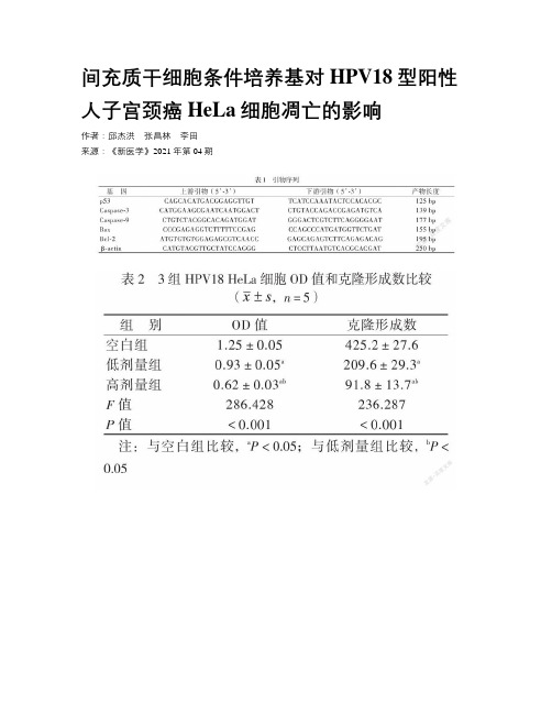
间充质干细胞条件培养基对HPV18型阳性人子宫颈癌HeLa细胞凋亡的影响作者:邱杰洪张昌林李田来源:《新医学》2021年第04期【摘要】目的观察人脐带间充质干细胞条件培养基(MSC-CM)对HPV18型阳性人子宫颈癌HeLa细胞(HPV18 HeLa细胞)凋亡相关基因的調控作用,以及对HPV18 HeLa细胞凋亡的影响。
方法制备MSC-CM,设置空白组、低剂量组和高剂量组,配制相应浓度分别为0%、20%和60%的MSC-CM处理HPV18 HeLa细胞,使用CCK-8法、克隆形成实验检测细胞活力和克隆形成能力;磷脂结合蛋白Ⅴ-异硫氰酸荧光素/碘化丙啶双染法凋亡染色和流式细胞术检测细胞凋亡的变化;实时荧光定量PCR检测凋亡相关基因mRNA表达量。
结果与空白组相比,在低剂量组或高剂量组的MSC-CM作用下,HPV18 HeLa细胞的生长活性和克隆形成能力均降低,细胞凋亡率升高(P均< 0.05),且高剂量组作用效果均比低剂量组明显(P均<0.05)。
与空白组相比,HPV18 HeLa细胞在低剂量或高剂量组MSC-CM的作用下,HPV18 HeLa细胞中p53、胱天蛋白酶-3(Caspase-3)、Caspase-9、Bax的mRNA表达量升高,而B 淋巴细胞瘤-2(Bcl-2)的mRNA表达量降低(P均< 0.05),高剂量组比低剂量组的作用效果更明显(P < 0.05)。
结论 MSC-CM能诱发HPV18 HeLa细胞凋亡,可能与p53、Caspase-3、Caspase-9、Bax以及Bcl-2等基因表达差异相关。
【关键词】人乳头瘤病毒18型;宫颈癌;HeLa细胞;间充质干细胞;细胞凋亡Effect of umbilical cord-derived mesenchymal stem cell-conditioned medium on apoptosis of HPV18-infected HeLa cell lines Qiu Jiehong, Zhang Changlin, Li Tian. Department ofGynecology and Pelvic Floor Disorders Center, the Seven Affiliated Hospital of Sun Yat-sen University, Shenzhen 518107, ChinaCorresponding author, Li Tian, E-mail:*******************【Abstract】 Objective To evaluate the effect of umbilical cord-derived mesenchymal stem cell-conditioned medium (MSC-CM) on regulating the apoptosis-related genes and determine the impact upon the apoptosis of human papillomavirus type18-infected HeLa cell lines. Methods Primary cervical cancer HeLa cells were divided into the blank, low-dose and high-dose groups. HeLa cells were cultured and treated with 0%, 20% and 60% of MSC-CM in three groups, respectively. The cell viability and clone formation ability of HeLa cells were detected by CCK-8 assay and clone formation assay. The apoptosis of HeLa cells was detected by Annexin V-Fluorescein isothiocyanate/Propidium iodide (V-FITC/PI) staining and flow cytometry. The expression levels of p53, Bax, Caspase-3, Caspase-9 and Bcl-2 mRNA in HeLa cells were quantitatively measured by RT-PCR. Results In the low- and high-dose groups, the cell viability and clone formation ability were significantly decreased, whereas the apoptosis rate was significantly elevated compared with those in the blank group (all P < 0.05). The effects in the high-dose group were more evident than those in the low-dose group (all P < 0.05). The expression levels of p53, Bax, Caspase-3 and Caspase-9 mRNA in the low- and high-dose groups were significantly up-regulated, whereas that of Bcl-2 mRNA was significantly down-regulated compared with those in the blank group (all P <0.05). The effects in the high-dose group were more pronounced than those in the low-dose group (all P < 0.05). Conclusions Under the effect of MSC-CM, the apoptosis of HPV18 cervical HeLa cells can be induced, probably associated with the differential expression patterns of p53, Bax,Bcl-2, Caspase-3 and Caspase-9 mRNA.【Key words】 Human papillomavirus type18;Cervical cancer;HeLa cell;Mesenchymal stem cells;Apoptosis人乳头瘤病毒(HPV)是一种闭合双链DNA病毒[1]。
骨碎补活性成分促进骨缺损后骨重建的机制及其组织工程学应用

骨碎补活性成分促进骨缺损后骨重建的机制及其组织工程学应用Δ邓志军1*,杨文龙2,杨智军1,赵斌1,李典1,杨凤云2 #(1.江西中医药大学研究生院,南昌 330004;2.江西中医药大学附属医院骨科,南昌 330006)中图分类号 R965;R318文献标志码 A 文章编号 1001-0408(2024)08-1023-06DOI 10.6039/j.issn.1001-0408.2024.08.22摘要骨缺损的治疗难度大、周期长,一直都是骨科临床面临的重大挑战。
骨碎补是我国中医骨伤科的常用药材,其活性成分(主要为黄酮类)可促进骨髓间充质干细胞成骨分化、成骨细胞增殖、成血管-成骨耦联,抑制破骨细胞活力,从而促进骨缺损区的骨质矿化和修复重建。
骨碎补活性成分是骨再生治疗药物的良好替代品,将其负载于组织工程支架材料上,可极大地提高药物的生物利用度。
同时,缓释微球进一步解决了支架药物的突发性释放等问题,应用其所制备的含骨碎补活性成分的复合支架具有较好的成骨活性和骨诱导性,骨修复效果确切,可满足骨移植物的多样化性能要求,临床应用前景广阔。
关键词骨碎补;活性成分;总黄酮;骨缺损;骨组织工程Mechanism and application in tissue engineering of the active ingredient of Drynariae Rhizoma promoting bone defect repairDENG Zhijun1,YANG Wenlong2,YANG Zhijun1,ZHAO Bin1,LI Dian1,YANG Fengyun2(1. Graduate School,Jiangxi University of Chinese Medicine,Nanchang 330004,China;2. Dept. of Orthopedics,the Affiliated Hospital of Jiangxi University of Chinese Medicine, Nanchang 330006, China)ABSTRACT Bone defect has always been a major clinical challenge because of its great difficulty and long period of treatment. Drynariae Rhizoma is a commonly used medicine in osteology and traumatology of traditional Chinese medicine,and its active ingredients(mainly flavonoids) facilitate osteoblast differentiation of bone marrow mesenchymal stem cells, osteoclast proliferation,vascular-osteogenic coupling,and inhibit osteoclast activity to promote bone mineralization,and repair and reconstruction of bone defect. As a good substitute for bone regeneration drugs,the active constituents of Drynariae Rhizoma can be loaded on scaffold materials of tissue engineering,which greatly improves the bioavailability of the drug. Meanwhile,the sustained-release microspheres also solve some problems such as sudden drug release from the scaffolds,and the composite scaffolds with active ingredient of Drynariae Rhizoma prepared by them have good ossification activity and osteoinduction,with precise bone repair effects, which meet the diverse performance requirements of bone grafts and have a promising clinical application prospect. KEYWORDS Drynariae Rhizoma; active ingredient; total flavonoids; bone defect; bone tissue engineering随着交通业和工业的迅速发展,创伤显现出高能、多发、复杂的特点,尤其是高能量创伤导致的急性骨丢失、复杂骨折,使得骨缺损的发生率大幅度升高[1]。
【论文】辛伐他汀对骨合成代谢影响及机制进展

*国家自然科学基金资助项目(编号:81170998)△通信作者。
E -mail :249200195@qq.com辛伐他汀对骨合成代谢的影响及机制的研究进展*王忠磊1,邓悦1,岳新新2,高岩2,赖春花2,周磊2,田芳华3△1山东省青岛市口腔医院口腔颌面外科(266003);2南方医科大学附属口腔医院种植科(广州510280);3山东省临沂市人民医院口腔科(276000)他汀类(statins )药物是最近20年治疗高胆固醇血症的新药,因为该药疗效显著,不良反应小,耐受性好,受到临床应用的重视和好评。
辛伐他汀是甲基羟戊二酰辅酶A 还原酶的特异性抑制剂,它是一种无活性的内脂类药物,通过抑制胆固醇合成的关键性限速酶甲基羟戊二酰辅酶A (HMG -CoA )还原酶,从而阻断肝细胞的内源性胆固醇合成。
辛伐他汀作为他汀类药物的代表,是一种能够降低血清胆固醇和三酰甘油的有效药物。
辛伐他汀在调脂过程中还能提高骨密度,促进骨生长。
近年来发现,以辛伐他汀为主的他汀类药物可促进成骨细胞增殖,骨形成增加,将不同载体携带的辛伐他汀局部应用于各种不同的动物模型可产生不同的促骨形成效果,但其确切的机制尚不清楚。
医学界开始高度关注该药在骨合成代谢方面的应用研究。
本文就辛伐他汀的特性及对骨合成代谢所产生的影响和机制等作一综述。
1骨合成代谢的相关机制1.1促进骨形成蛋白(BMP )的表达BMP 由骨基质产生,通过Smad 蛋白信号转导途径将成骨信号转导至骨髓间充质干细胞,诱导其向成骨、成软骨方向分化,并在骨和软骨的发育成熟过程中起重要作用[1-4];BMP 还与多种骨病(如骨折、骨质疏松、骨肿瘤)有密切关系。
1.2人甲状旁腺素(hPTH )促骨合成hPTH 的主要生理功能是调节钙磷代谢、促进维生素D 代谢、激活骨细胞,是调节钙、磷代谢及骨转换的重要肽类激素之一,能精确调节骨的合成及分解代谢过程。
hPTH 片段目前已成为重要的骨形成促进剂,与受体结合后通过活化cAMP 依赖的蛋白激酶A 及钙离子依赖的蛋白激酶C 信号转导途径发挥生物学作用。
抗坏血酸作用下兔骨髓间充质干细胞外基质分泌与结构改变

抗坏血酸作用下兔骨髓间充质干细胞外基质分泌与结构改变姚植业;刘玉梅;陈燕玲;陈亮;何少茹;张展松【摘要】BACKGROUND: The effect of extracellular matrix on stem cells is the focus of tissue engineering. However, there are few reports about the synthesis and secretion of extracellular matrix as well as its effects on cells. OBJECTIVE: To isolate, culture and identify rabbit bone marrow mesenchymal stem cells (BMSCs), and to explore the changes of extracellular matrix and whole structure under the intervention of ascorbic acid. METHODS: Rabbit BMSCs were isolated by differential adherent method of the bone marrow, and the expression of CD44, CD45 and CD31 was identified by flow cytometry. The BMSCs were cultured in the culture medium containing 20 mg/L ascorbic acid. Then the cell morphology, gross structure, ultrastructure, and histological changes of BMSCs were observed. The expression of extracellular matrix related genes was detected by RT-PCR. RESULTS AND CONCLUSION: Over 95% passage 2 BMSCs could express CD44, but the expression levels of CD45 and CD31 were extremely low. Intervention with ascorbic acid enhanced the proliferation of BMMSCs with unclear cell boundaries. A cell-sheet structure formed at 10-14 days after intervention. Hematoxylin-eosin staining results showed a layered cell arrangement, and Masson staining findings showed a large amount of extracellular matrix composition. Abundant endoplasmic reticula and vesicle-like structure were observed under the transmission electron microscope. RT-PCR findings showed thatascorbic acid significantly increased the expression of fibronectin mRNA in the BMSCs (P < 0.05), but slightly increased the mRNA expression of collagen type I. All these findings indicate that ascorbic acid not only increases the proliferation and transformation of rabbit BMSCs, but also promotes the synthesis and secretion of extracellular matrix, which has great potential in tissue engineering applications.%背景:细胞外基质对干细胞的影响是目前组织工程学的研究热点,而关于自身细胞外基质的合成、分泌及对细胞的影响却少有研究报道.目的:体外培养兔骨髓间充质干细胞,观察抗坏血酸对其细胞外基质与整体结构改变的影响.方法:通过差速贴壁法分离获取兔骨髓间充质干细胞,流式细胞技术检测CD44、CD45、CD31的表达.将兔骨髓间充质干细胞用含抗坏血酸(20 mg/L)的培养基进行干预,观察细胞形态、大体结构、超微结构及组织学改变,RT-PCR检测细胞外基质相关基因的表达.结果与结论:①经流式细胞学分析,第2代骨髓间充质干细胞中95%以上的细胞表达CD44,而CD45与CD31的表达极低;②抗坏血酸干预后,骨髓间充质干细胞增殖良好,细胞间界限难以分辨,干预10-14 d后,形成了"膜片状"结构;③苏木精-伊红染色后可见细胞分层排列,Masson 染色后可见大量细胞外基质成分,透射电镜下可见胞内含大量内质网与"囊泡样"结构;④与未经抗坏血酸干预的对照组相比较,经抗坏血酸干预的实验组纤维连接蛋白的mRNA表达显著升高(P < 0.05),而Ⅰ型胶原的mRNA表达虽稍提高,但两组间差异无显著性意义;⑤结果表明,抗坏血酸促进了兔骨髓间充质干细胞的增殖转化、细胞外基质的合成分泌、膜片状结构的形成,在组织工程应用中具有巨大潜能.【期刊名称】《中国组织工程研究》【年(卷),期】2018(022)009【总页数】7页(P1325-1331)【关键词】抗坏血酸;干细胞;骨髓间充质干细胞;细胞增殖;细胞接触抑制;细胞转化;细胞外基质;纤连蛋白;Ⅰ型胶原;细胞外基质稳态;国家自然科学基金【作者】姚植业;刘玉梅;陈燕玲;陈亮;何少茹;张展松【作者单位】汕头大学医学院,广东省汕头市 515041;广东省人民医院/广东省医学科学院,广东省广州市 510080;广东省人民医院/广东省医学科学院,广东省广州市510080;广东省人民医院/广东省医学科学院,广东省广州市 510080;广东省人民医院/广东省医学科学院,广东省广州市 510080;广东省人民医院/广东省医学科学院,广东省广州市 510080;广东省心血管病研究所,广东省广州市 510080【正文语种】中文【中图分类】R394.20 引言 Introduction骨髓间充质干细胞(bone marrow mesenchymal stem cells,BMSCs)在体外表现出活跃的增殖和分化能力,是组织工程种子细胞研究的热点[1]。
组蛋白赖氨酸甲基转移酶在糖尿病中的研究进展
组蛋白赖氨酸甲基转移酶在糖尿病中的研究进展包志伟; 杨天竹; 刘洋; 王志刚【期刊名称】《《中国医药导报》》【年(卷),期】2019(016)025【总页数】4页(P45-48)【关键词】组蛋白; 组蛋白甲基化; 赖氨酸甲基转移酶; 糖尿病【作者】包志伟; 杨天竹; 刘洋; 王志刚【作者单位】哈尔滨医科大学(大庆)医学检验与技术学院生物化学教研室黑龙江大庆 163319【正文语种】中文【中图分类】R587.1糖尿病是在遗传因素与环境因素相互作用下,以胰岛素的分泌和利用异常引发的高血糖为特征的代谢性疾病,目前尚无根治方法。
糖尿病分为两型,1 型糖尿病主要与免疫系统缺陷和遗传因素有关,2 型糖尿病主要与胰岛素抵抗和肥胖有关,在我国糖尿病患者中有90%是2 型糖尿病患者。
胰岛素的分泌和利用是糖尿病发生发展的关键因素,胰岛素分泌和发挥功能的过程又受到许多因素的调节,如胰岛素受体和胰岛素样生长因子1 的分子含量变化影响胰岛素亲和能力[1]。
许多组蛋白修饰酶在代谢疾病中起着至关重要的作用,包括糖尿病及其并发症。
组蛋白甲基化调节酶在代谢疾病中的作用也引起了人们的关注,多种组蛋白赖氨酸甲基转移酶与糖尿病及其并发症的发生发展有关,因此本文对组蛋白赖氨酸甲基转移酶在糖尿病及其并发症中的研究进展作一简要综述。
1 影响组蛋白甲基化的相关因素组蛋白甲基化是表观遗传学中组蛋白修饰的主要形式之一。
组蛋白甲基转移酶和组蛋白脱甲基酶共同完成组蛋白甲基化修饰这一动态过程。
组蛋白的甲基化主要发生在H3 和H4 的赖氨酸(lysine,K)和精氨酸(arginine,R)上,组蛋白赖氨酸甲基转移酶主要由SET(Su var 3-9,E z,Trithorax)结构域家族和非SET 结构域家族的甲基转移酶完成。
目前,对SET 结构域家族研究相对充分。
H3K4、H3K36、H3K79 的甲基化主要参与基因激活,而H3K9、H3K20、H3K27 的甲基化主要参与基因沉默。
成骨细胞特异性转录因子Osterix对骨形成作用的分子机制
成骨细胞特异性转录因子Osterix对骨形成作用的分子机制张驰【期刊名称】《北京大学学报(医学版)》【年(卷),期】2012(044)005【摘要】Bone formation is a complex developmental process involving the differentiation of mesenchymal stem cells to osteoblasts.Osteoblast commitment and differentiation are controlled through a multistep molecular pathway regulated by different transcription factors and signaling proteins,including Indian hedgehog,Runx2,Osterix (Osx),and Wnt pathway.Osx is an osteoblast-specific transcription factor required for bone formation.Osx was first discovered as a bone morphogenetic protein-2 inducible gene in mesenchymal stem cells.Osx knock-out mice lack bone completely,and cartilage is normal.This opens a new window to the whole field of how bone forms. The discovery that Osx inhibits Wnt pathway highlights the potential for novel feedback control mechanisms involved in bone formation.Several downstream targets of Osx during bone formation have been identified,including Satb2,vitamin D receptor and vascular endothelial growth factor as well as Dkk(1) and Sost.The delineation of the cascade of events leading to bone formation should provide a molecular basis for the development of new and specific anabolic therapeutic agents for bone deficit conditions,such as osteoporosis and osteonecrosis.This review summarizes the recentadvances in understanding the molecular mechanisms of Osx effect on bone formation.Studies since the Osx discovery have provided convincing evidences to demonstrate that Osx is the master gene that controls osteoblast lineage commitment and the subsequent osteoblast differentiation and proliferation.%骨形成是一个涉及到从间质干细胞向成骨细胞分化的复杂发育过程.成骨细胞定向分化受到一个多步骤的分子通路控制,通过不同的转录因子和信号蛋白调控,包括Indian hedgehog,Runx2,Osterix (Osx),以及Wnt 信号通路.Osx是骨形成所必需的成骨细胞特异性转录因子,Osx最早是在间质干细胞被骨形成蛋白-2诱导向成骨细胞分化过程中发现的基因.Osx基因敲除小鼠完全缺乏骨,而软骨是正常的,这为研究骨的形成打开了一个全新的窗口.Osx抑制Wnt信号通路的发现揭示了参与骨形成的一种新的反馈调控机制.骨形成过程中Osx下游的靶目标已经确定一些,包括Satb2、维生素D受体和血管内皮生长因子,以及Dkkl和Sost.骨形成过程一系列信号传导因子的揭示、阐明应当为一些新的特异性合成代谢治疗药物的研究开发提供分子理论基础,以便治疗骨缺失疾病(如骨质疏松症和骨坏死).本文综述了Osx在骨形成作用分子机制中的最新进展,自Osx 发现以来,各种研究证据表明Osx是决定成骨细胞定向的主导因子,继而调控成骨细胞的分化和增殖.【总页数】7页(P659-665)【作者】张驰【作者单位】Bone Research Laboratory,Texas Scottish Rite Hospital for Children,University of Texas Southwestern Medical Center Dallas,TX 75219,USA【正文语种】中文【中图分类】R336【相关文献】1.Osterix:与成骨细胞分化和骨形成有关的转录因子 [J], 乔建瓯;宁光;王铸钢2.成骨细胞特异性转录因子 Osterix 对骨形成作用的研究进展 [J], 吴添龙;程细高(审校)3.成骨细胞特异性转录因子Osterix的研究进展 [J], 徐鹏程;邱明才;王宝利4.成骨细胞分化、骨形成与修复中转录因子Osx和Satb2的调控作用 [J], 侯秋科;黄永铨;李昀骏;陈东风5.转录因子Osterix对成骨细胞Col1a1基因的反式激活作用 [J], 潘秋辉;马纪;于永春;孙奋勇因版权原因,仅展示原文概要,查看原文内容请购买。
干细胞离子通道的相关性研究

干细胞离子通道的相关性研究叶恭杰【摘要】离子通道在干细胞兴奋和兴奋传导上扮演重要角色.研究表明,离子通道在调控细胞有丝分裂、细胞分化和细胞周期进程中起重要作用.深入研究干细胞离子通道,了解其特性,将有望调控干细胞的某些功能,从而治疗一些用常规方法无法治愈的疾病.现就最近发现的干细胞离子通道在细胞增殖、分化调控中所起的作用予以综述.%Ion channels in stem cells play crucial roles in excitation genesis and impulse conduction in excitable cells. Recent studies have demonstrated that bioelectric properties can control cell mitotic activity, cell cycle progression and differentiation. The ability to control cell functions by modulating bioelectric properties would be an invaluable tool for directing stem cell behavior toward therapeutic goals. Here is to make a review focusing on the roles of recently found ion channels in regulation of cell proliferation and differentiation in stem cells.【期刊名称】《医学综述》【年(卷),期】2012(018)006【总页数】3页(P825-827)【关键词】干细胞;离子通道;增殖;分化【作者】叶恭杰【作者单位】宁波大学附属李惠利医院心血管内科,浙江,宁波,315041【正文语种】中文【中图分类】R541干细胞或祖细胞移植治疗冠状动脉粥样硬化性心脏病(简称冠心病),能明显改善心功能[1-2]。
肿瘤干细胞与EMT

肿瘤干细胞与EMT肿瘤干细胞(cancer stem cell , CSC学说认为,肿瘤实际上是由一小群具有无限增殖潜能和自我更新能力的干细胞样细胞及其产生的分化程度不均一的细胞团组成,其中具有自我更新能力并能产生异质性肿瘤细胞的细胞被称为肿瘤干细胞。
肿瘤干细胞的两个重要特性一是具有自我更新驱动肿瘤发生的能力,二是具有多向分化形成肿瘤的异质性的潜能1。
上皮间质转化(epithelial-to-mesenchymal transition ,EMT是具有极性的上皮细胞转化为具有移行能力的间质细胞,并获得侵袭和迁移能力的过程。
EMT是一个多步骤的动态变化过程,上皮细胞间相互作用消失,组织结构松散,立方上皮细胞转变为纺锤形纤维细胞形态,并表现出侵袭性。
实体肿瘤中央的细胞为上皮细胞表型,周围的细胞常常会呈间质细胞表型,其较强的运动能力使肿瘤细胞在局部产生浸润,并侵入血和淋巴管而转移至靶器官。
到达靶器官后,癌细胞可发生间质上皮转化(MET来重建细胞间连接及细胞骨架从而23形成转移灶。
EMT与肿瘤转移密切相关,而且也可以作为得到肿瘤干细胞的方法。
近年来,肿瘤干细胞与EMT之间的关联性逐渐受到研究者的关注,二者在肿瘤的复发、转移和耐药性上面有很多相似点4。
肿瘤干细胞模型和EMT的概念试图从不同的角度来揭示肿瘤的发展,但两者都不能独立地解释所有生物学事件。
诱导EMT能促使肿瘤细胞获得干细胞特性,通过诱导分化的肿瘤细胞最终形成肿瘤干细胞并维持干性,而肿瘤干细胞同样具有EMT寺征。
然而,EMT是通过何种分子机制转化干细胞样细胞的,目前尚不清楚。
下面向大家介绍目前已知的关于EMT和肿瘤干细胞间分子机制上的关联性。
连接EMT与肿瘤干细胞的信号通路:EMT和CSC的形成均是动态的过程,受到TGF B、Wnt / 3 -catenin 、Hedgehog、Notch5等多种信号通路的调控。
TGF3作为多功能的细胞因子,可诱导EMT的发生,另外,研究发现,用TGF3诱导EMI T生时可获得CD133+勺肿瘤起源干细胞(cancer initiating cells,6CICs)。
- 1、下载文档前请自行甄别文档内容的完整性,平台不提供额外的编辑、内容补充、找答案等附加服务。
- 2、"仅部分预览"的文档,不可在线预览部分如存在完整性等问题,可反馈申请退款(可完整预览的文档不适用该条件!)。
- 3、如文档侵犯您的权益,请联系客服反馈,我们会尽快为您处理(人工客服工作时间:9:00-18:30)。
gene expression was analyzed. METHODS: Gene expression of MSCs and diffuse-type GC cells were analyzed by microarray. Genes related to stem cells, cancer and the epithelial-mesenchymal transition (EMT) were extracted from human gene lists using Gene Ontology and reference information. Gene panels were generated, and messenger RNA gene expression in MSCs and diffuse-type GC cells was analyzed. Cluster analysis was performed using the NCSS software. RESULTS: The gene expression of regulator of G-protein signaling 1 (RGS1) was up-regulated in diffuse-type GC cells compared with MSCs. A panel of stem-cell related genes and genes involved in cancer or the EMT were examined. Stem-cell related genes, such as growth arrest-specific 6, musashi RNA-binding protein 2 and hairy and enhancer of split 1 (Drosophila), NOTCH family genes and Notch ligands, such as delta-like 1 (Drosophila) and Jagged 2, were regulated. CONCLUSION: Expression of RGS1 is up-regulated, and genes related to stem cells and NOTCH signaling are altered in diffuse-type GC compared with MSCs. Key words: Mesenchymal stem cells; Gastric cancer; Stem cells; Gene; Epithelial-mesenchymal transition
ORIGINAL ARTICLE Basic Study
Regulated genes in mesenchymal stem celri Tanabe, Kazuhiko Aoyagi, Hiroshi Yokozaki, Hiroki Sasaki
Submit a Manuscript: /esps/ Help Desk: /esps/helpdesk.aspx DOI: 10.4252/wjsc.v7.i1.208
World J Stem Cells 2015 January 26; 7(1): 208-222 ISSN 1948-0210 (online) © 2015 Baishideng Publishing Group Inc. All rights reserved.
Shihori Tanabe, Division of Safety Information on Drug, Food and Chemicals, National Institute of Health Sciences, Setagayaku, Tokyo 158-8501, Japan Kazuhiko Aoyagi, Hiroki Sasaki, Department of Translational Oncology, National Cancer Center Research Institute, Chuo-ku, Tokyo 104-0045, Japan Hiroshi Yokozaki, Department of Pathology, Kobe University of Graduate School of Medicine, Chuo-ku, Kobe 650-0017, Japan Author contributions: Tanabe S and Aoyagi K performed the experiments and analysis; Sasaki H coordinated and provided the collection of samples; Yokozaki H provided financial support and advice for this work; Tanabe S designed the study, wrote and was involved in editing the manuscript. Supported by Cancer Research from the Ministry of Health, Labour and Welfare Open-Access: This article is an open-access article which was selected by an in-house editor and fully peer-reviewed by external reviewers. It is distributed in accordance with the Creative Commons Attribution Non Commercial (CC BY-NC 4.0) license, which permits others to distribute, remix, adapt, build upon this work non-commercially, and license their derivative works on different terms, provided the original work is properly cited and the use is non-commercial. See: / licenses/by-nc/4.0/ Correspondence to: Dr. Shihori Tanabe, PhD, Division of Safety Information on Drug, Food and Chemicals, National Institute of Health Sciences, 1-18-1, Kami-yoga, Setagaya-ku, Tokyo 158-8501, Japan. stanabe@nihs.go.jp Telephone: +81-3-37001141 Fax: +81-3-37076950 Received: July 18, 2014 Peer-review started: July 19, 2014 First decision: September 4, 2014 Revised: September 18, 2014 Accepted: November 17, 2014 Article in press: December 16, 2014 Published online: January 26, 2015
