细胞自噬调控的研究进展
细胞生物学中的自噬调控机制研究
细胞生物学中的自噬调控机制研究细胞自噬是一个重要的细胞内降解和代谢途径,它参与调控多种生理和病理过程,包括供能调节、细胞死亡、炎症、免疫应答以及癌症、糖尿病、神经退行性疾病等的发病机制。
在自噬过程中,细胞通过酶依赖的机制将细胞内的蛋白质、DNA、RNA、脂质等分解为小分子,以供细胞再利用。
然而,是否发生自噬、自噬的程度、废弃物的选择等都需要受到调控。
本文将介绍当前细胞生物学中关于自噬调控机制的研究进展。
自噬体的结构及其形成机制在自噬过程中,细胞膜系统会重新排列,形成一个袋状结构,内部溶解有蛋白酶,被称为自噬体(autophagosomes)。
自噬体的形成过程是一个复杂的多步骤过程,一般包括动员、启动、扩张、闭合和运输等环节。
自噬的启动需要自噬相关基因(Atg)家族的蛋白介导。
在启动阶段,细胞负责适应性地调节正常代谢水平和响应外部因素的信号。
此后,细胞开始扩大自噬体的表面积,以便关闭自噬体囊泡,在最后阶段通过裂解次级溶酶体从自噬体中释放出消化酶,完成被降解物的降解。
自噬的调控机制自噬的正常发生及程度需要细胞中一系列分子、蛋白、信号通路等的协同作用。
在细胞生物学中,已经发现了许多影响自噬的蛋白。
这些蛋白可以通过多种方式影响自噬的发生和程度。
目前,已知的自噬调节机制主要可以分为两大类:一类是mTOR信号通路调节的自噬;另一类是masitlin等分子的介导。
mTOR信号通路调节的自噬mTOR是一种蛋白酶,参与调控细胞的代谢、生长、增殖等功能。
目前,mTOR有两个亚基组成,可以形成两种不同的蛋白酶。
其中,mTORC1主要调节自噬过程。
mTORC1会通过抑制自噬相关基因Atg13的磷酸化,从而抑制自噬体的形成。
此外,mTORC1还可以调节细胞中蛋白的合成、稳定性、翻译机制等,建立了自噬和其他细胞代谢过程的联系。
同样地,Akt还可以通过抑制Atg13的磷酸化作用,促进mTORC1信号通路,并抑制细胞自噬。
此外,其他信号通路如PI3K-Akt和AMPK等,也可以与mTOR信号通路相互作用,调控自噬的程度和发生。
细胞自噬的基础知识与研究进展
细胞自噬的基础知识与研究进展细胞自噬(autophagy)是指细胞自身分解和回收废弃物质的一种过程,具有维持细胞内环境平衡、细胞生长、代谢和身体适应力等方面的重要作用。
它是细胞生物学领域中的一大研究热点,得到了广泛关注。
一、细胞自噬的三种类型细胞自噬分为三种类型:微型自噬(microautophagy)、宏型自噬(macroautophagy)和小体自噬(chaperone-mediated autophagy,CMA)。
其中,微型自噬与宏型自噬是非选择性自噬,而小体自噬则是选择性自噬。
微型自噬是指细胞通过直接将废物分解成小的空泡来完成清除废物的过程。
宏型自噬则是通过将废物包裹进一个由双层膜组成的泡膜内,使其与溶酶体融合、分解的过程。
而小体自噬则是通过由Hsc70蛋白、LAMP-2A和HSP90组成的复合物来识别、捕获并分解特定蛋白质的过程。
二、细胞自噬的生化机制细胞自噬不仅涉及大量的细胞生物学蛋白质,还涉及到一些细胞内化学物质。
自噬的基本过程首先涉及由Atg(autophagy-related gene)基因编码的多种蛋白质在细胞内的调节作用。
这些蛋白质可以调节自噬与外环境的联系,以及与涉及的细胞运输相关的分解系统的作用。
细胞自噬的开始通常是由Atg1和Atg13等蛋白复合体的存在调节的,这些蛋白质作为自噬衍生的起点,启动成为自我糖化的起点。
蛋白复合体说大多是保存在细胞滋生蛋白(ER)突出物内或腺苷酸酰化酶(mTOR)等控制细胞自我代谢的重要酶中。
细胞自噬的早期主要涉及细胞内与mTOR有关的信号转导通路和PtdIns3K(磷脂酰肌醇3-激酶)通路。
其中,mTOR通路通过进一步活化Ras相关蛋白、主导蛋白(PKB或AKT)等蛋白的更多生物活性,使得下游的Atg1和Atg13蛋白被阻止,从而抑制细胞自噬的过程。
而PtdIns3K通路则是自噬开始的关键,它通过生成PtdIns3P(磷脂酰肌醇3-磷酸)在细胞的自噬小泡形成中发挥了作用。
细胞自噬机制的研究进展

细胞自噬机制的研究进展细胞自噬是一种重要的细胞代谢途径,通过分解和回收细胞内部的有害或无用物质,维持细胞内环境的稳定性,并起到调节细胞生长、维持生命活动的作用。
近年来,对细胞自噬机制的研究取得了许多重要的进展,从细胞自噬的启动、调控到自噬相关疾病的研究均有新的突破。
首先是细胞自噬的启动机制。
细胞自噬最早的启动信号是一种被称为ATG1/ULK1 kinase的蛋白酶,它能够与自噬剂源泡膜(phagophore)结合,激活其他ATG蛋白的功能,从而启动自噬。
最近的研究表明,ATG1/ULK1 kinase的激活还受环境因素和细胞代谢状态的影响,例如细胞内的营养水平和能量状态。
这些发现揭示了细胞自噬启动的新机制,为了解自噬调控提供了新的线索。
其次是细胞自噬的调控机制。
自噬过程需要大量的ATG蛋白参与,这些蛋白通过形成复合物,调控自噬各个阶段的发生和进行。
其中,两个关键复合物是PI3K-III复合物和ATG12-ATG5-ATG16复合物。
PI3K-III复合物通过合成一种称为PI(3)P的信号分子,在细胞膜上构建自噬剂源泡膜。
ATG12-ATG5-ATG16复合物则参与自噬剂源泡膜的扩张和囊泡的合并。
最近的研究还发现,一些细胞膜上的磷脂酰肌醇磷脂酰肌醇酶(PI(3)P)与ATG蛋白之间的相互作用也对自噬的调控具有重要作用。
这些调控机制的研究有助于我们进一步理解细胞自噬的分子机制。
此外,细胞自噬还与一些疾病的发生和发展密切相关。
许多疾病,如肿瘤、神经变性病和心血管疾病等都与细胞自噬的异常有关。
例如,自噬的减少会导致细胞内垃圾物质的堆积,进而引发细胞的恶变和肿瘤的发生。
而神经变性病如阿尔茨海默病和帕金森病则与自噬的缺陷有关。
近年来,针对自噬异常的调控策略也成为了疾病治疗的重要研究方向。
综上所述,细胞自噬机制的研究正迅速推进,从自噬的启动机制、调控机制到与疾病的关系,都有了许多新的进展。
随着技术的不断发展,相信细胞自噬机制的研究将为细胞生物学和疾病治疗提供更深入的见解和新的方向。
细胞自噬机制研究的新成果
细胞自噬机制研究的新成果自噬是一种重要的细胞降解和回收机制,能够将细胞内的损坏或陈旧的器官、蛋白质和细胞器进行分解,为细胞提供新的能量和基础物质。
近年来,细胞自噬机制的研究取得了新的突破,揭示了更多关于自噬过程的核心调控因子和作用机制。
本文将介绍细胞自噬机制研究的新成果,包括自噬信号通路的发现、自噬相关蛋白的调控以及自噬与疾病的关系等。
一、自噬信号通路的发现自噬过程受多个信号通路的精确调控,其中最为重要的是ATG(自家噬食基因)通路和mTOR(靶向雷帕霉素靶蛋白)信号通路。
最新研究发现,AMPK(5'腺苷酸单磷酸激活蛋白激酶)信号通路也参与了自噬的调控。
AMPK通路能够感应细胞内能量状态的变化,并通过磷酸化ATG1蛋白,进而激活自噬过程。
这一发现拓展了我们对自噬调控的理解,为进一步揭示自噬机制提供了新的思路。
二、自噬相关蛋白的调控自噬过程依赖于一系列的自噬相关蛋白,包括ATG家族蛋白和LC3(microtubule-associated protein 1 light chain 3)等。
在自噬的启动阶段,ATG家族蛋白调控着自噬囊泡的形成和扩张,并参与器官和蛋白质的选择性降解。
最新研究发现,脱髓鞘基因1(DCAF1)能够调控自噬的早期阶段。
DCAF1可以促进ATG12的泛素化修饰,从而增强ATG12与ATG5的结合,推动自噬囊泡的形成。
此外,研究还发现,RAB33B蛋白在自噬过程中起到了关键作用,调节细胞内自噬囊泡的合并和形成自噬体。
这些新发现为深入探索自噬调控机制提供了新的线索。
三、自噬与疾病的关系细胞自噬的异常调节与多种疾病的发生和发展密切相关。
近年来,越来越多的研究表明,自噬与心血管疾病、神经退行性疾病以及恶性肿瘤之间存在着紧密的联系。
例如,在心肌梗死发展的过程中,自噬过度激活可能导致心肌细胞的大量死亡。
而在神经退行性疾病中,异常的自噬调节可能导致神经元的功能损害和凋亡。
此外,一些研究还发现,在肿瘤发展中,自噬可能发挥着双重作用,既能抑制肿瘤的发生,也可能促进肿瘤的发展。
细胞自噬的研究进展及其功能
细胞自噬的研究进展及其功能自噬,是指细胞通过溶酶体促进细胞内容物的降解和再利用的过程。
自噬是细胞代谢的重要组成部分,细胞的健康与否与其自噬能力密切相关。
自噬与许多生理和病理过程有关,例如细胞增殖、免疫系统、细胞死亡和神经退化等。
而细胞自噬是指细胞在应对压力和维持稳态时主动降解自身蛋白和细胞器,以维持自身生存和功能。
近年来,细胞自噬的研究取得了突破性进展。
其中最重要的就是发现了自噬过程中的 Atg 基因家族。
Atg 基因家族包括 Atg1-Atg18、Atg29、Atg31 和Atg34 等多个基因,这些基因编码的蛋白质参与了自噬过程的不同阶段。
通过研究这些基因和蛋白质,我们可以更好地理解细胞自噬的启动和调控机制。
自噬的启动与调控机制包括自噬体的形成、自噬过程中的质膜转运和降解等多个环节。
自噬体的形成是指细胞膜从细胞表面向内形成一定的凹陷,并包裹细胞内容物的过程。
通过 Atg 基因家族蛋白的作用,细胞可以形成自噬体,并通过自噬体降解细胞内的不需要的或受损的细胞器和蛋白质。
这些自噬体内的物质会先后进入各种酶体进行降解,其中涉及到的酶体主要有溶酶体和核酸体等。
随着对自噬启动和调节机制的深入研究,科学家们也在不断发现自噬在许多生理和病理过程中的重要作用。
细胞自噬在免疫系统中的作用自噬与免疫系统密切相关,细胞通过自噬来消除细菌、病毒和细胞内异常蛋白等多种物质。
在某些病原体感染和癌变等情况下,自噬可以帮助基因修复和细胞生长,从而起到免疫保护作用。
同时,自噬也参与了自身抗感染和对自身组织的免疫。
细胞自噬在细胞增殖和生存中的作用近年来,科学家们还发现了细胞自噬在细胞增殖和生存中的作用。
细胞通过自噬清除过时或已死亡的细胞内部分,从而促进细胞生存。
另外,通过自噬调控并降解信号转导蛋白,细胞可以保持一定的平衡态,并保证细胞不会由于外部环境的变化而受到损伤。
细胞自噬在神经退行性疾病中的作用神经退行性疾病是一类与老化有关的、以神经原细胞死亡和脑部功能受损为特征的疾病。
细胞自噬的研究进展

细胞自噬的研究进展细胞自噬是细胞内部一种重要的基本代谢过程,是一种细胞质内自噬体膜包裹并降解包裹物的细胞生物学过程。
自噬既是细胞繁殖和分化的基本过程,也是机体应对氧化应激、营养胁迫、感染和腫瘤等外部或内部刺激的主要体内防御机制,同时还在许多疾病的发生和发展中发挥着举足轻重的作用。
目前,对于自噬的研究已经引起了广泛的关注。
本文将会详细介绍细胞自噬的研究进展。
一、自噬的发现历史及分子机制研究自噬这一现象最早由异物、细菌和用染料染色的细胞器等被发现。
20世纪50年代,贝尔格曼等人发现吞噬细菌的细胞器,而后来发现该细胞器从肝细胞发生,被称作“自噬体”;在20世纪60年代,巴塞尔大学的克里帕等人首次提出了自噬的概念,从那时起,自噬的研究进入了快速发展的阶段。
在分子机制研究方面,目前已经发现了许多关键蛋白,包括控制自噬的Atg蛋白家族。
Atg蛋白家族由Atg1-Atg36等蛋白针对自噬体的各个生理阶段而分化成不同的亚群。
目前已经确认的Atg蛋白中,Atg1、Atg13、Atg17、Atg29和Atg31形成复合体,已经在酿酒酵母中得到验证;Atg6、Atg5、Atg12、Atg16形成E3酶复合体,调控自噬体反应膜的扩增;Vps34, Beclin 1、Vps15和Atg14L可以形成复合体——PI3K复合体III,恰恰是在这个过程中,生产出了诱导自噬的信号Lipid-Dyct-4-P和毒性带有的酰化脂——Dyct-PE。
二、自噬与疾病2.1自噬与肿瘤自噬在抑制肿瘤发生和发展等方面具有重要作用。
研究发现,与恶性肿瘤细胞相比,正常细胞中自噬的水平更高,持续时间更长,而且触发自噬可以降低肿瘤细胞的代谢活性,减慢肿瘤细胞的增殖速度。
当细胞出现缺氧、营养不足、蛋白质聚集等应激情况时,自噬会被激活,减少代谢产物的积累,帮助细胞应对应激,降低细胞受到损伤的风险,从而有效抑制肿瘤的发生和发展。
同时,自噬还可以通过消化和降解有害物质,避免对细胞造成进一步的伤害。
细胞自噬机制的研究进展

细胞自噬机制的研究进展近年来,细胞自噬机制在生物学领域引起了广泛关注。
细胞自噬是一种与细胞新陈代谢密切相关的自我调节过程,通过吞噬并降解细胞内的废弃物、受损蛋白质和细胞器,从而维持细胞内环境的稳定。
细胞自噬对于细胞存活与死亡、器官发育与组织修复等过程具有重要调节作用。
本文将结合最新的研究进展,探讨细胞自噬机制的研究进展。
细胞自噬的基本过程可分为诱导、吞噬、运输及降解四个步骤。
诱导是指细胞应激或缺乏营养等刺激下,启动自噬相关途径的过程。
吞噬是指通过膜袋形成将细胞内废弃物包裹并封入液泡中的过程。
运输是指被封入液泡内的废弃物通过运输蛋白向溶酶体运送的过程。
降解是指废弃物在溶酶体内被水解酶降解为营养物质和其他有用物质的过程。
近年来,研究人员通过细胞自噬相关基因的敲除和过表达等方法,揭示了细胞自噬调控过程中的关键分子。
其中,原始自噬蛋白1(ATG1)和自噬蛋白12(ATG12)是自噬相关途径中最早被鉴定出的关键分子。
ATG1被认为是一个重要的自噬相关磷酸化激酶,ATG12与ATG5形成共价连接,参与丝裂原细胞器聚集和液泡形成过程。
此外,细胞自噬还需要通过ATG9介导的运输途径来调控。
ATG9是唯一已知参与自噬运输的跨膜蛋白,它通过囊泡-囊泡融合和内吞作为动态的驱动力。
ATG9的敲除研究表明,它在维持正常细胞自噬过程中具有不可替代的作用。
细胞自噬的调控机制非常复杂,与多个信号通路密切相关。
最近的研究表明,AMP激活的蛋白激酶激活蛋白激酶(AMPK)和线粒体信号通路与细胞自噬之间存在密切关系。
AMPK通过抑制mTORC1(哺乳动物雷帕霉素靶蛋白复合物1)的活化,从而促进细胞自噬的启动。
此外,线粒体信号通路也可以通过调节线粒体燃烧和ROS产生来参与细胞自噬的调节。
近年来,细胞自噬的研究不仅限于单个细胞,还扩展到组织和器官水平。
研究人员发现,在肿瘤发展和糖尿病等疾病进程中,细胞自噬的异常调控往往起到重要作用。
一些肿瘤细胞通过抑制细胞自噬来逃避免疫系统的检测和降解,从而促进肿瘤的生长和转移。
细胞自噬研究中的进展

细胞自噬研究中的进展近年来,细胞自噬这一重要的细胞代谢途径已经成为了生物学研究的焦点之一。
在细胞自噬中,细胞通过吞噬某些细胞成分并将其分解成基本有机物质,然后再利用这些有机物质来维持细胞的正常代谢活动。
自噬作为一种细胞代谢途径,在正常生理过程中扮演着重要的角色,同时也参与了许多病理过程的发生与发展。
在对自噬的研究中,许多新的成果与发现已经出现,这些成果不仅丰富了我们对细胞自噬的认识,也为临床治疗提供了一些新的思路。
一、氨基酸的利用与自噬氨基酸是构成蛋白质的基本单元,在细胞自噬中扮演着重要的角色。
在一般情况下,当人体内的蛋白质发生分解时,其产生的氨基酸会参与到新蛋白质的合成中,以维持正常的生理代谢活动。
不过,在一些特殊情况下,如饥饿状态下,人体会分解储存的蛋白质以获取能量,该过程称作蛋白质分解代谢。
当蛋白质分解代谢发生时,氨基酸会被释放出来,并进入人体内进行再生产或者参与到其他细胞代谢途径中。
最近有研究表明,自噬在氨基酸来源消耗极度有限的条件下能够起到维持细胞代谢活动的作用。
例如,当人体在饥饿状态下持续进行有氧运动,会导致血浆氨基酸水平迅速降低,从而影响肌纤维的合成和能量供应。
而自噬作为一种重要途径,可以通过吞噬和分解损耗的蛋白质来维持肌纤维的正常合成和能量供应,从而保证身体的正常代谢活动。
二、自噬在防治肿瘤中的应用肿瘤是当代医学面临的一个重要难题,不同的肿瘤发生部位和类型导致其治疗方法和效果都存在很大差异。
正因为如此,许多研究人员开始将细胞自噬这一途径作为一种新的治疗手段来探索。
在肿瘤治疗中,目前大部分的手段都是通过化疗或者放疗来消灭癌细胞,但是由于肿瘤细胞自身的特殊性质,该方法常常会导致癌细胞的死亡能力下降,从而诱导出相应的耐药性。
据研究人员统计,自噬作用在肿瘤细胞中可以通过若干途径协同发挥作用。
首先,通过吞噬膜受体,自噬系统可以针对性地吞噬某些靶向物质,从而有效地消灭肿瘤细胞。
其次,自噬作用在调节肿瘤细胞的代谢过程和细胞周期中扮演着重要角色,从而阻止细胞的生长和繁殖。
细胞自噬机制的研究现状及前景

细胞自噬机制的研究现状及前景随着科技的进步和人类对生命的深入探索,细胞自噬机制作为一个生命现象备受关注。
细胞自噬指的是一种细胞内自我降解的过程,它可以清除一些有害因子并回收内部储备物质以维持正常的代谢和能量需求。
细胞自噬对于生命体的健康和生存至关重要,能够控制细胞凋亡、细胞增殖以及对新陈代谢的调节。
本文将综述细胞自噬机制的研究现状和前景。
一、细胞自噬调控网络的主要研究进展细胞自噬是一个复杂的过程,它涉及多种信号通路和蛋白质的互作。
目前,已经发现了许多调控自噬的基因和通路,其中最著名的是TOR信号通路和AMPK信号通路。
TOR信号通路是一个负反馈的调控网络,它可以抑制细胞自噬的启动并促进细胞增殖。
AMPK信号通路则是一个正反馈的调控网络,它可以促进细胞自噬的启动并抑制细胞增殖。
除此之外,细胞自噬的启动还涉及其他的信号通路和分子机制,如Beclin-1-PIC3KC3复合物、LC3蛋白等等。
二、细胞自噬在代谢调节中的作用细胞自噬是细胞内部代谢调节的重要机制之一。
在细胞自噬过程中,一些有害因子和内部分解物会被分解为营养物质,以维持细胞内能量的平衡。
同时,细胞自噬还可以清除细胞内一些老化的细胞器,保持细胞的活力和健康。
近年来,细胞自噬还被发现参与了多种代谢性疾病的发生和发展,如糖尿病、代谢综合征、癌症等。
三、细胞自噬在人类疾病中的作用细胞自噬在人类疾病中的作用已经成为了广泛关注的话题。
研究显示,细胞自噬的异常调节与多种疾病的发生和发展密切相关。
例如,某些神经性疾病和心血管疾病会导致细胞自噬的异常,而癌症和代谢性疾病则会导致细胞自噬的持续激活。
因此,对细胞自噬机制的深入研究和探索将有助于更好的理解和治疗这些疾病。
四、细胞自噬研究面临的挑战和前景尽管目前我们已经在细胞自噬机制的研究上取得了许多的成果,但在研究过程中仍面临着许多挑战。
首先,细胞自噬的过程复杂而多样,无法通过单一的实验方法来描述和分析。
其次,细胞自噬和其他细胞的生命现象(如细胞增殖和凋亡)有着紧密的联系,难以确定具体的调控机理。
细胞自噬机制的研究进展

细胞自噬机制的研究进展细胞自噬是一种重要的细胞代谢途径,通过降解细胞内的蛋白质和脂质等,帮助细胞排除垃圾、更新组分、维持稳态等。
随着科技的进步,越来越多的细胞自噬机制被发现并得到深入研究。
本文将对细胞自噬机制的研究进展进行讨论。
一、自噬小体的形成和功能自噬小体是自噬途径中的关键结构,是由已折叠的蛋白质和脂质聚合形成的小囊泡。
它们包含了各种垃圾物质,如蛋白聚集体、囊泡、损伤细胞器等,进一步被降解成氨基酸和其他分子,以供细胞再利用。
自噬小体的形成是由一个叫做自噬体的结构基础,其中包含了多种蛋白质和配体,如Atg8、Atg9、Atg16L1等。
近年来,越来越多新的自噬蛋白质也被鉴定出来,它们的功能和作用机制也越来越清晰。
例如,最新的研究发现,在大肠杆菌中表达的蛋白Lso2,表现出在自噬体的核心位置聚集后与其他自噬蛋白相互作用的能力,推测Lso2参与了细胞自噬的调节。
二、自噬与疾病关系的研究细胞自噬在许多疾病的发病机制中扮演着重要角色,例如癌症、神经退行性疾病、心血管疾病等。
过去一段时间,自噬在癌症研究领域中引起了广泛的关注。
研究表明,自噬可能促进肿瘤的生长和扩散,并能够改变肿瘤细胞的代谢环境,使得肿瘤细胞在恶劣的环境下存活。
此外,自噬与哮喘、肝炎、肝纤维化等其他疾病之间的关系也受到了科学家们的关注。
通过研究自噬对这些疾病的作用,人们可能会找到一些新的治疗方法或者药物。
三、自噬与免疫正常功能的调控细胞自噬机制在调节免疫应答过程中也扮演了重要的角色。
在免疫响应中,T细胞和B细胞等可以通过细胞自噬途径将细胞内的抗原异位化,并将其呈现给其他细胞。
此外,免疫细胞如巨噬细胞在清除病毒和细菌等外来感染时,也可以利用自噬途径以更有效地消灭外来抗原。
四、自噬途径在细胞代谢和能量平衡调控中的作用自噬机制在代谢和能量平衡调节中也扮演着重要角色。
在机体处于饥饿状态下,细胞会自动启动自噬途径,通过降解自身内部的失效或老化器官,将储备物质释放出来以供机体使用。
细胞自噬的研究进展与应用前景
细胞自噬的研究进展与应用前景细胞自噬是指细胞利用自身的膜系统将无用或损坏的细胞器等细胞成分进行包涵和降解的生物学过程。
与细胞凋亡不同,细胞自噬是一种非常重要的生理过程,它参与细胞代谢、生长、发育和免疫应答,同时也是机体在抗击胁迫和环境变化中的重要适应性反应。
不断深入的细胞自噬研究使我们对该过程的理解越发深入,尤其是在其与多种重大疾病的关联研究中,细胞自噬显示出了广阔的应用前景。
1. 细胞自噬的基本机制及研究进展细胞自噬可以分为三个主要步骤:包涵、内向吞噬和降解。
包涵是指细胞膜在无菌部位向细胞内部闭合并形成自吞噬囊泡;内向吞噬是指囊泡被运动到溶酶体中,囊泡和溶酶体膜融合并向细胞内部释放其内容物;降解是指溶酶体内部的蛋白酶和酸性酶使包涵物质被降解成小分子。
在这个过程中,有一些基因和蛋白质起到了重要的调节作用。
例如蛋白质LC3(microtubule-associated protein 1 light chain 3)既是包涵囊泡膜的结构蛋白,也是细胞自噬的标志物;eukaryotic initiation fact or 2α (eIF2α)、mTOR调节相关蛋白、ATG(autophagy-related)蛋白等也是重要的调节因子。
虽然细胞自噬已经被发现数十年,但目前仍有许多未知领域等待我们去探索,例如自噬细胞膜的生成和分解、自噬体的运输和合并、自噬信号和调节之间的交流等。
这些问题的解答,将有助于我们更为深入地理解细胞自噬的全部过程。
2. 细胞自噬在多种疾病中的关联及其研究进展近年来,研究表明细胞自噬与多种疾病有关。
例如,Alzheimer 的病人大脑中的 Tau 蛋白纤维能刺激自噬并形成证据性的分子间电子传输,从而对细胞的稳定性和信号传递产生影响。
肝病及与脂质腺体相关的疾病研究表明,自噬能够对脂质代谢紊乱产生积极的调解作用。
结直肠炎是肠道炎症性疾病,其发病和自噬水平变化密切相关。
各种类型的癌症也与自噬有关,其中的确诊与治疗方案可进一步依靠自噬水平提供支持。
细胞自噬的研究进展

细胞自噬的研究进展细胞是构成生命的基本单位和最小结构基元,而细胞自噬则是一种重要的代谢途径,能够保证细胞内部环境的稳定,维持细胞对各种环境变化的适应能力,进而维持整个生物体的稳态。
细胞自噬是生命科学领域的重要研究方向之一。
本文将深入探讨细胞自噬的研究进展及其丰富多彩的科学应用。
一、细胞自噬的定义和重要性细胞自噬,指细胞分解自身过程的总称,是一种高度自我调控的生物代谢过程,能够既保护细胞免于损伤,又满足细胞长时间正常代谢的需要。
其主要机制是通过细胞溶酶体对损伤细胞器和无法修复的蛋白等进行分解。
细胞自噬对于维护正常的细胞代谢过程非常关键。
假如一个细胞出现某些损伤,如果不及时进行自噬处理,就会引发细胞凋亡,从而影响正常层级的组织和器官的发展。
细胞自噬还可以提供细胞在饥饿或缺氧环境中所需的能量和营养物质,尤其是在癌细胞的治疗中被广泛运用,因此研究细胞自噬机理和相关调控因子具有重要的临床意义。
二、细胞自噬的机制和调节过程细胞自噬机制主要包括“微型管相关的蛋白8”(MAPK8/JNK)信号通路、磷脂酰肌醇3激酶(PtdIns3K)/Akt信号通路等。
其中,最主要的是PtdIns3K/Akt信号通路,该通路直接调节自噬相关的蛋白靶标,即细胞自噬作用基因1(ATG1)和线粒体自噬相关的ATG32/NDP52。
此外,ATG蛋白家族是细胞自噬过程中的重要参与者, ATG1、ATG2、ATG3、ATG4、ATG5、ATG6、ATG9和ATG16L8等都是ATG体系中重要的组成部分。
此外,ATG体系的结构还可以通过不同的信号传导通路进行调控,如磷脂酰肌醇等信号通路。
不仅如此,还有一些细胞自噬中的调控过程,如细胞外部环境中的调控、器官谷体制和DNA损伤等,都是影响细胞自噬的因素。
细胞外部环境的调控方式关键是细胞表面上受体的调节,它可刺激ATG蛋白质相互结合和ATG体系在细胞内前进的快速进程。
与此同时,Nrf2、JNK等信号通路也直接或间接地参与进来,发挥着调节细胞自噬的作用。
细胞自噬调控机制的研究现状及未来发展趋势
细胞自噬调控机制的研究现状及未来发展趋势细胞自噬是一种重要的细胞代谢过程,其通过溶解损坏或功能失调的蛋白质和细胞器等细胞成分,使其得到循环再利用和清除。
自噬过程被广泛认为是一种细胞内靶向的修复机制,其异常调节与许多疾病的发生和进程密切相关,如神经系统疾病、代谢紊乱疾病等。
在过去的几十年中,关于细胞自噬的调控机制研究蓬勃发展,同时也展现出了未来的发展趋势。
本文将对细胞自噬调控机制的研究现状及未来发展趋势进行讨论。
自噬调控机制的基本过程包括自噬体形成、吞噬体的形成和降解。
在这个过程中,许多因素均参与了细胞自噬过程的调控。
目前已经鉴定到了大量的自噬调控因子。
其中,一些因子通过参与调控细胞环境和代谢途径之间的相互作用,直接或间接地控制自噬过程的启动和终止。
例如,在哺乳动物细胞中,mTOR被认为是一种自噬抑制剂,通过其活化后可抑制自噬的进程。
同时,ATG编码的蛋白也是自噬相关基因家族中重要的成员,其参与到自噬体的形成和下降解等多个方面。
这些因子的研究为我们提供了深入探讨细胞自噬调控机制的重要数据。
同时,在现有研究的基础上,一些新的发现也展现出了未来自噬调控机制的发展趋势。
例如,近年来新兴的纳米技术在自噬调控机制的研究中被广泛应用。
许多研究者在自噬细胞的靶向和成像方面进行了创新性的研究。
例如,一些纳米粒子通过特定的物理和化学特性来实现对自噬细胞的定位控制。
这种新兴的技术可为我们提供更加直观的自噬调控机制研究数据,并推进了更深层次的探究。
此外,基因工程技术是未来细胞自噬调控机制研究的又一重要发展趋势。
近年来,CRISPR/Cas9技术的出现带来了基因修饰和控制的新思路。
该技术能够对基因组进行精准的编辑和改组,可帮助我们调控自噬启动因子和抑制因子的表达,进而深入探究细胞自噬的调控机制。
在这方面,还有许多研究正在进行中。
综上所述,细胞自噬调控机制的研究正在快速发展。
在现有研究基础上,先进技术和创新研究思路的引入为其发展提供了新的思路和可能。
细胞自噬抑制剂的研究进展及在治疗中应用
细胞自噬抑制剂的研究进展及在治疗中应用细胞自噬抑制剂是一类近年来备受关注的药物,能够抑制细胞自噬,进而对多种疾病的治疗产生积极作用。
本文将从基础研究、药物研发、临床应用三方面论述细胞自噬抑制剂的研究进展及在治疗中的应用。
一、基础研究自噬是细胞内一种非特异性的破坏代谢过程,在正常细胞代谢中具有重要作用。
自噬基本过程包括自噬体的形成、自噬体与溶酶体的融合、自噬体的分解等步骤。
该过程在多种生理和病理状态下均发挥重要作用,细胞自噬异常与多种疾病发生有关。
在细胞自噬的调控过程中,自噬相关基因(autophagy-related genes,Atgs)起着重要的作用。
其中,有一些基因调控细胞自噬产生抑制作用,称为细胞自噬抑制基因或自噬抑制基因。
细胞自噬抑制剂的作用机制便是通过干预这些基因,调节自噬代谢,起到抑制细胞自噬的作用。
二、药物研发近年来,细胞自噬抑制剂的药物研发逐渐成为国内外药物研发的热门领域。
目前存在着多种细胞自噬抑制剂,如3-MA、CQ、Bafilomycin A1等,这些药物在预防和治疗多种疾病中已经有了不俗的表现。
除了目前已知的细胞自噬抑制剂,相关研究也在不断推进中。
例如在近期的一项研究中,科学家们利用酸性体外环境通过改变细胞膜脂质成分调节了自噬的过程,并且,结合对不同脂质分子的调节作用,发现一种具有高效的自噬抑制效果的化合物DMXL。
这一发现为将来的药物研发提供了有益参考。
三、临床应用细胞自噬抑制剂的临床应用涉及多种疾病的治疗。
比如癌症、神经退行性疾病等。
在癌症治疗中,细胞自噬抑制剂可通过减少细胞自噬对化疗药物的降解作用,提高化疗药物的疗效。
针对神经退行性疾病的治疗,早期自噬对细胞的保护作用往往被强调,而在晚期疾病中,细胞自噬的活跃反而可能会导致疾病的恶化。
这时,通过给予细胞自噬抑制剂的治疗,则可以抑制神经元的自噬谷并延缓疾病进程。
不过,细胞自噬抑制剂的应用也存在着一些问题。
例如,细胞自噬抑制剂的抑制效果可能因治疗剂量产生反作用,反而促进细胞自噬等。
细胞自噬研究进展
细胞自噬研究进展一、本文概述细胞自噬是一种在真核生物中广泛存在的生物学过程,它涉及到细胞内部受损、变性或多余的蛋白质及细胞器的降解和再利用。
自噬过程对于维持细胞内环境的稳态、促进细胞存活和适应环境变化具有重要意义。
近年来,随着分子生物学、细胞生物学和遗传学等研究领域的深入发展,细胞自噬的研究取得了显著的进展。
本文旨在综述细胞自噬的基本机制、调控网络以及其在疾病发生发展中的作用,并对当前细胞自噬研究的前沿和展望进行探讨。
我们将首先回顾细胞自噬的基本概念和分类,包括巨自噬、微自噬和分子伴侣介导的自噬等。
随后,我们将重点介绍细胞自噬的核心机制,包括自噬体的形成、成熟、与溶酶体的融合以及底物的降解过程。
在此基础上,我们将深入探讨自噬相关基因(ATGs)及其调控网络在细胞自噬过程中的作用,以及自噬与其他细胞过程(如凋亡、坏死等)之间的关系。
我们还将关注细胞自噬在疾病发生发展中的作用。
研究表明,细胞自噬的异常与多种人类疾病的发生发展密切相关,如神经退行性疾病、代谢性疾病、感染性疾病和肿瘤等。
因此,对细胞自噬在疾病中的具体作用及其机制进行深入探讨,有望为相关疾病的预防和治疗提供新的思路和方法。
我们将对细胞自噬研究的未来展望进行讨论。
随着研究的不断深入,人们对细胞自噬的认识将越来越深入,细胞自噬的研究将有望为生命科学领域带来更多的突破和创新。
二、细胞自噬的分子机制细胞自噬是一个高度保守且复杂的过程,涉及多个分子和信号通路的协同作用。
其核心分子机制主要包括自噬相关基因(ATG)的调控、自噬体的形成和成熟,以及自噬体与溶酶体的融合和降解。
自噬相关基因的调控:细胞自噬受到多种ATG的精确调控。
这些基因在自噬的不同阶段发挥着关键作用,如ATGATG5和ATG7等。
这些基因的表达和活性受到多种上游信号的调控,如mTOR信号通路和AMPK信号通路等。
自噬体的形成和成熟:自噬体的形成是自噬过程的关键步骤。
在这一过程中,细胞质中的一部分物质被双层膜结构包裹,形成自噬体。
细胞自噬调控机制的研究进展

细胞自噬调控机制的研究进展细胞自噬是一种细胞内垃圾清除和维持细胞稳态的重要机制。
它通过将细胞内的有害蛋白质、细胞器等物质包裹成囊泡,然后将其降解并回收利用。
近年来,对细胞自噬调控机制的研究取得了重要进展,为我们深入了解细胞自噬的调控机制提供了新的视角。
首先,研究人员发现细胞自噬的启动主要由ATG(自噬相关基因)家族蛋白质调控。
ATG蛋白质包括ATG1、ATG5、ATG7等,它们在自噬过程中起着重要的作用。
例如,ATG1蛋白质可以激活自噬起始复合物,从而促进自噬囊泡的形成。
ATG5和ATG7蛋白质则参与自噬囊泡的扩张和成熟。
这些发现揭示了细胞自噬调控的分子机制,为我们进一步研究细胞自噬提供了重要线索。
其次,研究人员还发现细胞自噬的调控与细胞能量代谢密切相关。
细胞自噬可以通过降解细胞内的有害蛋白质和细胞器,从而提供细胞所需的能量和营养物质。
而在能量不足的情况下,细胞自噬可以被激活,以满足细胞的能量需求。
研究人员发现,AMPK(5'AMP-activated protein kinase)和mTOR(mammalian target of rapamycin)等信号通路在细胞自噬的调控中起着重要作用。
AMPK可以通过抑制mTOR信号通路的活性,从而激活细胞自噬。
这一发现不仅揭示了细胞自噬与细胞能量代谢之间的密切联系,还为我们深入了解细胞自噬调控的分子机制提供了新的思路。
此外,细胞自噬的调控还与细胞生命周期和疾病发生发展密切相关。
研究人员发现,细胞自噬在细胞周期不同阶段表现出不同的调控模式。
例如,在细胞分裂过程中,细胞自噬被抑制,以确保细胞有足够的能量和营养物质完成分裂。
而在细胞凋亡过程中,细胞自噬被激活,以促进有害蛋白质的降解,从而保护细胞免受损伤。
此外,细胞自噬的异常调控与多种疾病的发生发展密切相关。
例如,细胞自噬的功能缺陷可能导致神经退行性疾病的发生,如阿尔茨海默病和帕金森病。
因此,深入研究细胞自噬的调控机制对于解析疾病的发生发展机制具有重要意义。
细胞自噬机制研究新进展
细胞自噬机制研究新进展细胞自噬机制是一种逐步引导细胞的完整或部分物质分解,从而实现细胞重新利用和功能维持的重要生物学过程。
自噬是一种常态生理现象,而当细胞生存环境出现异常的时候,如营养状况、缺氧、感染或者外源因素等,细胞自噬机制会随之调整以应对环境变化,从而维持细胞的正常生理状态。
随着对自噬机制的研究不断深入,发现了大量的自噬相关基因和调控因子,这些因子对于自噬过程的平衡是至关重要的。
例如,mTORC1是一个重要的自噬抑制因子,当细胞营养摄取充足时会被激活,从而抑制自噬过程。
而Beclin-1等自噬相关分子则是主要的自噬激活因子,它们调节自噬发生的关键节点。
近年来,针对自噬机制研究的新成果不断涌现,其中最值得一提的是杨氏体自噬机制的发现。
杨氏体是一种新发现的细胞内微生物,它能感染滋补单壁菌和马拉维病毒。
以往的研究表明,杨氏体能通过特定的侵染策略,在暴露于宿主的自噬机制后,尤其是磷酸酯酶VCIP135(也称为VLPS)和Atg5等基因找到自噬过程的漏洞。
这就促进了自噬过程的开启,从而加快细胞对细菌的死亡和分解,实现更快的清除作用。
由此可见,杨氏体自噬机制的发现对于细胞免疫和抗菌研究是具有重要意义的。
此外,还有近年来相关研究报道称,通过RNAi技术靶向调控自噬抑制因子mTORC1信号通路,也可以达到对肿瘤治疗的积极作用。
该技术可以抑制肿瘤细胞生长,并通过增强自噬过程降低人体对肿瘤的免疫抵抗力,从而减缓肿瘤细胞生长速度。
总的来说,细胞自噬机制的研究正处于一个快速发展的时期;这类研究的广泛应用领域也在逐步扩大,未来有望开拓更多的细胞测序、基因编辑、肿瘤等方面的应用前景。
当然,随着细胞自噬技术的推广与应用,也需要注意细胞自噬机制对于动植物的生长发育、免疫和代谢等的潜在影响,以便更好的保证该技术的安全性和稳定性。
细胞自噬调控机制研究

细胞自噬调控机制研究随着生命科学的发展,对于细胞自噬调控机制的研究也越来越深入。
细胞自噬是一种细胞内的降解和再利用方式,主要功能是把细胞内的垃圾、老化的蛋白质等分解成小的蛋白质,供能和新陈代谢需要。
这种结构奇特的细胞降解途径,受到越来越多的关注,它的研究可能会有助于开发新型的治疗方法,例如消除肿瘤细胞,预防脑部退化性疾病,治疗2型糖尿病等。
本文将对于细胞自噬调控机制的研究进展进行一些探讨。
一、细胞自噬的类型细胞自噬有三种类型:巨噬细胞容器介导的自噬( CMA )、微噬细胞介导的自噬 ( microautophagy ) 和膜介导的自噬 (MAC) 。
其中,CMA 是一种特殊的自噬通路。
当需要降解的蛋白质的膜内序列被一个介导分子 HSC70 定位到赤霉素酰化酶的表面时,HSC70 就会招募 LAMP2A。
LAMP2A 会形成开放的孔道使需要降解的蛋白质进入溶酶体。
与 CMA 不同,m icroautophagy 是一种非膜介导的细胞激活自噬,其中大量垃圾为通过内胞质过程在线粒体内聚。
二、细胞自噬的调控机制细胞自噬的调控机制非常复杂,其中核酸药物、蛋白质修饰、小的非编码 RNA、磷酸酸化、CLEC16A、Numb 以及肿瘤抑制因子等因素均可以调节自噬。
下面我们介绍一些关键的细胞自噬调控机制。
1. 基因调控自噬活性的调控有许多大量的基因调控,其中最为重要的是歧化相关基因 8 ( ATG8)。
它可以被脯氨酸酶和 E1 自噬酵素活化,成为后续的自噬反应物,并调节了细胞自噬过程。
2. 感受器调控目前已知的自噬负性调控感受器有 mTORC1、PI3K、Akt、AMPK 和 MAPK 这些自噬蛋白质与ATG 1 功能上最为相似,因为它们部分调节自噬的信号通路,如 AMPK 和 mTORC1 对于自噬的调节作用已经被证实。
3. 信号转导及其过程的调节在细胞自噬过程中,信号传导是一个重要的环节。
PINK1 、PARKIN 、 VPS34 等被证实参与自噬的调节过程中。
细胞自噬机制研究的新进展

细胞自噬机制研究的新进展自噬是一种细胞内垃圾处理和营养倒换的重要机制。
它可以通过溶酶体内清除受损的蛋白质、细胞器等,为新生代生命所需的原材料提供能量和营养物质,该过程是一个高度有序的程序。
在细胞生长、分化和脓肿形成等正常生理状态和疾病过程中,自噬机制起着重要的调节作用。
新进展关于自噬机制的研究,自2004年诺贝尔生理学或医学奖颁给了研究细胞死亡与自噬机制的三位科学家,此后,自噬机制的研究成为生命科学领域的热点研究方向,随着研究的不断深入,自噬机制的分子机理和生物学意义也逐渐被揭示。
2020年,科学家们在细胞自噬、疾病及其临床治疗方面又取得了不少重要的进展。
第一项是“续航自噬机制”研究。
细胞在应对氧气和营养缺乏以及危及其生存的其他压力刺激时,会通过续航自噬机制来抵御压力,同时维持基础代谢和生命机能。
此机制的作用是优先保护新生代或者生命更为重要的细胞器(如线粒体等),从而保证细胞自我重建的能力,同时防止细胞过早死亡。
在肿瘤和代谢疾病等多种疾病中,续航自噬机制发生失调,导致细胞死亡和疾病发生发展。
因此,进一步了解续航自噬机制的作用和调节机理,可以为疾病治疗提供新思路。
第二项是“自噬酶体网络与抗病毒免疫”。
在人体中,自噬-酶体网络是两种细胞内消化系统的综合体,它们往往被应用在环境或内部压力增加后淘汰和代替细胞内一些瘙痒物质和有害微生物等的处理。
研究发现,在抗病毒免疫系统中,自噬-酶体网络也扮演着重要角色。
例如,在感染宫介微生物病毒的过程中,细胞通过自噬-酶体网络分解病毒产生的蛋白质和病毒膜,从而限制病毒的感染和复制。
这些研究结果为人类预防治疗病毒性感染提供了新的途径。
第三项是“自噬修饰及其在疾病中的作用”。
在自噬机制中,修饰酶和调节蛋白可以通过磷酸化等多种途径进行修饰。
在这些修饰过程中,无论是酶促作用还是蛋白质互作,都往往是影响自噬机制功能的关键。
因此,对神经退行性疾病、肌肉萎缩症等与自噬异常相关的疾病中自噬修饰的研究,将为疾病发生的机制、治疗和抑制提供新的思路。
- 1、下载文档前请自行甄别文档内容的完整性,平台不提供额外的编辑、内容补充、找答案等附加服务。
- 2、"仅部分预览"的文档,不可在线预览部分如存在完整性等问题,可反馈申请退款(可完整预览的文档不适用该条件!)。
- 3、如文档侵犯您的权益,请联系客服反馈,我们会尽快为您处理(人工客服工作时间:9:00-18:30)。
Advances in Clinical Medicine 临床医学进展, 2019, 9(3), 163-179Published Online March 2019 in Hans. /journal/acmhttps:///10.12677/acm.2019.93027Advances in the Regulation of AutophagyDan XiaPathology Department of Shandong Medical College, Linyi ShandongReceived: Feb. 4th, 2019; accepted: Feb. 13th, 2019; published: Feb. 25th, 2019AbstractIn this review, we will describe the dynamic progress how cells form isolation membranes with the participation of various autophagy-related proteins under the stimulation of upstream signals such as MTOR and AMPK, and further extend to form autophagic characteristic structures “auto-phagosome”, and how mature autophagosomes combine with lysosome to complete the degrada-tion and reuse of cytoplasmic substances. In addition, the research progress of post-translational modification (including phosphorylation, glycosylation, ubiquitination, acetylation and mercaptan modification) in regulating autophagy was briefly reviewed. It was pointed out that post-translational modification of autophagic proteins played an important role in the process of autophagy. Understanding which amino acid residues in autophagic proteins are modified and confirming the expression of these modified amino acids in related diseases will provide impor-tant targets for disease diagnosis and treatment.KeywordsAutophagy, MTOR, Post-Translational Modification细胞自噬调控的研究进展夏丹山东医学高等专科学校病理教研室,山东临沂收稿日期:2019年2月4日;录用日期:2019年2月13日;发布日期:2019年2月25日摘要本文介绍了细胞在接受MTOR、AMPK等上游信号刺激下,在多种自噬相关蛋白参与下如何形成隔离膜、并进一步延伸形成自噬特征性结构“自噬体”以及成熟的自噬体如何与溶酶体结合完成胞浆物质的降解夏丹和再利用的动态过程,并简述了翻译后修饰(包括磷酸化作用、糖基化修饰、泛素化修饰、乙酰化修饰及硫醇修饰)如何调控自噬的研究进展,指出自噬蛋白的翻译后修饰在细胞自噬过程中发挥重要作用。
理解自噬蛋白中哪个氨基酸残基被修饰,以及证实这些修饰氨基酸在相关疾病的表达状态,将会为疾病的诊断和治疗提供重要的靶点。
关键词自噬,MTOR ,翻译后修饰Copyright © 2019 by author(s) and Hans Publishers Inc.This work is licensed under the Creative Commons Attribution International License (CC BY)./licenses/by/4.0/1. 引言细胞自噬(Autophagy)是一个进化保守的代谢过程,基于溶酶体自我消化损伤的细胞器、蛋白聚集体和胞浆组分(这种表述来源于希腊语,“auto ”-self 和“phagia ”-eating),从而形成自身营养池。
当细胞处于饥饿或应激状态时,通过自噬实现能量和营养再循环以做出对不同应激的适应性反应。
自噬受很多高度保守的自噬相关分子ATGs (Autophagy-related gene)调节,目前已被证实的有36个ATG 分子参与自噬的发生。
除了基本的“管家”功能外,还有一些刺激因素会诱导自噬发生,如细胞因子、应激、病原体、错误折叠或聚集的蛋白、损伤的细胞器甚至是蛋白合成的抑制[1] [2] [3]。
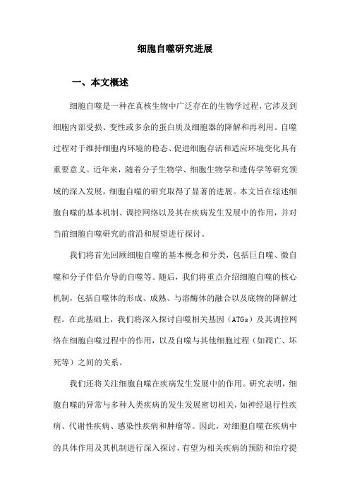
2. 细胞自噬的过程自噬的发生,从起始到自噬体形成经历一系列膜的动态变化。
首先,各种胞浆组分包括受损细胞器被“隔离膜”结构包裹,随后,新月形的隔离膜不断融合、延伸,直到形成完整的双层膜细胞器——自噬体(Autophagosome) (图1)。
Figure 1. The process of autophagy图1. 细胞自噬过程酵母中的自噬体形成时Atg 蛋白聚集到液泡膜附近的一个点,自噬体在这个特异位点形成,因此称它为自噬前体结构“PAS ”(pre-autophagosomal structure) [4]。
在哺乳动物细胞中是否存在PAS 结构仍不确定。
隔离膜形成、组装中一个重要复合物是Class III PI3K 复合物(由PIK3C3、p150、Atg14和BECN1组成),其主要功能是产生PI3P (phosphatidylinositol-3-phosphate),PIK3C3复合体招募一些PI3P 结合蛋白Initiation Nucleation PhagophoreLC3 (Atg8)Atg12-Atg5-Atg16L 长寿命蛋白损伤的细胞器Autolysosome Amphisome Autophagosome Elongation & Closure Degradation夏丹后进一步招募两个泛素样蛋白连接复合物,Atg12-Atg5-Atg16L1和LC3/Atg8-PE (phosphatidylethanola-mine)到隔离膜参与膜的进一步延伸[5]。
在正常条件下,大部LC3/Atg8主要在细胞质中,而当自噬被诱导发生时,LC3/Atg8则以LC3/Atg8-PE 连接体形式定位于隔离膜两侧。
LC3/Atg8可控制细胞自噬体的大小,因其始终存在于自噬体膜上,所以Atg8及其哺乳动物细胞同源物LC3的脂化水平被广泛用于衡量细胞自噬发生的指标。
目前的研究进展表明,多种细胞器和质膜为自噬体膜的形成提供了脂类或重要蛋白。
Ktistaki课题组通过研究ZFYVE1证实了内质网是自噬体膜的来源之一[6]。
Lippincott-Schwartz和他的同事通过对LC3及ATG5的观察发现饥饿诱导的自噬体可以出现在线粒体位点上,认为线粒体也是自噬体膜潜在的来源之一[7]。
最近的研究发现将细胞从4℃迅速转移到37℃,诱导其发生内吞的过程中,许多质膜标志物与自噬体早期结构出现共定位现象,且质膜标志物与隔离膜、自噬前体和自噬体都密切相关,表明质膜在由隔离膜发展为自噬体的过程中发挥了重要作用[8]。
通过对酵母的研究发现许多低聚的高尔基复合物可通过参与高尔基-内体间的运输从而影响自噬体合成,因此高尔基体可能在酵母系统的自噬中发挥重要作用[9][10],但至今尚不清楚高尔基体是否在哺乳动物的自噬过程中发挥作用。
有学者提出Atg9的穿梭可能为自噬体提供膜来源[11],因此明确Atg9在穿梭中的具体作用可能为研究自噬体膜来源提供更多有用信息。
近期的研究取得了很大进展,Hamasaki的研究证实内质网-线粒体接触位点(ER-mitochondria contact site)是自噬体形成的起始位点,当内质网-线粒体的接触位点被破坏后,自噬体形成受到严重抑制[12]。
Ge的课题组认为ERGIC (ER-Golgi intermediate compartment)可能是自噬体形成的另一起点,非细胞实验证实内质网-高尔基体间隔是LC3B脂化过程的膜底物,细胞实验也证实了ERGIC对于自噬体的形成是必须的[13]。
Graef等的研究认为内质网的排出位点(ER exit sites, ERES)是自噬体合成的起始以及膜来源的位点,ERES产生COPII被膜小泡,并能相互融合,形成“内质网–高尔基体中间体”,介导蛋白从内质网到顺面高尔基体的运输,为phagophore成核、延伸提供特定膜来源或结构平台[14],但具体有哪些关键分子参与这一过程尚不清楚,需进一步研究来确认。
完整的双层膜自噬体形成后,将内容物运送到溶酶体以完成最终的降解过程。
来自酵母的研究结果显示,Atg8从自噬体外膜释放可作为它解聚自噬发生的起始因子进而准备融合的信号[15][16]。
免疫电镜结果也表明多数Atg8只存在于成熟自噬体内膜上[17]。
目前哺乳动物细胞的LC3B从外膜的释放是否可以作为自噬体与内吞体或溶酶体融合的信号尚不清楚。
但与隔离膜相关蛋白如ATG16L1和ULK1不存在于成熟的自噬体上,表明它们在与溶酶体融合前已经从自噬体上分离。
其中的机制及其是否成为自噬体与溶酶体融合的先决条件有待进一步研究证实,因此,自噬体成熟是一个既复杂又受精细调控的过程。
溶酶体是细胞内降解蛋白质、脂质、糖类和核酸等的重要细胞器。
各种来源的物质(胞外、细胞表面或胞内的底物)通过胞内一些相互关联的运输途径运送到溶酶体进行降解,其终末产物被循环利用。
溶酶体功能受损将阻断各种底物的降解,导致废物大量堆积,引起溶酶体病变。
自噬体和溶酶体均是膜包裹的能够行使独立功能的细胞器,自噬体分布相对随机,而溶酶体常位于细胞核附近的中心位置。
因此,自噬体要与它们融合,先要运动到它们所在的位点,这个过程受到细胞内骨架蛋白及相关马达动力蛋白的调控[18]。
当自噬体运动到目的地时,它就开始完成与溶酶体的融合过程。