Kawasaki Disease - Yale medStation - Yale University
2024版川崎病课件

Chapter
2024/1/26
24
国内外研究现状及成果分享
1 2
国内外川崎病研究概述
介绍国内外在川崎病领域的研究现状,包括发病 机制、诊断方法、治疗手段等方面的研究进展。
重要研究成果分享
展示近年来在川崎病研究方面取得的重要成果, 如新的诊断技术、有效的治疗方法等。
3
国内外研究合作与交流
介绍国内外研究机构在川崎病领域的合作与交流 情况,以及国际学术会议和研究成果的发布平台。
包括无菌性脑膜炎、面神经麻痹、听力丧 失、急性播散性脑脊髓炎等。这些并发症 虽然发生率较低,但一旦发生,对患者的 危害较大。
2024/1/26
15
03
治疗与预后
Chapter
2024/1/26
16
治疗原则及方法
01
02
03
早期识别和治疗
川崎病的早期识别和治疗 对于预防并发症和改善预 后至关重要。
21
家庭护理指导建议
家庭环境优化
建议家长为患儿提供安静、舒适、 整洁的家庭环境,减少不良刺激。
饮食调整
指导家长为患儿提供营养均衡、 易消化的饮食,避免刺激性食物
和饮料。
规律作息
建议家长帮助患儿建立规律的作 息习惯,保证充足的睡眠和休息。
2024/1/26
22
学校及社会资源整合利用
学校支持
宣传教育
疗等前沿技术的应用前景。
26
提高公众认知度和关注度举措
2024/1/26
加强科普宣传
通过媒体、社交平台等途径加强川崎病的科普宣传,提高公众对 川崎病的认知度和关注度。
开展公益活动
组织相关机构开展川崎病公益活动,如义诊、讲座等,提高公众 对川崎病的认识和重视程度。
川崎病

性心肌炎中所见的心肌溶解
心包炎
3%~31%、血性渗出液、中性白细胞、免疫复合物水平高
心脏瓣膜病变
瓣膜的炎性浸润或乳头肌功能不全至二尖瓣反流 心内膜炎或主动脉炎症伸延导至主动脉返流
传导系统损害
临床表现
冠脉病变
冠状动脉炎
接触史
发热与皮疹的关系 口周苍白圈,帕氏线 脱皮 病原学检查阳性 抗感染治疗有效
鉴别诊断
败血症
局部感染病灶
中毒貌明显 血培养阳性 抗感染治显
发热与皮疹关节症状的一致性 结膜无充血 无脱皮
急性期治疗
标准方案
疾病10天内IVGG2g/kg(10~12小时以上)
发热持续5天以上。 四肢末端变化:急性期手足硬性水肿,掌跖及指趾
端红斑;恢复期,甲床皮肤移行处膜样脱皮
皮疹:多形性红斑,躯干部多,不发生水疱及痂皮
双眼球结膜充血
口腔粘膜:口唇潮红、皲裂,杨梅舌,口、咽部粘
膜弥漫性充血
急性非化脓性颈部淋巴结脓肿,直径>1.5cm
诊断标准
具有以上6项中有5项以上者,又不能被其他已知疾
病所解释可诊断
除外其他疾病尤其是病毒性感染、溶血性链球菌感
染(暴发型、猩红热)、葡萄球菌感染(中毒性休 克综合征)和耶尔森菌感染(耶尔森菌感染约有10% 病例临床表现符合川崎病诊断标准)
非典型KD
诊断标准6项中只有4项或3项,但在病程中心
超或心血管造影证明有冠状动脉瘤者(多见 于<6月婴儿或>8岁年长儿)
变,低电压,少数心律失常。心肌梗塞时可 见异常Q波,ST-T单向曲线
负荷试验
川崎病休克综合征的诊治进展

川崎病休克综合征的诊治进展陈夜肖东琼银四川大学华西第二医院急诊医学科,出生缺陷与相关妇儿疾病教育部重点实验室,四川成都610041[摘要]川崎病(KD)是一种以发热为主要表现的儿童常见血管炎。
川崎病休克综合征(KDSS)是KD的一种罕见表现,其特点是收缩压持续低于同龄儿童正常收缩压低值20%及以上,或伴有组织灌注不良的一种临床征象。
由于其早期症状不典型,临床上常常与感染性休克混淆,从而影响患儿的诊治,因此早期识别该病对于儿科临床医生尤为重要。
本文从KDSS的发病机制、临床表现、实验室指标、治疗和预后等方面对其进行归纳总结,以便尽早治疗,进而改善患儿的预后。
[关键词]川崎病;川崎病休克综合征;诊断;治疗冲图分类号]R725.9[文献标识码]A[文章编号]1674-4721(2021)3(a)-0020-04Progress in diagnosis and treatment of Kawasaki disease shock syndrome CHEN Ye XIA O Dong-qiong银DeparLmenL of Emergency Medicine,WesL China Second University Hospital,Sichuan University,Key Laboratory of BirLh Defects and Related Diseases of Women and Children of the Ministry of Education,Sichuan Province,Chengdu 610041,China[Abstract]Kawasaki disease(KD)is a common vasculitis in children with fever as the main manifestation.Kawasaki disease shock syndrome(KDSS)is a rare form of KD,characterized by sustained systolic blood pressure lower than20%' or more of normal low systolic blood pressure in children of the same age,or accompanied by a clinical sign of poor tissue perfusion.Because its early symptoms are not typical,it is often confused with septic shock in clinical practice, which affects the diagnosis and treatment of children.Therefore,early recognition of the disease is particularly important for pediatric clinicians.This article summarizes KDSS from its pathogenesis,clinical manifestations, laboratory indicators,treatment and prognosis,so as to facilitate early treatment and improve the prognosis of children. [Key words]Kawasaki disease;Kawasaki disease shock syndrome;Diagnosis;Treatment川崎病(KD)是一种影响儿童的急性、发热性血管炎,通常表现为持续发热,结膜充血,急性非脓性颈部淋巴结肿大,口唇发红、皲裂和四肢末端水肿叫在诊断KD的基础上,一些患儿在疾病的急性期可能会经历血流动力学不稳定,血压下降甚至休克,患儿收缩压持续低于同龄儿童正常收缩压低值的20%及以上,或合并组织低灌注的临床体征,Kanegaye等何于2009年首次描述了这一现象,并将其定义为川崎病休克综合征(KDSS),KDSS在KD患儿中的发病率为1.9%~7.0%。
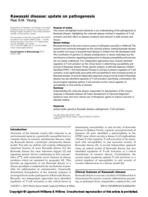
Kawasaki disease update on pathogenesis

Kawasaki disease:update on pathogenesis Rae S.M.YeungIntroductionActivation of the immune system after exposure to an environmental agent in a genetically susceptible host is a common theme in systemic autoimmune diseases,mak-ing Kawasaki disease a prototypic autoimmune disease model.Not only are arthritis and systemic inflammation important features of acute Kawasaki disease[1],but Kawasaki disease has clear infectious triggers[2]and important genetic factors contributing to host suscepti-bility[3 ],with tremendous racial variation in disease incidence which are unaltered by geography[4].This provides an opportunity to use Kawasaki disease as a disease paradigm to understand the mechanisms respon-sible for inflammation in autoimmunity.Genetically determined dysregulation of the immune response is an integral factor in the pathogenesis of Kawasaki disease. Recently,two independent approaches have identified regulation of T-cell activation as the critical factor in determining susceptibility to and severity of Kawasaki disease in children.Firstly,a genetic association study of Japanese sib pairs identified a polymorphism in the ITPKC gene which encodes a kinase(1,4,5-triphosphate 3-kinase C)that regulates T-cell activation,to be associ-ated with susceptibility to and increased severity of Kawasaki disease[5].A second independent approach using an animal model of Kawasaki disease has also identified regulation of T-cell activation as a critical determinant of coronary disease.Costimulation,the second signal regulating optimal T-cell activation,is a critical regulator of susceptibility to and severity of vascular inflammation in a disease model[6].Clinical features of Kawasaki disease Kawasaki disease is an acute vasculitis of childhood that is characterized by massive systemic inflammation present-ing as prolonged fever,nonpurulent conjunctivitis,oralDepartments of Paediatrics,Immunology,and Institute of Medical Sciences,University of Toronto,and Cell Biology Program,Hospital for Sick Children Research Institute,Toronto,Ontario,Canada Correspondence to Rae S.M.Yeung,MD,PhD,FRCPC, Hospital for Sick Children,MaRS Centre–Toronto Medical Discovery Tower East,101College Street, Toronto,ON M5G1L7,CanadaTel:+14168138964;fax:+14168138883;e-mail:rae.yeung@sickkids.caCurrent Opinion in Rheumatology2010,22:551–560Purpose of reviewThis review will highlight recent advances in our understanding of the pathogenesis of Kawasaki disease,highlighting the molecular players involved in regulation of T-cell activation and their affect on disease incidence and outcome in both humans and mouse.RecentfindingsKawasaki disease is the most common cause of multisystem vasculitis in childhood.The vessels most commonly damaged are the coronary arteries,making Kawasaki disease the number one cause of acquired heart disease in children from the developed world. The contribution of genetics to disease predisposition is clearly implicated,but the mechanisms involved in regulating predisposition to disease susceptibility and outcome are not clearly understood.Two independent approaches have recently identified regulation of T-cell activation as the critical factor in determining susceptibility and severity of Kawasaki disease.Firstly,genetic analysis of affected Japanese children identified ITPKC,1,4,5-triphosphate3-kinase C,a kinase involved in regulation of T-cell activation,to be significantly associated with susceptibility to and increased severity of Kawasaki disease.A second independent approach using an animal model of Kawasaki disease has also identified regulation of T-cell activation,specifically costimulation,the second signal regulating optimal T-cell activation as the critical regulator of susceptibility to and severity of disease.SummaryUnderstanding the molecular players responsible for dysregulation of the immune response in Kawasaki disease will foster development of improved diagnostic/ predictive tools and more rational use of therapeutic agents to improve outcome in affected children.Keywordsanimal model,genetics,Kawasaki disease,pathogenesis,T-cell activationCurr Opin Rheumatol22:551–560ß2010Wolters Kluwer Health|Lippincott Williams&Wilkins1040-87111040-8711ß2010Wolters Kluwer Health|Lippincott Williams&Wilkins DOI:10.1097/BOR.0b013e32833cf051mucosal inflammation,cervical lymphadenopathy, induration and erythema of the hands and feet,and a diffuse polymorphous skin rash.In addition to fever and thefive principal clinical features of the disease,there are a myriad of clinicalfindings attributed to the underlying multisystem vasculitis,including,most commonly,arthri-tis,aseptic meningitis,colitis,urethritis,anterior uveitis, and hydrops of the gall bladder[7].The most common age of occurrence is between12months and4years,although both younger children and adults can be affected.Early treatment with aspirin and intravenous immunoglobulin (IVIG)decreases the incidence of coronary artery damage [8–11],but despite appropriate treatment,aneurysms continue to develop in approximately5%of children [7,12].When coronary artery dimensions are adjusted for body surface area,the number of children with coronary artery lesions increase to20–30%of children[2,13]. Kawasaki disease is now recognized as the number one cause of acquired heart disease in children in the indus-trialized world[14].Etiology of Kawasaki diseaseKawasaki disease is endemic with seasonalfluctuations punctuated with epidemic outbreaks[15].Many of the clinical features of the disease are outbreak-dependent with a different spectrum of clinicalfindings in one mini-outbreak compared with another,and with cases having similar clinical phenotypes clustering temporally[16]. Kawasaki disease has been linked with many different causative agents ranging from bacteria to viruses,but no one causative agent has been consistently demonstrated. Kawasaki diseasefits nicely in the spectrum between an infectious disease and a true autoimmune disease,with an infectious trigger leading to a prolonged self-directed immune response.The debate regarding cause has centered around the mechanism of immune activation: conventional antigen versus superantigen(SAg)[17]. SAgs are a group of proteins which share the ability to stimulate a large proportion of T-cells(up to30%of the T-cell repertoire compared with one in a million T-cells for conventional antigens)by binding to a portion of the T-cell receptor b chain(TCRV b)in association with the major histocompatibility complex(MHC)class II mole-cules with no requirement for antigen processing[18]. SAgs have been identified in a variety of microorganisms including many of the bacteria and viruses isolated from children with Kawasaki disease[17].A superantigen is also responsible for induction of coronary artery disease in a murine model of Kawasaki disease in which Lactoba-cillus casei cell wall extract(LCWE)was shown to induce coronary arteritis[19].Although the debate continues regarding the mechanism of initial immune activation, the more likely scenario is that there is cooperation between different mechanisms and afinal common path-way of immune activation responsible for this clinical syndrome[17].Regardless of method of T-cell activation, the massive immune activation characteristic of Kawasaki disease is translated into the classicfindings of systemic inflammation manifested clinically as fever and the cardinal features of Kawasaki disease.Systemic inflam-mation progresses to local inflammation and persistence of the inflammatory response at the coronary artery leading to vessel wall damage and aneurysm formation. Studies in both mice and humans are remarkably in accord,identifying genes and gene products involved in regulation of T-cell activation as central to governing risk and outcome in Kawasaki disease.Lessons learned from a mouse model of Kawasaki diseaseExperimental mice develop coronary arteritis in response to intraperitoneal injections of LCWE[20].The resultant vasculitis is similar to Kawasaki disease in children [19–21].Young mice(4–5weeks old)are much more susceptible to LCWE-induced disease compared with older mice.Massive peripheral immune activation within hours of LCWE injection is followed by local infiltration into cardiac tissue at day3.The inflammatory infiltrate, comprising mainly T cells,intensifies and peaks at day28 post injection.This is accompanied by elastin breakdown with disruption of the intima and media and aneurysm formation at day42[19,22,23].A superantigen found within LCWE is responsible for development of vascular disease[19].As with all disease models,replication of the disease is not perfect,but this murine model of Kawasaki disease is an accurate model sharing many features with the human disease including an infectious trigger leading to massive immune activation;disease susceptibility in the young;a time course similar to that seen clinically in Kawasaki disease;similar pathology of coronary arteritis;and response to IVIG treatment allow-ing study of the pathogenic mechanisms driving local inflammation and damage to the coronary arteries.The disease model(Fig.1)starts with massive immune activation(step1)by a microbe with superantigenic activity.The novel SAg found in the cell wall of L. casei preferentially expands T-lymphocytes expressing TCRV b2,4,6and14positive T-cells,and superantigenic activity is directly correlated with the ability to induce coronary arteritis in mice[19].SAg activity is T-cell-dependent and T-cells are required for disease,with the inability to induce disease in RAG-1[24 ]and TCR a-deficient mice[22].What distinguishes this novel SAg from other bacterial SAgs is the presence of localized production of IFN g and TNF a in affected vascular tissue.This profile is not only important in the systemic immune response,but their divergent roles are important in regulating the local immune response at the coronary artery[22,23].Ablation of IFN g confirmed that IFN g552Pediatric and heritable disordersplays an important regulatory role in disease induction. Mice with absence of TNF a activity(therapeutic block-ade or TNFR1knockouts)do not develop coronary disease after LCWE stimulation.Despite a robust systemic T-cell response,there is neither local inflam-mation nor vessel wall breakdown in the absence of TNF a activity,thus TNF a is the critical proinflamma-tion mediator leading to heart disease.The T-cells found in affected vessels express SAg-reactive TCRV b families,afinding out of keeping with the fate of SAg-activated T-cells,which are actively deleted by apoptosis.We have found that costimulation can rescue SAg-stimulated T-cells from apoptosis[6].Costimulation is important in optimal T-cell activation,which requires engagement of the TCR and a second(costimulatory) signal.A subpopulation of the SAg-responsive T-cells is rescued from apoptosis because of increased costimula-tion(step2).The coronary endothelium is transformedPathogenesis of Kawasaki disease Yeung553Figure1Model of diseasepathogenesisinto a professional antigen-presenting cell(APC)by up-regulation of costimulatory molecules driven in part by the tissue-specific expression of Toll-like receptor (TLR)2(step3).Increased TLR2expression partnered with TLR2stimulation by the TLR2ligand in LCWE [25,26]lead to increased expression of costimulatory molecules facilitating rescue of SAg-activated T-cells and continued local production of proinflammatory cyto-kines,thus perpetuating inflammation at the coronary vessel wall.IFN g and TNF a are involved in transcrip-tional regulation of matrix metalloproteinases(MMPs), with TNF a up-regulating,and IFN g inhibiting pro-duction of MMP-9.The enzymatic activity of MMP-9 leads to elastin breakdown and aneurysm formation(step 4).Elastin degradation alone,without loss of smooth muscle cells,leads to vessel wall damage typical of Kawasaki disease.Blocking MMP-9activity has no effect on inflammation,but markedly decreases vessel damage [27].Inflammation can be dissociated from end-organ damage by the ablation of MMP-9.Thus MMP-9is oneof the critical downstream molecules responsible for aneurysm formation[28].TNF a-dependent leukocyte recruitment signals are a critical factor controlling the traffic of pathogenic T-cells to the coronary artery.A sustained local immune response coupled with persistent TNF a production and leukocyte recruitment lead to up-regulation of proteolytic activity,elastin degradation, vessel wall damage and the characteristic coronary artery lesions seen in Kawasaki disease.T-cell activation and costimulation:lessons from the disease modelOptimal T-cell activation requires engagement of the TCR(signal one)and a costimulatory signal(signal two). The second signal is dependent on soluble factors such as IL-2or the ligation of cell surface molecules[29,30].Most T-cell surface costimulatory molecules are members of the immunoglobulin and TNF superfamilies and are important components of the immunologic synapse (Fig.2)[31].Occurrence of both signal one and signal two leads to optimal T-cell activation evidenced by IL-2 production and survival of the T-cell.CD28and4–1BB are two examples of positive costimulatory receptors. Positive second signals(costimulators)enhance and sus-tain T-cell responses and coinhibitors or negative costi-mulatory molecules inhibit TCR-mediated responses and contribute to peripheral T-cell tolerance.Although well characterized,the CD28/B7pathway is complex because of the dual specificity of the B7.1(CD80)and B7.2(CD86)ligands for the stimulatory receptor CD28 and the inhibitory receptor CTLA-4(cytotoxic T lymphocyte-associated antigen4or CD152)[32].Upon binding its ligands,B7.1and B7.2on activated APCs, CD28cooperates with the TCR to enhance IL-2pro-duction,glucose metabolism and T-cell survival[32–34].Initial T-cell activation is usually dependent on CD28/B7 interaction[31],but the T-cell response is not a single event with activation followed by commitment to expan-sion,differentiation and migration to carry out their effector functions[31].The list of costimulatory mol-ecules with contributions to these different steps is growing.The need for inducible costimulatory receptors in addition to the constitutively expressed CD28mol-ecule may reflect the importance of differential regula-tion of T-cell activation on distinct T-cell subsets and in distinct locations(lymph nodes versus extra nodal). Moreover,immune system homeostasis makes it critical that T-cell survival is tightly regulated at each stage of the response,from initial activation,through the duration of the effector function in tissues,to the memory stage of the immune response.Indeed dysregulated lymphocyte survival is a critical factor contributing to autoimmunity with members of the TNFR family of costimulatory molecules emerging in multiple,well powered,genetic case-control studies as risk genes in various autoimmune diseases[35].4–1BB is of particular interest in Kawasaki disease.4–1BB is an inducible costimulatory molecule and member of the TNF family of receptors,and plays similar roles to CD28in cell survival[31].In-vivo studies in mice show that anti-4-1BB agonistic antibodies can rescue superantigen-activated T-cells from death[36] and our recent results show that anti-4-1BB agonist antibodies exacerbate the LCWE model of Kawasaki disease in mice[6].Additionally,4-1BB is a TRAF1 binding receptor and TRAF1has been strongly impli-cated as a risk gene in autoimmunity(Table1)[37,38]. Genes indirectly regulating costimulation Other genes/pathways indirectly regulating co-stimu-lation and T-cell activation have been implicated as risk554Pediatric and heritable disordersFigure2The immune synapse:participation of costimulatorymoleculesgenes/loci in autoimmunity and are intimately related to the pathogenesis of Kawasaki disease in the mouse model including the TLR pathway [25,26].As discussed above,the pathogenic population of superantigen-responsive T-cells is rescued from apoptosis because of increased costimulation.The coronary endothelium is transformed into a professional APC by up-regulation of costimulatory molecules driven in part by the tissue-specific expression of TLR2.TLRs belong to a class of evolutionarily conserved pattern recognition receptors important in initiating the innate immune response.TLR2recognizes defining properties of gram-positive bacteria.Cell surface expression of TLR2at the coronary artery is driven by TNF a ,IFN g ,and MHCII signaling mediated by SAg.TLR2plays a pivotal role in directing and driving the persistence of the SAg-initiated T-cell response in the LCWE model of Kawasaki disease.TLR2is preferen-tially expressed at the coronary artery and is rapidly up-regulated in the heart and at the coronary vessel during disease development.TLR2signaling results in increased expression of costimulatory molecules,linking the innate and adaptive arms of the immune system.Physiologically,enhanced costimulation,from TLR2stimulation,rescues SAg-activated T-cells from apoptosis leading to persistence of the inflammatory response.The effect of TLR2is directly relevant to the disease model.Although,TLR2ligand alone cannot induce coronary disease,TLR2stimulation together with superantigenic activity induces disease.In fact,TLR2directly modu-lates incidence and severity of LCWE induced coronary disease.These results demonstrate the need for both SAg and TLR2ligand for disease induction,and a novel role for TLR2in directing the specificity and persistence of inflammation at the coronary artery,a principle with wide ranging applications in other organ specific autoimmune diseases [25].Contribution of geneticsEpidemiologic data suggest a major role for genetics in Kawasaki disease risk and outcome.For example,whereas Kawasaki disease occurs in all ethnic groups and all regions of the world,the incidence of disease varies dramatically from region to region and between different ethnic groups.The annual incidence,reported as number per 1000000children under age 5years rangesPathogenesis of Kawasaki disease Yeung 555Table 1Candidate genes identified from an animal model of Kawasaki disease Candidate gene(s)FunctionRelevance to Kawasaki diseaseCD28Co-stimulatory molecule cooperating with TCR to enhance IL-2production and T-cell survival Important in TCR-induced T-cell activation and survival CD80(B7.1)CD86(B7.2)Ligands for CD28CD152(CTLA-4)Negative stimulator signal of T-cell activation,competing with CD28binding to their common ligands B7.1and B7.2Inhibition of T-cell activationRisk gene associated with rheumatoid arthritis 4-1BBTRAF1binding receptor resulting in activation of ERK/MAPK leading to phosphrylation and degradation of the proapoptotic molecule BIMImportant in T-cell survivalTRAF1TNFR associated factor promoting T-cell survival downstream of several TNFR family members including TNFR1,2,CD40and 4-1BB Specific risk associated with polymorphismin TRAF1,TNFAIP3,TNIP1and TNFRSF/MMEL1found in several studies of inflammatory and autoimmune diseasesTNFAIP3(A20)Involved in feedback inhibition of NF-k B TNIP1TNFAIP3-interacting protein Tnfrsf14(Mmel1)TRAF-interacting proteinCD40Receptor for CD40L or CD514mediating modulation of platelet function Mediating a variety of immune and inflammatory responses;risk gene associated with other inflammatory/autoimmune diseasesa IIb b 3a 5b 1Receptor for CD40L or CD514mediating modulation of platelet function Important not only in cellular immunity and inflammation but also in the pathophysiology of thrombosis CD514(CD40L)Thrombo-inflammatory moleculeTLR2Recognizing microbial-associated molecular patterns leading to induction of NF-k B and MAP gene activation,and production of pro-inflammatory cytokines and costimulatory moleculesPolymorphisms of TLR2found to be associated with several infectious,inflammatory and autoimmune diseasesMMP1Elastolytic matrix metalloproteinasesElastases up-regulated by inflammation leading to elastin break-down.Local activity in affected coronary arteries seen in mice and humans MMP-2MMP-9MMP-12MMP-13MMP-3Regulation of MMP activities Elevated levels of MMP3found in KDTIMP-1TNF a Tumor Necrosis Factor a Necessary for inflammation and elastin breakdown in coronary arteryIFNgInterferon gammaRegulatory cytokine-suppressing inflammatory responsefrom5in Denmark,8in New Zealand,26in Canada,39 in Hong Kong,55in China,105in Korea to over180in Japan[39–46].Siblings of affected children are at10-fold higher risk for Kawasaki disease compared with the general population,and incidence of Kawasaki disease is two-fold higher that normal in children of affected individuals[47,48].Genetic association studies have already identified several candidate genes for Kawasaki disease susceptibility and very recently,results of a genome-wide association study(GWAS)demonstrated significant association of Kawasaki disease risk and out-come in the Japanese population with variants in the ITPKC gene(see below).This association has been replicated in a small cohort of Caucasian American chil-dren[5],but was not reproduced in studies of other ethnic groups[49].Thus immune dysregulation observed clini-cally in Kawasaki disease in mice and humans are inti-mately linked to genetic variation.Genetic studies in childrenMost genetic studies in Kawasaki disease have focused on candidate genes,with genes selected for analysis based on their known function or potential role in disease pathogenesis.These genes can be broadly grouped into two:those involved in the inflammatory response and those involved in vascular biology(see Table2)[2,49–85,86 ],logical choices for a multisystem inflammatory disease of blood vessels.Candidate genes have been examined in patient cohorts with promisingfindings; however,conflicting results have often been found between published studies both within and between different ethnicities.Sometimes,even association studies,which have included validation cohorts within the same ethnicity,cannot be replicated in a cohort from another ethnic group.Potential contributing factors include differences in haplotype structures or linkage556Pediatric and heritable disordersTable2Candidate genes tested in children with Kawasaki diseaseGene symbol Gene name References I.Inflammatory responseAdaptive immunityT-cells ITPKC Inositol1,4,5-trisphosphate3-kinase C(ITPKC)[5,49]CASP3Caspase3[50]PD-1Programmed death-1[51]CD40L CD40ligand[52,53]B-cells FCGR2A Fc fragment of IgG,low affinity IIa,receptor(CD32)[54,55]FCGR2B Fc fragment of IgG,low affinity IIb,receptor(CD32)[54,55]FCGR3A Fc fragment of IgG,low affinity IIIa,receptor(CD16a)[55]FCGR3B Fc fragment of IgG,low affinity IIIb,receptor(CD16b)[54,55]MICA MHC class I polypeptide-related sequence A[56] Innate immunity CD14CD14antigen[57]MBL Mannose-binding lectin[58,59] Cytokines IL-1b Interleukin1,beta[60]IL-1Ra Interleukin1receptor antagonist[60]IL-4Interleukin4[60–62]IL-6Interleukin6[63]IL-10Interleukin10[64]IL-18Interleukin18[65]LTA Lymphotoxin alpha[66]TNF-a Tumor necrosis factor-a[66–70] Leukocyte recruitment CXCR2Chemokine(C-X-C motif)receptor2[71]CXCR1Chemokine(C-X-C motif)receptor1[71]CX3CR1Chemokine(C-X3-C motif)receptor1[71]CCR3Chemokine(C-C)receptor3[71]CCR2Chemokine(C-C)receptor2[71]CCR5Chemokine(C-C)receptor5[71–73]MCP1Monocyte chemoattractant protein-1[74]CCL3L1Chemokine(C-C motif)ligand3-like1[72]II.Vascular biologyExtra-cellular matrix breakdown MMP2Matrix metallopeptidase2[75]MMP3Matrix metallopeptidase3[75–77]MMP9Matrix metallopeptidase9[75,76]MMP12Matrix metallopeptidase12[75]MMP13Matrix metallopeptidase13[75]TIMP2Tissue inhibitor of metalloproteinase2[78] Vascular function ACE Angiotensin I converting enzyme[79]AGTR1Angiotensin II receptor,type I[80]VEGFA Vascular endothelial growth factor A[81–84]VEGFR2Vascular endothelial growth factor[81]eNOS Endothelial nitric oxidase synthase[85]iNOS Nitric oxide synthase2,inducible[85]CAMK2D Calcium/calmodulin-dependent protein kinase II delta[86 ]disequilibrium between different ethnicities.If the observed positive association was a result of a linkage disequilibrium between the marker tested and a genetic variant,the association would not be detected in a differ-ent ethnic groups in which the responsible genetic variant was not linked with the identical markers[3 ].A more detailed review of recent genetic studies in Kawasaki disease is provided in reference[3 ];the discussion in this update will be focused on molecular players involved in regulation of T-cell activation.T-cell activation:lessons from genetic studies in childrenITPKC,a gene directly regulating T-cell receptor(TCR) signaling,has recently been identified in GWAS in Kawasaki disease[5].ITPKC is one of the three isoforms of inositol1,4,5-triphosphate3-kinase(ITPK)that phos-phorylates inositol1,4,5-triphosphate(IP3).IP3is a key molecule in signaling in many cell types and recently found to play an important role in regulating TCR signaling and T-cell activation[87,88].Hydrolysis of phosphatidylinositol4,5-biphosphate by phospholipase C in response to various external stimuli,generates IP3.Specifically,TCR stimulation releases IP3resulting in increased intracellular Ca2þmediated by IP3receptors found on the endoplasmic reticulum.Ca2þinflux acti-vates nuclear factor of activated T-cells(NFAT),with nuclear translocation and transcriptional activation of cytokines involved in T-cell activation and proliferation including IL-2.ITPKC is a negative regulator of T-cell activation via down-regulation of the Ca2þ/NFAT signal-ing pathway in T-cells[5].A SNP(itpkc_3),located in the intron of ITPKC,was found to regulate splicing efficiency thus decreasing negative regulation of T-cell activation resulting in increased T-cell activation and IL-2production.The itpkc_3C allele was strongly correlated with Kawasaki disease and this hyperactive T-cell phenotype seemed to confer an increased risk of developing coronary artery lesions.A subanalysis showed correlation of itpkc_3C with resistance to IVIG therapy. This study was performed on78Japanese sib pairsfirst using linkage disequilibrium mapping identifying a peak at chromosome19,followed by screening of over1000 single-nucleotide polymorphisms(SNPs)in cases and controls to identify candidate genes.This was refined in an independent cohort of Japanese patients with Kawasaki disease and controls which identified three highly significant clusters of SNPs.The association of nine SNPs with Kawasaki disease was confirmed in an independent case-control set.Subsequently,trans-mission disequilibrium test(TDT)analysis of209US multiethnic trios showed asymmetric transmission of four of the nine SNPs.Subanalysis of the US cohort for only Americans of European descent confirmed asymmetric transmission of these four SNPs,all located within introns of known genes.Functional testing showed that ITPKC is the most likely candidate,given the biology and involvement in T-cell activation.An investigation of another region of interest from the work reported above by Onouchi’s group recently ident-ified genetic variants in caspase-3(CASP3),to be signifi-cantly associated with Kawasaki disease in both Japanese and European-American children.Caspases are a family proteins responsible for execution of the apoptotic pro-cess.Caspase activation usually occurs as a cascade of initiator and effector enzymes.The effector caspases, such as caspase-3,are responsible for most of the cleavage events observed during activation-induced cell death. Specifically,one of the commonly associated SNPs in CASP3associated with Kawasaki disease affects binding of NFAT[50].Caspase-3has also been reported to cleave the inositol1,4,5-triphosphate receptor,type1in apop-totic T-cells.ITPR1is a receptor for IP3,the substrate for ITPKC in T-cells.Interestingly,caspase-3activity has also been demonstrated in vascular smooth muscle cells and possess elastolytic activity[89],able to break down elastin,thus potentially a dual role in disease pathogenesis.Studies in children with Kawasaki have also identified costimulatory molecules as candidate genes governing susceptibility to disease and predisposition to poor cor-onary outcome.Specifically,genetic association with variants in the CD40ligand(CD40L)and programmed death-1(PD-1)genes,both members of the TNF super-family of costimulatory molecules,have been reported [51–53].Corresponding increases in CD40L protein expression on platelets and circulating T-cells have also been described during the acute phase of Kawasaki disease[90].In addition to its role in the regulation of the immune response,CD40L is now recognized to be thrombo-inflammatory and predictive of cardiovascular events[52,53,91–96].Platelets constitute the major source of circulating CD40L.Marked elevation of the platelet count is one of the pathognomic features of children with Kawasaki disease.ConclusionUntil the cause of Kawasaki disease is determined and the pathophysiology more exactly specified,the recog-nition and treatment of Kawasaki disease will remain largely empiric.Studies suggest that15–25%of children with Kawasaki disease do not respond to initial treatment with IVIG,and have persistent inflammation,putting them at increased risk for coronary artery damage.There are no additional consensus recommendations for man-agement,although further doses of IVIG,steroids,anti-cytokine therapy(TNF blockage)[7,97]and other agents have been used,with varying results.There have been noPathogenesis of Kawasaki disease Yeung557。
川崎病进展

Differential Diagnosis
Infectious
– Spirocheteal: Lyme disease, Leptospirosis – Parasitic: Toxoplasmosis – Rickettsial: Rocky Mountain spotted fever, Typhus
Edema, erythema, desquamation
2. Polymorphous exanthem, usually truncal 3. Conjunctival injection 4. Erythema&/or fissuring of lips and oral cavity 5. Cervical lymphadenopathy
Illness not explained by other known disease process
Modified from Centers for Disease Control. Kawasaki Disease. MMWR 29:61-63, 1980
Atypical or Incomplete Kawasaki Disease
Kawasaki Disease:
Mucocutaneous Lymph Node Syndrome
“A self-limited vasculitis of unknown etiology that predominantly affects children younger than 5 years. It is now the most common cause of acquired heart disease in children in the United States and Japan.” Jane Burns, MD*
kawasaki disease(川崎病) PPT课件

The main goal of treatment is to prevent coronary artery disease and relieve symptoms.: Full doses of salicylates (aspirin); intravenous gammaglobulin are the mainstays of treatment.
Acute stage (1-11 d)
1. High fever (temperature >39℃)
2. Nonexudative bilateral conjunctivitis (90%) 3. Polymorphous erythematous rash 4. Acral erythema and edema that impede ambulation 5. Strawberry tongue and lip fissures 6. Lymphadenopathy (75%), generally a single,
Differentials
Measles and other viral exanthems Leptospirosis Rickettsial disease Stevens-Johnson syndrome Drug reaction Juvenile rheumatoid arthritis
Lab Studies
Cardiac enzymes ↑ ( CK,CK-MB, cardiac troponin, LDH)
Radiography: rule out cardiomegaly or subclinical pneumonitis.
Imaging Studies
川崎病

Newburger JW ,Takahashi M,Gerber MA,et al. Diagnosis,treatment,and long-term management of Kawasaki disease:a statement for health professionals from the Committee on Rheumatic Fever,Endocarditis and Kawasaki Disease,Councilon Cardiovascular Disease in the Young,American Heart Association [J]. Circulation,2004,114(6):1708-
诊断标准(典型)
临床表现≥4条标准,可在起病第4天诊断为川崎病
双眼球结膜充血(无分泌物) 口唇及口腔改变:口唇绛红、皲裂,杨梅舌, 口腔和咽部弥漫性充血 皮肤改变:多形性红斑,皮疹
4/5
四肢末端改变,(急性期)手掌、足底及指(趾)端潮红、硬肿, (恢复期)指趾端甲床及皮肤移行处膜样脱皮
与川崎病发病及冠状动脉损伤相关的易感基因

China &Foreign Medical Treatment中外医疗川崎病(Kawasaki disease ,KD)是好发于6个月~5岁儿童常见的急性发热出疹性疾病,基本病理变化为主要累及冠状动脉的全身性血管炎,约15%~20%未经治疗的患儿发生冠状动脉损伤。
最新的报告显示,KD 的发病率在一些国家有逐年上升的趋势,现已成为发达国家儿童获得性心脏病最常见的原因[1]。
由于可以导致严重的心血管并发症严重影响儿童的健康,并且作为儿科住院的常见病之一受到人们的重视。
流行病学资料表明,KD 好发于某些特定人群,遗传背景决定KD 的发病,并且和冠状动脉损伤、难治性KD 有关。
现就近年来与KD 发病及冠状动脉损伤相关基因国内外研究现状作一综述。
1ITPKC 基因免疫细胞活性的改变是KD 发生发展的重要环节,主要表现为CD4/CD8比值的增加[2]。
ITPKC 基因定位于19q13.2,是Ca++/NFAT 信号通路的第二信使分子,作为T 细胞活化的负调节因子,表达的下调,引起Ca2+的流动,通过胞浆中NFAT 的去磷酸化最终导致T 细胞的激活或活性的增加进而分泌促炎症细胞因子,这可能与KD 的易感性和疾病的严重程度有关。
Onouchi 等[3]通过全基因组连锁分析确定,ITPKC 基因第1内含子rs28493229(G/C)位点的C 等位基因对ITPKC mRNA 的剪切效率明显较G 等位基因低,使ITPKC 表达减少,成熟mRNA 数量下降30%,引起IL-2等炎性介质的释放,C 等位基因作为KD 的易感基因并且增加冠状动脉损害的风险。
来自台湾学者的报道也显示该基因位点C 等位基因与KD 易感性相关,并且还与接种卡介苗部位的红肿有关[4]。
2CASP3基因CASP3是一种效应型半胱氨酸蛋白酶,是Ca++/NFAT 信号通路的负反馈调节因子,在T 细胞凋亡中起着关键作用。
研究发现,位于CASP3基因5′非翻译区的rs72689236(G/A)位点变异与日本及欧美KD 患儿明显相关,G 等位基因突变为A 后,引起NFAT 与DNA 相应结合位点结合能力下降和降低CASP3mRNA 转录,T 细胞凋亡减少,增加KD 的易感性[5]。
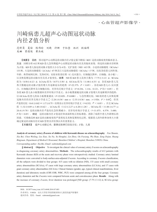
a川崎病患儿超声心动图冠状动脉内径Z值分析

•心血管超声影像学•川崎病患儿超声心动图冠状动脉内径Z值分析范舒旻 夏焙 陈伟玲 刘晓 许娜 于红奎 林洲 欧福祥吴姗 曾德俊 黄兵旋 【摘要】 目的 探讨超声心动图冠状动脉内径Z值定量川崎病(KD)冠状动脉病变的临床意义。
方法 回顾分析612例KD患儿急性期超声心动图冠状动脉内径及其临床表现。
将冠状动脉内径转换为Z值,KD患儿按冠状动脉Z值的大小分为4组,无扩张组(ND)415例、小冠状动脉瘤组(SCAAs)133例、大冠状动脉瘤组(LCAAs)47例及巨大冠状动脉瘤组(GCAAs)17例,比较各组患儿间性别、年龄、典型病例比例、发热时间、实验室检查结果(C-反应蛋白、红细胞沉降率、白细胞、血小板),以及恢复期冠状动脉内径及其Z值变化。
结果 ND组KD患儿发热天数为(7.75±3.12)d、SCAAs组为(8.50±4.12)d、LCAAs组为(8.57±3.58)d、GCAAs组为(11.88±4.33)d,各组KD患儿发热天数随冠状动脉Z值的增大有逐渐延长的趋势(F=22.375,P<0.05)。
各组KD患儿C-反应蛋白、红细胞沉降率及白细胞比较,差异均无统计学意义(F=0.236、1.116、0.121,P均>0.05);但各组KD患儿血小板数量间差异有统计学意义,血小板数量随冠状动脉Z值增大有逐渐增多的趋势,以GCAAs组患儿的血小板数量最高(F=22.029,P=0.000)。
与急性期比较,ND组的患儿在恢复期冠脉内径的差异无统计学意义[(2.24±0.34)mm vs(2.33±0.36)mm,t=1.926,P>0.05],但其Z值的比较(0.41±0.82 vs 1.17±0.75)结果显示差异有统计学意义(t=8.332,P<0.05);并且SCAAs组(1.32±0.89 vs 3.40±0.62)、LCAAs组(3.12±2.27 vs 6.20±1.28)、GCAAs组(11.88±6.77 vs20.4±9.70)冠状动脉内径Z值均比急性期减小,差异均有统计学意义(t=11.073、4.579、3.480,P均<0.05)。
川崎病的临床表现

(2)球结合膜充血 Conjestion of Ocular Conjunctivae
(3)唇及口腔改变 Changes of Lips and Oral
Dry and Cracked Lips
Strawberry Tongue
(4)手足症状 Symptom of Limbs
Indurative Edema
Desquamation at Fingertips
(5)皮肤改变 Changes of Derma
Annular Erythema
(6)颈淋巴结肿大 Cervical lymphadenopathy
The others:
间质性肺炎
Interstitial pneumonia
无菌性脑膜炎
aseptic meningitis
关节炎和关节痛
symptoms of arthritis
黄疸
jaundice
Examination
1.血液检查 Blood Test 白细胞增高 以中性粒细胞为主 血小板早期正常 第2-3周增多 血沉增快 C反应蛋白升高
波
Summary
Questions
1、川崎病的主要临床表现? 2、川崎病的临床表现和病理分期的关系?
川崎病的临床表现
Clinical Manifestations of Kawasaki Disease
Etiology and pathogenesis
Etiology and pathogenesis
川崎病合并冠状动脉瘤血管结构相关基因的研究进展

川崎病合并冠状动脉瘤血管结构相关基因的研究进展
戚慧茹;林瑶;石琳
【期刊名称】《国际心血管病杂志》
【年(卷),期】2024(51)2
【摘要】川崎病(KD)是好发于儿童的急性血管炎症性疾病,冠状动脉瘤(CAA)是KD 的严重并发症,标志着更严重的临床症状和不良的临床预后。
血管内膜层单核细胞浸润、内皮细胞降解以及弹性层破坏是KD合并CAA的基本病变,提示冠状动脉管壁结构存在炎症破坏。
不同遗传背景的KD患儿发生CAA的风险各异,提示遗传因素在CAA发病中发挥重要作用。
该文介绍KD合并CAA的血管结构相关基因和发病机制,为KD合并CAA的早期识别和机制探索提供理论依据。
【总页数】4页(P105-108)
【作者】戚慧茹;林瑶;石琳
【作者单位】北京大学首都儿科研究所教学医院;首都儿科研究所附属儿童医院心血管内科
【正文语种】中文
【中图分类】R54
【相关文献】
1.VKORC1基因多态性与川崎病并发冠状动脉瘤患儿华法林稳定剂量相关性研究
2.病程第3天发现川崎病巨大冠状动脉瘤合并急性左心衰1例
3.川崎病合并冠状动
脉瘤危险因素分析4.1例川崎病合并巨大冠状动脉瘤患儿的围手术期护理5.川崎病合并巨大冠状动脉瘤5例临床治疗分析
因版权原因,仅展示原文概要,查看原文内容请购买。
川崎病(kawasaki disease

Skin :Redness and crust at the site of BCG inoculation, Small pustules, Transverse furrows of the finger nails
Байду номын сангаас
PRINCIPAL SYMPTOMS
Fever: persisting 5 days or more (inclusive of those
cases in whom the fever has subsided before the 5th day in response to therapy)
Non-purulent cervical lymphadenopathy is less frequently encountered (approximately 65%) than other principal symptoms during the acute phase.
Echocardiogram documented coronary artery dilation or aneurysm.
Angiographically documented coronary artery dilation or aneurysm.
Elevated leukocyte count with a predominance of neutrophils or a normal leukocyte count with a left shift is typical on the acute phase of illness. Elevated ESR is almost universally present in acute phase. Thrombocytosis in the later stages is common.
2024年度-川崎病讲课ppt课件

对于IVIG无反应或耐药的患者,可考虑使用糖皮质激素、免疫抑制剂等 药物。同时,根据患者具体情况,可酌情使用抗生素、抗病毒药物等。
15
并发症预防与处理
冠状动脉病变的预防
通过早期识别和治疗,降低冠状动脉病变的发生率。对于已经发生冠状动脉病变的患者,需定期进行心脏超声 检查,评估病变进展情况,并采取相应治疗措施。
14
药物选择及剂量调整
01
丙种球蛋白(IVIG)
作为川崎病治疗的首选药物,可迅速控制炎症,减少冠状动脉病变风险
。剂量通常根据患者体重计算,一般为2g/kg,单次静脉注射。
02 03
阿司匹林
具有抗炎和抗血小板聚集作用,可减轻川崎病症状并预防血栓形成。初 始剂量通常为80-100mg/kg/d,分次口服,热退后3天逐渐减量至小剂 量维持。
川崎病讲课ppt课件
1
目录
• 川崎病概述 • 诊断与鉴别诊断 • 治疗与预后 • 患者教育与心理支持 • 研究进展与未来展望
2
01
川崎病概述
3
定义与发病机制
定义
川崎病(Kawasaki Disease,KD )是一种急性、自限性的全身性 血管炎,主要影响婴幼儿和儿童 。
发病机制
目前尚未完全明确,但普遍认为 与感染、免疫异常和遗传因素有 关。
12
03
治疗与预后
13
治疗原则及方法
早期识别和治疗
川崎病的治疗关键在于早期识别,一旦确诊 应立即开始治疗,以减轻血管炎症和降低并 发症风险。
抗炎治疗
使用大剂量丙种球蛋白(IVIG)和阿司匹林 等抗炎药物,控制全身血管炎症,减少冠状 动脉病变的发生。
对症治疗
针对患者的具体症状,如发热、皮疹、黏膜 充血等,采取相应的对症治疗措施,以缓解 患者不适。
- 1、下载文档前请自行甄别文档内容的完整性,平台不提供额外的编辑、内容补充、找答案等附加服务。
- 2、"仅部分预览"的文档,不可在线预览部分如存在完整性等问题,可反馈申请退款(可完整预览的文档不适用该条件!)。
- 3、如文档侵犯您的权益,请联系客服反馈,我们会尽快为您处理(人工客服工作时间:9:00-18:30)。
Cardiovascular Manifestations of Acute Kawasaki Disease
• Late
– Thrombocytosis – Elevated CRP
Cardiovascular Manifestations of Acute Kawasaki Disease
• EKG changes
– Arrhythmias – Abnormal Q waves – Prolonged PR and/or QT intervals – Low voltage – ST-T–wave changes.
• Musculoskeletal
– Myositis, arthralgia, arthritis
Kawasaki Disease: Labs
• Early – Leukocytosis – Left shift – Mild anemia – Thrombocytopenia/ Thrombocytosis – Elevated ESR – Elevated CRP – Hypoalbuminemia – Elevated transaminases – Sterile pyuria
• Infectious agent most likely
– Age-restricted susceptible population – Seasonal variation – Well-defined epidemics – Acute self-limited illness similar to known
Typhus
Differential Diagnosis
• Immunological/Allergic
– JRA (systemic onset) – Atypical ARF – Hypersensitivity reactions – Stevens-Johnson syndrot explained by other known disease process
Modified from Centers for Disease Control. Kawasaki Disease. MMWR 29:61-63, 1980
Atypical or Incomplete Kawasaki Disease
New Haven Coronavirus
• Identified a novel human coronavirus in respiratory secretions from a 6-month-old with typical Kawasaki Disease
• Subsequently isolated from 8/11 (72.7%) of Kawasaki patients & 1/22 (4.5%) matched controls (p = 0.0015)
and treatment algorithm
Differential Diagnosis
• Infectious
– Measles & Group A beta-hemolytic strep can closely resemble KD
– Bacterial: severe staph infections w/toxin release
Kawasaki Disease:
Mucocutaneous Lymph Node Syndrome
“A self-limited vasculitis of unknown etiology that predominantly affects children younger than 5 years. It is now the most common cause of acquired heart disease in children in the United States and Japan.”
• Explore the differential diagnosis of this illness • Consider the most important sequelae of this
disease • Discuss treatment & long-term management
recommendations
1. Changes in the extremities
▪ Edema, erythema, desquamation
2. Polymorphous exanthem, usually truncal 3. Conjunctival injection 4. Erythema&/or fissuring of lips and oral cavity 5. Cervical lymphadenopathy
1974, simultaneously recognized in Hawaii
What is Kawasaki Disease?
• Idiopathic multisystem disease characterized by vasculitis of small & medium blood vessels, including coronary arteries
• GI
– Diarrhea, vomiting, abdominal pain, hydrops of the gallbladder, jaundice
• Neurologic
– Irritability, aseptic meningitis, facial palsy, hearing loss
– Desquamation, may have persistent arthritis or arthralgias
– Gradual improvement even without treatment
• Convalescent (Months to years later)
Trager, J. D. N Engl J Med 333(21): 1391. 1995.
Kawasaki Disease: An Update
Bevin Weeks, MD Yale University School of Medicine
Objectives
• Review the historical & clinical features of a “common” but poorly understood childhood disease
Trager, J. D. N Engl J Med 333(21): 1391. 1995.
Han, R. CMAJ 162:807. 2000.
Kawasaki Disease: S&S
• Respiratory
– Rhinorrhea, cough, pulmonary infiltrate
• Seasonal variation
– More cases in winter and spring but occurs throughout the year
The Biggest Question:
What is the Etiology of Kawasaki Disease?
Etiology
• Present with < 4 of 5 diagnostic criteria • Compatible laboratory findings • Still develop coronary artery aneurysms • No other explanation for the illness • More common in children < 1 year of age • 2004 AHA guidelines offer new evaluation
Jane Burns, MD*
*Burns, J. Adv. Pediatr. 48:157. 2001.
History of Kawasaki Disease
• Original case observed by Kawasaki January 1961
– 4 y.o. boy, “diagnosis unknown”
– Mercury
Phases of Disease
• Acute (1-2 weeks from onset)
– Febrile, irritable, toxic appearing – Oral changes, rash, edema/erythema of feet
• Subacute (2-8 weeks from onset)
of occurrence in twins • Overall U.S. in-hospital mortality ≈ 0.17%
Epidemiology
• Annual incidence of 4-15/100,000 children under 5 years of age
• Over-represented in Asian-Americans, African-Americans next most prevalent
infections
• No causative agent identified
– Bacterial, retroviral, superantigenic bacterial toxin – Immunologic response triggered by one of several
microbial agents
– Viral: adenovirus, enterovirus, EBV, roseola
