人钠碘同向转运体(NIS)在肿瘤治疗中的研究进展
甲状腺微小癌组织与NIS(钠碘转运体)的相关性分析
甲状腺微小癌组织与NIS(钠碘转运体)的相关性分析甲状腺微小癌组织与NIS(钠碘转运体)的相关性分析引言:甲状腺癌是一种常见的内分泌肿瘤,其中微小癌是最常见的亚型之一。
微小癌指的是直径小于2厘米的甲状腺癌,尽管它在临床表现上相对较轻,但仍有一定的恶性潜力。
近年来,研究者们对甲状腺微小癌的发病机制进行了广泛的研究,其中一个备受关注的方向是甲状腺微小癌组织与NIS的相关性。
本文将探讨甲状腺微小癌组织与NIS之间的关系,并分析其潜在的临床意义。
一、甲状腺癌的发病机制甲状腺癌的发病机制十分复杂,目前尚未完全明确。
遗传因素、环境因素以及某些促进和抑制因子的作用可能导致甲状腺细胞的癌变。
理解甲状腺微小癌的发病机制对于临床预后评估和治疗方案的制定至关重要。
二、NIS的功能和表达NIS,即钠碘转运体,是甲状腺细胞内的一种膜蛋白。
它的主要功能是调节碘在甲状腺细胞内的吸收和浓集,从而合成和释放甲状腺激素。
NIS的表达对于维持甲状腺功能的正常运作至关重要。
一些研究表明,NIS的异常表达与甲状腺癌的发生和发展有关。
三、甲状腺微小癌组织中NIS的表达情况由于甲状腺癌患者多数在手术前接受碘摄取的限制,因此无法通过直接测量NIS功能来评估其表达情况,研究者们通常采用免疫组织化学等方法来检测并定量甲状腺微小癌组织中NIS的表达。
多数研究发现,甲状腺微小癌组织中NIS的表达较低,甚至完全丧失。
四、是什么导致了甲状腺微小癌组织中NIS的异常表达?当前研究认为,导致甲状腺微小癌组织中NIS异常表达的主要原因有两个:一是甲状腺微小癌细胞中既有NIS基因突变,导致其功能受损或完全丧失;二是既有NIS基因正常表达,但被肿瘤周围的肿瘤微环境因素所抑制。
五、甲状腺微笑癌组织中NIS异常表达的临床意义NIS是碘摄取和碘治疗的关键目标,甲状腺微小癌患者中NIS的异常表达可能导致碘治疗的失败,从而影响患者的临床预后。
因此,准确评估甲状腺微小癌患者中NIS的表达情况对指导临床决策具有重要意义。
钠/碘同向转运体在人不同组织学分型胃癌中的表达
碘钠同向转运体在分化型甲状腺癌组织的表达及临床意义论文
性肿瘤发病率的4%…。目前被国际社会所公认的分 化型甲状腺癌标准的综合治疗方式为手术切除治疗+
促甲状腺素(TSH)抑制治疗+放射碘治疗,但‘31 I放射 治疗也会引起一些不良反应,且并非所有分化型甲状 腺癌患者术后应用131 I放射治疗均有效,因此对于甲
状腺组织进行染色并观察这些组织中碘钠同向转运体 (NIS)的表达情况,特别是在分化型甲状腺癌组织中 的表达状况,为甲状腺癌的个体化13‘I放射治疗提供
【Key words】Thyroid cancer;Sodium/iodide
thyroid
cancer
symporter;”1 I radiation therapy;Differentiated
近几年,甲状腺癌的发病率逐年上升,约占全身恶
状腺癌的个体化131 I放射治疗研究显得尤为重要。本 研究采用免疫组织化学方法对滤泡细胞来源的不同甲
术切除+抑制TSH治疗复发率可降为11%,手术切 除+放射碘治疗+抑制TSH治疗复发率降至2.7%, 因此放射碘治疗是分化型甲状腺癌术后治疗的主要方 法之一心J。但并非所有分化型甲状腺癌都对131 I治疗 敏感,这是因为在其演进过程中可发生去分化。国外 研究结果发现,分化型甲状腺癌的摄碘缺陷可以是两 方面原因造成的,即NIS蛋白表达的减少、缺失或是其
was
hyperthyroidism was 12.69%.30.43%,46.67%and 100.00%respeetivrely.The staining intensity of NIS in the primary hyperthyroidism Was higher than thyroid adenoma and thyroid tissues adjacent to thy— roid cancer,and that in the thyroid adenoma and thyroid tissues adjacent to thyroid cancer Was higher than
hNIS基因转导黑色素瘤细胞介导放射性碘摄取的实验探究

symporter
cDNA for
for
it’S potential utility
as
a
radioiodide
treatment
gene
amplified with total RNA from human
was
thyroid tissue by RT—PCR.The hNIS eDNA
vector
means
for
diagnostic therapy
in
SCintigraphic hyperthyroidism
and
for.radioiodide cancer.The
and
thyroid
continned of
molecular
greateF
analysis of NIS clearly holds the potential
iodide
in
into
the
thyroid
tissues.NISplຫໍສະໝຸດ yskeyroles
thyroid
pathophysiology thyroid
hormone
the
route
by which iodide reaches the gland for and
as a
biosynthesiS
imaging
two
cell lines named B16一A and B16一B
Ⅳ
inserted into coloning expression
vector
pUCm-T
and
subcoloned
into eukaryotic
NIS蛋白表达对甲状腺癌诊断及 131I疗效预测的相关性研究
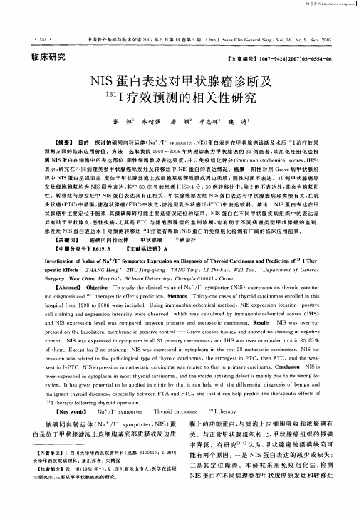
测 NI S蛋 白 在 细 胞 中 的 表 达 部 位 、 阳性 细胞 数 及 表 达 强 度 , 并 以免疫 组化评 分 ( i mmu n o h i s t o c h e mi c a l s c o r e s , I HS ) 表示 , 研 究 在 不 同病 理 类 型 甲状 腺 癌原 发 灶及 转 移 灶 中 NI S蛋 白的 表 达 情 况 。结 果 阳 性 对 照 Gr a v e 病 甲 状 腺 组 织 中 NI S蛋 白呈 强 表 达 , 定 位 于 甲 状腺 滤 泡 上 皮 细 胞 基 底 部 质 膜 或 周 边 质 膜 ;阴 性 对 照 不 表 达 3 1 例 甲状 腺 癌 原 发 灶 细 胞 胞 浆 均 为 NI S阳 性 表 达 , 其中 8 O . 6 5 %的患者 I HS  ̄4分 ; > 3 0例 转 移 灶 中 , 除2 例不表达外 , 其余为胞浆 阳 性 。转 移 灶 与 原 发 灶 中 NI S蛋 白表 达 具 有 正 相 关 ;甲 状 腺 癌 原 发 灶 NI S蛋 白表 达 与 甲 状 腺 癌 病 理 类 型 有 关 , 在 乳 头状癌( P TC ) 中最 强 , 滤泡状腺癌( F T c ) 中 次之 , 滤泡型乳头状癌 ( f v P TC ) 中表 达 较 弱 。结 论 NI S蛋 白 表 达 在 甲
临床 研 究
【 文 章 编 号 】 1 0 0 7 — 9 4 2 4 ( 2 0 0 7 ) 0白表 达 对 甲状 腺 癌 诊 断及 I 疗 效 预测 的相 关 性 研 究
张 恒 朱精 强 唐 颖 李 志辉 魏 涛
【 摘要】 目的
维普资讯
中 国普 外 基 础 与 l 临床 杂 志 2 0 0 7年 9月第 1 4卷 第 5期
碘转运体在分化型甲状腺癌及其淋巴转移灶中的表达和分布
钠/碘转运体(sodium /iodide symporter ,NIS )是甲状腺滤泡细胞基底膜上的一种跨膜糖蛋白,它由643个氨基酸组成,它能选择性、主动性地转运血液中的碘到滤泡上皮细胞[1]。
研究表明,NIS 对于活性碘的转运不仅是生理合成甲状腺激素的重要限速步骤,同时也是临床上利用放射性碘的方法来评价、诊断和治疗甲状腺疾病以及非甲状腺肿瘤的基础。
以往都认为导致甲状腺癌组织对放射性核素摄取降低的原因是NIS 的低表达或不表达,但近期少量文献报道,NIS 蛋白在甲状腺乳头状癌中是正常表达,甚至过度表达。
本实验组已用蛋白免疫印迹技术(Western blot 法)检测发现NIS 蛋白在分化型甲状腺癌原发灶、淋巴转移灶及癌旁组织中均有表达,并且NIS 在甲状腺癌细胞中的表达主要集中在细胞浆内,而功能靶位细胞膜上仅有小量表达[2]。
本钠/碘转运体在分化型甲状腺癌及其淋巴转移灶中的表达和分布胡阳英,沈美萍*(南京医科大学第一附属医院普外科,江苏南京210029)[摘要]目的:研究钠/碘转运体(sodium /iodide symporter ,NIS )在分化型甲状腺癌及其淋巴转移灶中的表达情况,探讨NIS与分化型甲状腺癌的关系及放射碘治疗分化型甲状腺癌的意义。
方法:用实时荧光定量聚合酶链反应(real time polymerasechain reaction ,RT-PCR )法及组织芯片免疫组化法检测分化型甲状腺癌原发灶,癌旁组织及其淋巴转移灶中NIS 表达情况。
结果:RT-PCR 示:癌旁组织和分化型甲状腺癌原发灶NIS 的mRNA 表达量无显著性差异,转移淋巴结组NIS mRNA 表达较其他组低。
免疫组化示:NIS 在分化型甲状腺癌原发灶、癌旁组织及淋巴转移灶中NIS 阳性表达率无统计学差异,但是NIS 在分化型甲状腺癌原发灶及淋巴转移灶胞膜中的表达比在癌旁组织胞膜中的表达低。
结论:在甲状腺分化型癌中,NIS 表达量正常,但NIS 蛋白在细胞中的定位发生了改变,主要表达在细胞浆内,并非表达在功能靶位细胞膜上,此结果对放射碘治疗甲状腺分化型癌有指导意义。
钠碘共同转运体在乳腺癌中的研究进展_董凤萍
钠碘共同转运体在乳腺癌中的研究进展收稿日期:2015-9-21;修回日期:2015-10-16通讯作者:聂建云,E -mail :njyvip@董凤萍,张杰,汤晗,李文斌,聂建云(昆明医科大学第三附属医院,云南昆明650100)摘要:钠碘共同转运体(sodium iodide symporter ,NIS )是甲状腺激素合成过程中转运碘的糖化膜蛋白,调控甲状腺细胞摄取碘,是甲状腺及其转移癌进行放射性碘治疗的基础。
研究显示放射性碘对乳腺癌也有治疗作用,乳腺癌能表达NIS 蛋白,研究NIS 在乳腺癌中的作用机制并进行干预,可能成为治疗乳腺癌的潜在靶点,NIS 介导放射性碘可能是治疗乳腺癌尤其是三阴性乳腺癌的一个新的方向。
关键词:钠碘共同转运体;乳腺癌;放射性碘中图分类号:R737.9文献标识码:A 文章编号:1004-0242(2016)04-0282-04doi :10.11735/j.issn.1004-0242.2016.04.A008Research Progress in Sodium Iodide Symporter in Breast CancerDONG Feng -ping ,ZHANG Jie ,TANG Han ,et al.(Affiliated Tumor Hospital of Kunming Medical University ,Kunming 650100,China )Abstract :Sodium iodide symporter (NIS)is a glycosylated membrane protein in the process of thy -roid hormone synthesis ,and NIS regulates iodine uptaken of thyroid cells ,which is the basis for the treatment of radioactive iodine in thyroid and its metastatic carcinoma.Studies have shown that radioactive iodine also has therapeutic effect on breast cancer.It has been reported that breast cancer can express NIS protein.To study the mechanism and intervention of NIS in breast cancer may become the potential target of treatment for breast cancer ,NIS mediated by radioactive iodine may be a new direction for the treatment of breast cancer ,especially three negative breast cancer.Key words :sodium iodide symporter (NIS);breast cancer ;radioactive iodine乳腺癌是全世界最常见的女性恶性肿瘤,约占女性恶性肿瘤的15%,致死率约为20%。
NIS蛋白在三阴性乳腺癌中的表达-4页word资料
NIS蛋白在三阴性乳腺癌中的表达钠/碘同向转运体(Na+/I-symporter,NIS)是一种介导碘转运的膜蛋白,主要位于甲状腺细胞基底膜,可使胞内碘含量浓聚为血浆的20~30倍。
NIS浓聚碘是甲状腺激素生物合成、诊断性甲状腺核素显像以及甲亢、甲状腺肿瘤放射性碘治疗的基础。
除甲状腺外,在乳腺、胎盘、唾液腺、消化腺和前列腺等组织中发现了NIS的存在;并经核酸序列测定证实非甲状腺组织的NIS 与甲状腺相同,只是表达的水平有差异。
其中乳腺组织中NIS的表达与功能正越来越引起研究者的关注。
经证实正常乳腺组织不表达NIS,但乳腺癌组织表达NIS[2]。
三阴性乳腺癌是指雌激素受体(ER)、孕激素受体(PR)和人表皮生长因子受体2(HER-2)均为阴性的乳腺癌,绝经前女性好发,约占乳腺癌的15%[1]。
由于缺乏雌孕激素受体,这种类型的乳腺癌主要依赖手术及全身化疗,不能采取有效的内分泌治疗,局部复发和远处转移率均较非三阴性乳腺癌高而成为近年来乳腺癌亚型中的研究热点。
如能证实NIS在其组织上强表达将为131Ⅰ治疗三阴性乳腺癌提供重要理论依据。
1 资料与方法1.1 一般资料选取2008年7 月至2010 年7月期间于河南科技大学附属第一医院外科行手术治疗的180 例患者的乳腺癌组织,从中选取三阴性乳腺癌(TNBC)30例作为实验组。
所有患者均为女性年龄31~59岁,中位年龄45岁。
绝经前女性16例,绝经后14例,肿瘤0.05),具有可比性。
1.2 操作步骤全部病例组织蜡块采用连续切片,切片厚度为4 μm,取相邻2张切片分别行HE染色及免疫组化染色。
免疫组化一抗为鼠抗人单克隆抗体(NIS),购自美国Labvision公司,SP 试剂盒购自福建迈新公司;设Grave病甲状腺组织作为阳性对照及PBS液代替一抗作为阴性对照。
1.3 结果判定由我院两位高职称病理医师盲法读片,在光镜下确定阳性细胞百分数及染色表达强度,不一致的结果通过共同协商讨论达成一致。
NIS基因在肿瘤显像和治疗中的应用
b o n e ma r r o w me s e n c h y ma l s t e m c e l l s , r B MS C ) , 转 染 细 胞 移 植
1 . 1 报 告 基 因显 像 报 告 基 因 显 像 主 要 是 利 用 基 因 融 合 、 双
顺 反子、 双 启 动 子 及 双 向 转 录等 重 组 技 术 , 构 建 表 达 报 告 基 因
基 因转 染 介 导 放 射 性 核 素 显 像 和 治 疗 是 将 基 因转 染 与 核 素 显 像 和治 疗 相 结合 的 方 法 , 基 因转 粱 使 肿 瘤 细胞 表 达 高水 平 的抗 原 、 受体或膜蛋 白, 特 异 性 地 摄 取 某 一 放 射 性 核 素 或 其 标 记物 , 达 到 实 现 放 射 性 核 素 显 像 诊 断 和 内照 射 治 疗 的 目的 。钠
MR I 、 生物发光和放射性核素显像 , 其 中 NI S作 为 报 告 基 因 可
直 接 静 脉 注 射 放 射 性 核 素 进 行 显 像 。胡 硕 等 l _ 5 将 A d — r NI S - E G F P重 组 腺 病 毒 载 体 转 染 至 大 鼠 骨 髓 问 充 质 干 细 胞 ( r a t
hNIS转染结肠癌SW480细胞介导放射性碘治疗的实验研究的开题报告
hNIS转染结肠癌SW480细胞介导放射性碘治疗的实验研究的开题报告一、研究背景和意义放射性碘治疗已成为结直肠癌等恶性肿瘤的重要治疗手段之一,但治疗效果存在一定局限性,主要原因是肿瘤细胞中内源性碘摄取量低,限制了放射性碘在肿瘤细胞内的积累和治疗效果。
因此,通过基因转移技术将人钠碘转运体(hNIS)基因导入肿瘤细胞,增强肿瘤细胞对碘的摄取能力,是提高放射性碘治疗效果的有效手段。
近年来,各种肿瘤细胞中的hNIS基因转移技术的研究越来越成熟,但对于结肠癌SW480细胞的hNIS基因转移尚缺乏深入研究。
因此,本研究旨在通过hNIS转染结肠癌SW480细胞,探讨hNIS转移对SW480细胞内碘的摄取能力及放射性碘治疗效果的影响,从而为进一步提高放射性碘治疗效果提供实验依据。
二、研究内容和方法本研究将采用SW480细胞作为研究对象,获得hNIS基因转染的SW480hNIS细胞株和对照组SW480细胞株。
首先,利用RT-PCR和Western blotting方法检测hNIS基因在细胞内的表达情况。
其次,使用125I标记的碘化物对SW480hNIS和SW480细胞进行体外摄取实验,评估hNIS转移后SW480细胞对碘的摄取能力的变化。
最后,分别对SW480hNIS和SW480细胞株进行放射性碘治疗,比较两个细胞株的生长程度和凋亡情况,评估hNIS转移对放射性碘治疗效果的影响。
三、预期结果和展望本研究预计通过SW480细胞的hNIS转染,获得表达hNIS的SW480hNIS细胞株,证实hNIS基因的转移对SW480细胞内碘的摄取能力有明显提高,进一步验证了hNIS基因转移技术可以增强肿瘤细胞对碘的摄取能力的假设。
同时,预期获得的结果将为优化放射性碘治疗方案提供实验依据,推进放射性碘治疗的研究和临床应用。
钠碘转运体在甲状腺乳头状癌中的表达
钠碘转运体在甲状腺乳头状癌中的表达摘要】目的探讨钠碘转运体(sodium/iodide symporter,NIS)蛋白在甲状腺乳头状癌表达。
方法收集甲状腺乳头状癌根治术后患者60例,结节性甲状腺肿15例。
通过免疫组化法对NIS蛋白表达进行分析,根据表达强弱分为0,1,2,3分,对两组患者NIS蛋白表达水平进行t检验,并观察NIS蛋白表达部位分布情况。
结果甲状腺乳头状癌患者NIS表达主要定位于细胞膜,而结节性甲状腺肿NIS蛋白阳性颗粒主要定位于细胞浆,表达阳性评分分别为1.45±0.77和1.07±0.47,甲状腺乳头状癌NIS蛋白表达强度显著高于结节性甲状腺肿组(P<0.05)。
结论甲状腺乳头状癌的NIS表达水平升高,但是存在功能定位障碍。
【关键词】甲状腺乳头状癌钠碘转运体钠碘转运体(sodium/iodide symporter,NIS)主要位于甲状腺滤泡上皮细胞的基底膜上,主要功能是作为碘泵转运血液内碘离子进入细胞,参与甲状腺激素的合成[1]。
甲状腺癌乳头状癌是最常见的甲状腺癌病理类型,占所有类型的80%。
131I治疗是甲状腺乳头状癌根治术后最主要的辅助治疗方法,131I通过钠碘转运体从血液中摄取后对肿瘤细胞进行放射治疗。
但并非所有甲状腺乳头状癌都对131I治疗敏感,这是因为在其演进过程中可发生去分化,NIS表达障碍,失去摄碘能力。
本文对我院甲状腺乳头状癌的NIS表达进行了研究。
1 资料与方法1.1 临床资料回顾分析我院2008.3-2011.1甲状腺乳头状癌根治术后60例患者完整临床资料,男性10例,女性50例,年龄17~68岁,平均(38.6±10.8)岁。
收集结节性甲状腺肿手术患者15例,男性4例,女性11例,年龄22~59岁,平均(36.4±11.5)岁。
所有患者术前均未行内、外放射治疗,标本经常规病理诊断。
1.2 NIS免疫组化染色全部病例石蜡标本均为10%中性甲醛固定,常规组织处理、石蜡包埋。
钠碘同向转运体相关文献4
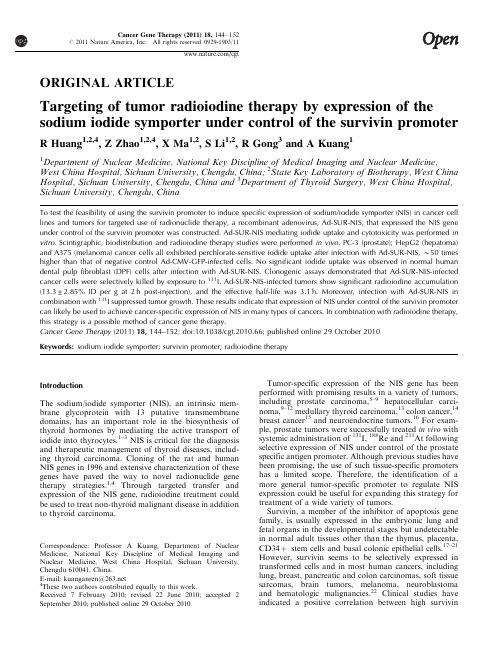
ORIGINAL ARTICLETargeting of tumor radioiodine therapy by expression of the sodium iodide symporter under control of the survivin promoterR Huang 1,2,4,Z Zhao 1,2,4,X Ma 1,2,S Li 1,2,R Gong 3and A Kuang 11Department of Nuclear Medicine,National Key Discipline of Medical Imaging and Nuclear Medicine,West China Hospital,Sichuan University,Chengdu,China;2State Key Laboratory of Biotherapy,West China Hospital,Sichuan University,Chengdu,China and 3Department of Thyroid Surgery,West China Hospital,Sichuan University,Chengdu,ChinaTo test the feasibility of using the survivin promoter to induce specific expression of sodium/iodide symporter (NIS)in cancer cell lines and tumors for targeted use of radionuclide therapy,a recombinant adenovirus,Ad-SUR-NIS,that expressed the NIS gene under control of the survivin promoter was constructed.Ad-SUR-NIS mediating iodide uptake and cytotoxicity was performed in vitro .Scintigraphic,biodistribution and radioiodine therapy studies were performed in vivo .PC-3(prostate);HepG2(hepatoma)and A375(melanoma)cancer cells all exhibited perchlorate-sensitive iodide uptake after infection with Ad-SUR-NIS,B 50times higher than that of negative control Ad-CMV-GFP-infected cells.No significant iodide uptake was observed in normal human dental pulp fibroblast (DPF)cells after infection with Ad-SUR-NIS.Clonogenic assays demonstrated that Ad-SUR-NIS-infected cancer cells were selectively killed by exposure to 131I.Ad-SUR-NIS-infected tumors show significant radioiodine accumulation (13.3±2.85%ID per g at 2h post-injection),and the effective half-life was 3.1h.Moreover,infection with Ad-SUR-NIS in combination with 131I suppressed tumor growth.These results indicate that expression of NIS under control of the survivin promoter can likely be used to achieve cancer-specific expression of NIS in many types of cancers.In combination with radioiodine therapy,this strategy is a possible method of cancer gene therapy.Cancer Gene Therapy (2011)18,144–152;doi:10.1038/cgt.2010.66;published online 29October 2010Keywords:sodium iodide symporter;survivin promoter;radioiodine therapyIntroductionThe sodium/iodide symporter (NIS),an intrinsic mem-brane glycoprotein with 13putative transmembrane domains,has an important role in the biosynthesis of thyroid hormones by mediating the active transport of iodide into thyrocytes.1–3NIS is critical for the diagnosis and therapeutic management of thyroid diseases,includ-ing thyroid carcinoma.Cloning of the rat and human NIS genes in 1996and extensive characterization of these genes have paved the way to novel radionuclide gene therapy strategies.1,4Through targeted transfer and expression of the NIS gene,radioiodine treatment could be used to treat non-thyroid malignant disease in addition to thyroid carcinoma.Tumor-specific expression of the NIS gene has been performed with promising results in a variety of tumors,including prostate carcinoma,5–9hepatocellular carci-noma,9–12medullary thyroid carcinoma,13colon cancer,14breast cancer 15and neuroendocrine tumors.16For exam-ple,prostate tumors were successfully treated in vivo with systemic administration of 131I,188Re and 211At following selective expression of NIS under control of the prostate specific antigen promoter.Although previous studies have been promising,the use of such tissue-specific promoters has a limited scope.Therefore,the identification of a more general tumor-specific promoter to regulate NIS expression could be useful for expanding this strategy for treatment of a wide variety of tumors.Survivin,a member of the inhibitor of apoptosis gene family,is usually expressed in the embryonic lung and fetal organs in the developmental stages but undetectable in normal adult tissues other than the thymus,placenta,CD34þstem cells and basal colonic epithelial cells.17–21However,survivin seems to be selectively expressed in transformed cells and in most human cancers,including lung,breast,pancreatic and colon carcinomas,soft tissue sarcomas,brain tumors,melanoma,neuroblastoma and hematologic malignancies.22Clinical studies have indicated a positive correlation between high survivinReceived 7February 2010;revised 22June 2010;accepted 2September 2010;published online 29October 2010Correspondence:Professor A Kuang,Department of Nuclear Medicine,National Key Discipline of Medical Imaging and Nuclear Medicine,West China Hospital,Sichuan University,Chengdu 610041,China.E-mail:kuanganren@ 4These two authors contributed equally to this work.Cancer Gene Therapy (2011)18,144–152r2011Nature America,Inc.All rights reserved 0929-1903/11 /cgtexpression levels and poor prognosis,accelerated rate of recurrence and increased resistance to therapy in many cancer patients.23Previous studies have identified a putative region of the survivin promoter that is likely responsible for the induction of specific,high-level expression in tumors.24,25The survivin promoter contains multiple cell cycle-dependent elements and a cell cycle gene homology region.This region has been suggested to control expression of various G2-M-regulated genes, including the survivin gene,in a manner correlating with G2-M cell cycle periodicity.24In this study,we have developed a tumor-targeting adenovirus that expresses the NIS gene under control of the survivin promoter.We investigated the utility of this system for molecular imaging and radioiodine gene therapy.The results reported herein demonstrate the potential efficacy and selectivity of a combination of radioiodine therapy and adenovirus-mediated expression of the NIS gene under the control of the survivin promoter for cancer therapy.Materials and methodsCell linesThe human prostate cancer cell line,PC-3;the human hepatocellular carcinoma cell line,HepG2;the human melanoma cell line,A375and the adenoviral transformed human embryonic kidney cell line,293,were purchased from the American Type Culture Collection(ATCC, Rockville,MD).All cancer cell lines were cultured in RPMI1640medium supplemented with10%calf serum (Gibco,Carlsbad,CA),2m M glutamine,100IU ml–1 penicillin and100ng ml–1streptomycin.Cells were main-tained at371C in a humidified atmosphere containing5% CO2.Both293cells and a control human dental pulp fibroblast cell line(DPF;a kind gift from Dr Peng Li, West China College of Stomatology,Sichuan University) were cultured in Dulbecco’s Modified Eagle Medium (high glucose)medium supplemented with10%FBS and 100IU ml–1penicillin/streptomycin(100ng ml–1).Construction of plasmids and recombinant adenovirus In order to generate the luciferase expression plasmid under the control of the survivin promoter(pSUR-LUC), an B260bp fragment of the human survivin gene promoter(B222toþ39,GeneBank Accession Number AY795969)was amplified from PC-3genomic DNA by PCR.This region contains two cell cycle-dependent elements and one cell cycle gene homology region.The sequences of the oligonucleotide primers were as follows: forward primer,50-AGATCTGCACGCGTTCTTTGAA AGC-30,and0-AAGCTTCCACCTCTG CCAACGGGTC-3,which allowed for the introduction of Bgl II and Hin dIII sites(underlined in the respective sequences).The survivin promoter was T/A cloned into the PMD-T18vector for sequencing,and then subcloned using the Bgl II and Hin dIII sites into the luciferase vector pGL3-basic(Promega,Madison,WI)to generate pSUR-Luc(Figure1a).In order to generate the NIS constructs,the coding region of NIS was amplified from normal human thyroid tissue by RT–PCR(nucleotides348to2280,GeneBank Accession Number,NM_000453),followed by T/A cloning into the PMD-T18vector for sequencing.The sequences of the oligonucleotide primers were as follows: forward primer,50-CCCAAGCTTATGGAGGCCGTG GAGACCGGGGAAC-30,and reverse primer,50-GCG TCTAGATCAGAGGTTTGTCTCCTGCTG-30,which allowed for the introduction of Hin dIII and Xba Isites(underlined in the respective sequences).The luciferase gene of pSUR-Luc was replaced with hNIS by subcloning into the Hin dIII and Xba I sites.The survivin promoter-hNIS-SV40polyA cassette was then isolated by digestion with Kpn I and Sal I and cloned into the pShuttle-1vector(Stratagene,Cedar Creek,MD)to generate pS-SUR-NIS(Figure1b).Subsequently,homo-logous recombination was performed using the Adeasier system(Stratagene)in BJ5183Escherichia coli to generatepAd-SUR-NIS.The Ad-SUR-NIS virus was packaged by transfecting4m g of Pac I-digested pAd-SUR-NIS DNAinto293cells using Lipofectamine2000(Invitrogen, Carlsbad,CA)and propagating in293cells according tothe standard procedure.After two-step purification onCsCl gradients,viral stocks were desalted using Pharma-cia G50columns(Orsay,France)and frozen atÀ801C in10mM Tris-HCl(pH7.5)containing2.5%glycerol.Viraltiters were calculated by a dilution plaque assay in293cells and expressed as PFU per ml.Purification and plaque assays were performed by SinoGenoMax,Beijing, China.The recombinant adenovirus,Ad-CMV-NIS, which uses the CMV promoter to drive NIS expression (previously described26),was used as a positive control.The recombinant adenovirus,Ad-CMV-GFP,which provided by SinoGenoMax,was used as a negativecontrol.Figure1Structures of plasmids used in this study.(a)Diagram represents the structure of the luciferase reporter plasmid.The survivin promoter(À222to39bp)was cloned upstream of the luciferase gene using the restriction sites indicated.(b)Diagram represents the structure of the adenovirus shuttle plasmid pS-SUR-NIS.The plasmid was constructed by inserting a DNA fragment intothe E1-deleted region using the Kpn I/Sal I restriction sites.This DNA fragment contained the survivin promoter,the full length NIS cDNA,and the Simian Virus40polyadenylation signal.Targeted radioiodine therapy for tumorsR Huang et al145Cancer Gene TherapyAnalysis of the survivin promoter activity in vitroIn order to determine the transcriptional activation activity of the survivin promoter,we utilized the Dual-Luciferase reporter assay system(Promega).The plasmid pRL-TK,which contains the Renilla luciferase gene under the control of the thymidine kinase promoter, was used as an internal control.In all,1Â106cells of each cancer cell line and normal control were plated in six-well tissue culture plates and24h later,the cells were transfected with4m g of the indicated plasmid DNA and 0.1m g of pRL-TK using Lipofectamine2000(Invitrogen) according to the manufacturer’s protocol.At48h post-transfection,luciferase activity was determined using the Reporter Lysis Buffer and Luciferase Assay System according to the manufacturer’s protocol(Promega). The dual luciferase ratio was defined as the luciferase activity of the indicated plasmid divided by the luciferase activity of pRL-TK.The pSV40-LUC vector(Promega) that contains Simian Virus40(SV40)promoter was used as a positive control.All experiments were performed in triplicate.In vitro125I and99m Tc pertechnetate uptake experimentsIn all,1Â106cells of each cancer cell line and control DPF cells were plated in six-well tissue culture plates and allowed to reach exponential growth for24h and infected with Ad-SUR-NIS,Ad-CMV-NIS(positive control)or Ad-CMV-GFP(negative control)viruses at MOI¼100in 1ml of medium without serum.After2h,the media was changed to complete medium.For each cell line,the medium used for infection was the same as the culture medium.Approximately28–30h after viral infection,cells were washed once with1ml of phosphate-buffered saline (PBS).Iodide uptake was then initiated by adding1ml of medium without serum containing3.7KBq of125I or 99m Tc pertechnetate per well.Following30min incuba-tion with radionuclide,cells were washed twice with ice-cold PBS and incubated for20min in1ml of ice-cold ethanol.The ethanol was then recovered,and radio-activity was quantified(c.p.m.)using a g-counter(No.262 Factory,Xi’an,China).For inhibition experiments,cells were incubated with both300m M NaClO4and125I for 30min,followed by quantification of125I uptake as described above.In vitro cell killing with131I and clonogenic assayCell killing with131I was performed according to the method of Boland et al.27described as following:cells were seeded in24-well tissue culture plates and divided into three groups.The first two groups were infected with the Ad-SUR-NIS adenovirus and the Ad-CMV-GFP virus,respectively.Thirty hours after infection,cells were washed once with PBS and incubated with1ml of serum-free medium containing370KBq131I.The third group was not infected and was incubated in serum-free medium.After7h,cells from all three conditions were washed twice with PBS,trypsinized,and counted.For each condition,cells were plated in triplicate in six-well tissue culture plates(1000cells per well)and incubated for 1week at371C.Cells were then washed once with PBS and stained with crystal violet solution(2g l–1crystalviolet,4%formaldehyde,20%ethanol).Colonies of430cells were counted,and the mean and s.d.of the number of colonies were determined for each condition.Resultswere expressed as the percentage of surviving cells,defined as the percentage of colonies obtained after treatment with131I compared with no intervention,and are representative of two independent experiments. Biodistribution of125I in tumor-bearing mice Approximately2Â107PC-3cells were subcutaneouslytransplanted into the left or right flank of6-week-old Balb/c nude mice and allowed to proliferate until the tumor reached a diameter of8–10mm.At this point, the Ad-SUR-NIS virus or the Ad-CMV-GFP virus (negative control)was injected into the tumors(both 1Â109PFU).Radionuclide uptake was assessed2days later by intravenous injection with370KBq of125I.At2, 4,6and12h post-injection,animals were killed,and selected organs(bone,muscle,heart,lung,liver,spleen, stomach,intestine,kidney,brain and tumors)were dissected,weighed and counted for radioactivity. Scintigraphic imaging of tumor-bearing mice in vivo Forty-eight hours after intratumoral injection of 1Â109PFU Ad-SUR-NIS or Ad-CMV-GFP(negative control),PC-3tumor-bearing mice were injected with 18.5MBq of99m Tc pertechnetate via the tail vein.After 2h,mice were anesthetized with ketamine and xylazine and scanned with a g-camera equipped with a pinhole collimator(Philips Medical Systems,Milpitas,CA). Effects of NIS expression on131I therapy in vivo Accordingly,the PC-3tumors were induced in Balb/c nude mice as described above.When the tumors reached 8–10mm in diameter,the mice were given thyroxine (Sigma-Aldrich,St Louis,MO)at a concentration of 5mg l–1in the drinking water for7days before treatment. Two days before treatment,the animals were divided into two groups of three animals each.One group received an intratumoral injection of1Â109PFU Ad-SUR-NIS.The other group received an intratumoral injection of 1Â109PFU Ad-CMV-GFP.Forty-eight hours later, both groups were given an intraperitoneal dose of 111MBq of131I(the dose same with Schipper et al.16at their NIS mediate-131I therapy experiment).The length and width of the tumors were measured with a caliper at 5-day intervals after treatment.Tumor size was calculated using the formula:lengthÂwidth2/2.All mice were followed for a total of30days and then euthanized. Tumor samples and immunohistochemical stainingIn order to detect NIS expression,resected tumors from nude mice were fixed in4%paraformaldehyde for24h. Samples were immunostained using a standard streptavidin-biotin labeling protocol.Briefly,after deparaffinization and antigen retrieval,5m m tissue sections were incubated with NIS antibody(mouse monoclonal,1:100,Chemicon, Temecula,CA)at371C for1h then at41C overnight.Targeted radioiodine therapy for tumorsR Huang et al 146Cancer Gene TherapyThe sections were then incubated with biotinylated goat anti-mouse immunoglobulin G(1:200,Zymed Labora-tories,San Francisco,CA)and subsequently incubated with horseradish peroxidase-labeled streptavidin(1:200, Zymed Laboratories).The cells were then counterstained with3,30-diaminobenzidine for analysis.Statistical analysisAll data are expressed as mean±s.d.Significance was determined by the Student’s t-test.P o0.05was considered to be statistically significant.ResultsCancer-specific activation of the survivin promoterin vitroTo assess whether the activation of the survivin promoter is cancer specific and to compare the transcriptional activation activity between the survivin promoter and the SV40promoter,we transfected both cancer(PC-3, HepG2,A375)and normal(DPF)cells with plasmids containing the luciferase gene under control of either the survivin promoter(pSUR-Luc)or the SV40promgfeoter (pSV40-Luc).Survivin promoter activity was generally very high in cancer cell lines and very low in normal cells.The mean dual reporter ratio for cancer and normal cell lines was 6.76±0.83and0.33±0.23,respectively (Figure2).However,the activity of the SV40promoter was similar in both cancer cell lines and normal cells,with a mean dual reporter ratio of2.23±0.08and2.23±0.19, respectively(Figure2).These data suggest that the survivin promoter is selectively active in cancer cells compared with normal cells.Ad-SUR-NIS is functional in various human cancer cell linesAn adenovirus that expresses NIS under control of the survivin promoter(Ad-SUR-NIS)or the CMV promoter (Ad-CMV-NIS)and a control virus expressing GFP under control of the CMV promoter(Ad-CMV-GFP) were generated and used to infect cancer cell lines(PC-3, HepG2,A375)and non-malignant control cells(DPF).In order to test the expression of NIS in these cell lines, functional assays quantifying radioiodine uptake were performed.Thirty hours after adenovirus infection,iodide uptake was measured by incubating the cells for30min with3.7KBq of125I.As shown in Figure3,all cancer cell lines infected with Ad-SUR-NIS exhibited significant iodide uptake,with a mean value of12092±977c.p.m. This value represented a45–56-fold increase over control Ad-CMV-GFP-infected cells(237±36c.p.m.,P¼0.000). No significant iodide uptake was evident in DPF cells infected with Ad-SUR-NIS(300±42c.p.m.),consistent with luciferase reporter assays indicating the specificity of the survivin promoter for cancer cells(Figure3).Both cancer and normal cell lines infected with the general NIS expression virus Ad-CMV-NIS exhibited significant iodide uptake.The mean amount of iodide uptake for all Ad-CMV-NIS-infected cells was23256±3806c.p.m.,B85–120times higher than Ad-CMV-GFP-infected cells, indicating the efficacy of NIS expression for induction of radioiodine uptake(Figure3).In addition to125I uptake, cancer cell lines infected with Ad-SUR-NIS also exhibited selective and increased99m Tc pertechnetate uptake.The mean amount of99m Tc pertechnetate uptake was 11906±1424c.p.m.for cancer cells(Figure3).In order to further demonstrate that uptake of125I wasdependentFigure2In vitro analyses of transgene expression of the survivinand SV40promoters by transient transfection.The transcriptional activity of the survivin and SV40promoters was measured in various cancer and normal cells.The data shown are the means±s.d.of atleast three independentexperiments.Figure3In vitro radioiodine and99m Tc pertechnetate uptake.PC-3,HepG2and A375cancer cell lines and the control DPF cell linewere infected with Ad-SUR-NIS,Ad-CMV-NIS or Ad-CMV-GFP viruses and subjected to either125I or99m Tc pertechnetate uptake.Ad-SUR-NIS-infected PC-3,HepG2and A375cancer cell lines allshow significant125I and99m Tc pertechnetate uptake ability compared with Ad-CMV-GFP-infected controls.No significant uptakewas observed in Ad-SUR-NIS-infected DPF cells.Ad-CMV-NIS infection resulted in increased125I uptake in all cell lines.Cotreat-ment with NaClO4completely inhibited125I uptake in Ad-SUR-NIS-infected cancer cells.Targeted radioiodine therapy for tumorsR Huang et al147Cancer Gene Therapyupon NIS expression in Ad-SUR-NIS-infected cells,cells were treated with the specific NIS inhibitor,sodium perchlorate during iodide uptake.Figure3shows that sodium perchlorate treatment inhibited iodide accumula-tion by92.5±1.2%(P¼0.000).Ad-SUR-NIS-infected human cancer cells are efficiently killed by131I in vitroIn order to demonstrate selective killing of cancer cells that could be obtained using Ad-SUR-NIS infection in combination with radioactive iodide treatment,131I uptake experiments were performed on Ad-SUR-NIS-infected PC-3,A375,HepG2and DPF cells.Ad-CMV-GFP-infected cells treated with131I were used as controls. After131I treatment,clonogenic assays were performed, and results were expressed as the percentage of surviving cells compared with untreated controls.Following ex-posure to131I,o10%of Ad-CMV-GFP-infected cells were killed compared with untreated controls.However, 86±3.5%,92±3.0%and89±4.5%of PC-3,A375and HepG2cells,respectively,were killed by131I following infection with Ad-SUR-NIS.No significant cell death was observed in Ad-SUR-NIS-infected DPF cells treated with131I,as91±2.3%of cells were recovered.These results demonstrate that coupling Ad-SUR-NIS infection and131I treatment efficiently and specifically leads to cancer cell killing in vitro.125I biodistribution studies in tumor-bearing miceIn order to extend our in vitro results using the Ad-SUR-NIS virus to an in vivo model,we used nude mice bearing tumors generated by subcutaneous transplantation of PC-3cells as a model.Tumors were injected with either the Ad-SUR-NIS virus or the control Ad-CMV-GFP virus,and mice were subjected to125I treatment48h later. Quantitation of the uptake of125I(%ID per g)in tumors and various organs was evaluated at2,4,6and12h after 125I administration(Table1).PC-3tumors infected with Ad-SUR-NIS accumulated13.3±2.85%ID per g at2h, 5.61±1.38%ID per g at4h,2.74±0.21%ID per g at6h and0.68±0.05%ID per g at12h,with a mean effective half-life of 3.1±0.6h.All other organs accumulated negligible amounts of isotope(below2.5%ID per g at all times)except stomach.The biodistribution data are summarized in pared with Ad-SUR-NIS-infected tumors,negative control tumors infected with Ad-CMV-GFP accumulated0.66±0.24%ID per g125I at 2h after administration(P¼0.000).At4,6and12h, they accumulated0.42±0.44%ID per g(P¼0.001), 0.34±0.04%ID per g(P¼0.000)and0.06±0.01%ID per g(P¼0.014),respectively.These data indicate that infection of tumors with the Ad-SUR-NIS virus leads to selective uptake of125I in vivo.Visualization of Ad-SUR-NIS-infected tumors by99m Tc pertechnetate scintigraphyIn order to test whether Ad-SUR-NIS-infected tumors could be visualized in vivo,scintigraphy was performed on tumor-bearing mice using99m Tc pertechnetate,allowing imaging with a pinhole collimator.99m Tc pertechnetate (18.5MBq)was injected intraperitoneally,and images were taken after2h(Figure4).As expected,tissues naturally expressing NIS(stomach and thyroid)or involved in iodide elimination(bladder)were visualized. The Ad-SUR-NIS-infected tumor was clearly visible,with intensity comparable to that of the thyroid or the bladder. However,the Ad-CMV-GFP-infected PC-3tumor was not visible.Decreased tumor growth induced by Ad-SUR-NIS infection in combination with131I treatmentIn vitro studies suggested that Ad-SUR-NIS infection combined with radioiodine treatment can inhibit growth of cancer cells.In order to extend these results to an in vivo model,tumors were generated in Balb/c nude mice and infected with either Ad-SUR-NIS or Ad-CMV-GFP. After48h,mice were treated with131I as described in the Materials and methods.As shown in Figure5,tumors inTable1Biodistribution of125I pertechnetate in mice with Ad-SUR-NIS-injected PC-3xenografts(%ID per g tissue;mean and s.d.after intravenous injection)Organ2h4h6h12h Stomach14.7±0.729.42±1.59 4.44±0.860.54±0.12 Tumor13.3±2.85 5.61±1.38 2.74±0.210.68±0.05 Heart 1.25±0.230.31±0.270.29±0.100.14±0.01 Liver 1.21±0.260.45±0.250.46±0.170.20±0.04 Spleen 1.00±0.310.53±0.260.56±0.280.12±0.01 Lung 1.48±0.340.66±0.250.53±0.260.15±0.02 Kidney 2.10±0.510.89±0.190.66±0.270.16±0.03 Intestinal 1.78±0.690.37±0.430.53±0.290.17±0.03 Muscle0.60±0.160.40±0.090.37±0.010.07±0.02 Bone 1.44±0.330.84±0.090.48±0.080.28±0.01 Brain0.16±0.040.04±0.110.07±0.010.05±0.03Figure499m Tc pertechnetate scintigraphic images of PC-3 xenografts infected with either Ad-CMV-GFP or Ad-SUR-NIS.Mice harboring PC-3xenograft tumors infected with either Ad-CMV-GFP (a)or Ad-SUR-NIS(b)were imaged with a pinhole collimator2h after injection of18.5MBq99m Tc pertechnetate.(a)The Ad-CMV-GFP-infected tumor is not visible using99m Tc pertechnetate imaging.(b)The Ad-SUR-NIS-infected tumor is clearly visible,with an intensity comparable to that of the thyroid or the bladder.Targeted radioiodine therapy for tumorsR Huang et al 148Cancer Gene Therapyanimals that received 131I and Ad-CMV-GFP continued to grow until the end of the experiment.In contrast,tumors in animals that received injections of both Ad-SUR-NIS intratumorally and 131I intraperitoneally began to show a slight reduction in growth on day 10and day 15,and from day 20to day 30,tumor volumes in this group were statistically different from Ad-CMV-GFP-infected controls.The volume reduction rates at days 20,25and 30were 32.9±11.3%(P ¼0.029),46.2±4.4%(P ¼0.001)and 51.4±15.9%(P ¼0.012),respectively.These results suggest that expression of NIS in combina-tion with 131I treatment may prove to be a possible strategy for reduction of tumor growth in vivo .Membrane expression of NIS in tumors injected with Ad-SUR-NISFor in vivo experiments,expression of the NIS protein in the tumor was characterized by immunohistochemical staining.Tumors injected with Ad-SUR-NIS werespecifically labeled with anti-NIS beling was clearly confined to cell membranes,indicating that expression of NIS driven by the Ad-SUR-NIS virus leads to correct localization of the NIS protein in vivo .Representative tumor sections are shown in Figure 6.DiscussionConsistent with the role of NIS in the success of radioiodine therapy for thyroid cancer,targeted expres-sion of NIS provides the possibility of NIS-targeted radionuclide therapy for treatment of non-thyroidal tumors.27–29In contrast to other potential therapeutic genes,therapeutic use of NIS has several advantages.The potential function of NIS as a diagnostic gene allows non-invasive radioiodine imaging to confirm and localize functional NIS expression before proceeding to the application of a therapeutic 131I dose.NIS gene therapy may also be more effective due to a substantial bystander effect caused by treatment with the b -emitter 131I,which has a path length of up to 2–4mm,and can cause the death of NIS-expressing cells as well as neighboring non-transfected cells.30The ultimate goal of cancer therapy is to maximize cancer cell-specific cytotoxicity and minimize toxic side effects in non-malignant cells.Gene therapy using tissue-specific promoters provides a means of selectively targeting therapeutic genes to malignant cells.31Several cancer-specific targeting promoters are available for targeting of individual cancers,including the prostate specific antigen promoter (prostate cancer),the albumin enhancer and promoter (hepatocellular cancer),the CEA promoter (medullary thyroid cancer,colorectal cancer),the MUC-1promoter (breast cancer,pancreatic cancer)and the chromogranin A promoter (neuroendocrine tumors).These promoters are cancer-type specific,but are limited in scope.The expression of survivin is noted in common human cancers but not in normal adult tissues.As the increased survivin activity is controlled transcriptionally,it has been suggested that the survivin promoter might control the transgene expression in a cancer-specific manner.32,33Figure 5Therapeutic effects of Ad-SUR-NIS infection in combina-tion with 131I treatment in PC-3xenografts.Tumor size was assayed after 131I treatment in nude mice bearing tumors infected with either Ad-SUR-NIS or Ad-CMV-GFP.Growth of Ad-SUR-NIS-infected PC-3tumors was significantly retarded (*,**,***P o 0.05)at days 20,25and 30after injection of 111MBq 131I.Figure 6Representative immunostaining of NIS in tumors from nude mice.Note the distinct cell membrane staining of NIS in the PC-3tumor infected with Ad-SUR-NIS (a ).In contrast,the non-infected PC-3tumor is negative for NIS immunoreactivity (b ).Original magnification for each panel was Â400.Targeted radioiodine therapy for tumors R Huang et al149Cancer Gene Therapy。
分化型甲状腺癌NIS与TSHR研究进展
分化型甲状腺癌NIS与TSHR研究进展茹晓婷;刘勤江;杨荣【摘要】目前,放射性核素131I已成为甲状腺癌治疗的一种重要手段.但对于某些碘抵抗的甲状腺癌患者来说,这种治疗方法已经失去了疗效.研究发现,NIS和TSHR 与甲状腺癌组织的碘代谢密切相关,通过将NIS基因转染到恶性肿瘤细胞中,会增加癌细胞的摄碘能力,从而使放射性核素治疗有意义.本文通过复习相关文献对NIS和TSHR在DTC中的研究进展进行综述.【期刊名称】《甘肃医药》【年(卷),期】2016(035)004【总页数】3页(P262-264)【关键词】分化型甲状腺癌;钠碘同向转运体;促甲状腺激素受体【作者】茹晓婷;刘勤江;杨荣【作者单位】730000甘肃兰州,兰州大学生命科学学院;730050甘肃兰州,甘肃省肿瘤医院头颈外科;730050甘肃兰州,甘肃省肿瘤医院病理科【正文语种】中文1.1 NIS的功能、分布与调控NIS主要分布于甲状腺滤泡上皮细胞的基底膜,借助于钠、钾离子交换所产生的离子浓度梯度将血液中的碘离子转运入甲状腺滤泡上皮细胞,是甲状腺合成甲状腺激素的一个限速步骤[1]。
NIS并不是甲状腺所特有,在其它正常组织器官如唾液腺、泪腺及胃黏膜和结肠黏膜以及乳腺癌中也表达,并且有聚碘能力。
人的NIS基因定位于染色体19p13,TSH通过不同的机制刺激甲状腺滤泡细胞的碘摄入,TSH可上调NIS的mRNA及蛋白表达。
其次,碘的过多摄入会抑制NISmRNA及蛋白的表达。
另外,一些细胞因子如甲状腺转录因子-1/2(TTF-1/2)、配对盒转录因子-8(Pax8)等正调节因子以及肿瘤坏死因子(TNF)、白细胞介素-6(IL-6)和干扰素等负调节因子都会不同程度影响NIS的表达。
除此之外,其他因子如维甲酸、雌激素还有阿霉素会促进NIS的表达。
1.2 NIS与甲状腺癌的诊断众所周知,BRAFT1799A(V600E)突变是甲状腺癌最常见的基因突变之一,有研究认为其过高的突变已经对患者预后评估价值不大。
新的甲状腺自身抗原钠/碘同向转运体与甲状腺疾病
新的甲状腺自身抗原钠/碘同向转运体与甲状腺疾病
李连喜
【期刊名称】《国外医学:内科学分册》
【年(卷),期】2000(027)010
【摘要】甲状腺钠/磺同向转运体(NIS)是主要存在于甲状腺细胞的一种糖化膜蛋白,它在甲状腺激素合成中起重要作用。
目前人NIS(hNIS)与鼠NIS(rNIS)的分子结构及其cDNA序列已经明确,并发现NIS异常与许多甲状腺疾病有关,近年更发现NIS是一种新的甲状腺自身抗原,与自身免疫性甲状腺病的发生密切相关。
对NIS研究有着诱人的临床应用前景。
【总页数】5页(P419-422,430)
【作者】李连喜
【作者单位】上海第二医科大学附属瑞金医院
【正文语种】中文
【中图分类】R581
【相关文献】
1.人钠碘转运体mRNA在良恶性甲状腺疾病中表达的研究 [J], 帅红霞;张木勋
2.钠/碘同向转运体在甲状腺癌131Ⅰ治疗中的应用 [J], 张一帆;李彪
3.碘钠同向转运体在新疆维吾尔族甲状腺乳头状癌中的表达及其临床意义 [J], 木扎帕尔·阿不都克热木;马斌林;阿里比亚提·艾尼
4.自身免疫性甲状腺疾病钠/碘同向转运体的相关性研究 [J], 王利强;郜风丽;刘淑娟;杨芬;周会杰;陈派强;郄永强;姚文森
5.甲状腺乳头状癌钠碘同向转运体基因启动子区域CpG岛异常甲基化状态检测及其意义 [J], 殷凯;张筱骅;王瓯晨;闫东升;陈晓燕;李权
因版权原因,仅展示原文概要,查看原文内容请购买。
分化型甲状腺癌病人NIS基因突变的检测

分化型甲状腺癌病人NIS基因突变的检测位俊慧;李贤;郝风云;林东亮;刘世国;孙振青【期刊名称】《齐鲁医学杂志》【年(卷),期】2017(32)5【摘要】目的研究分化型甲状腺癌(DTC)病人钠碘同向转运体(NIS)基因突变类型和特点,初步探讨其基因型和表现型的关系。
方法从50例DTC病人的甲状腺癌组织及正常甲状腺组织中提取基因组DNA,采用PCR扩增和直接测序的方法对NIS 基因全部外显子进行基因突变的检测。
结果在2例病人甲状腺癌组织中发现位于NIS基因第10外显子的1个基因突变(c.1571C>T,p.Leu 408=),尽管密码子发生变化,但是两组密码子均编码亮氨酸。
正常甲状腺组织中未发现基因突变。
甲状腺癌组织和正常甲状腺组织NIS基因突变频率差异无统计学意义(P>0.05)。
结论NIS基因突变可能与青岛地区DTC无相关性,检测到的同义突变在甲状腺摄碘过程中不起主要作用。
【总页数】4页(P508-511)【关键词】甲状腺肿瘤;钠碘同向转运体;突变【作者】位俊慧;李贤;郝风云;林东亮;刘世国;孙振青【作者单位】青岛大学附属医院普外科;城阳人民医院甲乳外科;青岛大学附属医院病理科;青岛大学附属医院产前诊断中心【正文语种】中文【中图分类】R736.1【相关文献】1.NIS和TSHR在分化型甲状腺癌中的表达及其与BRAF V600E突变的关系研究[J], 茹晓婷;刘勤江;杨荣2.NIS mRNA 在分化型甲状腺癌中的表达及其临床意义 [J], 许建林;梁晓燕;南永刚;赵华;施常备3.分化型甲状腺癌患者的NIS表达情况及其与甲状腺超声特征的关系 [J], 吕彦利;康秀梅;岳胜南;王燕4.NIS、Oct-4和CD133在碘难治性分化型\r甲状腺癌中的表达分析 [J], 李德宇;李娜5.谈谈有关分化型甲状腺癌的BRAF和TERT基因突变 [J], 王琳萍;李惠霖因版权原因,仅展示原文概要,查看原文内容请购买。
NIS基因转染肿瘤细胞介导131I治疗
NIS基因转染肿瘤细胞介导131I治疗
黄蕤
【期刊名称】《国际放射医学核医学杂志》
【年(卷),期】2001(025)005
【摘要】钠/碘同向转运体(NIS)是一种跨膜糖蛋白,具有主动运输碘的能力.NIS的异常与许多甲状腺疾病有关.将NIS基因转染到肿瘤细胞使其表达NIS并摄取131I 已获成功,但131I在细胞或病灶内停留的时间很短,达不到治疗剂量,故延长131I的有效半衰期和提高NIS转染率是今后研究的方向.
【总页数】6页(P204-209)
【作者】黄蕤
【作者单位】四川大学华西医院核医学科,四川,成都,610041
【正文语种】中文
【中图分类】R817.5
【相关文献】
1.hNIS基因转导黑色素瘤细胞介导放射性碘(125I,131I)摄取的体内实验 [J], 陈立波;朱瑞森;陆汉魁;余永利;罗全勇;于建芳
2.慢病毒转染造血干细胞介导的肿瘤基因治疗的研究进展 [J], 马巍;吴学元;陈炜;刘广宇
3.hNIS基因的靶向性转染介导肝癌131I治疗 [J], 陈立波;罗全勇;余永利;袁志斌;陆汉魁;郭礼和;朱瑞森
4.人钠/碘转运体基因转染结肠癌SW480细胞介导放射性碘治疗的实验研究 [J],
杜凡;周佰林;张红征;李焕斌;正伟;王玲;张琦
5.联合应用成纤维细胞介导的G-CSF基因疗法及IL-2基因疗法对肿瘤的治疗作用[J], 于益芝;曹雪涛;雷虹;董虹;章卫平
因版权原因,仅展示原文概要,查看原文内容请购买。
钠碘转运体与分化型甲状腺癌
钠碘转运体与分化型甲状腺癌
袁耿彪;匡安仁
【期刊名称】《中国介入影像与治疗学》
【年(卷),期】2005(002)001
【摘要】钠碘转运体(NIS)是甲状腺滤泡细胞基底膜上的一种糖蛋白,具有选择性和主动性转运碘(I-)到甲状腺滤泡细胞内的功能.NIS不仅存在于甲状腺,也存在于腮腺、胃黏膜、乳腺等多种组织.有报道,将NIS基因转染到NIS低表达的甲状腺癌细胞和其他类型的肿瘤细胞内,肿瘤细胞表达NIS,增加了131I的摄取.在分化型甲状腺癌(DTC)和其他肿瘤,131I治疗展现了良好的前景.
【总页数】4页(P6-9)
【作者】袁耿彪;匡安仁
【作者单位】四川大学华西医院核医学科,四川,成都,610041;四川大学华西医院核
医学科,四川,成都,610041
【正文语种】中文
【中图分类】R736.1;R814.42
【相关文献】
1.分化型甲状腺癌钠碘转运体的表达及其与甲状腺超声表现关系的研究 [J], 王伟伟;王斐;房世保;宁春平;阎胜利
2.碘对大鼠体内及体外甲状腺钠碘转运体mRNA表达的调节 [J], 聂秀玲;孙云;李
兰英;阎玉芹;陈祖培
3.人钠/碘转运体基因转染结肠癌SW480细胞介导放射性碘治疗的实验研究 [J],
杜凡;周佰林;张红征;李焕斌;正伟;王玲;张琦
4.人钠/碘同向转运体基因转染肺癌A549细胞介导放射性碘摄取实验研究 [J], 樊怿辉;樊冀伟;张裕东;王晓谭;严煜
5.碘对大鼠钠碘转运体基因启动子区甲基化的影响 [J], 董小婉;陈婷婷;吴翌;金秀兰;汪靓婧;周换丽;王佑民
因版权原因,仅展示原文概要,查看原文内容请购买。
人钠碘转运体基因转染人大细胞肺癌介导^(131)I治疗的实验研究
人钠碘转运体基因转染人大细胞肺癌介导^(131)I治疗的实验研究郑薇;李玮;李宁;谭建【期刊名称】《中国肺癌杂志》【年(卷),期】2009(12)6【摘要】背景与目的肺癌是严重危害人民生命和健康的常见病。
目前世界上倾向于将两类生物学行为不同的肺癌分为小细胞肺癌(SCLC)和非小细胞性肺癌(NSCLC)。
而NSCLC中的大细胞肺癌治疗效果多年来一直没有显著提高,一方面由于其生物学特性十分复杂、恶性程度高且多药耐药。
另一方面还在于约80%大细胞肺癌患者确诊时已属晚期而失去手术机会。
所以化疗在大细胞肺癌的治疗中是无可取代的,但多药耐药的出现令人沮丧。
于是,我们设想将转基因技术与放射性核素治疗相结合,将hNIS基因转染至人大细胞肺癌H460细胞系中,使原本不摄碘的H460细胞摄取碘。
然后利用放射性核素^131I的辐射生物效应,干扰H460细胞的生物代谢,抑制肿瘤细胞增值活性,最终使癌细胞死亡。
【总页数】2页(P468-469)【关键词】^131I治疗;人钠碘转运体;大细胞肺癌;基因转染;实验研究;放射性核素治疗;非小细胞性肺癌;介导【作者】郑薇;李玮;李宁;谭建【作者单位】天津医科大学总医院核医学科【正文语种】中文【中图分类】R581.1;R392.11【相关文献】1.裸鼠模型的人钠/碘转运体基因转染人大细胞肺癌介导放射性核素显像 [J], 郑薇;谭建;李玮;李宁;许静2.MR扩散加权成像在体评估人钠碘转运体转染鼻咽癌介导131Ⅰ治疗效果的实验研究 [J], 钟兴;史长征;弓健;郭斌;黄力;徐浩3.人钠/碘同向转运体基因转染胶质瘤细胞介导放射性碘治疗的研究 [J], 谭建;李玮;刘晓华;肖茜;贾强;李宁4.人钠/碘转运体基因转染结肠癌SW480细胞介导放射性碘治疗的实验研究 [J], 杜凡;周佰林;张红征;李焕斌;正伟;王玲;张琦5.人钠/碘同向转运体基因转染肺癌A549细胞介导放射性碘摄取实验研究 [J], 樊怿辉;樊冀伟;张裕东;王晓谭;严煜因版权原因,仅展示原文概要,查看原文内容请购买。
- 1、下载文档前请自行甄别文档内容的完整性,平台不提供额外的编辑、内容补充、找答案等附加服务。
- 2、"仅部分预览"的文档,不可在线预览部分如存在完整性等问题,可反馈申请退款(可完整预览的文档不适用该条件!)。
- 3、如文档侵犯您的权益,请联系客服反馈,我们会尽快为您处理(人工客服工作时间:9:00-18:30)。
Ab ta t sr c :Th o im o i es mp re ( I ) i n iti scp a ma m e b a ep o en t a e s d u idd y o t r N S Sa n rn i ls m r n r ti h t
m e a e c i e i di e t a s dit s a tv o d r n por n o t hy o d g a nd s v r le t a h o da i s s,i ti t he t r i l nd a e e a x r t yr i lts ue n p r i u a he l c a i n m a ma y glnd.Be a e o he c o i h r c e ia i n o S,is a tc l r t a t to m r a c us ft l n ng c a a t rz to fNI t
染技术 , 有望 将 ” I 疗 应 用 于 其 他 肿 瘤 , 在 治 疗 的 同时 对 肿 瘤 进 行 显 像 和 监视 。 治 并
关 键 词 : 碘 同 向 转 运 体 ; 瘤 ; 射 性 碘 治 疗 ; 因治 疗 钠 肿 放 基
中图 分 类 号 :R 1 ; 7 o 4 8 7 R 3. 4 文 献 标 志 码 :A 文 章 编 号 : 007 1 (0 8 0-1 70 1 0 -5 2 2 0 ) 30 7-7
维普资讯
第2卷 第 3 1 期
20 0 8年 8月
同 位
素
V0 . 1 No 3 I2 .
A u 20 g. 08
J u n l fIo o e o r a s tp s o
人钠 碘 同向转 运体 ( S 在 肿 瘤治 疗 中的研 究进 展 d p t o o y a d p y i l g o l e i v s i a e . Th r g e s wo l e e o e i h r i a h l g n h so o y c u d b n e tg t d ep o r s ud b
.
t p o c s c l xt nd t e ofr d o o de t e t ntt hy o d c nc ra on hv wo a pr a he ou d e e he US a i i di r a me o t r i a e nd n t — r da a e .One i y usng t i e ha im s i o vi umor us t a f r a o o oi lc nc r s b i he ma n m c n s nv l ng t o r ns o m r n t l t e tt u o ,b s d on t endu i S xp e so n t r i n a e T heo he s r a he t m r a e he r i cng NI e r s i n i hy o d a d c nc r t r i
刘 耘 ,赵 明
( 中科 技 大 学 同济 医学 院 附属 同济 医 院 核 医 学 科 , 北 武 汉 华 湖 403) 3 0 0
摘 要 :钠碘 同 向 转 运 体 (o im dd y otr NI) 一 种 内 源 性 细 胞 膜 蛋 白质 , 可 以 间 接 激 活 甲状 Sd u I ieS mp r , S 是 o e 它
sg fc nti h c n s S e pr s i n l c a i m ma y gl n nd b e s a — i nii a ft e me ha i msofNI x e son i a t tng ma r a d a r a tc nc e r l i t d,i i h mo et n 80 ofc s se r s n ge usNI I her ur ra e eucda e n wh c r ha a e xp e se do no S n t ut e。
Pr g e s i Tu o o r s n m r The a y W ih Hu a o i m o i y po t r r p t m n S d u I d de S m re
LI Yun,ZH AO i U M ng
( p rme t f Nu la d cn ,T n j s i l f T n j d c l o l e De a t n ce rMe iie o g i o Hop t o g i a o Me ia l g , C e Hu z o gU i es y o ce c n eh oo y, h n 4 0 3 , h n ) a h n n v ri f S i e d T c n l t n a g Wu a 3 0 0 C ia
能 将 促 使 此 类 疾 病 在 病 理生 理 学 和 临 床 治 疗 方 面 有 重 大 进 展 。在 将 来 , 能 通 过 两 种 方 法 达 到 利 用” I 可 治 疗 甲状 腺 癌 及 非 甲状 腺 肿瘤 的 目的 : 种 是 基 于 重 新 诱 导 内 源 性 NI 因 在 乳 腺 癌 和 甲状 腺 癌 中 的 表 达 , 一 S基 利 用 肿 瘤 转 移 和去 分 化 的 机 制 对 肿 瘤 进 行 治 疗 ; 一 种 是 采 用 N S 的新 型 细 胞 减 少 性 的 基 因 治 疗 战 略 。 另 I NI 因具 有 独 一 无 二 的优 势 , , 可 以 同 时作 为 受 体 和 治 疗 基 因在 体 内发 挥 作 用 , 此 , 用 NI 因转 S基 即 它 因 利 S基
腺 和 少 数 几 个 非 甲 状 腺 腺体 ( 有 泌 乳 功 能 的 乳 腺 ) 碘 的 摄 取 过 程 。 利 用 其 分 子 克 隆 的 特 征 , 以研 究 其 如 对 可
在 甲状 腺 病 理 和生 理 中 的重 要 作 用 。 类 似 的 , 过 8 的 乳 腺 癌 病 人 体 内都 表 达 N S 阐 明 其 根 本 机 制 可 超 O I,