颈静脉孔的应用解剖学
颈静脉孔的解剖及临床应用

颈静脉孔的解剖及临床应用颈静脉孔的解剖及临床应用摘要:颈静脉孔由于位置深,解剖结构复杂,比较难理解,手术也难到达(5.6.7)。
因其在大小和形态在不同颅骨的差异,在同一颅骨上两侧的不同,同一孔道颅内端与颅外端的不同,以及其形态不规则行程曲折、由两块颅骨构成,有诸多的颅神经和静脉管穿行其间,所以很难将其概念化。
1 颈静脉孔的位置形态颈静脉孔是位于测颅底的枕、颞之间较大的不规则裂隙,外形和大小变异较大。
由颞、枕骨共围成,位于颅底枕髁的外侧,左右各一,呈不规则的椭圆形。
其内有后组脑神经和颈内静脉穿行,该孔为颅底最低点,有利于颅内静脉引流至颈内静脉。
颞骨岩部下面有一深窝,为颈静脉窝,构成颈静脉孔的前内界及外界,窝内容纳颈静脉球。
枕骨颈静脉突的前缘有一深而宽的切迹,为颈静脉切迹,构成颈静脉孔的后内界。
在孔的外侧壁,有乳突小管存在,迷走神经耳支穿过此管。
在其前缘有鼓室小管开口,舌咽神经鼓室支经此入鼓室。
颈静脉孔内存在颞骨和枕骨向孔内突出的颈静脉内突,分别称为颞突和枕突,部分融合成骨桥。
骨桥在影像学检查中具有一定意义。
2 颈静脉孔的结构毗邻颈静脉孔外口的前方为颈动脉管外口,外侧为茎突、茎乳孔,再向后外侧为乳突。
内侧为舌下神经管、枕髁和枕骨茎突。
茎突是咽旁间隙的中心解剖标志,能保护其深面的颈内动脉、静脉和后组颅神经。
颈静脉孔内口前内侧部的前外上方为内耳门,后内下方为舌下神经管,三者连线近乎一条直线。
其后外侧为前庭导水管外口,后内侧可出现髁管。
颈静脉孔前内侧部有岩下窦沟从颞骨岩尖向下沿岩枕裂延伸,其内有岩下窦至颈静脉孔注入颈内静脉。
颈静脉孔后外侧部则有乙状窦沟从横窦外端沿颞骨乳突部延伸。
颈静脉孔与周围结构的距离,与其本身的大小有关,主要与颈静脉窝的大小有关,颈静脉窝较大时,顶部与鼓室仅隔一层很薄的骨板。
3 颈静脉孔内结构颈静脉孔内口前内侧部的前外上方为内耳门,后下方为舌下神经管,三者连线近乎成一直线。
其后侧为前庭导水管外口,后内侧可出现髁管。
颈静脉孔的应用解剖学研究

颈静脉孔的应用解剖学研究梁树立;漆松涛;彭林;张嘉林;冯文峰【期刊名称】《解剖学杂志》【年(卷),期】2001(024)006【摘要】目的:研究颈静脉孔的形态结构及毗邻关系,为临床手术提供解剖学依据.方法:在32个颅底标本观察颈静脉孔的形态、在10个完整头颅标本上观察颈静脉孔内穿梭结构间关系,并测量其的长度、宽度及其到颞骨岩骨嵴、前庭小管外口、内耳门下后缘、后半规管弓峰的距离.结果:颈静脉孔位于岩枕交界处,形态不规则,可有1~3个孔,其中1孔占75%,2孔21.9%;Ⅸ~Ⅹ间均有纤维分隔;岩下窦穿颈静脉孔时90%位于Ⅸ~Ⅹ神经间,10%位于Ⅹ~Ⅺ神经间,其95%汇入颈静脉球、45%分支同时汇入颈静脉,测量结果均值依次为15.45、9.08、13.79、9.51、4.77、5.21 mm,其中颈静脉孔长度、宽度及其到前庭小管外口、后半规管下缘弓峰的距离有统计学差异(P<005).结论:颈静脉孔是颅底一个明显的标志性结构,与毗邻的各个重要结构有着一定的解剖关系,颈静脉孔内结构变异较大,右侧颈静脉孔较对侧大,高颈静脉球以右侧多见.【总页数】4页(P580-583)【作者】梁树立;漆松涛;彭林;张嘉林;冯文峰【作者单位】神经外科,;南方医院神经外科,;南方医院神经外科,;南方医院神经外科,;南方医院神经外科,【正文语种】中文【中图分类】R32【相关文献】1.内镜辅助下枕下远外侧髁上-髁旁入路颈静脉孔区的解剖学研究 [J], 修俊刚;付万新;江耿思;陈桂增2.虚拟现实系统对乙状窦前入路微创显露颈静脉孔区的解剖学研究 [J], 汤可;周青;周敬安;赵亚群;刘策3.颈静脉孔区静脉窦开口的解剖学研究 [J], 孙慧颖;高志强;田旭;赵杨;魏兴梅;奥登苏日塔;于姝婷;宋雯洁;冯国栋4.骨性颈静脉孔区的显微解剖学研究 [J], 庞刚;韩卉;孟庆玲;胡玉婷5.颈静脉孔区的薄层断面解剖学研究 [J], 靳颖;刘津平;田德润;李云生因版权原因,仅展示原文概要,查看原文内容请购买。
颈静脉孔的应用解剖及其在外科手术中的应用

s e s o f a d u l t s k u l l s( 1 2 6 s i d e s ) w e r e o b s e r v e d a n d me a s u r e d f r o m s h a p e , d i a m e t e r s i z e o f t h e j u g u l a r f o r a m e n a n d a d j a c e n t r e l a t i o n s h i p .Re —
静脉孔 内外 口的 口径大小和毗邻关系进行观察和测量 。结果
孔 内外 口间差异显著 ( P< 0 . 0 5 ) ; 毗邻关系左右无 明显差异 ( P> 0 . 0 5 ) 。结论
疗 中应加 以注意。 [ 关键词 ]颈静脉孔 ; 应用 解剖 ; 外科手术
[ 中图分类号 ]R 3 2 3 . 1 ; 1 1 5 4 3 . 4; R 6 5 1 . 1 1 [ 文献标识码 ]A [ 文章编号 ]1 6 7 2 — 5 0 4 2 ( 2 0 1 3 ) 0 1 4 3 0 5 8 - 0 2
d i f f e r e n c e b e t w e e n i n s i d e a n d o u t s i d e t h e m o u t h o f t h e j u g u l a r  ̄ r a me n ( P< 0 . 0 5 ) , nd a he t r e w a s n o s i g n i i f c nt a d i f f e r e n c e b e t w e e n l e f t nd a
A p p l i e d a n a t o my o f t h e j u g u l a r f o r a me n a n d i t s a p p l i c a t i o n i n s u r g e r y
颈静脉孔区的应用解剖学研究

颈静脉孔区的应用解剖学研究摘要:颈静脉孔区是颈部重要的解剖结构,其解剖特点对颈部肿瘤、血管外科手术、神经外科手术等领域具有重要的临床应用价值。
本文主要对颈静脉孔区的解剖结构、生理功能、临床应用及相关问题进行综述,以期为临床医生提供参考。
关键词:颈静脉孔区;解剖结构;生理功能;临床应用一、简介颈静脉孔区是颈部重要的解剖结构之一,位于颈部胸锁乳突肌和斜角肌之间,是颈部血管、神经、淋巴等结构的交汇处。
其解剖特点对颈部肿瘤、血管外科手术、神经外科手术等领域具有重要的临床应用价值。
本文主要对颈静脉孔区的解剖结构、生理功能、临床应用及相关问题进行综述。
二、颈静脉孔区的解剖结构颈静脉孔区是由胸锁乳突肌、斜角肌、颈内静脉、迷走神经、副神经、颈部淋巴结等组成的一个三角形结构,其顶部为胸锁乳突肌,底部为斜角肌,内侧为颈内静脉,外侧为颈部淋巴结。
颈静脉孔区的大小和形状因人而异,但一般为椭圆形或三角形,其面积约为2-3平方厘米。
(一)胸锁乳突肌胸锁乳突肌是颈部肌肉中的重要肌肉之一,其起点为胸骨和锁骨,止点为颈椎横突和第一肋骨,属于颈肩部肌肉。
胸锁乳突肌在颈静脉孔区的位置较高,其上缘为颈静脉孔区的顶部。
(二)斜角肌斜角肌是颈部肌肉中的重要肌肉之一,起点为胸锁乳突肌和第一肋骨,止点为颈椎横突和第二肋骨。
斜角肌在颈静脉孔区的位置较低,其下缘为颈静脉孔区的底部。
(三)颈内静脉颈内静脉是颈部重要的血管之一,起点为颅内静脉窦,止点为锁骨下静脉汇合处。
颈内静脉在颈静脉孔区内穿过斜角肌和胸锁乳突肌之间的裂隙,进入胸腔。
(四)迷走神经迷走神经是颈部神经中的重要神经之一,起点为延髓,止点为胃肠道。
迷走神经在颈静脉孔区内从颈内静脉的外侧穿过,进入胸腔。
(五)副神经副神经是颈部神经中的重要神经之一,起点为延髓和颈髓,止点为肌肉和腺体。
副神经在颈静脉孔区内从颈内静脉的内侧穿过,进入胸腔。
(六)颈部淋巴结颈部淋巴结是颈部淋巴系统的重要组成部分,位于颈部的深浅两层,与颈动脉、颈静脉、迷走神经、副神经等结构密切相关。
颈静脉孔区解剖

颈静脉孔区解剖
HELLO NOVEMBER
颈静脉孔由枕骨基底部和颞骨岩部下面围成,近似呈尖向前内的三角形,内侧壁为枕骨基底部,后外侧壁为颈静脉窝,前外侧壁为颈静脉内突和颈动脉管内侧壁。
内主要有岩下窦、颅神经及乙状窦等重要结构。
Hovelague将颈静脉孔分为两部分:前内侧部较小,内含有舌咽神经,也叫作神经部;后外侧部较大,内含有颈静脉球体、迷走神经和副神经,叫作静脉部。
这两部分通常被纤维桥或骨桥分隔。
在颈静脉孔上覆盖有硬脑膜,在硬脑膜上有两个孔洞,一个孔为舌咽神经经过的通路,此神经经孔进入神经部;另一个是迷走神经孔,迷走神经和副神经由此孔进入静脉部。
上述诸结构在颈静脉孔内的位置与相互毗邻关系, 国内外有关文献描术不一。
岩下窦在颈静脉孔注入颈内静脉。
舌咽神经、迷走神经及副神经经颈静脉孔出颅。
乙状窦经颈静脉孔延续为颈内静脉。
前世今生:颈静脉孔区入路——概述

前世今生:颈静脉孔区入路——概述颈静脉孔区手术入路,这是颅底外科界一个让人望而却步但又不得不理清的话题。
首先,颈静脉孔本身就是颅底解剖中最为复杂的一个区域,相较于仅仅涉及单一颅骨的海绵窦区(蝶骨)和枕骨大孔区(枕骨),颈静脉孔区涉及了两块颅骨——颞骨和枕骨,而它们又分别牵扯到了两个学科——颞骨是耳鼻喉(ENT)的灵魂之地,而神外对枕骨更为熟悉。
再来说手术入路,由于该区域位置深在,结构复杂,无论对于哪个学科来说,都必然经历很长的时间去完善自己的体系,这过程中就诞生了无数混乱不堪的理念和命名,这是纵向的复杂;而由于隶属于不同学科范畴,涉及不同的病变,处理的方式和理念自然有很大区别,两个学科从互不相认到发生碰撞到逐渐融合,又“杂交”出了更多的混沌,这是横向的复杂。
即使是当今世界级的中心,似乎也因受限于某一主导专业与生俱来的学科天性,在处理该区域病变时,仍是“个性鲜明”而非“海纳百川”,相应地,其发表的学术著作自然而然带上了某些印记,深入有余而广度不足。
这些都造成了开篇所说的“望而却步”。
要解决这一难题,唯有思考阅读实践。
对于实践机会严重有限的我们,借用沈李奎师兄说的一句话,“唯有阅读和思考是超越时空的捷径”。
接下来就跟随百余篇历史文献来理一理这个难题——颈静脉孔区手术入路。
概述首先需明确,颅底所有的“孔”(foramen)都不是二维的开口,而是三维的管道;贯穿颈静脉孔这一管道的,并不是颈静脉,而只有IX-XI后组颅神经。
颈静脉孔(jugular foramen)又称“后破裂孔”(posterior foramen lacerum)[1],因此同破裂孔一样,对于颈静脉而言,开口只有一个,即颅外口,其只是在颅内口的外侧“擦身而过”。
可以假想,如果在颅内面不存在颈静脉孔,对乙状窦和颈静脉球来说丝毫没有影响。
故颈静脉球和乙状窦都属于颈静脉孔内口处脑膜层以外的硬膜间结构。
图1:蓝色虚线为颈静脉孔内空间范围,红色箭头和黄色箭头为颅内口(分别进入乙状部和岩部),红色实线为颅外口,大小对比可理解下文的“别有洞天”和“管中窥豹”。
颈静脉孔区解剖课件

04
术后护理:预 防感染、保持 1 诊断方法:CT、MRI、超声等影像学检 查
02 治疗方法:药物治疗、手术治疗、介入治 疗等
03 颈静脉孔区病变的临床表现:头痛、头晕、 耳鸣、视力下降等
04 颈静脉孔区病变的预防:保持良好的生活 习惯,避免长时间低头、颈部受凉等。
03
颈内静脉的主 要功能是收集 头部和颈部的 血液,并将其 输送到心脏
04
颈内静脉在颈 部的位置和走 行与颈动脉相 似,但比颈动 脉更靠近中线
迷走神经
01
02
03
04
迷走神经损伤可能导致心脏、 呼吸、消化等功能障碍,需 要及时治疗。
迷走神经在颈静脉孔区与颈 动脉、颈静脉等血管紧密相 邻,容易受到损伤。
热等症状
颈静脉孔区损伤: 如外伤、手术损 伤等,可引起局 部出血、神经损
伤等症状
颈静脉孔区畸形: 如先天性颈静脉 孔区畸形、颈静 脉孔区发育不良 等,可引起局部 畸形、功能障碍
等症状
颈静脉孔区的手术
01
手术目的:治 疗颈静脉孔区 病变,如肿瘤、 畸形等
02
手术方式:微 创手术、开放 手术等
03
手术风险: 损伤神经、 血管等
颈静脉孔是颈部的重要结构,位于颈部前外 侧,是颈部血管、神经和淋巴管的通道。
颈静脉孔是颈部血管的重要通道,包括颈内 静脉、颈外静脉和颈总动脉。
颈静脉孔是颈部神经的重要通道,包括颈神 经、臂神经和胸神经。
颈静脉孔是颈部淋巴管的重要通道,包括颈 淋巴管、臂淋巴管和胸淋巴管。
颈静脉孔区的神经 血管
颈内动脉
谢谢
颈静脉孔区解剖课件
演讲人
目录
01
颈静脉孔区解剖结 构
《颈内静脉应用解剖》课件

通过影像学检查如CT、MRI等可确诊颈内静 脉压迫综合征。
症状
颈内静脉压迫综合征可能出现头颈部肿胀、 疼痛,以及颅内压增高等症状。
治疗
治疗方式包括去除病因、改善血液循环等, 严重时可考虑手术治疗。
颈内静脉的损伤与处理
病因
症状
颈内静脉损伤通常由创伤、手术操作等因 素导致。
颈内静脉损伤可能出现出血、肿胀、瘀斑 等症状,严重时可能导致休克。
04
CATALOGUE
颈内静脉的解剖变异
颈内静脉的解剖变异类型
颈内静脉位置变异
颈内静脉的位置可能发生改变,偏离 正常位置,如高位、低位或中位。
颈内静脉口径变异
颈内静脉属支变异
颈内静脉的属支(如甲状腺下静脉、 胸廓内静脉等)可能存在变异,包括 属支的数量、起源和汇入位置等。
颈内静脉的口径大小存在差异,有的 可能较粗,有的可能较细。
是颈内静脉的属支,引流甲状腺区域的血液。
副半奇静脉
右侧副半奇静脉汇入右颈内静脉,左侧汇入左颈内静脉。
奇静脉
奇静脉引流胸腹壁、食管、胃等部位的血液,右侧奇静脉汇入右 颈内静脉,左侧汇入左颈内静脉。
02
CATALOGUE
颈内静脉的生理功能
颈内静脉的血流动力学
颈内静脉是颈部主要的静脉通道 ,负责收集头面部、颈部的血液
颈内静脉是临床上常用的血管通路之一,其临床意义在于 为患者提供长期的血液透析、药物治疗、营养支持等治疗 手段。颈内静脉具有管径粗、变异小、位置表浅等优点, 便于穿刺和置管,且并发症发生率较低。
颈内静脉的临床应用需根据患者的具体情况选择合适的操 作技术和护理措施,以确保治疗的安全和有效性。同时, 医护人员应具备扎实的解剖知识和操作技能,以应对可能 出现的并发症和意外情况。
颈静脉孔区的应用解剖学研究

颈静脉孔区的应用解剖学研究
颈静脉孔区是指颈静脉在咽鼓管入口处存在的一种狭窄现象,这
种狭窄多发生在中老年人身上,主要是由于老化所致。
它是由粘膜覆
盖的颈静脉出口引起,它们中间可能存在一个或多个狭窄的群体,且
容易受到外界的影响,有的时候会收紧或扩张。
因此,研究颈静脉孔
区的应用解剖学具有重要的意义。
颈静脉孔区的应用解剖学研究主要包括以下几个方面。
首先,探
讨颈静脉孔区的解剖特点,比如它的大小、形状、位置、粘膜厚度等。
此外,研究还可以分析颈静脉孔区内或周围部位肿瘤或其他疾病的分
布情况以及影响因素。
此外,研究还可以针对颈静脉孔区开展功能性
研究,比如考察它是否会影响血流量和血液循环,或者它是否会影响
呼吸等等。
此外,颈静脉孔区的应用解剖学研究还可以探讨其临床治疗价值。
比如,可以通过测定颈静脉孔区的尺寸来预测患者的血流量,从而为
临床制定定制化的治疗方案。
另外,可以通过研究颈静脉孔区的状况
来准确判断患者的健康状况,以便对患者采取有效的治疗措施。
总而言之,颈静脉孔区的应用解剖学研究具有重要的意义,它可
以准确判断患者健康状况,帮助更好地管理患者,并有助于更好地制
定治疗方案。
颈静脉孔区薄层断层解剖学研究

37
中国耳鼻咽喉颅底外科杂志
第8卷
两侧颧弓点, 作垂直于横断面的冠状切面; 平行于此 冠状平面 , 过乳突后缘作另一冠状平面 ; 再作一正中 矢状切面, 最后得到一包含颈静脉孔区及相邻组织 结构在内的组织块, 一共取 9 个组织块。 1. 2 生物塑化标本的制作 采用生物塑化技术, 将上述 9 例组织块分别制 成塑化薄层连续断面标本, 横断、 冠状、 矢状每个方 位各 3 套, 片间距 1. 5mm, 其中片厚 1. 2mm, 锯路损 耗0. 3mm 。断层标本 逐一用肉眼、 手术显 微镜、 光 学显微镜观察和测量 , 分片记录。 2 结果 2. 1 横断断面 颈静脉孔位于颞骨岩部 ( P ) 与枕骨 ( Oc) 之间 ,
颈静脉孔( JF) 是颅底重要解剖结构 , 位置较深 , 有舌咽神经、 迷走神经、 副神经及血管结构穿过 , 神 经与血管之间相互关系复杂。尽管对颈静脉孔区的 显微解剖已有较多研究
[ 1, 2]
t ion)
[ 5]
, 重点进行了颈 静脉孔区的供详细的断层 解剖学依据。 1 材料与方法
[ 1, 2]
邱明国 , 等 : 颈静脉孔区薄层断层解剖学研究
第1期
血管部两个部分 , 神经部为第 为第
脑神经占据, 血管部
静脉孔及相关神经、 血管能够连续显示并准确分辨, 与 HRCT 、 MRI 图象有良好的对应关系, 对颈静脉 孔及相关结构的正常断层解剖研究有较高的价值, 可以为临床颈静脉孔区的影像学诊断提供较准确的 断层解剖学资料。 ( 本文图 1~ 6 见插页第 2 页 )
突小房。舌下神经管( HC) 位于乙状窦的内下方 , 管 内有大量的静脉结构。在面神经垂直段层面 , 颈静 脉球顶部位于颞骨下部的颈静脉窝内, 其上方紧邻 后半规管下脚 , 外上方为鼓窦 , 外侧为 面神经垂直 段 , 内侧部有舌咽神经束、 迷走神经束、 副神经束进 入。颈静脉孔内下方为舌下神经管( 图 3) 。在茎突 根部层面 , 颈静脉孔上方为前庭及面神经垂直段的 起始部 , 舌咽神经束、 迷走神经束、 副神经束沿枕骨 表面进入颈静脉孔 , 位于颈静脉孔的内下方, 沿颈内 静脉向前下走行。在颈静脉孔内口处可见岩下窦由 内上向外下走行, 位于神经束的前内侧, 进入颈静脉 孔后, 逐渐向下走行, 在颈静脉孔外口处与颈静脉球 交汇 , 开口于颈静脉球的内侧壁。同时可见舌下神 经向外下伴随舌咽神经束、 迷走神经束、 副神经束走 行( 图 4) 。在前庭窗层面 , 颈内静脉位于颈内动脉 的外侧 , 上方为鼓室的下壁 , 仍可见岩下窦于颈内动 脉的后方汇入颈内静脉。此层面可清楚显示颈内静 脉、 颈内动脉、 颈静脉孔及颞骨内结构的相互关系。 2. 3 矢状断面 颈静脉孔10~ 11层面显示, 其中 6~ 7 个层面可 显示内外口及管道全程。管道前上壁为颞骨岩部构 成 , 后下壁由枕骨构成。颈静脉球与乙状窦的移行 部可全程显示 , 两者几乎成直角。颈静脉球向下延 续为颈内静脉。颈静脉窝成光滑的穹隆状 , 其上方 有前庭( Vs) 、 耳蜗 ( Co) 等结构, 内耳道内面神经、 前 庭神经 ( VN) 及蜗神经 ( CoN) 可清楚显示。颈静脉 孔的前方为颈内动脉, 在颈内动脉的前方可见棘孔、 卵圆孔及下颌神经等结构。颈静脉孔内神经结构在 矢状断面难以观察, 仅在颈静脉孔内口处可见舌咽 神经束、 迷走神经束、 副神经束呈上下方向排列, 位 于脉络膜丛的前下方( 图 5, 6) 。 3 讨论
颈静脉孔区的显微解剖学对策.pdf

1、JF后壁2、颈突3、枕髁4、枕骨大孔5、舌下神经管外口6、斜坡下部(基底部)7、髁管外口图1枕骨底面观l、JF后内侧缘2、乙状窦末段3、颈静脉结节4、舌下神经管内口5、斜坡6、小脑窝7、枕内嵴8、枕内隆突9、横窦沟图2枕骨内面观颞骨以外耳门为中心分三部。
前上方、呈鳞片状的鳞部。
内有脑膜中动脉沟、颧突、下颌窝、关节结节等结构;从前、下、后三面围绕外耳道的弯曲骨片称鼓部;岩部也称锥部,尖指向前内对着蝶骨体。
岩举成JF的前外侧边。
’结构见图3,图41、内耳门2、蜗水管外口3、锥形窝4、颞突5、乙状窦沟6、乳突孔7、顶切迹8、岩上窦沟9、弓状隆起10、岩浅大神经沟11、脑膜中动脉沟12、前庭水管外口13、三叉神经压迹图3颞骨的内面观31、乳突孔2、二腹肌沟3、乳突4、鼓乳裂5、外耳门6、岩鼓裂7、下颌窝8、颧突9、茎乳孔10、颈动脉管11、颈静脉窝图4颞骨的底面观JF位于颅底枕髁的前外侧和岩骨的后内侧,左右各一。
从颅内看,JF位于颞骨岩部外1/3的后面,岩枕裂的后端,岩部与枕骨结合处(图7)。
从颅外看,JF位于颈动脉管外口、茎突和枕髁3点连线形成的三角区内,居颈动脉管外口之后,茎突的前讷侧,枕骨髁的外‘侧。
和咽旁间隙相对应(图8)。
2.JF形态和大小JF位于后颅窝,+既不是严格的矢状位,也不是严格冠状位,其长轴由后外至前内。
它。
的后外侧部分较大,而前内侧部较小,JF外口呈不规则的三角形,偏向外侧。
内口位于后颅窝的中部斜坡的两侧,形似鸟状,头部相当于前内侧部,腹部相当于后外侧部,两端分别连与与鞍区相连的岩下窦沟和与横窦沟相连的乙状窦沟。
内口和外口之间是膨大的一形状不规则深窝,称颈静脉窝,内容纳颈静脉球(jugIllarbulk,JB)(图9.2)。
镜下对每个标本测量JF的长径、前内侧部的最大横径及后外侧部的最大横径,然后以三径之和作为比较的指标,比较JF左右侧的大小。
见表1表1JF左右侧的大小比较3.分型颞骨岩部和枕骨在发育过程中向JF’各发出大小不等骨性突起,称颈静脉内突(intrajugularpress,uP),其中发自颞骨岩部的称颞突,发自枕骨的称枕突,前者细长、后者短粗,二者发育程度的不同可形成几种情况:1、枕突和颞突均明显发育,伸向JF内(图5,图9·1);2、颞突发育明显,并向颈静脉孔外口方向延伸,形成颈静脉窝部分内侧壁,枕突不发育(图7);3、枕突发育明显,伸向颈静脉孔内,颞突不明显。
颈静脉孔区显微解剖研究

颈静脉孔区显微解剖研究[摘要] 目的研究颈静脉孔区显微解剖,为手术入路的选择提供形态学资料。
方法手术显微镜(15倍)下对15个头颅标本,30侧颈静脉孔进行显微解剖和观察。
结果颈静脉孔分为三部,即岩部、神经部、乙状部。
岩部为接收岩下窦、舌下神经管静脉、岩斜裂静脉和椎静脉丛分支的静脉窦;神经部由位于结缔组织鞘中的舌咽、迷走、副神经所组成,行于颈静脉球上方的内侧,舌咽神经位于最前方,神经间被纤维或骨性结构隔开;乙状部接收乙状窦血流。
结论颈静脉孔区结构复杂,熟悉颈静脉孔区的显微解剖有利于手术中保护重要的神经和血管。
[关键词]颈静脉孔显微解剖The Mieroanatomical Study of the Jugular ForamenWang Lu, Ruan CaiLian, Han ZhenkuiAbstract:Objective To provide morphological basis for the selection of operation approach by studying the microanatomy of jugular forainen. Methods 30 jugular foramens of 15 specimenswere studied under the microscope.Results The jugular foramen was divided into three parts, the petrosa part, the neutral part and the sigmoid part. The jugular bulb and adjacent part of jugular vein received drainage from the sigmoid and inferior petrosal sinuses,the vertebral venous plexus,the venous plexus of the hypoglossal canal,the condylar emissary vein,and the vein coursing along the inferior aspect of the petroclival fissure. Conclusion The jugular foramen is a complex area. The vital nerves and vascular structures can be protected in operation if the microsurgical anatomy of the jugular foramen is familiarized by the doctor.Key words: jugular foramen;microanatomy颈静脉孔(iugular foramen,JF) 在颅底各孔中结构复杂,位置深在,内容物多,差异较大[1]。
颈静脉孔区显微应用解剖研究
颈静脉孔区的占位会导致该区域内的神经以及血管之间的位置产生变化,特别是因占位的推挤会造成血管以及神经之间的潜在间隙发生显著扩大,所以利用显微解剖来对孔内和颅外部分神经和血管的解剖关系进行阐述,尤其是其间是否存在能够分离的解剖间隙对于手术中切除占位保留正常神经以及血管的功能具有显著意义。
该研究中对10例头颅标本,20侧颈静脉孔展开了详细地显微解剖研究,现将研究结果汇报如下。
1资料与方法1.1一般资料研究对象为成人尸颅10具,经福尔马林固定,在颈5-6间离断,双侧颈内动脉及椎动脉采用60%红色硅胶灌注,蓝色乳胶灌注双侧颈内外静脉。
研究中所应用的设备主要包括有:三钉头颅固定架、ZEISS显微镜、常规手术器械、医用显微高速磨转、数码照相机、显微手术器械、两脚规、游标卡尺(精度为0.02mm)。
1.2解剖方法解剖成人尸颅10例的双侧颈静脉孔区,切口采用Fisch入路,大“C”形皮肤切口前起自颧弓根部,围绕耳廓到乳突根向前弯向胸锁乳突肌前缘,翻起皮瓣时应注意保护面神经额支和颞浅动脉、耳大神经、耳后神经。
1.3数据处理数据用百分率表示,应用SAS6.0软件进行统计,行χ2检验。
2结果2.1颈静脉孔的位置和形态该组10例尸颅中有16侧从颞骨岩部与枕骨的颈静脉突各发出一大小不等的颈静脉内突伸入颈静脉孔内,前者相对细长,而后者则相对粗短;有4侧发育完全,出现骨桥,将颈静脉孔分成前内与后外两部分。
一般情况下,岩骨与枕骨的颈静脉内突之间通过纤维组织形成纤维桥。
该组10例尸颅双侧共20个颈静脉孔的解剖中,舌咽神经和迷走神经之间存在硬膜或骨性分隔者18侧(90%),有16侧岩下窦经舌咽神经以及迷走神经之间入颈静脉球,两者同迷走神经以及副神经之间存在锥形切迹。
枕突及其硬膜返折分隔,划分颈静脉孔包括前内侧的岩下窦与舌咽神经。
后外侧的乙状窦以及两者之间的迷走神经与副神经。
2.2颈静脉孔区的硬脑膜结构在颈静脉孔区,硬脑膜产生两个通向颅外的通道,处在前内的舌咽通道以及后外的迷走通道。
颈静脉孔区的应用解剖学研究

颈静脉孔区的应用解剖学研究**颈静脉孔区的应用解剖学研究***引言*颈静脉孔区是颈部解剖学中一个重要而复杂的区域,涉及到颈动脉、颈静脉等多个血管和神经结构。
对颈静脉孔区进行深入的应用解剖学研究,对于理解颈部血管循环、神经分布以及手术操作等方面具有重要的临床意义。
本文将从颈静脉孔区的解剖结构、应用价值以及研究方法等方面展开讨论,旨在全面呈现颈静脉孔区的应用解剖学研究。
*一、颈静脉孔区的解剖结构*颈静脉孔区位于颈部的后三角区域,是一个由多个解剖结构组成的区域。
主要包括颈静脉、颈动脉、神经丛等多个重要结构。
颈静脉孔区内的结构相互交错,密集的血管和神经网络构成了一个复杂的解剖学区域。
详细了解这些结构的位置、走向和相互关系,对于在临床实践中进行准确的解剖和操作具有重要的指导价值。
1. **颈静脉**:颈静脉是颈部最重要的静脉之一,负责将头颈区域的血液输送至上腔静脉。
其在颈静脉孔区的走向以及与其他血管的关系直接影响到手术操作的安全性和效果。
2. **颈动脉**:颈动脉是颈部主要的动脉之一,通过颈静脉孔区向上供应大脑血液。
对颈动脉在颈静脉孔区的分支、变异以及与周围结构的关系进行深入研究,有助于在手术中避免不必要的损伤。
3. **神经丛**:颈静脉孔区包含丰富的神经丛,其中既有迷走神经,也有来自交感神经系统的神经纤维。
了解这些神经的分布情况,对于手术操作中的神经保护和功能维持具有关键性的作用。
*二、颈静脉孔区的应用价值*深入研究颈静脉孔区的解剖学结构,不仅有助于提高手术的安全性和成功率,还对多个临床领域具有广泛的应用价值。
1. **外科手术导航**:颈静脉孔区的解剖结构研究可为颈部外科手术提供重要的导航信息。
在颈部肿瘤切除、颈动脉瘤修复等手术中,深入了解颈静脉孔区的结构有助于外科医生更准确地定位和操作。
2. **介入治疗**:对颈静脉孔区的解剖学研究有助于指导介入性治疗,如颈静脉导管插入、球囊扩张术等。
准确地了解血管结构和神经分布,可以最大程度地避免并发症。
颈静脉孔区的应用显微解剖研究
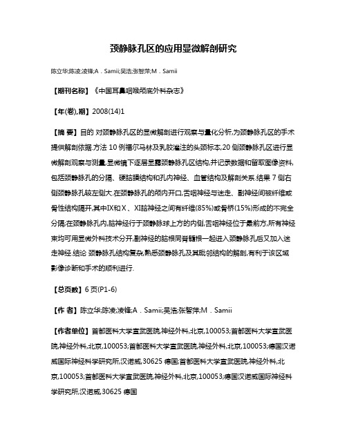
颈静脉孔区的应用显微解剖研究陈立华;陈凌;凌锋;A.Samii;吴浩;张智萍;M.Samii【期刊名称】《中国耳鼻咽喉颅底外科杂志》【年(卷),期】2008(14)1【摘要】目的对颈静脉孔区的显微解剖进行观察与量化分析,为颈静脉孔区的手术提供解剖依据.方法 10例福尔马林及乳胶灌注的头颈标本,20侧颈静脉孔区进行显微解剖观察与测量,显微镜下逐层显露颈静脉孔区结构,并记录数据和留取图像资料,包括颈静脉孔的分隔、硬脑膜结构和孔内神经、血管结构及解剖关系.结果 7侧右侧颈静脉孔较左侧大.在颈静脉孔的颅内开口,舌咽神经与迷走、副神经间被纤维或骨性结构隔开,其中Ⅸ和Ⅹ、Ⅺ脑神经之间有纤维(85%)或骨桥(15%)形成的不完全分隔;在颈静脉孔内,脑神经行于颈静脉球上方的内侧,舌咽神经位于最前方,所有神经束均可用显微外科技术分开,副神经的脑根同脊髓根一起进入颈静脉孔后又加入迷走神经.结论颈静脉孔结构复杂,熟悉颈静脉孔及其毗邻结构的解剖,有利于该区域影像诊断和手术的顺利进行.【总页数】6页(P1-6)【作者】陈立华;陈凌;凌锋;A.Samii;吴浩;张智萍;M.Samii【作者单位】首都医科大学宣武医院,神经外科,北京,100053;首都医科大学宣武医院,神经外科,北京,100053;首都医科大学宣武医院,神经外科,北京,100053;德国汉诺威国际神经科学研究所,汉诺威,30625德国;首都医科大学宣武医院,神经外科,北京,100053;首都医科大学宣武医院,神经外科,北京,100053;德国汉诺威国际神经科学研究所,汉诺威,30625德国【正文语种】中文【中图分类】R323.1【相关文献】1.骨性颈静脉孔区的应用显微解剖及临床意义 [J], 韩云志;娄卫华;袁林林2.枕下经颈-颈静脉突入路达颈静脉孔区的显微解剖研究 [J], 刘庆;袁贤瑞;姜维喜;罗端午;潘亚文3.枕下远外侧入路到达颈静脉孔区的骨性结构的显微解剖研究 [J], 付万新;康春华;彭志强4.颈静脉孔区的显微解剖及定位标志研究 [J], 刘庆;袁贤瑞;潘亚文;姜维喜;罗端午5.国人颈静脉孔区神经血管的显微解剖研究—颞下窝径路、乙状窦后径路应用解剖学 [J], 周慧芳;王宏亮;邱明哲;王长月;李崇谦因版权原因,仅展示原文概要,查看原文内容请购买。
- 1、下载文档前请自行甄别文档内容的完整性,平台不提供额外的编辑、内容补充、找答案等附加服务。
- 2、"仅部分预览"的文档,不可在线预览部分如存在完整性等问题,可反馈申请退款(可完整预览的文档不适用该条件!)。
- 3、如文档侵犯您的权益,请联系客服反馈,我们会尽快为您处理(人工客服工作时间:9:00-18:30)。
#应用解剖#颈静脉孔的应用解剖学肖 明, 丁 炯, 韩群颖, 王鹤鸣, 左国平(南京医科大学解剖学教研室,江苏南京210029) =摘要>目的:为与颈静脉孔相关的影像诊断和临床治疗提供解剖学资料。
方法:从颅底内、外面,对80具成年颅骨的颈静脉孔进行观测;并对20具成人尸头进行解剖,观察该区域神经血管解剖关系。
结果:¹62.3%右侧颈静脉孔较左侧大,15.9%左侧较大,21.8%两侧大小一致;º14.38%的颈静脉孔有骨桥,85.62%无骨桥;»颈静脉孔内、外侧缘距正中矢状面两侧的平均距离颅外均较颅内大:颅外分别为26.11mm 和33.41mm,颅内分别为22.29mm 和27.52mm 。
¼Ù脑神经多沿颈静脉孔前上缘,Ú、Û脑神经沿内侧缘出颅,两者被纤维索(占87.5%)或骨桥(占12.5%)隔开。
½Ù脑神经多经颈静脉孔外口前上缘向前下越过颈内动脉表面;Ù脑神经经颈内静脉深面(占57.5%)或其浅面(42.5%)行向后下。
结论:右侧颈静脉孔通常较左侧大,左右不对称;影像学观测该区域血管、神经应选择恰当的层面。
=关键词>颈静脉孔; 颈内静脉; 脑神经; 应用解剖学=中图分类号>R323.1 =文献标识码>A =文章编号>1001-165X(2001)02-0159-03Applied anatomy of jugular foramen XIAO Ming,DING J ong ,HAN Qun -ying,et al.De p a rtment o f A natom y ,Nan j ing Medical University ,Nan j ing 210029,China=Abstract >Objective:To provide anatomic data for imaging diagnosis and microsurgical treatment of jugular fora -men (JF)lesions.Methods:The JF was observed and measured from internal and external aspects in 80adult skulls.The anatomic relationships between the nerves and vessles in this region were observed i n detail by dissecting 20adult cephalic specimens.Results:¹In 62.3%of all these cases the right JF was larger than the left.In 15.9%the left was larger and in 21.8%they were equal in size.ºBone bridges could be seen in 14.38%and could not in 85.62%.»Measured from extracranial aspect,the average distance from midsagi ttal plane to the medial and lateral border of the JF (medial 26.11mm,lateral 33.41mm),was larger than those measured from intracramial aspect (medial 22.29mm,lat -eral 27.52mm).¼Ùcranial nerve made its ex i t through the anterior superior border of the JF in most cases,meanwhile Úand Ûcranial nerve through the medial border,they were distinctly separated from each other by a band of fibrous tissue (account for 87.5%)or a bone bridge (12.5%).½Jus t outside the J F,Ùcranial nerve appeared at the anter-ior border and made a loop downward and forward superficial to the internal carotid artery.Ùcranial nerve run down ward and back ward deep (account for 57.5%),or superficially (42.5%)to the internal jugular vei n.Conclusions:T he right J F is usually larger than the left,and not symmetry on both sides.The key to imaging diagnosi s of nerves and ves -sels in the JF region is to select the sectional plane correctly.=Key w ords >Jugular foramen; Internal jugular vein; Cranial nerve; Applied anatomy颈静脉孔为枕骨与颞骨岩部之间的一骨性孔道,位于岩枕缝的后端,被颈内静脉结节分为二部或三部[1]。
前部有岩下窦注入,与Ù~Û脑神经通过;后部较大,乙状窦经此延续为颈内静脉。
上述诸结构在颈静脉孔内的位置与相互毗邻关系,国内外有关文献描述甚不统一[2,3]。
本实验目的是观察颈静脉孔的形态、内容物的相互关系,具体测量其孔径大小两侧对称性,为影像学观测提供相应的形态学资料。
1 材料和方法材料为教研室标本室提供的80只成年男女颅骨(不分性别),经耳颞线将颅骨锯开。
对颈静脉孔大体形态进行观察,并着重观察有无骨桥(完整与否)及其类型与出现率;根据Doclo 制定的原则将骨桥进行分类:位于舌下神经管前上方者为Ñ型,位于舌下神经管后方的为Ò型。
分别从颅内测量颈静脉孔的最大、最小横径,矢状径(图1);从颅外测量横径与矢状径,并从颅内、外面对颈静脉孔内、外侧缘距正中矢状面距离进行测=收稿日期>2000-03-11=作者简介>肖 明(1972-),男,江苏盐城人,硕士,讲师,主要从事临床解剖学研究,Tel:(025)6662879,E -mail:renjie@ 。
量。
测量仪器为颅骨测径器。
另20例成年尸头标本(男12例,女8例),于颞骨乳突中部与耳颞线平行锯断,移去颅骨的上端,细心将Ù、Ú、Û脑神经从脑干背面剪断,剔除脑组织,保留颈静脉孔周围的硬脑膜,观察上述神经在孔内口的位置及与岩下窦相互毗邻关系;去除颅底肌肉等软组织,暴露颈内静脉孔外口,观察颈内静脉与Ù、Ú、Û脑神经的解剖关系。
AB.横径(transverse diameter)CD.最大矢状径(maximum sagittal diameter)E F.最小矢状径(minimum sagi ttal diameter)图1 颈静脉孔的各径线颅内测量示意图Fig.1 Intracranial measuremen ts of the schematic drawing of diameters of the jugular foramen#159#中国临床解剖学杂志2001年第19卷第2期2 结果2.1 静脉孔的形态观察在160侧颈静脉孔中,无骨桥者占85.62%(137侧);有骨桥者占14.38%(23侧),其中位于右侧者占3.16%,位于左侧者占8.75%,双侧均有者占2.47%。
23侧骨桥分类如下:13侧属于Ñ型;7侧属于Ò型;介于Ñ、Ò型之间形状不规则者共3侧。
2.2 颈静脉孔各径测量测量数据显示:160侧颈静脉孔中,62.3%为右侧较大,15.9%为左侧较大,21.8%两侧大小一致。
左右颈静脉孔各径线测量值分别见表1、表2。
表1 颅内两侧颈静孔横径、矢状径的测量值 x ?s (min~max)mmTab.1 Intracranial measurement of the treansverse and sagittal diameter (mm)of the jugular foramen in two sides Mean ?SD (min~max)mm项 目(item)例数(case)右 侧(ri ght)左 侧(left)t P 横径(transverse d.)8015.15?2.34(10.96~19.92)14.22?2.78(8.32~20.32) 1.56>0.05最大矢状径(max.s agittal d.)808.82?1.28(6.12~11.10)7.28?1.61(4.20~10.92) 3.26<0.05最小矢状径(min.sagittal d.)59*4.30?1.26(2.46~7.74)4.55?1.42(1.52~12.70)0.75>0.05*有骨桥者不再测量最小矢状径(The mi n.s agittal d.were not meas ured when bone bridges existed)表2 颅外两侧颈静孔横径、矢状径的测量值 x ?s (min~ma x )mmTab.2 Extracranial measurement of the transverse and sagittal diameter(mm)of the jugular foramen in two sides Mean ?SD (min~max)mm项 目(item)例数(case)右 侧(ri ght)左 侧(left)t P 横径(transverse d.)8018.12?1.92(11.30~21.36)16.36?2.32(9.08~21.58) 3.51<0.05矢状径(sagittal d.)8010.45?2.04(6.12~11.10)9.58?2.09(1.00~12.78)3.35<0.05颅内测量数据显示:右侧颈静脉孔的最大矢状径较左侧大,两侧的横径与最小矢状径无显著性差异;颅外测量数据显示:右侧颈静脉孔的横径、矢状径均大于左侧。