医学影像学简答题
(完整版)医学影像学简答题(全)

一、星形细胞瘤的CT表现。
1.病变多位于白质。
2.Ⅰ级肿瘤平扫多呈低密度灶,边界清楚,占位效应轻,增强检查无或轻度强化。
3.Ⅱ~Ⅳ级肿瘤平扫多呈高、低或混杂密度肿块,边界不清,占位效应和瘤周水肿明显,增强检查多呈不规则花环样强化或附壁结节强化。
二、脑膜瘤的好发人群、好发部位、CT、MRI、鉴别诊断。
(非常重要)1.好发人群:中年女性。
2.好发部位:多位于脑外(矢状窦旁、大脑凸面、蝶骨嵴、嗅沟、桥小脑角、大脑镰、小脑幕)。
3.CT表现:平扫肿块呈等或稍高密度,类圆形,边界清楚,多以广基底与硬脑膜相连,瘤周水肿轻或无,增强检查病变多呈均匀明显强化。
4.MRI:平扫肿块在T1WI和T2WI上均呈等或稍高信号,增强T1WI 肿块呈均匀明显强化,邻近脑膜增厚并强化而形成脑膜尾征。
5.鉴别:星形细胞瘤,脑转移瘤,脑脓肿。
三、硬膜外血肿的CT表现。
(非常重要)1.颅板下梭形或半圆形高密度灶。
2.常伴有骨折。
3.血肿范围局限,不跨越骨缝。
4.占位效应较轻。
四、硬膜下血肿的CT表现。
(非常重要)1.颅板下新月形或半月形高密度影。
2.常伴有脑挫裂伤或脑内血肿。
3.脑水肿和占位效应明显。
五、脑梗死的分型及各自的CT表现。
(熟悉)缺血性梗死:1.低密度梗死灶,部位和范围与闭塞血管供血区一致。
2.皮髓质同时受累。
3.占位效应较轻。
4.增强扫描可见脑回状强化。
出血性梗死:1.低密度的梗死灶内可见高密度的出血灶。
2.占位效应明显。
腔隙性梗死:1.低密度梗死灶。
2.无占位效应。
六、鼻咽癌的CT表现。
(非常重要)1.平扫表现为患侧咽隐窝变浅、消失或隆起。
2.咽顶、后、侧壁肿块突向鼻咽腔。
3.颈深淋巴结肿大。
4.增强检查病变呈不均匀明显强化。
七、癌性空洞、结核空洞和脓肿空洞的鉴别。
(一般重要)1、癌性空洞:多见于老年患者。
多位于肺上叶前段和下叶基底段。
多为厚壁偏心空洞。
内壁不光整,可有壁结节,外壁可有分叶征及毛刺征。
常伴肺门、纵隔淋巴结增大。
医学影像学简答题(Medical imaging simplified answer )

医学影像学简答题1(Medical imaging simplified answer 1)3. Short answergeneral1. Brief description of medical X-ray characteristicsThe X ray is an electromagnetic wave with penetration; Fluorescence effect; Photographic effects and biological effects. Its penetration is related to material density, thickness and wavelength of X ray, and fluorescence effect is the basis of fluoroscopy. Photographic effect is the basis of X-ray photography; The ionization effect, which involves changes in human biology, is the basis of radiological protection and radiotherapy.2. The basic principle of X-ray imaging is describedOn the one hand, it is based on the penetration of the X ray, the fluorescence effect and the photographic effect, and on the other hand, the difference between the density and thickness of the body tissue. When the X rays penetrate various tissues of the human body, it is absorbed in different degrees so that there is a difference in the amount of X on the screen or in the X-ray. This allows for contrast between the black and white contrast on the screen or in the X-ray.Bones, joint systems1. The X-ray performance of acute and chronic suppurative osteomyelitisThe suppurative osteomyelitis is caused by staphylococcus aureus in the bone marrow, good hair in children and juvenile, long backbone epiphysis good hair. The early (2 weeks) may have the following soft tissue changes: 1. Two, subcutaneous tissue and muscle intersections blurred, bone may not have obvious change. Bone changes were seen after 2 weeks. The localized osteoporosis was started in the epiphyseal cancellous bone. Subsequently, most dispersed irregular form of bone destruction. Edge blur. In the future, the bone destruction area may merge into a large area of destruction. And gradually extend to the backbone. Can be accompanied by pathological fracture. Osteocortical destruction can form the subperiosteum abscess and stimulate the periosteum to cause periosteal hyperplasia. The new bone with low density is parallel to the backbone. Later, as the course of illness extended. The new osteogenesis is obvious and can form the shell. Osteonecrosis is caused by the emergence of periosteum and thrombotic arteritis. The X-ray shows the dead bone formed along the axis of the bone, which is very dense. If the lesion is close to the joint, the abscess can destroy the bone cortex of the dry epiphysis and enter the joint synovial card. Cause suppurative arthritis. The X line is the swelling of the joint capsule. The gap in the joint is widened early and even dislocated. Late narrowing. Osteopenia. When acute suppurative osteomyelitis is not treated promptly and adequately. It can be transformed into chronic suppurative osteomyelitis. The X-ray showed a large number of osteogenic hyperplasia, thickening of periosteum and fusion with cortex, which was stratified or lacy, thickening of the bony cortex, and narrowing of the medullary cavity. The backbone thickened. Irregular appearance, if not recovered,can still be seen bone destruction and dead bone.2. Describe the X - ray performance of the spine tuberculosisIt is the most common person of bone and joint tuberculosis. Good for children and young people. With lumbar multiple hair. The X - line performance is mainly osteoporosis and cancellous bone fracture. The attachment is less cumulative. Vertebral bodies often collapse due to bone damage, flattening or wedges. When the lesion is involved in the vertebral body, the lower margin of the bone. The rupture of the intervertebral cartilage plate is caused by the use of the broken cortex. When the intervertebral disc is invaded, the intervertebral space is narrowed. Even disappearing, the adjacent vertebral bodies are embedded and fused. At the same time, the lesion can produce a large number of caseous necrosis material in the destruction of bone, and the cold abscess is formed in the soft tissues around the spinal column.The X line is characterized by the presence of a fusiform soft shadow on both sides of the vertebral body, known as a lateral abscess. In addition, due to pathological fracture. The lateral spine of the spine can be seen to change the curvature of the spine. The post-emergence deformity.3. Test the X-ray performance of vitamin D deficiency ricketsBecause vitamin D is not a cause of calcium and phosphorus metabolism, osteoid tissue in bone is deficient in calcium salt deposits. Systemic metabolic bone disease. The X-ray showed a decrease in bone density in the general bone. Bone trabeculaeare rare, fuzzy, margin roughness, bony cortex thinned, stratified change. In the areas where bone metabolism is more active, such as the occurrence of bone epiphysis, the low density edge is blurred, and the epiphyseal calcification zone is irregular, blurred, thin and disappeared. In the middle of the epiphyses, there is a tortuous deformation in the middle of the epiphysis, with a very irregular margin and a hairbrush shape. The gap between the epiphysis and the epiphysis is widened. The corner of the epiphysis was altered by bone spur. The front of the thoracic ribs is a wide mouth. At the same time, the weight-bearing long bone is often bent and deformed. (O leg, X leg, etc.), a small number of patients can have a blue branch fracture healing X-ray performance: the temporary calcification belt reappears, the cup mouth shape depression and brush change are relieved and disappeared. The epiphyseal space is normal. Bone density increases and bone cortex thickens. Bone epiphysis increases, the density increases, and the bone deformation is prolonged.4. Test the differential diagnosis of benign and malignant bone tumors from the characteristics of X-ray.Benign:No transfer: no transfer.Growth condition: slow growth, non-invasion and adjacent tissues, but can be oppressive.Local bone changes: swelling bone damage, clear line with normal bone, sharp edge, thinning of the bony cortex, andexpansion can maintain continuity.Periosteum hyperplasia: generally no periosteal proliferation, can have a small amount of periosteum hyperplasia after pathological fracture, and periosteal new bone is not damage the surrounding soft tissue changes: no swelling or lump shadow more, if there are any lump, the edge is clear.Angiography: vascular differentiation is normal, and the tumor can be used to compress blood vessels.Malignant:Transfer: transfer.Growth: rapid growth, transsexual and adjacent tissue organs.Local bone changes: invasive bone fracture, blurred boundary and normal bone boundary, uneven edges, irregular fracture and defect, and bone formation of tumor.Periosteal hyperplasia: multiple forms of periosteal hyperplasia, and can be destroyed by tumor.The surrounding soft tissue changes: the growth of the soft tissue is not clear from the surrounding tissue.Angiography: it can be seen that tumor blood vessels are more and more disordered, the tumor staining and arteriovenous fistula, and the blood supply artery thickening and the blood vessel erosion become rigid, and the edge damage and so on.5. Take the femoral neck fracture as an example.1. Delayed healing or non-healing of fracture; X - ray showed delayed bone scab, with few or no presence, delayed or prolonged fracture line.Two, false joint formation: the X ray shows the bone bushy with the end of the bone, and there is a clear line between the two sides of the broken end.3. Fracture deformity healing: X - ray shows bone formation Angle, rotation, shortening deformity.4. After trauma, osteoporosis.5. Bone and joint infection; For acute chronic bone, arthritis X ray performance.6. Bone ischemic necrosis: increased femoral bone density and deformation.7. Joint rigidity: it is caused by adhesion to the joint, often with osteoporosis and soft tissue atrophy.Viii. Degeneration of joints: change after chronic bone injury.9. Ossified myositis: calcification in different degree of soft tissue after fracture.The respiratory system1. What methods are used in chest imaging examination?1. Chest perspective 2, (positive and lateral) 3, high - kilovol-meter 4, body layer photography 5, bronchography 6, CT 7, MRI2. What are the basic X-ray manifestations of lung lesions?A, exudative lesions: show the edge blur, density uniform shape shadow, range from flocculus to big leaf, when lesions involving the big leaf, its shape is in line with lung and sharp edges, air-bronchogram and visible.2. Fibrosis change: the expression is high density, the boundary is clear, walking rigid, irregular shape of the line shape.Iii. Proliferative lesion: localized nodules or petals, with high density, relatively clear edges, and generally no fusion trend.4. Calcified venereal changes: the appearance is sharp, the density is extremely high, the shape is different, the size of the speckle shape or plaque shape.Voids: 1. Wormwood vacuous cavity: manifested in a large number of pulmonary real changes with multiple small permeable areas. The form is irregular, it is wormlike. 2. Thin wall cavity: hollow wall thickness < 3mm, boundary clear, smooth circular light zone. 3. Thick wall hole: wall thickness > 3mm, the holeis round or irregular, peripheral or unreal change area, the inner wall is smooth and neat or concave, the hole can have or airless plane.Six, mass lesions, benign tumor characterized by round or oval, smooth boundary, density uniform spherical density shadow, malignant tumors are lobulated, the boundary is not sharp, can have a short nap or umbilical concave), central necrosis.3. With a solid shadow on one side of the chest, which diseases should be considered? What aspects should be analyzed in the identification?One, a large number of pleural effusion, one side of the lung, the one side of the lung, the one side of the pleural hypertrophy, the one side of the pleural hypertrophy, the one side of the lung, the one side of the lung and the one side of the lungShould note: when identifying a, mediastinal position 2, diaphragmatic level three, five, four, thoracic rib gap width size on a flat piece of six, observe whether air-bronchogram, observe whether the main bronchus is unobstructed in layer 7, combined with clinical data4. What are the direct and indirect X-ray signs of bronchial lung cancer (central type)?1. Lumps, located in the lung area, are rounded or lobule.2. Endobronchial polyps filling defect.3. The bronchial wall thickened and the lumen was narrow or blocked, with rat tail or cup.Ii. Indirect symptoms: 1. Obstructive pulmonary disease,The horizontal and pulmonary masses of the upper lobe of the upper lobe of the upper lobe of the upper lobe.2. Obstructive pneumonia: repeated attacks and slow absorption of exudative lesions.3. Obstructive emphysema: the air volume of the blocked lung is increased, and the brightness is increased.5. Typical X-ray manifestations of large leaf pneumonia?Lobule pneumonia may involve most or all of the lobes. The former is characterized by uniformity of density, and the shadow of the edge is indistinct. The edge is clear, with the interleaf crack as the boundary, its shape with the pulmonary lobe, the contour is consistent, its inside visible bronchi meteorology. Different forms of lobule pneumonia vary.6. Typical X-ray manifestations of acute hemorrhagic disseminated tuberculosis?The early two lung density of the lesion showed a change of hair glass. In about 10 days, the two lungs showed diffuse uniform distribution, the same size, uniform density of miliary nodules. The two lung textures are not clear.The circulatory system1. Simple mitral stenosis X-ray performance?The heart increases, the left atrium and right ventricle are enlarged, and the left heart is often significantly enlarged.The main reason for the reduction of the general aortic ball is the reduction of left ventricle blood elimination, aortic dysplasia or the left rotation of the heart and big blood vessels, and the aortic arch folds.The left ventricle shrank, the apex of the heart moved, the lower part of the heart was straight.4. Mitral membrane calcification, direct sign.5. Pulmonary congestion or interstitial edema, upper pulmonary vein dilation, lower pulmonary veins. Sometimes it can be seen that the diameter of 1 ~ 2mm in diameter can be seen in the lung field, which is composed of hemosiderosis.2. The X line of high blood heart disease is shown as?One, the heart is aortic type, the left ventricle segment increases, becomes round, the heart apex is in the phrenic, the cardiac phrenic horn shows acute Angle, the left ventricle is prominent, overlaps with the spine.Second, the left ventricle is increasing to the left, and the apex of the heart is often under the diaphragm.3. The perspective can be seen that the opposite pulsation.When left heart failure, the left atrium increases, and pulmonary congestion and pulmonary edema appear.5. Severe, the heart is generally enlarged, but the left ventricular enlargement is the main.The aorta has dilation, extension, and circuity.3. X-ray performance of pulmonary heart disease?Changes in pulmonary hypertension and chronic pulmonary diseaseOne, pulmonary hypertension, often occurs before the heart shape changes.Second, the right ventricle enlarges, the heart is in the mitral valve type, the heart rate is more than the normal person not much. Some cases: the heart is smaller than normal, and is related to the low level of the pulmonary emphysema.Three, chronic pulmonary disease, chronic bronchitis, extensive lung tissue fibrosis and emphysema.4. X ray performance of congenital heart disease atrial septal defect?When the defect is small, the size and shape of the heart andshape are normal or change.The heart is of mitral valve type, often moderate increase.Two, right atrium and right ventricular enlargement,The major characteristic changes of atrial septal defect were significantly increased in the right atrium.3. The pulmonary artery protruding, the pulsating enhancement, the pulmonary portal angiectasia. There are often lungmen dancing.In the left atrium, the left ventricle and aorta decreased, while the first left ventricle enlarged.5. Pulmonary hyperemia and later pulmonary hypertension.5. The X ray performance of common Fallot tetralogy?1. The heart is generally not enlarged, the heart is blunt, the upper warped is a sheep's nose, the heart lumbar depression, if there is a third ventricle forming, the heart is flat, or slightly raised.Second, the right ventricle increases.The left ventricle narrowed with decreased blood flow, the left atrium was generally unchanged, and the right atrium was mild to moderate due to increased blood flow and increased right ventricular pressure.4. The lung door shrinks and the lung vessels are slim.The aorta is widened and shifted to the right.The digestive system1. According to what characteristics can the organ of the digestive system be divided into two categories? Where are the organs?According to the characteristic of the digestive organ is the real organ or the hollow viscera, the digestive organ is divided into two categories. The liver and pancreas belong to the substantial organ. Esophagus, stomach, duodenum, large, small intestine and biliary system belong to hollow viscera.2. What kind of inspection methods and imaging methods are used in the two main types of digestive tract and cavity?The liver and pancreas of parenchyma were mainly used for CT, ultrasound and mri. After the general sweep; When necessary, CT iodine contrast agent was enhanced, and magnetic resonance was enhanced with gadolinium contrast agent.The hollow viscera was mainly used for routine X-ray examination, the gastrointestinal tract was radiographed by barium, and the bile was used for the contrast of iodine3. The X-ray signs of benign and malignant ulcers are identified.A benign ulcer protrudes from the gastric cavity. The ulcer is located within the contour of the stomach2. The shape of the shadow: the benign ulcer is relatively small and round, and the malignancy is larger and more shallow.Three, niche mouth: benign ulcer with mucosal edema, width is consistent, sometimes under pressure to change form malignant ulcer niches mouth cancer tissue invasion, forming ring levee involuntary pressure, change or more cancer nodules form refers to the indentation, sharp corners.Iv. Benign ulcer stomach constriction peristaltic direct niches, malignant ulcer is more than 1 cm from the niches, peristalsis disappears.4. Differentiation of esophageal foreign body and trachea foreign bodyTake the coin foreign object as an example, because the diameter of the esophagus is small, the left and right diameters are wider, so the esophageal foreign body is in a circular position, and the lateral position view is striped. The trachea foreign body is opposite, because the trachea half annular cartilage is absent is facing the rear, so the maximum diameter of the foreign body is the front and rear direction. The positive view is long and long, while the lateral position is round.5. Identification of jejunum, ileum and intestinal obstruction? How to diagnose low - level intestinal obstruction based on flatslice?The intestinal mucosa is a fish-bone arrangement perpendicular to the vertical axis of the intestinal tube. The mucosa of ileus is only two intestinal wall lines. The most significant expansion of the obstruction tube diameter is the semilinar fold.The high intestinal obstruction is mainly manifested in the left middle and upper abdominal multiple qi level, the stomach also sees the liquid level, the lower abdomen and the pelvic cavity of the lower abdomen and the lower gas. Low intestinal obstruction, the expression is the whole abdomen several stair - shaped gas levelUrinary system1. Differential diagnosis of urinary calculus. (points)1. Gallstones: the form is polygon, the surrounding density is high, the central density is low, sometimes there is the high density core. Lateral photography is located in front of the spine.Lymph node calcification: form irregular punctate, structure, and has no fixed position, to move a large degree (e.g., mesenteric lymph node calcification) imaging of the renal pelvis can understand outside or in the urinary tract.3. Intestinal contents (coprolites or drugs) : the position is not constant, the repeated photo position can be changed ordisappeared, and the bowel will disappear.Iv. Venous stone (pelvic cavity) : small, round, circular or concentric round dense shadow, the edges are neat, often for both sides and multiple, the position is more than partial, when necessary retrograde contrast imaging is identified.2. X-ray manifestations of renal tuberculosis. (points)Flat slice: the kidney contour area can protrude, terminal form shrinksCalcification: diffuse, cloudy, spottedAngiography: wormhole destruction, renal cortical abscess and vacuous formation, pyelonephrosis, renal pelvis, renal calyx (peripheral imformation, deformed stenosis), renalself-truncation3. The X-ray of typical urinary calculi. (points)Kidney: sliced: mulberry, layered, antlerContrast: density, higher density, filling defect, obstructionUreter: flat slice: the long axis is consistent with the ureterAbdominal segment: side of the lumbar spineThe sacroiliac segment: the sacroiliac jointPelvic segment: roughly parallel to the pelvic rimLower end of ureter: polymorphismContrast: positive, negative and catheter relationship, obstruction of waterBladder calculi: above the symphysis pubis, the midline of the pelvic cavity changes with positionUrethra: the posterior urethra: the symphysis of the pubic bone and the posterior urethra4. Several common radiographic reflux X - ray manifestations.1. Tubule reflux: the radiate dense shadow radiated from the center of the kidney to the cortex.Second, the kidney sinus reflux: it appears as the irregular Angle or band dense shadow around the fornix, and the author appears in an irregular shape.Iii. Circumfluence of the blood vessels: the arch of the arch is shown as the arch of the arch.Iv. Lymphatic reflux: it is shown as a slender, meandering, curved silhouette that walks in the direction of the renal gate.5. Various imaging examinations and USES of urinary system. (points)IVP: the shape of the renal pelvis, renal calices, ureters, and bladder, and the function of the renal excretion2. Retrograde pyelography: used for IVP display (such as renal dysfunction) or not for IVP (such as liver and kidney function, iodine allergy)Bladder angiography: excretory method: the urethral stricture cannot be intubated or at the same time, the upper urinary tract should be examinedRetrograde: observe bladder size, shape, position to diagnose bladder disease4. Urethrography: mostly used for urethral stricture, calculi, congenital malformation, etc5. Retroperitoneal aerated angiography: showing the renal, adrenal profile and retroperitoneal mass and the relationship with the kidney6. Arteriography: diagnosis of vascular lesions and adrenal neoplastic lesionsCentral, five official system1. Evaluation of CT in the treatment of sinus tumors.CT diagnosis of smaller tumors is of great value and can be determined in its origin and scope. Benign tumor margins are clear and orderly, without bone damage. But it is difficult todetermine the pathological nature. The mucous cyst showed an enlarged sinus cavity and increased density. CT is of great value in diagnosis of malignant tumor. In the early stage of osteopenia, there was a shadow of mass in the sinus cavity, and the sinus cavity was seen in the sinus cavity. The sinus wall can be damaged early, and the adjacent structure can be shown as the nasal cavity, the invasion of the orbit and the scope.2. CT manifestations of meningiomas.The CT findings of typical meningiomas are high in density, with clear edges, spherical or subleaf lesions, and cranial bones, which are connected to the cerebellum. There was no edema or slight edema in the oven. The general performance of the enhanced scanning was significantly enhanced.。
医学影像考试简答题
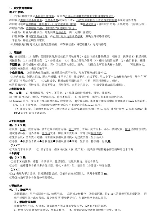
1、原发性肝细胞癌答:CT表现:⑴平扫示肿瘤大多呈不均匀低密度影,癌灶内合并坏死和囊变或脂肪变性区则密度更低。
⑵肿块呈类圆形或不规则形,边界不清楚(浸润生长者),少数呈膨胀性生长者边缘有假包膜形成则边界清楚。
⑶肿瘤可造成局部膨隆,肝叶增大,肝内管道和肝门推移。
⑷肝硬化表现(肝叶比例失调、肝裂增宽、门脉高压等)。
⑸增强扫描:动态增强扫描:造影剂呈“快进快出”表现。
动脉期:肝癌为动脉供血,此期病灶明显强化,高于周围肝脏密度。
门静脉期:肿块密度开始下降,而正常肝组织逐渐明显强化,肿块呈等或略低密度平衡期:肿瘤呈明显低密度。
⑹肝癌侵犯门脉时可见血管内充盈缺损. ⑺转移征像:淋巴结肿大;远处转移等。
2、胃溃疡答:直接征象:1)龛影:类园形钡斑,切线位位于胃轮廓外2)龛影口部水肿带:线征、项圈征、狭颈征3)粘膜纠集间接征象:(1)痉挛性改变(2)分泌增加(3)胃动力及张力异常(4)瘢痕收缩胃变形(5)幽门狭窄、梗阻早期胃癌:需用低张双对比造影。
胃小区粘膜结构紊乱、消失。
切线位上可见刺突样小龛影。
可见颗粒状、小圆形充盈缺损,表面毛糙不平。
中晚期胃癌:⑴胃腔内充盈缺损:缺损边缘轮廓不光整,形态不规则或呈分叶状。
⑵腔内龛影:龛影大而浅,形态不规则,多呈半月形,外缘平直,内缘不整,呈大小不一尖角样指向外周,常伴有“环堤”征及“半月综合征”。
⑶粘膜改变:粘膜皱襞局限性破坏、中断,周围粘膜粗大、僵直。
⑷胃轮廓改变:胃腔变形,边缘不整齐,胃壁僵硬,容积小且固定。
⑸病变部蠕动减弱或消失。
3.椎间盘突出答:X线:1)椎间隙异常:变窄、不等宽。
2)椎体后缘骨质增生:骨赘、骨唇等。
3)脊柱曲度异常:侧弯,平腰或后凸,脊柱不稳等。
4)游离骨块:椎管内有小骨块或钙化块。
5)Schmorl结节:椎体上下缘局限性凹陷、边缘硬化。
6)脊髓造影:椎间盘平面硬膜囊前外侧压迹>3mm即可诊断。
CT:(1)直接征象:①椎间盘局限性后突②突出间盘钙化③Schmorl结节。
医学影像学——名词解释、选择题、简答题全套
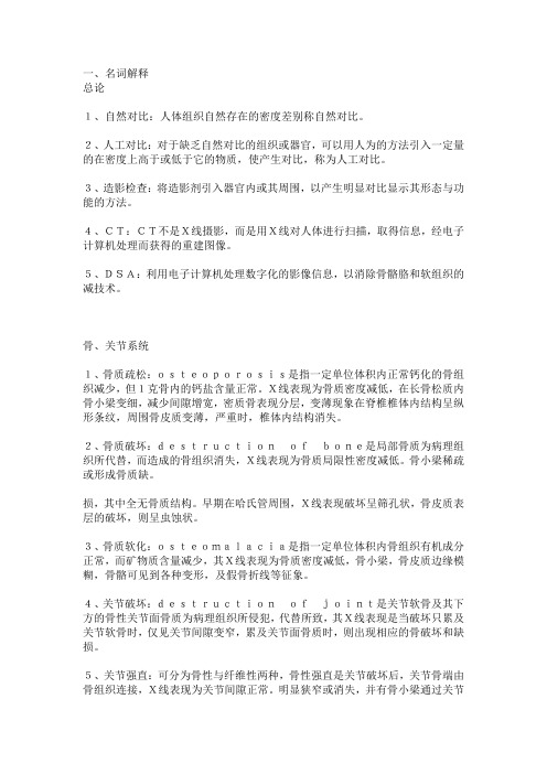
一、名词解释总论1、自然对比:人体组织自然存在的密度差别称自然对比。
2、人工对比:对于缺乏自然对比的组织或器官,可以用人为的方法引入一定量的在密度上高于或低于它的物质,使产生对比,称为人工对比。
3、造影检查:将造影剂引入器官内或其周围,以产生明显对比显示其形态与功能的方法。
4、CT:CT不是X线摄影,而是用X线对人体进行扫描,取得信息,经电子计算机处理而获得的重建图像。
5、DSA:利用电子计算机处理数字化的影像信息,以消除骨骼胳和软组织的减技术。
骨、关节系统1、骨质疏松:osteoporosis是指一定单位体积内正常钙化的骨组织减少,但1克骨内的钙盐含量正常。
X线表现为骨质密度减低,在长骨松质内骨小梁变细,减少间隙增宽,密质骨表现分层,变薄现象在脊椎椎体内结构呈纵形条纹,周围骨皮质变薄,严重时,椎体内结构消失。
2、骨质破坏:destructionofbone是局部骨质为病理组织所代替,而造成的骨组织消失,X线表现为骨质局限性密度减低。
骨小梁稀疏或形成骨质缺。
损,其中全无骨质结构。
早期在哈氏管周围,X线表现破坏呈筛孔状,骨皮质表层的破坏,则呈虫蚀状。
3、骨质软化:osteomalacia是指一定单位体积内骨组织有机成分正常,而矿物质含量减少,其X线表现为骨质密度减低,骨小梁,骨皮质边缘模糊,骨骼可见到各种变形,及假骨折线等征象。
4、关节破坏:destructionofjoint是关节软骨及其下方的骨性关节面骨质为病理组织所侵犯,代替所致,其X线表现是当破坏只累及关节软骨时,仅见关节间隙变窄,累及关节面骨质时,则出现相应的骨破坏和缺损。
5、关节强直:可分为骨性与纤维性两种,骨性强直是关节破坏后,关节骨端由骨组织连接,X线表现为关节间隙正常。
明显狭窄或消失,并有骨小梁通过关节连接两侧骨端。
纤维性强直X线表现可见狭窄的关节间隙,并且无骨小梁贯穿,但临床功能丧失。
6、骨质坏死:是骨组织局部代谢的停止,坏死的骨质称为死骨,死骨的X线表现为骨质局限性密度增高。
医学影像学简答题集锦
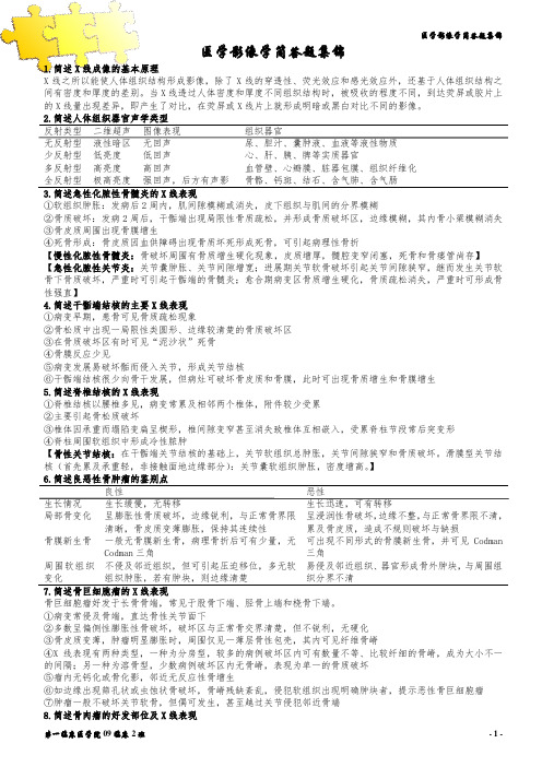
医学影像学简答题集锦1.简述X线成像的基本原理X线之所以能使人体组织结构形成影像,除了X线的穿透性、荧光效应和感光效应外,还基于人体组织结构之间有密度和厚度的差别。
当X线透过人体密度和厚度不同组织结构时,被吸收的程度不同,到达荧屏或胶片上的X线量出现差异,即产生了对比,在荧屏或X线片上就形成明暗或黑白对比不同的影像。
2.简述人体组织器官声学类型反射类型二维超声图像表现组织器官无反射型液性暗区无回声尿、胆汁、囊肿液、血液等液性物质少反射型低亮度低回声心、肝、胰、脾等实质器官多反射型高亮度高回声血管壁、心瓣膜、脏器包膜、组织纤维化全反射型极高亮度强回声,后方有声影骨骼、钙斑、结石、含气肺、含气肠3.简述急性化脓性骨髓炎的X线表现①软组织肿胀:发病后2周内,肌间隙模糊或消失,皮下组织与肌间的分界模糊②骨质破坏:发病2周后,干骺端出现局限性骨质疏松,并形成骨质破坏区,边缘模糊,其内骨小梁模糊消失③骨皮质周围出现骨膜增生④死骨形成:骨皮质因血供障碍出现骨质坏死形成死骨,可引起病理性骨折【慢性化脓性骨髓炎:骨破坏周围有骨质增生硬化现象,皮质增厚,髓腔变窄闭塞,死骨和骨瘘管尚存】【急性化脓性关节炎:关节囊肿胀、关节间隙增宽;进展期关节软骨破坏引起关节间隙狭窄,继而发生关节软骨下骨质破坏,严重时可引起干骺端的骨髓炎;愈合期病变区骨质增生硬化,骨质疏松消失,严重时可形成骨性强直】4.简述干骺端结核的主要X线表现①病变早期,患骨可见骨质疏松现象②骨松质中出现一局限性类圆形、边缘较清楚的骨质破坏区③在骨质破坏区有时可见“泥沙状”死骨④骨膜反应少见⑤病变发展易破坏骺而侵入关节,形成关节结核⑥干骺端结核很少向骨干发展,但病灶可破坏骨皮质和骨膜,此时可出现骨质增生和骨膜增生5.简述脊椎结核的X线表现①脊椎结核以腰椎多见,病变常累及相邻两个椎体,附件较少受累②主要引起骨松质破坏③椎体因承重而塌陷变扁呈楔形,椎间隙变窄甚至消失致椎体互相嵌入,受累脊柱节段常后突变形④脊柱周围软组织中形成冷性脓肿【骨性关节结核:在干骺端关节结核的基础上,关节软组织总肿胀,关节间隙狭窄和骨质破坏。
医学影像学简答题全

医学影像学简答题全引言医学影像学作为现代医学的重要组成部分,通过应用各种影像学技术,可以直观地观察和诊断人体内脏器官的形态和功能,为临床医生提供重要的辅助诊断信息。
本文将回答一些关于医学影像学的简答题,帮助读者更好地了解这个领域。
问题一:什么是医学影像学?回答:医学影像学是一门研究利用物理学、生物学和医学知识,通过各种影像学技术来诊断和治疗疾病的学科。
它包括了放射学、超声学、核医学和磁共振成像等多种技术和方法。
问题二:医学影像学有哪些主要应用领域?回答:医学影像学广泛应用于临床医学的各个领域,包括但不限于以下几个主要方面:1. 诊断:通过影像学技术可以观察人体内脏器官的形态和结构,对疾病进行准确的诊断。
2. 治疗规划:医学影像学可用于辅助手术规划,特别是在手术前对疾病进行先期评估和虚拟手术操作,减少手术风险。
3. 评估疗效:通过比较术前和术后的影像学表现,可以评估治疗的效果,并为调整治疗方案提供依据。
4. 临床研究:医学影像学技术可以用于疾病研究和新药试验,对疾病的发生机制和治疗方法进行探索。
问题三:医学影像学的常用技术有哪些?回答:医学影像学技术主要包括以下几种:1. 放射学:包括X射线摄影、计算机断层扫描(CT)、磁共振成像(MRI)和数字化减影血管造影(DSA)等技术,可用于观察人体内脏器官的形态和结构。
2. 超声学:通过利用超声波在人体组织中的传播和反射原理,产生图像来观察器官和组织的形状、结构和功能。
3. 核医学:利用放射性示踪物质追踪人体内的代谢过程,并通过测量射线释放的能量来形成影像,其中包括放射性核素摄取显像和正电子发射断层扫描(PET-CT)等技术。
4. 磁共振成像:通过利用磁场和电磁波的相互作用原理,观察和诊断人体内器官的结构和病变。
问题四:医学影像学的安全性如何?回答:医学影像学技术在临床应用中是相对安全的,但仍需注意以下几个方面:1. 辐射安全:放射学技术涉及的X射线和其他射线在高剂量下可能对人体产生损害。
医学影像诊断重点简答题
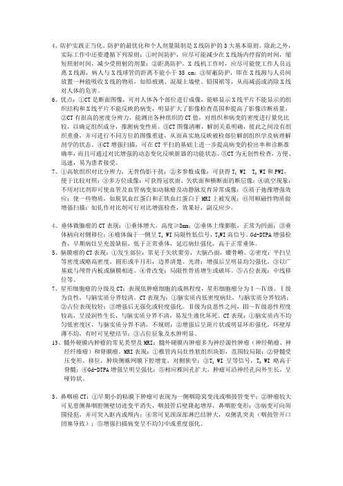
4、防护实践正当化、防护的最优化和个人剂量限制是X线防护的3大基本原则。
除此之外,实际工作中还要遵循下列原则:①时间防护,应尽可能减少在X线场内停留的时间,缩短照射时间,减少受照射的剂量;②距离防护,X线机工作时,应尽可能使工作人员远离X线源,病人与X线球管的距离不能小于35 cm;③屏蔽防护,即在X线源与人员间放置一种能吸收X线的物质,如铅玻璃、混凝土墙壁、铅围裙等,从而减弱或消除X线对人体的危害。
6、优点:①CT是断面图像,可对人体各个部位进行成像,能够显示X线平片不能显示的组织结构和X线平片不能反映的病变,明显扩大了影像检查范围和提高了影像诊断质量。
②CT有很高的密度分辨力,能测出各种组织的CT值,对组织和病变的密度进行量化比较,以确定组织成分,推测病变性质。
③CT图像清晰,解剖关系明确,彼此之间没有组织重叠,并可进行不同方位的图像重建,从而真实地反映被检部位解剖组织学及病理解剖学的状态。
④CT增强扫描,可在CT平扫的基础上进一步提高病变的检出率和诊断准确率,而且可通过对比增强的动态变化反映脏器的功能状态。
⑤CT为无创性检查,方便、迅速,易为患者接受。
7、①高软组织对比分辨力,无骨伪影干扰;②多参数成像:可获得T1 WI T2 WI和PWI,便于比较对照;③多方位成像:可获得冠状面、矢状面和横断面的断层像;④流空现象:不用对比剂即可使血管及血管病变如动脉瘤及动静脉发育异常成像;⑤质子弛豫增强效应:使一些物质,如脱氧血红蛋白和正铁血红蛋白于MRI上被发现;⑥用顺磁性物质做增强扫描:如钆作对比剂可行对比增强检查,效果好,副反应少。
4、垂体微腺瘤的CT表现:①垂体增大,高度≥8mm;②垂体上缘膨胀,正常为凹面;③垂体柄向对侧移位;④瘤体偏于一侧呈T1 WI局限性低信号,T2WI高信号。
Gd-DTPA增强检查,早期病灶呈充盈缺损,低于正常垂体,延迟病灶强化,高于正常垂体。
5、脑膜瘤的CT表现:①发生部位:常见于矢状窦旁,大脑凸面,蝶骨嵴。
医学影像学简答题

医学影像学简答题公司内部编号:(GOOD-TMMT-MMUT-UUPTY-UUYY-DTTI-简述肺充血与肺淤血的区别肺充血:肺动脉分支成比例地增粗且向外周伸展,边缘清晰锐利,肺野透明度正常,长期易导致肺动脉高压。
见于房间隔缺损,动脉导管未闭等左向右分流的先天性心脏病。
肺淤血:肺野透明度减低,肺门增大,边缘模糊,肺纹理增多增粗且边缘模糊。
简述房间隔缺损的X线表现肺血增多,肺动脉段突出,肺门动脉扩张,心影增大呈二尖瓣型,右房右室增大,主动脉结正常或缩小。
简述肝硬化的CT表现①全肝萎缩,肝各叶大小比例失常,尾叶与左叶较大,右叶较小②肝轮廓凹凸不平③肝门、肝裂增宽④出现脾肿大、腹水、胃底与食管静脉曲张等门静脉高压征象食管癌x线表现①粘膜破裂消失、中断、破坏代之以癌瘤杂乱不规则影像②管腔狭窄:为浸润型ca,管壁僵硬,上方扩张③腔内充盈缺损:见增生型ca,向腔内突出,不规则,大小不等充盈缺损;④不规则龛影:轮廓不规则长形龛影,溃疡型癌⑤受累段食管局限性僵硬,及形成纵隔内肿块影脑膜瘤CT表现CT 平扫:呈圆形等或略高密度,边界清晰,常见斑点状钙化广基底与硬膜相连,类圆形,周围水肿轻,静脉或静脉窦受压可出现中重度水肿侵犯相邻颅板引起增生或破坏。
增强:明显均匀强化。
简述大叶性肺炎的CT表现①充血期:病变呈磨玻璃样影,边缘模糊,病变区血管隐约可见②实变期:可见沿大叶或肺段分布的致密实变影,内有“空气支气管征”③消散期:实变影密度随病变吸收而减低,呈散在、大小不等的斑片状影。
简述绞窄性小肠梗阻的X线表现特点:①具有单纯性小肠梗阻征象:肠腔扩张积气、积液呈多发气液平征象;胃、结肠内气体减少或消失。
②假肿瘤征③咖啡豆征④肠袢卷曲固定征⑤长液平征⑥空回肠换位征⑦肠壁厚、液平面无波动、腹水等征象。
胸部影像学检查常采用哪些方法一、胸部透视二、拍片(正、侧位)三、高仟伏拍片四、体层摄影五、支气管造影六、CT七、MRI急性血性播散型肺结核的典型X线表现病变早期两肺密度增高呈毛玻璃样改变。
影像诊断学简答题

影像诊断学简答题1、X线的物理特性包括哪些?答:①穿透性;②荧光效应;③感光效应;④电离效应。
2、X线成像具备的基本条件?答:①X线具有一定的穿透力,能穿透人体的组织结构;②被穿透的组织结构必须存在着密度和厚度的差异,X 线在穿透的过程中被吸收的量不同,以至于剩余的X线量有差别;③这个有差别的剩余X线仍是不可见的,还必须经过显像这一过程。
3、X线的检查方法包括哪些,各种方法的临床应用范围?答:(1)普通检查:①荧光透视(简称透视),常用于胸部检查,配合胃肠钡餐、钡剂灌肠或心血管造影等检查。
②X线摄影,X线摄片广泛应用于临床,如胸部、腹部、四肢、头颅、骨盆及脊柱等部位的检查。
(2)特殊检查:特殊检查包括体层摄影、软射线摄影、放大摄影和荧光摄影等。
自CT等新的现代成像技术应用以来,只有软射线摄影还在应用(主要用来检查软组织,如女性乳腺)。
(3)造影检查:对缺乏自然对比的组织结构或器官,将密度高于或低于改结构或器官的物质引入结构或器官内或其周围间隙,使之产生对比以显影。
主要用于食管、胃肠道、血管造影等4、X线的诊断原则是什么?答:全面观察,具体分析,结合临床,作出诊断。
5、CT与X线的临床应用有什么不同?答:胃肠道主要使用X线检查,骨骼系统和胸部多首选X线检查;CT主要用于中枢神经系统、头颈部、胸部、心脏与血管、腹部、盆部,骨骼系统(显示骨变化,如骨破坏与增生的细节)。
6、MRI的成像原理是什么?答:磁共振成像是利用体内氢原子核在强磁场内发生磁矩,用射频发生共振提供能量,改变磁矩,停止射频,恢复磁矩,释放能量,产生信号,经计算机处理,形成MR图像。
7、DSA的成像原理是什么?答:数字荧光成像是DSA的基础。
DSA是用碘化铯探测器将穿过人体的信息X线进行接收,使不可见的信息X线变为光学图像。
经影像增强器亮度增强后,再用高分辩率的摄像机进行扫描,所得到的图像信号经模/数转换器后进行储存,此时将对比剂注入前所采集的蒙片与对比剂注入后所采集的血管充盈像进行减影处理,再经数模转换器后形成只留下含对比剂的减影血管像。
医学影像简答题

医学影像学试题答案A一、填空题:1、关节基本病变包括(关节肿脓)(关节破坏)(关节退行变)(关节强直)(关节脱位)五选其三种。
2、MRI对(钙化)(细小骨化)的显示不如X线和CT。
3、异常心脏形态是(二尖瓣型心脏)(主动脉型心脏)(普大型心脏)4、正常成人心胸比是(0.5左右)横位心心脏纵轴与胸廓水平面夹角是(>45度)(45度)5、肾结石典型的X线表现(桑椹状)(鹿角状)(分层状)6、肺纹理由(肺动脉)、(肺静脉)组成,其中主要是(肺动脉分支),(支气管)、(淋巴管)及(少量间质组织)也参与肺纹理的形成。
7、肺叶间裂在普通CT上表现为(少量间质组织),在高分辨力CT图像上表现为(细线状或窄带状致密影)。
8、X线与医学成像有关的基本特性有(穿透作用)、(荧光作用)、(感光作用)、(电离作用/生物效应)。
9、肝癌CT增强扫描的特点是块进(快出)。
10、单纯性小肠梗阻的典型X线表现有(肠管扩张)、(阶梯状液气平面)。
二、名词解释:1、骨龄:骨的原始骨化中心和继发骨化中心的出现时间,骨骺与干骺端骨性愈合的时间有一定的规律性,用时间来表示即骨龄。
2、关节破坏:是关节软骨及其下方的骨性关节面骨质为病理组织所侵犯、代替所致。
3、骨质软化:是指一定单位体积内骨组织有机成分正常,而矿物质含量减少,组织学上显示骨样组织钙化不足。
4、冠心病定义:指冠状动脉硬化及功能性改变导致心肌缺血缺氧而引起的心脏病变5、肺充血:肺动脉内血容量增多6、法四:(1)肺动脉狭窄(2)室间隔缺损(3)主动脉骑跨(4)右室肥厚7、支气管气象:在X线胸片及CT片上,实变的肺组织中见到含气的支气管分支影(1分)。
可见于大叶性肺炎和小肺癌中(1分)。
8、充盈缺损:消化管腔内因隆起性病变而致使钡剂不能在该处充盈,该区域形成钡剂缺损表现。
常见于消化道占位性病变或异物。
9、半月综合征:溃疡型胃癌钡餐造影检查见到下列印象称为半月综合征:1、胃腔内充盈缺损肿块;2、肿块表面不规则半月形或盘状龛影,位于胃腔内;3、龛影周围围绕环堤,伴有指压迹状充盈缺损。
医学影像学精选简答题100题(附答案)

以下是医学影像学精选简答题100 题(附答案):一、X 线部分(20 题)1.X 线是如何产生的?答案:X 线是在真空管内高速行进成束的电子流撞击钨(或钼)靶时而产生的。
2.X 线的基本特性有哪些?答案:穿透性、荧光效应、感光效应、电离效应。
3.简述透视的优缺点。
答案:优点:可转动患者体位,进行多方向观察;操作简便;费用较低。
缺点:影像对比度及清晰度较差;不能留下永久性记录。
4.X 线摄影的基本原理是什么?答案:利用X 线的穿透性、感光效应,使人体结构在胶片上形成潜影,经显影、定影等处理后形成可见影像。
5.什么是高千伏摄影?有何特点?答案:高千伏摄影是用120kV 以上的管电压产生的X 线进行摄影。
特点:穿透力强,可减少胸壁软组织、肋骨对肺组织的遮挡,使肺纹理显示更清晰;层次丰富,但对比度下降。
6.什么是体层摄影?答案:体层摄影是通过特殊装置和操作获得某一选定层面上组织结构的影像,而使非选定层面的结构被模糊掉。
7.简述X 线造影检查的原理。
答案:将造影剂引入器官或其周围间隙,使之产生密度差异,从而显示出其形态和功能。
8.常用的X 线造影剂有哪些类型?各举一例。
答案:阳性造影剂如钡剂(硫酸钡),阴性造影剂如空气、二氧化碳等。
9.碘造影剂的不良反应有哪些?答案:轻度反应有恶心、呕吐、荨麻疹等;中度反应有喉头水肿、呼吸困难、血压下降等;重度反应可出现休克、呼吸心跳骤停等。
10.简述X 线在骨骼系统检查中的应用价值。
答案:可显示骨的形态、结构、密度变化;发现骨折、脱位、骨质破坏、增生等病变;对一些先天性骨发育异常也有诊断意义。
11.骨折的基本X 线表现有哪些?答案:骨折线(透光带)、骨小梁中断、断端移位、成角畸形、软组织肿胀等。
12.关节脱位的X 线表现是什么?答案:关节正常关系丧失,组成关节的骨端发生移位,可伴有骨折。
13.骨质破坏的X 线表现是什么?答案:局部骨质密度减低,骨小梁稀疏或形成骨质缺损,骨皮质破坏可呈虫蚀状、筛孔状或大片骨质缺损。
医学影像学名词解释与简答题库

医学影像学名词解释与简答题库一、名词解释1.螺旋CT(SCT): 螺旋CT扫描就是在旋转式扫描基础上,通过滑环技术与扫描床连续平直移动而实现得,管球旋转与连续动床同时进行,使X线扫描得轨迹呈螺旋状,因而称为螺旋扫描。
2.CTA:就是静脉内注射对比剂,当含对比剂得血流通过靶器官时,行螺旋CT容积扫描并三维重建该器官得血管图像。
3.MRA:磁共振血管造影,就是指利用血液流动得磁共振成像特点,对血管与血流信号特征显示得一种无创造影技术。
常用方法有时间飞跃、质子相位对比、黑血法。
4.MRS:磁共振波谱,就是利用MR中得化学位移现象来确定分子组成及空间分布得一种检查方法,就是一种无创性得研究活体器官组织代谢、生物变化及化合物定量分析得新技术。
5.MRCP:就是磁共振胆胰管造影得简称,采用重T2WI水成像原理,无须注射对比剂,无创性地显示胆道与胰管得成像技术,用以诊断梗阻性黄疽得部位与病因。
6.PTC:经皮肝穿胆管造影;在透视引导下经体表直接穿刺肝内胆管,并注入对比剂以显示胆管系统。
适应症:胆道梗阻;肝内胆管扩张。
7.ERCP:经内镜逆行胆胰管造影;在透视下插入内镜到达十二指肠降部,再通过内镜把导管插入十二指肠乳头,注入对比剂以显示胆胰管;适应症:胆道梗阻性疾病;胰腺疾病。
8.数字减影血管造影(DSA):用计算机处理数字影像信息,消除骨骼与软组织影像,使血管成像清晰得成像技术。
9.造影检查:对于缺乏自然对比得结构或器官,可将高于或低于该结构或器官得物质引入器官内或其周围间隙,使之产生对比显影。
10.血管造影:就是将水溶性碘对比剂注入血管内,使血管显影得X线检查方法。
11.HRCT:高分辨CT,为薄层(1~2mm)扫描及高分辨力算法重建图像得检查技术12.CR:以影像板(IP)代替X线胶片作为成像介质,IP上得影像信息需要经过读取、图像处理从而显示图像得检查技术。
13.T1:即纵向弛豫时间常数,指纵向磁化矢量从最小值恢复至平衡状态得63%所经历得弛豫时间。
医学影像学名词解释与简答题库

医学影像学名词解释与简答题库一、名词解释1.螺旋CT(SCT): 螺旋CT扫描就是在旋转式扫描基础上,通过滑环技术与扫描床连续平直移动而实现得,管球旋转与连续动床同时进行,使X线扫描得轨迹呈螺旋状,因而称为螺旋扫描。
2.CTA:就是静脉内注射对比剂,当含对比剂得血流通过靶器官时,行螺旋CT容积扫描并三维重建该器官得血管图像。
3.MRA:磁共振血管造影,就是指利用血液流动得磁共振成像特点,对血管与血流信号特征显示得一种无创造影技术。
常用方法有时间飞跃、质子相位对比、黑血法。
4.MRS:磁共振波谱,就是利用MR中得化学位移现象来确定分子组成及空间分布得一种检查方法,就是一种无创性得研究活体器官组织代谢、生物变化及化合物定量分析得新技术。
5.MRCP:就是磁共振胆胰管造影得简称,采用重T2WI水成像原理,无须注射对比剂,无创性地显示胆道与胰管得成像技术,用以诊断梗阻性黄疽得部位与病因。
6.PTC:经皮肝穿胆管造影;在透视引导下经体表直接穿刺肝内胆管,并注入对比剂以显示胆管系统。
适应症:胆道梗阻;肝内胆管扩张。
7.ERCP:经内镜逆行胆胰管造影;在透视下插入内镜到达十二指肠降部,再通过内镜把导管插入十二指肠乳头,注入对比剂以显示胆胰管;适应症:胆道梗阻性疾病;胰腺疾病。
8.数字减影血管造影(DSA):用计算机处理数字影像信息,消除骨骼与软组织影像,使血管成像清晰得成像技术。
9.造影检查:对于缺乏自然对比得结构或器官,可将高于或低于该结构或器官得物质引入器官内或其周围间隙,使之产生对比显影。
10.血管造影:就是将水溶性碘对比剂注入血管内,使血管显影得X线检查方法。
11.HRCT:高分辨CT,为薄层(1~2mm)扫描及高分辨力算法重建图像得检查技术12.CR:以影像板(IP)代替X线胶片作为成像介质,IP上得影像信息需要经过读取、图像处理从而显示图像得检查技术。
13.T1:即纵向弛豫时间常数,指纵向磁化矢量从最小值恢复至平衡状态得63%所经历得弛豫时间。
医学影像学名词解释选择题简答题全套
医学影像学名词解释选择题简答题全套一、背景介绍医学影像学是现代医学中非常重要的一门学科,它通过应用各种成像技术,如X光、CT、MRI等,对人体内部进行观察和诊断。
在医学影像学的学习中,很多同学可能会遇到一些名词解释选择题和简答题,在这篇文章中,我将为大家提供一套全面的医学影像学名词解释选择题和简答题,以帮助大家更好地理解和掌握这门学科。
二、名词解释选择题1. 什么是放射线?A. 是电磁波的一种B. 是有电荷粒子的流动C. 是由高能粒子发出的光线D. 是电子从高能态返回低能态时发出的光线答案:A. 是电磁波的一种2. 什么是CT扫描?A. 通过探测人体的电磁感应现象,获取图像信息B. 通过射入X射线,经过人体后获取图像信息C. 通过磁场的改变,获取人体内部的图像信息D. 可以通过声波的反射来获取图像信息答案:B. 通过射入X射线,经过人体后获取图像信息3. 什么是MRI?A. 通过探测人体的电磁感应现象,获取图像信息B. 通过射入X射线,经过人体后获取图像信息C. 通过磁场的改变,获取人体内部的图像信息D. 可以通过声波的反射来获取图像信息答案:C. 通过磁场的改变,获取人体内部的图像信息4. 什么是超声波检查?A. 通过探测人体的电磁感应现象,获取图像信息B. 通过射入X射线,经过人体后获取图像信息C. 通过磁场的改变,获取人体内部的图像信息D. 可以通过声波的反射来获取图像信息答案:D. 可以通过声波的反射来获取图像信息三、简答题1. 请解释一下什么是X射线?X射线是一种高能电磁波,能够穿透人体组织,被不同密度的组织吸收的程度不同,通过探测X射线的吸收情况,可以得到人体内部的图像信息。
2. 请简要解释一下CT扫描的工作原理。
CT扫描通过射入X射线,经过人体后被不同组织吸收,探测器会接收到经过人体后剩余的X射线,并将其转化为电信号。
计算机会对这些电信号进行处理,生成一系列断层图像,再通过图像重建算法,将这些断层图像合成为一个整体的图像,以供医生进行诊断。
医学影像学简答题全

医学影像学简答题全在医学影像学领域,简答题是检验学生对于影像学知识的理解和应用能力的一种常见考试形式。
本文将回答几个常见的医学影像学简答题,帮助读者更好地了解相关知识。
1. 什么是医学影像学?医学影像学是一门通过使用放射学、超声学、磁共振等技术,对人体内部结构和功能进行非侵入性的可视化检查和诊断的医学学科。
它包括了各种影像技术的原理、应用和解读。
2. 什么是放射学?放射学是医学影像学的一个分支,利用放射线产生的影像来检查人体内部的结构和病变。
常见的放射学技术包括X射线、CT扫描和核医学等。
3. 什么是超声学?超声学利用高频声波在人体内部产生回音,通过接收和解读回音来得到人体内部结构和病变的影像。
它是一种安全、便捷和非侵入性的影像技术,常用于妇产科、心脏病学等领域。
4. 什么是磁共振成像(MRI)?磁共振成像是一种利用强磁场和无害的无线电波来产生人体内部图像的影像技术。
它可以提供高分辨率的解剖图像,并且对软组织的对比度较好,常被用于检查脑部、关节和脊柱等部位。
5. 什么是计算机断层扫描(CT)?计算机断层扫描是一种利用X射线和计算机技术来生成人体内部横断面图像的影像技术。
它可以提供高分辨率的解剖结构图像,并且对于检测肿瘤、受伤和感染等有很好的效果。
6. 什么是核医学?核医学是一种利用放射性药物并结合放射性探测器来检查人体器官和功能的影像技术。
它可以用于检查代谢活动、器官功能和疾病的变化,常用于心脏、肾脏和甲状腺等的诊断。
7. 影像学在临床上有什么应用?影像学在临床上有广泛的应用,可以用于疾病的早期诊断、病变的定位和评估治疗效果。
它常被用于检查肿瘤、骨折、器官损伤、内脏病变等,是现代医学诊断的重要工具。
8. 医学影像学的发展趋势是什么?随着科学技术的不断进步,医学影像学也逐渐向更加精准、智能化的方向发展。
例如,基于人工智能的影像解读系统可以帮助医生自动诊断,提高诊断的准确性和效率。
此外,更加个性化的影像检查和治疗方案也将成为未来发展的趋势。
医学影像学简答题
1.论述X线的成像原理X线之所以能使人体组织在荧屏上或胶片上形成影像,一方面是基于X线的穿透性、荧光效应和感光效应;另一方面是基于人体组织之间有密度和厚度的差别。
当X线透过人体不同组织结构时,被吸收的程度不同,所以到达荧屏或胶片上的X线量即有差异。
这样,在荧屏或X线片上就形成明暗或黑白对比不同的影像。
X线影像的形成,是基于以下三个基本条件:首先,X线具有一定的穿透力,能穿透人体的组织结构;第二,被穿透的组织结构,存在这密度和厚度的差异,X线在穿透过程中被吸收的量不同,以致剩余下来的X线量有差别;第三,这个有差别的剩余X线,是不可见的,经过显像过程,例如经过X线片、荧屏或电视屏显示,就能获得具有黑白对比、层次差异的X线图像。
2.骨肉瘤的诊断要点及X线表示最早出现的临床症状是疼痛,偶有局部创伤史,起初多为间断性、隐性疼痛,活动后加重;渐渐变为持续性、剧烈的疼痛,且夜间痛较白天明显,患者有时半夜疼醒或无法睡眠。
恶性程度越高的骨肉瘤,患者的疼痛发生越早且较剧烈。
患部出现包块,多位于近关节处,肿块大小不等,硬度不一,局部伴有压痛,如骨肉瘤穿破骨皮质,可出现固定的软组织肿块,表面光滑或者凹凸不平,常于短期内形成较大的肿块。
少数骨肉瘤如果向骨骺蔓延,甚至突破关节软骨,侵入关节囊,导致关节功能障碍。
X线片表现:骨质致密度不一。
有不规则的破坏,表面模糊,界限不清,病变多起于骺端,因肿瘤生长及骨膜反应高起形成考德曼氏三角,有与骨干垂直方向的放射形3.肺结核的分型及相应的X线表示发性肺结核,血行播散性肺结核,继发性肺结核X线表示:原发综合征:典型的病变表现为哑铃状双极现象,一端为肺内原发灶,另一端为同侧肺门和纵隔肿大的淋巴结,中间为发炎的淋巴管。
肺部原发结核病灶一般为单个,开始时呈现软性、均匀一致、边界比较明确的浸润改变,如果病变再行扩大,则可累计整个肺叶。
淋巴管炎为一条或数条自病灶向肺门延伸的条索状阴影。
血行播散性肺结核:表现为两肺广泛均匀分布的密度和大小相近的粟粒状阴影,即所谓“三均匀”X线征。
- 1、下载文档前请自行甄别文档内容的完整性,平台不提供额外的编辑、内容补充、找答案等附加服务。
- 2、"仅部分预览"的文档,不可在线预览部分如存在完整性等问题,可反馈申请退款(可完整预览的文档不适用该条件!)。
- 3、如文档侵犯您的权益,请联系客服反馈,我们会尽快为您处理(人工客服工作时间:9:00-18:30)。
简述肺充血与肺淤血的区别
肺充血:肺动脉分支成比例地增粗且向外周伸展,边缘清晰锐利,肺野透明度正常,长期易导致肺动脉高压。
见于房间隔缺损,动脉导管未闭等左向右分流的先天性心脏病。
肺淤血:肺野透明度减低,肺门增大,边缘模糊,肺纹理增多增粗且边缘模糊。
简述房间隔缺损的X线表现
肺血增多,肺动脉段突出,肺门动脉扩张,心影增大呈二尖瓣型,右房右室增大,主动脉结正常或缩小。
简述肝硬化的CT表现
①全肝萎缩,肝各叶大小比例失常,尾叶与左叶较大,右叶较小②肝轮廓凹凸不平③肝门、肝裂增宽
④出现脾肿大、腹水、胃底与食管静脉曲张等门静脉高压征象
食管癌x线表现
①粘膜破裂消失、中断、破坏代之以癌瘤杂乱不规则影像
②管腔狭窄:为浸润型ca,管壁僵硬,上方扩张
③腔内充盈缺损:见增生型ca,向腔内突出,不规则,大小不等充盈缺损;
④不规则龛影:轮廓不规则长形龛影,溃疡型癌
⑤受累段食管局限性僵硬,及形成纵隔内肿块影
脑膜瘤CT表现
CT 平扫:呈圆形等或略高密度,边界清晰,常见斑点状钙化
广基底与硬膜相连,类圆形,周围水肿轻,静脉或静脉窦受压可出现中重度水肿侵犯相邻颅板引起增生或破坏。
增强:明显均匀强化。
简述大叶性肺炎的CT表现
①充血期:病变呈磨玻璃样影,边缘模糊,病变区血管隐约可见
②实变期:可见沿大叶或肺段分布的致密实变影,内有“空气支气管征”
③消散期:实变影密度随病变吸收而减低,呈散在、大小不等的斑片状影。
简述绞窄性小肠梗阻的X线表现特点:
①具有单纯性小肠梗阻征象:肠腔扩张积气、积液呈多发气液平征象;胃、结肠内气体减少或消失。
②假肿瘤征③咖啡豆征④肠袢卷曲固定征⑤长液平征⑥空回肠换位征⑦肠壁厚、液平面无波动、腹水等征象。
胸部影像学检查常采用哪些方法?
一、胸部透视二、拍片(正、侧位)三、高仟伏拍片四、体层摄影五、支气管造影六、CT七、MRI
急性血性播散型肺结核的典型X线表现?
病变早期两肺密度增高呈毛玻璃样改变。
约10天后两肺呈弥漫性均匀分布,大小相同,密度均匀一致的粟粒状结节影。
两肺纹理显示不清。
根据什么特点可将消化系统器官分为两大类?各器官分属哪一类?
根据消化器官是实质器官,还是中空脏器这一特点,将消化器官分为两大类。
肝脏、胰腺属于实质性脏器。
食道,胃,十二指肠,大、小肠以及胆道系统属于中空脏器。
