生物显微镜产品使用说明书
XDS-1B倒置生物显微镜使用说明书

ISO9001国际质量体系认证
ISO14001环境体系认证
从眼前做起
XDS-1系列倒置生物显微镜
使用说明书
医疗器械生产企业许可证编号:渝食药监械生产许号
医疗器械注册号:渝食药监械(准)字2011第2220106号
产品标准:YZB/ 渝 0048-2007;GB/T2985-2008
在您使用XDS-1系列倒置生物显微镜之前,请您仔细地阅读本使用说明书。
它可以指导您正确使用,免除错误操作造成仪器
损坏,同时帮您获得最佳观察效果。
XDS-1系列倒置生物显微镜使用说明书
申明:此说明书中内容,如产品因技术改进发生变更,恕不预告,敬请客户谅解!
从眼前做起
地址:重庆市北碚区歇马镇沪渝村82号
电话:(023)
传真:(023)
网址:邮编:400712
生产地址:重庆北碚歇马(重庆光学仪器厂)
请珍惜您周围的环境,杜绝可以避免的污染,在产品开箱安装以后,
请将产品包装废弃物分类妥善处置。
衷心感谢您的合作!
6. 常见故障与排除方法
7. 标准配套表。
显微镜DMS中文说明书word精品文档15页

数码液晶生物显微镜使用说明书使用前须知简介:数码生物显微镜是显微镜、数字液晶屏、高清晰专业数码相机及大容量存储卡科学地融为一体高科技产品。
数码显微镜具有视觉舒适,操作简便直观等特点。
数码图像与观察同步,可方便地观察生物图像,明显降低视觉压力,减少头晕、目眩等生理反应;很适合长时间的观察操作。
同时强大的图像处理技术,为您的实时拍照、存储、输出、打印提供最优化。
一、安全注意事项1.开箱时应小心,防止镜头等附件跌落损坏。
2.避免将显微镜放置在有阳光直射、高温或高湿、多尘、以及容易受到强烈震动的地方,确保载物台平坦、水平并足够坚固。
3.需要移动显微镜时,双手分别始终紧握显微镜体机架二侧。
4.工作时,显微镜体灯室附近会有些发热,应确保灯室周围有足够的散热空间。
5.使用本公司提供的专用电线。
将本机接地,避免雷击。
6.为保证安全,更换卤素灯或保险丝前,一定要确保主开关已处于“O”(断开)状态,并切断电源,同时等待灯泡及灯室完全冷却后进行。
(指定灯泡:6V/20W 卤素灯泡)。
7.输入电压检查:显微镜背部标明的输入电压与供电电压一致,否则将会导致显微镜严重损坏(注意双目液晶显示观察头的供电一定要用标配的12V电源)。
8.载物台上不得放置过重物体,以免栽物台变形甚至损坏。
二、维护和保养1.本光学仪器所有镜头均经装校调整,请勿自行拆装。
2.物镜转换器和粗微动调焦机构,结构精密,请不要轻易拆装。
3.仪器应保持清洁,经常清除灰尘,清洁时应特别注意不要污染光学部件。
4.透镜的污迹如指纹、油脂可使用干净的软棉布、镜头纸或纱布蘸上无水(纯)酒精(乙醇)或二甲苯轻轻拭去。
(注意:乙醚和酒精都是易燃化学物品,应远离明火,勿将这些化学物品接近明火。
尽量在通风良好的房间使用这些化学品。
)5.不要使用有机溶剂擦拭显微镜,特别是液晶屏等,如要清洁,请使用中性去除污剂。
6.使用时,如果显微镜被液体沾湿,应立即切断电源,并擦干。
7.仪器应放置在阴凉,干燥的地方,不使用显微镜时,应用防尘罩罩上。
显微镜DMS中文说明书

1.开箱时应小心,防止镜头等附件跌落损坏。
2.避免将显微镜放置在有阳光直射、高温或高湿、多尘、以及容易受到强烈震动的地方,确保载物台平坦、水平并足够坚固。
3.需要移动显微镜时,双手分别始终紧握显微镜体机架二侧。
4.工作时,显微镜体灯室附近会有些发热,应确保灯室周围有足够的散热空间。
5.使用本公司提供的专用电线。将本机接地,避免雷击。
A:图像尺寸
B:图像质量
C:设置模式
通过上下键选择每项功能的选项;
A、图像尺寸
B、图像质量
C、设置模式
3.3.5设置模式详解
A.默认设置:本相机所有设置好的功能将会自动保存,即使关闭电源也能保存相机当前的设定(包括所做的任何变更)。若要将相机重设为出厂预设设定,请在菜单里选择默认设置选项的是。
B.日期:日期和时间信息可以与图像一起记录在SD卡上,文件名也会包括日期和时间信息。使用相机之前,请务必设定正确的日期和时间。设定方法如下:
6.为保证安全,更换卤素灯或保险丝前,一定要确保主开关已处于“O”(断开)状态,并切断电源,同时等待灯泡及灯室完全冷却后进行。(指定灯泡:6V/20W 卤素灯泡)。
7.输入电压检查:显微镜背部标明的输入电压与供电电压一致,否则将会导致显微镜严重损坏(注意双目液晶显示观察头的供电一定要用标配的12V电源)。
1.目镜、物镜组合后的总放大率:
目镜
10×
10×
10×
10×
物镜
4×
10×
40×
100×
总光学放大率
40×
100×
400×
1000×
总数码放大率
63.5×
158×
635×
1580×
显微镜使用说明

生物显微镜使用说明书(一)明场显微观察使用方法步骤1.接通电源,调节亮度:打开电源开关,调节亮度旋钮。
2.置入观察所需样本:把切片放到载物台上,调节载物台手轮,将标本移入光路。
3.将10×物镜转入光路对标本进行调焦:转动物镜转换器,将10×物镜转入光路;转动粗微调手轮进行调焦。
4.调节双目瞳距与视度:调节双目镜筒的两转轴目镜筒瞳间距,使两眼视场重合;调节屈光度调节圈使左右两眼目镜视度一致。
5.调节聚光镜孔径光栏:调节聚光镜(对焦旋钮)及孔径光栏(孔径光栏拨杆)。
6.将所需物镜转入光路对标本进行齐焦,开始观察(二)油浸物镜的使用先将40×物镜调焦清晰后移出光路,在标本上方光斑处滴一滴浸油,然后将100×物镜移入光路。
轻轻转动物镜转换器或微微转动载物台的纵横移动手轮,同时稍微转动微动调焦手轮,以排除浸油中的气泡,否则气泡会严重影响成像效果。
✧用浸油观察完毕后,应立即用脱脂棉、镜头纸、纱布等蘸适量工业酒精与乙醚的混合液(比例1:4)将仪器及切片上的浸油擦拭干净。
✧浸油虽然无毒,但当触及皮肤时,应用水和肥皂彻底冲洗,如进入眼睛应用清水彻底冲洗15分钟以上。
(三)注意事项1)观察样本时,物镜应先从低倍镜开始观察。
2)切忌不要同时用力反向旋转左右粗、微动手轮,造成调焦机构损坏。
3)变换物镜时,勿直接扳动物镜,应手持转换器的齿纹部分转动转换器。
4)瞳距因人而异,故使用前均应重新调整双目的瞳距。
5)使用完毕,应将亮度调节旋钮调到最暗,载物台调到最低,关闭电源,拔下电源插头。
若使用过浸油,则应及时将物镜和切片擦拭干净。
最后将仪器用防尘罩遮盖严密。
6)如仪器停用时间较长,应将物镜、目镜从主机上取下,并放入干燥容器(如防潮缸)中并放置干燥剂。
同时主机上应盖好相应的防尘罩。
重庆奥特光学仪器有限责任公司B302系列生物显微镜使用说明书

系列生物显微镜使用说明书重庆奥特光学仪器有限责任公司建议您在使用本仪器前,全面细致地阅读本说明书,它可以指导您正确使用显微镜,掌握仪器操作方法,免除错误操作而造成对仪器的损坏,同时帮您获得最佳观察效果。
1···注 意 事 项 !!1、产品使用目的本显微镜仅用于显微观察,不可用作其他目的。
否则可能导致本仪器的损坏。
2、请勿自行拆卸本仪器为精密仪器,出厂前已经过精密调校,随意拆卸可能会触电或损坏仪器。
除非本说明书所提及的需用户动手的部分,请不要拆卸其它任何部件。
如您有疑问或发现仪器有故障,请与厂家或就近的销售商联系。
3、注意输入电压是否相符本仪器设计为宽电压(100V~240V 50/60Hz)输入,可适用于绝大部分地区及国家电压情况。
但如果供电电压超出此范围,仪器将会严重损坏。
4、防止烫伤和着火仪器通电使用时,灯泡及附近的集光镜等部分温度会急剧上升,直至达到一个热平衡状态。
此时这些部位温度较高,使用时千万注意不要灼伤自己。
不要将酒精、汽油、纸张等易燃物靠近灯泡,以防引起火灾!!5、更换灯泡应选用厂家同种规格的灯泡,否则可能导致仪器损坏。
灯泡更换应待其完全冷却之后方可进行,同时应切断电源拔去电源插头!!6、仪器的搬运搬运前应切断电源,拔下电源插头,收好电源线。
放置时注意不要压伤手指。
本仪器属精密仪器,应轻拿轻放,使用时谨慎操作。
剧烈震动或强行硬性操作会导致仪器的相关部件严重损坏。
7、仪器的安装请参考说明书中,相关显微镜安装操作说明进行,以免自行安装损坏仪器。
8、使用环境本仪器正常使用的环境要求为:室内温度:0℃~40℃ 最大相对湿度:85%温度过高或湿度过大均会引起光学部件生霉、起雾或结露,使仪器不能正常使用。
9、包装废弃物的处理请将显微镜包装的废弃物(如:纸箱、泡沫等)分类后妥善处理,或送至废品收购站,即保护环境节约资源!目 录注意事项 1………………………………………………………………简介与用途 2………………………………………………………………一、性能及参数 3………………………………………………………………二、各部份名称 4………………………………………………………………三、安装与搬运 5………………………………………………………………四、操作与使用 6………………………………………………………………明场显微观察使用方法步骤 (6)聚光镜及孔径光栏的调节 (6)浸液物镜的使用 (7)偏光装置的安装与使用 (7)暗场装置的使用 (7)使用完毕注意事项 (7)五、摄影、摄像装置的安装与使用 8………………………………………………………………六、维护与保养 9………………………………………………………………附:常见故障与排除 10………………………………………………………………B302系列生物显微镜集合国内外优势资源,具备国际先进水平的全新一代O P T E C卓越的O T I C S无限远校正光学系统确保出色的分辨率和清晰度 O P T E C开拓性的100×水浸物镜取代传统油浸物镜,极具划时代意义 O P T E C独家创新的低倍减光物镜,高、低倍物镜间转换无需亮度调节 水滴式造型设计,新颖的设计理念,建立舒适的操作环境,时尚工作空间 人体工程学设计,低位布局,手臂置于桌面操作,长时间工作不易疲劳本仪器 可用于学校教学及医院临床检验等领域,是各种实验室研究的理想助手。
生物显微镜BB.1152-PLi和BB.1153-PLi产品说明书

BB.1152-PLiBB.1152-PLi™A NT I MI C R O B I A L PR O T E C T I O NL A Y E R SModelos binoculares y trinoculares de tipo Siedentopf inclinados 30°, giratorios 360º con tornillo de fijación y con tubos porta-oculares Ø 30 mm. La distancia inter-pupilar es ajustable entre 48 y 75 mm. Corrección de ±5BB.1153-PLPHi BB.1153-PLiEuromex Microscopen bv • Papenkamp 20 • 6836 BD Arnhem • The Netherlands • T +31 (0) 26 323 22 11 • info@ • | C I E N C I A S D E L A V I D A || E D U C A C I ÓN |FIC HA T ÉC NI C AACCESORIOS Y REC AMBIOSO C U L A R E SBB.6010 Ocular de gran campo WF10x/20 mm. Para porta-ocular de 30 mm ØBB.6015 Ocular de gran campo WF15x/15 mm. Para porta-ocular de 30 mm ØBB.6020 Ocular de gran campo WF20x/11 mm. Para porta-ocular de 30 mm ØBB.6110 O cular de gran campo WF10x/22 mm. con escala micrométrica.Para porta-ocular de 30 mm ØBB.6099 Pareja de protectores de gomaO B J E T I V O S - 45 M M PA R F O C A L E SBB.8804 Objetivo DIN 45 mm. plano 4x/0.10BB.8810 Objetivo DIN 45 mm. plano 10x/0.25BB.8820 Objetivo DIN 45 mm. plano 20x/0.40BB.8840 Objetivo DIN 45 mm. plano S40x/0.65BB.8860 Objetivo DIN 45 mm. plano S60x/0.80BB.8800 Objetivo DIN 45 mm. plano S100x/1.25 (inmersión en aceite)BB.7204 Objetivo DIN 45 mm. corregido a infinito (IOS) plano 4x/0.10BB.7210 Objetivo DIN 45 mm. corregido a infinito (IOS) plano 10x/0.25BB.7220 Objetivo DIN 45 mm. corregido a infinito (IOS) plano 20x/0.50BB.7240 Objetivo DIN 45 mm. corregido a infinito (IOS) plano S40x/0.65BB.7260 Objetivo DIN 45 mm. corregido a infinito (IOS) plano S60x/0.80BB.7200 O bjetivo DIN 45 mm. corregido a infinito (IOS) plano S100x/1.25(inmersión en aceite)M I S C E L ÁN E ABB.9602 A nalizador en lámina 25 mm. Ø. Se coloca en el revólver porta-objetivosBB.9707 Filtro esmerilado 32 mm. Ø. Se coloca en el soporte porta-filtros BB.9732 F iltro polarizador de 32 mm. de diámetro. Se instala en elsoporte porta-filtros del condendadorBB.9880 Sistema Köhler adaptable BB.9900 Maleta de aluminioAE.9919 Bolsa de microscopio de nylon (58 x 32 x 24 cm)AE.9916 Euromex funda protección anti polvo, extra grande (50 x 68 cm)BB.9993 NeoLED de recambio 3WAE.5227 Fusibles de vidrio de 1A, caja de 10 unidadesACC E S O R I O S D E C A M A R AAE.5130 A daptador universal SLR con lente de proyección 2x para tubotrinocular de 23,2 mm. de diámetro. Requiere anillo T2AE.5025 Anillo T2 para cámara digital SLR NIKON D AE.5040 Anillo T2 para cámara digital SLR CANON EOSA R T ÍC U L O D E CO N S U M OPB.5155 L áminas porta-objetos 76 x 26 mm de cristal semi-blanco conbordes no cortados. Suministrado en paquetes de 50 unidades.PB.5157-W P orta-objetos de 76x26 mm., cantos pulidos y banda colorblanco (caja de 50 unidades)PB.5157-B P orta-objetos de 76x26 mm., cantos pulidos y banda color azul(caja de 50 unidades)PB.5160 L áminas porta-objetos 76 x 26 mm de cristal semi-blanco conconcavidad (profundidad de la cavidad 0,25 mm) y con bordes no cortados. Suministrado en paquetes de 50 unidadesPB.5168 C ubre objetos de 22x22mm, grosor de 0.13-0.17mm.Suministrado por paquetes de 100 unidadesPB.5165 C ubre-objetos 18 x 18 mm, grosor de 0.13-0.17 mm. Suministradopor paquetes de 100 unidadesPB.5255 A ceite de inmersión, índice de refracción n= 1,515. Frasco de 25 ml PB.5274 Alcohol isopropílico 99%, botella de 200 mlPB.5245 Papel de limpieza de lentes, paquete de 100 unidades PB.5275 K it de limpieza compuesto por líquido de limpieza de lentes,gamuza, papel de limpieza de lentes, cepillo, pera de aire y bastoncillos de algodónPB.5276 K it de limpieza y mantenimiento de microscopios, 16 piezas:cepillo de limpieza, juego de destornilladores 6 piezas, soplador de aire, tres llaves Allen de 1,5, 2, 2,5 mm, líquido de limpieza de lentes de 20 ml, paño de limpieza de 140 x 140 mm, papel de limpieza de lentes 100 piezas, tubo de grasa de mantenimiento, botella de aceite de 10 ml, embalada en una bonita caja de herramientas。
显微镜使用调节说明书

显微镜结构示意图显微镜使用说明您现在使用的是1600倍生物显微镜,配有三个目镜(5X、10X、16X)和三个物镜(10X、40X、100X)1.低倍目镜和低倍物镜的使用(低倍镜容易找到目标):(1)准备:将显微镜取出,平放于桌面上,转动粗调焦旋钮,将镜筒略升高使物镜转换器与载物台距离拉开,以便安装物镜,按任意顺序装上三个物镜(物镜转换器有一个孔上有防尘盖,装物镜时需取下),物镜需顺着螺纹方向旋到底;取下目镜筒上的防尘盖并插上一个低倍的目镜,安装完毕将显微镜置于前方略偏左侧,必要时使镜臂倾斜以便观察。
再旋转物镜转换器,将低倍物镜对准载物台中央的通光孔(可听到“咔哒”声)。
(2)对光:打开光圈,转动聚光镜使其上升(其作用是把光线集中到所要观察的标本上),双眼同时睁开,以左眼向目镜内观察,同时调节反光镜的方向,直到视野内光线明亮均匀为止。
反光镜的平面镜易把其他景物映入视野,一般用凹面镜对光,光线不足时,可使用电光源(电光源的安装见下文)。
(3)放标本片:标本片朝上放到载物台上用移动尺的两个夹子夹住标本片(640倍的显微镜是用压片压住标本),调节移动尺的两个旋钮把要观察的部分移到通光孔的正中央。
(4)调节焦距:从显微镜侧面注视物镜镜头,同时旋转粗调焦旋钮,使镜筒缓慢下降,大约低倍镜头与玻片间的距离约5mm时,再用左眼从目镜里观察视野,左手慢慢转动粗调焦旋钮,使镜筒缓缓上升,直至视野中出现物像为止。
如物像不太清晰,可转动细调焦旋钮,使物像更加清晰。
如果按上述操作步骤仍看不到物像时,可能由以下原因造成:①转动调焦旋钮太快,超过焦点。
应按上述步骤重新调节焦距.②物镜没有对正,应对正后再观察。
③标本没有放到视野内,应移动标本片寻找观察对象。
④光线太强,尤其观察比较透明的标本片或没有染色的标本时,易出现这种现象,应将光线调暗一些后,再观察。
2.低倍物镜转高倍物镜(40倍物镜)的使用(1)依照上述操作步骤,先用低倍镜找到清晰物像。
奥林巴斯CHD型生物显微镜使用说明书
III Locking of the Pre-focusing Lever 1m Use of Immersion Objectives
6. OPTICAL DATA
7. TROUBLE SHOOTING
@ Do not disassemble any part of microscope, since the integrated performance may be impaired.
@ When not in use, the microscope should be covered with the dust cover provided or contained in a storage case, and kept in a place free from humidity and mold.
fJ Main18nance and .;;.Sto..;..ra
_
CD Use a clean brush or lens tissue paper to clean the lens surfaces. If the lens surfaces are soiled with oil or
fingerprints, wipe them off carefully with gauze moistened with a small amount of alcohol and ether (3: 7) solution, or xylene.
1) Insert the screw into a flat washer and one of the two holes (8 mm dia.) in the base plate of the case as illustrated at the right.
生物显微镜操作步骤OLYMPUS-CX23
生物显微镜操作步骤OLYMPUS CX23和解决方法:1.光线不足:调节LED照明亮度或使用聚光镜提高亮度。
2.载物台下降:调节粗调旋钮的张力。
3.对焦困难:使用微调旋钮和预对焦旋钮,或检查物镜是否正确插入。
4.镜头表面脏污:使用吹风球或软毛刷轻轻清洁。
5.其他故障:请参考使用说明书或联系售后服务。
OLYMPUS CX23生物显微镜操作步骤明场观察操作步骤如下:1.打开LED照明,旋转光强度调节旋钮调节亮度。
2.放置标本:旋转粗调旋钮,完全降低载物台,打开标本夹,将标本从前向后滑到载物台上,轻轻返回标本夹。
3.调节瞳距:根据双眼之间的距离调节两个目镜之间的距离。
4.调节屈光度:补偿观察者左右眼之间视力差异。
5.使用眼罩:(1)戴着眼镜:在正常的折叠位置使用眼罩。
(2)不戴眼镜:拉出眼罩,阻止外部光线进入目镜与眼睛之间,使观察更舒适。
6.移动标本:通过旋转Y-轴和X-轴旋钮移动标本。
记录X-轴和Y-轴刻度(坐标),移动标本后,也可以检索到标本的原始位置。
7.对焦:标本出现在视野中时,旋转粗调和微调旋钮,获取精确的对焦。
8.工作距离(WD):表示获取标本的精确对焦时,物镜与标本之间的距离。
物镜放大倍数 WD ( mm )4× 27.810× 8.040× 0.6100× 0.139.调节粗调旋钮的张力:如果载物台因其自身的重量而下降,或采用微调旋钮获取的对焦很快消失,可以用平头螺丝刀调节粗调对焦调节旋钮的张力。
10.使用预对焦旋钮:可以防止标本碰撞物镜,导致标本损坏。
注意:建议总是使用预对焦旋钮,但如果没有必要,将预对焦旋钮设置在最高的位置。
否则,可能无法对焦标本。
11.调节聚光镜位置:通常在最高位置使用聚光镜。
如果整个观察视场亮度不够,可以稍微降低聚光镜,提高亮度。
12.切换物镜:握住并旋转物镜转换器,使物镜准确处于标本上方。
注意:不可握住物镜镜头旋转物镜转换器。
星特朗生物显微镜说明书

CM800-B 使用说明书#44128-B目录简介 (2)参数 (2)部件 (3)显微镜所含标准附件 (3)放大率(倍率)表 (4)安装显微镜 (4)调焦和改变放大倍数(放大率) (4)观测标本 (5)显微镜操作 (5)制作切片 (6)保养、维护 (6)保修条款 (7)1祝贺您正确得选择并购买了星特朗显微镜产品系列。
您新购买的这款生物显微镜是我们精心研制的精密光学仪器,精选优质材料,经久耐用。
其特别的设计,在最大程度上免除了您维护的烦恼。
在使用本款生物显微镜之前,请仔细阅读本说明书,熟悉产品的功能和操作,以便让您更顺心和更方便地使用本产品。
本手册中所涉及的各部件请参见显微镜图示。
本款显微镜具有40倍至1600倍的放大率。
非常适合霉菌、酵母菌、微生物、动植物组织、纤维以及细菌等标本载片的检验。
同时,您也可以使用低倍放大来检验小的或薄的物件,比如:硬币、岩石、昆虫、PCB板及各种其它物件。
本显微镜已配备预制的切片帮助您开始学习使用。
另外,通过使用空白载玻片和工具来自行创建标本载片,您还可以探索属于自己的令人激动的微观世界。
本手册的最后一个章节为您提供了简单的保养和维护技巧,您只需依照步骤执行即可确保显微镜能长年保持优良的性能和使用上的方便,为您带来乐趣。
注意:本产品为14岁或以上人员设计和使用的。
参数#44128-B参数载物台双压片载物台(88 mm x 88 mm)目镜WF10X,WF20X巴洛镜2X调焦器单速调焦物镜4X,10X,40X照明器—顶部和底部LED使用3节AA电池(用户自备)机头单筒45°倾斜物镜转换器三孔限位照明上/下光源照明聚光镜数值孔径0.65重量(带电池) 1.1 kg尺寸127x152x279mm23显微镜所含标准附件目镜目镜筒物镜转换器物镜载物台底部照明器样品夹架臂顶部照明器调焦旋钮电源开关机头底座•WF 10X 目镜•WF 20X 目镜•4X、10X、40X 物镜•2X 巴洛镜•顶部照明器—LED 可调亮度•底部照明器—LED 可调亮度•光阑—6位•制好的切片•自制切片工具41.从纸箱中取出手提箱,按指示的方向打开手提箱。
徕卡 DM100 用户手册说明书

Leica DM100用户手册祝贺您!恭喜您购买Leica DM100生物显微镜。
该型号产品设计独特,附件配置齐全,为您带来真正功能全面的高品质仪器。
虽然徕卡显微镜的可靠性和耐用性已在实践中得到证实,但仍需小心谨慎操作。
因此我们建议您先阅读本用户手册。
手册中包含您所需要的操作、安全和维护信息。
只需遵守几条规则,就可以确保即使在频繁使用多年以后,您的显微镜仍然像第一天使用时一样稳定可靠。
预祝您在工作中获得成功!本用户手册中使用的符号 5重要安全说明 6使用说明 8健康风险和使用安全 10仪器负责人须知 11附件、维护和维修 12电气数据和环境条件 13拆箱 15找出镜筒 16安装镜筒 17拆除和装入物镜 18安装反光镜 (选装) 20安装偏光套装 (选装) 21开启显微镜 23选择物镜 24准备观察 25调焦 26调节双目镜筒 27调节眼罩 29油镜 (仅限100×物镜) 30维护说明 33常规维护 34尺寸 37目录重要提示本用户手册中使用的符号警告!有安全方面的危险!该符号表示特别重要的信息,必须仔细阅读并严格遵守。
否则可能会导致以下后果:O人身伤害。
O仪器故障和损坏。
危险电压警告该符号表示必须阅读和遵守的信息。
否则可能会导致以下后果:O人身伤害。
O仪器故障和损坏。
重要信息该符号表示附加的信息或解释,使说明更清晰明了。
重要安全说明本用户手册中描述的仪器和附件已通过安全性和潜在危险测试。
原始状态为了使仪器保持其原始状态,并确保操作安全,用户必须遵守本用户手册中的说明和警告。
非指定用途使用本用户手册所述之外的方法操作本仪器可能导致人身伤害和财产损失。
这种做法会影响仪器上提供的保护功能。
如要对此仪器进行改装、改良或将其与本用户手册提及范围之外的非徕卡组件一起使用,必须先咨询徕卡的相关机构!未经授权而对仪器进行改装或不按照规定使用仪器都将导致所有保修失效。
“安全概念”册子“安全概念”册子包含了有关显微镜、电气与其它附件的维修工作、要求与操作的附加安全信息,以及常规的安全说明。
微蓝生物显微镜系列产品技术参数说明书
MB.1001E NF O Q U ESos modelos binoculares tienen tubos inclinados de 45 °, distancia inter-pupilar ajustable entre 48 y 75 mm y un ajuste de ± 5 dioptrías en el tubo izquierdo os modelos trinoculares tienen tubos inclinados de 30 °, la distancia inter-pupilar es ajustable entre 48 y 75 mm y un ajuste de ± 5 dioptrías en el tubo izquierdo l cabezal trinocular tiene un divisor de haz de trayectoria de luz fija (50:50)MB.1051-LCDMB.1155 MB.1153MB.1152 MB.1051I LU M I N AC I ÓN• T odas las versiones monoculares se suministran con una iluminación LED 1W de intensidad ajustable• L as versiones binoculares y trinoculares se suministran con un sistema de iluminación NeoLED ™ 1W de intensidad ajustable• T odos los modelos tienen baterías internas recargables con un cargador de batería externo de 100-240 V / adaptador de redCO N T E N I D O I N C LU I D O• S e suministra con cargador de adaptador de red, filtro blanco, cubierta anti-polvo, manual de usuario y aceite de inmersión de 5 ml para modelos con objetivo S100x. Software incluido para modelos digitales y para los modelos LCD• Todo empacado en una caja de poliestirenoMODELOS DIGI TALESC ÁM A R A• C ámara integrada CMOS USB-2 de 1.3 MP• R esolución máxima de 1272 x 952, profundidad de color de 24 bits, hasta 30 fotogramas por segundo• S e entrega con el software ImageFocus 4, cable USB-2 y un patrón de calibración de 1 mm / 100 partes • La garantía para la cámara es de dos añosS O F T WA R E• E l software ImageFocus 4 de captura y análisis permite guardar imágenes en formatos .jpg, .tif o .bmp, así como videos en formato .avi• L as imágenes se pueden anotar y las mediciones se pueden realizar en imágenes en vivo o capturadas• C ompatible con Windows 7, 8 y 10, configuraciones de 32 y 64 bits • L a versión Mac OS Light también está disponible para capturar imágenes • L as actualizaciones se pueden descargar en nuestro sitio web PA N TA L L A LC DTodos los microscopios MicroBlue LCD vienen con una pantalla LCD de 5.6”• Resolución de la pantalla 640 x 480• Las imágenes y los videos se guardan directamente en la tarjeta SD • R esolución de imagen 2048 x 1536, 1600 x 1200, 1280 x 960, 640 x 480 en formato .JPEG• Resolución video 640 x 480 en formato .AVI• Suministrado con el programa Imagebasic et cable USB.2• Garantía de 2 años para la cámaraP R O G R A M A I M AG E B A S I C• E l programa de captura permite guardar imágenes en formato.jpg y videos en formato .avi• Compatible con Windows 7, 8 et 10, en configuraciones de 32 y 64 bits • L as actualizaciones se pueden descargar desde nuestra página web EuromexMicroscopenbv•Papenkamp20•6836BDArnhem•TheNetherlands•T+31(0)263232211•F+31(0)263232833•****************•ACCESORIOS Y REPUES TOSMB.6010 Ocular de gran campo WF10x/18 mmMB.6010-P Ocular de gran campo WF10x/18 mm con puntero MB.6010-M Ocular de gran campo WF10x/18 mm micrométrico MB.6015 Ocular de gran campo WF15x/11 mm MB.6020 Ocular de gran campo WF20x/9 mm MB.6099 Pareja de protectores de gomaMB.7004 Objetivo acromático 4x/0.10, para-focal 35 mm MB.7010 Objetivo acromático 10x/0.25, para-focal 35 mm MB.7020 Objetivo acromático 20x/0.40, para-focal 35 mm MB.7040 Objetivo acromático S40x/0.65, para-focal 35 mm MB.7060 Objetivo acromático S100x/1.25, para-focal 35 mm MB.9710 Filtro esmeriladoMB.9900 Maleta de transporte de aluminioAE.9918 Bolsa de microscopio de nylon, dimensiones 26 x 18 x 400 mm MB.9975 Adaptador de red / cargador externo 100-240 Vca / 5 Vcc (50/60 Hz)MB.9981 Recambio 1W LED MB.9991 Recambio 1W NeoLEDM O D E LO SMono Bino TrinoObjetivos máximos Objetivos 4/10/S40xObjetivo S60xObjetivo S100x Platina mecánica X-YLED NeoLED BateríasMB.1001 •3•••MB.1051•4••••MB.1651•4•••••MB.1151•4•••••MB.1052•4••••MB.1652•4•••••MB.1152•4•••••MB.1053•4••••MB.1653•4•••••MB.1153•4•••••M O D E LO SDigital LCD Digital mono Objetivos máximos Objetivos 4/10/S40x Objetivo S60x Objetivo S100x Platina mecánica X-YLED NeoLED BateríasMB.1001-LCD*•3•••MB.1051-LCD*•4••••MB.1055•4••••MB.1655•4•••••MB.1155•4•••••* modelos con pantalla LCDPB.5155 P orta-objetos de 76x26 mm., cantos pulidos(caja de 50 unidades)PB.5157-W P orta-objetos de 76x26 mm., cantos pulidos. Banda blanca(caja de 50 unidades)PB.5157-B P orta-objetos de 76x26 mm., cantos pulidos. Banda azul(caja de 50 unidades)PB.5160 P orta-objetos de 76x26 mm., cantos pulidos. Con cavidad(caja de 10 unidades)PB.5165 C ubre-objetos de 18x18mm., 0.13-0.17 mm.(caja de 100 unidades)PB.5168 C ubre-objetos de 22x22mm., 0.13-0.17 mm.(caja de 100 unidades)PB.5245 Papel de limpieza de lentes (paquete de 100 hojas)PB.5255 Aceite de inmersión, n=1.482 (25 ml.)PB.5274 Alcohol isopropilico 99% (200 ml.)PB.5275 K it de limpieza compuesto por líquido de limpieza de lentes,gamuza, papel de limpieza de lentes, cepillo, pera de aire y bastoncillos de algodón.。
CX23生物显微镜作业指导书

CX23生物显微镜作业指导书(第一版)文件控制状态:受控□非受控□文件持有人:版号:第一版编制人:批准人:控制编号:发布日期:年月日实施日期:年月日华大质量检测中心有限责任公司发布1.目的保证每次使用均能按使用说明书要求规范操作,避免损坏仪器,保证个人及样品的安全。
2.适用范围适用于本实验室CX23生物显微镜仪器。
3.职责本实验室所有技术人员,必须按照本文件相关的操作规程进行操作。
4.操作程序4.1打开LED照明1、将电源开关打开;2、旋转光强度调节旋钮,调节光强度;4.2将标本放置在载物台上1、顺时针旋拧粗调旋钮,完全降低载物台;2、打开标本夹固定部件,设置标本时将其从前向后滑到载物台上;3、设置标本后,轻轻返回标本固定部件;4、旋拧上端Y-轴旋钮,向Y-轴方向移动标本,并旋拧下端X-轴旋钮,向X-轴方向移动标本;注:请不要用手直接握住标本夹,移动标本,否则可能损坏旋钮的旋转结构;如果标本的移动达到Y-轴和X-轴移动范围的极限,Y-轴和X-轴旋钮各自的旋转张力会变得沉重。
在此情况下,停止旋拧旋钮。
4.3调节对焦1、从侧面面对显微镜,旋拧粗调旋钮,使物镜尽可能靠近标本;2、一边通过目镜观察标本,一边调节粗调旋钮,降低载物台,并调节到适当的亮度;3、标本出现在视图中时,旋拧微调旋钮,获取精准的对焦;4.4调节瞳距根据双眼之间的距离调节两个目镜之间的距离。
4.5调节聚光镜位置和孔径光阑1、旋拧聚光镜高度调节旋钮,将聚光镜移动到最高位置;2、孔径限位杆指示了物镜的放大倍数。
旋拧孔径限位杆,使与在用物镜相同的放大倍数指示朝前。
4.6调节屈光度1、旋拧目镜上端的屈光度调节环,将标线调节到[-1](两侧);2、调节目镜瞳距,从而可以用双眼进行观察;3、放好标本;4、将10×物镜转入光路,并旋拧粗调/微调旋钮,对焦标本;5、将物镜镜头切换为40×物镜镜头,并旋拧粗调/微调旋钮,对焦标本;6、将物镜镜头切换为10×物镜镜头。
生命科学显微镜BS.1152-PLi产品说明书

BS.1152-PLiZ E R N I K E CO N D E N S E R F O R P H A S E CO N T R A S TThe in height adjustable Zernike NA. 1.25 phase contrast disc condenser comes with phase annuli for 10/20/S40x and S100x phase contrast objectives, an iris diaphragm, a swing-out filter holder and also a BF position for bright field contrast. Phase contrast objectives in either EPL-PH, EPL-PHi or PL-PHi phase contrast versions. Supplied with alignment telescope and green filterCO N D E N S E R W I T H S L I D E R(S)F O R P H A S E CO N T R A S T The in height adjustable NA. 1.25 simple phase contrast condenser has a free slot for either a slider for 10/S40x phase contrast objectives OR a slider for20/S100x phase contrast objectives. The condenser has an iris diaphragm and filter holder. The condenser also includes a BF position for bright fieldcontrast. Phase contrast objectives in either EPL-PH, EPL-PHi or PL-PHi versions. Supplied with alignment telescope and green filterP O L A R I S AT I O NThe bScope has an integrated slot above the nosepiece for an optional polarization filterI L L U M I N AT I O NThe microscopes of the bScope are equipped with a 3 W NeoLED adjustable illumination system for increased light output and a 100-240 Vac integrated power supply. On request, rechargeable batteries are also availableN E O L E D™ I L L U M I N AT I O NThe 3 W adjustable Köhler NeoLED diascopic illumination is powered by an internal 100-240 V power supply making it suitable for worldwide use.The innovative NeoLED design offers larger apertures, allowing the optical system of the bScope microscope to produce images at higher resolutions, very close to the theoretical diffraction limit of the optics. Other benefits of the NeoLED is the low energy consumption, no heating and a long operating lifetimeKÖH L E R I L L U M I N AT I O NA Köhler illumination ensures for all infinity corrected IOS models the highest possible contrast and the maximum achievable resolving power. It generates a uniform illumination of the sample and eliminates all interference from dust on lenses and side glare of the light sourceThe Köhler illumination is optional for non-IOS modelsCO R D L E S S U S EThe optional rechargeable batteries turn the bScope into a cordlessC S S–C A B L E S T O R AG E S Y S T E MAllows users to easily stow away excess cable length into the back of the instrument during operation and to roll up the power cable for easy storageC A R R Y I N G G R I PThe integrated carrying grip at the back of the microscope ensures safe transportation of the microscope Carrying gripA N T I-T H E F T S L O TAt the back of the microscope a Kensington Security Slot is placed, which can be used to secure the instrument from theftPAC K AG E CO N T E N TSm art Styrofoam packaging ensures a low environmental footprint while maintaining maximum safety during transport. Supplied with power cord, dust cover, tools, a spare fuse, white filter, user manual and 5 ml immersion oilPhase contrast models are supplied with green filter and alignment telescope. An optional aluminum case can be suppliedBS.1153-PLiwith cameraBS.1153-PLPHi BS.1151-EPL BS.1152-PLiB S CO P E F O R B R I G H T F I E L D M O D E L SBinoTrinoHWF 10x/20 mm eyepiecesQuadruple nosepiece E-plan Phase 10/20/S40 S100xQuintuple nosepiece E-plan Phase IOS 10/20/S40 S100xQuintuple nosepiece Plan Phase IOS 10/20/S40 S100xKöhler NeoLED™NeoLED™2-position swivelling ergo headRechargeable batteriesBS.1152-EPLPH ••••o BS.1153-EPLPH ••••o BS.1152-EPLPHi •••••o BS.1153-EPLPHi •••••o BS.1152-PLPHi •••••o BS.1153-PLPHi •••••oo = optionalAll models are equipped with a 152/197 x 131 mm stage with integrated 75 x 36 mm X-Y rackless mechanical stageB S CO P E F O R P H A S E CO N T R A STACCE SS O R I E S A N D SPA R E PA R T S E YE P I E C E SBS.6010 HWF 10x/20 mm eyepieceBS.6010-P H WF 10x/20 mm eyepiece with pointerBS.6010-C H WF 10x/20 mm eyepiece with crosshairsBS.6010-CM H WF 10x/20 mm eyepiece with 10/100 micrometerand crosshairsBS.6012 WF 12.5x/14 mm eyepieceBS.6015 WF 15x/11 mm eyepieceBS.6020 WF 20x/11 mm eyepieceBS.6099 Pair of eyecupsO B J E C T I V E SBS.7104 E-plan EPL 4x/0.10 objective. WD 37.0 mmBS.7110 E-plan EPL 10x/0.25 objective. WD 6.61 mmBS.7120 E-plan EPL 20x/0.40 objective. WD 1.85 mmBS.7140 E-plan EPL S40x/0.65 objective. WD 0.64 mmBS.7160 E-plan EPL S60x/0.85 objective. WD 0.20 mmBS.7100 E-plan EPL S100x/1.25 oil immersion objective. WD 0.19 mmBS.7204 Plan PL 4x/0.10 objective. Working distance 17.9 mmBS.7210 Plan PL 10x/0.25 objective. WD 8.8 mmBS.7220 Plan PL 20x/0.40 objective. WD 8.6 mmBS.7240 Plan PL S40x/0.65 objective. WD 0.56 mmBS.7260 Plan PL S60x/0.85 objective. WD 0.25 mmBS.7200 Plan PL S100x/1.25 oil immersion objective. WD 0.33 mmBS.7510 E-Plan Phase EPLPH 10x/0.25 objective. WD 6.61 mmBS.7520 E-Plan Phase EPLPH 20x/0.40 objective. WD 1.85 mmBS.7540 E-Plan Phase EPLPH S40x/0.65 objective. WD 0.64 mmBS.7500 E-Plan Phase EPLPH S100x/1.25 oil immersion objective.WD 0.19 mmBS.8204 E-plan EPLi 4x/0.10 infinity corrected IOS objective. WD 18.9 mm BS.8210 E-plan EPLi 10x/0.25 infinity corrected IOS objective.WD 5.95 mmBS.8220 E-plan EPLi 20x/0.40 infinity corrected IOS objective.WD 2.61 mmBS.8240 E-plan EPLi S40x/0.65 infinity corrected IOS objective.WD 0.78 mmBS.8200 E-plan EPLi S100x/1.25 oil immersion infinity corrected IOS objective. WD 0.36 mmBS.8404 P lan PLi 4x/0.10 infinity corrected IOS objective. WD 21.0 mm BS.8410 P lan PLi 10x/0.25 infinity corrected IOS objective. WD 5.0 mm BS.8420 P lan PLi 20x/0.40 infinity corrected IOS objective. WD 8.8 mm BS.8440 P lan PLi S40x/0.65 infinity corrected IOS objective. WD 0.66 mm BS.8460 P lan PLi S60x/0.85 infinity corrected IOS objective. WD 0.46 mm BS.8400 P lan PLi S100x/1.25 oil immersion infinity corrected IOSobjective. WD 0.36 mm BS.8510 E-Plan Phase EPLPHi 10x/0.25 infinity corrected IOS objective.WD 5.95 mmBS.8520 E-Plan Phase EPLPHi 20x/0.40 infinity corrected IOS objective.WD 2.61 mmBS.8540 E-Plan Phase EPLPHi S40x/0.65 infinity corrected IOS objective.WD 0.78 mmBS.8500 E-Plan Phase EPLPHi S100x/1.25 oil immersion infinity corrected IOS objective. WD 0.36 mmBS.8710 P lan Phase PLPHi 10x/0.25 infinity corrected IOS objective.WD 5.00 mmBS.8720 P lan Phase PLPHi 20x/0.40 infinity corrected IOS objective.WD 8.80 mmBS.8740 P lan Phase PLPHi S40x/0.65 infinity corrected IOS objective.WD 0.66 mmBS.8700 P lan Phase PLPHi S100x/1.25 oil immersion infinity corrected IOS objective. WD 0.36 mmCO N D E N S E R S A N D ACC E S S O R I E SBS.9102 A bbe condenser 1,25 NA with slot for darkfield and phasecontrast slidersBS.9105 Swing-out Abbe condenser 0,9/1,25 NAP H A S E CO N T R A S T ACC E S S O R I E SBS.9118 Z ernike phase contrast kit with E-plan EPLPH 10/20/S40 and S100 oil phase contrast objectives, Zernike rotating condenser,telescope and green filterBS.9120 Z ernike phase contrast kit with E-plan EPLPHi 10/20/S40 and S100 oil phase contrast IOS infinity corrected objectives, Zernikerotating condenser, telescope and green filterBS.9123 Z ernike phase contrast kit with plan PLPH 10/20/S40 and S100 oil phase contrast objectives, Zernike rotating condenser, telescopeand green filterBS.9126 Z ernike phase contrast kit with plan PLPHi 10/20/S40 and S100 oil phase contrast IOS infinity corrected objectives, Zernikerotating condenser, telescope and green filterBS.9148 Alignment telescope for bScope IOS version with 30 mm tubes BS.9149 A lignment telescope for bScope non IOS version with 23.2 mm tubesBS.9156 P hase contrast kit with Abbe condenser with slot for slider.E-plan EPLPH 10/S40x phase contrast objectives, slider with 10and 40x annuli, telescope and green filterBS.9157 P hase contrast kit with Abbe condenser with slot for slider.E-plan EPLPH 20/S100x phase contrast objectives, slider with 20and 100x annuli, telescope and green filterBS.9158 P hase contrast kit with Abbe condenser with slot for slider.E-plan EPLPH i 10/S40x phase contrast infinity correctedobjectives, slider with 10 and 40x annuli, telescope andgreen filterEuromexMicroscopenbv•Papenkamp20•6836BDArnhem•TheNetherlands•T+31(0)263232211•F+31(0)263232833•****************•BS.9159P hase contrast kit with Abbe condenser with slot for slider. E-plan EPLPHi 20/S100x phase contrast infinity corrected objectives, slider with 20 and 100x annuli, telescope and green filterBS.9162P hase contrast kit with Abbe condenser with slot for slider. Plan PLPHi 10/S40x phase contrast infinity corrected objectives, slider with 10 and 40x annuli, telescope and green filterBS.9163P hase contrast kit with Abbe condenser with slot for slider. Plan PLPHi 20/S100x phase contrast infinity corrected objectives, slider with 20 and 100x annuli, telescope and green filterP O L A R I Z AT I O N AT TAC H M E N T SBS.9647 P olarization polarizer 16 mm diameter for slider under bScopeheadBS.9651 P olarization attachment for lamphouse of bScope with 360°scaleBS.9660P olarization kit: analyzer under head and polarizer on lamphouseC A M E R A ACC E S S O R I E SAE.5130 U niversal Ø 23.2 mm tube adapter with built-in 2x lens for SLR photo camera with APS-C sensor. Needs T2 adapter AE.5025T2 ring for Nikon D SLR digital cameraAE.5040 T2 ring for Canon EOS SLR digital cameraM I S C E L L A N E O U SBS.9102A bbe condenser 1.25 NA with slot for darkfield and phase contrast sliders. Without darkfield slider and without phase contrast slidersBS.9170 S lider with darkfield stop for Abbe condenser with slot for bScope. Needs condenser with slot (BS.9102)BS.9880 K öhler attachment for bScope BS.9900 Aluminum transport case for bScope BS.9993 NeoLED™ replacement unit AE.1370 Set rechargeable batteriesD I S P O S A B LE SPB.5155 Microscope slides 76 x 26 mm, ground edges, 50 pieces PB.5157-W M icroscope slides 76 x 26 mm, ground edges and frosted sidewhite color, 50 pieces per packPB.5157-B M icroscope slides 76 x 26 mm, ground edges and frosted sideblue color, 50 pieces per packPB.5165 Cover glasses 18 x 18 mm, 0.13-0.17 mm, 100 pieces PB.5168 Cover glasses 22 x 22 mm, 0.13-0.17 mm, 100 pieces PB.5245 Lens cleaning paper, 100 sheets per pack PB.5255 Immersion oil, 25 ml. Refraction index n = 1.482PB.5274 Iso propyl alcohol 99%, 200 mlPB.5275C leaning kit: lens fluid, lint free lens tissue/paper, brush, air blower, cotton swabsPB.5276 M icroscope maintenance and servicing kit, 16pcs: cleaningbrush, 6 pcs screwdriver set, air blower, 3 pcs Allen key, 1.5, 2, 2.5 mm, lens cleaning fluid 20 ml, cleaning cloth 140 x 140 mm, 100 pcs Lens tissue sheets, tube of maintenancegrease, 10 ml bottle of oil, packed in a nice toolbox100 Lauman Lane, Suite A, Hicksville, NY 11801Tel: (877) 877-7274 | Fax: (516) 801-2046Email:*****************'LVWULEXWHG E\。
ZEISS Axioscope 5智能生物学显微镜商品说明书
ZEISS Axioscope 5Your Smart Microscope for Biomedical Routine and ResearchProduct InformationVersion 1.0In the past, documenting samples with multiple fluorescent labels in your routine lab could be time consuming. To get best image quality, you needed to manually switch filters, adjust illumination intensities and exposure times and to snap each single channel image. For three different channels, this could sum up to 15 steps and clicks. With Smart Microscopy from ZEISS, this is a thing of the past.Your Axioscope 5 with Axiocam 202 mono and Colibri 3 LED illumination takes this workload from you. You don't even need to move your hands from them icroscope stand anymore. All you have to do is focus and press Snap – and you're done! You can now concentrate on the essence of your job and let your Axioscope 5 work for you. You'll work more efficiently, save time and produce high contrast images with best image quality. What's more: this even works without any PC involved.Your Smart Microscope for Biomedical Routine and ResearchClick here to explore all features in an interactive infographic.J› In Brief › The Advantages › The Applications › The System› Technology and Details ›ServiceAnimationSimpler. More Intelligent. More Integrated.Capture Four Fluorescence Channelswith Just One ClickAcquiring fluorescent images has never been so easy. Combine Axioscope 5 with the high perfor-mance LED light source Colibri 3 and the sensitive, standalone microscope camera Axiocam 202 mono to have the perfect setup for easy multi-channel fluorescence d ocumentation. Switche ffortlessly between the channels for UV, blue, green and red excitation. Just select the relevant channels and press Snap. The system then takes over and automatically a djusts the exposure time, acquires the image, switches the channel and starts again. That's it: you get your overlayedm ultichannel fluorescence image including scale bar – even without a PC.Benefit from Smart LED IlluminationAxioscope 5 uses its transmitted white light LEDto provide powerful illumination with high colorfidelity. You will clearly see the subtle d ifferencesin your sample. And experience all the advantagesof LED illumination such as stable color tempera-ture, low energy consumption and long lifetime.Axioscope 5 comes with a light intensity managerthat produces uniform brightness at all magnifica-tions. Adjusting lamp brightness when you changemagnification is a thing of the past. That savesyou time and reduces eye fatigue, too.Smart Microscopy Makes Your DigitalD ocumentation FasterAxioscope 5 makes documenting your specimensvery efficient. The color impression shows up inthe camera image exactly the same as it appearsthrough the eyepieces. The smart Axioscope 5s ystem makes automatic adjustments for bright-ness and white balance to keep digital documen-tation easy. All you have to do is focus on yoursample, press the ergonomic Snap buttonon the microscope, and that's it. Acquiring highquality images with high color fidelity has neverbeen easier – and faster.› In Brief› The Advantages› The Applications› The System› Technology and Details ›ServiceExpand Your PossibilitiesUsed in combination with the microscope cameras Axiocam 202 mono or Axiocam 208 color, you have the full advantage of a smart standalonem icroscope solution.Camera settings such as white balance, contrast and exposure time are done automatically.W ithout needing additional imaging software or even a computer, you can:Stand-alone for Basic Routine Imaging ZEISS Axioscope 5 operates i ndependently of acomputer system.VZEISS Labscope for Advanced Routine Imaging Operating ZEISS Axioscope 5 with ZEISS Labscope imaging software is ideal for c onnected microscopyand standard multichannel fluorescence imaging.VZEISS ZEN for Research ApplicationsUse ZEN imaging software to perform advanced imaging tasks with ZEISS Axioscope 5.VVV• Snap images and record videos directly from your stand• Use mouse (and optionally keyboard) to control your c amera via OSD (on screen display)• Save settings• Store images with all metadata of the m icroscope and camera as well as scaling i nformation • Predefine the name or rename your imageThis is Smart Microscopy – Digital Documentation Made Easy › In Brief› The Advantages › The Applications › The System› Technology and Details › ServiceExpand Your PossibilitiesBoost your Efficiency – with Smart MicroscopyEfficiency and quality are key in your lab, but it can take a lot of time to acquire detail-rich, true-color images. You know the drill: place the sample, focus your region of interest, switch to the computer,a djust settings such as white balance, exposure time and gain, then acquire an image, insert a scale bar, switch back to the microscope … and so on. That's what a typical documentation workflowlooks like. Now, with the Axioscope 5 system, you can stay focused on your sample at all times, thanks to smart microscopy. Digital documentation is inherent in the system design. Just press thee rgonomic Snap button on the microscope and you're done. The procedure integrates perfectly with your established microscopy workflow and boosts your efficiency tremendously.› In Brief› The Advantages › The Applications › The System› Technology and Details › ServiceExpand Your PossibilitiesRat kidney, acquired in transmitted light brightfield,objective: Plan-Apochromat 20× / 0.8Rabbit muscle, acquired in DIC contrast,objective: Plan-Apochromat 63× / 1.4Trout cartilage acquired in phase c ontrast, objective: Plan-Apo-chromat 63× / 1.4Crystal, acquired in polarization c ontrast,objective: Plan-Neofluar 20×Whether unstained cells, histologically staineds ections, or other samples: transmitted light tech-niques continue to be the standard for manye xaminations.With Axioscope 5 you can use a sheer variety ofc ontrasting techniques for your applications:the classical methods of brightfield, darkfield,phase contrast, but also Differential I nterferenceContrast (DIC) and polarization c ontrast.A xioscope 5 can also be equipped with P lasDIC,the cost effective interference contrasting tech-nique.› In Brief› The Advantages› The Applications› The System› Technology and Details› ServiceExpand Your PossibilitiesMink Uterus Endometrium Epithelial Cells, vimentin – red, F-actin – green, nucleus – blue; acquired with ZEISS Axioscope 5, Colibri 3 andAxiocam 202 mono in stand-alone mode, objective: Plan-Apochromat 40× / 0.95ZEISS Colibri 3 LED IlluminationComplement your Axioscope 5 with the optional fluorescence LED illumination Colibri 3, and acquire brilliant fluorescence images with ease. Colibri 3 delivers the right wavelength and intensity to ex -cite fluorescent dyes and proteins in a gentle way.• Save time and money thanks to the long LED lifetime and adjustment-free operation. • Choose up to four configurable wavelengths to fit your needs. Upgrade anytime you need to.• Individually control and switch between c hannels for UV, blue, green and red excitation – or use selected wavelengths simultaneously.• With direct visual status feedback, you area lways sure which FL-LED is in use.• The integrated design saves space and makesfor easy and ergonomic operation.100 µm50 μmIndian muntiac, fibroblasts, F-actin – red, nucleus – green objective: Plan-Apochromat 20× / 0.8Mouse kidney in fluorescence, cryosection, AF 488 – WGA, AF 568 Phalloidin, DAPI, objective: Plan-Apochromat 20× / 0.8› In Brief› The Advantages › The Applications › The System› Technology and Details › ServiceHistological specimen, CDx immunohistological stain;Red: immunoreactive antigens in cytoplasm;Blue: nuclear counterstaining Ziehl-Neelsen-Färbung, objective: EC Plan-Neofluar 63× / 0.95 Korr.Chromosome specimen, Giemsa stain, objective: Plan-Apochromat 63× / 1.4 Renal tissue, Trichrome stain,objective: Plan-Apochromat 40× / 0.95Tailored Precisely to Your Applications› In Brief › The Advantages › The Applications › The System› Technology and Details › Service1235341 Microscope• ZEISS Axioscope 5, transmitted light, LED • ZEISS Axioscope 5, transmitted light, Hal 50• ZEISS Axioscope 5, fluorescence 2 Recommended Objectives • Plan-Apochromat • Plan-Neofluar • N-Achroplan5 Software • Stand-alone• Labscope imaging app • ZEN imaging softwareYour Flexible Choice of Components3 Illumination Transmitted light:• LED 10W, Hal 50, Hal 100Reflected light, fluorescence: • Colibri 3, HXP 120, and other4 Recommended Microscope Cameras • ZEISS Axiocam 202 mono • ZEISS Axiocam 208 color› In Brief › The Advantages › The Applications › The System› Technology and Details › ServiceSystem Overview› The Advantages› The Applications› The System› Technology and Details› ServiceSystem Overview› The Advantages› The Applications› The System› Technology and Details› ServiceTechnical Specifications› In Brief› The Advantages› The Applications› The System› Technology and Details› ServiceTechnical Specifications› The Advantages› The Applications› The System› Technology and Details› ServiceTechnical Specifications› The Advantages› The Applications› The System› Technology and Details› Service>> /microserviceBecause the ZEISS microscope system is one of your most important tools, we make sure it is always ready to perform. What’s more, we’ll see to it that you are employing all the options that get the best from your microscope. You can choose from a range of service products, each delivered by highly qualified ZEISS specialists who will support you long beyond the purchase of your system. Our aim is to enable you to experience those special moments that inspire your work.Repair. Maintain. Optimize.Attain maximum uptime with your microscope. A ZEISS Protect Service Agreement lets you budget for operating costs, all the while reducing costly downtime and achieving the best results through the improved performance of your system. Choose from service agreements designed to give you a range of options and control levels. We’ll work with you to select the service program that addresses your system needs and usage requirements, in line with your organization’s standard practices.Our service on-demand also brings you distinct advantages. ZEISS service staff will analyze issues at hand and resolve them – whether using remote maintenance software or working on site. Enhance Your Microscope System.Your ZEISS microscope system is designed for a variety of updates: open interfaces allow you to maintain a high technological level at all times. As a result you’ll work more efficiently now, while extending the productive lifetime of your microscope as new update possibilities come on stream.Profit from the optimized performance of your microscope system with services from ZEISS – now and for years to come.Count on Service in the True Sense of the Word› In Brief › The Advantages › The Applications › The System› Technology and Details › ServiceN o t a l l p r o d u c t s a r e a v a i l a b l e i n e v e r y c o u n t r y . U s e o f p r o d u c t s f o r m e d i c a l d i a g n o s t i c , t h e r a p e u t i c o r t r e a t m e n t p u r p o s e s m a y b e l i m i t e d b y l o c a l r e g u l a t i o n s .C o n t a c t y o u r l o c a l Z E I S S r e p r e s e n t a t i v e f o r m o r e i n f o r m a t i o n .E N _41_011_205 | C Z 05-2019 | D e s i g n , s c o p e o f d e l i v e r y , a n d t e c h n i c a l p r o g r e s s s u b j e c t t o c h a n g e w i t h o u t n o t i c e . | © C a r l Z e i s s M i c r o s c o p y G m b HCarl Zeiss Microscopy GmbH 07745 Jena, Germany ******************** /axioscope。
XSP-06显微镜使用说明书
XSP-06显微镜使用说明书一、用途:本系列生物显微镜是供各医疗机构、化验室,研究所及高等院校等部门一般生物学、细菌学研究,临床试验及在教学中实验用,选择不同倍率物镜与不同倍率目镜配合得到不同的总放大率。
二、规格:1.机械筒长;160毫米2.三个物镜:放大倍数分别为10倍、40倍、100倍3.三个目镜:放大倍数分别为5倍、10倍、16倍4.粗动调焦范围为50毫米,微动调焦范围1.8-2.2毫米。
5.工作台面积为120毫米×110毫米。
6.反光镜直径为50毫米,一面为平面,一面为凹面。
7.移动尺:纵向调节范围:≥25毫米;横向调节范围:≥50毫米;游标读数精度:0.1毫米。
三、使用:1、将标本切片的放在工作台上,用切片压片压紧。
2、将各倍率物镜顺序装入物镜转换器上,目镜插入目镜筒中。
3、观察时先用低倍物镜寻找需观察物,然后将被观察物移至视场中心,再转换高倍率物镜观察。
4、调焦时一般先用粗调焦旋钮调节物镜至能看到标本轮廓,然后再用微调焦旋钮调节,直至物象最清晰时为止。
在使用高倍率物镜时最好由下而上进行调节,这样可以避免镜头因碰到切片而损坏。
5、转动反光镜,使照明光线射入镜筒,获得明亮的视物,然后调节光阑的孔径大小,以便获得最清晰的物象。
四、维护保养1、使用完毕后,仪器应装回防尘袋,并放在干燥、清洁、通风良好和无酸碱蒸气的地方。
2、显微镜已经过仔细地检验和高校,物镜和目镜不要自行拆卸,如有任何灰尘,先用吹风球吹去,然后用软毛刷拂除。
3、显微镜各机械部分如沾附灰尘,也应该将灰尘拂除,然后用干而清洁的软细布擦试干净。
4、物镜用后必须装入物镜盒中,以防碰损和沾污。
5、需使用100X物镜时在镜头与切片之间需加少许香柏油。
6、为了防止调时物镜压破载玻片,有定位螺钉作限位,但限位螺钉只能限制粗调最低点,当微调下降到一定位置时,物体仍有可能压到切片,使用时仍需注意。
XSZ-G显微镜说明书正文
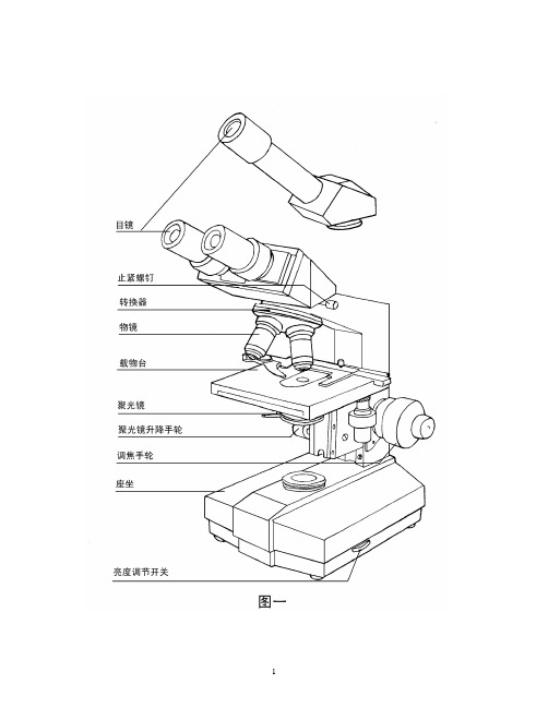
一、用途XSZ-G系列生物显微镜是重庆光电仪器有限公司对社会的最新奉献。
她将帮助您在生物学、植物学、细胞学、病理学、药物化学、医疗卫生、环境监测、学校教学、农业科研等领域获得更多更大的成功。
二、规格与技术参数1、总放大倍率:40×~1600×2、目镜:10×平场目镜16×平场目镜3、物镜:消色差物镜4×/0.1、10×/0.25、100×/1.25和半平场物镜40×/0.654、机械筒长:160毫米5、机械移动载物台横向移动范围60毫米纵向移动范围30毫米游标格值0.1毫米6、粗微动调焦范围25毫米:微动调焦手轮格值为0.002毫米7、阿贝式聚光镜数值孔径NA=1.258、滤色片:黄、绿、蓝三、结构和性能G系列生物显微镜的基本结构如图一所示。
10×、16×平场目镜为您提供了宽广平坦的视场。
所有物镜符合DIN标准,40×、100×(油)物镜均设有弹簧装置,能避免因使用不慎而损坏物镜及标本;新型的40×半平场物镜大大提高了成象质量。
四孔滚珠轴承内定位式物镜转换器可快速、准确地更换不同放大倍率的物镜。
粗微动调焦机构为同轴式(图二),该机构设有调节手轮,以调整粗动手轮的松紧,以适应不同操作者的手感和防止因长期使用而产生的载物台自动下滑现象。
此机构还设有限位圈,以便确定载物台上限位置,既方便使用又安全可靠。
矩形机械移动式载物台上的标本片夹,可方便地装夹标本。
搬动片夹手柄,放入标本后松开手柄,标本就被夹持在载物台。
纵、横向移动手轮同轴安装,使用方便。
载物台上刻有记取纵、横向移动量的刻尺和游标。
(如图三所示)。
载物台下面的阿贝式聚光镜(图四)采用齿轮、齿条机构传动,使聚光镜升降平稳。
调整三个紧定螺钉,可使聚光镜中心和光轴重合。
拨动聚光镜下的光栏调节杆,可改变孔径光栏的大小。
转动滤色片框,可方便地更换不同颜色的滤色片。
XDS-1B倒置生物显微镜使用说明书(重庆光电仪器有限公司)

ISO9001国际质量体系认证ISO14001环境体系认证从眼前做起XDS-1系列倒置生物显微镜使用说明书医疗器械生产企业许可证编号:渝食药监械生产许20060009号医疗器械注册号:渝食药监械(准)字2011第2220106号产品标准:YZB/ 渝0048-2007;GB/T2985-2008页脚内容0页脚内容1XDS-1系列倒置生物显微镜使用说明书申明:此说明书中内容,如产品因技术改进发生变更,恕不预告,敬请客户谅解!从眼前做起请珍惜您周围的环境,杜绝可以避免的污染,在您使用XDS-1系列倒置生物显微镜之前,请您仔细地阅读本使用说明书。
它可以指导您正确使用,免除错误操作造成仪器页脚内容2地 址:重庆市北碚区歇马镇沪渝村82号 电 话:(023)67959666 68287856 传 真:(023)68283256 网 址:http//: 邮 编:400712生产地址:重庆北碚歇马(重庆光学仪器厂)目 录1. 用途 (1)2. 规格............................................................1 3. 安装 (3)4.使用 (5)5.维护保养 (7)6.常见故障与排除方法 (9)7.标准配套表 (10)页脚内容310 25 4010 25 40WF101020 WF161614 S 559.5 S 6.3 6.37.6 S0.650.65DZ 1111PF1017.4 102.3 总放大倍数物镜总放大倍数目镜1254观察WF 10(φ20)100250400 WF 16(φ14)160400640摄影S 55125200S 6.3631582522.4 载物台纵横方向行程50mm×75mm,刻尺格值1mm,2.7粗微调焦组粗微调焦手轮同轴安装,粗动行程10mm,微动手轮格值0.002mm。
2.8双目瞳距调节范围55~75mm.2.9 摄影幅面24mm36mm.2.10 摄像装置选配2.11 电源输入: 220V50Hz (或110V60Hz)保险丝管: 520 1A游标格值0.1mm。
MT-40系列生物显微镜操作手册说明书

MT-40 Series Biological MicroscopeManualThis manual expatiates the using method, troubleshooting and maintenance about MT-40 series biological microscope. Please study this manual thoroughly before operating, and keep it with the instrument. The manufacturer reserves the rights to the modifications by technology development. On the basis of operation ensured, technical specifications may be subject to changes without notice.Contents MT-40 Series Before Use1. Components (1)2. Assembling (3)2-1 Assembling Scheme (3)2-2 Assembling Steps (4)3. Operation (6)3-1 Set Illumination (6)3-2 Place the Specimen Slide (6)3-3 Adjust the Focus (7)3-4 Adjust the Focusing Tension (7)3-5 Adjust the Interpupillary Distance (7)3-6 Adjust the Field Diaphragm (Iris Diaphragm Koehler Illuminator Condenser Optional) (8)3-7 Adjust the Aperture Diaphragm (8)3-8 Use the Oil Objective (100X) (9)3-9 Use the Filter (9)3-10 Replace the Fuse (10)4. Technical Specifications (11)4-1 MT-40 Series Biological Microscope Technical Parameters (11)4-2 Parameters of objective (11)5. Troubleshooting (12)1. Operation Notice1. As the microscope is a high precision instrument, always operate it with care, and avoid physical shake during the operation.2. Do not expose the microscope in the sundirectly, either not in the high temperature, damp, dust or acute shake. Make sure the worktable is flat and horizontal.3. When moving the microscope, use both hands to hold its back hand-clasping ① and the front base ②, and lay it down carefully (see Fig. 1). ★ It will damage the microscope by holding the stage, focusing knob or head when moving.4. When working, the surface of condenser will be very hot. Make sure there is enough room for the heat dissipating around the condenser ③ (see Fig. 2).5. Connect the microscope to the ground to avoid lightning strike.6. For safety, make sure the power switch ④ is at “0” (off) and power it off before replacing the bulb or fuse, and wait until the lamp cools down (see Fig. 2).★ Bulb selected only: Single 3W LED light.7. Wide voltage range is supported as 100~240V . Additional transformer is not necessary. Make sure the voltage is in this range.8. Use the special wire supplied by our company.Fig. 1Fig. 22. Maintenance1. Wipe the lens gently with a soft tissue. Carefully wipe off the oil marks and fingerprints on the lens surfaces with a tissue moistened with a small amount of 3:7 mixture of alcohol and ether or dimethylbenzene.★ As the alcohol and ether is flammable, don’t place these chemical near to fire or fire source. For example, when turning on or turning off the electrical device, please use these chemical in a ventilated place.2. Don’t use organic solution to wipe the surfaces of the other components. Please use the neutral detergent if necessary.3. If the microscope is damped by liquid when using, please power it off immediately and wipe it dry.4. Never disassemble the microscope, otherwise the performance will be affected or the instrument will be damaged.5. After using, cover the microscope with a dust cover.3. Safety SignSign Signification Study the instructions before use. Unsuitable operation would lead to person hurt or instrument faulty. | Main switch ON OMain switch OFF- 1 -- 2 -- 3 -- 4 -- 5 -- 6 -- 7 -3-3 Adjust the Focus1. Move the objective 4X to the light path.2. Observe the right eyepiece with right eye. Rotate the coarse focusing knob until the ① image appears (see Fig. 10).3. Rotate the fine focusing knob ③ for clear details.★The position screw ② can stop the objective touching the clips.3-4 Adjust the Focusing TensionIf the handle is very heavy when focusing or the specimen leaves the focus plane after focusing or the stage declines itself, please adjust the tension adjustment ring ① (see Fig. 11).To tighten the focusing arm, rotate the tension adjustment ring ① according to the arrowhead pointed; to loosen it in the reverse direction.3-5 Adjust the Interpupillary DistanceWhen observe with two eyes, hold the base of the prism and rotate them around the axis until there is only one field of view.“。
- 1、下载文档前请自行甄别文档内容的完整性,平台不提供额外的编辑、内容补充、找答案等附加服务。
- 2、"仅部分预览"的文档,不可在线预览部分如存在完整性等问题,可反馈申请退款(可完整预览的文档不适用该条件!)。
- 3、如文档侵犯您的权益,请联系客服反馈,我们会尽快为您处理(人工客服工作时间:9:00-18:30)。
XSP-5C系列生物显微镜使用说明书
目录
用途 (1)
主要规格 (1)
结构 (2)
工作原理 (3)
操作 (3)
保养 (4)
型号组合 (5)
七、型号组合:
部件名称
各种常归产品型号正常配备
XSP-5C XSP-5CV XSP-5CA 机架* * *
单目头*
单目V型头*
铰链式双目头*
WF10X广角目镜* * *
WF16X 目镜* * *
195 4X物镜* * *
六、保养:
1.仪器使用完毕后,用吹风球吹去镜头上的灰尘,用细软布蘸二甲苯擦拭镜头上的油污。
用清洁的细软布擦拭整机上的灰尘和污迹。
运动部分应随既涂上薄薄一层无腐蚀性的润滑剂,然后放入塑料袋中,装回木箱,并放在干燥,清洁,通风良好的和无酸碱蒸汽的地方。
2.显微镜出厂前已经仔细的检验和调整,物镜和目镜请不要自行拆卸。
3.物镜使用后必须装入物镜盒中,以防碰损和沾污。
一、用途:
本系例显微镜可供高等院校、中等专科学校及其它有关部门教学示范、课程实验及其它微观世界的研究。
选择不同倍率的物镜和目镜配合
可得到不同的总放大倍率。
其最大放大倍率为:40X/、64X 、100X、160X、
400X、640X、1000X、1600X。
用户根据需要可选择不同的组合型。
二、主要技术规格:
机械筒长:160mm
物像黄轭距:195mm
消色差物镜:4X 、10X、40X(弹簧)、100X(油弹)
54
目镜:WF10X(广角)、WF16X
倍率组合:
目镜
4X 10X 40X(弹簧)100X(油弹)
倍率
物
镜
10X广角40X 100X 400X 1000X
16X 64X 160X 640X 1600X
照明形式:反光镜(直径50mm)电照明装置(220V,20W)
载物台:载物台面积(120mmX130mm)
5c型载物台,带单透镜聚光镜(NA.0.65),拨盘光栏
5CV型,5CA型载物台,带阿贝聚光镜(NA1.25),可变光栏
目镜筒:斜筒式,可作360度旋转
双目筒,瞳距55-75mm
12
三、结构:四.工作原理:
由物镜装被照明的标本物进行线时放大若干倍,再由目镜进行视角
放大,从而使观察者得到所需的部放大倍率.
五、操作:
1.将标本切片放在载物台上,用切片压片压紧。
2.将各倍率的物镜顺序装入物镜转换器,将所选用的目镜插入目镜
筒中,使用双目时应将双目间距调至适合瞳距。
3.转动反光镜,使照明光线射入镜筒(或接通电照明光源),然后调
节聚光镜下光栏的孔径大小,使视场明暗适度。
4.观察时,先用低倍物镜寻找观察物,然后将观察物移至视场中心,再换高倍的物镜观察。
当使用100X物镜时,应先用干净的细木棒蘸少许
香柏油滴于物镜端部,再转入视场。
必须使物镜端部和盖玻片之间充满
香柏油液体,方可观察操作。
5.调焦时,一般先用粗调焦旋钮调节物镜至能看到标本轮廓,然后
再用微调旋钮进行调节,直至物像最清晰为止。
使用高倍物镜时最好由
下到上进行调节,以避免镜头因碰到切片而损坏。
6.调节聚光镜的高低和孔径光栏口径大小,至标本像对比度适宜,
像质清晰.
3。
