肺炎的X线表现和鉴别诊断
常见肺炎的影像诊断1

病理变化:
镜下不同的发展阶段,病变的表现和严重程度不 一致。早期病变的细支气管充血、水肿,表面附 着粘液性渗出物,随病情进展,病变支气管、细 支气管几周围肺泡腔出现中性及红细胞、脱落的 上皮细胞,周围肺组织充血,可有浆液渗出,部 分肺泡过度扩张;中性粒细胞渗出增多时,支气 管及周围肺组织破坏,呈完全化脓性炎症改变。
(3)灰色肝样变期 第5—6天,病变肺叶仍 肿大,充血消退,由红色转为灰白色,称 灰肝期。肺泡腔内渗出的纤维素增多,相 邻肺泡纤维素丝连接成网,纤维素网中有 大量中性粒细胞,肺泡腔毛细血管受压, 肺泡腔内红细胞几乎很少见到。此期渗出 物中不易检出细菌。
大叶性肺炎
灰色肝样变期 肺泡腔内充满渗出的纤维素及中性粒细胞, 箭头示相邻肺泡腔内纤维素经肺泡间孔互相连接.
支气管树整体观
右主叶支气管 右上叶支气管
右中叶支气管
气管 左主支气管
左上叶支气管
左下叶支气管
右下叶支气管
支气管树
支气管肺段
每一肺段支气管及其分支分布区的全部肺组织总 称支气管肺段,它是构成肺的形态学和功能学的 基本单位。
通常左右肺各有10个肺段。有时因左肺尖段与后 段、前底段与内侧底段支气管共干,此时左肺只 有8个支气管肺段。
大叶性肺炎
病变肺叶肿胀,色灰黄, 质实如肝.
(2)红色肝样变期 第3—4天,肿大的肺 叶充血呈暗红色,质地变实,切面灰红, 似肝脏外观,故称此。此期肺泡腔内充满 纤维素及大量红细胞,间杂少量中性粒细 胞和巨噬细胞,肺泡腔内的红细胞被巨噬 细胞吞噬、崩解后,形成含铁血黄素随痰 液咳出,致使痰液呈铁锈色。病变波及胸 膜时,引起纤维素性胸膜炎,发生胸痛, 随呼吸和咳嗽而加重
CT表现 一侧或双下肺多发渗出性病灶,呈 斑片状模糊影。
肺炎的X线表现和鉴别诊断
肺炎的X线表现和鉴别诊断肺炎是一种常见的呼吸道感染疾病,通常表现为发热、咳嗽、胸痛等症状。
临床医生经常使用X线检查来观察肺部病变情况,并通过X线表现和分析鉴别诊断肺炎的类型。
本文将介绍肺炎的X线表现和鉴别诊断。
X线肺炎表现肺炎的X线表现通常是肺部实变和病变灶的存在。
肺炎X线表现分为以下几种情况:1. 大叶性肺炎 (Lobar pneumonia)大叶性肺炎是肺泡和支气管的感染,通常表现为局限性的肺实变,影响一个或两个肺叶。
X线表现通常显示阴影区域由上至下扩展。
炎症一般在第2~3个病日呈现,且在处理过程中发生的暖反应更具特征性。
另一特征是阴影边界模糊。
2. 支气管肺炎 (Bronchopneumonia)支气管肺炎表现为肺泡和细支气管的感染,通常影响肺实质。
可在肺野内形成多个散在的、较小的实变灶,分布不均匀,且通常没有明确的边界。
X线表现可显示分布广泛的肺部浸润影。
3. 间质性肺炎(Interstitial pneumonia)间质性肺炎是肺间质和肺泡壁的炎症。
因肺泡内炎症灶太小而难以在X线上检测到。
X线表现为大的增生交界线,特别在肺尖及胸膜下边缘清晰可见。
肺炎的鉴别诊断肺炎的鉴别诊断是基于肺部X线表现,患者的临床症状和生化指标等多种因素综合判断。
常见肺炎和其他肺部疾病的鉴别诊断包括:1. 肺癌 (Lung cancer)肺癌通常表现为同侧肺野内的肿物影,通常位于肺野的边缘。
X线图像还可以显示类似肺为轻度肺实变的斑点浸润。
2. 支气管哮喘 (Bronchial asthma)支气管哮喘通常表现为双肺散在的、小的透亮区。
在发作期间和发作后,纵隔可能搏动,使X线表现出胸膜下阴影。
3. 细胞变态反应、肺结核和肺栓塞等还有其它细节和表现形式,需要结合医生临床经验进行判断。
肺炎的X线表现因病变类型不同而异。
肺部的大叶性肺炎和支气管肺炎通常具有不同的X线表现,但间质性肺炎因其小的病变灶且不易检测。
因此,鉴别诊断通常需要结合患者的临床症状、生化指标、影像学表现等多种因素进行分析,并由医生进行最终的诊断。
球形肺炎的X线表现与鉴别诊断

误诊率最高 , 可能与肺癌常见 、 年龄偏大有关 。( 球形肺 炎 1 ) 形 态上虽大体呈 球形 、 类球形 , 较多为方 形或不 规则三角 形 , 但
其 中贴 近胸膜 的方形病灶 有特征性 ; 肺癌 形态呈较规 整球形 , 方 形少见 。f1 2球形肺炎边缘毛糙 , 可见长毛刺和棘状改变 , 且模糊 , 有 时可见晕征 , 其周 围可见淡薄小 片状影 , 映 了病变急性 渗出 反 性 改变 。肺癌周 围毛刺细短 , 边缘多较清楚 , 不见晕征 , 可合并肺 门及纵 隔淋 巴结肿大 。( 球形肺炎密度 较淡 , 3 ) 且增强后病灶中央 可见规则 、 面清晰 的无强 化 区, 界 反映 了炎性坏死 的特点 , 征 此 少见于肺癌 , 具有特征性 。而肺癌增 强后 如为不均 匀强化 , 强化
21 0 0年 1 月 中 第 2 第 3 期 1 卷 2
Oe o e 2 0 t b r 0Βιβλιοθήκη V012 . No_2 3
中国中医药咨 讯
J u a fC i aTrdto M iee Me iieI fr t n o r lo h n a iin Chn s d cn nomai n o ・1 9 ・ 5
治 疗 后 , 形 阴影 不 易 消 散 。 而 球 形 肺 炎 经 抗 感 染 治 疗 后 , 形 球 球
形肺炎 的机 制 目前 尚无详细 的解 释 , 可能与下列 因素有关 : ①炎 性渗出物通过孔 氏孔 和兰勃 氏管 向周围均匀扩散。② 由于抗生 素 的应用和 自身免疫力使 大叶性肺 炎和节段肺炎发 展受限而形 成 。③大叶性肺 炎和节段性肺炎 的吸收从边缘开始 , 可使病灶呈
这与球形肺炎容易相鉴别 。 26 机 化 性 肺 炎 .
改变且较模糊 , 系支气管周围炎所致。⑥病灶内见粗大血 管纹 贯 穿, 围及近肺 门侧血管纹增 多 、 周 增粗 、 扭曲呈“ 围充血征” 周 但无
肺炎X线表现与诊断

肺炎X线表现与诊断肺炎(pneumonia)为常见肺疾病,X线检查对病变的发现、部位、性质以及动态变化,可提供重要的诊断资料。
按病变的解剖分布可分为大叶性肺炎、支气管肺炎(小叶性肺炎)及间质性肺炎。
按病原菌的肺炎分类法对X线诊断无实用价值。
1.大叶性肺炎大叶性肺炎(lobar pneumonia)多为肺炎双球菌致病。
好发于冬春季,多见于青壮年。
起病急,以突然高热、恶寒、胸痛、咳嗽、咳铁锈色痰为临床特征。
血白细胞总数及中性粒细胞明显增高。
大叶性肺炎可累及肺叶的一部,也可从肺段开始扩展至肺叶的大部或全部,偶可侵及数叶。
X线征象较临床出现晚3~12小时。
其基本X线表现为不同形态及范围的渗出与实变。
自应用抗生素以来,典型的大叶性实变已不多见,病变多呈局限性表现。
大叶性肺炎的早期,即充血期,X线检查可无阳性发现,或只表现为病变区肺纹理增多,透明度略低或呈密度稍高的模糊影。
病变进展至实变期(包括红肝样变期及灰肝样变期),X线表现为密度均匀的致密影,如病变仅累及肺叶的一部分则边缘模糊。
由于实变的肺组织与含气的支气管相衬托,有时在实变区中,可见透明的支气管影,即支气管气像。
炎症累及肺段表现为片状或三角形致密影,如累及肺叶的轮廓一致(图3-1-11)。
不同肺叶的大叶性实变形状各不相同。
消散期的表现为实变区的密度逐渐减低,先从边缘开始。
由于病变的消散是不均匀的,病变多表现为散在、大小不等和分布不规则的斑片状致密影。
此时易被误认为肺结核,应予注意。
炎症进一步吸收可只遗留少量索条状影或完全消散。
临床上,症状的减轻常较肺内病变吸收为早,病变多在两周内吸收。
少数患者可延迟吸收达1~2个月,偶可机化而演变为机化性肺炎。
2.支气管肺炎支气管肺炎(bronchopneumonia)又称小叶性肺炎(lobular pneumonia),常见致病菌为链球菌、葡萄球菌和肺炎双球菌等。
多见于婴幼儿、老年及极度衰弱的患者或为手术后的并发症。
肺炎的影像学诊断简版
肺炎的影像学诊断肺炎的影像学诊断引言肺炎是一种常见且严重的呼吸系统感染疾病,其特点是肺实质发生炎症,导致气体交换功能受损。
影像学在肺炎的诊断和评估中起着重要的作用。
本文将介绍肺炎的影像学诊断的方法和常见特征。
X线检查X线检查是肺炎影像学诊断的最常用方法。
它是一种非侵入性、快速且经济实惠的检查方式。
在常规胸部X线检查中,肺炎的典型表现是肺实质的浸润和阴影增加。
通常可以观察到以下几个方面的表现:1. 斑片状浸润:肺炎的早期表现是局限性斑片状浸润,常见于胸片的下叶和胸外区域。
2. 大片状浸润:随着炎症的进展,肺炎可以呈现为较大范围的片状浸润,常见于胸片的肺野。
3. 肺叶实变:肺炎的极端表现是肺叶的实变,常见于胸片的上叶或下叶。
此外,X线检查还可以观察到气管位置和支气管的异常,如支气管炎树芽样阻塞。
CT扫描CT扫描是一种更加敏感和准确的肺炎影像学诊断方法。
它可以提供更多的细节和横断面图像,有助于描绘肺部的解剖结构和病变的范围。
常见的CT表现包括:1. 浸润阴影:与X线检查类似,CT扫描可以显示肺实质的浸润阴影,但更加清晰和详细。
浸润阴影可以是斑片状、大片状或结节状。
2. 空洞形成:在某些情况下,肺炎可能导致空洞形成,这些空洞可以通过CT扫描进行定位和评估。
3. 胸腔积液:在肺炎的严重情况下,胸腔积液也是一种常见的表现,CT扫描可以准确显示积液的情况。
此外,CT扫描还可以评估其他肺部病变,如肺不张、肺气肿等,并在误诊、复发或治疗监测中提供更多帮助。
磁共振成像相对于X线检查和CT扫描,磁共振成像(MRI)在肺炎的影像学诊断中应用较少。
然而,MRI可以提供更好的软组织对比度,并在某些特殊情况下提供有价值的信息。
MRI在儿童或孕妇等特殊人群中可能更适用,避免了X线辐射的风险。
结论肺炎的影像学诊断对于疾病的早期发现、定位和评估起着至关重要的作用。
X线检查、CT扫描和MRI是常见的肺炎影像学诊断方法。
每种方法都有其特点和适应症,医生应根据具体情况选择合适的影像学检查方法。
肺炎的影像学诊断

巨细胞病毒性肺炎
肾移植术后3个月,巨细胞病毒性肺炎。 胸部平片见两上肺散在斑片状淡泊渗出影
3天后胸片,出现大片实变 并且进展迅速
巨细胞病毒性肺炎同一病例。次日CT示两上网状 模糊影,肺间质浸润为主病变
甲型H1N1流感病毒肺炎的鉴别诊断
• 细菌性肺炎:叶或段的实变影,病变较局 限,一般多为一段或以一叶病变发生,很 少发生两肺或一肺弥漫性病变。 • 支原体肺炎:病变以间质改变为主。早期 表现肺纹理增多模糊及网状纹理,进展时 呈局限或者广泛的片状磨玻璃影,自肺门 向肺野外围伸展的大片扇形阴影。CT可显 示早期小叶中心磨玻璃影或实变影,肺间 质炎症、网状阴影及小叶间隔增厚影。
入院检查
• 体温 39.5℃,脉搏106次/分,呼吸 20次/分,血压 120/70mmHg。双侧扁桃体1°肿大,双肺呼吸音增粗, 未闻及干湿性啰音 • 白细胞9.7109/L,中性粒细胞百分比77.9% • 肝功能异常,谷丙转氨酶(ALT)达76U/l,血糖 8.4mmol/L • 心肌酶指标提示明显升高:肌酸激酶(CK)达 1240U/L,肌酸激酶-心肌同功酶(CK-MB)88U/L, LDH达587U/L • 痰培养:草绿色链球菌 • 口咽含漱液及气管吸取液送省疾病中心检测,RTPCR 法检测出乙型流感病毒
完全大叶性实变
大叶性肺炎典型影像学表现为实变起自肺叶外周、 紧邻胸膜,然后向肺野中心扩散。
抗炎后复查病灶基本吸收
球形肺炎CT表现
• 边缘清楚、光滑,呈球形或类球形的病灶,密度 较均匀。有的灶中央密度低于周边(液化坏死), 有时可形成小空洞,钙化、支气管气相偶尔可见; • 增强扫描示病灶软组织部分明显强化,呈均匀强 化、周边环形强化或部分强化,少数不强化; • 肿块边缘不规则,毛刺多较长.部分为索条状,可 有周围充血征(晕环)、血管集束征等; • 病灶多贴近胸膜呈广基相贴,伴局部胸膜增厚但 少有胸水及胸壁改变,有时可见胸膜下脂肪层增 厚,部分病灶见典型胸膜凹陷征.
肺炎的影像诊断和鉴别诊断

xx年xx月xx日
《肺炎的影像诊断和鉴别诊断》
CATALOGUE
目录
引言肺炎的影像诊断肺炎的鉴别诊断结论
01
引言
1
目的和背景
2
3
肺炎是常见的呼吸系统疾病,准确诊断对治疗和预后至关重要。
影像学检查在肺炎的诊断和鉴别诊断中具有重要价值。
本综述旨在系统总结和探讨肺炎的影像学表现、诊断和鉴别诊断。
研究方法和范围
X线影像呈区域性肺血管纹理减少,有时可见尖端指向肺门的楔形阴影。
少见肺炎的鉴别
肺真菌病
肺水肿
肺栓塞
肺寄生虫病
04
结论
肺炎影像诊断准确性
肺炎鉴别诊断的重要性
不同类型肺炎的影像学表现
研究成果总结
对临床实践的建议
临床医生应重视影像学检查在肺炎诊断中的应用,并选择合适的影像学检查方法。
重视影像学检查
关注病情变化
立克次体肺炎
X线影像呈弥漫性支气管炎,可伴肺脓肿和脓胸。
其他病原体所致肺炎
肺结核
X线影像呈上叶尖后段、下叶背段和后基底段为主的浸润影,可伴空洞和纤维化。
X线影像呈支气管肺炎、融合性支气管肺炎、肺脓肿和脓胸等表现,可伴空洞。
X线影像呈粟粒性肺结核、肺脓肿等表现,可伴胸腔积液。
X线影像呈双肺弥漫性浸润影,以肺门为中心,由内向外逐渐变淡。
CT诊断
MRI
在显示肺实质和肺血管病变方面,MRI具有较高的诊断价值,但价格昂贵,一般不作为首选。
其他影像技术
如超声、核素扫描等,在特定情况下也可用于肺炎的诊断和鉴别诊断,如超声心动图可观察有无合并心力衰竭等并发症。
MRI和其他影像技术
03
肺炎的鉴别诊断
肺部感染鉴别诊断

1.干酪性肺炎:急性结核性肺炎临床表现与肺炎球菌肺炎相似,X线亦有肺实变,但结核病常有低热乏力,痰中易找到结核菌。
X线显示病变多在肺尖或锁骨上下,密度不均,历久不消散,且可形成空洞和肺内播散。
而肺炎球菌肺炎经青霉素治疗3~5天,体温多可恢复正常,肺内炎症也较快吸收。
该患者临床表现不符,暂不考虑。
2.急性肺脓肿:早期临床表现与肺炎球菌肺炎相似,但随着病程的发展,大量脓臭痰为肺脓肿的特征,致病菌有金葡菌、克雷白杆菌及其他革兰阴性杆菌和厌氧菌。
X线显示脓腔和液平,较易鉴别。
该患者胸部CT未见类似病灶,故暂不考虑。
1.肺结核:该病可表现为发热、咳痰、咳痰,胸部CT可有肺内多发结节病灶表现,但患者病程中无午后低热、乏力、盗汗、食欲减退等结核中毒症状。
故目前该诊断依据不足,需入院后行PPD、痰找结核等相关检查以行明确。
CRP与降钙素原下呼吸道感染的新进展:下呼吸道感染是常见的感染性疾病,其临床诊断主要依据疾病诱因、症状、体征、白细胞计数和分类、痰病原菌培养及胸部X线检查等。
但是,呼吸系统疾病的症状与体征往往十分相似,难以鉴别。
因为多种炎症反应均可引起白细胞计数的改变,据国外专家研究发现,在各种临床表现中,白细胞升高并不是感染的独立预测指标,白细胞计数用于诊断感染的准确性很低。
而痰病原菌培养的检出率低,且培养结果的得出往往需要较长时间,影响疾病的早期治疗。
事实上,正是由于缺乏诊断感染的可靠的临床指标,临床常常出现延误或过度使用抗生素的情况。
早期应用正确的抗生素治疗,可以明显降低下呼吸道感染性疾病患者的病死率。
另一方面,滥用抗生素将显才增加耐药性细菌感染的危险,增大临床治疗难度。
因此,正确、及时诊断下呼吸道感染的类型,确定感染的严重程度,对于下呼吸道感染性疾病的治疗具有非常重要的意义。
5.金葡菌肺炎:金黄色葡萄球菌感染引起的肺炎多为急性起病,中毒症状严重,常有高热、畏寒、寒战,咳黄脓痰,血白细胞总数明显增高,肺部影像学可见单个或多发脓腔形成,痰或者血培养出该菌可确诊。
各种肺炎的X线检查有何特征

各种肺炎的X线检查有何特征1.肺炎球菌肺炎:早期仅见肺纹理增粗或受累的肺段、肺叶稍模糊。
近年由于抗生素的应用,典型的大叶实变少见。
实变阴影中可见支气管气道征,肋膈角可有少量胸腔积液征。
肺炎消散期,X线浸润逐渐吸收,可有片块区域吸收较早,呈现“假空洞”征。
2.葡萄球菌肺炎:X线阴影的易变性是金葡菌肺炎的一个重要特征。
X线显示肺段或肺叶实变,或呈小叶样浸润,可有单个或多发的液气囊腔,形成阴影内伴有空洞和液平。
3.克雷白杆菌肺炎:X线显示肺叶或小叶实变,有多发性蜂窝状肺脓肿,叶间隙下坠。
4.军团菌肺炎:早期为单叶斑片状肺泡内浸润,继有肺叶实变,可迅速发展至多肺叶段,下叶多见,单侧或双侧,可伴少量胸腔积液。
偶有肺内空洞及脓胸形成。
5.肺炎支原体肺炎:肺部多种形态的浸润影,呈节段性分布,以肺下野为多见,也有从肺门附近向外伸展者。
6.肺念珠菌病:(1)支气管型:双肺中、下野纹理增重。
(2)肺炎型:两肺中下野有弥漫性小片状或斑点状阴影,亦可融合成大片肺炎阴影,边缘模糊,形态多变,还可有多发性脓肿。
少数病例伴胸膜改变。
7.病毒性肺炎:多见双肺下叶弥漫性密度均匀的小结节状浸润阴影,边缘模糊,少数患者可见叶性浸润或弥漫性网状结节性浸润灶。
8.厌氧菌性肺炎:双下肺底纹理增多粗乱,夹杂有边缘模糊的斑片状阴影,或同时伴有脓胸、胸膜积液等征象。
肺炎是由多种病源菌引起的肺充血,水肿,炎性细胞浸润和渗出性病变.临床上常见,可发生于任何的人群。
临床表现主要有发热,咳嗽,咳痰,呼吸困难,肺部X线可见炎性浸润阴影。
细菌性肺炎占成人各类病原体肺炎的80%。
在儿童、老年人和免疫抑制患者中病死率极高。
多有畏寒、发热、咳嗽、咳痰、胸痛等症状,少数有咯血和呼吸困难;其它症状有恶心呕吐、周身不适、肌肉酸痛等。
病毒性肺炎是由多种病毒感染引起的支气管肺炎。
多发生于冬春季节。
临床表现一般较轻。
主要症状为干咳、发热、呼吸困难、紫绀和食欲减退。
支原体肺炎是由肺炎支原体引起的肺炎,曾称原发性非典型性肺炎。
肺炎的X线表现和鉴别诊断

目录
• 肺炎的X线表现 • 肺炎的鉴别诊断 • 肺炎的X线诊断标准 • 肺炎的X线检查方法 • 肺炎的预防与治疗
01
肺炎的X线表现
早期X线表现
肺纹理增粗
由于炎症刺激,早期X线表现为肺 纹理增粗,这是肺炎的常见表现。
肺实质浸润
炎症细胞浸润肺实质,导致肺实质 密度增高,X线表现为片状或斑片 状阴影。
特殊类型肺炎的诊断标准
支原体肺炎
X线检查可见肺部出现斑片状阴 影,密度不均,边缘模糊或清 晰,以双下肺明显。
病毒性肺炎
X线检查可见肺部纹理增粗、模 糊,可能出现磨玻璃样改变。
军团菌肺炎
X线检查可见肺部出现斑片状或 大片状实变影,密度增高,边 缘模糊。
衣原体肺炎
X线检查可见肺部出现斑片状阴 影,密度不均,边缘模糊或清 晰,以双下肺明显。
04
肺炎的X线检查方法
正位胸片检查
正位胸片是肺炎诊断的基本检查 方法,能够显示肺部的基本病变,
如炎症、浸润、实变等。
正位胸片可以观察到肺部纹理的 改变,如增粗、紊乱等,以及肺 部实质的改变,如斑片状、大片
状阴影等。
正位胸片对于发现肺部病变的部 位、范围和程度有一定的帮助, 但分辨率相对较低,对于一些细
胸腔积液
X线检查可见胸腔内出现液体 密度影。
重症肺炎的诊断标准
肺部病变广泛
肺实变
胸腔积液增多
肺门淋巴结肿大
X线检查可见肺部病变广 泛,累及多个肺叶或肺
段。
X线检查可见肺部出现大 片状实变影,密度增高,
边缘模糊。
X线检查可见胸腔内液体 密度影增多,且可能伴
有胸膜增厚。
X线检查可见肺门淋巴结 肿大,边缘清晰或模糊。
肺炎的影像诊断和鉴别诊断

7天 12天
26天 39天
ห้องสมุดไป่ตู้
9天 15天
30天
4.X线表现:
肺纹理增重,模糊, 局限或广泛的的片状模糊影像, 常为磨玻璃密度,也可为肺实变密度,可按或不 按肺叶及肺段分布。肺段或大片阴影。 可位于肺野的内带、中带和外带。 单发或多发,中下肺野多见。
4. CT表现
男,53岁,03-10-14受雨淋,17日发热39.5度,WBC6000-5000,
•H5N1甲型禽流感患者
•图b为图a 5天后胸片,显示病变进展较快,双肺全部 受累,为ARDS。
(1)临床表现最为严重时,如病人的精神状况及体温,白细胞
计数等指标较高时,肺部影像最为严重和广泛。
(2)当临床状况好转时,肺部影像也减轻。
(3)但也有报道肺部影像正常及病变较轻者而死亡的病例。
(4)在病变吸收过程中, 影像的吸收时间迟于临床症状的 改善时间,即临床表现已经明显 好转,肺内仍然可见异常影像 (与一般的肺炎相似)。
在单侧或双侧肺弥漫性浸润,呈大片 状毛玻璃样影及肺实变影像,其内可见含气支 气管征。
病变后期为双肺弥漫性实变影, 少数可合并单侧或双侧胸腔积液。
•住院第五天咳嗽、气短,胸片表现为双下肺斑片状肺浸润病灶(图A)。 •24小时后,肺炎快速进展,表现为双肺弥漫性肺炎,符合ARDS表现,患者死亡(图B)。
肺炎的影像诊断和 鉴别诊断
•急性肺炎鉴别。
•根据X线及病理大体形态,肺炎分为 大叶性肺炎、支气管肺炎和间质性肺 炎。
X线平片对于肺炎的价值
可确定肺内有无病变, 可确定病变的部位 可确定病变的范围, 了解病变的动态变化, 了解有无合并症, 观察治疗效果和判断预后。
•肺炎的病原诊断需根据临床及病 原学检查,
肺炎影像学鉴别诊断
肺炎影像学鉴别诊断
肺炎是指肺部感染引起的炎症性疾病,通常由细菌、病毒或真菌引起。
肺炎在临床上是比较常见的疾病之一,但不同病原体引起的肺炎
在影像学表现上有所不同,因此对肺炎的影像学鉴别诊断显得尤为重要。
一、细菌性肺炎
细菌性肺炎是最常见的肺炎类型之一,主要由肺炎支原体、流感嗜
血杆菌、肺炎链球菌等细菌引起。
在X线胸片上,细菌性肺炎的特点
是叶片状、节段状浸润影,伴有支气管充气征象。
CT表现为感染灶周
围的浸润区,有时出现支气管充气征、空洞形成等。
二、病毒性肺炎
病毒性肺炎是由呼吸道病毒感染引起的,如流感病毒、腺病毒等。
在影像学上,病毒性肺炎的X线胸片表现为双肺弥漫性间质性改变、
叶片状浸润影、磨玻璃影等。
CT表现为双肺多发的斑片状、浸润影和
磨玻璃影,可伴有胸腔积液等。
三、真菌性肺炎
真菌性肺炎是由真菌感染引起的肺部疾病,常见的真菌包括曲霉、
念珠菌等。
在影像学上,真菌性肺炎的X线胸片表现为多发性结节影、斑片状浸润影、空洞等。
CT表现为双肺多发的结节、空洞、斑片状浸
润影等。
四、肺部结核
肺部结核是由结核分枝杆菌引起的慢性感染性疾病,在影像学上表现为肺尖和上叶的病变较多。
X线胸片可见肺门纵隔淋巴结增大、斑点状、节段状浸润影等。
CT表现为结节、空洞、胸膜增厚等。
总结而言,通过对肺炎不同类型的影像学特征进行鉴别诊断,有助于明确病原体的类型,从而制定更加针对性的治疗方案,提高治疗效果。
因此,医生在诊断肺炎时应充分重视影像学检查,结合临床表现进行综合分析,确保患者能够得到及时有效的治疗。
肺炎的影像诊断和鉴别诊断

肺炎的影像诊断和鉴别诊断肺炎是指肺部组织发生感染性炎症的疾病,常见症状包括咳嗽、咳痰、发热等。
在临床诊断中,影像学检查是一种常用的方法,可以帮助医生准确判断病变部位、性质,进行鉴别诊断。
本文将对肺炎的影像诊断和鉴别诊断进行讨论,旨在提供一些参考。
一、X线检查X线是最常见的肺部影像学检查方法之一,它能够对肺脏进行较为全面的观察,包括肺野的透明度、边缘模糊程度等。
在肺炎的影像表现上,常见的是肺纹理增加、阴影模糊等。
不同类型的肺炎在X线上有一些特征性的表现。
非典型肺炎,如军团菌肺炎、支原体肺炎等,X线影像上的表现相对于典型肺炎来说较为轻微,主要表现为斑片状浸润阴影,常见于两肺中下野。
典型肺炎多以肺叶或肺段为主,表现为实变,融合性浸润和空洞等。
二、CT检查CT(计算机断层扫描)是一种高级影像学检查技术,通过多个方向的断层图像构建出三维图像,可以提供更为详细的解剖信息。
在肺炎的诊断中,CT检查有着不可替代的优势。
CT影像常见的肺炎表现包括结节状浸润、斑片状浸润、空洞形成等。
肺炎的CT表现具有一定的多样性,而且会有一些特征性的表现。
例如,结核性肺炎的CT影像通常表现为结节状密度增高的病灶,中心坏死可以形成空洞;病毒性肺炎常表现为双侧多形状斑片状、磨玻璃样阴影的浸润。
三、核磁共振检查核磁共振(NMR)利用核磁共振现象进行成像,与CT相比,其分辨率更高,不需要使用放射线,对于某些特定患者如孕妇、儿童等更为安全。
在肺炎的影像诊断中,核磁共振是一种辅助手段。
四、PET-CT检查PET-CT检查是一种结合了正电子发射断层扫描(PET)和计算机断层扫描(CT)的技术,它可以提供肺炎发炎区域的代谢信息和解剖信息,对于非特异性影像学表现的肺炎诊断有着较高的准确性。
肺炎的影像诊断和鉴别诊断需要综合考虑患者的临床表现、实验室检查以及影像学检查结果。
不同类型的肺炎在影像学上具有一些特异性表现,但也有一些交叉现象,因此需要综合分析。
肺炎的X线表现和鉴别诊断优秀课件
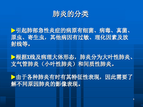
(5)合并症:易发生急性呼吸窘迫综合征(ARDS)合并ARDS时两肺出现广泛实变影像。 辅助通气治疗的患者可发生气胸和纵隔气肿。少数可合并胸腔积液。 (6)影像与临床的联系:临床表现严重时,肺部影像改变也最为广泛。当临床状况好转时,肺部影像也逐渐吸收。但也有报道肺部影像正常及病变较轻者而死亡的病例。
13.01.04
Case 2
14.01.04
16.01.04
Case 2
19.01.04
Case 2
21.01.04
20.01.04
Case report 3
Patient P.T.B. female, 30 y/o living in Ha Nam province. Admitted on 05/01/04, died on 09/01/04. Having history of contact with flu chicken. She looked after her daughter who had died of acute atypic pneumonia (H5N1 confirmed) at the Pediatrics Hospital just 2 days before. Main complains: Feeling fatigue, fever, chest discomfort and shortness of breath. She went to provincial hospital and CXR was performed but no change on film. However, on the 3rd of the illness, respiratory distress quickly developed and she was refered to NICRTM. On admission: temp 380C, pulse 90, BP 80/40, RR 30/min. Oxygen saturation 64%. Lab findings : WBC 1.7 G/L, (63.5% neutrophils), platelets 66 G/L, BUN 14.3mmol/l. Chest X-ray : changes with opacities unclear boundary infiltrations over 2 lungs . Management: Oxygen therapy with mask but no effect, BiPAP ventilation was applied. SaO2 was around 83-90%. Antibiotics Axepim(头孢吡肟), Solumedrol,Tequin and Dopamin(多巴胺) were ministered also.
过敏性肺炎的X线诊断和鉴别
过敏性肺炎的X线诊断和鉴别发表时间:2017-10-31T16:45:01.680Z 来源:《医药前沿》2017年10月第30期作者:郭强蕊[导读] 过敏性肺炎是由过敏性反应所导致的肺部炎性改变,该疾病的临床症状不够明显。
(甘肃省平凉市第二人民医院放射科甘肃平凉 744000)【摘要】目的:分析过敏性肺炎的X线诊断和鉴别。
方法:选取2000年01月-2015年12月在我院行X线检查的过敏性肺炎患者30例,回顾性分析患者的相关临床资料,分析过敏性肺炎的X线诊断和鉴别方法。
结果:过敏性肺炎的X线显示:病灶的形态、发生部位及范围等均有一定的规律。
病灶分布以肺外围部为主,肺尖部很少受累。
病灶形态以浸润性阴影、小结节状阴影等表现为主。
中心透亮,有时会出现假性空洞,由内向外吸收消散。
其中急性过敏性肺炎表现:可见弥漫性结节影,边界不清,或者斑片状毛玻璃影,或者实变影,分布以双侧中下肺野为主。
亚急性过敏性肺炎表现:可见网结节影,由细线条纤维束、结节共同形成的;慢性过敏性肺炎表现:可见网状影、粗线条状纤维影、结节影,分布以中上肺野为主。
结论:对于过敏性肺炎患者来说,应用X线检查具有一定的临床价值,可进一步诊断与鉴别过敏性肺炎,为临床下一步治疗提供依据。
【关键词】X线;过敏性肺炎;诊断;鉴别【中图分类号】R563.1 【文献标识码】A 【文章编号】2095-1752(2017)30-0108-02过敏性肺炎是由过敏性反应所导致的肺部炎性改变,该疾病的临床症状不够明显,在临床上诊断过程中,极易出现漏诊、误诊等情况。
对于过敏性肺炎的诊断方法有很多,如X线、CT、实验室检查、肺功能检验等,均是临床常见的检验方法,但其在临床诊断、鉴别均存在着不同程度的优点以及缺点[1-2]。
本研究为了更好地分析过敏性肺炎的X线诊断和鉴别价值,特在我院选取2000年01月-2015年12月在我院行X线检查的过敏性肺炎患者30例,收集资料进行分析,现汇报如下。
大叶性肺炎的X线诊断

1、病因
为细菌引起的急性肺部炎症,主要致 病菌为肺炎双球菌。冬、春发病高。多见 于青壮年。
2、病理
炎性渗出主要发生于肺泡,支气管和间质很少 受累。病理改变分四期:
• (1)充血期:12小时~24小时,肺泡毛细血管
充血扩张,肺泡内有少量浆液性渗出液,细胞不 多,肺泡内仍含气体。
5、鉴别诊断
• (1)大叶性肺不张:同为大叶性实变,但肺叶的
容积缩小,叶间裂凹陷,临近的器官向病变区移 位,应以鉴别。
• (2)干酪性肺炎:多见于右上叶,实变系多心性
团絮状干酪灶的融合,其密度较大叶性肺炎高, 但不均匀,其中可见不规则的空洞。常有支气管 播散灶,痰内可查到结核菌。
• (3)胸膜炎:与下叶的大叶性肺炎鉴别:其肋膈
见一片状密实阴影,上界锐利平直,从此向下的阴影逐 渐变淡,肋膈角保持清晰。侧位片尖端指向肺门,底部 达前胸壁或者肋膈角,上缘以横裂为界,下缘以斜裂的 前下部为界的三角形阴影。
• 左上叶大叶性肺炎,表现相当于右上叶与中叶表现
之和。正位片其高密度影几乎占全部肺野,仅肺基底部 和肋膈角保持透亮,下缘模糊不清。侧位后界锐利止于 斜裂。
• 炎变肺叶的容积一般不增大,由于肺泡内充满炎性
渗出物,有时可略大于正常,叶间裂可稍凸,伴有肺不 张时,容积可略小于正常,叶间裂可稍凹。实变区一般 看不到肺纹理,由于含气的支气管与实变的肺组织相互 衬托,可显示“空气支气管征”。
• (3)吸收消散期:病灶密度逐渐变淡,呈散在的大小
不一的分布不规则的斑片状阴影。进一步吸收仅遗留条 索状阴影,或完全恢复正常。
• 下叶大叶性肺炎,正位整个中下肺野呈大片浓密阴
影,上缘高于横裂模糊不清,下界浓密直达膈,完全遮 盖肋膈角和心缘。侧位呈一大三角形浓密区,位于后下 方,前缘止于横裂,后缘直达后胸壁,下面与膈影融合。
关于肺部炎症的X线表现

关于肺部炎症的X线表现【关键词】肺部炎症 X线(一)大叶性肺炎[图像特征]X线正位胸片:(1)左上肺可见大片高密度影,密度均匀;(2)病灶边界欠清楚。
CT平扫(肺窗):(1)右肺下叶背段大片高密度影,病变累及整个肺段;(2)内可见通气支气管;(3)左肺下叶见小片状高密度影,境界清。
[影像诊断] 左肺上叶大叶性肺炎;两肺下叶大叶性肺炎,右肺下叶背段较明显。
[临床提醒] X线胸片及CT均可用于大叶性肺炎的诊断,胸片较CT在临床的应用更为广泛。
大叶性肺炎典型的影像表现为累及整个肺叶或肺段的实变影,受累肺叶或肺段的体积不缩小,内可见“含气支气管征”。
结合典型的影像学表现及临床症状和相关实验室检查,诊断本病较容易,但因抗生素的广泛使用,典型的大叶性肺炎已不常见,偶见于老年人、使用免疫抑制剂等免疫力低下的病人。
大叶性肺炎早期在X线上缺乏特异征象,部分可表现正常。
CT可显示肺炎早期的充血改变,并能更好的显示病变的范围。
X线胸片及CT检查的目的还在于及时反映病情变化,从而观察治疗效果,指导临床治疗。
(二)支气管肺炎[图像特征]X线正位胸片:左下肺见小片状模糊影,左侧心缘模糊不清。
CT平扫(肺窗):两下肺见多发斑片状高密度影,呈局灶性分布。
[影像诊断] 两下肺支气管肺炎。
[临床提醒] 支气管肺炎的X线表现多样,大多数表现为两下肺野中、内带沿支气管分布的不规则斑点状或小片状致密影,境界较模糊。
病变密度不均匀,中心密度较高,多伴有肺纹理增粗。
病变可以比较散在且较小,也可集中呈大片融合趋势,但多不局限于某一肺段或肺叶。
由于粘液堵塞支气管,病变区域可夹杂有小叶性肺不张或局限性肺气肿。
当细支气管阻塞时,也可形成小三角形肺不张影。
小儿多首先发生在脊柱旁,然后向心缘发展,因此早期易被心影所掩盖。
CT是本病的主要检查手段,结合病史一般能够确诊。
少数病例仅表现为不规则粟粒样病变或仅为肺纹理增强。
此时,影像诊断比较困难。
(三)病毒性肺炎[图像特征]X线正位胸片:(1)两肺纹理增加,以两下肺野中内带明显;(2)沿肺纹理分布见密度不均匀斑片状模糊阴影。
间质性肺炎鉴别诊断

间质性肺炎鉴别
1.特发性肺纤维化
多表现为进行性呼吸困难伴刺激性干咳,双肺可及Velcro音,伴有杵状指(趾),胸部X线提示双肺弥漫性网状阴影,肺功能为限制性通气障碍。
该患者既往肺部影像学及肺功能检查不支持,暂不考虑。
2.嗜酸细胞性肺炎
多有发热、咳嗽、气促,偶有少量咯血,可有体重减轻,周围血嗜酸细胞比例增多,胸片示双侧肺野外带片状阴影。
患者病史不符,影像表现不典型,嗜酸细胞正常,故可以鉴别。
3.结缔组织病所致双肺间质性疾病
如类风湿性关节炎、SLE、Wenger肉芽肿等,多有各个相关系统如关节、皮肤、鼻咽、呼吸道等受累。
患者病史不符,暂不考虑。
- 1、下载文档前请自行甄别文档内容的完整性,平台不提供额外的编辑、内容补充、找答案等附加服务。
- 2、"仅部分预览"的文档,不可在线预览部分如存在完整性等问题,可反馈申请退款(可完整预览的文档不适用该条件!)。
- 3、如文档侵犯您的权益,请联系客服反馈,我们会尽快为您处理(人工客服工作时间:9:00-18:30)。
影像学检查在肺炎诊疗中的作用价值
X线平片是诊断肺炎的主要方法,其价值为: ▶可确定肺部有无病变 ▶可确定部位 ▶可确定范围 ▶了解病变的动态变化 ▶了解有无合并症 ▶观察治疗效果和判断预后
▶肺炎主要采用X线平片检查。
▶ CT检查主要病例。
▶肺炎的病原诊断需根据临床及病原学检查
Case 3
Case 3
Case 3
Case report 4
• Patient T.V.C male, 19 y/o living in Bac Giang province. • Admitted on 16/01/04. Discharged on 30/01/04. • Having no history of contact with flu chicken.. But many ill chicken died around area where patient was living. • Present history: 5 days of illness at home. High fever, sputum coughing and shortness of breath. He Admitted to Bac Giang provincial hospital, CXR showed serious lesions. The next day, condition became more critical with difficulty in breath and he was refered to NICRTM. • On admission: temp 38.50C, pulse 84, BP 110/70, RR 54/min, crackle rales in both 2 sides of lung. SaO2 82%. • Lab findings : WBC 3.9 G/L, (75.5% neutrophils), platelets 127 G/L, BUN 6.6mmol/l. • Chest X-ray : Opacities with unclear boundary over 2 lungs. • Management: Oxygen therapy with mask, Tamiflu, Axepim(头孢吡肟) Solumedrol. SaO2 was improved 91-95%. • Clinical course: After 2 days of treatment: Condition was improved. No longer fever or shortness of breath. SaO2 95-99%. Lab findings: WBC 10.8 G/l, GOT 148, GPT 194, LDH 1014. + Discharged on the day of 14th.
(3)病变的范围:早期病变为局限性片状阴影, 进展后呈多灶性或弥漫性分布,可累及一个或 多个肺段、肺叶,也可位于一侧肺或双肺。但 一般不按肺叶或肺段分布。 病灶进展快 ,重症 患者很快出现双肺弥漫性病变。 (4)动态变化:胸部影像表现变化较快。重症 患者肺内病变 进展迅速 ,短期内病灶明显增多, 从小片到大片,从单侧到双侧。病变密度也发 生明显的变化。在恢复过程中肺内病变的 吸收 也较快。
•
• •
Case 2
13.01.04
14.01.04
Case 2
16.01.04
19.01.04
Case 2
20.01.04
21.01.04
Case report 3
• Patient P.T.B. female, 30 y/o living in Ha Nam province. • Admitted on 05/01/04, died on 09/01/04. • Having history of contact with flu chicken. She looked after her daughter who had died of acute atypic pneumonia (H5N1 confirmed) at the Pediatrics Hospital just 2 days before. • Main complains: Feeling fatigue, fever, chest discomfort and shortness of breath. She went to provincial hospital and CXR was performed but no change on film. However, on the 3rd of the illness, respiratory distress quickly developed and she was refered to NICRTM. • On admission: temp 380C, pulse 90, BP 80/40, RR 30/min. Oxygen saturation 64%. • Lab findings : WBC 1.7 G/L, (63.5% neutrophils), platelets 66 G/L, BUN 14.3mmol/l. • Chest X-ray : changes with opacities unclear boundary infiltrations over 2 lungs . • Management: Oxygen therapy with mask but no effect, BiPAP ventilation was applied. SaO2 was around 83-90%. Antibiotics Axepim (头孢吡肟), Solumedrol,Tequin and Dopamin(多巴胺) were ministered also.
Case 1
12.01.04
14.01.04
Case 1
16.01.04
19.01.04
Case 1
21.Jan.o4
23.Jan.o4
Case report 2
• • • • • Patient N.L.Hh. female, 23 y/o living in Thai Binh province. Admitted on 13/01/04, died on 23/01/04. She was older sister of the case 1. Main complains on admission: Fever, dry cough and diarrhea, no shortness of breath. Physical examination: temp 39.60C, pulse 100, BP 100/60, RR 20/min, no rales in both lungs. Oxygen saturation 99%. Other signs were normal. Lab findings on admission: WBC 5.6 G/L, (69% neutrophils), platelets 131 G/L, BUN 3.4mmol/l. Chest X-ray : no remarkable changes. Clinical course: On the 4th day : Became worse with continuous fever 390C, coughing, shortness of breath RR 40/min, crackle rales in both lungs, SaO2 was 86%. BP 100/60. WBC 3.5 G/l. GOT 77, GPT 40 U/l. Addition treatment with oxygen therapy by mask and antibiotics Axepim(头孢吡肟), Tavanic, Zithromax and Solumedrol(甲强龙),But it seemed to be uneffected.
Case 4
Case 4
Case 4
Case report 5
• Patient L.T.H female, 20 y/o living in Bac Ninh province. • Admitted on 16/01/04. Discharged on 11/02/04. • Having history of contact with flu chicken.. Many ill chicken died around area where patient was living. • Present history: 7 days of illness at home. High fever, sputum coughing and shortness of breath. • On admission: temp 38.20C, pulse 120, BP 100/60, RR 58/min, crackle rales in both 2 sides of lung. SaO2 80%. • Lab findings : WBC 7.2 G/L, (87.3% neutrophils), platelets 211 G/L, BUN 3.7mmol/l. • Chest X-ray : Opacities with unclear boundary over 2 lungs. • Management: Oxygen therapy with mask, Tamiflu; Axepim(头孢吡肟) • Solumedrol(甲强龙) ,SaO2 was improved 91-95%. • Clinical course: After 3 days of treatment: Condition was improved. No longer fever or shortness of breath. SaO2 95-99%. Lab findings: WBC 10.8 G/l, GOT 37, GPT 84+ Discharged on the day of 26th.
