后肾腺瘤临床病理及免疫组化观察分析
肾肿瘤病理报告
肾肿瘤病理报告
一、患者基本信息
•姓名:XXX
•性别:男
•年龄:XX岁
•就诊日期:XXXX年XX月XX日
二、临床病史
XXX先生因上腹部不适,伴有血尿前往就诊,经过详细检查后就诊于本院。
三、病理检查结果
1. 肾部CT扫描
肾部CT扫描结果显示在左肾上部可见一个直径约为Xcm的肾肿块。
2. 病理标本检查
收到来自左肾部的手术切除标本一份,标本编号为XXX。
切片镜下所见
镜下观察可见切片呈团块状,肿块直径约为Xcm。
肿块由囊性和实质性病变构成。
囊性区域内含淡黄色透明液体,液体中可见散在的细胞碎片。
实质性病变区内可见瘤细胞,细胞形态不规则,大小不一。
瘤细胞胞质丰富,核染色质深染,核仁明显,细胞核分裂象较少。
免疫组化染色
免疫组化染色结果显示瘤细胞表达Vimentin,CD10,同时表达PAX8,CK7和EMA阴性。
诊断结果
根据以上病理检查结果,我们结合临床表现和免疫组化染色结果,最终诊断为左肾肿瘤,病理类型为良性肾细胞瘤。
四、治疗方案
根据肾肿瘤为良性肾细胞瘤的诊断结果,建议XXX先生采取观察治疗,密切关注肿瘤的生长情况。
如肿瘤生长迅速或出现恶变的迹象,可能需要手术切除。
五、结论
通过肾肿瘤的病理检查,我们确认了XXX先生左肾的肿瘤为良性肾细胞瘤,建议进行观察治疗,并定期复查肿瘤的生长情况。
如有需要,在进一步的治疗方面可采取手术切除等方法。
以上为肾肿瘤病理报告的详细内容,希望对XXX先生的诊断和治疗提供参考。
如有其他疑问,请及时咨询医生。
后肾腺瘤的临床及病理分析并文献复习

周 利群
周 亮亮 李 学松
金杰 郭应禄
姚林
何群
何志 嵩
【 要】 目的 摘
探讨后 肾腺瘤 ( A 的临床及 病理 学特点 。方法 M )
回顾 性分 析 20 ~ 00年 06 2 1
女性 1例 , 男性 1
在北京大学第一 医院诊治 的 2例 M A患者的临床及病理学资料并复习文献 。结果
【 关键词 】 肾肿 瘤 ; 腺瘤 ; 肿瘤 , 组织 学类 型
后 肾腺 瘤( eaehi ae o , m tnp r dnmaMA) c 是非 常罕见 的肾脏 良性肿瘤。MA虽然具有特 殊 的临床和病 理学 特征 , 但术前
行 开 放 肾部 分 切 除 术 。
3 .组织 病理及免疫 组化 结果 : 切除 的标本 经 1 % 甲 对 0 醛溶液 固定 , 石蜡包埋 , m 厚 连续切 片 , 5汕 常规 HE染 色 , 光 镜 观察 ; n io E Vs n法 免 疫 组 化 染 色 。抗 体 选 用 波 形 蛋 白 i ( ie t , i 、 ls瘤蛋 白( 1 、 Vm ni Vm) Wi n m WT ) 角蛋 白单克 隆抗 体 A 1 A 3 神经特异性烯醇化酶 ( S ) 角蛋 白 7 C 7 以及 E/E 、 NE 、 (K )
后 肾腺 瘤 , 瘤 细 胞 小 而 一 致 , 密 排 列 , 梁 索 状 、 肿 紧 呈 团块 状 、
例 2 男 ,3岁 , , 3 体检发现 右肾肿瘤 1 入 院。无 临床症 周
状及 阳性体征 , 血常规 、 常规 、 尿 肾功能 正常 ; 超声 : 右肾 中下 极探及 等 回声结 节 ,. m ×3 6a 边 界清 , 3 6c . m, 内部 回声均 匀 , 周可见暗带 , 内未见 明显血 流信 号。C : 肾实质可 其 其 T右
免疫组化肾上腺疾病结果判读标准

免疫组化肾上腺疾病结果判读标准一、绪论肾上腺疾病是一类常见的内分泌疾病,常见的有肾上腺皮质功能亢进、肾上腺皮质功能减退等。
免疫组化技术在肾上腺疾病的诊断和治疗中发挥着重要作用,本文将对免疫组化肾上腺疾病结果判读的标准进行详细介绍。
二、免疫组化在肾上腺疾病诊断中的应用免疫组化是通过特异性抗体与组织标本中的相应抗原结合,然后通过染色或发光等方式来检测目标蛋白的表达水平。
在肾上腺疾病的诊断中,免疫组化能够帮助医生发现肿瘤细胞的来源、类型,识别细胞的生物学行为等,为临床诊断和治疗提供重要依据。
三、免疫组化在肾上腺疾病中的标记物在肾上腺疾病的免疫组化诊断中,常用的标记物主要有肿瘤抑制基因p53、Ki-67、S-100、Cytokeratin、Synaptophysin等。
这些标记物的表达水平与肿瘤的生长、浸润、转移等密切相关,能够为医生判断肿瘤的类型和恶性程度提供帮助。
四、免疫组化肾上腺疾病结果判读的标准1.肿瘤抑制基因p53的表达肿瘤抑制基因p53在肿瘤中的异常表达与肿瘤的发生、发展密切相关。
免疫组化检测肾上腺疾病样肿瘤患者p53蛋白的表达情况,如果p53的阳性率高,说明肿瘤细胞增殖活跃,预后不佳,需密切观察和治疗。
2. Ki-67的表达Ki-67是细胞增殖标记物,其在肿瘤细胞中的表达水平能够反映肿瘤的活跃程度。
免疫组化检测Ki-67的表达水平,能够帮助医生判断肿瘤的生长速度和转移风险,指导临床治疗方案的制定。
3. S-100的表达S-100是一种神经系统特异性标记物,其在肿瘤组织中的表达水平能够帮助医生判断肿瘤的来源和类型。
免疫组化检测S-100的表达情况,对于肾上腺神经内分泌肿瘤的诊断和鉴别诊断具有重要意义。
4. Cytokeratin的表达Cytokeratin是上皮细胞特异性标记物,对于肾上腺皮质样肿瘤和鳞状细胞癌的诊断和鉴别诊断具有重要意义。
免疫组化检测Cytokeratin的表达情况,能够帮助医生明确肿瘤的类型和来源,为临床治疗提供重要依据。
免疫组化实验结果分析
免疫组化实验结果分析免疫组化实验是一种常用的生物学实验方法,用于检测细胞或组织中特定蛋白质的表达情况。
通过特定的抗体与目标蛋白质结合的方式,可以在光学显微镜下观察到颜色反应,从而得出关于该蛋白质表达水平的结论。
本文将对免疫组化实验结果进行详细分析。
一、实验结果描述在分析免疫组化实验结果之前,首先需要对实验结果进行描述。
描述应该包括实验样本的来源、处理方法、实验抗体及其稀释倍数、染色方式以及显微镜下观察到的颜色和形态特征等信息。
例如,可以描述实验使用的抗体是针对肿瘤标志物CA125的,样本来源于人体卵巢癌组织,实验过程中使用了免疫组化染色试剂盒,并观察到染色结果具有明显的细胞膜表达。
二、结果分析1. 强阳性表达:强阳性表达意味着目标蛋白质在细胞或组织中高水平表达。
在显微镜下观察到明亮的染色,通常是在细胞膜、细胞质或核内。
这种结果表明目标蛋白质与相关疾病或生物学过程密切相关,可以作为潜在的治疗靶点或疾病诊断标记物。
2. 弱阳性表达:弱阳性表达意味着目标蛋白质在细胞或组织中低水平表达。
在显微镜下观察到的染色较为淡色,并且通常在少数细胞中出现。
这种结果可以表示目标蛋白质在特定细胞类型或条件下的表达受到抑制,或者表达水平较低对于疾病进展的影响较小。
3. 阴性表达:阴性表达意味着目标蛋白质在细胞或组织中未被检测到。
在显微镜下观察不到明显的染色。
这种结果可能表明目标蛋白质在该细胞类型或该疾病中不表达,或者实验方法存在技术问题导致无法检测到目标蛋白质。
4. 零值控制:在免疫组化实验中,通常会设置阴性对照或零值对照来验证实验结果的准确性。
零值控制是指阴性对照样本在实验中未出现任何染色反应。
这一结果证明实验方法的特异性良好,不会对非特异性信号产生干扰。
三、结果解释对实验结果进行解释是免疫组化实验结果分析的重要环节。
在解释时,需要结合实验目的、已有的相关研究和临床实际进行综合考虑。
根据实验结果的阳性或阴性表达情况,可以推测目标蛋白质在疾病发生发展中的作用以及其潜在的临床应用前景。
肾脏、睾丸肿瘤病理学

达+的异型大细胞。
全肾被黄色 肿瘤占据, 仅肾右缘残 余月牙形肾 实质,肿瘤 内大量出血。
Masson 三色 染色,可见血 管、纤维、肌 肉、脂肪组织。
膀胱内癌肿呈乳头状,浸润膀胱壁,并向膀胱外发展。
膀胱内结节 状肿物,浸 润膀胱壁, 阻塞双侧输 尿管,引起 肾盂积水。
镜下
肿瘤细胞有间变,细胞大小不等,排列不 规则,核有异形。根据其细胞形态、排列及其 肿瘤的侵袭性,常可分为三级: Ⅰ级:肿瘤乳头尚规则,但表面的移行细胞层次 增加,细胞密集,核大、染色质丰富,有异形 ,可见核分裂相,侵袭性少见。 Ⅱ级:肿瘤细胞异型性大,核分裂相多见,排列 可呈乳头状,但分布不均,常形成巢团状,肿 瘤有侵袭性。 Ⅲ级:肿瘤细胞呈高度异型性或未分化,核分裂 相多见,肿瘤坏死明显,常浸润深层组织。
大量的超微结构及免疫组化研究表明肾血 管平滑肌脂肪瘤的前体细胞是从一种生长 于血管周围的细胞发展来的
免疫组化研究已表明在肾脏的其他肿瘤中 无HMB-45表达。提示HMB-45免疫组化 的应用可以有效地将肾血管平滑肌脂肪瘤 从含有梭形细胞的其他肾肿瘤中(如平滑 肌瘤、肉瘤样肾细胞癌)鉴别出来,并使 术前穿刺活检更有意义。
CGA
SYN
CK/CD10
甲状腺滤泡癌样肾细胞癌
FIGURE 1. The mass shows multinodular appearance with extensive necrosis, and it is protruding into the renal pelvis. FIGURE 2. Histologic findings of the tumor. The most part of tumor consists of microfollicles and macrofollicles, in which inspissated colloid-like material is present in the lumina (A). Some luminal materials in the follicles are calcified, but no definitive psammomatous calcifications are present (B). The tumor cells of follicles have a moderate amount of clear or eosinophilic cytoplasm and round to oval nuclei with fine chromatin (C). The tumor cells show high nuclear grade with prominent nucleoli and frequent abnormal mitoses in some areas (D). FIGURE 3. Immunohistochemical stainings. The tumor cells show intensive staining for cytokeratin AE1/AE3 (A). CD10 immunostain shows positive reaction along the cytoplasmic membrane (B).
后肾性腺瘤一例报告
后 肾性 腺 瘤 是 一 种 罕 见 的 肾 脏 良性 肿 瘤 , 1 9 9 6年 召 开
的分子遗传 学对 肾细胞肿 瘤分类影 响 的工作会 议案报道 ,
未 见 大 宗 病 例 。后 肾性 腺 瘤 女 性 常 见 , 男 女 比例 l : 2 。临床 上 可有腰痛 、 血尿 , 镜 下血 尿为 常见 表 现 。 文献 报 道 此 病 患 者 中 1 2 %伴 有 红 细 胞 增 多 症 , 是 所 有 肾肿 瘤 中数 量 最 高 的 。C T多
后 肾性 腺 瘤 临 床 表 现 为 良性 过 程 , 需 与 Wi l ms 瘤、 肾 细
后 肾性 腺 瘤 ( m e t a n e p h r i c a d e n o ma , MA) 是 一 种 罕 见 的 肾 脏 良性 肿 瘤 , 仅 占 肾脏 上 皮 性 肿 物 的 0 . 2 %f — I 。至 今 国 外 文 献 报道不 足 1 0 0例 , 国 内仅 十余 例 , 认 识 不 多 。 为此 , 笔 者 就 我
2 0 0 3 , 2 6 ( 3 ) :1 8 2 一 l 8 4 .
参考文献
[ 1 l陶玉年 , 郭立新. 双缩脲试剂对 血清铜测定有携带 污染 【 J ] . 临床
( 2 0 0 6 — 0 7 — 1 0收稿
2 0 0 6 — 1 2 . 3 0修 回)
孙东建 )
( 本文 编 辑
短 篇 与 病 例 报 告
后 肾性 腺 瘤 一 例 报 告
李
关键词 肾肿瘤 腺瘤
卉
蔡 文娟
李
艳
王政禄
张 淑英
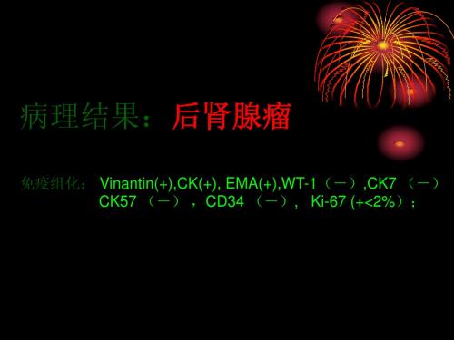
病例报告【 文献类型] 诊断 该病免疫组化染 色大多呈 V i m e n i f n , S - 1 0 0 , l e u 7阳性 ,瘤细 胞L e u Ml 和 HMB - 4 5阴性 。 其免疫组化特点 与胎儿 肾脏 的 s 状小体很相似。
后肾腺瘤
影像学表现
目前,关于MA的影像表现只限于个例报道 , 无 特 异 性,难以其他肾肿瘤相鉴别,需术后病理明确诊断 有报道称: B 超呈高回声, CT 表现为边界清楚,密 度均匀,外生性生长明显,平扫时肿瘤密度高于周 围肾组织,有的可伴少量钙化,增强后低于周围肾 组织密度,并可有延迟增强,可高度怀疑此肿瘤存 在的可能,值得注意但有待于进一步研究 术前肾穿病理检查假阴性可能性大,故所有MA术前 均被误诊,且有报道MA存在远处转移可能,故有人 建议行根治性患肾切除术
病 例 ( 一 ) 岁
6 M
病 例 ( 一 )
病 例 ( 一 )
病 例 ( 一 )
病 例 ( 二 ) 岁 男
3
病 例 ( 二 )
肾 母 细 胞 瘤 病 例 ( 二 )
小
结
后肾腺瘤非常少见,临床、影像及生物学表现复杂 多变,缺乏特征性 影像学检查,典型肾癌、错构瘤、肾母细胞瘤易与 之鉴别,转移瘤结合临床亦可鉴别;但难与不典型 肾癌、乏血管性错构瘤等鉴别 影像学检查难以诊断该肿瘤!如具有:B超呈高回声; CT表现为界清,密度均匀,平扫时密度高于周围肾 组织,增强后低于周围肾组织,伴有红细胞增多症 等表现中有两种以上者可考虑后肾腺瘤可能
患发 者现 女右 肾 岁上 ,极 体占 检位
47
CT值:平扫—33HU,动脉期—57HU 静脉期—67HU,延迟—65HU
鉴别诊断
肾癌(RCC)--乳头状癌 肾血管平滑肌脂肪瘤(AML)--乏血管性 肾母细胞瘤(Wilms瘤)--小儿 肾转移瘤
肾癌—一般情况
最多见的肾恶性肿瘤,约占肾全部肿瘤的85%, 病理分类:①透明细胞癌( 70%~80% )②乳头状 癌( 10%~15% ) ③嫌色细胞癌( 5% )④少见 -集合管癌、未分类性肾癌等 来源于肾小管上皮细胞,易发生于肾上、下极,呈 实质性不规则肿块,常有出血和坏死,少数可有钙 化;边界清楚或不清楚,可有假包膜 临床症状早期不明显,典型表现:腰痛、肾区肿块、 无痛性血尿;40岁以上多发,男性多于女性
1例无实体肿瘤的后肾腺瘤并文献复习
查 体 : 7 5 , 1 / n R2 T 3 .。P9 次 mi, 0次 / i, P14 8 mH , a r nB 2 /4m g 神
(g 6 .0mg L 微 量 白蛋 白 ( IU)0 9 / 、 MA) 8 . / 、 转 铁 蛋 白 触诊包块 、 痛性 肉眼血尿 以及间歇性发热。约 1 %的患者可 5 20mg L 尿 无 0 ( R 4 . 0mg L 2 T U) 3 9 / , 4 h尿 蛋 白 定 量 : 8 . g 总 蛋 白 10 3 2r ; a 见红细胞增 多症 , 因可能与 MA细胞可产生并分泌促红细胞 原 ( P 6 . / 白蛋 白 ( l)9 7gL 球 蛋 白( B 2 . / 、 生成素及其他多种细胞 因子有关… , T )9 2g L、 Ab 3 . / GL )9 5g L 一些病例 可 出现 血压高 ,
诊断 。
声 : 、 、 、 未见 明显异 常 ; 肾、 肝 胆 胰 脾 双 输尿 管未 见 明显 占位 ,
临 床 资 料
E G胸片未见异常 。人 院后在 B超引导 下定位 右 肾下 极 , C 选 用 1G肾活检针 , 4 巴特 自动肾活检枪进行穿刺。 活检取 出肾组织送往上海 长征 医院肾脏病研究 所肾病实
C T增强造影未发现实体肿 瘤 , 同时合 并慢性 肾小球 肾炎。现 A C A A阴性 ; N A、 C 乙肝三 系 : s g +)H eg( , U A 船 A ( , BA +) H c g
后肾腺瘤五例诊治分析
Di g o i n a a e n fM e a e h i e o a n ssaБайду номын сангаасd M n g me to t n p rcAd n ma: Re o to i eCa e n ie a ur v e p r fF v s sa d L t r t e Re iw Z AO We , H i
Thes m po s lboa oy ts r s t y tm , a r tr e t e ul s, i gng in d n s, p t oo ia e a nains te t n , a d o c me f 5 aint ma i f g i g a h lg c l x mi t o , r ame t n uto s o p te s
i f me s mea e h i a e o y p to o i a a d i r d a tn p r d n ma b ah l gc l n mmu o i o h mia x mia in . Al t e e 5 p t n su d r n u gc l c n h s c e c le a n t s t o l h s a i t n e we ts r ia e
C
S 娶 e
・
・
l 7l・
诊 治 分 析 ・
后 肾腺 瘤 五 例 诊 治 分 析
赵 伟 ,李振 华 ,孔 垂 泽
【 摘要 】 目的 探讨后 肾腺瘤的临床 症状 、影像 学及病理学特点 、治疗及 效果 ,提 高其诊治水平 。方法 回顾
性 分析 我 院 20 - 2 1 收 治 的 5例后 肾腺 瘤 患者 的 临床 症状 、 实验 室检 查 、影 像 学 检 查 、 病 理 检 查 结 果 、 治疗 方案 00 0 0牟 及 效 果 。结 果 患者 均 经 彩 色 多普 勒 超 声检 查 发 现 ,经 C T检 查诊 断 为 肾 肿 瘤 ,经 病 理 及 免 疫 组 化 检 查 证 实 为 后 肾腺 后 肾腺 瘤 是 一 种 非 常 罕 见 的 肾脏 良性 肿 瘤 ,保 留 肾 单 位 的肿
免疫组化肾上腺疾病结果判读标准

免疫组化肾上腺疾病结果判读标准英文回答:Immunohistochemistry (IHC) is a commonly used technique in the diagnosis and classification of adrenal diseases. It involves the use of specific antibodies to detect and localize specific proteins within the adrenal tissue. The results of IHC staining are interpreted based on the intensity and distribution of the staining.In the case of adrenal diseases, there are several key proteins that are commonly targeted for IHC staining. These include adrenocorticotropic hormone (ACTH), cortisol, aldosterone, androgen receptor (AR), and steroidogenic enzymes such as 21-hydroxylase and 17α-hydroxylase. The expression and distribution of these proteins can provide valuable information about the underlying pathology.For example, in a patient with suspected adrenal adenoma, the IHC staining for cortisol and aldosteronewould be expected to show strong positive staining in the tumor cells. This would indicate that the tumor is functioning and producing excessive amounts of these hormones. On the other hand, in a patient with suspected adrenal carcinoma, the IHC staining for cortisol and aldosterone may be negative or weak, indicating that the tumor is non-functioning.In addition to hormone-related proteins, IHC can also be used to detect other markers of adrenal diseases. For example, in patients with suspected pheochromocytoma, IHC staining for chromogranin A and synaptophysin can help confirm the diagnosis. These markers are typically expressed in the chromaffin cells of the adrenal medulla and are absent in normal adrenal tissue.The interpretation of IHC results in adrenal diseases is based on the comparison of staining patterns between the diseased tissue and normal tissue. The intensity ofstaining is graded on a scale from 0 to 3, with 0indicating no staining and 3 indicating strong staining. The distribution of staining can also provide importantinformation, such as whether the staining is diffuse or focal, and whether it is present in specific cell types.In summary, the interpretation of IHC results in adrenal diseases involves assessing the intensity and distribution of staining for specific proteins. This information can help diagnose and classify different adrenal pathologies. Examples include the use of IHC staining for cortisol and aldosterone in the diagnosis of adrenal adenoma and carcinoma, and the use of IHC staining for chromogranin A and synaptophysin in the diagnosis of pheochromocytoma.中文回答:免疫组化(Immunohistochemistry,IHC)是一种常用的诊断和分类肾上腺疾病的技术。
33391050

实用医学杂志 2 1 0 0年第 2 6卷 第 5期
后 肾腺瘤 的临床病 理 观察
孟红 蒋春 樊 蒋俊 孟寒 韩 义明
摘要 目的 : 对后 肾腺瘤的病理诊断、 鉴别诊断和预后进行分析:方法 : 3例后 肾腺 瘤 . 复习其 临床 资料 , 病理 学检查 包括常规病理学 、 免疫组织化学 . 并进行 随访 结果 : 光镜 下肿瘤细胞小 , 匀一致 , 大质少 , 均 核 无并型性及 核分裂像 . 肿瘤细胞排 列成小管状 、 粱索状 、 团块状 、 乳头状及 肾小球样 结构 出血 、 坏死、 囊性 变常见 。 并可见 沙粒
处 有 圆形肿 块 凸 出于右 肾表 面 , 肿块剜 除 术 。术后 行 随访 1 年余 状况 良好 。例 3 ,2岁 , B超发 现左 女 5 因
肾肿瘤 6 周人院。 患者无任何临床症状及体征。 T C 示
左 肾上 极不 规 则 混 杂密 度 肿 块 灶 ,考 虑 多 为 良性 肿 瘤 。术 中见左 肾上 极肿 瘤大 小约 8c m×7c m×5c m, 质 较硬 , 局部 呈囊 性 变 , 与周 围组 织粘 连 , 易与左 肾 不
合 文献 主要对 其病 理诊 断及 鉴别 诊 断进 行讨 论 , 以提 高对 本病 的正确 诊断 。 1 资料 与方 法 11 病 例来 源 2 0 . 0 4年 7月 至 20 09年 7月 我 院收 治 3例 M A。
1 病 理检 查 对切 除 的标 本经 4 . 2 %甲醛溶 液 固定 , 石 蜡 包 埋 , m厚 连 续 切 片 , 4 常规 H E染 色 , 光镜 观 察。 1 免 疫组 织化 学 对 所有 标 本进 行 了 E Vs n法 . 3 n io i
体 钙 化 。免 疫 组 化 Vm ni( )C 7 + 、 K1( )C 5 ( )E A( )S N( ) D s i( ) 例 患 者 术后 状 态 良 iet + 、 K ( ) C 8 + 、 D 7 + 、 M 一 、Y 一 、 em n 一 。结论 : 肾腺瘤是一种 罕见的 。 良性肿瘤 , 织学具有独特的 后 肾脏 组
后肾腺瘤2例并文献复习
ห้องสมุดไป่ตู้丁AN
nh n sa
L Ga — n
g in qa g
ZH0U n a Ya n n
1 De a t n f Pa h lg . p rme to to o y,Zh n s a s ia 。 Fu a ie st o g h n Ho p t l d n Unv riy,
Ke o ds M e a e h i d n ma W i S t mo ; P p l r e a c r i o : M e a e h i a e o i r ma yW r t n p rca e o ; l u r m a i a y r n l a c n ma l tn p r d n f o c b
S a g a 2 0 3 ; . p rme t f Pa h l g h n h i 0 0 2 2 De a t n t oo y,t e4 5 Ho p t lo o h 5 s ia f PLA ,S a g a 2 0 5 h n h i 00 2
Ab t a t 0betv : x lr h l ia et r sa dhso ah lgc l h rce fmea e h i a e o . to sTor — src jcie Toe poet eci c lfa u e n it p t oo ia aa tro tn p rc d n ma Meh d : e n c
维普资讯
46 2
Cicl d aJ unl f hn ,0 7Vo 1 , o3 li i lo ra o i 20 . 1 4 N . n a Me c C a .
中国临床医学
20 0 7年 6月 第 1 4卷 第 3期
后肾腺瘤临床诊治分析(附2例报道并文献复习)
[bt c O j t e o i v siae t e c n c l a ilgc l a d h s p t 00 i fau e f mea e h i A s at r ] b ci T n e t t h l ia ,rd oo i n i o a 1gc e t r s o t n p r e v g i a t h c a eo ( d n maMA) Meho s 2 c ss f . t d a e o MA we e e o t d n l e a u e wee e iwe r rp re a d i r t rs t r rv e d.Th ciia , e l c l n
r di lgi a a hit pa hOlgi f a ur s, a w el s i gno i a te t e of a oo c l nd sO t O c et e s l a d a ss nd r a m nt M A w e e lo ic s d. r as d sus e R e u t M A ha no ypial lnial s ls s t c ci c and a o o c l e ur s r di l gi a f at e but uni e qu hit pat s0 hol c t uc ur s o sr t e .
内见 分 隔 ,约 5 5× 54m ,C . .c T值 囊 性 部 分 2 Hu, 7
罕少疾 病杂志 2 1 0 0年 1 0月 第 1 7卷 第 5期 5
实性部分 4 Hu 4 ,增强后实性部分可见强化。M 示 :
[ y wo dlmea e hrc d n ma e a n o ls Ke r s t n p i a e o ;rn l e pa m
后肾腺瘤的病理特征(2例报告并文献复习)
诊 断及 预 后 。 结果
瘤, 确 诊依 赖 组 织 病 理 学 检 查 , 其 免 疫 组 化 尚无 统 一 定 论 , 需 与 Wi l ms 瘤及乳头状 肾细胞癌相 鉴别 。 手 术 切 除术 后 患 者需 长期 随访 。 【 关 键 词 】 肾肿 瘤 ; 病 理 学 ; 后 肾腺 瘤
现代泌尿生殖肿瘤杂志 2 0 1 3年 2月第 5卷第 1期 J C o n t ! ! Nhomakorabea!
! ! 里 ! !
! ! : :
・
临 床 研 究
・
后 肾腺 瘤 的病 理 特征 ( 2例 报 告并 文献 复 习 )
王翔 赵 彬 李 黎 承杨 周 立权 邓耀 良
t h e me t a n e p h r i c a d e n o ma ( M A) . Me t h o d s To r e p o r t t wo c a s e s o f M A a n d s u mma r i z e t h e h i s —
vi e wi n g t he l i t er a t ur e s . Re s u l t s M A ha d t yp i c a l p at hol og i c f e a t u r e s . Co n c l u s i on s M A i s r a t e
Hi s t o p at ho l o g i c a l c ha r a c t e r i s t i c s o f t he me t a ne ph r i c a de no m a:r e p o r t o f t wo c a s e s a nd l i t e r a t u r e r e v i e w
免疫组织化学在肾脏肿瘤病理诊断中的应用

免疫组织化学在肾脏肿瘤病理诊断中的应用现今,免疫组织化学(IHC)技术已经广泛应用于肾脏肿瘤的病理诊断上,它的应用不仅提高了肿瘤病理学家们的准确性,而且还对对肿瘤的治疗有着至关重要的作用。
本文旨在阐明免疫组织化学技术在肾脏肿瘤病理诊断中的应用。
首先,要明白免疫组织化学在肾脏肿瘤病理诊断中的作用,我们必须了解该技术本身。
免疫组织化学是一种基于抗原-抗体反应(antigen-antibody reaction)的技术,利用一抗,对肿瘤细胞内的抗原进行检测,从而诊断肿瘤的类型和性质。
因为它的准确性和高度的特异性,在肾脏肿瘤的病理诊断领域里,免疫组织化学技术受到了广泛的应用。
该技术可以在活检中用于诊断,也可以用于判断肿瘤细胞的增殖活动、浸润、侵袭和转移等。
此外,免疫组织化学技术能够检测多种不同的肿瘤抗原,包括胚抗原、癌抗原、肿瘤标志物等,它可以帮助医生区分同一类癌症的不同亚型,以及检测恶性肿瘤的转移,从而给患者的治疗选择提供依据。
其次,免疫组织化学技术在肾脏肿瘤病理诊断中的主要应用是检测肾脏肿瘤的表型及其病理演变,并为鉴定肿瘤类型和性质提供依据。
首先,免疫组织化学技术可以用来检测肾脏肿瘤的细胞内抗原,从而明确诊断肿瘤病理类型。
其次,该技术可以帮助确定恶性肿瘤的侵袭深度、浸润程度和远处转移情况,从而帮助医生选择合适的治疗方案。
最后,免疫组织化学技术还可以用来检测肿瘤的表型,如肿瘤细胞的增殖活动、死亡情况等,为病理演变提供重要的线索。
总的来说,免疫组织化学技术在肾脏肿瘤病理诊断上起着不可替代的作用。
它不仅提高了肿瘤病理学家们的准确性,而且还可以用于检测恶性肿瘤的转移情况和改变肿瘤细胞表型。
另外,该技术也可以帮助把肿瘤分为不同的类型,从而提高患者的治疗效果。
因此,免疫组织化学技术在肾脏肿瘤病理诊断中的作用不可忽视。
至此,本文总结了免疫组织化学技术在肾脏肿瘤病理诊断中的应用。
免疫组织化学可以很好地检测肿瘤抗原,帮助确定肿瘤的病理类型、侵袭深度、浸润程度和转移情况,以及检测肿瘤细胞表型,从而为患者选择正确的治疗方案奠定基础。
后肾腺瘤的临床、病理和MRI表现(附2例报道并文献复习)
后肾腺瘤的临床、病理和MRI表现(附2例报道并文献复习)贺新华;丁玉芹;黄健峰;缪熙音;罗荣奎;曹颖丽;周建军;曾蒙苏【摘要】目的:探讨后肾腺瘤(MA)的临床、病理和MRI表现,提高对MA的诊断水平.方法:回顾性分析2例经病理证实的MA患者的临床、病理及MRI资料,结合相关文献进行分析和总结.结果:本组2例MA患者均为女性,均表现为单侧肾脏实质性肿块,T1WI肿瘤实质部分信号与肾皮质相仿,T2WI为等信号和略低信号,DWI均为明显高信号,1例瘤内可见出血、坏死、囊变、钙化和假包膜,信号不均匀,另1例瘤内可见中心瘢痕,除瘢痕外肿瘤信号较均匀,增强后1例表现为中度不均匀延迟强化,另1例表现为“快进快出”的强化方式.病理学特点:肿瘤细胞非常丰富,排列紧密,常可形成肾小球样或花蕾状结构,常见砂粒体形成.免疫组化常见WT-1、CD57阳性,EMA和AMACR阴性.结论:MA典型的MRI表现为T1WI和T2WI低或等信号,DWI明显高信号,瘤内常见出血、坏死,部分伴有囊变和钙化,信号不均匀,边界清晰,有或无假包膜,增强后病灶呈轻中度持续性或渐进性强化.因其MRI表现形式多样,最终诊断依赖于病理学检查.【期刊名称】《中国临床医学影像杂志》【年(卷),期】2015(026)001【总页数】3页(P56-58)【关键词】肾肿瘤;腺瘤;磁共振成像【作者】贺新华;丁玉芹;黄健峰;缪熙音;罗荣奎;曹颖丽;周建军;曾蒙苏【作者单位】江苏省如皋市人民医院影像科,江苏如皋226500;复旦大学附属中山医院,上海200032;复旦大学附属中山医院,上海200032;复旦大学附属中山医院,上海200032;复旦大学附属中山医院,上海200032;复旦大学附属中山医院,上海200032;复旦大学附属中山医院,上海200032;复旦大学附属中山医院,上海200032【正文语种】中文【中图分类】R737.11;R445.2后肾腺瘤(MA)是一种罕见的肾脏原发性良性肿瘤,来源于肾胚胎发育过程中的残留组织,约占成人肾脏上皮源性肿瘤的0.2%。
后肾腺瘤临床诊治分析(附2例报告)

【 关键词 】 后 肾 瘤 ・ 特点 腺S u y o i g ssa t d n d a no i ndte t n fmea e h i de o a, e o to 2 c ss S Jn ,LNG S eg —to r ame to t n p rca n m ar p r f ae . U i I hn a ,GUIZh —rig,e a.De r- i un t 1 pat
hep u o t i e n ild a n ss t r g o i swe1 Co c u i n Mea e h i d n ma i i d o r e i u ro gn t g fo r m- l f lfr i d f r ta ig o i .I p o n ssi l. n l so s f e s t n p r a e o s a k n fr e b n g t mo r i a i r m e c a n i n
腺 瘤患者 临床资料 、 手术切 除后 标本行病理检查 、 免疫组 化检 测结果进 行分析 , 并就其核 型特 点、 治疗方案和 预后进行
文献复 习。结果 镜下该肿瘤 由大小不 一的细胞排列 成密集 小管状 、 腺样 结构 , 胞无 明显异型性 、 细 无核 分裂相 ; 疫 免 组化染 色有助 于鉴别诊 断; 肾腺 瘤预后 较好 。结论 后 肾腺 瘤是 一种 罕见 的肾脏胚胎 性上皮 源性 良性肿瘤 , 后 常被误 诊 为恶性肿瘤 , 与肾母 细胞 瘤、 头状 肾细胞癌 等鉴 别。 应 乳
临床和实验 医学杂志 2 1 年 3月 第 1 卷 第5 02 1 期
・3 9 ・ 4
后 肾腺 瘤 临床 诊 治 分 析 ( 2例 报 告 ) 附
苏劲 凌 生涛 桂 志明 许 志坚 ( 东 医学院 附属 医院泌尿 外科 广 广东 湛江 540 ) 20 1
免疫组化肾上腺疾病结果判读标准

免疫组化肾上腺疾病结果判读标准免疫组化(immunohistochemistry,IHC)是一种用来检测组织中特定蛋白质的方法,常用于肾上腺疾病的诊断和治疗。
通过对肾上腺组织中的特定蛋白质进行染色和分析,可以帮助医生确定疾病的类型和严重程度,从而制定更精准的治疗方案。
本文将介绍免疫组化在肾上腺疾病诊断中的应用及结果判读标准。
一、免疫组化在肾上腺疾病诊断中的应用免疫组化在肾上腺疾病诊断中起着重要作用,它可以帮助医生确定肾上腺组织中是否存在特定蛋白质的表达及其表达水平。
常用的标记物包括醛固酮合酶(aldosterone synthase)、17-羟化酶(17-hydroxylase)、21-羟化酶(21-hydroxylase)、肾上腺皮质醇果糖转移酶(11-beta-hydroxylase)等。
通过对这些标记物的检测,医生可以对患者的肾上腺疾病进行更准确的诊断和分析。
免疫组化的应用不仅局限于对肾上腺疾病的诊断,同时还可以帮助确定疾病的病理类型,如嗜铬细胞瘤、肾上腺皮质腺瘤、肾上腺皮质增生症等。
这对于制定个性化的治疗方案具有重要意义。
二、免疫组化在肾上腺疾病结果判读标准1.阳性判定:阳性染色通常表现为细胞核和胞浆呈棕黄色或棕红色的染色颗粒,颗粒大小不等。
根据颗粒的数量和染色强度,可对阳性结果进行定量分析。
2.阴性判定:阴性结果通常为细胞核和胞浆无染色颗粒或染色颗粒极少,染色强度非常弱。
3.染色强度判定:免疫组化结果的染色强度通常分为四个等级:无染色(0级)、弱染色(+1级)、中等染色(+2级)、强染色(+3级)。
根据染色强度的不同,可以对阳性结果进行定量分析。
4.染色面积判定:染色面积是指肾上腺组织中出现阳性染色的面积比例,通常用百分比表示。
根据染色面积的不同,可以进一步确定阳性结果的强度和分布情况。
5.阳性细胞类型判定:肾上腺组织中的阳性细胞类型有所不同,染色颗粒主要分布在细胞核或胞浆。
根据阳性细胞的类型和分布情况,可以对阳性结果进行细化分析。
免疫组化在肿瘤病理诊断中的应用培训课件
(六)肿瘤细胞增殖活性与细胞凋亡标记抗体
瘤细胞增殖指数反应细胞增殖活性,是瘤细 胞新生(恶性)状况的客观指标,与凋亡指数 呈反比关系。常用抗体有:
1.增殖细胞核抗原(proliferative neuclear Antigen,PCNA),核阳性。
2.Ki-67,Brdu抗体:原要求新鲜组织,新 近有Ki-67石蜡切片标记抗体问世。
11
3.神经内分泌肿瘤标记:包括类癌、胰岛、
垂体瘤、甲状腺髓样癌、皮肤Merkel细胞癌、
原始神经外胚层瘤(PNET)等,目前应用
NSE、嗜铬蛋白颗粒A(CgA)及触突素
(Syn)三种抗体同时或两种一起用,因NSE
不够特异。
免疫组化在肿瘤病理诊断中的应用
12
(五)淋巴网状组织肿瘤(淋巴瘤)标记
1.B细胞标记抗体:CD20(L26),Ig重链 (IgG、M、A、E)、轻链(λ、κ)、CD79a。
(一)上皮性肿瘤标记
1.角蛋白(Keratin):一种中间丝蛋白,分
(1)表皮角蛋白(Epidermal keratin,
EK)——鳞状上皮或高分化鳞癌标记;(2)
细胞角蛋白(cytokeratin,CK)——低分子
角蛋白,对腺癌、移行细胞部及低分化鳞癌
标记。一般用广谱型角蛋白抗体为好,常用。
免疫组化在肿瘤病理诊断中的应用
2.T细胞标记抗体:CD45RO(UCHL-1), CD3较好。CD56
3.组织细胞标记抗体:Mac387、CD14、抗 溶菌酶(lysozyme)、抗糜蛋白酶(α-1 Antichymotrypsin)等。
免疫组化在肿瘤病理诊断中的应用
13
4.霍奇金淋巴瘤(Hodgkin’s lymphoma, HL)R-S细胞(Reed-Sternberg cell)标记抗 体:CD15、CD30
6例后肾腺瘤的临床与病理学特点分析
a
l U n iv
it y
,
T ia
n
j in
I n s t it u t e
o
f U r o l o g y T ia
,
n
3 0 0 2 1 1 C h in
o bj e c tiv
e x
p lo
re
t h e c li n i c a l a n d h i s t o p a t h 0 Io g i c a I f e a t u r e s o f m e t a n e p h r i c a d e n o m a
ll
.
ll s
c
w
it h
m
re
u la r n u c le
i th a t fo
s
d
a t u b u la r o
c e
de
n o
id p a t
e
m
te
rn
.
i M it o t i c f g
n e
u re s
w e re
bs
e n
r ra re
Im
c
m u n o
o
h is t o
he
ic
s e
The
a v e ra
te
r o u n
f tu if b
m o r w a s rm c e
c m
M ic g
a
ro s c o
p ic
l
o
e rv a rm e
d tha t the tu
r a
m o rs
—
w e re
c o m
- 1、下载文档前请自行甄别文档内容的完整性,平台不提供额外的编辑、内容补充、找答案等附加服务。
- 2、"仅部分预览"的文档,不可在线预览部分如存在完整性等问题,可反馈申请退款(可完整预览的文档不适用该条件!)。
- 3、如文档侵犯您的权益,请联系客服反馈,我们会尽快为您处理(人工客服工作时间:9:00-18:30)。
文章编号:100025404(2008)2122047204论著后肾腺瘤临床病理及免疫组化观察分析孙慧勤,郭德玉,章 容,赵友光,阎晓初,于冬梅,柳凤轩 (第三军医大学西南医院病理学研究所,重庆400038) 摘 要:目的 探讨后肾腺瘤(metanephric adenoma,MA)的病理特征及鉴别诊断方法。
方法 应用常规病理、免疫组化方法观察本所近年诊断的2例MA手术切除标本,结合相关临床资料(从病史中摘取)分析其病理特点。
结果 2例大体均为类圆形灰白色肿块,大小各约1、4c m,有薄层包膜,与周围组织分界清楚。
组织学上具有独特的形态结构特点:瘤细胞较小,核大质少,无异型性,呈规则的小管、小团状及肾小球样等结构排列。
免疫组化分析:V i m entin阳性,W T1阳性或部分阳性,CK灶性阳性,C D57有1例散在和小灶性阳性,另1例片状强阳性,E MA、CK7、Cg A、CD10均为阴性。
2例均发生于中老年女性,其中1例在同一肾脏的另一部位还有肾细胞癌。
结论 作为WHO分类中一种少见的良性肾肿瘤类型,MA在形态学上具有独特的组织结构特点,免疫表型表现为W T1、C D57、V i m entin、CK多有表达,其中V i m entin恒定出现,W T1、CD57、CK表达强度和范围各有不同,而E MA、CK7、Cg A、C D10多无表达;要注意与W li m s瘤、肾乳头状细胞癌、后肾腺纤维瘤等进行鉴别。
关键词:后肾腺瘤;临床病理;免疫组化表型特征 中图法分类号:R730.21;R730.261;R737.11 文献标识码:AC li n i copa tholog i c and imm unoh istochem i ca l fea tures of m et anephr i c adenomaS UN Hui2qin,G UO De2yu,Z HANG Rong,ZHAO You2guang,Y AN Xiao2chu,Y U Dong2mei,L I U Feng2xuan(I nstitute of Pathol ogy,South west Hos p ital,Third M ilitary Medical University,Chongqing400038,China) Abstract:O b j ec ti ve T o study the clinicopathol ogic,i m munohist oche m ical features and differential diag2 nosis of metanephric adenoma(MA).M e tho d s T wo cases ofMA recently diagnosed in our institute were ob2 served and analyzed in the gr oss,hist ol ogically and i m munohist oche m ically.Pathol ogical features were conclu2 ded based on clinical inf or mati on extracted fr om the medical records and literature revie w.R e su lts Both tumors were masses of r ound in shape,gray2white in col or,and1or4c m in size,envel oped with a thin cap sule with a clear boundary in macr oscop ic vie w.They als o had distinct feature in hist ol ogy as s mall cells with high rati o of nucleus t o cyt op las m,no nuclear atyp ia or m it otic figures.The tumor exhibited well2organized tubules, s mall cell nests,‘gl omerul oid bodies’,and shar p ly de marcated fr o m the adjacent renal parenchy ma.I m muno2 hist oche m ical phenotype of exp ressi on p r ofiles were as diffused vi m entin,diffused or partially positive W T1,fo2 calized CK,s potty or s mall focal of CD57in one case while str ong patchy exp ressi on in another case.The ex2 p ressi on of E MA,CK7,Cg A,CD10were negative.Both cases were m iddle t o old2aged fe males.I n one case there was renal cell carcinoma l ocated else where in the sa me kidney.Co nc lu s i o n MA,a rare novel f or m of benign renal neop las ma in the WHO classificati on,has distinct architecture in tissue structure.MA has charac2 teristic phenotypes,as consistent exp ressi on of vi m entin and frequent exp ressi ons of W T1,CD57,and CK, which are vari ous in both intensity and distributi on,but E MA,CK7,Cg A,CD10are negative.So far,it is be2 lieved t o a benign tumor and should be differentiated fr om W il m s tumor,pap illary renal cell carcinoma (PRCC),metanephric adenofibr o ma etc. Key words:metanephric adenoma;clinical pathol ogy;i m munophenotyp ic feature 作者简介:孙慧勤,女,云南省昆明市人,博士,副主任医师,副教授,主要从事肿瘤病理学及血管生成方面的研究。
电话:(023)68765455,E2mail:huiqinsun02@yahoo.co 收稿日期:2008203203;修回日期:2008204210 后肾腺瘤(metanephric adenoma,MA)是一种少见的肾脏肿瘤,1992年由B risigotti命名[1],其后被归为一独立的腺瘤,2004年版WHO肾脏肿瘤分类中新增加了“后肾肿瘤”(metanephric tu mors)大类,其中包括7402第30卷第21期2008年11月 第 三 军 医 大 学 学 报ACT A AC ADE M I A E ME D I C I N AE M I L I T AR I S TERTI A EVol.30,No.21Nov.2008了MA以及后肾腺纤维瘤、后肾间质肿瘤[2]。
虽然目前国内外已有数10例病例报道,但对其临床病理、免疫表达、生物学和基因变化特点尚未得到全面认识。
迄今MA被认为是一种生长缓慢,无转移且预后良好的良性肿瘤,但近来有2例转移报道[3],且发现其细胞基因存在异常,其中的染色体异常区域9p24恰好是新近确定的一种肿瘤抑制基因K ANK(kindey ankyrin re2 peat2containing p r otein)所在位置[4],所以有关该肿瘤的临床病理学、细胞生物学及遗传特点,以至诊断和治疗仍值得进一步探讨。
现将近年本所2例诊断病例就其临床病理、免疫组化特征进行分析,同时结合该肿瘤相关的研究进展进行讨论。
1 资料与方法1.1 病例与标本来源 收集本所2003年1月至2007年12月5年内手术切除送检的肾脏肿块标本,病理诊断为MA的病例共2例,从相关病例病史中摘取获得相关临床病史资料(主诉、临床表现、体格检查、实验室及影像学检查资料等)。
1.2 方法 对手术切除送检的肾脏标本进行大体观测,取部分瘤组织,经10%福尔马林固定,常规石蜡包埋、切片、HE染色,同时采用即用型En V isi on二步法,二氨基联苯胺(DAB)显色,苏木精对比染色,对2例标本的石蜡切片行免疫组织化学染色。
选用抗体:波形蛋白(V i m entin,1∶400)、W T1(即用型)、细胞角蛋白(CK,1∶200)、上皮膜抗原(E MA,1∶200)、Cg A、C D117、P504S (即用型)均购于迈新公司,即用型CD57、CD10、CKH、CK8/18、CD15购于中杉公司,CK7(即用型)、S2100(1∶800)、购于DAK O 公司。
阳性结果判定:抗体阳性反应呈棕黄色,CD57、V i m en2 tin、CK、CK7、Cg A、E MA、CKH、CK8/18、C D117、P504S及S2100均为细胞质阳性,W T1为细胞质与细胞核阳性,CD10、C D15为细胞膜阳性。
2 结果2.1 临床特征 病例1:患者,女性,35岁,因“反复右腰部疼痛1年,体检发现右肾肿瘤2个月”入院。
患者入院前1年无明显诱因出现右腰部隐痛不适,休息后缓解。
症状反复发作,但较轻。
多次于院外行B超发现右肾囊肿,未经特殊治疗。
入院前2个月于北京某医院行CT发现“右肾良性实性占位”。
病程中无血尿、脓尿,无畏寒、发热,无尿频、尿急、尿痛等不适症状。
入院CT 检查示:右肾门处一约3.6cm×4.6cm实质占位。
术中见:右肾脏中极肾门处一直径3~4c m灰白色包块,表面光滑,自肾实质向外突出。
病例2:患者,女性,54岁,因“反复潮热6个月,右腰胀不适1个月,外院B超提示右肾占位”入院手术,术中见右肾上中部3.5c m×3.0c m实质占位,中央坏死,下极亦见一直径1c m 的灰白色肿块,作右肾切除送检。