lipocalin-2在肝再生中的作用
血清脂质运载蛋白2在小鼠视网膜血管内皮细胞血管生成中的作用

血清脂质运载蛋白2在小鼠视网膜血管内皮细胞血管生成中的作用姚国敏;李蓉;田瑾【摘要】目的:观察脂质运载蛋白2(lipocalin-2,LCN-2)对体外培养的小鼠视网膜内皮细胞(retinal vascular endothelial cells,RVECs)增殖、迁移和管腔形成的影响及其在视网膜新生血管中的作用.方法:取生长状况良好的RVECs分为不同浓度组:分别以0、5、10μmol/L LCN-2作用细胞48h.采用EdU法检测细胞增殖,Transwell法检测细胞迁移,Matrigel法检测管腔形成.结果:不同浓度LCN-2作用的细胞增殖率大于对照组,差异有统计学意义(P<0.05),不同浓度LCN-2组RVECs细胞迁移数目多于对照组,差异有统计学意义(P<0.05),不同浓度LCN-2组细胞管腔形成数明显多于对照组,差异有统计学意义(P<0.05),且细胞增殖、迁移和管腔形成均随着LCN-2浓度的升高而增加.结论:LCN-2能够明显促进RVECs的血管生成过程,提示LCN-2是一种重要的促视网膜新生血管形成的因子.【期刊名称】《国际眼科杂志》【年(卷),期】2018(018)009【总页数】5页(P1578-1582)【关键词】脂质运载蛋白2;血管生成;视网膜内皮细胞【作者】姚国敏;李蓉;田瑾【作者单位】710077 中国陕西省西安市,西安医学院第一附属医院眼科;710077中国陕西省西安市,西安医学院第一附属医院眼科;714000 中国陕西省渭南市中心医院眼科【正文语种】中文0引言糖尿病视网膜病变(diabetic retinopathy, DR)是一种糖尿病的严重微血管并发症,也是全世界劳动人群发生低视力和致盲的主要原因[1-2]。
在DR病情发展的晚期,患者出现病理性视网膜新生血管,即进入增殖性糖尿病视网膜病变期(proliferative diabetic retinopathy, PDR),继而发生玻璃体出血和牵拉性视网膜脱离[3],给患者带来严重的视力损害和视力残疾。
华东医院肝病治疗推荐用药四
以上是临沂肝病医院华东医院专家根据多年临床经验总结出的各类肝病用药,极具参考价值。但具体用法用量需按照各位患者具体情况而定,临沂肝病医院华东医院提醒广大患者一定要到正规医院就医,切勿擅自服药。
二:益肝泰
适应症:清热解毒、健脾补肾。用于慢性乙型、丙型肝炎之湿热毒侵、脾肾亏虚证。见乏力、纳差、胁痛、腹胀、腰膝酸软等。
三、疫功能,有使乙肝抗原转阴,促使乙肝抗体形成之功效。用于治疗乙肝、乙肝病毒携带者、肝硬化、肝肿大、肝损伤、急慢性肝炎、迁延性肝炎、肝癌等。对乙肝抗原三阳者、特别是对乙肝抗原转阴效果最佳。
七:注射用核糖核酸(援健)
适应症:用于慢性迁移性肝炎、活动性肝炎,肝硬化及其它肝脏疾病。
药理作用:肝炎辅助药,可提高机体细胞免疫功能,促进肝细胞合成蛋白质,降低谷一丙转氨酶。
八、注射用促肝细胞生长素(苷炎灵)
适应症:各型重型病毒性肝炎(急性、亚急性、慢性重症肝炎的早期或中期)或有发展为重型肝炎趋势的慢性活动性肝炎的治疗。
临沂肝病医院华东医院肝病专家根据多年临床治疗经验,总结出以下肝病用药:
一:促肝细胞生长素
适应症:用于重型肝炎(病毒性肝炎、肝衰竭早期或中期)、慢性肝炎活动期、肝硬化的综合治疗。
药理作用:本品系从新鲜乳猪肝脏中提取纯化制备而成的小分子多肽类活性物质,具备以下生物效应:①能明显刺激新生肝细胞的DNA合成,促进损伤的肝细胞线粒体、粗面内质网恢复,促进肝细胞再生,加速肝脏组织的修复,恢复肝功能。②改善肝脏枯否细胞的吞噬功能,防止来自肠道的毒素对肝细胞的进一步损害,抑制肿瘤坏死因子(TNF)活性和Na+,K+-ATP酶活性抑制因子活性,从而促进肝坏死后的修复。同时具有降低转氨酶、血清胆红素和缩短凝血酶原时间的作用。③对四氯化碳诱导的肝细胞损伤有较好的保护作用。④对D-氨基半乳糖诱致的肝衰竭有明显的提高存活力的作用。
肝脏再生和保护的分子机制研究进展

肝脏再生和保护的分子机制研究进展董育玮;陆伦根【摘要】肝脏再生是大多数肝损伤恢复所必需的一个过程.再生通过包括肝脏细胞和肝外器官在内的复杂的相互作用网络来实现.肝脏体积的恢复依赖于肝细胞增殖,该过程包括启动相、增殖相及终止相.肝细胞主要通过TNF-α和IL-6等细胞因子由库普弗细胞启动,随后在细胞因子及生长因子的刺激诱导下发生增殖和细胞生长.肝脏再生过程中,IL-6/STAT3和PI3K-PDK1-Akt通路发挥关键性作用.IL-6/STAT3通路通过cyclinD1/p21调节肝细胞增殖,通过上调FLIP、Bcl-2、Bcl-xL、Ref1、MnSOD防止细胞死亡.PI3K-PDK1-Akt通过mTOR等下游分子调节细胞大小,并具有生存、抗凋亡、抗氧化特性.虽然肝脏再生的分子机制已被广泛研究,但仍需进一步阐明以用于肝脏疾病的治疗.【期刊名称】《国际消化病杂志》【年(卷),期】2012(032)003【总页数】5页(P148-152)【关键词】肝脏再生;肝脏保护;凋亡;IL-6/STAT3;PI3K-PDK1-Akt【作者】董育玮;陆伦根【作者单位】200080,上海交通大学附属第一人民医院消化科;200080,上海交通大学附属第一人民医院消化科【正文语种】中文虽然目前肝胆胰手术已达到很高水平,大多数肝脏损害的恢复主要还是依赖于肝脏内在的再生能力。
肝脏的再生过程很复杂,诸如部分肝切除术(PH)或部分肝移植造成肝体积原发性减少,或者因病毒性肝炎、药物性肝损伤或脂肪肝等损伤性肝病诱导的有活性肝组织的继发性减少,都可诱导或触发肝再生过程[1]。
在这些病理状态下,肝脏再生的进展通常平行于肝实质及结构的逐渐损害而发生。
因此,在肝脏疾病的治疗中,为了达到最大程度的肝脏再生和功能恢复,有必要阐明肝脏保护以及在正常及病理条件下肝脏再生的分子生物学机制。
1 肝脏再生的分子机制1.1 肝脏再生的肝内和肝外调节肝脏是由包括肝细胞、库普弗细胞、窦内皮细胞(SEC)、肝星状细胞(HSC)、胆管细胞和干细胞在内的多种细胞类型所组成的。
器官移植药普乐可复的优点(与环孢素比较)
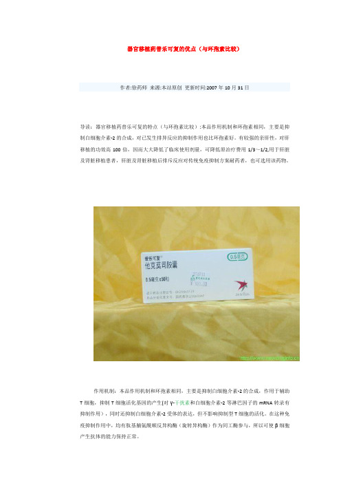
器官移植药普乐可复的优点(与环孢素比较)作者:徐药师来源:本站原创更新时间:2007年10月31日导读:器官移植药普乐可复的特点(与环孢素比较):本品作用机制和环孢素相同,主要是抑制白细胞介素-2的合成,对已发生排异反应的抑制作用也比环孢素好。
有较强的亲肝性,对肝移植的功效高100倍,因而大大降低了临床使用剂量,可降低原治疗费用1/3~1/2,用于肝脏及肾脏移植患者,肝脏及肾脏移植后排斥反应对传统免疫抑制方案耐药者,也可选用该药物。
作用机制:本品作用机制和环孢素相同,主要是抑制白细胞介素-2的合成,作用于辅助T细胞,抑制T细胞活化基因的产生(对γ-干扰素和白细胞介素-2等淋巴因子的mRNA转录有抑制作用),同时还抑制白细胞介素-2受体的表达,但不影响抑制型T细胞的活化。
在这种免疫抑制作用中,均有肽基脯氨酰顺反异构酶(旋转异构酶)作为同工酶参与,所以可使β细胞产生抗体的能力保持正常。
药物特点:与环孢素相比,本品有如下特点:①该药的免疫抑制作用无论在体外还是在体内试验都是环孢素的数十倍到数百倍。
在临床上本品1%的浓度就有效。
另外对已发生排异反应的抑制作用也比环孢素好。
②以往以环孢素为基础的免疫抑制方案中,不可逆的慢性排斥反应问题依然如旧,增加剂量虽然有进一步效果,但是带来严重的不良反应,本品可望能减少肝肾移植受体的急、慢性排斥反应。
③用本品的患者,细菌和,病毒感染率也较环孢素治疗者低,尤其是本品有较强的亲肝性,对肝移植的功效高100倍,因而大大降低了临床使用剂量,可降低原治疗费用1/3~1/2(因使用本品的患者可在术后2周出院而使用环孢素者则要4周),同时不良反应也明显降低,它具有低死亡率、高移植存活率、移植物具良好成功率及对类甾醇的相对非依赖性等优点。
适应证:用于肝脏及肾脏移植患者,肝脏及肾脏移植后排斥反应对传统免疫抑制方案耐药者,也可选用该药物。
禁忌证:禁用于对本品过敏的患者及孕妇,哺乳妇女使用本品时应停止授乳,2岁以下患者使用本品前应作有关EBV病毒血清学方面检查。
脂质运载蛋白2在肝脏疾病发生发展中的作用

2脂质运载蛋白2在肝脏疾病发生发展中的作用廖争光,卫诗蕙,杜丹玉,孙 立,袁胜涛中国药科大学药物科学研究院,江苏省新药筛选重点实验室,南京210009摘要:脂质运载蛋白2(LCN2)最初是从感染猴空泡病毒40的小鼠肾细胞培养物中纯化得到的一种分泌糖蛋白,在炎症期间对细胞稳态控制以及应对细胞应激或损伤的反应中发挥着关键作用,被认为是风湿病、癌症、肝脏疾病和炎症性疾病的一种潜在的生物标志物。
已有研究表明,LCN2在肝实质细胞和非实质细胞中表达并分泌入血,与急性肝损伤、肝硬化、病毒性肝炎、酒精性肝病、非酒精性脂肪性肝病以及肝细胞癌等的发生发展密切相关。
本文总结了LCN2与肝脏疾病发病机制相关的动物实验和临床研究,以期为肝脏疾病的防治提供新思路和治疗靶点。
关键词:肝疾病;脂质运载蛋白2;病理过程基金项目:国家自然科学基金(81773766)Roleoflipocalin-2inthedevelopmentandprogressionofliverdiseasesLIAOZhengguang,WEIShihui,DUDanyu,SUNLi,YUANShengtao.(JiangsuKeyLaboratoryforNewDrugScreening,InstituteofPharmaceuticalResearch,ChinaPharmaceuticalUniversity,Nanjing210009,China)Correspondingauthor:YUANShengtao,yuanst1967@163.com(ORCID:0000-0002-5626-6532)Abstract:Lipocalin-2(LCN2)isasecretedglycoproteinoriginallypurifiedfrommousekidneycellsinfectedwithsimianvirus40andplaysakeyroleinthecontrolofcellularhomeostasisduringinflammationandtheresponsetocellularstressorinjury,anditisconsideredapotentialbiomarkerforrheumaticdiseases,cancer,liverdiseases,andinflammatorydiseases.StudieshaveshownthatLCN2isexpressedinhepaticparenchymalandnonparenchymalcellsandissecretedintothebloodstream,anditiscloselyassociatedwiththedevelopmentandprogressionofacuteliverinjury,livercirrhosis,viralhepatitis,alcoholicliverdisease,nonalcoholicfattyliverdisease,andhepatocellularcarcinoma.ThisarticlesummarizestheanimalexperimentsandclinicalstudiesontheassociationofLCN2withthepathogenesisofliverdis eases,inordertoprovidenewideasandtherapeutictargetsforthepreventionandtreatmentofliverdiseases.Keywords:LiverDiseases;Lipocalin-2;PathologicProcessesResearchfunding:NationalNaturalScienceFoundationofChina(81773766)DOI:10.3969/j.issn.1001-5256.2022.09.044收稿日期:2022-02-18;录用日期:2022-03-20通信作者:袁胜涛,yuanst1967@163.com 糖蛋白作为细胞膜的结构成分以及免疫细胞的抗原决定簇,在抵御多种疾病中起着关键作用。
二氢槲皮素帮你养肝——肝净,皮肤一定娇嫩!
二氢槲皮素帮你养肝——肝净,皮肤一定娇嫩!原标题:二氢槲皮素帮你养肝——肝净,皮肤一定娇嫩!肝脏是人体最重要的代谢和解毒器官人体胃肠道所吸收的各种物质,包括营养物质和毒素等,经胃肠道的血管系统后,都由肝脏的门脉进入肝脏内进行分解、合成及解毒。
因为肝脏是唯一没有痛感神经的器官,所以无论它累成怎么样,它也从不呻吟叫苦,也不会喊痛,这也是人们经常忽略它健康状况的根本原因。
这就是为什么肝癌一发现,就是晚期的原因。
肝脏其实是一个非常重要的脏器,它掌管着糖、脂肪、蛋白质的解毒、代谢,人体大部分的新陈代谢和有毒物质的转化,因此,它也是最易污染的部门肝脏解毒功能的原理:在机体代谢过程中,人体吃进去的食物药物、呼吸进去的各种有毒害气体、以及无法及时排出体外的宿便,都要先通过门静脉收集,然后在肝内被解毒和清除。
它有两大功能:1肝主疏泄。
所谓疏泄,就是让你能够疏通、畅达。
比如说人生气了,时间久了,肝气郁结,就影响脾胃的运化功能,也就是说消化功能变差,所以人生气了,就不想吃饭。
2肝主藏血。
肝脏不只管藏血,还管着全身调节气血运行,管血液分配。
比如说,人在运动的时间,肝脏就把血液分配到四肢,女性朋友生理期前,肝脏就把血液分配到血海,即冲脉,那个时候肝脏的血液少了,柔韧性格就下降了,所以女性朋友总有那么几天脾气不好。
“肝”苦与共如今,人们生活节奏快、工作压力、生活压力越来越大,加班、应酬、熬夜是家常便饭,饮食不规律、配搭失衡、缺乏运动等不良生活方式,使我们的肝脏承受巨大的负荷,造成日积月累的损伤,甚至导致常见的酒精肝、脂肪肝、肝硬化和病毒性肝炎等肝的病变。
当肝脏这个解毒器官出现问题时,人体就会出现以下一些症状:无端感到疲倦、无端感到烦躁、焦虑和忧郁。
眼睛干涩或者死鱼眼、口干口苦、偏头痛。
头发经常很油。
出现粘便、有体臭。
腰部赘肉增加、脾气特别大、失眠多梦。
前胸后背有红痣。
指甲有明显竖条纹、脸庞两边有肝斑。
乳腺增生和妇科问题。
如果以上症状,你已经满足了一项以上,那你就必须要开始调理肝胆的功能了。
脂蛋白相关磷脂酶a2正常值范围
脂蛋白相关磷脂酶a2正常值范围脂蛋白相关磷脂酶A2(lipoprotein-associated phospholipase A2,简称Lp-PLA2)是一种与动脉粥样硬化相关的生物标志物,其正常值范围可以用于评估患者的动脉粥样硬化风险。
本文将介绍Lp-PLA2的正常值范围以及与其相关的临床意义。
Lp-PLA2是一种磷脂酶A2家族的酶,主要存在于动脉粥样硬化斑块中的巨噬细胞和内皮细胞。
它参与了动脉粥样硬化病变的形成和发展过程。
通过水解氧化低密度脂蛋白(ox-LDL)中的脂质分子,Lp-PLA2促进了斑块内炎症反应和斑块不稳定性的增加。
因此,Lp-PLA2的水平与动脉粥样硬化的严重程度和冠心病的风险密切相关。
Lp-PLA2的正常范围在不同的研究中可能会有所差异,但一般认为正常值范围为1.5-3.8 ng/mL。
这个范围是通过对大量健康人群的测定得出的。
需要注意的是,不同实验室可能使用不同的试剂盒和测定方法,因此正常范围可能会有所偏差。
因此,在进行Lp-PLA2的检测时,应该参考实验室提供的参考范围,以进行准确的评估。
Lp-PLA2的测定可以通过血液样本进行,一般采用酶联免疫吸附试验(ELISA)或免疫荧光法进行测定。
这些方法具有高灵敏度和高特异性,可以准确地测定Lp-PLA2的水平。
Lp-PLA2的水平与动脉粥样硬化的风险密切相关。
研究表明,Lp-PLA2水平的升高与冠心病、脑卒中和周围动脉疾病的发生和发展有关。
在冠心病患者中,Lp-PLA2的水平常常明显升高。
此外,Lp-PLA2还可以作为判断冠心病患者病情稳定性和预后的指标,高水平的Lp-PLA2表明冠心病患者的病情较为严重,预后较差。
除了动脉粥样硬化,Lp-PLA2的水平还与其他疾病的发生和发展有关。
研究发现,Lp-PLA2的水平与炎症性疾病、肾脏疾病、糖尿病等的发生和预后密切相关。
因此,Lp-PLA2的测定不仅可以用于动脉粥样硬化的评估,还可以作为其他疾病的辅助诊断和预后评估的指标。
磷脂酶a2的作用
脂蛋白磷脂酶A2(Lp-PLA2)Lp-PLA2是磷脂酶超家族中的亚型之一,也被认为是血小板活化因子乙酰水解酶,由血管内膜中的巨噬细胞、T细胞和肥大细胞分泌。
动脉粥样硬化斑块中Lp-PLA2表达上调,并且在易损斑块纤维帽的巨噬细胞中强表达。
Lp-PLA2可水解ox-LDL中的氧化磷脂,生成脂类促炎物质,如溶血卵磷脂和氧化游离脂肪酸,进而产生多种致动脉粥样硬化作用,包括内皮细胞死亡和内皮功能异常,刺激粘附因子和细胞因子的产生。
这些物质可通过趋化炎症细胞进一步产生自我强化的循环,产生更多促炎物质。
临床意义1、脂蛋白磷脂酶A2是针对血管内皮炎症即AS 严重程度的特异性炎症标记物,可以有效预警、评估心脑血管栓塞性疾病的发生风险。
2、Lp-PLA2在动脉粥样硬化形成和发展中有促进作用,是一个新的独立的预测冠心病意外的危险因素。
与hsCRP相比,Lp-PLA2的优点是与其他危险因素的相关性很小,Lp-PLA2与hsCRP相结合,可以提高对冠心病危险的预测水平。
3、Lp-PLA2不仅仅是一个危险标志物,还可能为心血管病的治疗提供一个新靶点,降低血浆中和血管壁的酶活性,它将代表一个新的极有发展前途的抗动脉粥样硬化的方法。
最后联合传统风险因子检测,可提高预测健康人未来患心血管疾病的风险的准确性。
性能参数(1) 方法学:免疫增强比浊法;(2) 线性范围:在0-800ng/mL范围内相关系数r≥0.990;线性偏差:浓度≤200ng/mL时,线性绝对偏差在±30ng/mL以内;浓度>200ng/mL时,线性相对偏差在±15.0%以内;(3) 精密度:批内精密度≤8%,批间精密度≤10%;(4) 准确度:相对偏差≤15%;(5) 样本类型:血清;(6) 参考范围:0-175ng/mL。
枯否细胞前列腺素E2的分泌与肝脏再生

枯否细胞前列腺素E2的分泌与肝脏再生富有;段芳龄;等【期刊名称】《胃肠病学和肝病学杂志》【年(卷),期】1993(002)002【摘要】本文用大鼠从体内,外观察了前列腺素E2(PGE2)对肝再生的作用。
重点集中在肝部分切除后所分离的离体枯否细胞(KC)在大肠杆菌脂多糖(LPS)的刺激下合成及分泌PGE2的能力。
结果显示,给大鼠体内注射消炎痛(5mg/kg)抑制C的PGE2产生,从而显著地阻碍了肝脏的再生。
其DNA合成率明显低于未使用组(P<0.01)。
而在DNA合成高峰期(术后24h)所分离的再生肝之KC,其合成PGE2的能力明显强于假手术组对等数量的KC(PGE2浓度分别为15ng/ml及6.5ng/ml,P<0.01)。
据此我们得出结论,KC通过其内源性PGE2的产生从而启动肝细胞的再生。
【总页数】4页(P18-21)【作者】富有;段芳龄;等【作者单位】MedicalResearch(151),VAMedicalCenterLongBeach,CA90822;河南医科大学消化疾病研究所【正文语种】中文【中图分类】R333.4【相关文献】1.IRF3基因干扰对LPS刺激原代枯否细胞早期细胞因子分泌动态变化的影响 [J], 朱彤;涂文娟;谈志丽;刘亮明2.p38MAPK在重症急性胰腺炎大鼠枯否细胞分泌促炎细胞因子中的作用 [J], 任洪波;李兆申;许国铭;屠振兴;贾一韬;施新岗;龚燕芳3.Toll样受体在高迁移率族蛋白B1诱导严重烧伤后枯否细胞分泌促炎性细胞因子中的作用 [J], 陈旭林;孙立;郭峰;王飞;刘晟;梁勋;王仁素;王永杰;孙业祥4.小鼠角膜基质细胞分泌的转化生长因子β2及前列腺素E2对树突状细胞成熟过程的抑制作用 [J], 卢建民;王会芳;李小磊;连玲艳;宋秀君5.小鼠角膜基质细胞分泌的转化生长因子β2及前列腺素E2对树突状细胞成熟过程的抑制作用 [J], 卢建民;王会芳;李小磊;连玲艳;宋秀君因版权原因,仅展示原文概要,查看原文内容请购买。
血清Lipocalin-2、CEA、CA125联合检测对肺癌的诊断价值
血清Lipocalin-2、CEA、CA125联合检测对肺癌的诊断价值作者:黄远虹张知刘小勇来源:《中国现代医生》2021年第33期[摘要] 目的探讨血清脂质运载蛋白-2(Lipocalin-2)、癌胚抗原(CEA)和糖类抗原125 (CA125)联合检测对肺癌的诊断价值。
方法选取2020年1—12月于本院就医的54例确诊肺癌患者及60名健康体检者作为试验组及对照组。
采用ELISA法检测血清Lipocalin-2水平,用罗氏cobas8000全自动生化分析仪检测CEA和CA125含量,并计算受试者工作特性曲线(ROC曲线)。
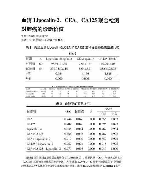
结果肺癌患者血清Lipocalin-2(239.04±98.15)ng/mL、CEA(6.04±5.21)ng/mL和CA125(25.64±22.98)U/mL的浓度均显著高于健康体检人群(P<0.05)。
肺癌患者血清Lipocalin-2、CEA和CA125及三者联合检测ROC曲线下面积分别为0.848、0.744、0.784及0.970,三者联合检测的诊断效能显著高于三者单独及CEA+CA125联合的诊断效能。
结论血清Lipocalin-2、CEA和CA125水平对肺癌的诊断有重要意义,联合检测可显著提高肺癌的诊断效率。
[关键词] 脂质运载蛋白-2;癌胚抗原;糖类抗原125;肺癌;联合检测[中图分类号] R446.11 [文献标识码] B [文章编号] 1673-9701(2021)33-0146-04[Abstract] Objective To explore the diagnostic value of combined detection of serum lipocalin-2, carcinoembryonic antigen (CEA) and carbohydrate antigen 125(CA125) in lung cancer (LC). Methods A total of 54 patients confirmed as lung cancer and 60 healthy people admitted to our hospital from January to December 2020 were selected as the experimental group and control group. Serum Lipocalin-2 level was detected by ELISA, CEA and CA125 content were detected by cobas8000 automatic biochemical analyzer (Roche), and the receiver operating characteristic (ROC) curve was calculated. Results The levels of serum Lipocalin-2(239.04±98.15) ng/mL,CEA(6.04±5.21) ng/mL and CA125 (25.64±22.98) U/mL in LC patients were significantly higher than those in healthy people(P<0.05). The areas under the ROC curve of serum Lipocalin-2, CEA and CA125 in LC patients were 0.848, 0.744, 0.784 and 0.970, respectively. The diagnostic efficiency of the combined detection of the three indexes was significantly higher than that of the combined detection of the three alone and the combined detection of CEA and CA125. Conclusion Serum levels of Lipocalin-2, CEA and CA125 are important for the diagnosis of LC,and combined detection can significantly improve the diagnostic efficiency of LC.[Key words] Lipocalin-2; Carcinoembryonic antigen; Carbohydrate antigen 125; Lung cancer; Combined detection肺癌是全球最常見的消化道恶性肿瘤之一,严重威胁人类的生命和健康,其发病率及病死率居各类恶性肿瘤的首位[1]。
cpla2 抑制 原理
cpla2 抑制原理
CPLA2抑制剂是一类针对磷脂酶A2(Phospholipase A2,简称PLA2)的药物或化合物,其作用是抑制PLA2的活性。
PLA2是一种参与磷脂代谢的酶,它在细胞膜上催化磷脂的水解,产生游离脂肪酸和磷脂酰胺。
这些产物在炎症和细胞信号传导中发挥重要作用。
CPLA2抑制剂的原理主要是通过干扰PLA2的活性,从而调节磷脂代谢和相关的细胞信号通路。
通过抑制PLA2活性,CPLA2抑制剂可以减少游离脂肪酸的产生,从而抑制炎症反应和细胞膜的破坏。
这对于一些炎症性疾病如风湿性关节炎、炎症性肠病等具有重要意义。
另外,PLA2在一些肿瘤细胞中也发挥重要作用,因此CPLA2抑制剂也被研究用于肿瘤治疗领域。
通过抑制PLA2活性,可以影响肿瘤细胞的增殖、侵袭和转移能力,因此具有潜在的抗肿瘤作用。
总的来说,CPLA2抑制剂的原理是通过抑制PLA2酶的活性,调节磷脂代谢和相关的细胞信号通路,从而对炎症性疾病和肿瘤具有一定的治疗作用。
当然,具体的药物作用机制和临床应用还需要进一步的研究和探讨。
lipin2基因名
lipin2基因名LIPIN2基因(lipin-2)是一种细胞内脂质代谢的重要调控因子。
它是一个非常重要的基因,在人体的营养代谢和代谢方面发挥重要作用。
它可以调节血液中的脂肪水平,参与脂肪酸合成,调节脂质的分解和合成,还可以调节糖尿病和肥胖的发生。
LIPIN2基因位于人类第6号染色体的11.1百万基因组位置上。
它的别名有AFF3、PAF-AH、PAFAH1B2、PAP7和ALPK1。
LIPIN2基因是一种多功能蛋白,它可以促进脂质代谢,参与脂肪酸合成,调节脂质的分解和合成,还可以调节血液中的脂肪水平,从而发挥重要作用。
LIPIN2基因编码一种脂质运输蛋白,也就是所谓的LIPIN2蛋白,它是一种多功能性蛋白,它不仅可以参与脂肪酸的合成,还可以参与脂质的分解和合成,还可以调节血液中的脂肪水平,从而发挥重要作用。
LIPIN2蛋白可以结合脂质体,作为脂质的运输载体,参与脂质的转运和调节。
它可以参与调控细胞内脂质代谢,调节脂质的分解和合成,还可以参与细胞内DNA的转录,从而调节多种生理和病理过程。
此外,LIPIN2蛋白还可以通过调节细胞的凋亡来调节炎症反应,从而参与抗炎和抗感染的过程。
它还可以通过调节细胞膜可渗性Na+/K+-ATP酶的活性,来调节细胞的渗透压,从而参与调节血压的过程。
LIPIN2基因可以通过调节脂质的合成和分解来控制血液中的脂肪水平,进而可以防止和治疗糖尿病和肥胖等疾病。
目前,研究表明,LIPIN2基因的突变可能会导致肥胖,糖尿病和心血管疾病等。
总之,LIPIN2基因是一种重要的基因,可以参与细胞内的脂质代谢,参与脂质的合成和分解,参与调节血液中的脂肪水平,并可以参与抗炎和抗感染的过程,从而发挥重要作用,预防和治疗糖尿病、肥胖和心血管疾病。
氟维司群联合戈舍瑞林对绝经前晚期乳腺癌患者血清LCN-2及Six1表达的影响
氟维司群联合戈舍瑞林对绝经前晚期乳腺癌患者血清LCN-2及Six1表达的影响曹主根【摘要】目的探讨氟维司群联合戈舍瑞林对绝经前晚期乳腺癌患者血清脂质运载蛋白-2(LCN-2)及Six1蛋白表达的影响.方法 40例绝经前晚期乳腺癌患者随机分为观察组和对照组,每组20例,对照组采用戈舍瑞林治疗,观察组采用氟维司群联合戈舍瑞林治疗,治疗后观察患者临床疗效及不良反应,检测其LCN-2和Six1水平变化.结果治疗后对照组,观察组客观有效率为75.00%,优于对照组的45.00% (P<0.05).治疗后2组患者LCN-2和Six1水平均有明显下降(P<0.05),但观察组患者更明显,差异有统计学意义(P均<0.05).2组患者恶心呕吐、厌食、潮热、肌肉酸痛等不良反应发生率差异均无统计学意义(P均>0.05).结论氟维司群联合戈舍瑞林对绝经前晚期乳腺癌患者疗效显著,可显著改善患者激素水平,且安全性高,值得临床推广.【期刊名称】《肿瘤基础与临床》【年(卷),期】2017(030)006【总页数】3页(P537-539)【关键词】晚期乳腺癌;氟维司群;戈舍瑞林;脂质运载蛋白-2;Six1【作者】曹主根【作者单位】泗洪县人民医院肿瘤内科,江苏宿迁223900【正文语种】中文【中图分类】R737.9乳腺癌的发病率在全球一直呈上升趋势,而且我国乳腺癌发病率的增长速度高于其他高发国家,目前乳腺癌已成为重大公共卫生问题[1]。
随着对乳腺癌认识的加深,其治疗理念也随之改变,根据患者病情和身体状况采取合理的诸如放疗、化疗、内分泌治疗等综合治疗手段[2]。
内分泌治疗方案是治疗激素异常型乳腺癌的首选方案,临床常用药物为他莫昔芬,然而对于绝经前的晚期乳腺癌疗效并不佳,研究[3]报道氟维司群联合戈舍瑞林可对该类患者有较好的治疗效果。
脂质运载蛋白-2(lipocalin-2, LCN-2)和Six1蛋白都是参与肿瘤发生和发展的重要细胞因子,在肿瘤的侵袭和转移过程中发挥重要作用[4]。
抑制铁死亡可减轻脓毒症小鼠的心肌损伤:脂钙蛋白-2的作用
脓毒症全球死亡率为10%,中国每年约有500万脓毒症患者,死亡率超过30%[1,2]。
心肌功能损伤是脓毒症住院患者中常见并发症[3],脓毒症引起的心功能不全是脓毒症死亡率高的主要原因[4],但其确切机制尚不完全清楚。
铁死亡最早被观察到铁离子螯合剂能抑制Erastin 诱导的癌细胞死亡不同于已知的细胞死亡途径,如凋亡、焦亡和坏死[5]。
铁死亡的机制主要涉及脂质过氧化物增加和脂质活性氧的进一步释放。
当谷胱甘肽过氧化物酶4(GPX4)或谷胱甘肽活性降低时,可诱导Inhibiting ferroptosis attenuates myocardial injury in septic mice:the role of lipocalin-2HUANG Yuhui 1,ZHANG Gongpeng 2,LIANG Huan 1,CAO Zhenzhen 3,YE Hongwei 1,GAO Qin 11Department of Physiology,2Department of Clinical Medicine,Bengbu Medical College,Bengbu 233000,China;3Department of Respiratory and Critical Care Medicine,First Affiliated Hospital of Bengbu Medical College,Bengbu 233000,China摘要:目的观察铁死亡在盲肠结扎与穿孔(CLP )法诱导的脓毒症小鼠心肌损伤中的作用,并探讨脂钙蛋白-2(Lcn2)在铁死亡中的可能作用。
方法选取8周龄雄性C57BL/6小鼠,采用CLP 法诱导脓毒症心肌损伤模型。
小鼠随机分为3组(10只/组):假手术组、脓毒症组(CLP ,小鼠接受CLP 手术)、脓毒症+铁死亡抑制剂Ferrostain-1组(CLP+Fer-1,小鼠腹腔注射浓度为5mg/mL 的Fer-15mg/kg ,1h 后接受CLP 手术)。
血清AOPP、LCN-2水平在妊娠期糖尿病孕妇不良妊娠结局中的预测分析
血清AOPP、LCN-2水平在妊娠期糖尿病孕妇不良妊娠结局中的预测分析杨萍;余文;王彩霞【期刊名称】《四川生理科学杂志》【年(卷),期】2024(46)5【摘要】目的:研究血清晚期氧化蛋白产物(Gestational diabetesmellitus,AOPP)、人脂质运载蛋白2(Lipocalin 2,LCN-2)水平在妊娠期糖尿病(Gestational diabetes mellitus,GDM)孕妇不良妊娠结局中的预测价值。
方法:回顾性收集2021年10月-2022年6月在我院分娩的89例GDM孕妇的临床资料。
根据妊娠结局将孕妇分为妊娠结局良好组(n=67)和妊娠结局不良组(n=22)。
分析不良妊娠结局情况;对比两组的血清AOPP、LC-2水平;分析血清AOPP、LCN-2水平与GDM孕妇不良妊娠结局的相关性及其对不良妊娠结局的预测价值。
结果:妊娠结局不良组血清AOPP、LCN-2较妊娠结局良好组显著提高(P<0.05)。
经双变量Pearson分析,血清AOPP、LCN-2水平与GDM孕妇不良妊娠结局呈正相关(r=0.568、0.599,P<0.001)。
绘制ROC曲线显示,得到AUC分别为:0.852、0.890、0.915,其中联合检验的预测价值最高。
结论:GDM孕妇血清AOPP、LCN-2高表达与不良妊娠结局具有密切关系,且具有一定预测价值。
【总页数】3页(P1109-1111)【作者】杨萍;余文;王彩霞【作者单位】信阳市平桥区妇幼保健院产科【正文语种】中文【中图分类】R71【相关文献】1.血清LCN-2及AOPP与妊娠期糖尿病并发子痫前期的相关性研究2.妊娠期糖尿病患者并发子痫前期血清LCN-2、AOPP水平变化及意义3.血清LCN-2、AOPP 与妊娠期糖尿病不良妊娠结局的相关性分析4.妊娠期糖尿病孕妇血清SHBG水平与胰岛素分泌及不良妊娠结局关系5.血清ACA、AOPP以及抗-β2-GPⅠ水平与妊娠期糖尿病孕妇不良妊娠结局的关系因版权原因,仅展示原文概要,查看原文内容请购买。
携带lipocalin 2基因的溶瘤腺病毒对胰腺癌细胞生长的体外抑制作用

( 同济 大学 附 属 第 十 人 民医 院 普 外科 上海 2 0 7 ;! 旦 大 学 附 属 中 山 医 院普 外 科 002 复 上海 2 0 3 002
中 国科 学 院 上 海 生 命科 学研 究 院生 物 化 学 与 细 胞 生 物 学研 究 所
[ s a t Obet e To c n tu trpiains lcieo c lt d n vr se p e s g l o ai Abt c] r jci v o sr c e l t -eet n oyi a e o iu x r si i cl 2 c o v c n p n
上海
20 3 ) 0 0 1
【 要 】 目的 摘 抑制作用。方法
构 建 携 带 人 l oai 2 因且 删 除 E B 5基 因 的溶 瘤 腺 病 毒 , 究 其 体 外 对胰 腺 癌 细 胞 生 长 的 i cl 基 p n 15 研 利 用 R —C 技 术 获 得 l oa n2基 因 并 克 隆 至 p A1 TP R i cl p i C 3质 粒 ,构 建 p A1一p cl 用 C 3l oa n2 i i
g n n n e tg t t n iu o c iiy i ir . M eh d Li o ai e ewa lne n o D e ea d iv s ia eisa tt m ra tvt nv to to s p c l 2 g n sco di t CA 1 n 3
Fu a Unv J M e S 2)8 N。,3 ( dn i M e S i ( o u , 6 d u ’ 5 u u 。 、J ) d c
ai i c l 2基 因的溶瘤 腺 病 毒 对 胰 腺癌 p n 细胞 生 长 的体 外 抑 制作 用
二酰甘油 磷脂酶a2 花生四烯酸

二酰甘油、磷脂酶a2和花生四烯酸是生物化学领域中非常重要的概念,它们在人体健康和疾病发生中起着至关重要的作用。
在本文中,我们将深入探讨这些概念的深度和广度,以便更好地理解它们在生物体内的重要作用。
一、二酰甘油二酰甘油是一种重要的生物分子,它在人体内起着关键的代谢和能量调节作用。
在细胞内,二酰甘油是三酸甘油酯的代谢产物,它通过三酸甘油酯水解酶的作用,在脂肪细胞中被分解成脂肪酸和甘油。
这些产物参与到机体的脂肪代谢中,对于保持机体的能量平衡和体重稳定起着非常重要的作用。
二酰甘油还是血液中甘油三酯的主要组成部分之一,它的水平对人体健康有着重要的影响。
二、磷脂酶a2磷脂酶a2是一种重要的酶类蛋白,它在人体内参与到磷脂代谢和脂质代谢中起着关键的调节作用。
磷脂酶a2主要通过水解磷脂酰胆碱和磷脂酰乙醇胺,产生游离脂肪酸和溶栓活性磷脂。
这些产物对于细胞膜的稳定和炎症反应有着重要的影响,同时也参与到血小板的凝集和血液凝固中。
磷脂酶a2的活性异常与许多疾病的发生和发展密切相关,它被认为是预防心血管疾病和炎症性疾病的潜在靶点之一。
三、花生四烯酸花生四烯酸是一种重要的多不饱和脂肪酸,它在人体内起着多种生理功能,对于人体的健康有着重要的影响。
花生四烯酸是ω-6系列不饱和脂肪酸的代表物质,它在人体内通过代谢产生前列腺素和白三烯素等生物活性成分,调节细胞信号转导和炎症反应。
花生四烯酸还是细胞膜的重要组成部分,对于保持细胞膜的流动性和通透性具有重要意义。
花生四烯酸的摄入量和代谢产物水平与很多慢性疾病的发生有着密切的联系,它被认为是人体健康的重要指标之一。
二酰甘油、磷脂酶a2和花生四烯酸是生物体内非常重要的概念,它们在细胞代谢和健康调节中起着不可替代的作用。
进一步的研究发现将有助于我们更好地理解它们的作用机制和相关疾病的发生机制,为未来的疾病预防和治疗提供重要的理论支持。
希望我们的文章对你有所帮助,更好地理解这些重要的生物概念。
二酰甘油、磷脂酶a2和花生四烯酸在人体健康和疾病发生中的重要性已经得到充分的论证。
中性粒细胞明胶酶相关脂质运载蛋(NGAL)
中性粒细胞明胶酶相关脂质运载蛋白一、临床适应症背景情况中性粒细胞明胶酶相关脂质运载蛋白(Neutrophil gelatinase-associated lipocalin,NGAL)又称人脂质运载蛋白2(lipocalin2,Ln2)或噬铁蛋白(siderocalin),是人脂质运载蛋白家族中的一个新成员。
近年来,NGAL作为一种新的肾损伤标志物倍受关注。
早期急性肾损伤、慢性肾病、心肾综合症等疾病血清和尿液中NGAL会显著升高,且其水平监测对病情轻重、疗效及预后判断具有较好的诊断价值。
临床检测NGAL主要有免疫层析法、免疫比浊法、速率色散比浊法、乳胶增强免疫比浊法等。
胶乳增强免疫比浊法是近年来出现的一种较为稳定、准确的体液蛋白均相免疫比浊检测方法。
肾脏损伤约占所有泌尿生殖道损伤的65%左右。
急性肾损伤是一组临床综合征,是指突发(1-7d 内)和持续(>24h)的肾功能突然下降,定义为血清肌酐至少上升0.5mg/dl 1,表现为氮质血症、水电解质和酸碱平衡以及全身各系统症状,可伴有少尿(<400ml/24h或17ml/h)或无尿(<100ml/24h)。
肾病的发病迁延难愈,时间超过三个月,病人尿液和相关的血液指标出现异常,肾脏病理学、影像学发现异常,或肾脏的肾小球有效滤过率低于60%,都可统称为“慢性肾病”。
慢性肾病如未能及时有效救治,导致病情恶化进展,则随病程迁延,慢性肾病患者将发展成为慢性肾功能不全、肾衰竭,最终形成尿毒症。
心肾综合征(cardio-renalsyndrome,CRS)即心脏或肾脏对另一器官的损害不能代偿时,互为因果,形成恶性循环,最终加速心脏和肾脏功能的共同损害和衰竭。
其发病率、死亡率高,给社会和患者带来沉重的负担。
目前对心肾综合征的认识有限,缺乏有效的治疗措施,是临床处理的难题。
早期诊断急性肾功能损伤时血、尿NGAL浓度通常会迅速升高,2h最为明显( 比临界值升十至几百倍),血清肌酐、尿酶等传统指标往往要在24~72h才后明显升高,因而NGAL可用于急性肾功能损伤的早期诊断。
Lipocalin-2基因表达产物对人胚肾细胞增殖的影响
Lipocalin-2基因表达产物对人胚肾细胞增殖的影响帖儒修;朱道银;李娜;王爽【期刊名称】《中国生物制品学杂志》【年(卷),期】2008(21)11【摘要】目的构建Lipocalin-2(Lcn-2)基因真核表达质粒PL-2,并检测其及前期原核表达的重组Lcn-2蛋白对人胚肾细胞HEK293增殖的影响。
方法利用RT-PCR 从鼠巨噬细胞RAW264.7中扩增Lcn-2基因,克隆入真核表达质粒pEGFP-C1中,构建重组质粒PL-2。
转染HEK293细胞,通过荧光观察和RT-PCR鉴定Lcn-2基因的表达。
将重组质粒PL-2和前期原核表达的重组Lcn-2蛋白作用于HEK293细胞,通过MTT法检测细胞增殖情况,Westernblot法检测细胞增殖核抗原(PCNA)的表达。
结果重组真核表达质粒PL-2经酶切和测序鉴定,证明构建正确。
经荧光观察和RT-PCR鉴定,转染重组质粒PL-2的HEK293细胞中有Lcn-2基因的表达。
加入重组Lcn-2蛋白后,HEK293细胞较对照组明显增殖,细胞中PCNA的表达也较对照组明显升高,而重组质粒PL-2对HEK293细胞增殖以及细胞中PCNA的表达均无影响。
结论已成功构建了Lcn-2基因真核表达质粒,其对HEK293细胞的增殖无影响,而原核表达的重组Lcn-2蛋白对HEK293细胞的增殖有一定的促进作用,推测Lcn-2基因的表达产物可能通过与细胞膜受体结合促进HEK293细胞的增殖。
【总页数】5页(P972-976)【关键词】Lipocalin-2基因;真核表达;原核表达;人胚肾细胞;增殖【作者】帖儒修;朱道银;李娜;王爽【作者单位】重庆医科大学免疫学教研室【正文语种】中文【中图分类】Q813.5【相关文献】1.PDX-1基因对人胚肾细胞胰岛发育相关基因表达的影响 [J], 夏华强;徐小明;刘律君;卢光琇2.同型半胱氨酸、同型半胱氨酸诱导基因抗体及叶酸对人胚心肌细胞同型半胱氨酸诱导基因的表达及细胞增殖的影响 [J], 王光明;吴俊;李英;毕振武;吕丹瑜;董静霞3.ARG1基因对人胚肾细胞293砷染毒前后增殖及凋亡的影响 [J], 杨元元;李媛媛;李莉;张冰荫;王瑞;慕晓玲4.构建人SMARCAL1基因过表达慢病毒载体调控胚肾细胞增殖 [J], 石淑娟;李诚;乔凌燕;杨槟伊;李堂5.构建人SMARCAL1基因过表达慢病毒载体调控胚肾细胞增殖 [J], 石淑娟;李诚;乔凌燕;杨槟伊;李堂因版权原因,仅展示原文概要,查看原文内容请购买。
- 1、下载文档前请自行甄别文档内容的完整性,平台不提供额外的编辑、内容补充、找答案等附加服务。
- 2、"仅部分预览"的文档,不可在线预览部分如存在完整性等问题,可反馈申请退款(可完整预览的文档不适用该条件!)。
- 3、如文档侵犯您的权益,请联系客服反馈,我们会尽快为您处理(人工客服工作时间:9:00-18:30)。
O R I G I N A L A R T I C L EThe role of lipocalin-2in liver regenerationKatrin Kienzl-Wagner 1*,Alexander R.Moschen 2,3*,Sabine Geiger 2,3,Alexandra Bichler 3,Felix Aigner 1,Gerald Brandacher 4,Johann Pratschke 1and Herbert Tilg 2,31Department of Visceral,Transplant and Thoracic Surgery,Center of Operative Medicine,Innsbruck Medical University,Innsbruck,Austria 2Christian Doppler Research Laboratory for Gut Inflammation,Innsbruck Medical University,Innsbruck,Austria3Department of Internal Medicine I (Gastroenterology,Endocrinology &Metabolism),Innsbruck Medical University,Innsbruck,Austria 4Department of Plastic and Reconstructive Surgery,Johns Hopkins University School of Medicine,Baltimore,MD,USAKeywordsbiomarker –hepatic proliferation –LCN2–lipocalin-2–liver regeneration –partial hepatectomyCorrespondenceHerbert Tilg,MD,Department of Internal Medicine I,Innsbruck Medical University,Anichstrasse 35,Innsbruck 6020,Austria Tel:+4351250423540Fax:+4351250423538e-mail:herbert.tilg@i-med.ac.at Received 13November 2013Accepted 1July 2014DOI:10.1111/liv.12634AbstractBackground &Aims:Various immune mediators such as interleukin-6(IL-6)have been implicated in the process of liver regeneration.Lipocalin-2(LCN2)has been recently characterized as a prototypic immune mediator produced by various cell types being involved mainly in host defence.In addition,numerous studies have demonstrated its clinical value as a biomar-ker.This study aimed at defining the role of LCN2in liver regenera-tion.Methods:We studied LCN2expression in wild-type mice in a model of partial hepatectomy (PH).Furthermore,we evaluated liver regeneration after PH in LCN-deficient mice compared to littermate controls.Serum levels of LCN2were assessed in a small group of patients undergoing hepatic resec-tion.Results:LCN2is dramatically induced in livers and sera of wild-type mice after PH,whereas liver LCN2-receptor expression was decreased.Sham operations did not affect hepatic and serum LCN2expression.Although LCN2-deficient mice exhibited increased baseline liver expression indices,LCN2-deficient mice did not differ from wild-type mice with respect to hepatic proliferation suggesting that this molecule is not involved in hepatic repair.Only serum IL-1b levels were slightly lower in LCN À/Àmice,whereas IL-6serum levels did not differ between various tested animal groups.In humans undergoing hepatic resection,LCN2levels increased significantly within 24h following surgery.Conclusions:LCN2,although massively induced in mice after PH,is not relevant in murine hepatic regeneration.Further,human studies have to define whether LCN2could evolve as biomarker after liver surgery.The liver has the unique ability to fully regenerate after major tissue loss induced by viruses,toxins,autoim-mune disease,resection of primary or metastatic tumours as well as in the context of partial liver trans-plantation (1).Enhancing the regenerative capacity of the liver is crucial for the development of specific thera-pies for chronic liver disease and expanding indications for liver resection.Even though the last years have seen remarkable progress in characterizing the molecular and cellular mechanisms of liver regeneration,the multiple components and complex networks that orchestrate the regeneration process remain to be further elucidated.The current understanding of liver regeneration encompasses various cytokines,growth factors and met-abolic networks that link liver function with cell growth and proliferation.In normal liver regeneration as seenafter partial hepatectomy (PH),the liver mass is replen-ished by replication of existing hepatocytes without involvement of stem or progenitor cells (2).Upon liver resection,expression of TNF-a and IL-6is induced in Kupffer cells by various mechanisms including lipopoly-saccharide resulting in activation of STAT3in hepato-cytes and subsequent entry into the cell cycle.Key mitogenic factors that drive cell cycle progression are hepatocyte growth factor,epidermal growth factor and vascular endothelial growth factor.Termination of hepatocyte proliferation is controlled by the action of transforming growth factor b and activins (1,3–5).Mounting evidence suggests that lipocalin-2(LCN2)is an important modulator of proliferative and regenera-tive processes.LCN2is a 25kDa glycoprotein of a b -barrel shaped structure with the ability to bind small hydrophobic ligands and to form complexes with soluble macromolecules (6).LCN2so far has been ascribed great functional diversity.It has been shown to*Both authors contributed equally to this study.Liver International (2014)©2014John Wiley &Sons A/S.Published by John Wiley &Sons Ltd1Liver International ISSN1478-3223be a component of the innate immune system(7),it is highly induced during the acute phase response(8),and it acts as an immunomodulator in inflammatory colo-rectal diseases(9).Furthermore,LCN2regulates the inflammatory response during ischaemia and reperfu-sion of transplanted hearts(10).It was further demon-strated that hypoxia and anaemia induce LCN2 expression(11),whereas it inhibits differentiation of erythroid progenitor cells(12)and induces apoptosis (13).Numerous studies have proposed LCN2as a potent biomarker for acute renal injury(14–16).Recent evidence further implicates a role for LCN2in obesity, insulin resistance and hyperglycaemia(17).Previous studies have established LCN2as a growth regulator.LCN2has been associated with tissue involu-tion(18),it induces the epithelial to mesenchymal tran-sition,thereby actively influencing cancer progression by affecting tumour invasion and metastasis(19),and LCN2facilitates mucosal regeneration by promoting cell migration(20).In the developing kidney,LCN2pro-motes the organization of epithelial cells into tubular structures(21),whereas in chronic kidney disease it seems to be a key effector in aberrant growth(22).Dur-ing the regenerative process after renal ischaemia-reper-fusion injury,LCN2acts both as an inducer and inhibitor of renal cell proliferation(23,24).Data on the potential role of LCN2in liver homoeo-stasis and liver disease are limited.LCN2expression is strongly induced in models of acute and chronic liver injury(25–28)and it is significantly increased in liver biopsies from patients with chronic HCV(29)or HCC (30).To define the role of LCN2in liver regeneration, we characterized the regulation of LCN2in wild-type mice after2/3PH.Furthermore,we investigated the impact of heterozygote or homozygote LCN2deficiency on liver regeneration.MethodsMiceAll animal experiments described were performed in accordance with Austrian legal requirements after approval by the Austrian authorities(licensed to AR Moschen No BMWF-66.011/0040-II/10b/2009).All mice were maintained at the central animal facility of the Medical University Innsbruck under specific patho-gen-free conditions with ad libitum access to standard chow and water.The generation of LCN2À⁄Àmice has been described elsewhere(7).For all experiments,male littermates were used.At the time of surgery,mice were 12–14-week old with a body weight of20–30g.Murine2/3PHAll surgical procedures were performed under isoflurane inhalation anaesthesia using a vaporizer for laboratory animals(Provet,Lyssach,Switzerland)and a micro-scope with25-to50-fold magnification(Leica MZ12.5, Wetzlar,Germany).The2/3PH was performed as described previously(31,32).Briefly,the liver is exposed through midline laparotomy and the gallbladder is removed.For70%hepatectomy,the right and left upper lobe(median lobe)and the left lateral liver lobe are ligated at the pedicle of the respective liver lobe(5/0silk) and resected.Resected material was preserved as index liver tissue for further work-up.After irrigating the abdomen with sterile saline,the abdomen is closed in two layers(running5/0PDS suture for peritoneum and fascia,running5/0seralon suture for skin).To replace intra-operativefluid loss,the mouse is given2–3ml sterile saline subcutaneously.Sham procedures consisted of anaesthetizing mice followed by midline laparotomy and manipulating but not resecting the liver.Sample preparationMice were injected intraperitoneally with 2.5mg/ml BrdU(BD Biosciences,San Diego,CA,USA)2h before organ retrieval.After the indicated time points,mice were anaesthetized with isoflurane.Following relaparot-omy,blood was collected from the vena cava inferior. The remnant regenerative liver was inspected in situ, carefully resected,and processed according to protocol. Both index and follow-up liver tissue were systematically worked up to befixed in10%buffered formalin,stored in RNA-later,or snap-frozen for protein work-up.ImmunohistochemistryFour micrometre sections were prepared from formalin fixed,paraffin-embedded specimens of liver tissue sam-ples.Sections were deparaffinized in xylene and rehy-drated in graded alcohols.Antigen retrieval was achieved by microwaving slides for10min in antigen unmasking solution(Vector Laboratories,Burlingame,CA,USA). Endogenous peroxidase activity was quenched with3% H2O2in PBS for10min.Non-specific protein binding sites were blocked using serum-free protein block(Dako, Glostrup,Denmark).The followingfirst antibodies were applied:specific rabbit polyclonal anti-LCN2antibody (provided by Professor Marit Nilsen-Hamilton,Iowa State University,Ames IA,USA)and anti-PCNA(Prolif-erating Cell Nuclear Antigen)Ab-1,clone:PC10 (Thermo Lab Vision,Rockford,IL,USA).Specificity was demonstrated by using either isotype-specific con-trol antibodies or recombinant LCN2for blocking experiments.HRP-conjugated anti-mouse or anti-rabbit pAb(Dako)were incubated for60min at room temper-ature.LCN2or PCNA were visualized with3,30-diam-inobenzidine(DAB)as a chromogen.Slides were counterstained with haematoxylin QS(both Vector La-borytories),dehydrated and mounted permanently in Eukitt(O.Kindler GmbH,Freiburg,Germany). Nuclear incorporation of BrdU was visualized on paraffin-embedded tissue sections using a BrdU In-SituLiver International(2014)©2014John Wiley&Sons A/S.Published by John Wiley&Sons Ltd2Lipocalin-2in liver regeneration Kienzl-Wagner et al.Detection Kit(BD Pharmingen,San Diego,CA,USA) according to the manufacturer’s instructions.Pictures were acquired on a Zeiss AX10(Imager A1,Zeiss,Ober-kochen,Germany)with high-resolution camera and Ax-ioVision software.The number of BrdU-or PCNA-positive nuclei was determined using an Optimas image analysis package(Weiss imaging and solutions GmbH, Bergkirchen/Guending,Germany).mRNA and real-time qPCRTotal RNA was extracted from liver tissue using1ml/ 100mg TRIZOL reagent according to the manufac-turer’s instructions(Life Technologies,Carlsbad,CA, USA).One microgram of total RNA was reverse tran-scribed into cDNA using200units of moloney murine leukaemia virus reverse transcriptase(Life Technolo-gies)with random hexanucleotide primers(Roche, Mannheim,Germany)as previously described(33). Primers were designed using Primer Express software (Applied Biosystems,Foster City,CA,USA)and Fast-PCR professional(University of Helsinki). Quantitative real-time PCR(qPCR)was performed in a total volume of25l l in40cycles(95°C for30s fol-lowed by60°C for60s)using a Mx3000P bioanalyzer (Stratagene,now Agilent Technologies,Santa Clara,CA, USA).Master mixes were from Eurogentec(Seraign,Bel-gium).Expression levels were calculated using the stan-dard-curve method using serial dilutions of positive control cDNA(from stimulated splenocytes)as a stan-dard.The abundance of each mRNA was expressed as ratio to beta glucuronidase(Gusb)mRNA expression. The following primers have been used:Lcn2forward:50-GCC TCA AGG ACG ACA ACA TCA-30,Lcn2reverse: 50-CAC CAC CCA TTC AGT TGT CAA-30,Lcn2probe: 50-FAM-TTC TCT GTC CCC ACC GAC CAA TGC BHQ1-30,Lcn2R forward:50-GGC GAT TTC TAC AGC GAA TGA-30,Lcn2R reverse:50-CTA TCA GCC ACC GTG CAG ACT-30,Lcn2R probe:50-FAM-CCT CTT CCT GTT TTA TGG CTG GCC TGG T-BHQ1-30.Analysis of circulating LCN2and cytokines(IL-1b,IL-6, TNF-a)Venous blood was collected under microscopic guid-ance from the vena cava inferior and centrifuged at 2500g.Serum was stored atÀ80°C until assayed.Mouse LCN2(NGAL)was assayed using commercially avail-able antibody pairs from R&D Systems(DuoSet Devel-opment Kit,Minneapolis,MN,USA).IL-1b,IL-6and TNF-a were determined using BD OptEIA TM assays con-sisting of capture and detection antibodies along with streptavidin-horseradish peroxidase(HRP)conjugate. The respective recombinant cytokines served as a stan-dard(BD Biosciences,PharMingen).The absorption was read with a PowerWave HT Microplate Spectropho-tometer(Biotek,Winooski,VT,USA)at450nm with a reference wavelength of570nm.Western BlotProtein was extracted homogenizing liver tissue in M-PER Mammalian Protein Extraction Reagent con-taining Halt Protease Inhibitor Cocktail(Thermo Scien-tific,Pierce,Rockford,IL,USA)according to the manufacturer’s instructions.Tissue lysates were sepa-rated under reducing conditions on12.5%acrylamide gels using a mini-PROTEAN3electrophoresis system (Bio-Rad,Hercules,CA,USA).Gels were transferred to Hybond-P PVDF membranes(GE Healthcare,Uppsala, Sweden),blocked with 2.5%BSA containing0.1% Tween.Blots were incubated at4°C o/n applying one of the following primary antibodies:specific rabbit poly-clonal anti-LCN2(provided by Professor Marit Nilsen-Hamilton,Iowa State University)or anti-GAPDH (clone14C10;Cell Signaling Technology,Danvers,MA, USA).HRP-conjugated anti-rabbit secondary antibodies were used for secondary detection(Dako).Bands were visualized using a chemiluminescent substrate on a Hyperfilm ECL(both GE Healthcare)that was automat-ically processed on an X-rayfilm developer.Densitome-try was performed with ImageJ(34).Human serum samplesFrom March2014to May2014,five patients undergoing hepatic resection were enrolled at the Department of Visceral,Transplant and Thoracic Surgery,Innsbruck Medical University.This study protocol was reviewed and approved by the local ethics committee(AN2014-0024).All patients provided written informed consent for the participation of this study.Human serum sam-ples were collected the day prior to surgery as well as24 and48h post-operatively.Blood samples were centri-fuged at2500g.Serum was stored atÀ20°C until assayed.Human LCN2(NGAL)was assayed using com-mercially available human LCN2ELISA from R&D Sys-tems(according to manufacturer’s instructions).StatisticsThe differences among groups were analysed by Mann–Whitney U-test and,where appropriate,by Kruskal–Wallis ANOVA.Significance was assumed for P<0.05. All data analyses were carried out using the IBM SPSS Statistics19.0software package(IBM,Armonk,NY, USA)and graphical illustrations were prepared using GraphPad Prism version6(GraphPad Software, Jolla,CA,USA).ResultsLipocalin-2is strongly induced during liver regeneration We investigated the regulation of LCN2during liver regeneration.LCN2was strongly induced as early as6h after PH and peaked after12h reaching52.9-foldLiver International(2014)©2014John Wiley&Sons A/S.Published by John Wiley&Sons Ltd3 Kienzl-Wagner et al.Lipocalin-2in liver regenerationhigher levels over baseline controls.Thereafter,expres-sional levels declined until 48h,plateaued but remained 7.9-fold elevated at 96h (Fig.1A).The expression of the LCN2receptor (LCN2R)was slightly reduced (2.1-fold after 6h,2.7-fold after 12h,2.2-fold after 24h and 2.5-fold after 48h)after PH but returned to baseline levels at 72h (Fig.1B).RNA data were confirmed by Western blot analysis.Accordingly,protein concentra-tions peaked at 12h as shown by Western blot analysis (Fig.1D).LCN2protein levels gradually declined until(A)(B)(C)(D)(E)(G)(F)Fig.1.(A)mRNA was extracted from liver tissue at the indicated time points after PH and relative LCN2expression was determined by quan-titative real-time PCR (purple line,n =5per group).Steady state levels were assayed from index liver tissue that was obtained at the time of resection (blue line,n =5per group);*P <0.05,**P <0.01.(B)Index and follow-up liver tissue mRNAs as mentioned above were assayed for Lipocalin receptor (LCN2R)mRNA (n =5per group,*P <0.05,**P <0.01).(E)Serum was collected at the indicated time points after PH and circulating LCN2concentrations were determined by ELISA.(C)Hepatic LCN2and LCN2R mRNA expression were compared between mice that underwent PH (black bars)and sham-operated controls (white bars).(D)Liver tissue was collected at the indicated timepoints after PH and protein was extracted and LCN2content determined by Western Blot.Protein content of Gapdh served as a loading control.Protein concentration was quantitated by densitometry.(F)Hepatic LCN2protein expression was determined by immunohistochemistry (brown:LCN2,blue:nuclei).(G)Circulating LCN2levels in patients undergoing liver resection.Serum samples were obtained prior to hepatic surgery,24and 48h after liver resection.Concentration of circulating LCN2was determined by ELISA.Liver International (2014)©2014John Wiley &Sons A/S.Published by John Wiley &Sons Ltd4Lipocalin-2in liver regeneration Kienzl-Wagner et al.96h(Fig.1D).As determined by immunohistochemis-try,hepatic LCN2positivity was strongest after24h (Fig.1F).In addition to strong LCN2regulation,we observed significant microvesicular steatosis following PH occurring12–24h after hepatic resection(Fig.1F). Circulating LCN2levels were determined by ELISA.As outlined in Fig.1E,the highest serum levels were found 24h after PH.To rule out a significant contribution of the surgical manipulation on LCN2and LCN2R levels, we performed sham operations.As depicted in Fig.1C, laparotomy on its own did not significantly impact hepatic LCN2or LCN2R expression.To elucidate whether LCN2is also regulated following liver resection in humans,we assessed LCN2serum levels infive patients undergoing hepatic resection of different extent for variable aetiologies.Patient characteristics are pro-vided in Table1.LCN2was readily detectable by ELISA in all samples.There was a significant increase in circu-lating LCN2expression24h post-liver resection com-pared to pre-operative LCN2levels(P<0.05;Fig.1G).Impact of LCN2deficiency on liver regenerationWe next investigated the impact of LCN2deficiency on liver regeneration.Therefore,wild-type mice(LCN2+⁄+), mice heterozygote for LCN2(LCN2+⁄À)and LCN2 knockout mice(LCN2À⁄À)were subjected to PH.As shown in Fig.S1,the number of BrdU-positive cells peaked48h after PH indicating the time point of maxi-mum hepatic regeneration and was consequently used for further analysis.At the same time point,we also defined the maximum positivity for PCNA(data not shown).Interestingly,LCN2À/Àmice demonstrated sig-nificantly elevated baseline liver regeneration compared to LCN2+/+and LCN2+/Àlittermates as demonstrated by increased numbers of PCNA-positive hepatic nuclei (Fig.2A).However,no significant differences in hepatic proliferation were observed at48h after PH between wild-type(LCN2+⁄+),LCN2+⁄Àand LCN2À⁄Àlittermates with regard to number of PCNA-positive cells(Fig.2B) and BrdU-positive nuclei(Fig.2C).Impact of LCN2genotype on circulating cytokine levels Pro-inflammatory cytokines have been shown to be potent inducers of the initiation phase of liver regeneration.As LCN2has been implicated in the regu-lation of immune functions,we hypothesized that the LCN2genotype might affect the cytokine profile after PH.As shown in Fig.2D–F,TNF-a serum concentra-tions peaked6h after PH,whereas IL-1b and IL-6 showed their peaks after12h.Interestingly,the overall IL-1b response was higher in wild-type mice compared to LCN2+⁄Àand LCN2À⁄Àmice(Fig.2D).While IL-1b was detectable until48h in wild-type mice,it was unde-tectable in knockout mice after12h.No significant dif-ferences were seen for IL-6concentrations(Fig.2E). Regarding circulating TNF-a concentrations,lowest lev-els were observed in mice heterozygote for LCN2 (Fig.2F).Liver cytokine expression data after12,24 and48are depicted in Fig.S2and did not show any differences between study groups.DiscussionRecent evidence demonstrates that LCN2expression shows clear correlations with liver damage.LCN2 expression was significantly increased in acute and chronic liver injury models(26,27)as well as in chronic HCV infection(29).A recent study observed a substan-tial increase in LCN2liver expression after PH in rats (25),although the role of LCN2remains unclear.We now also observe that PH is followed by an impressive LCN2induction.As extensive LCN2overexpression did not influence hepatocyte proliferation at48h post-PH in LCN2À/Àmice,the reasons for this prominent LCN2 upregulation need to be explored:whether it is just a bystander effect or whether it is necessary for liver regeneration.One possible explanation is that LCN2is in fact a mediator of liver regeneration and constitutes another redundant pathway(4).The idea that LCN2influences liver regeneration is supported by the fact that LCN2À/Àmice exhibit significantly higher baseline regeneration compared to their wild-type littermates.Consequently, LCN2was supposed to act as an inhibitor of hepatocyte proliferation in a way that under physiological condi-tions LCN2prohibits excess hepatocyte proliferation to maintain liver homoeostasis.In line with these data,Mi-harada et al.demonstrated in a mouse model of acute haemolytic anaemia that under normal physiological conditions,LCN2secreted by erythroid progenitor cellsTable1.Characteristics of patients undergoing liver surgeryPatient Age Sex Diagnosis PVE/PVL Liver resection158Male HCC No Segment II/III extended to IVa 272Male Recurrent HCC Yes Right hemihepatectomy334Female Donation for LDLT No Segment II/III471Male Hepatic metastases from CRC No Atypical resection II/III+VI 562Female CCC Yes TrisegmentectomyPVE,portal vein embolization;PVL,portal vein ligature;HCC,hepatocellular carcinoma;CCC,cholangiocellular carcinoma;CRC,colorectal cancer; LDLT,living donor liver transplantation.Liver International(2014)©2014John Wiley&Sons A/S.Published by John Wiley&Sons Ltd5 Kienzl-Wagner et al.Lipocalin-2in liver regenerationin bone marrow suppressed the survival and differentia-tion of erythroid progenitors.Once an urgent haemato-poiesis is required,the erythroid progenitors appear to escape from LCN2-mediated suppression thereby per-mitting rapid production of mature cells (12).However,considering LCN2an inhibitor of liver regeneration under physiological conditions neither explains the dramatic increase in LCN2expression following 70%hepatectomy nor the absent impact on hepatocyte proliferation at 48h post-PH in LCN2À/Àmice.Hypothetically,LCN2could exert a dual role in the process of liver regeneration as has been proposed for renal regeneration.Depending on the phase of reper-fusion (early te)LCN2acts as an inducer and inhibitor of renal cell proliferation.LCN2blockade with anti-LCN2antibody in the late regenerative phase(A)(B)(C)(D)(E)(F)Fig.2.(A)Liver tissue was resected from wild-type,LCN2+⁄Àand LCN2À⁄Àmice at the time of PH.Tissue sections were stained for prolifera-tive cell nuclear antigen (PCNA).Pictures were acquired at 1009magnification and the number of PCNA-positive nuclei was determined using an Optimas software package.(B)Livers were harvested from wild-type,LCN2+⁄Àand LCN2À⁄Àmice 48h after PH.Paraffin-embedded tissue sections were stained for PCNA.The number of PCNA-positive cells was determined using an Optimas software package.(C)Mice were injected with BrdU 2h before livers were collected and processed.Liver sections were stained for BrdU.Absolute numbers of positive nuclei per 1009high-power field was determined with Optimas.(D –F)Serum samples were obtained 6,12,24and 48h after PH.Concen-trations of circulating cytokines were determined by ELISA.(D)IL-1b ,(E)IL-6,(F)TNF-a concentrations.*P >0.05.Liver International (2014)©2014John Wiley &Sons A/S.Published by John Wiley &Sons Ltd6Lipocalin-2in liver regeneration Kienzl-Wagner et al.reduced renal cell proliferation,whereas anti-LCN2 antibody administration in the early inflammatory phase had the opposite effect with increased expression of proliferation markers(23).Adopting thesefindings from renal regeneration to our data would propose LCN2as an inhibitor of hepatocyte proliferation in physiological liver homoeostasis and an inducer–though redundant–of liver regeneration upon PH. Alternatively,the fact that LCN2induction does not alter hepatocyte regeneration at48h post-PH which represents the time of maximum proliferative activity suggests that LCN2upregulation in the liver and serum upon major hepatic resection simply is a bystander effect.Already in1995,Liu et al.identified SIP24/24p3 as an acute phase protein(8).Major hepatic surgery with resection of70%of the liver parenchyma is of course an appropriate stimulus that elicits an acute phase response.However,as shown in our sham opera-tions,midline laparotomy and manipulation of the vis-ceral organs on its own did not significantly induce hepatic LCN2expression.Furthermore,LCN2has been shown to be strongly upregulated during anaemia.In a mouse model of acute hypovolaemic anaemia Jiang et al.found LCN2mRNA dramatically increased in the liver of phlebotomized mice and LCN2protein levels were increased in the cor-responding sera(11).Major surgery such as70%hepa-tectomy is inevitably accompanied by a certain blood loss,solely because of the fact that a significant amount of portal,arterial and venous blood is trapped within the three liver lobes that are being resected.This state of post-operative hypovolaemia may therefore very well explain the striking induction of LCN2that we observed upon PH.In addition to strong LCN2regulation,we observed significant microvesicular steatosis following PH occur-ring12–24h after hepatic resection.Liver regeneration following PH is physiologically associated with transient microvesicular steatosis(35–37).which seems to be required for the regenerative process.The injured liver produces cytokine signals that trigger the release of fatty acids from adipose tissue into circulation that are then taken up by hepatocytes and stored as triacylglycerols. Their hydrolysis releases fatty acids for mitochondrial b-oxidation and ATP production and provides substrate for the biosynthesis of membrane phospholipids to sup-port rapid cell division(38).Strikingly,LCN2expres-sion has been linked to obesity and non-alcoholic fatty liver disease(17,39).As the peak of steatosis within the regenerating liver correlates with the peak of hepatic LCN2expression upon PH,the striking LCN2induction that we observed could be interpreted as an epiphenom-enon of the transient microvesicular steatosis that is physiologically associated with liver regeneration. Previous studies have established the role of IL-1b to induce LCN2expression via the NF-j B pathway (40–42).In line with thesefindings,serum IL-1b concentrations peaked at12h after PH.As IL-1b is known to exert a mitoinhibitory effect on hepatocyte proliferation,the fact that the overall IL-1b response was markedly higher in wild-type mice compared to LCN2-deficient littermates might explain why LCN2À/Àmice demonstrated significantly elevated baseline liver regeneration.In summary,our data convincingly demonstrate that LCN2is potently expressed after PH.We further observed a significant increase in circulating LCN2levels 24h post-liver resection in humans suggesting that lar-ger prospective studies using LCN2as a potential bio-marker for liver regeneration should be initiated. Studies in LCN2-deficient mice,however,clearly dem-onstrate that this acute phase protein is not involved in hepatic regeneration.AcknowledgementsConflict of interest:The authors do not have any disclo-sures to report.References1.Bohm F,Kohler UA,Speicher T,Werner S.Regulation ofliver regeneration by growth factors and cytokines.EMBO Mol Med2010;2:294–305.2.Zaret KS,Grompe M.Generation and regeneration of cellsof the liver and pancreas.Science2008;322:1490–4.3.Fausto N,Campbell JS.The role of hepatocytes and ovalcells in liver regeneration and repopulation.Mech Dev 2003;120:117–30.4.Fausto N,Campbell JS,Riehle KJ.Liver regeneration.Hepatology2006;43(2Suppl.1):S45–53.5.Clavien PA,Petrowsky H,Deoliveira ML,Graf R.Strate-gies for safer liver surgery and partial liver transplantation.N Engl J Med2007;356:1545–59.6.Flower DR.The lipocalin protein family:structure andfunction.Biochem J1996;318(Pt1):1–14.7.Flo TH,Smith KD,Sato S,et al.Lipocalin2mediates aninnate immune response to bacterial infection by seques-trating iron.Nature2004;432:917–21.8.Liu Q,Nilsen-Hamilton M.Identification of a new acutephase protein.J Biol Chem1995;270:22565–70.9.Nielsen BS,Borregaard N,Bundgaard JR,et al.Inductionof NGAL synthesis in epithelial cells of human colorectal neoplasia and inflammatory bowel diseases.Gut1996;38: 414–20.10.Aigner F,Maier HT,Schwelberger HG,et al.Lipocalin-2regulates the inflammatory response during ischemia and reperfusion of the transplanted heart.Am J Transplant 2007;7:779–88.11.Jiang W,Constante M,Santos MM.Anemia upregulateslipocalin2in the liver and serum.Blood Cells Mol Dis 2008;41:169–74.12.Miharada K,Hiroyama T,Sudo K,Nagasawa T,Nakam-ura Y.Lipocalin2functions as a negative regulator of red blood cell production in an autocrine fashion.FASEB J 2005;19:1881–3.13.Devireddy LR,Gazin C,Zhu X,Green MR.A cell-surfacereceptor for lipocalin24p3selectively mediates apoptosis and iron uptake.Cell2005;123:1293–305.Liver International(2014)©2014John Wiley&Sons A/S.Published by John Wiley&Sons Ltd7 Kienzl-Wagner et al.Lipocalin-2in liver regeneration。
