眼球分析课件
合集下载
眼球的解剖与生理ppt课件

容易出现破裂;视神经周围最厚(1.0mm)。 v 与视N交接处 外2/3 视神经鞘膜
内1/3 筛板
扫描电镜下巩膜筛孔
10
整理ppt
3、角 巩 膜 缘
角膜和巩膜的移行带。角巩膜缘内侧即前房角。 前房角: 从角膜后弹力膜止端至巩膜突。在前房角内依次可见 到如下结构:schwalbe线、小梁网和Schlemm管、巩膜突。
整理ppt
(2)角膜生理学特点
维持眼球的完整及对眼内容物的保护 透明:无血管——营养供应靠房水、泪膜、角膜缘血管网
纤维排列整齐 相对脱水的恒定状态 屈光力强:43D(总屈光力的70%) 感觉敏锐:丰富的神经末梢,自我保护。
9
整理ppt
2、巩 膜
① 质韧,乳白色,由致密的胶原纤维和弹力纤维构成。 ② 厚度各处不一,眼外肌附着处最薄(0.3mm),受伤
26
整理ppt
房水循环主要途径
小梁网 Schlemm 管
21
视神经盘
整理ppt
视
神
经
乳
中央凹
头
(
生
理
盲 点22
)
整理ppt
视网膜组织结构
➢ 视网膜神经上皮层(感觉层)由三级神经元组成 第一神经元:光感受器—感光
1.视锥细胞:司明视觉、色觉,主要分布在黄斑区。此区受损则 可发生中心视力和色觉异常
2.视杆细胞:司暗视觉,主要分布在视网膜周边部。视杆细胞功 能障碍时可产生夜盲症
13
睫状肌
后房
前房
虹膜
瞳孔开大肌 瞳孔扩约肌
整理ppt
虹膜根部
晶状体
14
整理ppt
(2) 特 点:
➢ 根部脆弱,外伤易离断。 ➢ 富含血管:炎症时渗出多,外伤易出血。 ➢ 色素细胞多:外伤、炎症、缺血从虹膜上游离。 ➢ 神经未梢丰富:病变时疼痛明显。
眼科学讲眼部解剖图课件PPT

第四节:眼附属器
睫毛、 眼睑、 结膜、 泪器、 眼外肌、 眼眶。
(一)睫毛
位于上下眼睑的边缘,向前上、前下方伸出, 排列整齐。根部有丰富的感觉神经丛,
对触觉非常敏感。睫毛的平均寿命为3~5个月。
(二)眼睑
眼睑位于眼眶出口,覆盖眼球前部,分为上睑和下睑, 中间称为睑裂,边缘为睑缘,上下睑缘交接处称为内 眦和外眦。内眦包围着一个肉状隆起,称为泪阜,泪 阜的上下缘处各有一小孔,为上、下泪小点。 眼睑通过瞬目作用将泪液散开,均匀地湿润角膜,填 补角膜表面的不平之处,使角膜形成良好的光学表面。 眼睑闭合可以保护眼睛免遭外伤。上下睑板有高度发 育的皮脂腺(睑板腺)埋藏其中,其出口位于睑缘上。 睑板腺分泌脂肪性油脂,形成泪液的最表层,在配戴 隐形眼镜时脂肪成份会沉淀在镜片上。
连 鼻泪管:位于骨性鼻泪管的管道内,上接泪囊,向下开口于
下鼻道
5mm---12mm,垂直10. 由7块颅骨组成,深4-5cm 6》分部:皮质,中央玻璃体,中央管 眼睑位于眼眶出口,覆盖眼球前部,分为上睑和下睑,中间称为睑裂,边缘为睑缘,上下睑缘交接处称为内眦和外眦。
敏锐有视锥细胞和视杆细
眼球近球形,前后径为24mm,水平径 虹膜 根据光线的强弱使瞳孔缩小或开大,保证视网膜成像清晰
约23mm,此时如发育不完全±1.00mm
时就可以出现±3.00D的屈光度。
第二篇:眼球解剖
第一节 眼球的解剖及生理
•眼球壁 •外层 •中层 •内层
一、眼球壁
•外膜:由坚韧的纤维组织所组成,构 成眼球的完 整的封闭的外壁,起到保护 眼内组织,维持眼球形 状的作用
•角膜 前1/6 •巩膜 后5/6 •中膜 又称葡萄膜,色素膜,血管膜 •虹膜 •睫状体 •脉络膜 •内膜 视网膜
初中生物七年级《眼球的结构及功能》公开课优秀课件

虹膜:有色素,中央的小孔叫瞳孔
眼球壁 中膜 睫状体:由睫状肌构成,调节晶状体曲度
眼
脉络膜:营养眼球,使眼球内部形成暗室
球
的
内膜:又叫视网膜,含有对光线敏感的细胞,能感
结
受光的刺激
构 房 水:折射光线
内容物 晶状体:透明,有弹性,像双凸透镜,能折射光线
玻璃体:透明胶状物质
想一想:
• 在光线到视网膜之前,经过哪些结构?
讨论一:观察同桌的眼睛, 说说眼睛有哪些结构?
讨论二:拆装眼球模型,讨论归纳眼球 的结构和功能
一、眼球的结构
测测你的记忆力!
睫状体1
虹 2膜 角 3膜 瞳 孔4
巩7 膜 脉8络膜 视9网膜 视1神0 经
晶状5体 6玻璃体
眼球的结构和功能图解:
角膜:无色透明,可以透过光线
外膜 巩膜:白色,坚固,保护眼球的内部结构
4 第23题
第12题
第34题
第51题
1.人们常说“眼观六路,耳听八方。” 这说明眼和耳都是人认识世界的感觉器 官。眼的主要结构是( )
A.眼球 B.晶状体
C.瞳孔 D.视网膜
2.外界物体反射来的光线,依次经过下 列哪些结构到达视网膜( ) A.瞳孔、角膜、晶状体、玻璃体 B.角膜、瞳孔、晶状体、玻璃体 C.瞳孔、角膜、玻璃体、晶状体 D.角膜、瞳孔、玻璃体、晶状体们常
眼
脉络膜:营养眼球,使眼球内部形成暗室
球
的
内膜:又叫视网膜,含有对光线敏感的细胞,能感
结
受光的Байду номын сангаас激
构 房 水:折射光线
内容物 晶状体:透明,有弹性,像双凸透镜,能折射光线
玻璃体:透明胶状物质
眼球结构及视觉PPT课件

坐姿要端正:不可弯腰驼背,越靠近或趴着 做功课易造成睫状肌紧张过度,进而造成近视
• 睡眠不可太少,作息有规律:睡眠不足身体 容易疲Fra bibliotek,易造成假性近视。
2019/8/2
18
角膜、房水、晶状
光 体、玻璃体 线 折光系统
成像于视网膜
感光细胞 接受刺激 产生兴奋
视神经
(纠正倒像) 视觉中枢
产生视觉
正常眼
看近物 睫状肌收缩晶状体曲度变大 物像在视网
膜上
看近远物视 眼 戴凹透镜舒张
变小
晶状体曲度过大或眼球前后径过长 物像在视网膜前方
远 视 眼 戴凸透镜 晶状体曲度过小或眼球前后径过短 物像在视网膜后方
用眼卫生:三要三不要
2019/8/2
19
失去正常人辨别颜色能力 的先天性色觉障碍。
红色盲、绿色盲、红绿色 盲、黄蓝色盲和全色盲。
眼球 (主要) 眼
角膜:透明 外膜 巩膜:白色(眼白)
睫状体:调节晶状体曲度
的
眼球壁 中膜 虹膜(眼球颜色):瞳孔
结
(受虹膜平滑肌控制)外界
光线强瞳孔缩小反之扩大
构
脉络膜:营养
内膜:视网膜 有感光细胞
附属结构:眼睑、结膜等
2019/8/2
8
近视和远视
2019/8/2
近视
远视 9
不同曲度的凸透镜折光情况示意图
(D)角膜、房水、晶状体和玻璃体组成
2、我们通常说的蓝眼睛是指人的眼球中( B )
(A)角膜是蓝色的(B)虹膜是蓝色的
(C)晶状体是蓝色的(D)玻璃体是蓝色的
2019/8/2
29
3、人眼相当于凸透镜,当人观察物体时, 物体在视网膜上形成的像是( D ) A 正立缩小的虚象 B 正立缩小的实象 C 倒立缩小的虚象 D 倒立缩小的实象
《眼科学》眼部解剖结构ppt课件

血管膜
视网膜
最内层,贴于眼球内壁,结构复杂, 功能重要,对视觉的形成至关重要。
中间层,富含血管和色素细胞,有营 养作用,也称为葡萄膜。
眼内容物
01
02
03
房水
充满于角膜和晶状体之间 的透明液体,有营养角膜 、晶状体和维持眼压的作 用。
晶状体
透明、富有弹性的双凸透 镜,有折射光线和调节焦 距的作用。
眼运动神经
支配眼球运动,包括动眼神经、滑车神经和外展神经。
感觉神经
传递眼部感觉,如三叉神经和面神经。
视路
视网膜神经节细胞
01
将视觉信息传递至大脑皮质。
视神经纤维
02
汇聚成视神经束,穿过视交叉进入大脑。
大脑皮质
03
处理视觉信息,产生视觉感知。
06
眼部疾病与防治
常见眼部疾病
近视
由于眼轴过长或角膜、晶状体曲率过 大,使平行光线聚焦在视网膜前面, 导致远视力降低。
04
泪器解剖结构
泪腺
泪腺
是产生泪液的器官,位于眼球的 外上方,分为上、下两对,分别
称为上泪腺和下泪腺。
泪腺的生理功能
主要分泌泪液,润滑眼球表面,保 持角膜湿润,防止感染。
泪腺的解剖结构
泪腺由腺泡和导管组成,腺泡分泌 泪液,导管将泪液引流到眼球表面 。
泪道
泪道
是泪液排出的通道,包括泪小点 、泪小管、泪囊和鼻泪管。
《眼科学》眼部解剖 结构ppt课件
目录
• 眼部概述 • 眼球解剖结构 • 眼睑解剖结构 • 泪器解剖结构 • 眼部血管与神经解剖结构 • 眼部疾病与防治
01
眼部概述
眼部的位置与功能
位置
《眼球的结构与功能》课件

控制用眼时间
适当休息,每隔20-30分钟休息一下眼睛,远离电子屏幕,眺望远 处,缓解眼部疲劳。
调整屏幕亮度与对比度
根据环境光线调整屏幕亮度与对比度,使屏幕光线柔和、不刺眼。
科学合理地使用电子产品
1 2
遵循“20-20-20”法则
每隔20分钟,将目光转向20英尺(约6米)远的 物体,持续注视20秒。
《眼球的结构与 功能》ppt课件
目录
• 眼球的解剖结构 • 眼球的功能 • 眼球的疾病与治疗 • 眼球的保健与护理 • 眼球的未来发展与展望
01
眼球的解剖结构
眼球壁的构成
01
02
03
前层
由角膜和巩膜组成,角膜 透明,有屈光作用,巩膜 白色,保护眼球内部结构 。
中层
由虹膜、睫状体和脉络膜 组成,富含血管和色素细 胞,为眼球提供营养。
包括泪腺和泪道,分泌泪液并排出, 保持眼球湿润和清洁。
结膜
覆盖在眼睑内面和眼球表面的薄膜, 具有分泌和润滑作用。
02
眼球的功能
视觉的形成
视觉的形成过程
光线通过角膜、晶状体等结构, 在视网膜上形成倒置的图像,大 脑皮层对图像进行处理,形成清 晰的视觉。
视觉的重要性
视觉是人类感知外界的主要方式 ,对于生存、学习和工作都至关 重要。
ቤተ መጻሕፍቲ ባይዱ
03
眼球的疾病与治疗
常见的眼球疾病
青光眼
眼内压升高导致的 视神经损伤。
结膜炎
眼部炎症,常见症 状包括红肿、流泪 和视力模糊。
白内障
晶状体浑浊导致的 视力下降。
视网膜脱离
视网膜与眼球壁分 离,导致视力丧失 。
角膜炎
角膜炎症,可能导 致视力下降和疼痛 。
眼的解剖及生理ppt课件

膜,前膜比后膜后约一倍。一生中晶状体纤维不断生成并将原先的 纤维挤向中心,逐渐硬化而形成晶状体核,晶状体核外较新的纤维 称为晶状体皮质。
晶状体的生理功能
晶状体无血管,营养来自房水和玻璃体, 主要通过无氧糖酵解途径来 获取能量。 作用:1.正常眼无调节状态下晶状体相当于19D的凸透镜,是最主要的
眼的屈光介质之一。 2.具有独特的屈光通透和折射功能,且可滤去部分紫外线,对
呈环形走向,收缩时可使瞳孔缩小,受 动眼神经的副交感纤维支配。 瞳孔开大肌:位于虹膜深层紧贴色素上皮层处,肌纤 维呈放射状排列,从虹膜根部一直延伸 到瞳孔缘,收缩时瞳孔变大,受交感神 经支配。
睫状体
睫状体:位于虹膜根部与脉络膜之间的、宽约6-7mm的环状组 织,其矢状面略呈三角形,巩膜突是睫状体基底部 附着处。
睫状冠:睫状体前1/3较肥厚,宽约2mm,富含血管。 睫状突:睫状冠内表面有70-80个纵行放射状嵴样皱褶。 睫状体扁平部:睫状体后2/3薄而平坦。 锯齿缘:扁平部与脉络膜连接处。 睫状体主要由睫状肌和睫状上皮细胞组成。睫状肌是平滑肌, 受副交感神经支配。睫状上皮细胞层由外层的色素上皮和内 层的无色素上皮两层细胞组成。
OD
视网膜
分层:分为十层
外层是视网膜色素上
皮层
内九层为视网膜神经
上皮层
视网膜神经上皮层和 色素上皮层之间有潜 在的间隙,在病理情 况下容易分离,形成 视网膜脱离。
二、眼球内容物
•包括房水、晶状体和玻璃体,与角膜一起构成眼的屈光系统。
•房水:为眼内透明液体,充满前房与后房。
前房:角膜后面与虹膜和瞳孔区晶状体前面之间的眼球内腔。容积 约0.2ml,前房中央部深度约2.5-3mm,周边渐浅。
视网膜
位置:是一层透明的膜,位于脉络膜的内侧。 黄斑:视网膜后极部有一无血管的凹陷区,解剖上称为中心
眼球解剖生理课件

眼球及眼睑正面图
眼
眼球 眼附属器 视路与视
中枢 血管与神
经
文档仅供参考,不能作为科学依据,请勿模仿;如有不当之处,请联系本人改正。
眼球及眼眶(矢状面)
眼
眼球 眼附属器
视路与视 中枢
血管与神 经
文档仅供参考,不能作为科学依据,请勿模仿;如有不当之处,请联系本人改正。
眼胚胎
人眼的形成
文档仅供参考,不能作为科学依据,请勿模仿;如有不当之处,请联系本人改正。
角巩膜缘
角膜与巩膜交界之处 宽约1mm(房角及房水引流系统的所在部位) 巩膜静脉窦:房水循环(房水静脉)
眼球壁
中层——葡萄膜
虹膜 睫状体 脉络膜
文档仅供参考,不能作为科学依据,请勿模仿;如有不当之处,请联系本人改正。
睫状体
前方连结虹膜根部、后端以锯齿缘与脉络膜分界 宽6mm,黑色环状带(睫状肌) 前1/3较厚睫状冠--70个睫状突分泌房水 与晶体赤道部之间透明小带联络(悬韧带)环状
肌收缩-悬韧带松弛-晶体变厚 后2/3较平坦--睫状环(平坦部或扁平部)
文档仅供参考,不能作为科学依据,请勿模仿;如有不当之处,请联系本人改正。
文档仅供参考,不能作为科学依据,请勿模仿;如有不当之处,请联系本人改正。
角膜组织结构与特点
角膜内皮层
具有角膜—房水屏障的功能,可使角膜处于 相对脱水状态,从而保持角膜的透明,损伤 后常常可引起基质层 的水肿混浊。
内皮细胞损伤后 不能再生
文档仅供参考,不能作为科学依据,请勿模仿;如有不当之处,请联系本人改正。
文档仅供参考,不能作为科学依据,请勿模仿;如有不当之处,请联系本人改正。
教学目的与要求
掌握眼球的解剖与生理(重点) 熟悉视神经、视路的解剖与生理 熟悉眼附属器的解剖与生理 了解眼的血循环及神经支配为科学依据,请勿模仿;如有不当之处,请联系本人改正。
眼
眼球 眼附属器 视路与视
中枢 血管与神
经
文档仅供参考,不能作为科学依据,请勿模仿;如有不当之处,请联系本人改正。
眼球及眼眶(矢状面)
眼
眼球 眼附属器
视路与视 中枢
血管与神 经
文档仅供参考,不能作为科学依据,请勿模仿;如有不当之处,请联系本人改正。
眼胚胎
人眼的形成
文档仅供参考,不能作为科学依据,请勿模仿;如有不当之处,请联系本人改正。
角巩膜缘
角膜与巩膜交界之处 宽约1mm(房角及房水引流系统的所在部位) 巩膜静脉窦:房水循环(房水静脉)
眼球壁
中层——葡萄膜
虹膜 睫状体 脉络膜
文档仅供参考,不能作为科学依据,请勿模仿;如有不当之处,请联系本人改正。
睫状体
前方连结虹膜根部、后端以锯齿缘与脉络膜分界 宽6mm,黑色环状带(睫状肌) 前1/3较厚睫状冠--70个睫状突分泌房水 与晶体赤道部之间透明小带联络(悬韧带)环状
肌收缩-悬韧带松弛-晶体变厚 后2/3较平坦--睫状环(平坦部或扁平部)
文档仅供参考,不能作为科学依据,请勿模仿;如有不当之处,请联系本人改正。
文档仅供参考,不能作为科学依据,请勿模仿;如有不当之处,请联系本人改正。
角膜组织结构与特点
角膜内皮层
具有角膜—房水屏障的功能,可使角膜处于 相对脱水状态,从而保持角膜的透明,损伤 后常常可引起基质层 的水肿混浊。
内皮细胞损伤后 不能再生
文档仅供参考,不能作为科学依据,请勿模仿;如有不当之处,请联系本人改正。
文档仅供参考,不能作为科学依据,请勿模仿;如有不当之处,请联系本人改正。
教学目的与要求
掌握眼球的解剖与生理(重点) 熟悉视神经、视路的解剖与生理 熟悉眼附属器的解剖与生理 了解眼的血循环及神经支配为科学依据,请勿模仿;如有不当之处,请联系本人改正。
眼球的结构与功能课件

集合:当眼注视近物时两眼必须向内转动, 使两眼视轴集合于一点,才能产生双眼单 视,称为集合。
屈光不正概论
正视眼:当眼调节静止时,5米以外的平行 光线经过眼的屈光系统屈折后,焦点在视 网膜黄斑中心凹上,这种屈光状态为正视
屈光不正:(非正视眼) 当眼调节静止时,5米以外的平行光线经过 眼的屈光系统屈折后,焦点不能在视网膜 黄斑中心凹上,这种屈光状态为屈光不正
晶状体:主要由水和蛋白质组成,本身无 血管其营养来自于房水。
晶状体参与眼球的屈光作用、调节功能,还 能吸收部分紫外线,起到保护视网膜的主 作用。随着年龄增长,晶状体核逐渐浓缩, 弹力减弱,调节力下降而出现老视。
玻璃体:为透明的胶质体,主要成分为水。 玻璃体除有屈光作用外,也起到支撑视网 膜的作用。
近视眼的分类:1 轴性近视 (眼轴长) 2 曲率性近视3 屈光指数性近视 4调节性近 视
近视眼的临床表现
视力降低:远视力降低而近视力多为正常 视疲劳:从事近距离工作时出现头痛及眼
疲劳。 近视眼的矫正:戴凹透镜(负镜片) 近视眼的验配原则:
最好的矫正视力最低的配镜度数
远视眼的形成与分类
远视眼:当眼调节静止时,5米外平行光线 经眼的屈光系统屈折后聚焦在视网膜后, 叫远视眼。
巩膜与角膜一同构成眼内容物的外屏障, 其主要功能为维持眼球外形,保护眼内组 织以稳定视力,避光形成暗盒。
中层
虹膜:虹膜为圆盘状薄膜,色素丰富,位 于角膜和晶状体之间,中间有一小孔即瞳 孔,平均直径为2.5-4mm,其大小可随光线 的强弱而改变,调节入眼光线。
睫状体:位于虹膜与视网膜的锯齿缘之间, 宽约6mm的环状组织,分前后两部分,前 部称睫状突,主要由睫状肌和丰富的血管 组成,由睫状小带与晶状体相连,后部为 睫状环。
眼球知识PPT课件
–婴儿时期富有弹性几乎透明 –年龄增大 弹性下降 晶体变硬稍大
• 新纤维产生后老纤维聚往中间
–晶体进一步老化失去光学透明度而变混浊影响视力 称白内障
16
眼球解剖和生理-玻璃体
• 位置:介于晶状体和视网膜之间 • 形态:透明的胶冻状 • 成分:水 99% • 功能:
–占眼球腔隙2/3 维持眼球形态 –固定视网膜与脉络膜的附着 –透明的屈光介质,导光
• 含杯状细胞 免疫功能 保护眼球免受感染
21
眼球解剖和生理-泪液
• 眨眼后形成均匀的泪膜 • 泪膜组成:3层
–类脂层(外层) –水质层(中间层) –粘液层(内层)
22
眼球解剖和生理-泪液
• 类脂层
–眼睑睑板腺分泌 –减少下面的水质层蒸发率 –类脂沉淀在隐形眼镜的前后表面易引起舒适度和视
力下降
• 水质层
–中间最厚的一层 占90% –泪腺和副泪腺分泌 –运输氧气、蛋白质、酶类、电解质无机物等营养物
质给角膜 –维持软镜含水量(对软镜配戴者来说)
23
眼球解剖和生理-泪液
• 粘液层
–结膜杯状细胞分泌 –粘附于角膜上皮层 维持角膜光
滑 –水质层和粘液层的蛋白质(溶菌
酶 乳铁蛋白 淀粉酶等)在隐形 眼镜前后面沉淀引起戴镜不适和 视力下降
• 维持角膜透明度
–水肿则透明度下降
• 形态固定,使角膜具有屈光性
7
眼球解剖和生理-角膜
• 后弹力层
–介于基质层和内皮层之间
• 内皮层
–角膜最内层 单层扁平六角形细胞 –损伤后不可再生 –功能
• 机械泵水作用
–维持角膜厚度、透明和水平衡
• 通透营养物质
8
眼球解剖和生理-角膜代谢
眼球1 PPT课件
眼球
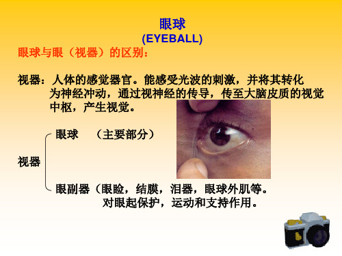
(EYEBALL) 眼球与眼(视器)的区别:
视器:人体的感觉器官。能感受光波的刺激,并将其转化 为神经冲动,通过视神经的传导,传至大脑皮质的视觉 中枢,产生视觉。
眼球 视器 眼副器(眼睑,结膜,泪器,眼球外肌等。 对眼起保护,运动和支持作用。 (主要部分)
一:眼球的位置 类似球形。位于眶腔的前部,后端通过视神经 连于间脑 二:眼球的组成:
2、睫状体 产生房水 调节视力 睫状肌 睫状突 睫状小带
3、脉络膜
3、脉络膜
丰富的血管(营养作用) 色素细胞(暗室作用) 吸收散射光线
虹膜部
盲部 视网膜 睫状体部 视部 (眼底、视神经盘、黄斑、中央凹)
眼 底:视网膜的后部 视神经盘(视神经乳头)有视网膜中央血管出入, 是视神经节C轴突汇集而成。又称生理性 盲点。 黄 斑 :视神经盘外侧约3.5mm处黄色小区。 黄斑中央凹 :为黄斑的中央凹陷,此处感光最敏锐
眼球璧 眼球
内容物
房水 晶状体 玻璃体
三:眼球壁的构造
外 纤维膜(角膜,巩膜)
眼球壁 中 血管膜 (虹膜,睫状体,脉络膜)
内 视网膜
纤维膜(角膜,巩膜) 1、角膜: 无色透明 无血管 丰富的神经末梢
2、巩膜 乳白色 不透明 厚而坚韧 保护眼球及 内容物
血管膜(虹膜,睫状体,脉络膜)
1、虹膜 色ቤተ መጻሕፍቲ ባይዱ细胞 瞳孔 瞳孔开大肌 瞳孔括约肌
《眼科学讲眼部解剖》课件

白内障
晶状体浑浊导致视力下降。
眼部疾病的诊断方法
01
视力检查
通过检查患者的远视力和近视力, 初步判断眼部疾病。
眼底镜检查
观察视网膜和视神经,了解眼底病 变情况。
03
02
裂隙灯检查
使用裂隙灯观察角膜、晶状体、玻 璃体等结构,发现病变。
眼压测量
测量眼球内部的压力,辅助诊断青 光眼等疾病。
04
眼部疾病的治疗方法
03
晶状体循环是指晶状体内部的营养物质和代谢废物的交换过程,通过 晶状体囊和晶状体上皮完成。
04
玻璃体循环是指玻璃体内的营养物质和代谢废物的交换过程,通过玻 璃体膜完成。
CHAPTER
03
眼附属器解剖
眼睑的结构和功能
眼睑的结构
眼睑分为上睑和下睑,由皮肤、皮下 组织、肌肉和睑结膜等组成。
眼睑的功能
CHAPTER
05
眼部疾病与治疗
常见的眼部疾病类型
远视
由于眼轴过短或角膜曲率过于 平坦,导致近视力模糊,远视 力清晰。
青光眼
眼内压升高导致视神经损伤和 视野缺损。
近视
由于眼轴过长或角膜曲率过陡 ,导致远视力模糊,近视力清 晰。
散光
由于角膜或晶状体表面曲率不 规则,导致视力模糊,可能出 现眩光和视疲劳。
保护眼球、调节光线进入量、湿润眼 球表面和防止异物进入等。
泪器的结构和功能
泪器的结构
泪器包括泪腺和泪道,泪腺分泌泪液,泪道负责泪液的引流 。
泪器的功能
湿润眼球表面、去除眼球表面的灰尘和微生物、参与形成泪 膜等。
眼眶的解剖结构
眼眶的结构
眼眶是一个容纳眼球和附属器的骨性腔室,由上壁、下壁、内壁和外壁等组成。
- 1、下载文档前请自行甄别文档内容的完整性,平台不提供额外的编辑、内容补充、找答案等附加服务。
- 2、"仅部分预览"的文档,不可在线预览部分如存在完整性等问题,可反馈申请退款(可完整预览的文档不适用该条件!)。
- 3、如文档侵犯您的权益,请联系客服反馈,我们会尽快为您处理(人工客服工作时间:9:00-18:30)。
Thin, semitransparent, inner layer of the eyeball.
视网膜(Retina内层)P67
正常眼底结构:
黄斑(Macula) 黄斑中心凹反射/光 Fovea 视盘(Optic Disc) (视乳头) 视杯(生理凹陷) 生理盲点 视网膜血管:A:V=2:3 The central retinal artery 颞上、鼻下枝 动静脉交叉压迹
体腔内。其主要成分为水
(占98%)。 玻璃体除有屈光功能外,主要是对视网膜
和眼球壁起支持作用。玻璃体无血管和神 经,代谢缓慢,不能再生,其营养来自脉 络膜和房水,因外伤或手术造成玻璃体丢 失时,其空间由房水充填。玻璃体液化
The Accessory Apparatus(The ocular adnexa)
房水流出的主要通道
Phacoemulsification extraction of cataract
白内障超声乳化手术切口
Scalpel:2.5mm width
forceps
葡萄膜(中):虹膜、睫状体、脉络膜
Uvea (P57)
Iris
结构:
虹膜纹理,隐窝, 虹膜小环(卷缩轮),瞳孔领
Lacrimal Apparatus : Lacrimal Gland &
Lacrimal Passage
lacrimal passage泪道
a. Puncta:泪点 b. Canaliculus泪小管 c. Lacrimal sac泪囊 e. Nasolacrimal duct:鼻泪管
Hasner lamella
组成。 前接角膜,在后部与视神经交接处巩膜分内外两
层,外2/3移行于视神经鞘膜,内1/3呈网眼状, 称巩膜筛板。此板很薄,视神经纤维束由此处穿 出眼球。 厚度: O.3~1mm
最薄 最厚 有保护眼内组织、 维持眼球形状的作用,避光, 眼外肌附着点
角膜缘(Limbus)
位置:为角膜与巩膜的移行区,一般认为角 膜缘前界起于角膜前弹力层止端,后缘为 后弹力层止端,宽约l.5-2.5mm。
前房,后房,玻璃体腔 The anterior chamber The posterior chamber Vitrous cavity
THE CORNEA 角膜(outer layer) :
位置:眼球最前端 形状:横椭圆形 大小:横径11~12mm,
直径10~11mm、 <10mm 或>13mm者为异常。 曲率半径:前7.8mm后6.8mm 屈光度:+43D,2/3。 厚度:中央0.5mm,
视网膜三级神经元:
光感受器 Photoreceptor layer ( rod and cone)
视锥细胞:强光、色觉 视杆细胞:弱光、无色视觉,
中心凹缺乏。 感光色素:视紫蓝、视紫、视青质
视紫红质
双极细胞 神经节细胞
视网膜脱离:
视网膜色素上皮层(RPE) / 视网膜神经感觉层
Contents of the Eye
dacryocystorhinostomy
The accessory glands 副泪腺
(situate beneath fornical Conjunctiva,secreting tears)
Krause gland
Wolfring gland
Extraocular Muscles:6眼外肌
周边约1mm 感觉神经丰富(三叉),无血管
Histology 5layers:(P50)
上皮细胞层 with regenerative ability
前弹力层
可再生
基质层
Transverse section of cornea
后弹力层 内皮细胞
Physiology(P59)
Protect, Transparency, Refract light,渗透作用, 感知环境及外界刺激
重要结构: 小梁网及Schlemm管等重要结构位于此区内。 角膜缘干细胞有增殖,再生能力。 眼前段手术常用切口部位。
Anterior Chamber Angler
前房角(Anterior Chamber Angler)P67
位于周边角膜与虹膜根部的连接处,前外壁为 角膜缘
Schwalbe线,巩膜突,小梁网,Schlemm管, 睫状带和虹膜根部
Nourishment supply by meshwork of blood vessels at the corneal limbus角膜缘 and aqueous humor房水.
The oxygen needed
by metabolism:
80% come from atmosphere 15% from vascular meshwork at
and Orbit(P72)
-- Eye lids, Conjunctiva,
Lacrimal Apparatus , Extraocular Muscles
The External Landmarks of The Eye
Lid margin , Lacrimal punctum , Lacrimal caruncle, Plica semilunaris , Limbus, Orbital section of lid, Tarsal section of lid,
limbus 5% from aqueous humor
Change the superficial curvatrue
PRK (Photorefractive keratectomy): corneal refractive surgery. Correct and treat myopia and astigmatism. laser. tissue dissolve
The globe protrud来自s(眼球突出度) about 12-14 mm
beyong the external orbital rim (instrument)
2mm
Anterior segment Posterior segment
equator
Eyeball 眼球
The eyeball
Ocular refractive media:,
cornea
aqueous humor
lens vitreous body
Aqueous Humor
produced by--, the circulation route P68 房水闪辉现象 血-房水屏障 Function
调节入眼光亮度,保证成像清楚; 神经血管丰富,炎症时渗出为主
睫状体( Ciliary Body )P58
睫状体冠部:睫状突分泌房水
血-房水屏障
睫状肌(眼内肌) Ciliary muscle :
调节作用,副交感神经支配
睫状体平坦部:
后段手术切口
止端:锯齿缘
ora serrata
posterior border
Visual Organ: (P50)
the Eyeball (globe) its Accessory apparatus (The ocular adnexa) the Visual pathway Visual cortex
视觉器官:眼球、眼附属器、视路、视中枢
The Eyeball:屈光系统(介质)和感光系统 The anteroposterior diameter of the globe:
脉络膜( Choroid,P60)
为血管膜的后部,前起锯齿缘,后止于视乳头周 围,介于视网膜与巩膜之间,有丰富的血管和色 素细胞。
Bruch膜 脉络膜上腔 占眼球90%的血流,
营养视网膜外层。 暗箱作用
Anatomy of Retina视网膜
Anterior margin: ora serrate ciliary body Posterior margin: round the optic disc outside: closely neighbors with the choroid inside: vitreous
LASIK
Tear Film泪膜
3 layers: 脂质层Monomolecular lipid layer--
(originated from the tarsal glands and Zeis gland) evaporation
水液层Watery layer--(originated from major
and accessory lacrimal glands)
粘蛋白层Glucoprotein mucous layer--
(corneal and conjunctival epithelium
Tear Film泪膜
Ingredient: IgA、IgG, mineral, etc.
bacteriolysis
Medial canthus, Lateral canthus, Eyelash, Eyebrow, Orifices of Merbomian glands, Palpebral fissure
眼睑(eye lids)
眼睑从外向内分5层:
①皮肤层:皮脂腺(Zeis腺), 变态汗腺(Moll腺)
瞳孔:2.5~4mm
调节进入眼内光线亮度
虹膜
眼内肌(3)
Intraocular muscle:
瞳孔扩约肌
Sphincter pupillae muscle 缩瞳,副交感神经
瞳孔开大肌Dilator muscle of pupil