手持式应力镜HD-1705说明书
手持式合金分析仪操作规程(中英文)

手持式合金分析仪操作规程Operating regulation for handy-alloy analysis一、范围1. Range本规程适用于厂内型号为Niton XLT898W手持式合金分析仪的使用操作。
This operating regulation is applied for handy-alloy analysis in HBG named Niton XLT898W.二、工作原理及适用范围:2. Working principle and scope of application采用X射线荧光(XRF)技术进行多元素分析。
用于各种高低合金钢、不锈钢、工具钢、铬/钼钢、镍合金、钴合金、镍/钴耐热合金、钛合金、铜合金等,可分析Ti V Cr Mn Fe Co Ni Cu Zn Nb Zr Mo Ag Pd Sn Hf Ta W Re Pb Bi Se Sb 等元素。
Using fluorescent X-ray technology to have multi-element analysis. this devie is applied for kinds of alloyed steel, non-corrosive steel, tool steel, chrome-molybdenum steel, nickel alloy, cobalt alloy, nickel/cobalt heat-resisting alloy, titanium alloy, copper alloy and so on. it can also analysis kinds of elements like Ti V Cr Mn Fe Co Ni Cu Zn Nb Zr Mo Ag Pd Sn Hf Ta W Re Pb Bi Se Sb.三、工作条件:3.Working condition工作温度:-20℃-50℃;样品温度:0℃-50℃。
PENTAX医学端息镜产品概述说明书

PRODUCT OVERVIEWDiscover the fascinating world ofPENTAX Medical endoscopyWith a combination of technological innovations and medical responsibility, PENTAX Medical sets new standards for endoscopic diagnosis and therapy.State-of-the-art medical and technological equipment of the highest quality that come with intelligent system compatibility as well as a comprehensive system program. Furthermore, an efficient endoscope drying and storage system, in addition to an efficient service program provides the highest level of investment security for endoscopists of all relevant disciplines.Solutions for every discipline and market4Upper GI and Lower GI solution:8Advanced diagnosis and treatment 8Diagnostic and standard therapeutics 18 Basic diagnosis and treatment 26 Peripherals and Accessories 30 C2 CryoBalloon Ablation for Barrett's Esophagus 32Biliopancreatic solution: 34 Advanced diagnosis and treatment 34 Diagnostic and standard therapeutics 36 Basic diagnosis and treatment 38 Peripherals and Accessories 40Pulmonology solution: 42 Advanced diagnosis and treatment 42 Basic diagnosis and treatment 44 Peripherals and Accessories 46 Intubation solution: 48 Advanced diagnosis and treatment 48 Basic diagnosis and treatment 50 Peripherals and Accessories 52ENT solution: 54 Advanced diagnosis and treatment 54 Diagnostic and standard therapeutics 56 Basic diagnosis and treatment 58 Peripherals and Accessories 60 Urology solution: 62 Diagnostic and standard therapeutics 62 Basic diagnosis and treatment 64 Peripherals and Accessories 66Endoscope automated pre-cleaning,drying and storage: 68 AquaTYPHOON 68 PlasmaTYPHOON+ and PlasmaBAG ECO 70 PlasmaTYPHOON and PlasmaBAG 71 Product Compatibility Chart 72* Available for specific countries only. Please contact your PENTAX Medical sales representative.** Available with PENTAX Medical INSPIRA™*** Available with i20c Video Endoscopes **** ED34-i10cT20TrueTorque™Adjustable stiffness***DISCOVERY™PENTAX Medical Stella™, HemoStat WideCup™, SCOPEPILOT ™, P ENTAX Medical Dis-covery™ and PENTAX Medical INSPIRA™ are registered trademarks of HOYA Corporation, PENTAX Medical Division. G-EYE ® is registered trademark of Smart Medical Systems Ltd.02PENTAX Medical Stella™ and PENTAX Medical INSPIRA™ are registered trademarks of HOYA Corporation, PENTAX Medical Division.DEC™ and PENTAX Medical INSPIRA™ are registered trademarks of HOYA Corporation, PENTAX Medical Division.* EB11-S01, EB15-S01** EB11-S01D I AEPENTAX Medical ONE Pulmo™*PENTAX Medical ONE Pulmo™ is a registered trademark of HOYA Corporation, PENTAX Medical Division.0USB Camera KitVIVIDEO™ is a registered trademark ofHOYA Corporation, PENTAX Medical Division.EC 0P 004&G /E 03 Automated Pre-Cleaning PlasmaTYPHOON™, PlasmaBAG™ and AquaTYPHOON™ are registered trademarks of PLASMABIOTICS SAS.Upper GI EndoscopesAdvanced diagnosis and treatment | INSPIRA SystemAdvanced diagnosis and treatment | INSPIRA System ProcessorLight source LEDAdvanced modalities Digital and optical enhancement (IE/OE)Digital output DVI, 3G-SDI, HD-SDI, 12G-SDI (4K)Analog output Y/CUser Interface7 inch customizable touchscreen with individual profile setting Digital Zoom Y esImage Enhancement i-scan (Surface Enhancement, Contrast Enhancement, ToneEnhancement), OE-Optical Enhancement (Mode 1 & 2);Auto-HDR; D-Range expansionFreeze scan Y esRed Density Y es*Compatible video e ndoscopes90K, 90i, J10, i10, i10c and i20c series; Not all models arecompatible. For details, contact your local PENTAX Medicalservice facility.Dimensions (W x H x D) / Weight400 x 205 x 520 mm, 24 kg* Activation requiredAdvanced diagnosis and treatment | G-EYE ®SystemG-EYE ® EndoscopesInflation SystemNaviAid™ SPARK 2C (Compatible with G-EYE ® endoscopes and NaviAid ™ AB-F)100 - 24050-60280 x 195 x 90±10Anchoring pressure 70.Unique Controlled Withdrawal ™pressure- 3 intermediate levelsNaviAid™ AB-F (Advancing Balloon)3,500≥ 3.74060± 10100-240 VAC;50-60 HzAdvanced diagnosis and treatment | SCOPEPILOT SystemPENTAX Medical Navigation Control Unit NCU-7000PowerVoltage100 - 230VAC Voltage fluctuation ±10%Frequency 50 - 60Hz Rated input150VA Operating EnvironmentAmbient temperature 10°C - 30°C Relative humidity 30% - 75%Air pressure700 - 1,060hPa Storage and Transportation Environment Ambient temperature -20°C - 60°C Relative humidity 10% - 85%Air pressure 700 - 1,060hPaVideo Signal Digital output: DVI Resolution of the entire monitor screen: 1920 x 1080p (1 set)Control SignalRCU 1 set Keyboard 1 setUSB1 set (USB 2.0 compliant) USB flash memory Image Recording Functions Recording media USB flash memory*USB hard drive is not available Recording format JPEGDimensions (NCU)Excluding protrusions 400mm (W) x 100mm (H) x 400mm (D)Maximum dimensions400mm (W) x 115mm (H) x 400mm (D)SCOPEPILOT Video ColonoscopeProcessorLight source LEDAdvanced modalities Digital and optical enhancement (IE/OE)Digital output DVI, 3G-SDI, HD-SDI, 12G-SDI (4K)Analog output Y/CUser Interface 7 inch customizable touchscreen with individual profile setting Digital Zoom Y esImage Enhancementi-scan (Surface Enhancement, Contrast Enhancement, ToneEnhancement), OE-Optical Enhancement (Mode 1 & 2);Auto-HDR; D-Range expansion Freeze scan Y es Red DensityY es*Compatible video e ndoscopes90K, 90i, J10, i10, i10c and i20c series; Not all models are compatible. For details, contact your local PENTAX Medical service facility.Dimensions (W x H x D) / Weight400 x 205 x 520 mm, 24 kg* Activation requiredAdvanced diagnosis | Artificial IntelligencePENTAX Medical DISCOVERY™ PENTAX Medical Smart Assistant System SAS-M10 PENTAX Medical DISCOVERY™ assists endoscopists in finding potential polyps during a colonoscopy examinationDisplay32” LCD capacitive touch-screenMax. resolution3,840 × 2,160Max. colors10 bits (8 bits + FRC) / 1.07B colorsPixel Pitch (mm)0.1845 x 0.1845Luminance (cd/m2)350 (TYP)Mounting/Holder VESA 100/200 mmSize and weight778 × 478 × 102 mm (W × H × D); 20.6 kgPower supply100 – 240 V AC, 50 – 60 HzDegree of protection against electricClass I / IPX0shock / ingress of waterCompatible Video Processors EPK-i7010, EPK-i7000, EPK-i7000(A), EPK-i5500c, EPK-i5000,i8020c INSPIRADiagnostic and standard therapeutics Upper GI EndoscopesLower GI EndoscopesDiagnostic and standard therapeuticsProcessorLight source150W XenonAdvanced imaging modalities Digital image enhancement (i-scan SE, CE, TE) Digital output DVI-DAnalog output RGB, Y/C, compositeUser Interface Control panel, keyboardDigital Zoom yesDimensions (W x H x D)350 x 180 x 485 mmDiagnostic and standard therapeutics | INSPIRA SystemUpper GI Endoscope INSPIRALower GI Endoscopes INSPIRAProcessor INSPIRAEPK-i8020c INSPIRA™ Video ProcessorLEDDigital and optical enhancement (IE/OE)DVI, 3G-SDI, HD-SDI, 12G-SDI (4K)Y/C7 inchcustomizable touchscreen with individual profile settingY esi-scan (SurfaceEnhancement, Contrast E nhancement, Tone Enhancement), OE-OpticalEnhancement (Mode 1 & 2); Auto-HDR; D-Range expansionY esY es*90K, 90i, J10, i10, i10c and i20c series; Not all models are compatible. For details, contact your local PENTAX Medical service facility.400 x 205 x 520 mm, 24 kgDiagnostic and standard therapeutics | IMAGINA System Upper GI Endoscope IMAGINALower GI Endoscopes IMAGINAIMAGINA G-EYE® EndoscopesProcessor IMAGINAEPK-i5500c IMAGINA Video Processor LED (build into theendoscope, not into theDigital imageenhancement (SE, CE,DVI, VGA DVI Y/C7 inch customizable,smartphone-likeUp to x 2.0400 x 172.6 x 471mm/ 13kgBasic diagnosis and treatment | DEFINA systemUpper GI EndoscopesLower GI EndoscopesProcessorLight source150W XenonAdvanced imaging modalities Digital image enhancement (i-scan SE, CE, TE)Digital output DVI-DAnalog outputs RGB, Y/C, composite User Interface Control panel, keyboard Digital ZoomyesDimensions (W x H x D)350 x 180 x 485 mmBasic diagnosis and treatment | VERSA System*Upper GI EndoscopesLower GI EndoscopesProcessorVideo output Y/C, DVI, RGB, SYNC, AVImaging enhancement E-stressed mucosal morphology or profile for increasing thesharpness of endoscopic images. Profile enhance level hasfour gears to choose from.Light source LEDDimensions (W x H x D / weight)452 x 167 x 456 mm / 11.5 kgColor tone adjustment“R”, “B” adjustment: ±25 stepsBrightness adjustment“Y” adjustment: ±25 stepsFreeze, save, review Endoscopic images can be frozen and saved, so that imagescan be reviewedGain control(OFF, 1, 2 and 3)Metering mode Average; PeakWhite balance adjustment Press the white balance switch to perform the white balanceadjustment.Voltage100 - 240VACPeripherals and AccessoriesElectrosurgeryW ith C2, I can offer my patients the latest and most effective treatment options and develop a more comprehensive Barrett’s center.Harshit S. Khara, MD. FACG, FASGE Clinical Associate Professor of Medicine Director of EndoscopyFG-1028 / FG-1024C2 CryoBalloon™ Standard and Pear CathetersFG-1018C2 CryoBalloon™Foot PedalFG-1013C2 CryoBalloon™ Cartridge (Box of 5)FG-1017C2 CryoBalloon™ControllerRecommended EndoscopeAdvanced diagnosis and treatmentERCP EndoscopesEndoscopic Ultrasound (EUS) EndoscopesProcessor IMAGINAEPK-i5500c IMAGINA Video Processor with i-scan and LED T echnology LED (build into the endoscope, not into the processor)Digital imageenhancement (SE, CE, TE)DVI, VGADVIY/C7 inch customizable, smartphone-like touchscreenUp to x 2.0400 x 172.6 x 471mm / 13kgProcessor INSPIRAEPK-i8020c INSPIRA™ Video ProcessorLEDDigital and optical enhancement (IE/OE)DVI, 3G-SDI, HD-SDI, 12G-SDI (4K)Y/C7 inchcustomizable touchscreen with individual profile settingY esi-scan (SurfaceEnhancement, Contrast E nhancement, Tone Enhancement),OE-Optical Enhancement Y esY es***90K, 90i, J10, i10, i10c and i20c series; Not all models are compatible. Fordetails, contact your 400 x 205 x 520 mm, 24 kgDiagnostic and standard therapeuticsERCP EndoscopesEndoscopic Ultrasound (EUS) EndoscopesProcessor IMAGINAEPK-i5500c IMAGINA Video Processor with i-scan and LED T echnology LED (build into the endoscope, not into the processor)Digital imageenhancement (SE, CE, TE)DVI, VGADVIY/C7 inch customizable, smartphone-like touchscreenUp to x 2.0400 x 172.6 x 471mm/ 13kgProcessor INSPIRAEPK-i8020c INSPIRA™ Video ProcessorLEDDigital and optical enhancement (IE/OE)DVI, 3G-SDI, HD-SDI, 12G-SDI (4K)Y/C7 inchcustomizable touchscreen with individual profile settingY esi-scan (SurfaceEnhancement, Contrast E nhancement, Tone Enhancement),OE-Optical Enhancement Y esY es***90K, 90i, J10, i10, i10c and i20c series; Not all models are compatible. Fordetails, contact your 400 x 205 x 520 mm, 24 kgBasic diagnosis and treatmentERCP EndoscopeEndoscopic Ultrasound (EUS) EndoscopesProcessor IMAGINAEPK-i5500c IMAGINA Video Processor with i-scan and LED T echnology LED (build into the endoscope, not into the processor)Digital imageenhancement (SE, CE, TE)DVI, VGADVIY/C7 inch customizable, smartphone-like touchscreenUp to x 2.0400 x 172.6 x 471mm/ 13kgProcessor INSPIRAEPK-i8020c INSPIRA™ Video ProcessorLEDDigital and optical enhancement (IE/OE)DVI, 3G-SDI, HD-SDI, 12G-SDI (4K)Y/C7 inchcustomizable touchscreen with individual profile settingY esi-scan (SurfaceEnhancement, Contrast E nhancement, Tone Enhancement),OE-Optical Enhancement (Mode 1 & 2); Auto-HDR; Y esY es***90K, 90i, J10, i10, i10c and i20c series; Not all models are compatible. Fordetails, contact your local PENTAX Medical 400 x 205 x 520 mm, 24 kg4041Peripherals and AccessoriesElectrosurgery4243Advanced diagnosis and treatmentEndoscopesProcessorsLight source300W XenonAdvanced imaging modalities Digital image enhancement (i-scan SE, CE, TE)Optical image enhancement (i-scan OE)Digital output HD-SDI, DVIAnalog outputs RGB, S-Video, composite video User interface Touch screen, TwinMode Digital zoomyesDimensions (W x H x D)400 x 205 x 520 mmLight source150W XenonAdvanced imaging modalities Digital image enhancement (i-scan SE, CE, TE)Digital output DVI-DAnalog outputs RGB, Y/C, composite User Interface Control panel, keyboard Digital ZoomyesDimensions (W x H x D)350 x 180 x 485 mm*Real-time Tissue Elastography**For details on frequency, refer to the instructions for use provided with the ultrasound scanning unit4445Basic diagnosis and treatmentEndoscopesLight SourceDimensions 32 mm (W) × 32 mm (H) × 88 mm (D)Weight 0.11 kgPower supplyLithium Battery 3V DC Power consumption [W]250 mW mpWhite LED46Peripherals and Accessories474849Advanced diagnosis and treatmentEndoscopeProcessorLight source150W XenonAdvanced imaging modalities Digital image enhancement (i-scan SE, CE, TE)Digital output DVI-DAnalog output RGB, Y/C, composite User Interface Control panel, keyboard Digital ZoomyesDimensions (W x H x D)350 x 180 x 485 mm5051Basic diagnosis and treatmentEndoscopesLight SourceDimensions 32 mm (W) × 32 mm (H) × 88 mm (D)Weight 0.11 kgPower supply Lithium Battery DC3V Power consumption 250 mW mpWhite LED52Peripherals and Accessories53Recommended T rolleyWeight [kg]70Dimensions - H x W x D [mm]1309 x 614 x 625Cart load capacity1505455Advanced diagnosis and treatmentEndoscopesProcessorLight source300W XenonAdvanced imaging modalities Digital image enhancement (i-scan SE, CE, TE)Optical image enhancement (i-scan OE)Digital output HD-SDI, DVIAnalog output RGB, S-Video, composite video User interface Touch screen, TwinMode Digital zoomyesDimensions (W x H x D)400 x 205 x 520 mmDigital Capturing ModuleProduct Name9372HD Digital CaptureProcessor and Memory Intel ® Core ™ i7 CPU 2.6 GHz; 16GB system RAMStorageFour removable hard drives (1 TB each); internal hard drive (1 TB)Operating System Microsoft ® Windows 10 and Microsoft ® Word for reports Additional Hardware DVD+RW drive with CD-R/RW writing capability Connectivity DICOM (option)Software Video and audio capture, processing, and playback; patient examination database management features; printing FlouroscopyOptional scan converter allows conversion of various and specialized input video formats to standard formats for recording and processing Dimensions (W x H x D / weight)211 x 444 x 495mm / 13.7kg5657Diagnostic and standard therapeuticsEndoscopesProcessorsVideo output DVI-D, VGAImage Format SXGA [1280 x 1024@60p], XGA [1024 x 768@60p], 720p [1280 x 720@60p] HDTV , 1080p [1920 x 1080@30p] HDTV Image Recording Integrated function, external USB storage device Dimensions (W x H x D)75 x 340 x 380 mm Digital Capturing ModuleProcessor and Memory Intel ® Core ™ i7 CPU 2.6 GHz; 16GB system RAMStorageFour removable hard drives (1 TB each); internal hard drive (1 TB)Operating System Microsoft ® Windows 10 and Microsoft ® Word for reports Additional Hardware DVD+RW drive with CD-R/RW writing capability Connectivity DICOM (option)Software Video and audio capture, processing, and playback; patient examination database management features; printing FlouroscopyOptional scan converter allows conversion of various and specialized input video formats to standard formats for recording and processing Dimensions (W x H x D / weight)211 x 444 x 495mm / 13.7kg5859Basic diagnosis and treatmentEndoscopes60Peripherals and Accessories616263Diagnostic and standard therapeuticsEndoscopeProcessorLight source150W XenonAdvanced imaging modalities Digital image enhancement (i-scan SE, CE, TE)Video output DVI-DAnalog output RGB, Y/C, composite User Interface Control panel, keyboard Digital ZoomyesDimensions (W x H x D)350 x 180 x 485 mm6465Basic diagnosis and treatmentEndoscope66Peripherals and Accessories6768Automated Brushless Endoscope ChannelsPre-Cleaning SolutionAquaTYPHOON™ReferenceAquaTYPHOON Power supply / electrical network 100-240 V Maximal power 100W Frequency50-60 Hz Pressure regulator - delivery pressure 0 to 5 bar (75.52 psi)Medical air inlet pressure 3 bar (43.51 psi)Minimal gas flowrate 70 l/min Minimal water inlet pressure 3 bar (43.51 psi)Dimensions (length / width / height)300 / 280 / 260 mm (11.8 / 11.0 / 10.2 in)Net Weight10.7 kg (16.53 lb)Ingress protection ratingIPX2AquaTYPHOON ™ Cycle TimesEBUS, bronchoscopes,naso-laryngoscopes, cystoscopes, ureteroscopes2 min Gastrointestinal (GI) endoscopes5 min EUS endoscopes, twin channel GI endoscopes7 min69C 0P 004&G /E70Premium Endoscope Drying and Storage SolutionPlasmaTYPHOON+Reference TYPHOON+Power supply / electrical network110-240VFrequency50-60 HzPressure regulator - delivery pressure0 to 5 bar (72.52 psi)Medical air inlet pressure 3 bar (43.51 psi)Minimal gas flowrate60 l/minDimensions (length / width / height)300 / 280 / 260 mm (11.8 / 11.0 / 10.2 in)Net Weight10.7 kgPlasmaTYPHOON+ and PlasmaBAG ECO System Cycle TimesUreteroscope, cystocope 1 min 5 sBronchoscope, ultrasound bronchoscope (EBUS),nasolaryngoscope1 min 30 s 5 sGastroscope, duodenoscope, enteroscope, colonoscope 2 min 30 s 5 sGastrointestinal ultrasound endoscope (EUS), gastrointestinalendoscope with double operating channel3 min 5 sEndoscopes without channels and transeophageal (TEE/TOE)ultrasound probes- 5 s71 This system is fully compatible with all major endoscope brands.PlasmaBAG is a single-use bag specifically designed for endoscope storage up to 31 days1), used incombination with the PlasmaTYPHOON or PlasmaTYPHOON+. Carbon emissions from PlasmaBAGproduction are compensated in a reforestation project in T ogo.The next generation PlasmaBAG is additionally produced from at least 80% recycled plastic and certifiedwith the Blue Angel label.1) Validated for up to 744 storage hours (31 days) according to NF EN 16442 norm. The maximumstorage time may be subject to local regulations on endoscope storage.PlasmaTYPHOON™ and PlasmaBAG™ are registered trademarks of PlasmaBiotics SA. All rightsreserved.PlasmaTYPHOONReference Typhoon V2TDPower supply / electrical network220-240VFrequency50-60 HzPressure regulator - delivery pressure0 to 5 bar (72.52 psi)Medical air inlet pressure 3 bar (43.51 psi)Minimal gas flowrate60 l/minDimensions (length / width / height)250 / 250 / 250 mm (9.8 / 9.8 / 9.8 in)Net Weight7.5 kgPlasmaBAGStandard PlasmaBAG PBag size: 60cm x 50cm, packaging: 400 unitsXL PlasmaBAG PBagXL size: 70cm x 64cm, packaging: 250 unitsXXL PlasmaBAG PBagXXL size: 84cm x 60cm, packaging: 300 unitsPlasmaBAG ECO (Blue Angel certified)PBag ECOsize: 62cm x 52cm, packaging: 400 unitsPlasmaTYPHOON and PlasmaBAG System Cycle TimesPediatric bronchoscope, nasolaryngoscope, ureteroscope,cystoscope (channel diam. <1.5mm)1 min5sBronchoscope, ultrasound bronchoscope (EBUS),nasolaryngoscope, ureteroscope, cystoscope (channel diam.> 1.5mm)1 min 30 s5sGastroscope, duodenoscope, enteroscope, colonoscope 2 min 30 s5sGastrointestinal ultrasound endoscope (EUS) 5 min5sEndoscopes without channels and transeophageal (TEE/TOE)ultrasound probes-5sProduct Compatibility Chart7372*Limited compatibilityL C M /01/07/21/320002/02 M K G E N -3003E N -E M E A R e v 11 (v 11.0)© Copyright 2023 HOYA Corporation. All rights reserved.These products must be used only by healthcare professionals. Before usage and for detailed product specifications, please refer to the instructions for use. In the interest of technical process, specifications may change without test update: April 2023DistributorsEMEA Headquarters GermanyPENTAX Europe GmbH Julius-Vosseler-Straße 10422527 HamburgT +49 40 / 5 61 92 - 0F +49 40 / 5 60 42 13***************************ItalyPENTAX Italia S.r.l.Via Clemente Prudenzio, 1620138 MilanoT +39 02-5099581F +39 02-49600535**********************************SpainPENTAX Medical Iberia S.A.U.Avenida del Sistema Solar 2528830 San Fernando de Henares MadridT +34 91-301-6240F +34 91-751-3115**************************************United Kingdom PENTAX U.K. Limited PENTAX HouseHeron Drive, Langley Slough SL3 8PNT +44 17 53 / 79 27 33F +44 17 53 / 79 27 94****************************FrancePENTAX France Life Care S.A.S.116 Quai de Bezons B.P. 20495106 ARGENTEUIL CEDEX T +33 1 / 3025 7575F +33 1 / 3025 7445****************************RussiaMoscow Representative officeof PENTAX Europe GmbH (Germany)1-ya Tverskaya-Yamskaya ulitsa, 25,stroenie 1, 6th floor 125047, Moscow T +7 495 995 10 91F +7 495 995 10 91 (fax ext. 234)TurkeyPENTAX TurkeyVeko Giz Plaza, Meydan Sokak No:3/43 343396Maslak – IstanbulT +90 212 / 705 05 26F +90 212 / 705 05 0NetherlandsPENTAX Nederland B.V.Edisonring 66669NB Dodewaard T +31 88 / 5 30 30 30F +31 88 / 5 30 30 40*************************。
手持式应力测试仪安全操作及保养规程
手持式应力测试仪安全操作及保养规程手持式应力测试仪是一种用于测量物体表面或内部应力的仪器。
在使用手持式应力测试仪的过程中,为了保证操作安全,避免设备故障和减少使用寿命,需要遵守一些安全操作和保养规程。
安全操作规程1. 熟悉设备在操作手持式应力测试仪之前,了解设备的使用方法、性能和结构等是非常重要的。
请仔细阅读设备的说明书,并严格按照使用说明进行操作,特别是注意安全时的注意事项。
2. 检查设备在使用手持式应力测试仪之前,应进行全面检查。
确认设备的所有零部件是否处于完好状态、电源是否正常、探头是否接触良好、接口是否加固等,以确保设备能正常工作。
3. 选择适当的探头手持式应力测试仪可配备多种不同类型和形状的探头,应按照需要合理选择探头。
探头的选择应与被测物体的形状和表面结构相匹配,以确保测试结果的准确性。
4. 避免接触电源带电部分手持式应力测试仪使用电源作为能源。
在检查设备时,必须确保电源线连接牢固且不易松动。
在更换探头时,需要切断电源。
在使用设备时,要避免手部接触电源带电部分,以免触电事故的发生。
5. 注意测量范围手持式应力测试仪选择不同型号探头测量时应注意测量范围。
超出仪器测量范围的测试会导致测试数据失真,从而影响测量的准确性。
同时,应避免在含有电磁干扰的环境中测试,以免影响测试结果的准确性。
6. 避免在潮湿的环境中使用手持式应力测试仪是一种电子设备,不宜在潮湿的环境中使用。
高湿度和低温环境会导致仪器故障和组件老化,从而降低测试的准确性。
使用过后,请将设备存放在干燥的环境中。
保养规程1. 定期校准设备手持式应力测试仪在使用一段时间后,测试的准确性会有一定的误差。
为了确保测试精度,需要定期校准仪器。
校准应由专业人员进行,定期检查设备是否正常工作,并对设备进行必要的校正和调整。
2. 保养设备手持式应力测试仪要长期正常工作,需要进行保养。
在使用过程中,要及时清洁探头和机身,以避免探头上积聚的尘土和石膏粉等杂质影响测量。
应力仪操作使用标准

4-4视场照度不均匀,可拆下灯罩,松开压圈四只螺钉,左、右、上、下调整光源至照度最大且又
均匀为止。调整时,应注意光源灯丝平面应和光轴垂直。
3-4仪器应用柔软丝绵揩擦;用毕后,用塑料套套上。
四.异常排除:
4-1偏振镜褪色变质,灵敏度显著下降。解决办法是更换偏振镜。
4-2全波片位置走动,灵敏度下降。此时可旋下盖板,先用校正棒旋入全波片金属框的螺丝内,再
把右边的紧固螺钉放松,转动全波片直至视场中呈现最亮的紫红色为止。
4-3检偏镜位置走动,灵敏度下降。此时可取出全波片,松开检偏镜架上的六只紧固螺钉,旋转检
标准书名称
编号
页次
1/1
一.适用范围:
应力仪是用来测定透明物体由于内应力而产生的双折射现象。
二.使用方法:
仪器最好放置在半暗室内工作。
操作步骤如下:
2-1把光源的插头插在220V交流电源上。
2-2把待测试样放在台面玻璃样,不论怎样旋转,视场中始终是紫红色。
2-3-2有应力的试样,旋转时会出现二种亮度最大的干涉色。
三.仪器的维护:
3-1偏振镜应防潮湿,以免变质、褪色。
3-2起偏镜应防止受热过甚。仪器连续使用时间过长时,应停用15-30分钟,以延长起偏镜的使用
寿命。
3-3被测件放到仪器工作台上时要小心轻放,不要擦伤玻璃台面。如遇损坏,不能随便拿有应力的
玻璃代用;否则会影响仪器的测量精度。
应力仪使用说明
1目的为了更好地让员工使用应力仪。
2 适用范围本说明适用于WYL-2应力仪的使用。
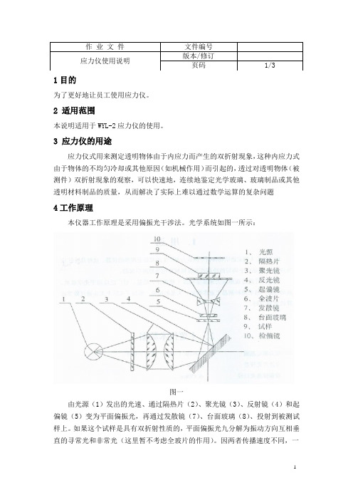
3 应力仪的用途应力仪式用来测定透明物体由于内应力而产生的双折射现象,这种内应力式由于物体的不均匀冷却或其他原因(如机械作用)而引起的,透过对透明物体(被测件)双折射现象的观察,可以快速地,连续地鉴定光学玻璃、玻璃制品或其他透明材料制品的质量,从而解决了实际上难以通过数学运算的复杂问题4工作原理本仪器工作原理是采用偏振光干涉法。
光学系统如图一所示:图一由光源(1)发出的光速、通过隔热片(2)、聚光镜(3)、反射镜(4)和起偏镜(5)变为平面偏振光,再通过发散镜(7)、台面玻璃(8)、投射到被测试样上。
如果这个试样是具有双折射性质的,平面偏振光九分解为振动方向互相垂直的寻常光和非常光(这里暂不考虑全玻片的作用)。
因两者传播速度不同,一个快,一个慢,透出试样后,就产生了一个光程差,最后通过检偏(10)将看到寻常光和非常光在同一个平面内产生的具有应力特征的干涉色图画。
干涉色决定于光程差的大小,光程差可由以下公式表示:R=(n1-n2)d,即R/d=(n1-n2),式中 R--光程差单位:豪微米(mu)d——被测试样的厚度单位:厘米(cm)(n1-n2)——寻常光与非常光的折射率差以上讲的系统(没有全玻片)有一个主要的缺点——仪器不灵敏。
就是当光程差在200-300mu以下时,干涉色只程现或明或暗的灰白色,光程差越小,灰白色越不易显示出来。
同时光程差有微小改变时,干涉色辨别不出变化,所以不能满足较小光程差的精确测量。
为此,在系统中放入一块附加光程差为560mu的全玻片(7),它起到灵敏片的作用,在视场中呈现一级紫红色。
在放入全拨片的一级紫红视场中引进被测试样,转动被测试样至最大亮度的位置,呈现一种干涩色。
再把被测试样转动90度,又呈现另一种颜色。
这是由于被测试样的光程差与全拨片的光程差相互迭加或相互减少的缘故。
被测试样的干涉色与光程差的关系如下表:根据上表,可由干涩色对紫红的偏离程度来决定被测试样光程差R的大小。
温捷手持光源 OLS-1315 说明书
Table of Contents1. Introduction ..................................................................... 1,21.1 Summary (1)1.2 Product Features (2)1.3 Technical Parameters (2)1.4 Main Applications (2)2. Operation Instructions (3)2.1 Powering On / Off (3)2.2 Backlight Operation (3)2.3 Control Panel Description (3)3. LCD Display (4)4. Maintenance (5)4.1 Light Emitter Cleaning (5)4.2 9V Battery Replacement (5)4.3 Calibration (6)5. Troubleshooting (6)6. Warranty Information (7)6.1 General Information (7)6.2 Included in Package (8)1. INTRODUCTIONAll the details including operation procedure, technical parameters as well as any others related can be found in this Operation Manual for the Toner OLS1315 Handheld Light Source as shown in the following diagram:1.1 SUMMARYThe OLS1315 Handheld Light Source is designed as a test device for measuring optical loss in systems, or to proof test installed optical plant when used with an optical power meter such as the Toner OPM1315. The light source provides two opticalwavelengths; these are 1310 and 1150 nm which are the standard singlemode wavelengths used in the CATV or RF industries.DC In Support Hanging Hole Optical Interface Sheath Body1.2 PRODUCT FEATURES• Providing two wavelength outputs• Optional CW, 270Hz, 1KHz, 2KHz modulation output • High stability of the output power• SC/PC adapter standard• Compact size and ergonomic design• Large LCD display and easy operation1.3 TECHNICAL PARAMETERS1.4 MAIN APPLICATIONS• Loss test of optical fiber link• Test Insertion loss of fiber jumpers or couplers• Installation and maintenance of fiber optic network2. OPERATION INSTRUCTIONS2.1 TURNING OLS1315 ON AND OFFPress the button to turn on the OLS1315. When the OLS1315 is turned on, the auto-off function is activated and the meter will shut off in 15 minutes. To deactivate this function momentarily press the a second time. Pressing the button when the light source isswitched on will toggle the auto off function on and off. An auto off icon will appear in the LCD display when this function is active.To turn the OLS1315 off press and hold the for about 3 to 5 seconds.2.2 BACKLIGHT OPERATIONWhen the light source is turned on, the back light will automatically activate. After a short period, the back light will switch off to conserve battery. Pressing the light button will turn the back light back on.2.3 CONTROL PANEL DESCRIPTIONPower ButtonUsed to turn meter on and off. Auto-off function operates through the useof this button.Wavelength ButtonThis button is used to select thewavelength, 1310 or 1550 nm.Modulation ButtonUsed to turn the different lightmodulaton modes on and off. When amodulation mode is selected, a dotappears in the LCD display.3. LCD DISPLAYOnce the power is turned on the following information will display on the LCD screen:(1) When the meter is on battery power, a battery level indicator willbe displayed in the lower left hand corner. When the indicator goes down to one bar, it is time to replace the battery.(2) When operating on the AC adapter, a plug symbol will be displayed above the battery indicator.(3) In the lower left hand corner of the LCD screen next to the battery indicator is the auto-off indicator. When the meter is turned on this is automatically activated and will turn the meter off after 15 minutes if the meter is not used.(4) In the main area of the screen (the center) the light wave frequency is displayed.(5) In the upper right part of the screen, the modulation mode in frequency is displayed in Hz.4. MAINTENANCEAs a highly sensitive electronic & optical instrument, the OLS1315 must be cared for properly so that it maintains accuracy.• Keep away from dust and dirt as this can damage the emitter• Only keep the meter in a dry clean place away from direct sunlight • Do not allow the light source to become overheated or extremely cold as this will affect the optical output• Keep away from vibration and do not drop the light source• Keep away from moisture, do not use in the rain or high humidity areas4.1 LIGHT EMITTER CLEANINGClean the emitter of the optical light source regularly.(1) Open the dust proof cap.(2) Screw off the adapter.(3) Use 2.5mm cotton swab with some anhydrous alcohol to clean the surface of the emitter gently.WARNING: Do not use anything hard or abrasive when cleaning the surface of the emitter. Do not drop since it may crack the emitter and cause the light source to not work.ATTENTION: Remember to cover the dust-cap when not using the meter.4.2 9V BATTERY REPLACEMENTOpen the back cover in order to remove and install the battery. Following is some useful information for better operation:(1) Install a new Alkaline 9V battery when the icon is displayed on the LCD screen.(2) Check the condition of the battery if the unit has been out of use for some time.4.3 CALIBRATIONThe OLS1315 is properly calibrated when shipped from the factory. The optical output level is set at -7 dBm ±0.5 dBm. If the unit is dropped or damaged, it should be returned for calibration or if you suspect erroneous readings.5. TROUBLESHOOTING6. WARRANTY PERIOD6.1 GENERAL INFORMATIONThe meter has a one year free from defects warranty period. This warranty is from the date of delivery and shall be guaranteed for any defects or faults caused by material quality or non-performance. Performance under normal operating conditions is fully guaranteed. Under this guarantee the company reserves the right to carry out any maintenance it deems necessary to restore the meter to optimal performance. If maintenance fails then the unit will be replaced. One free calibration is included on any warranty item. Note: Any damage that is caused by improper use of any kind will be charged for any maintenance or necessary replacement. The company will not be responsible for any accidental damage caused by the use of this meter. WARNING: If any of the following conditions takes place, the warranty shall be null and void.(1) If the warranty label is removed.(2) Case bolts (not mentioned in the manual) have been removed.(3) Improper use of the meter.(4) If the serial number has been altered or removed.(5) If the meter has been damaged.(6) If the meter has been exposed to moisture.6.2 INCLUDED IN PACKAGEOLS1315 Handheld Light Source ................................... 1 piece Operation Manual ........................................................... 1 piece 9V Battery ....................................................................... 1 piece Cotton Swab ................................................................... 1 piece SC/PC Optical Adapter ................................................... 1 piece OptionsAC Power Adapter model OPM-PSFC Optical Adapater model OLS-FCST Optical Adapater model OLS-STSC Optical Adapater model OLS-SC969 Horsham Road, Horsham, Pennsylvania 19044 USA800-523-5947 FAX 215-675 7543 • TEL 215 675 2053 email:*******************• 。
NEITZ纳宜慈5A手术放大镜使用说明
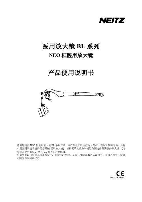
医用放大镜BL系列NEO框医用放大镜产品使用说明书感谢您购买NEO框医用放大镜BL系列产品,本产品是在以医疗为目的扩大观察对象物方面,具有小型医用眼镜功能的医疗器械(医用放大镜),请根据放大倍数和视野范围选择所满意的放大镜。
(请参照本说明书「1.2 型号BL系列的产品线」)。
为避免难以预料的不幸事故发生,在使用产品前,必须仔细阅读本产品说明书,并用心保管,做到可随时再次阅读状态。
目录(一)产品名称、型号、规格; (1)1.1产品名称: (1)1.2型号: (1)(二)备案、产品代理及售后服务信息; (1)2.1备案人名称: (1)2.2售后服务单位名称 (1)2.3中国代理人名称 (2)(三)生产企业信息: (2)3.1生产企业名称 (2)(四)备案凭证编号; (2)4.1备案凭证编号: (2)(五)产品技术要求编号; (2)5.1产品技术要求编号: (2)(六)产品性能、主要结构组成或者成分、适用范围; (2)6.1产品性能: (2)6.1.1 规格 (2)6.2包装内容的确认 (4)6.3主要结构组成或者成分 (5)6.3.1 带NEO框放大镜 (5)6.4适用范围 (6)6.5预期用途: (6)(七)禁忌症、注意事项、警示以及提示的内容; (7)7.1禁忌症: (7)7.2注意事项: (7)7.2.1 安全上的注意事项 (8)7.3警示及提示内容: (8)(八)事前准备及操作方法 (9)8.1调整鼻梁架的高度 (9)8.2镜筒角度及位置的调整 (9)8.3PD(瞳孔距离)的调整 (10)8.4观察距离的调整 (10)8.5系带的使用 (10)8.6防护镜片的安装与使用 (11)8.7放大镜的定制 (11)8.7.1减轻鼻梁的疼痛 (11)8.7.2 将鼻梁架贴近鼻梁 (11)8.7.3 调整弯曲合页的弯曲程度 (12)8.7.4 使用弯曲合页的止柱 (12)8.8查找故障原因 (13)(九)产品维护和保养方法、特殊储存、运输条件、方法; (14)9.1产品维护和保养方法: (14)9.1.1 清洁 (14)9.1.2 前额软垫的交换額 (14)9.2特殊储存条件、方法: (14)9.2.1 保管及运输 (14)9.2.2 保管及使用环境 (14)9.2.3 保管及运输 (14)(十)生产日期,使用期限或者失效日期; (15)10.1生产日期: (15)10.2使用期限: (15)(十一)配件清单,包括配件、附属品、损耗品更换周期以及更换方法的说明; (15)11.1可选配件: (15)11.2配件(附属品、损耗品)更换周期及更换方法: (15)(十二)医疗器械标签所用的图形、符号、缩写等内容的解释; (16)12.1包装标签所用图形(符号、缩写)的解释说明 (16)(十三)说明书的编制或者修订日期; (16)(十四)其他应当标注的内容; (16)14.1废弃 (16)(一)产品名称、型号、规格;1.1 产品名称:医用放大镜1.2 型号:BLS-1、BLS-2、BLS-3、BLD-3、BLP-4、BLP-6BL系列的产品线医用手术放大镜BL系列根据扩大倍数及视野范围,有几个种类。
Armasight Helios HD 热成像双眼镜说明书

© Armasight Inc. 2015This item is subject to export control under US Department of State. Contact Armasight Inc. for export information and regulations.The Armasight Helios HD is a family of Thermal Imaging bi-oculars with professional 75mm and 100mm Germanium (Ge) objective lenses. The bi-ocular design is better for sustained viewing periods and improved depth perception. The Helios HD product line is based on the latest FLIR Tau 2 VOx microbolometer core technology, and includes a variety of different models, each optimizing performance, price, and state-of-the-art capabilities for a variety of uses and missions. The Helios HD is a solid-state, uncooled, long-wave infrared, magnified, dedicated handheld thermal imager intended for day and nighttime missions.Helios HD Thermal Imaging bi-oculars are characterized by their simple but intuitive controls, functions, and features that are layered among direct button adjustments, direct combination button functions, and electronic menu selections. The Helios HD has the ability to record imagery and is equipped with video-out capabilities.The Helios HD Thermal Imaging bi-ocular represents one of the most sophisticated devices for long-range observation on the market today, due to its superior price-to-performance value.Thermal Imaging Bi-OcularHelios HDMultiple versions with optical magnifications ranging from 3x to 8x75mm and 100mm F1.0 Germanium objective lens options High-performance thermal imaging camera Lightweight and robust design Easy to operateManually adjustable eyepiece and objective lens Real-time displayDigitally controlled features:• Palette• Enhancement • Settings• Display Brightness• Electronic Magnification• User-Controlled Manual Non-Uniformity Correction/ Flat-Field Correction (UCMNUC/ FFC)Current operational state information display (battery status, active profile, palette setting)Wireless remote controlAnalog video input and output (NTSC/PAL) Powered by two standard CR123A batteries Power input capabilityDigital video recorder (optional)Serviceability under severe conditionsFilled with dry nitrogen to prevent internal fogging Water and fog-resistant Limited 3-year warranty10-year warranty on FLIR detectorPart No. ATAM000003Part No. ATAM000004Hd dVr digital VideoreCorderPart No. ATAM000005extended Battery PaCkPart No. ATAM000008Hard sHiPPing/Part No. ANHC000001oPerator manual*Default setting (may be altered at the customer’s request)** Rechargeable batteries with voltage 3.0V-3.7V can be used only in devices with serial number starting from 140885。
手持式光学仪器用户手册说明书

Product DescriptionThe ergonomically designed imager houses the imaging optics, detector, drive electronics, optical modulator,laser pointer and four standard or rechargeable AA size batteries. The system includes an optional pistol grip handle which holds virtually any ‘Pocket PC’or compatible ‘Palm’device as a combined processing, display unit and image storage device. As an alternative, the output of the imager can be displayed and processed in real time using a PC.OperationThe system is designed for either one or two handed operation. For one handed operation both the imager and the user provided ‘Pocket PC’/ ‘Palm’device can be attached to the handle to form a single integrated lightweight unit. For two handed operation the imager can be quickly detached from the handle leaving the ‘Pocket PC’/ ‘Palm’device attached for ease of operation. This latter configuration enables the imager to be pointed at awkward angles or used in confined places. Alternatively, instead of using a ‘Pocket PC’/ ‘Palm’device the imager can be linked to a PC or laptop computer using the RS232 serial cable supplied.Dec 2004IPU 40055 issue 3IRI 1011Universal Thermal ImagerThe IRI 1011 is a groundbreaking thermal imager product which brings the benefits of this versatile technology to the professional, the trades person and the non-specialist alike.The flexibility, ease of use and above all, the low cost of this product extend the normal application areas for thermal imaging from military and professional use, to wider use in industrial,commercial and domestic applications .Typical applications for the IRI 1011 include:Predictive and Preventative MaintenanceProcess MonitoringResearch and Development HV AC Troubleshooting Vehicle MaintenanceGeneral Industrial/Domestic......ABDWorld Leaders inArray-Based DetectorsThe IRI 1011 Universal Thermal ImagerPERFORMANCETemperature Measurement range:-10˚C to +300˚C Field of view (FOV):20˚x 20˚Spectral Response:8 to 14 micrometers Sensitivity:~0.3K @ 30˚C Displayed Image:96 x 96 pixels Detector:16 x 16 pixel array Frame rate:8HzIMAGE STORAGEUp to 1000 images per MB of SER POINTERA built in Class II laser is supplied to highlight the reference pixel.IMAGER POWER SUPPLYBattery:4 x AA type removable batteries.Lithium cells are recommended for operation at low temperatures.Operation time:Up to 8 hours.AC operation:AC adaptor, supplied.MECHANICALHousing: Impact Resistant Plastic.Dimensions: 120mm x 125mm x 80mm.Weight:< 600g not including ‘Pocket PC’/‘Palm’device and handle.Mounting: Handheld & Tripod mounting.IRI 1011 INCLUDESImager, software for ‘Pocket PC’, ‘Palm’& PC, 2m RS232 connection cable - imager to PC, user manual, AC power supply, carrying case.OPTIONA pistol grip handle for attaching imager and ‘Pocket PC’/ ‘Palm’Device for single handed operation.SPECIFICATIONABDWorld Leaders inArray-Based DetectorsInfraRed Integrated Systems Ltd, Towcester Mill,Towcester, Northants, NN12 6AD, UK Telephone:+44 (0) 1327 357824Fax:+44 (0) 1327 357825e-mail:***************.uk web site:Whilst IRISYS endeavour to ensure that all descriptions, weights, temperatures, dimensions and other statistics contained in this product information are correct, they are intended to give a general idea of the product only and IRISYS do not warrant their accuracy or accept liability for any reliance on them. IRISYS have a policy of continuous product improvement and reserve the right to change the specification of the products and descriptions in this data sheet. Prior to ordering products please check with IRISYS for current specification details. This product is protected by patents EP 0 853 237 B1 and US 6,239,433 B1. Other patents pending.All brands and product names are acknowledged and may be trademarks or registered trademarks of their respective holders.ENVIRONMENT Temp. operating range:-5˚C to +50˚C Humidity:10% to 90% non condensing Temp. storage range:-20˚C to +80˚CCE Mark (Europe):Complies with EMC directiveSETTINGS AND CONTROLS:User selectable sensitivity er selectable offset control (range).Auto adjust sensitivity/range.Display palettes: red/blue, green/blue and greyscale.‘Pocket PC’/ ‘Palm’device: two moveable temperature measurement cursors.PC: up to ten moveable temperature measurement cursors.User selectable emissivity values.User selectable image integration: 1 to 10 frames.Readout in ˚C, ˚F and K.Image snapshot.Image label.FEATURES - ‘POCKET PC’/ ‘PALM’SOFTWAREReal time image and temperature measurement display Multiple image storage and retrieval.Image browser.Battery Charge indicator‘Pocket PC’/ ‘Palm’device controlled by navigator button and touch sensitive screen controls. Reflected ambient temperature compensation.Temperature difference measurement.FEATURES - PC SOFTWAREMultiple image storage and retrieval.Time / Temperature display for up to ten user defined pixels.Save all 256 temperature values to Microsoft Excel.Copy & Paste images into other Microsoft applications.Reflected ambient temperature compensation.Real time image and temperature measurement displayCOMPUTER REQUIREMENTSPocket PC: Compatible with most ‘Pocket PC’devices running Microsoft ‘Pocket PC’2000, 2002 and 2003. e.g. HP iPAQ 2210, O2 XDA - (See IRISYS website for compatible Pocket PC’s). RS 232 to ‘Pocket PC’communication cable or CompactFlash RS 232 adaptor where applicable.Palm :Palm devices conforming to OS5 or higher, double density screen, 320 x 320 display resolution.eg. Palm Zire 71, Palm Tungsten T3. (See IRISYS website for compatible Palms)PC :IBM compatible PC with a minimum of:300MHz processor, MS Windows 2000 and XP (see IRISYS website for current list of operating systems supported). RS 232 serial port (115k Baud),16 bit colour graphics capability.........................CAUTIONCLASS II LASER PRODUCT635nm 0.9mW。
莱卡 d600 手持回弹硬度计 使用说明书
1065
Hardness test \ Mobile hardness testing devices
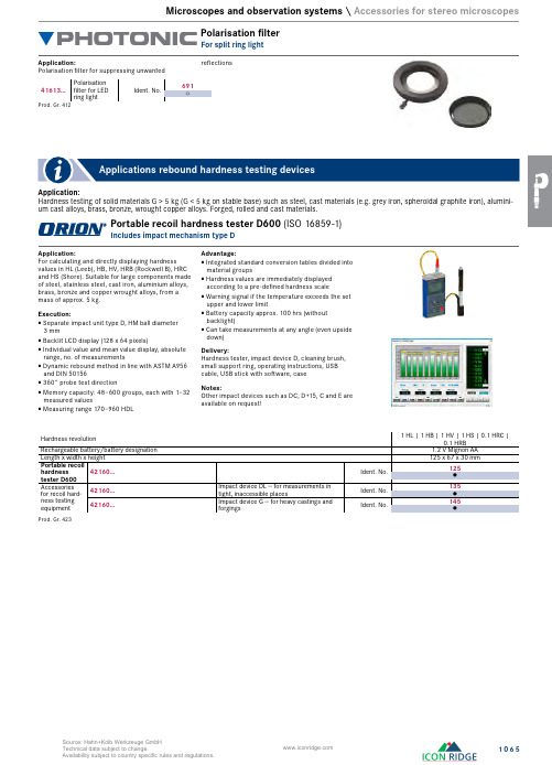
Mobile UCI UCI-3000 hardness tester (DIN 50159)
Separate manual sensor
Application: For determining and directly displaying hardness values in line with HB (Brinell), HV (Vickers), HRA (Rockwell A), HRB (Rockwell B), HRC (Rockwell), HS (Shore) on metallic materials. Ideal for fine-grain materials and alloys. The 50N standard sensor included in the scope of delivery allows e.g. hardness tests to be performed on nitride-hardened, case-hardened and high-frequency hardened parts, as well as coating tests (from 30 Micron with 10N probe). Suitable test attachments are optionally available for curved surfaces.
Execution: Includes separate 50 N manual probe, Vickers
136° indenter Manual probe 145 mm long Colour LCD display, backlit Individual, min./max and mean value display, with
玻璃应力测试仪操作指导书
1.目的规范操作员正确操作,减少事故发生率,确保产品符合要求。
2.范围:适用于检验和判定所有玻璃制品的应力。
3.操作步骤:3.1 应力仪外型图:3.2 打开电源开关:3.3把待测试样板放在台面的玻璃中心;3.4脚踩测试踏板,仪器通电测试;3.5旋转试样使视场中出现亮度最大的干涉色(没有应力的试样,不论怎样旋转,视场中始终是紫红色,有应力的试样旋转时会出现二种亮度最大的干涉色)4.判定标准:4.1 外观:无破损,裂纹,不允许有明显的玻璃条纹、模线、严重气泡、凹凸、铁锈等疵点;4.2 其他要求:被测物件夹扁处和压头处不能有明显的应力。
将物件置入干涉视场中,通过检偏镜观察物件整个表面,通过干涉色定性地判断退火质量。
如果放入光路后,视场的颜色基本不变(仍为紫色)或者只有轻微的变化(由暗红到紫色),说明退火质量良好,判定合格。
如果物件夹扁处或者压头处干涉色变化较大(绿色或者黄色),说明退火质量差,判定不合格。
5.注意事项:5.1 仪器应放置在通风干燥处。
偏振镜应防潮、防湿,以免变质褪色;5.2 起偏镜应防止受热过甚。
仪器连续使用时间不宜过长,如时间过长时,应用15-30分钟,等仪器内部降温或冷却后再用。
5.3 仪器工作台面是应力极小的玻璃制成,被测件放到工作台面上时,要小心轻放,不要擦伤台面玻璃。
如遇损坏,不能随便拿有应力的玻璃代用。
否则将会影响仪器的测量精度。
5.4 仪器应用柔软丝绒揩擦,用毕后,用塑料套套上。
6.设备点检:设备操作人员于正常工作日每天使用前,按《设备点检记录表》中各项要求进行点检。
7. 参考文件:《使用说明书》6. 相关记录表格:《设备点检记录表》。
PENTAX Medical i10系列端作用镜说明书

i10 Series EndoscopesAdvanced HD+ Visualization Enhanced Therapeutic Capabilities Improved Ergonomic ControlPENTAX Medical i10 Series EndoscopesEngineered and designed in partnership with healthcare professionals, the i10 SeriesEndoscopes provide high-quality, clinically relevant innovation with the intent to improve patient outcomes and reduce healthcare costs.The development of the i10 Series Endoscopes demonstrates PENTAX Medical’s commitment to the advancement of quality endoscopy with more therapeutic and specialized treatment capabilities.Raising the Standards of Clinical Acceptancei10 Series Endoscopes provide vivid HD+ imaging when combined with the state-of-the-art EPK-i7010 video processor.Note: Cart availability can vary by region. Cart shown does not reflect what is currently available in the United States.140° Effective Field of View (EFOV ™)CloseFocus ™ Examination Rangei10 Series Endoscopes with CloseFocus™ Examination Range allow physicians to get as close as 2mm from the mucosa for even greater resolving power and magnified visualization of the tissue and capillary networks.Additionally, this can be achieved automatically without the need for button activation.i10 Series Gastroscopes and Colonoscopes are designed with 140-degree Effective Field of View (EFOV ™) to optimize the endoscopic view with emphasis on full frame brightness,resolution, and minimized edge distortion. 140-degree EFOV yields greater magnification than other wider-angled endoscope designs for better recognition of even slight changes in mucosal surface within a field of view.i-SCAN is available with the EPK-i5010 and EPK-i7010 video processor. OE is available with the EPK-i7010 video processor. i-SCAN and OE image enhancement technologies are intended to be used as an optional adjunct following traditional white light endoscopy and is not intended to replace histopathological sampling. i-SCAN and OE are compatible with PENTAX Medical video gastrointestinal endoscopes.Advanced HD+ VisualizationHD+ High Definition imaging of a colon with ulcerative colitisSource: PENTAX Medical Global Image ArchiveHigh-definition endoscopy has become the standard in clinical care. An increasing number of endoscopists regularly make use of advanced technologies in their daily work. PENTAX Medical surpasses this standard with high definition CCDs and advanced video processing capabilities, for exceptional image clarity and detail resolution.OE Optical Enhancement filters the light source to provide focused wavelength bands that match the high efficiency absorption characteristics of hemoglobin. The resulting effect is a high visual contrast image between blood vessels and surrounding mucosa.PENTAX i-SCAN ™ mode 2 Source: PENTAX Medical Global Image Archivei-SCAN ™ Image EnhancementOE Optical EnhancementPENTAX i-SCAN ™ is a digital, post-processing image enhancement technology that provides the user with an enhanced view of the texture of the mucosal surface and the blood vessels. i-SCAN ™ has three different default settings: i-SCAN 1, i-SCAN 2, and i-SCAN 3, all of which can be simply accessed with the touch of a button.STAGE IIIHighRigidityModerate RigidityManeuverable RigiditySTAGE IISTAGE Ii-FLEX ™ Graduated Stiffness DesignTrueTorque ™ Insertion Tube DesignTrueTorque ™ Design offers a double coil of proprietary material precisely spaced to efficiently transfer torque from thephysician’s hand to the scope. Endoscopists typically utilize torque for precision steering through the colon, effectively managing loop formations, and introducing additional stiffness into the scope when advancing through the colon.Ergonomic Control Bodyi10 Series Endoscopes are equipped with a new and lightweight ergonomic control body design that provides improved comfort and minimized repetitive use fatigue. It is more compact and lighter — 30g lighter — than its 90i Series predecessor.* Innovatively designed angulation knobs and a six spoke R/L knob enable comfortable one-handed control, while repositioned buttons facilitate more intuitive access.i-FLEX Graduated Stiffness has three stages of Insertion Tube rigidity. During colonoscopy procedures, this allows for optimal maneuverability at the distal end when navigating the turns and bends of the colon, as well as increased stiffness in later stages to effectively transfer forward forces to advance the scope and minimize loop formations. This occurs without the need of additional mechanisms to add stiffness into the scope.ERCP T herapeutic CapabilitiesThe C.A.P . HD Duodenoscope is equipped with a 4.2mm instrument channel and redesigned elevator to enhance stability and address therapeutic needs related to ERCP .Note: i10 Series Colonoscopes are equipped with T rueT orque and Graduated Stiffness Design.* Note: the C.A.P . HD Duodenoscope control body is 40g lighter than that of its predecessor ED-3490TKLarger Instrument ChannelsThe Standard Gastroscope (EG29-i10) and Slim Standard Colonoscope (EC34-i10L) feature larger instrument channels — 3.2mm and 3.8mm respectively. This results in increased suction power and the opportunity to use larger accessories.Enhanced T herapeutic CapabilitiesImproved Ergonomic Control11.5 m m3.8 m m9.9 m m3.2 m mEC 34-i10LEG 29-i10Improved braking of elevator to maintain device angleAdvanced HD+ VisualizationAs trends in gastroenterology continue to drive innovations toward better patient care with the introduction of quality indicator measurements and tracking, image quality remains the common fundamental requirement for clinical acceptance and outcome relevance.The i10 Series Endoscopes deliver HD+ imaging to support physicians’ needs for improved detection and treatment ofGI diseases.Enhanced TherapeuticCapabilitiesWith increased pressure to reduce the costsof healthcare delivery, there is a growingdemand for interventional endoscopy ratherthan traditional surgical management of someGI disorders.The i10 Series Endoscopes are designedto bring more therapeutic capability tothe everyday endoscopic procedure. Newfeatures, such as larger instrument channels,are intended to expand the physician’s deviceoptions and support complex skill requirementsfor therapeutic treatment of GI disorders.Improved ErgonomicControlThe endoscopist’s daily procedure volumeis expanding and putting more strain on theendoscopist to do more cases and reduceprocedure times.The i10 Series Endoscopes are designed withthe latest ergonomic control body to bettersupport higher case volumes and overallcomfort in the physician’s hand. Additionally,PENTAX Medical Insertion Tubes are designedto balance efficiency, consistency, and patientcomfort in endoscopy.ColonoscopesDuodenoscopesi10 Series Endoscopes OverviewFOR USE IN THE UNITED STATES ONL Y .All referenced data was sourced from PENTAX Medical data on file.Copyright 2017. All Rights Reserved. All company and product names and marks contained within are federallyregistered trademarks, trademarks, or service marks of PENTAX Medical, Inc. Product specifications are subject to change without notice, and without any obligation on the part of the manufacturer.Raising the Standards of Clinical AcceptanceDemonstrating PENTAX Medical ’s commitment to the advancement of quality endoscopy with more therapeutic and specialized treatment capabilities, with the intent to improve patient outcomes and reduce healthcare costs.MK-618 Rev: EPENTAX MedicalAmericas Head Office 3 Paragon Drive Montvale, NJ 07645 Phone +1 800 431 5880 Fax +1 201 391 4189。
操作说明 - 手持带护身墙监测仪说明书
Vor Inbetriebnahme lesenDas Benutzerhandbuch ist von allen Personen, die für Gebrauch, Wartung oder Pflege des Produkts verantwortlich sind, sorgfältig zu lesen. Dieses Produkt funktioniert nur wie vorgesehen, wenn es nach den Anweisungen des Herstellers verwendet, gewartet und repariert wird.Sollte die Ladung der Batterie unter einen Ein- und Ausschalten des GerätsWechselstrom-adaptersteckerKabel vonAC-AdapterLadegerätLadevorgangsstatusLEDGaseintritts-Lade- und Kommunikationsanschlüssean der UnterseiteDrehknöpfezum AnziehenSonde (alle außer UltraRAE 3000+)halterungHerunterladen des BenutzerhandbuchsDas kostenlose Handheld PID Monitors Benutzerhandbuch ist das komplette Handbuch für den Betrieb und die Wartung aller Handheld PID-Monitore. Es muss von allen Personen, die für die Verwendung, Wartung oder Reparatur des Produkts verantwortlich sind oder sein werden, sorgfältig gelesen werden. Das Handheld PID Monitors Benutzerhandbuch können Sie hier herunterladen:https:///customer-care/resource-center/manualsOder verwenden Sie die Kamera Ihres Smartphones:Hygiene- und SuchmodiDas Gerät verfügt über zwei Hauptbetriebsarten: Hygienemodus: Nach dem Aufwärmen entnimmt dasGerät kontinuierlich Proben.Suchmodus: Nach dem Aufwärmen schaltet sich die Pumpe aus und es wird …Ready“ angezeigt. Dies ist idealfür die Kopfraumabtastung. Komponentenspezifische MessungenDer UltraRAE 3000+ kann neben der allgemeinen VOC-Messung auch komponentenspezifische Messungen durchführen. Dies erfordert die Verwendung eines RAE-Sep™ Separationsrohrs (Butadien oder Benzol) und den Einsatz des UltraRAE 3000+ im Röhrenmodus mit einer9,8eV-Lampe.Im Folgenden werden die 3 Schritte beschrieben, um spezifische Messungen durchzuführen:1. Starten Sie das Gerät und messen Sie in TVOC.2. Wenn der Messwert 0ppm beträgt, ist es nichterforderlich, in einen bestimmten Messmodus zuwechseln.3. Wenn der Messwert höher als die zulässigen Werte ist,nehmen Sie ein ungeöffnetes RAE-Sep™ Rohr undbrechen Sie beide Enden ab.4. Führen Sie das Rohr gemäß dem Pfeil ein.5. Lösen Sie den spezifischen Messmodus aus.Empfohlene VorgehensweisenWichtig! Testen Sie die Alarme vor der Verwendung. Wenn dieAlarme nicht alle funktionieren, lesen Sie die Anweisungen imBenutzerhandbuch. Verwenden Sie das Gerät nicht ohnefunktionstüchtige Alarme.Überprüfen Sie die Pumpe: Starten Sie das Gerät undblockieren Sie die Eintrittsöffnung, um sicherzustellen, dassder Alarm ausgelöst wird und Geräusche und Lichterfunktionieren. Wenn die Gaseintrittsöffnung verstopft ist, diePumpe jedoch nicht abschaltet, oder wenn die Pumpe auchbei geringfügigen Verstopfungen am Gaseinlass schonabschaltet, dann ist möglicherweise der Schwellenwert derPumpe zu hoch oder zu niedrig eingestellt. Lesen Sie imBenutzerhandbuch nach, wie Sie dies anpassen können.Überprüfen Sie, ob das Gerät auf Feuchtigkeit reagiert:Legen Sie Ihre Hand 10 bis 20 Sekunden über dieEintrittsöffnung (blockieren Sie dabei nicht den Durchfluss).Wenn das Gerät >2 ppm anzeigt oder wenn der ppbRAE3000+ >500 ppb anzeigt, muss der Sensor gereinigt werden.Immer externen Filter verwendenBevor Sie das Gerät in Betrieb nehmen, befestigen Sie einenexternen Filter an der Probenahmesonde, der verhindert,dass Feuchtigkeit, Schmutz und andere möglicherweiseverstopfende Materialien in die Sonde eindringen, der einenvorzeitigen Ausfall der Pumpe verhindert und den Sensorund die Lampe sauber hält.SondeFilterRev. A P/N: 059-4027-000November 2018。
应力仪中文说明书
1 Exicor GEN5 双折射测量系统概况 .............................................................................. 13 Exicor GEN5 双折射测量系统描述......................................................................... 13 大致的尺寸.............................................................................................................. 16
4 软件用户界面 ............................................................................................................... 29 概述图形用户界面 ................................................................................................... 29 菜单栏................................................................................................................................ 31
- 1、下载文档前请自行甄别文档内容的完整性,平台不提供额外的编辑、内容补充、找答案等附加服务。
- 2、"仅部分预览"的文档,不可在线预览部分如存在完整性等问题,可反馈申请退款(可完整预览的文档不适用该条件!)。
- 3、如文档侵犯您的权益,请联系客服反馈,我们会尽快为您处理(人工客服工作时间:9:00-18:30)。
北京绿野创能机电设备有限公司
010-567418620
HD-1705玻璃制品应力检查仪使用说明书
一、用途
玻璃制品应力检查仪是应用偏振光干涉原理检查玻璃内应力或晶体双折射效应的仪器。
由于仪器备有灵敏色片,因此本仪器可以根据偏振场中的干涉色序,定性或半定量的测量玻璃的内应力。
因此本仪器适合光学仪器厂、玻璃厂、玻璃制品长作测量光学玻璃、玻璃制品及其他光学材料的应力。
二、主要技术参数:
1、偏振片通光口径:150毫米
2、眼镜偏振片对角尺寸:48毫米
三、检测原理:
在正交的起偏镜与检偏镜之间放入双折射物质,视场中便会出现干涉色,一定的干涉色对应于一定的双折射光程差,其关系如表所示。
位于起偏镜与检偏镜之间的全波长是用高分子材料做成的薄片,其双折射光程差﹠为565毫微米,由表知道视场中的干涉色为紫红色。
如果在正交偏振镜之间除了全波片而外加试件,则二者的组合光程差,将大于或小于565毫微米,干涉色也相应地发生变化,根据干涉色查表得到组合光程差的数值。
有应力的玻璃试件也是双折射物质,将这样的试件放入应力检查仪光路中,也会引起干涉色的变化,就像上面所见附加试件放入光路的情形一样,只是由于玻璃试件应力不是均匀分布的,因此试件各点双折射光程差也不一样,结果视场中各点的干涉色变化情况也不相同。
四、测量方法:
将试件放放干涉视场中,通过检偏镜观察整个试件表面,根据干涉色定性地判断玻璃试件应力大小(玻璃试件退火质量)如试件颜色基本不变或者只有轻微的变化(由紫红到紫色),说明退火质量良好;如果试件某些部件上干涉色变化较大(例如出现绿色或黄色),说明退火质量较差。
然后参考国家标准《玻璃瓶罐内应力检验方法》。
五、仪器维护与使用注意事项:
1、仪器使用环境应干燥,注意防尘及防腐蚀性气体。
使用后收好。
2、仪器中各光学件表面不得用手摸,如有尘土可用毛刷轻轻拂去。
附表:偏振镜正交时干涉色与光程差的关系(毫微米)
*色序随光程差增大而升高,例如由紫红变蓝色时,色序升高;由紫红变红色,色序降低,参看表。