猫瘟热病毒抗原cdv-agelisa试剂盒使用说明书.doc
抗原检测试剂盒使用说明
抗原检测试剂盒使用说明
抗原检测试剂盒使用说明
一、适用范围:
本试剂盒专为检测抗原提供的试剂,用于实验室研究、样本检测和抗原检测项目的结构及性能分析。
二、原理:
本试剂盒采用免疫双抗血清方法,即采用抗原抗体免疫反应的原理,检测抗原的浓度,以间接检测抗原浓度的方法分辨抗原的存在与否。
三、主要材料及试剂:
1、主要试剂:免疫双抗血清
2、其他试剂:抗原检测试剂盒,包括抗原抗体、标准抗原、测定液、冰冻试剂和培养基等;
3、试剂设备:试管、温度计、pH计、洗涤器、滴定管、冰箱、凝固仪等。
四、使用方法:
1、抗原检测试剂盒使用前,请先清洗所有试剂设备,然后再按说明书上所示,将各试剂稀释或配制至实验室评定值;
2、抗原检测试剂中的抗原抗体( antibody )和抗原将按照相应的比例混合,充分混合后,转移至试管内;
3、在室温下反应30分钟,并将反应液放入凝固仪中冷凝20分钟,以便开始检测;
4、按说明书上所述,根据检测到的抗原的数量,进行抗原检测。
五、注意事项:
1、请勿将未使用的试剂放入试剂室;
2、每次实验完毕,务必及时清洗所有试剂设备;
3、使用时请穿实验衣和手套,并注意避免接触皮肤;
4、试剂使用完毕,请及时将其妥善存放;
5、请勿将废弃物倒入下水口,应妥善处理;
6、请勿将试剂盒接触酸性物质或温度过高的物质;
7、请勿将检测液接触空气,实验过程中应加以封闭。
犬瘟热金标快速检测试纸条(CDV-Ag)

犬瘟热金标快速检测试纸条(CDV-Ag)简介犬瘟热,俗称狗瘟,是由犬瘟热病毒引起的一种烈性传染病。
临床特征为双相热型、消化道、呼吸道炎症,眼、鼻流出脓样分泌物,少数病例可发生非化脓性脑炎。
多发生于3-6个月龄幼犬,青年犬也有感染,潜伏期为3-6天。
病毒单独感染症状轻微,若继发细菌感染症状加重,病死率较高。
检测原理本试纸为免疫层析法对病毒进行双抗体夹心检测。
检测样品为眼部及结膜分泌物,鼻液,唾液。
产品组分1.20份CDV Ag 试纸卡2.20份样品稀释液3.20份消毒棉签4.一份产品说明书保存及有效期限室温保存,不可冷冻。
生产日期起18个月有效。
样品收集及准备1、用生理盐水沾湿的棉签收集狗眼部及结膜分泌物,鼻液,唾液;2、采集样品时注意多部位同时收集新鲜及有效样品,充分在试管中搅拌稀释;;3、静置5分钟,用一次性滴管取上清液;4、样品一般须当即进行检测,否则应冷藏保存,超过24小时的,应该冷冻保存。
检测步骤1、试纸条恢复室温;2、取出试纸,开封后平放在桌面,从滴管中缓慢而准确地逐滴加入3-5滴混合液。
3、加样品液后,红色的液体从靠样品孔的观察窗边缘涌出,朝另一方向流动。
4、5-10分钟后判断结果。
结果判定阳性(+):当位置C显示出红色线条,而位置T同时显示出红色线条时,判为阳性。
阴性(-):当位置C显示出红色线条,而位置T不显色时,判为阴性。
无效:当位置C不显示出红色线条,则无论位置T显示出红色线条与否,该试剂盒判为无效。
阳性阴性注意事项1.仅用作体外诊断。
2.注意样品具有潜在传染性,注意防止交叉感染。
3.该试纸及配套试管、棉签均为一次性产品,不可交叉及重复使用。
4.包装袋破损或产品过期请勿使用。
Canine Distemper Virus Rapid Test CardIntroduceCanine distemper is caused by the canine distemper virus (CDV).It is a contagious, incurable, often fatal, multisystemic viral disease that affects the respiratory, gastrointestinal, and central nervous systems. Infected dogs shed the virus through bodily secretions and excretions, especially respiratory secretions. Young puppies between 3 and 6 months old are most susceptible to infection and disease and are more likely to die than infected adults. PrincipleThe test card is based on immunochromatography of double-antibody sandwich. Testing sample is the eye and conjunctival secretions,nasal fluid.Content1 CDV diagnostic Card: 20 pcs2 Dilution Buffer: 20 pcs3 Swab: 20 pcs4 Instruction: 1 insertProcedure1. Recover the dipstick to room temperature. remove as many of the test devices from the foil pouches as needed for testing.ing the disposable sample collection swab take a portion of secretion of eye mucus,nasal fluid.insert the collection swab into specimen tube containing the sample diluent.3.Mix the swab until the sample has been dissolved into the sample diluent.Discard the swab and repeat procedure for each sample. Adding 4 drops containing the sample diluent into the sample-well on the card.4. Read results after 10 minutes.ResultsPositive:The test zone and control zone have a pink/purple band visible.This indicates that the presence of canine distemper virus antigens.Negative:Only the control zone has a pink/purple band visible.The sample do not contains canine distemper virus antigens.Void:No color reaction on C line.Positive NegativePrecautions− Handle all biological materials as though capable of transmitting CDV.− Do not eat, drink, smoke or prepare foods, or apply cosmetics within the designated work area.− Do not use components past expiration date.− Optimal results will be obtained by strict adherence to this protocol. Careful washing and pipetting throughout this procedure are necessary to maintain precision and accuracy.− Do not use components past expiration date.− Only for veterinary use.The entire risk as to the performance of these products is assumed by the purchaser. We shall not be liable for indirect,special or consequential damages of any kind resulting from use of the products。
elisa试剂盒实验说明书

PRRSV N蛋白抗体检测间接ELISA方法的建立1 材料:1.1 蛋白、血清、酶标二抗原核表达PRRSV N蛋白;标准阳性血清和阴性血清,待检血清;自制HRP标记兔抗猪IgG;1.2 主要试剂和溶液包被液(50 mmol/L pH9.6的碳酸盐缓冲液)Na2CO3 1.59 gNaHCO3 2.93 g准确称取后,溶于950 mL的蒸馏水中,调pH值为9.6,定容到1 L;PBS-T洗涤液1000 mL 10 mmol/L pH7.4的PBS中加入0.5 mL Tween-20;封闭液和血清稀释液含10%小牛血清的PBS-T缓冲液,10%马血清的PBS-T缓冲液,含5%BSA的PBS-T缓冲液,含10%BSA的PBS-T缓冲液,含5%脱脂奶的PBS-T缓冲液;底物溶液100 mmol/L的柠檬酸溶液(21 g柠檬酸C6H6O7•H2O溶于去离子水,定容至1 L)24.3 mL,200 mmol/L Na2HPO4•12H2O(71.6 g Na2HPO4•12H2O溶于去离子水,定容至1 L)25.7 mL混匀,加入50 mg的四甲基联苯胺(TMB),临用前加入50 μL的30% H2O2;终止液(2 mol/L H2SO4)每200 mL终止液,由蒸馏水177.8 mL和浓硫酸22.2 mL 混和而成。
1.3 主要仪器设备电热恒温培养箱;4℃冰箱;酶标仪。
2 步骤:2.1抗原包被浓度和二抗稀释度的确定将原核表达的N蛋白分别作2.0 μg/mL,1.0 μg/mL,0.5 μg/mL,0.25 μg/mL,0.1μg/mL,0.05 μg/mL连续稀释6个稀释度,每个稀释度重复两孔,100 μL/孔,4℃包被酶标反应板过夜。
将自制HRP标记的兔抗猪二抗分别做1:50、1:100、1:200和1:400一系列倍比稀释,南京金益柏生物技术有限公司Tel:025-********Fax*************每个稀释度重复两孔,100 μL/孔,组成方阵确定重组蛋白的最佳包被浓度和二抗稀释度。
试剂盒使用说明书
大肠杆菌菌体蛋白残留量(E.coli DH-5α)酶联免疫分析(ELISA)试剂盒使用说明书本试剂盒仅供研究使用。
1使用目的:本试剂盒用于饲料、鱼、虾和肉类组织(如鸡、牛肉和猪肉),药物、血清和尿样中大肠杆菌菌体蛋白残留量(E.coli DH-5α)残留的定量检测。
2实验原理本试剂盒采用竞争ELISA方法,在微孔板包被有大肠杆菌菌体蛋白残留量(E.coli DH-5α)偶联抗原,加入大肠杆菌菌体蛋白残留量(E.coli DH-5α)标准品或样品,游离大肠杆菌菌体蛋白残留量(E.coli DH-5α)与微孔条上预包被的大肠杆菌菌体蛋白残留量(E.coli DH-5α)偶联抗原互相竞争抗大肠杆菌菌体蛋白残留量(E.coli DH-5α)抗体酶标记物,用TMB底物显色,加入终止液后颜色由蓝色变为黄色,用酶标仪在450nm波长下进行检测,吸光值与样品中大肠杆菌菌体蛋白残留量(E.coli DH-5α)含量成反比,通过标准曲线计算样品中大肠杆菌菌体蛋白残留量(E.coli DH-5α)的含量。
3试剂盒组成3.1预包被的大肠杆菌菌体蛋白残留量(E.coli DH-5α)偶联抗原的可拆酶标板:1块(12孔×8条)。
3.2大肠杆菌菌体蛋白残留量(E.coli DH-5α)标准品:6瓶(1ml/瓶),含量分别是:0ug/ml, 0.1ug/ml,0.3ug/ml,0.9ug/ml,2.7ug/ml,8.1ug/ml3.3抗大肠杆菌菌体蛋白残留量(E.coli DH-5α)抗体酶结合物:1瓶(6ml)。
3.4显色液A:1瓶(6ml)。
3.5显色液B:1瓶(6ml)。
3.6终止液:1瓶(6ml),2M硫酸。
3.7样本稀释液:1瓶(10×,6ml),用于样品稀释用。
3.8浓缩洗涤液:1瓶(20×,20ml),用于洗板。
3.9说明书一份。
4需要而未提供的材料4.1设备4.1.1波长450nm酶标仪。
CG,甘胆酸,CG,ELISA试剂盒说明书
CG,甘胆酸,CG,ELISA试剂盒说明书1)用弃的试剂盒及样本都应按污染物妥善处理。
试剂盒内终止液有腐蚀性,须小心使用。
2)孵育时,推荐使用水浴箱。
如无水浴箱,可取密封湿盒放入37℃温箱内使用。
3)瓶盖每次使用后要盖紧,各瓶盖之间不可混用。
不同批号CG,甘胆酸,CG,ELISA试剂盒说明书是以免疫学反应为基础,将抗原、牽9体的特异性反应与酶对底物的高效催化作用相结合起来的一种敏感性很高的试验技术。
由于抗原、抗体的反应在一种固相载体──聚苯乙烯微量滴定板的孔中进行,每加入一种试剂孵育后,可通过洗涤除去多余的游离反应物,从而保证试验结果的特异性与稳定性。
CG,甘胆酸,CG,ELISA试剂盒说明书种属:人、大小鼠、豚鼠、兔子、猪、犬、猴、马、牛、羊;标本:血清、血浆、细胞上清液、尿液、体液、灌洗液、脑脊髓、心房水、胸房水等等;公司主营业务:试剂盒:免疫组化试剂盒、ELISA试剂盒、分子生物学试剂盒。
细胞:原装进口细胞、国产肿瘤细胞、国产正常细胞。
耗材:细胞培养耗材、普通实验耗材。
生化试剂:原装进口生化试剂、进口分装生化试剂、国产生化试剂。
抗体:免疫组化、免疫荧光、流式细胞检测、免疫细胞化学。
服务:荧光定量PCR、测序、多肽合成、引物合成。
甘胆酸从冷藏环境中取出(2-8℃),然后放置室温平衡后可以使用。
1. 试剂盒提供6管标准品,每管已标定浓度,并且冻干。
实验前在每个标准品管中加入0.5mL样本稀释液,盖好后静置10分钟以上,然后反复颠倒/搓动助其溶解,使其恢复为每个标准品管身标注的浓度。
2. 20×洗涤缓冲液的稀释:蒸馏水按1:20稀释,即1份20×洗涤缓冲液加19份蒸馏水。
犬猫快速检测试纸的注意事项及说明书

现有试纸板包括犬猫两种
犬:CPV犬细小病毒,CCV犬冠状病毒,CDV犬瘟热病毒,C.Brucella Ab布鲁氏杆菌,SNAP CPL 犬胰腺炎
猫:FPV猫瘟,FIV Ab猫艾滋,FeLV Ag猫白血病,SNAP FPL猫胰腺炎
通用板:TOXO.Ag弓形虫
试纸的一些注意事项:
1、不同批号的试纸不能混用
2、采料不宜过多,棉签头½-⅔最好
3、妥善处理用过的试纸板,避免交叉传染
4、严格按照说明书执行
5、在规定时间内判读结果(记录时间)
6、专人操作(一人负责到底)
7、加样不宜过快(一滴滴加入观察液体行进速度)(加样保持滴管垂直于试纸板)
8、加样不宜过粘稠,吸取稀释液的上清液
9、试纸有可能产生误差,诊断需结合临床化验诊断
10、遵从医嘱
11、对于加液后液面不前进问题,可用吸管或细针在加样窗口
SNAP® cPL™犬胰腺炎快速检测试剂使用简单:
得检测结果颜色模糊,需进行重复检测。
无颜色出现——如果参考斑点无颜色,需进行重复检测。
犬瘟热病毒(CDV)核酸检测试剂盒(RT-PCR-荧光探针法)产品技术要求标准2023版
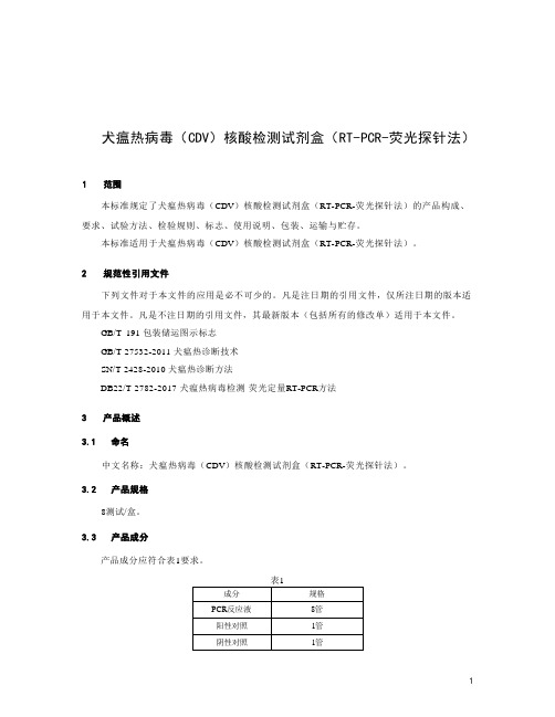
犬瘟热病毒(CDV)核酸检测试剂盒(RT-PCR-荧光探针法)本标准规定了犬瘟热病毒(CDV)核酸检测试剂盒(RT-PCR-荧光探针法)的产品构成、要求、试验方法、检验规则、标志、使用说明、包装、运输与贮存。
本标准适用于犬瘟热病毒(CDV)核酸检测试剂盒(RT-PCR-荧光探针法)。
下列文件对于本文件的应用是必不可少的。
凡是注日期的引用文件,仅所注日期的版本适用于本文件。
凡是不注日期的引用文件,其最新版本(包括所有的修改单)适用于本文件。
GB/T 191 包装储运图示标志GB/T 27532-2011 犬瘟热诊断技术SN/T 2428-2010 犬瘟热诊断方法DB22/T 2782-2017 犬瘟热病毒检测荧光定量RT-PCR方法中文名称:犬瘟热病毒(CDV)核酸检测试剂盒(RT-PCR-荧光探针法)。
8测试/盒。
产品成分应符合表1要求。
4.1.1 试剂盒各组分齐全,无破损,标签和说明书字迹清晰,内容完整。
4.1.2 试剂无明显浑浊或沉淀。
检测企业阳性参考品P1~P3 ,结果应均为阳性。
检测企业阴性参考品N ,结果应为阴性。
检测企业最低检出限参考品L ,重复10次,结果应均为阳性。
检测企业精密度参考品R1/R2各重复10次,结果应均为阳性,且FAM通道Ct值的变异系数(CV ,%)应不高于5.0%。
检测企业线性参考品(S1~S5)结果均为阳性,线性相关系数| r | >0.980。
经人员目测检测,结果应符合4. 1的要求。
取3份企业阳性参考品(P1~P3),按照犬瘟热病毒(CDV)核酸检测试剂盒(RT-PCR-荧光探针法)说明书进行检测,结果应符合4.2的要求。
取企业阴性参考品N ,按照犬瘟热病毒(CDV)核酸检测试剂盒(RT-PCR-荧光探针法)说明书进行检测,结果均为阴性,应符合4.3的要求。
取企业最低检出限参考品L ,按照犬瘟热病毒(CDV)核酸检测试剂盒(RT-PCR-荧光探针法)说明书进行检测,重复10次,结果应符合4.4的要求。
抗原试剂盒的用法
抗原试剂盒的用法1抗原试剂盒的定义抗原试剂盒又称ELISA试剂盒,是利用酶联免疫吸附反应(ELISA)检测,可用于检测特定抗原物质(如抗体、蛋白、抗原、细胞因子或其它外源能够与抗体结合的物质)在样品中的相互作用。
2各种抗原试剂盒抗原试剂盒可以分为常规ELISA试剂盒、全自动ELISA试剂盒、多因子ELISA试剂盒和金属ELISA试剂盒几种。
常规ELISA试剂盒是反应回路最简单的试剂盒,通常包含一种抗体或抗原、几种杂交液、一种酯化底物,用于检测特定抗原或抗体,能简便、快捷地进行单一或相互间因子检测,具有较多的应用。
全自动ELISA试剂盒是在常规ELISA试剂盒上完善后出现的,添加了稳定和吸附等因素,并采用微量无毒技术,该试剂盒的特点是环境友好、方便快捷。
多因子ELISA试剂盒是在全自动ELISA试剂盒的基础上添加多个组分,具有检测多种抗原物质的能力。
金属ELISA试剂盒是在金属氧化物的显色底物和稳定抗体反应的基础上发展起来的,可用于检测有机金属污染物。
3抗原试剂盒的用法(1)准备检测样品:首先将抽取后的样品稀释至稳定液,经过多次混匀,将水溶液中的Tween20去除。
(2)添加抗原:将抗原溶液加入模拟ELISA反应盒,添加抗原可通过改变抗原浓度来优化检测效率。
(3)添加抗体:将抗体溶液加入模拟ELISA反应盒,添加抗体可以提高检测的灵敏度。
(4)添加抗体复合物:将抗体复合物溶液加入模拟ELISA反应盒,抗体复合物是由抗原与抗体结合的复合物。
(5)调整混合液:将模拟ELISA反应盒中的混合液调节至pH值6.5-7.5范围内,以保证ELISA反应过程顺利进行。
(6)激发ELISA反应:把前述模拟ELISA反应混合液加入反应盒,使用适宜的温度、时间激发ELISA反应,在该过程中,抗体与抗原复合物会形成交联反应。
(7)处理ELISA反应:将ELISA反应处理后的混合液中的抗体复合物回收,然后采用恒定温度、时间处理ELISA反应,完成ELISA反应最后一步。
C反应蛋白ELISA试剂盒说明书
C反应蛋白ELISA试剂盒说明书C反应蛋白ELISA试剂盒说明书产品规格:96T/48T。
本产品只用于科研实验。
C反应蛋白ELISA试剂盒说明书Elisa试剂盒标本要求:1.标本采集后尽早进行提取,提取按相关文献进行,提取后应尽快进行实验。
若不能马上进行试验,可将标本放于-20℃保存,但应避免反复冻融2.不能检测含NaN3 的样品,因NaN3 抑制辣根过氧化物酶的(HRP)活性。
Elisa kit:3.1 samples were collected as soon as possible after the extraction, extraction by the relevant literature, as soon as possible after the extraction experiment. If you can not4.Test immediately, can put the sample at -20 deg.c for preservation, but should avoid repeated freezing and thawing5.2 could not detect the NaN3 containing samples, because NaN3 inhibited the activity of horseradish peroxidase (HRP).C反应蛋白ELISA试剂盒说明书Calculation:To the standard concentration as the abscissa, OD value as the ordinate, draw the standard curve on coordinate paper, according to the OD value of samples from the standard curve found corresponding concentration; multiplied by the dilution multiple; or with the standard concentration and the OD value calculated straight line regression equation of the standard curve, the sample OD value in the equation, calculate the sample concentration, multiplied by the dilution factor is the actual concentration of the sample.Experimental principle:This kit is used to measure the level of the chorionic gonadotropin in the sample of small and medium rats by double antibody sandwich method. With purified mouse chorionic gonadotropin? Antibody coated microtiter plate, made of solid phase antibody, to package is the micropore of monoclonal antibody in turn added to collagenase, and then with HRP labeled collagenase? Antibody binding, forming antibody, antigen and enzyme labeled antibody complexes, after thorough washing, TMB substrate added color. TMB under the catalysis of the HRP enzyme into blue, and under the action of acid into the final yellow. The depth of color and the collagenase in the sample were positively correlated. Enzyme standard instrument in 450 nm wavelength was used to measure the absorbance (OD) and the standard curve calculation of a small sample in chorionic gonadotropin concentrations.C反应蛋白ELISA试剂盒说明书上海乔羽生物有限公司专业的提供elisa试剂盒,人elisa试剂盒,猪elisa试剂盒,大鼠elisa试剂盒,小鼠elisa试剂盒,羊elisa试剂盒,植物elisa试剂盒等等,更多优惠,来电咨询!谢谢!C反应蛋白ELISA试剂盒说明书Matters needing attention:1 kit from the cold storage environment should be removed at room temperature for 15-30 minutes after the use of the enzyme labeled package is not used up after the plate, the plate should be stored in a sealed bag.2 washing buffer will crystallization, heated the water solubilization dilution, washing does not affect the results.3 each step sample should be used, and often to check its accuracy, in order to avoid the test error. A sample within 5 mins, ifthenumberofsampleismuch, recommend to use volley like.4 please make the standard curve at the same time, it is better to do the hole. Such as samples to be measured matter content is too high (the sample od high standard product the first hole hole OD), please first sample dilution multiples (n times) were measured and calculated please then multiplied by the total dilution ratio (n * * 5).5 closure plate membrane only a one-time use, to avoid cross contamination.6 substrate please avoid light preservation.7 in strict accordance with the instructions of the operation, the results of the test must be determined in accordance with the enzyme standard instrument readings.8 all samples, washing liquid and all kinds of waste should be treated as infectious agents.9 different batches of this reagent can not be mixed.10 if there is a difference between the specification and the English specification. C反应蛋白ELISA试剂盒说明书友情提醒:公司经营的产品均为科研实验用,不可用于临床应用。
犬瘟热病毒抗原(CDV)金标快速检测卡使用说明书

犬瘟热病毒抗原(CDV) 金标快速检测卡使用说明书犬瘟热是由犬瘟热病毒(CDV)引起犬的一种高度接触传染性传染病,早期表现双相热、急性鼻卡他以及随后的支气管炎、卡他性肺炎、严重的胃肠炎和神经症状为特征,少数病犬的鼻和足垫可发生角化过度。
病毒主要从鼻咽和呼吸道散布到支气管淋巴结和扁桃体进行原发性增殖,引起病毒血症,病毒分布到全身淋巴器官、骨髓和上皮结构的固有膜。
约50%的犬于感染后迅速产生抗体,但不出现临诊症状。
另一半的感染犬体内病毒继续增殖,广泛侵害多个系统的上皮细胞,而呈现特征性的症状。
病毒通过脑膜巨噬细胞扩散到脑,并在3~4周时出现神经症状。
有些感染犬仅有神经症状,这是由于病毒在抗体达到保护水平之前,已扩散到脑所致。
研究证明,在犬瘟热的临诊表现中,继发性细菌感染也起有很重要的作用。
[原理]本产品采用免疫层析金标双抗体夹心检测技术,可迅速,准确,简单地定性检测出犬类眼部分泌物(或结膜)、鼻涕、唾液、生殖器分泌物、肛门(粪便)或血清中的犬瘟热病毒抗原(CDV)。
所需验体量少,时间短(仅需10-15分钟)。
在检测卡判断窗中有结果判断线“T线”和检测控制线“C线”。
在未添加样品前,“T线”和“C线”都不显示。
“C线”被用于确认样品在检测卡中是否正常展开。
“T线”被用于确认样品中是否含有CDV抗原。
1)当“C线”显示时(一条紫红色线),证明样品在检测卡中正常展开了,可以进行“T线”判定。
当“C线”不显示时,证明样品在检测卡中没有正常展开,检测结果无效。
2)在“C线”和“T线”同时显示时(二条紫红色线),证明样品中含有CDV抗原。
当“C 线”显示而“T线”不显示时(一条紫红色线),证明样品中不含有CDV抗原。
[包装规格]1 10份CDV抗原快速检测卡、一次性采样棉签和一次性滴管2 10份装有反应缓冲液的样品收集管3 1 份使用说明书。
[功能,效果]用于犬类眼部分泌物(或结膜),鼻涕,唾液,生殖器分泌物及肛门(粪便)、血清中CDV抗原的验出。
ELISA检测试剂盒使用指南
ELISA检测试剂盒使用指南ELISA(酶联免疫吸附试验)是一种常用的免疫学实验方法,用于检测病原体、抗体或其他分子的存在和浓度。
它具有高灵敏度、高特异性、易操作和较低成本的优势,被广泛应用于生物医学研究、临床诊断和免疫学领域。
本篇文章将介绍ELISA检测试剂盒的使用指南,包括实验准备、试剂的使用步骤和结果解读。
一、实验准备1.阅读检测项目的说明书:在使用试剂盒前,仔细阅读说明书,了解试剂的使用方法、灵敏度和特异性等关键信息。
2.样本准备:根据实验要求,准备样本。
如果需要检测血清中的抗体水平,可以采集血液样本,离心分离血清;如果需要检测细胞培养上清中的分子,将上清收集,并使其清晰,避免细胞或杂质的污染。
3.样本预处理:根据实验需求,对样本进行必要的预处理。
例如,可以通过加热、稀释、酶解等方式来处理样本,以改变样本组分和浓度。
4.准备品质控制样品:制备阳性和阴性控制样品,用于评估试剂盒和实验操作的稳定性和准确性。
5.实验器材准备:准备所需实验器材,如酶标板、移液器、微孔板洗涤仪等。
确保器材的干净和完整,并按照说明书要求对其进行预处理。
二、试剂使用步骤1.试剂准备:根据说明书要求,将试剂从冰箱中取出,并在室温下放置一段时间使其恢复到室温。
确保试剂瓶盖紧闭,并避免暴露在直接阳光下。
2.实验操作:按照说明书的要求,将试剂加入到酶标板中,并根据实验设计进行标准曲线的设置。
标准曲线用于测量未知样品的数量,并计算出浓度。
3.孵育:根据试剂盒的要求,将酶标板放入孵育箱中进行孵育。
孵育温度和时间应根据实验要求进行调整。
4.洗涤:使用洗涤缓冲液对酶标板上的不特异性结合物进行洗涤。
洗涤过程应准确控制洗涤孔板次数和洗涤液的体积。
5.补液:在洗涤完成后,加入辣根过氧化物酶标况稀释液,促进酶标物与特异性结合物的反应。
6.孵育:根据试剂盒的要求,将酶标板放入孵育箱中进行二次孵育。
7.反应停止:根据试剂盒的要求,加入相应的停止液,停止酶反应。
ELISA 检测试剂盒 说明书
本试剂盒只能用于科学研究,不得用于医学诊断人(Human Human))17-17-羟皮质类固醇(羟皮质类固醇(羟皮质类固醇(17-OHCS 17-OHCS 17-OHCS))ELISA 检测试剂盒使用说明书检测原理试剂盒采用双抗体一步夹心法酶联免疫吸附试验(ELISA)。
往预先包被17-羟皮质类固醇(17-OHCS)抗体的包被微孔中,依次加入标本、标准品、HRP标记的检测抗体,经过温育并彻底洗涤。
用底物TMB显色,TMB在过氧化物酶的催化下转化成蓝色,并在酸的作用下转化成最终的黄色。
颜色的深浅和样品中的17-羟皮质类固醇(17-OHCS)呈正相关。
用酶标仪在450nm 波长下测定吸光度(OD 值),计算样品浓度。
样品收集、处理及保存方法1.血清:使用不含热原和内毒素的试管,操作过程中避免任何细胞刺激,收集血液后,3000转离心10分钟将血清和红细胞迅速小心地分离。
2.血浆:EDTA、柠檬酸盐或肝素抗凝。
3000转离心30分钟取上清。
3.细胞上清液:3000转离心10分钟去除颗粒和聚合物。
4.组织匀浆:将组织加入适量生理盐水捣碎。
3000转离心10分钟取上清。
5.保存:如果样本收集后不及时检测,请按一次用量分装,冻存于-20℃,避免反复冻融,在室温下解冻并确保样品均匀地充分解冻。
自备物品1.酶标仪(450nm)2.高精度加样器及枪头:0.5-10uL、2-20uL、20-200uL、200-1000uL3.37℃恒温箱操作注意事项1.试剂盒保存在2-8℃,使用前室温平衡20分钟。
从冰箱取出的浓缩洗涤液会有结晶,这属于正常现象,水浴加热使结晶完全溶解后再使用。
2.实验中不用的板条应立即放回自封袋中,密封(低温干燥)保存。
3.浓度为0的S0号标准品即可视为阴性对照或者空白;按照说明书操作时样本已经稀释5倍,最终结果乘以5才是样本实际浓度。
4.严格按照说明书中标明的时间、加液量及顺序进行温育操作。
5.所有液体组分使用前充分摇匀。
抗原检测试剂盒使用说明书

抗原检测试剂盒使用说明书
一、产品简介
本抗原检测试剂盒是一种用于检测抗原的专用试剂盒。
它可以检测
样品中的抗原,并将检测结果反馈到实验室,帮助诊断师进行有效的
鉴别诊断。
本试剂盒包含多种实验材料,可以快速检测样品中的抗原,以准确,灵活地检测抗原,为临床诊断提供重要支持。
二、使用目的
本抗原检测试剂盒用于实验室将样品中的抗原快速,准确地检测出来,临床医生拿到抗原检测结果,可以根据需要选择治疗方案,快速
恢复病人的健康状态。
三、产品组件
1.主检测液:用于定性或定量检测一种或多种抗原的标准液;
2.标准物质:用于定性或定量检测抗原的参照物;
3.样品处理试剂:用于抗原检测的样品处理和固定液;
4.样品调节剂:用于调整样品的体积。
四、操作步骤
1、将主检测液,标准物质和样品均匀混合;
2、将混合物中的抗原用均质液均质;
3、在室温下进行反应,检测活性抗原表位;
4、将活性抗原表位与标准物质进行适当比例的混合,得出抗原比例值;
5、根据抗原比例值进行临床分析。
五、注意事项
1、请在实验室内正确使用本抗原检测试剂盒;
2、使用前要先熟悉使用说明书,并遵守相关操作规程;
3、请勿将主检测液中的化学物质和抗原接触到皮肤上;
4、操作过程中请穿戴手套,并确保操作区域通风良好;
5、检测完成后,请将试剂盒放置在实验室定期处理的区域内。
1、ELISA 试剂盒使用说明书(5 孔板格式)
ELISA kitInstruction Manual5-plate formatJuly, 2006For research use only.Not for use in diagnostic or therapeutic procedures.ContentsAbbreviations 2 Introduction 3 Contents of the kit 4 Hazard information 4 Materials and reagents required but not provided 4 Working solutions 4 General procedure 5 Coating antibodies 5 Blocking 5 Test samples and standards 5 Biotinylated detector antibodies 5 SPP conjugate 5 Substrate 5 Cytokine standards 6 Storage kit reagents 6 Directions for washing 7 Trouble shooting 7 References 8AbbreviationsAPC Antigen presenting cellsBSA Bovine serum albuminCD Cluster of differentiationCSB Cytokine stabilization bufferDMSO Dimethyl sulfoxideELISA Enzyme linked immunosorbent assayGM-CSF Granulocyte-Macrophage Colony Stimulating Factor IFN InterferonIL InterleukinMHC Major histocompatibility complexOD Optical densityPB Phosphate bufferPBS Phosphate buffered salinePBST PBS containing 0.05% Tween-20PBST-B PBST containing 0.5% bovine serum albuminSPP Streptavidin-HRP polymerT h T helper subsetTMB TetramethylbenzidineTNF Tumor necrosis factorIntroductionCytokines are a group of regulatory proteins critically involved in many physiological processes such as immune recognition, cell differentiation and cell proliferation. They have been identified in many vertebrate species and are produced by a variety of different cell types. Cytokines are usually produced transiently and locally, acting in a paracrine or autocrine manner. They interact with high affinity cell surface receptors specific for each cytokine or cytokine group and are active at very low concentrations mostly in the picogram range.It is well known now that the type of an antigen-specific immune response largely depends on the selection or preferential activation of defined CD4+T cell subsets (i.e. T h1 and T h2). Activation of these subsets is characterized by the secretion of distinct patterns of cytokines. T h1, but not T h2 cells, primarily secrete IL-2 and IFN-γ while T h2, but not T h1 cells, produceIL-4, IL-5, IL-6, IL-10 and IL-13. Other cytokines, such as TNF-α and GM-CSF are produced by both T h subsets. In addition, the production of IL-12 and IL-10, produced by antigen presenting cells (APC) such as macrophages and dendritic cells, critically contributes to the preferential expansion of T h1- or T h2-type of cells. For instance, early production of IL-12 is considered essential for the development of T h1 cells. On the other hand, the absence or low concentrations of IL-12 and IFN-γ in the early phase of an immune response and concomitant production of IL-4 by cells of the mastcell/basophil lineage or T cells themselves is known to favor the development of T h2 cells. In addition to their regulatory effects on T h subset differentiation, the cytokines released by the two types of T h cells also produce distinct effector functions. For instance, IL-4 and IFN-γhave differential or antagonistic activities on immunoglobulin isotype selection or MHC class II expression. Therefore, the properties of an immune response can be best studied by determining the amounts of cytokines produced by the responding T cells and APC.Contents of the kitItemsQuantity(5-plate format)StorageconditionsCoating antibodies 1 vial 4ºC (39ºF)Cytokine standard 5 vials 4ºC (39ºF)Biotinylated detector antibodies 1 vial 4ºC (39ºF)SPP conjugate (Streptavidin-HRP polymer) 1 vial ≤ -20ºC (-4°F)TMB substrate tablets 5 4ºC (39ºF)Substrate buffer capsules 5 Rt*BSA stock solution (10%) 2 vials (24 ml) 4ºC (39ºF)Cytokine stabilization buffer (CSB)** 1 vial (5 ml) 4ºC (39ºF)Tween-20 1 vial (5 ml) Rt*ELISA plates8 Rt*Adhesive cover slips 10 Rt** Room temperature** For serum and plasma samples only; see under “Test samples and standards”Materials and reagents required but not provided•PB stock: dissolve 96.0 g Na2HPO4.2H2O plus 17.5 g KH2PO4in 1.0 L distilled water and adjust pH to 7.4•Sterile distilled water•H2SO4•Dimethyl sulfoxide (DMSO)•Pipetting devices for the accurate delivery of volume required for the assay performance •Plate washer: automated or manual (squirt bottle, manifold dispenser, etc)•Reading device for microtiter-plate set to 370, 450 and/or 655 nmWorking solutions•PBS: add 10 ml PB stock and 8.8 g NaCl to 1 L distilled water. Adjust pH to 7.4.Alternatively, use commercially available liquid PBS from Invitrogen or other suppliers.Do not use commercially available PBS tablets for the preparation of the coating solution (the filler in the tablets interferes with the coating process).•PBST: 0.5 ml Tween-20 dissolved in 1 L PBS.•PBST-B: 2 ml BSA stock solution (10%) added to 38 ml PBST.•Blocking buffer: 2 ml BSA stock solution (10%) added to 18 ml PBS (for 1 ELISA plate). •Substrate buffer: the contents of one capsule is dissolved in 100 ml distilled water (takes approximately 5 minutes). For optimal performance, the buffer solution should be used within60 minutes.•Stopping solution: 2 M H2SO4TMB (tetramethylbenzidine) and sodium perborate (in substrate buffer)General procedureCoating antibodies•Reconstitute the lyophilized antibodies by injecting 250 µl of sterile distilled water into the vial. Mix the solution gently for approximately 15 seconds and allow it to stand for 2 minutes at room temperature. Avoid vigorous shaking. To coat 96 wells of an ELISA plate 50 µl is pipetted out of the vial (or use a frozen aliquot of 50 µl; see "Storage kit reagents") and added to 5 ml PBS. Mix gently.•Add 50 µl of diluted antibody solution to each well of the ELISA plate and fill up to 100 µl with PBS.•Seal the plate to prevent evaporation.Incubate overnight at 4ºC or alternatively 1 to 2 hours at 37ºC.Blocking•Remove the coating antibody solution and wash the wells at least six times with PBST. •Add 200 µl of blocking buffer.•Seal the plate and incubate at 37ºC for 1 hour.Test samples and standards•Remove the blocking buffer but do not wash.•Add 1/20 volume of CSB to serum or plasma samples but not to other samples such as cell culture supernatants; CSB inhibits the degradation of cytokines in pure serum or plasma. •Dilute standards and test samples in an appropriate diluent (see “Cytokine standards”). •Add 100 µl to each well.•Seal the plate and incubate at 37ºC for 2 hours or overnight at 4ºC.Biotinylated detector antibodies•Remove test samples/standards and wash the wells at least six times with PBST. •Reconstitute the lyophilized antibodies by injecting 0.5 ml of sterile distilled water into the vial. Mix the solution gently for approximately 15 seconds and allow it to stand for 2 minutes at room temperature. Avoid vigorous shaking. Hundred microliter is pipetted out of the vial (or use a frozen aliquot of 100 µl; see "Storage kit reagents") and added to 10 ml PBST-B.Mix gently.•Add 100 µl of diluted antibody solution to each well.•Seal the plate and incubate at 37ºC for 1 hour.SPP conjugate•Remove detector antibody solution and wash the wells at least six times with PBST. •Reconstitute the contents of the vial by injecting 0.5 ml of sterile distilled water into the vial.Mix the solution gently for approximately 15 seconds and allow it to stand for 1 minute at room temperature. Avoid vigorous shaking. Hundred microliter is pipetted out of the vial (or use a frozen aliquot of 100 µl; see "Storage kit reagents") and added to 10 ml PBST-B. Mix gently.•Add 100 µl to each well.•Seal the plate and incubate at 37ºC for 1 hour.Substrate•Remove SPP conjugate and wash the wells at least six times with PBST.•Dissolve one TMB tablet in 1.0 ml DMSO (vortex at high speed for 5 minutes for complete dissolution)and than add 10 ml substrate buffer.•Mix thoroughly and immediately dispense 100 µl into each well. Leave the plate on the laboratory bench at room temperature (color development between 10 and 30 minutes).The substrate produces a soluble end-product that is blue in color and can be read spectrophotometrically at 370 or 655 nm. The reaction can be stopped by adding 50 µl of2 M H2SO4 (resulting in a yellow solution which can be read at 450 nm).Cytokine standardsFor maximum recovery, the vial with lyophilized cytokine standard should be reconstituted in 0.5 ml distilled water and allowed to stand for 1 minute at room temperature. Thereafter, the reconstituted cytokine standard (stock solution) is placed on melting ice and is immediately diluted as indicated below (preferentially within one hour). Use vials with cytokine standards only once.Please note that temperature of buffers and standard solution(s) should now be kept at 0-4ºC until use in the ELISA.The total amount of cytokine standard is indicated on the label of the vial (ng/vial). After reconstitution in 0.5 ml water, the concentration (ng/ml) will become twice the amount on the label [e.g. amount on label is 4.8 ng/vial; after reconstitution, the concentration becomes9.6 ng/ml = 9600 pg/ml].The standard stock solution is diluted to 320 pg/ml in PBST-B (highest concentration cytokine to be used in the standard range).The linear region of the cytokine standard curve is now obtainable in a series of two-fold dilutions in PBST-B ranging from 320 to 5 pg/ml. Always include a blank control (PBST-B only) in the standard range.Before establishing the standard curve, the OD value of the blank control (OD.bl) is subtracted from the measured OD values of the different standard solutions. The standard curve is now plotted as the standard cytokine concentration versus the corresponding (measured) OD value minus OD.bl. In addition, the actual OD values of the test samples are determined by subtracting OD.bl from the measured OD values.The concentration of the cytokine in the test sample can then be interpolated from the standard curve. It is useful to prepare a series of dilutions of the unknown test sample to assure that the OD will fall in the linear portion of the standard curve.Note 1: The OD value measured for the blank control (OD.bl) must be below 0.2.Note 2: for measuring cytokines in cell culture supernatant, samples should be diluted inPBST-B. However, when measuring cytokines in pure serum or plasma, the diluent for the standard and blank control should preferentially be control serum or plasma originating from the same species.Storage kit reagentsThe vials with lyophilized coating antibodies and biotinylated detector antibodies can be safely stored in a refrigerator for a defined length of time (expiry date indicated on the vial). After reconstitution, the antibodies remain fully active for minimal 6 months at 4ºC (39ºF) when kept sterile. However, it is strongly recommended to divide the reconstituted antibody solutions into small aliquots for single use. These aliquots should be stored at ≤-20ºC. Under these conditions the antibodies are stable for at least one year.Upon arrival, the vial with lyophilized SPP conjugate should be stored at ≤ -20°C. Storage of the vial at room temperature or at 4ºC for several months may lead to lower OD readings in the ELISA. After reconstitution, the SPP solution is stable for 2 months at 4°C but rapidly looses activity when stored at room temperature. It is strongly recommended that after reconstitution, the solution is immediately divided into small aliquots for single use and stored at ≤-20°C. Under these conditions SPP is stable for minimal 12 months.Directions for washing•Incomplete washing will adversely affect the assay. All washing must be performed with wash buffer (PBST).•Washing can be performed manually as follows: completely aspirate the liquid from all wells by gently lowering an aspiration tip (aspiration device) into each well. After aspiration, fill the wells with at least 300 µl wash buffer. Let soak for 10 to 20 seconds, then aspirate the liquid. Repeat as directed under "General procedure". After washing, the plate is inverted and tapped dry on absorbent paper.•Alternatively, the wash buffer may be put into a squirt bottle. If a squirt bottle is used, flood the plate with wash buffer, completely filling all wells. After washing, the plate is inverted and tapped dry on absorbent paper.•If using an automated washing device, the operating instructions should carefully be followed.Trouble shooting•Poor consistency of replicates can be overcome by increasing the stringency of washes particularly after the incubation step with detector antibody.•High values of the blank control (optical density > 0.2) can be overcome by shortening the incubation time with the substrate solution or is caused by improper washing procedures. •Inconsistent replicates may be due to cross-contamination of wells by improper pipetting procedures.•If no signal is observed in the wells with the standards•try a new vial with cytokine standard•check the pH of the substrate solution (between 5.0 and 5.5)•verify whether the antibody, SPP conjugate and standardpreparations were properly diluted•Avoid sodium azide in wash buffers and diluents, as this is an inhibitor of peroxidase activity.•Storage of reconstituted SPP at room temperature for several days can lead to a significant loss of SPP activity and consequently low OD readings.ReferencesBooks:•Practice and theory of enzyme immunoassays 1985In: Laboratory techniques in biochemistry and molecular biology, Vol.15 (eds R.H.Burdon and P.H. van Knippenberg)Science Publishers bv, Amsterdam, The Netherlands•ELISA and other Solid Phase Immunoassays.Theoretical and Practical Aspects 1988(eds D.M.Kemeny and S.J.Challacombe)John Wiley & Sons Ltd, Chichester, UK• A practical guide to ELISA 1991(ed D.M.Kemeny) Pergamon Press, Oxford, UKReview of U-CyTech ELISA references:Human cytokines:•Arend, S.M. et al. 2000 J. Infect. Diseases 181: 1850-1854 •Demirkiran, A. et al. 2006 Liver Transpl. 12: 277-284 •Hoogendoorn, M. et al. 2005 Clin. Cancer Res. 11: 5310-5318 •Tang, Y-M. et al. 2006 World J. Gastroenterol. 11: 4575-4578•de Waal, L. et al. 2004 J. Virol. 78: 1775-1781Monkey cytokines:•Fallon, P.G. et al. 2003 J. Infect. Dis. 187: 939-945•Hartman, G. et al. 2005 Vaccine 23: 3310-3317•Kornfeld, C. et al. 2005 J. Clin. Invest. 115: 1082-1091 •Mascarell, L. et al. 2006 Vaccine 24: 3490-3499•Polakos, N.K. et al. 2001 J. Immunol. 166: 3589-3598•de Swart, R.L. et al. 2002 J. Virol. 76: 11561-11569Mouse cytokines:•Eijkelkamp, N. et al. 2004 J. Neuroimmun. 150: 3-9•Kavelaars, A. et al. 2005 J. Neuroimmun. 161: 162-168•Vroon, A. et al. 2005 J. Immunol. 174: 4400-4406Rat cytokines:•Dieleman, J.M. et al. 2006 Life Sci. 79: 551-558•Pacheco-López, G. et al. 2005 J. Neurosci. 25: 2330-2337•Sajti, E. et al. 2004 Brain Behav. Immun. 18: 505-514•Teunis, M.A.T. et al. 2002 J. Neuroimmun. 13: 30-38。
动物疫病ELISA检测试剂盒产品技术标准2024年

动物疫病ELISA检测试剂盒1 范围本文件规定了动物疫病ELISA检测试剂盒的产品规格、技术要求、试验方法、检验规则、标志、包装、说明书、运输和贮存。
本文件适用于动物疫病ELISA检测试剂盒。
2规范性引用文件下列文件中的内容通过文中的规范性引用而构成本文件必不可少的条款。
其中,注日期的引用文件,仅该日期对应的版本适用于本文件;不注日期的引用文件,其最新版本(包括所有的修改单)适用于本文件。
GB/T 1.1-2020 标准化工作导则第1部分:标准化文件的结构和起草规则GB/T 20001.10 标准编写规则第10部分:产品标准GB/T 191 包装储运图示标志3 术语和定义本文件没有需要界定的术语和定义。
4 技术要求4.1 外观a)外观整洁完好、各组份齐全无错漏。
b)液体组份澄清、无沉淀/絮状物;瓶盖密封紧实;包被板无漏气。
c)各标识标签正确、清晰。
4.2 性能4.2.1 特异性用试剂盒检验病毒阴性样品及特异性阳性样品,结果应均为阴性。
4.2.2 敏感性用试剂盒检验敏感性质控品,结果应符合相应产品质量标准要求。
4.2.3 精密度用试剂盒平行测定精密性参考品,试剂盒变异系数不大于15%。
4.2.4 稳定性配套组分于37℃置7天后检测,结果应符合4.2.1~4.2.3项要求。
5 试验方法5.1 外观用目测法测定,结果应符合4.1的要求。
5.2 性能5.2.1 特异性对待检阴性样品及特异性阳性样品按照试剂盒说明书进行检验,每种样品至少做两个平行,结果应符合4.2.1的要求。
5.2.2 敏感性取经确证的敏感性质控品,按照检测试剂盒的说明书进行检验,每种样品至少做两个平行,结果应符合4.2.2的要求。
5.2.3 精密度使用待检试剂盒平行测定精密性参考品,计算变异系数,确定试剂盒的精密度,结果应符合4.2.3的要求。
CV (%)=√[∑(xi−x ̅)2]n i=1/(n−1)x ̅×100%式中:CV ——变异系数xi ——每个检测数据x ̅——所有检测数据平均值n ——检测数量6 检验规则6.1 组批和抽样以同批原料在同一生产线、一次性制备的产品为同一批次。
- 1、下载文档前请自行甄别文档内容的完整性,平台不提供额外的编辑、内容补充、找答案等附加服务。
- 2、"仅部分预览"的文档,不可在线预览部分如存在完整性等问题,可反馈申请退款(可完整预览的文档不适用该条件!)。
- 3、如文档侵犯您的权益,请联系客服反馈,我们会尽快为您处理(人工客服工作时间:9:00-18:30)。
猫瘟热病毒抗原(CDV-Ag)ELISA试剂盒使用说明书南京森贝伽现货供应,质量保证,价格优惠,试剂盒首选森贝伽。
本试剂仅供研究使用目的:本试剂盒用于测定猫血清,血浆及相关液体样本中猫瘟热病毒抗原(CDV-Ag)水平。
实验原理:
本试剂盒采用双抗体夹心酶联免疫法(ELISA)测定标本中猫瘟热病毒抗原(CDV-Ag)。
用纯化的猫瘟热病毒(CDV)抗体包被微孔板,制成固相抗体,可与样品中猫瘟热病毒抗原(CDV-Ag)相结合,经洗涤除去未结合的抗原和其他成分后再与HRP标记的瘟热病毒(CDV)抗体结合,形成抗体-抗原-酶标抗体复合物,经过彻底洗涤后加底物TMB显色。
TMB在HRP酶的催化下转化成蓝色,并在酸的作用下转化成最终的黄色。
用酶标仪在450nm波长下测定吸光度(OD值),与CUTOFF值相比较,从而判定标本中猫瘟热病毒抗原(CDV-Ag)的存在与否。
试剂盒组成:
样本处理及要求:
1. 血清:室温血液自然凝固10-20分钟,离心20分钟左右(2000-3000转/分)。
仔细收集上
清,保存过程中如出现沉淀,应再次离心。
2. 血浆:应根据标本的要求选择EDTA或柠檬酸钠作为抗凝剂,混合10-20分钟后,离心
20分钟左右(2000-3000转/分)。
仔细收集上清,保存过程中如有沉淀形成,应该再次离心。
3. 尿液:用无菌管收集,离心20分钟左右(2000-3000转/分)。
仔细收集上清,保存过程
中如有沉淀形成,应再次离心。
胸腹水、脑脊液参照实行。
4. 细胞培养上清:检测分泌性的成份时,用无菌管收集。
离心20分钟左右(2000-3000转/
分)。
仔细收集上清。
检测细胞内的成份时,用PBS(PH7.2-7.4)稀释细胞悬液,细胞浓度达到100万/ml左右。
通过反复冻融,以使细胞破坏并放出细胞内成份。
离心20分钟左右(2000-3000转/分)。
仔细收集上清。
保存过程中如有沉淀形成,应再次离心。
5. 组织标本:切割标本后,称取重量。
加入一定量的PBS,PH7.4。
用液氮迅速冷冻保存备
用。
标本融化后仍然保持2-8℃的温度。
加入一定量的PBS(PH7.4),用手工或匀浆器将标本匀浆充分。
离心20分钟左右(2000-3000转/分)。
仔细收集上清。
分装后一份待检测,其余冷冻备用。
6. 标本采集后尽早进行提取,提取按相关文献进行,提取后应尽快进行实验。
若不能马上
进行试验,可将标本放于-20℃保存,但应避免反复冻融.
7. 不能检测含NaN3的样品,因NaN3抑制辣根过氧化物酶的(HRP)活性。
操作步骤:
1.编号:将样品对应微孔按序编号,每板应设阴性对照2孔、阳性对照2孔、空白对照1
孔(空白对照孔不加样品及酶标试剂,其余各步操作相同)
2.加样:分别在阴、阳性对照孔中加入阴性对照、阳性对照50μl。
然后在待测样品孔先
加样品稀释液40μl,然后再加待测样品10μl。
加样将样品加于酶标板孔底部,尽量不触及孔壁,轻轻晃动混匀,
3.温育:用封板膜封板后置37℃温育30分钟。
4.配液:将30(48T的20倍)倍浓缩洗涤液加蒸馏水至600ml后备用
5.洗涤:小心揭掉封板膜,弃去液体,甩干,每孔加满洗涤液,静置30秒后弃去,如此
重复5次,拍干。
6.加酶:每孔加入酶标试剂50μl,空白孔除外。
7.温育:操作同3。
8.洗涤:操作同5。
9.显色:每孔先加入显色剂A 50μl,再加入显色剂B 50μl,轻轻震荡混匀,37℃避光显色
15分钟
10.终止:每孔加终止液50μl,终止反应(此时蓝色立转黄色)。
11.测定:以空白空调零,450nm波长依序测量各孔的吸光度(OD值)。
测定应在加终止
液后15分钟以内进行。
结果判定:
试验有效性:阳性对照孔平均值≥1.00; 阴性对照平均值≤0.10
临界值(CUT OFF)计算:临界值=阴性对照孔平均值+0.15
阴性判定:样品OD值< 临界值(CUT OFF)者为猫瘟热病毒抗原(CDV-Ag)阴性
阳性判定:样品OD值≥临界值(CUT OFF)者为猫瘟热病毒抗原(CDV-Ag)阳性
注意事项
1.操作严格按照说明书进行,本试剂不同批号组分不得混用。
2.试剂盒从冷藏环境中取出应在室温平衡15-30分钟后方可使用,酶标包被板开封后如未用完,板条应装入密封袋中保存。
3.浓洗涤液可能会有结晶析出,稀释时可在水浴中加温助溶,洗涤时不影响结果。
4.封板膜只限一次性使用,以避免交叉污染。
5.底物请避光保存。
6.试验结果判定必须以酶标仪读数为准,使用双波长检测时,参考波长为630nm
7.所有样品,洗涤液和各种废弃物都应按传染物处理。
终止液为2M的硫酸,使用时必须
注意安全。
保存条件及有效期
1.试剂盒保存:;2-8℃。
2.有效期:6个月。