image J使用教程
imagej用户使用手册第三部分
imagej用户使用手册第三部分摘要:一、ImageJ 概述1.ImageJ 的定义与作用2.ImageJ 的发展历程二、ImageJ 的安装与配置1.安装环境要求2.下载与安装步骤3.配置ImageJ三、ImageJ 基本操作1.文件打开与保存2.图像调整与处理3.图像测量与分析四、ImageJ 的高级功能1.批量处理2.插件安装与使用3.脚本编写与运行五、ImageJ 的应用领域1.生物医学研究2.材料科学3.环境科学4.其他领域六、ImageJ 的社区与资源1.官方论坛与邮件列表2.ImageJ 教程与文档3.第三方插件与资源七、ImageJ 的更新与维护1.版本更新2.问题修复与改进3.未来发展方向正文:ImageJ 是一款功能强大的图像处理软件,适用于各种领域。
它不仅免费、开源,而且具有丰富的插件和大量的社区资源。
本文将介绍ImageJ 的概述、安装与配置、基本操作、高级功能、应用领域、社区与资源以及更新与维护。
ImageJ 是一款基于Java 的图像处理软件,最早由美国洛斯阿拉莫斯国家实验室开发。
它具有强大的图像处理、分析和可视化功能,适用于生物医学、材料科学、环境科学等多个领域。
ImageJ 的发展历程可追溯到1997 年,至今已经过多次更新与改进,拥有了丰富的功能和应用。
要使用ImageJ,首先需要满足一定的安装环境要求,如Java 运行环境等。
然后从官方网站下载合适版本的软件并按照安装向导完成安装。
安装完成后,用户可以根据需要对ImageJ 进行配置,如更改默认设置、安装插件等。
ImageJ 的基本操作包括文件打开与保存、图像调整与处理、图像测量与分析等。
用户可以通过菜单栏、工具栏和快捷键轻松完成各种操作。
此外,ImageJ 还提供了许多高级功能,如批量处理、插件安装与使用、脚本编写与运行等,方便用户进行更复杂图像处理任务。
ImageJ 广泛应用于生物医学研究、材料科学、环境科学等领域。
imagej使用手册
imagej使用手册
ImageJ是一款功能强大的图像处理和分析软件,使用手册如下:
1. 安装与运行:ImageJ可以在线或下载后运行,只要安装了Java 或更高
版本虚拟机的计算机即可。
支持Windows、Mac OS X以及Linux系统。
2. 显示、编辑、分析、处理、保存和打印图像:ImageJ可以显示、编辑、
分析、处理、保存和打印8位、16位和32位图像。
可读取的图片格式包括TIFF、GIF、JPEG、BMP、DICOM、FITS以及“原始图件”。
支持“堆栈”(以及多维的堆栈),一系列的图片共用一个窗口。
3. 图像处理和测量计算:可以计算用户自定义选择的面积和像素值统计,可以测量距离和角度,可以创建密度直方图和线图。
所有功能在任意倍数下均可使用。
此外,ImageJ是多线程的程序,因此,像图片读取这种较耗时的操作可与
其他操作同时进行。
希望以上信息对您有帮助。
imagej用户使用手册第三部分

imagej用户使用手册第三部分摘要:一、ImageJ软件简介二、ImageJ 3D可视化技巧1.三维重建2.体积测量3.表面可视化4.透明度调整三、ImageJ实用技巧——常见问题汇总1.图像处理与分析2.插件安装与使用3.系统优化与调整四、结论与展望正文:imageJ用户使用手册第三部分一、ImageJ软件简介ImageJ是一款广泛应用于生物医学图像分析的开放源软件。
它具有强大的图像处理和分析功能,可以满足科研人员在实验研究中的需求。
本部分将为您介绍ImageJ的基本操作和实用技巧,帮助您更好地应用这款软件。
二、ImageJ 3D可视化技巧1.三维重建在ImageJ中,可以使用三维重建功能对扫描获得的立体图像进行可视化。
通过调整视角和光照,可以获得更为直观的三维效果。
此外,还可以对重建结果进行定量分析,为科研工作提供有力支持。
2.体积测量借助ImageJ的体积测量功能,用户可以对三维图像中的物体进行精确测量。
例如,在细胞成像研究中,可以测量细胞的大小、形状以及细胞内结构的体积变化等。
3.表面可视化表面可视化是另一种三维可视化技术,可以直观地展示物体表面的形态特征。
在ImageJ中,通过对图像进行表面重建,可以实现物体表面的可视化。
4.透明度调整在三维可视化过程中,可以通过调整图像的透明度来实现不同组织或结构之间的区分。
这对于观察透明或半透明的样品(如细胞和细胞器)非常有用。
三、ImageJ实用技巧——常见问题汇总1.图像处理与分析在ImageJ中,可以利用各种图像处理工具对图像进行预处理,如平滑、锐化、滤波等。
此外,还可以使用测量工具、ROI(感兴趣区域)工具等进行图像分析。
2.插件安装与使用ImageJ具有丰富的插件资源,用户可以根据需要安装和使用这些插件。
安装完成后,插件会自动集成到ImageJ软件中,方便用户进行相关操作。
3.系统优化与调整为了获得更好的图像处理和分析效果,可以对ImageJ系统进行优化和调整。
imagej用法
imagej用法ImageJ是一款强大的图像处理软件,广泛应用于生命科学、医学和材料科学等领域。
它具有开源、跨平台、丰富的插件支持等特点,使其成为科研人员和工程师们常用的图像分析工具。
本文将介绍ImageJ的基本用法,帮助用户更好地利用这一工具进行图像处理和分析。
1. ImageJ简介1.1 ImageJ的特点•开源:ImageJ是一款免费、开源的软件,用户可以自由获取、使用和修改源代码。
•跨平台:ImageJ支持多种操作系统,包括Windows、macOS和Linux,保证用户在不同平台上都能方便地使用。
•插件支持:ImageJ具有强大的插件体系,用户可以通过安装插件扩展功能,满足不同领域的需求。
1.2 安装与启动1.下载:在ImageJ官方网站上下载适用于您操作系统的版本。
2.安装:解压下载的压缩包,将文件夹移动到合适的位置即可。
3.启动:进入ImageJ文件夹,找到并运行可执行文件(如ImageJ.exe),即可启动ImageJ。
2. 基本操作2.1 打开图像在ImageJ中,可以通过以下方式打开图像:•文件-> 打开:选择要打开的图像文件。
•拖放:将图像文件直接拖放到ImageJ窗口中。
2.2 图像显示与导航•放大与缩小:利用工具栏上的放大和缩小工具,或者使用鼠标滚轮进行图像的放大和缩小。
•导航:在ImageJ窗口中,可以使用滚动条或者拖动图像来导航图像。
2.3 测量与标注•测量工具:通过工具栏上的测量工具,可以测量图像中的长度、面积等。
•标注:利用文本工具在图像上添加文字标注,方便说明和标记。
3. 图像处理3.1 图像滤波与增强•滤波器:ImageJ提供了多种滤波器,包括平滑、锐化、边缘检测等,可通过“处理-> 滤波器”进行选择。
•对比度与亮度:可以通过“图像-> 对比度/亮度”调整图像的对比度和亮度。
3.2 阈值处理与分割•阈值处理:可以通过“图像-> 调整-> 阈值”设置阈值,将图像转换为二值图像。
imagej使用方法
imagej使用方法ImageJ是一个开源的图像处理软件,可以用来进行图像的处理、分析和可视化等操作,广泛应用于生命科学、医学、工程和科学教育等领域。
本文将为您介绍ImageJ的使用方法,从安装、打开图像、图像处理、分析和保存等步骤进行详细介绍。
一、安装ImageJ首先,需要从官网下载ImageJ的安装包,并根据自己的操作系统选择对应的版本。
下载完成后,将文件解压到合适的目录下即可。
如果已经下载了Java环境,便可以直接双击ImageJ.exe文件打开软件。
二、打开图像ImageJ支持各种格式的图像,包括JPEG、PNG、GIF、TIF等等。
打开图像的方法有两种:1.点击菜单栏的“File”选项,然后选择“Open”即可打开本地图像。
2.将图像文件直接拖动到ImageJ窗口中即可自动加载图像。
三、图像处理ImageJ拥有丰富的图像处理工具,可以对图像进行各种操作,比如旋转、翻转、裁剪、缩放等等。
下面是图像处理的步骤:1.在菜单栏中选择“Process”选项,可以看到各种图像处理的功能。
2.选择相应的图像处理工具,进行对应的操作。
比如选择“Rotate”工具,可以旋转图像。
四、图像分析ImageJ可以进行各种图像分析,如测量图像的长度、面积、亮度等等。
下面是图像分析的步骤:1.在菜单栏中选择“Analyze”选项,可以看到各种图像分析的功能。
2.选择相应的图像分析工具,进行对应的操作。
比如选择“Measure”工具,可以测量图像的长度和面积等。
五、图像保存完成图像处理和分析后,需要将结果保存下来。
下面是保存图像的步骤:1.在菜单栏中选择“File”选项,然后选择“Save As”即可。
2.选择要保存的图像格式和路径,最后点击“保存”即可完成保存。
总之,ImageJ是一个非常方便且实用的图像处理工具,无论是对图像进行处理还是图像分析,它都能够满足您的需求。
通过本文的介绍,希望能够帮助您更好地使用ImageJ软件,为您的工作和学习提供帮助。
imagej用法 -回复
imagej用法-回复ImageJ是一款功能强大且易于使用的开源图像处理软件。
它由美国国立卫生研究院(NIH)开发,广泛应用于科学研究领域和生物医学图像处理。
本文将一步一步介绍ImageJ的基本用法,帮助读者快速上手使用该软件。
一、ImageJ的安装和启动首先,我们需要下载ImageJ软件并安装到计算机中。
可以通过ImageJ 官方网站(安装完成后,双击桌面上的ImageJ图标启动软件。
软件启动后,会弹出一个欢迎界面,显示软件的版本号和一些基本信息。
点击“OK”按钮关闭欢迎界面,进入软件的主界面。
二、打开和保存图像ImageJ可以打开常见的图像格式,如JPEG、PNG、BMP等。
在主界面中,点击“File”菜单,选择“Open”选项,弹出文件选择对话框。
在对话框中选择要打开的图像文件,并点击“打开”按钮。
软件会将选中的图像文件加载到主界面中显示。
如果想要保存图像,可以选择“File”菜单,然后选择“Save As”选项。
弹出保存对话框后,选择保存的文件路径和文件名,并选择保存的文件格式,最后点击“保存”按钮完成保存操作。
三、基本图像处理操作ImageJ提供了丰富的图像处理操作,包括调整亮度和对比度、裁剪图像、旋转图像等。
下面我们来介绍几个常用的图像处理操作。
1.调整亮度和对比度:点击“Image”菜单,选择“Adjust”选项,然后选择“Brightness/Contrast...”选项。
弹出的对话框中可以通过调整滑动条来改变图像的亮度和对比度。
2.裁剪图像:点击“Image”菜单,选择“Crop”选项。
然后使用鼠标在图像上拖拽一个矩形框,表示要裁剪的区域。
释放鼠标后,软件会自动裁剪选定的区域,并更新显示。
3.旋转图像:点击“Image”菜单,选择“Transform”选项,然后选择“Rotate...”选项。
在弹出的对话框中,可以通过填写角度来指定旋转的角度,然后点击“OK”按钮完成旋转操作。
软件Image J使用教程
作图软件:Image J介绍:该软件可以计算选定区域内分析对象的一系列几何特征,分析指标包括:长度,角度,周长,面积,长轴,短轴,圆度等。
参考:解螺旋-套路课01-第三章-第3节两种应用:免疫组化定量分析,计数免疫组化定量分析3种方法:手动、自动1、自动2【1】手动定量1.打开图片File –open –找到图片2.选择完所有阳性结果后,再同一进行计算Analyze –tools –ROI manger(感兴趣区域)然后标签栏选择第3个标签(以任意形状选取感兴趣区域)【2】自动定量11. 打开图片File –open –找到图片2. 彩图转换为8bit灰度图Image –type –8-bit3. 设定阈值Image –adjust –threshold系统会自动设定一个阈值,并将阈值之上的面积进行统计,此时打开免疫组化原始图片,手动调节阈值大小,当我们设定的阈值能够比价好的反应免疫组化的生物学意义时,就使用此时的阈值统计4. 统计Analyze –measure得到4个参数:area(阳性区域总面积),mean(平均光密度),min(最小光密度),max(最大光密度),后续作图用。
【3】自动定量21. 打开图片File –open –找到图片2. 彩图转换为RGBImage –type –RGB stack在得到的图片中,通过拖动下方的进度条,选取对比度最显著的那张3. 设定阈值Image –adjust –threshold系统会自动设定一个阈值,并将阈值之上的面积进行统计,此时打开免疫组化原始图片,手动调节阈值大小,当我们设定的阈值能够比价好的反应免疫组化的生物学意义时,就使用此时的阈值统计4. 统计Analyze –measure得到4个参数:area(阳性区域总面积),mean(平均光密度),min(最小光密度),max(最大光密度),后续作图用。
计数例如:克隆形成实验定量分析两种方法:手动计数、自动计数【1】手动计数1. 打开图片File –open –找到图片2. 点击工具栏第7个按钮“多点计数工具”在每个克隆上进行点击,每点一下,图上就会多一个软件加上的点3. 所有的点都选上之后,点击analyze –measure软件会生成一个结果图拉到最下面,就可以看到一共有多少点【2】自动计数1. 打开图片File –open –找到图片2. 彩图转换为8bit灰度图Image –type –8-bit3. 设定本张图中最大克隆和最小克隆设置最大克隆:点击工具栏第3个按钮,然后圈出本图中最大的克隆,再analyze –measure,看这个克隆的面积,例如area = 276设置最小克隆:点击工具栏第3个按钮,然后圈出本图中最小的克隆,再analyze –measure,看这个克隆的面积,例如area = 544. 设定阈值Image –adjust –threshold系统会自动设定一个阈值,并将阈值之上的面积全部用红色标记如果不合适,可手动调节因为阴影的存在,软件把这些阴影也识别为阳性结果,我们需要通过对克隆大小的限定,从而将阴影排除在阳性结果之外5. 通过对克隆大小的限定,从而将阴影排除Analyze –analyze particles在size(pixel^2)处填入克隆的大小范围,结合第3步的结果设置:30---300,点击“OK”这样,阴影就被排除了6. 拉到最低,便可以看到克隆的数量。
image j中文使用方法
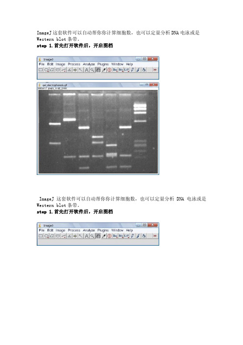
ImageJ这套软件可以自动帮你你计算细胞数,也可以定量分析DNA电泳或是Western blot条带。
step 1.首先打开软件后,开启图档ImageJ这套软件可以自动帮你你计算细胞数,也可以定量分析DNA电泳或是Western blot条带。
step 1.首先打开软件后,开启图档step 2.请先做校正,选择Analyze底下的Calibrate选项,再选择校正的模式,使用Uncalibrate OD,再按ok按下ok之后会出现校正的图形Step 3.在要分析的第一条(first lane)加上一个长型框(工具列第一个选项),再按下Analyze/Gels/select first Lane快速键(Ctr+1),此时框架中会出现一个号码1,之后可以移动框架到第二个lane再选择Analyze/Gels/select second Lane快速键(Ctr+2),当然可以一直加下去,最后按Analyze/Gels/plot Lanes快速键(Ctr +3)。
Step 4.分析以后会出现图型表示你刚选择的框内的影像强度,此时可以看到有几个比较高的区段,就是我们想定量的band,使用直线工具(工具列第五个选项)先将图形中高点为有band的区域和没有band的区域分开再,使用魔术棒工具(工具列第八个选项)点选要分析的区域。
Step 5.当我们点选分析时,在result的对话视窗会出现分析的数据,依序点选就会出现每个band的值。
注:当我们选择分析的条带也可以是横向选取,就可以只比较相同大小的DNA 的含量,同样也可以应用在western blot或其它类似实验条带的分析上。
使用ImageJ 分析图像中的颗粒数[] 原创教程,转载请保留此行1,到本站资料下载-实用小工具栏目下载 ImageJ 并安装。
2,打开ImageJ并打开要分析的图片。
请看演示图片。
3,把图像二值话或者设定阈值。
选择Image - Adjust - Threshold...根据提示设定你需要的阈值。
imagej使用教程
imagej使用教程ImageJ是一款免费开源的图像处理软件,被广泛应用于生物医学图像的分析和处理中。
本教程将向您介绍如何使用ImageJ进行基本的图像处理。
1. 安装ImageJ首先,您需要从ImageJ的官方网站(https:///ij/)下载并安装ImageJ软件。
根据您的操作系统,选择适合的安装程序进行安装。
2. 打开图像在ImageJ的菜单栏上,选择"File"(文件),然后点击"Open"(打开)。
浏览您的计算机,选择要处理的图像文件并点击"Open"(打开)按钮。
3. 调整图像大小如果您需要调整图像的大小,可以在菜单栏上选择"Image"(图像),然后点击"Adjust Size"(调整尺寸)。
在弹出的对话框中,输入所需的宽度和高度,并选择插值算法。
点击"OK"(确定)按钮应用更改。
4. 调整亮度和对比度您可以在菜单栏上选择"Image"(图像),然后点击"Adjust"(调整)来调整图像的亮度和对比度。
在弹出的对话框中,拖动亮度和对比度滑块,直到您满意为止。
点击"OK"(确定)按钮应用更改。
5. 进行滤镜处理ImageJ提供了多种滤镜工具,以改变图像的外观。
在菜单栏上选择"Process"(处理),然后点击"Filters"(滤镜)来选择所需的滤镜效果。
在弹出的对话框中,您可以调整滤镜的参数,然后点击"OK"(确定)按钮应用更改。
6. 测量图像特征ImageJ允许您测量图像中的各种特征,如面积、长度、角度等。
在菜单栏上选择"Analyze"(分析),然后点击"Measure"(测量)来测量图像的特征。
在弹出的结果窗口中,您可以查看测量结果。
imagej基本操作
imagej基本操作
ImageJ是一个强大的图像处理软件,以下是其基本操作:
1. 打开图像:选择“File”菜单中的“Open”选项,然后选择要打开的图像文件。
2. 图像显示:在“Display”菜单中选择不同的颜色通道或颜色模式来显示图像。
3. 调整图像大小:选择“Image”菜单中的“Resize”选项,然后输入新的图像大小。
4. 裁剪图像:选择“Edit”菜单中的“Crop”选项,然后选择要裁剪的区域。
5. 调整亮度/对比度:选择“Image”菜单中的“Adjust”选项,然后选择“Brightness/Contrast”进行调整。
6. 滤镜处理:选择“Process”菜单中的“Filter”选项,然后选择不同的滤镜进行处理。
7. 测量工具:选择“Analyze”菜单中的“Measure”选项,然后选择要测量的长度、角度等参数。
8. 绘制线条或标记:选择“Draw”或“Label”工具,然后在图像上绘制线条或标记。
9. 保存图像:选择“File”菜单中的“Save As”选项,然后选择要保存的文件格式。
10. 退出程序:选择“File”菜单中的“Exit”选项,然后保存所有未保存的更改。
以上仅是ImageJ的基本操作,它还有更多高级功能和工具,可以进一步探索并掌握。
imagej操作流程
imagej操作流程下载温馨提示:该文档是我店铺精心编制而成,希望大家下载以后,能够帮助大家解决实际的问题。
文档下载后可定制随意修改,请根据实际需要进行相应的调整和使用,谢谢!并且,本店铺为大家提供各种各样类型的实用资料,如教育随笔、日记赏析、句子摘抄、古诗大全、经典美文、话题作文、工作总结、词语解析、文案摘录、其他资料等等,如想了解不同资料格式和写法,敬请关注!Download tips: This document is carefully compiled by theeditor.I hope that after you download them,they can help yousolve practical problems. The document can be customized andmodified after downloading,please adjust and use it according toactual needs, thank you!In addition, our shop provides you with various types ofpractical materials,such as educational essays, diaryappreciation,sentence excerpts,ancient poems,classic articles,topic composition,work summary,word parsing,copy excerpts,other materials and so on,want to know different data formats andwriting methods,please pay attention!ImageJ操作流程详解:从新手到专家的进阶指南ImageJ是一款广泛应用于生物学、医学、物理学等领域,专门处理和分析图像的开源软件。
imagej使用方法2篇
imagej使用方法2篇ImageJ是一款功能强大的图像处理软件,它提供了丰富的图像处理和分析工具,被广泛应用于各个领域,如生物学、医学、材料科学等。
本篇文章将介绍ImageJ的基本使用方法,帮助读者快速上手。
第一篇:ImageJ使用方法(上)一、ImageJ的安装与启动在使用ImageJ之前,首先需要下载并安装该软件。
ImageJ的官方网站提供了Windows、Mac和Linux系统的安装包,用户可以根据自己的操作系统选择相应的版本进行下载。
安装完成后,双击打开软件图标即可启动ImageJ。
在启动过程中,系统可能会提示安装Java环境,请按照提示进行操作。
二、图像的导入与显示ImageJ支持多种图像格式,包括常见的BMP、JPG、PNG等格式,用户可以通过以下几种方式将图像导入ImageJ中:1. 点击菜单栏中的“File”,选择“Open”选项,然后在弹出的文件选择窗口中找到并选择要导入的图像文件。
2. 直接将图像文件拖拽到ImageJ的界面上。
3. 使用快捷键Ctrl+O(Windows系统)或Command+O(Mac系统)打开图像文件。
导入图像后,ImageJ会自动显示图像的缩略图,并在软件窗口的上方显示图像的完整路径和名称。
三、图像处理与分析1. 亮度、对比度调整:点击菜单栏中的“Image”,选择“Adjust”子菜单,然后可以对图像的亮度和对比度进行调整。
2. 图像缩放:点击菜单栏中的“Image”,选择“Scale”选项,在弹出的对话框中输入缩放比例,即可对图像进行放大或缩小。
3. 噪声去除:点击菜单栏中的“Process”,选择“Noise”,然后选择“Despeckle”选项,可以去除图像中的噪声。
4. 图像滤波:点击菜单栏中的“Process”,选择“Filters”选项,然后可以选择不同的滤波算法对图像进行滤波处理。
5. 边缘检测:点击菜单栏中的“Process”,选择“Find Edges”选项,可以对图像进行边缘检测。
imagej使用方法
imagej使用方法ImageJ是一款用于数字图像处理和分析的开源软件。
它提供了广泛的功能,如图像增强、测量、分割、滤波、校准、图像重建等。
以下是ImageJ的使用方法:1. 安装ImageJ:从ImageJ官方网站下载适合您操作系统的版本,然后安装。
2. 打开图像:在ImageJ的菜单栏中选择File-->Open,选择您要处理的图像。
3. 图像增强:ImageJ提供了许多图像增强工具,如对比度增强、亮度增强、直方图均衡化、中值滤波等。
在菜单栏中选择Process-->Enhance,选择适当的工具进行增强。
4. 图像测量:ImageJ可以快速准确地测量图像中的长度、面积、角度等。
在菜单栏中选择Analyze-->Measure,将光标移动到需要测量的对象上单击即可。
5. 图像分割:ImageJ可以将图像分成不同的区域,从而进行进一步的分析。
在菜单栏中选择Process-->Binary,选择适当的工具进行分割。
6. 图像重建:ImageJ可以通过各种算法来重建图像,例如反卷积、投影重建等。
在菜单栏中选择Process-->Reconstruct,选择适当的工具进行重建。
7. 图像校准:ImageJ可以校准图像以纠正图像畸变,例如通过标定棋盘校准相机等。
在菜单栏中选择Analyze-->Calibrate,选择适当的工具进行校准。
8. 插件:ImageJ提供了丰富的插件,可以扩展其功能。
在菜单栏中选择Plugins-->Install插件,选择适当的插件进行安装。
以上是ImageJ的基本使用方法,您可以通过阅读ImageJ的帮助文档来进一步了解其更高级的功能。
imagej用户使用手册第三部分
imagej用户使用手册第三部分【原创版】目录1.概述2.图像处理基本操作3.图像处理高级操作4.常见问题与解决方案5.总结正文【概述】本部分将详细介绍 ImageJ 图像处理软件的使用方法。
ImageJ 是一款功能强大的开源图像处理软件,广泛应用于生物医学、化学、物理等多个领域。
在本文中,我们将从基本操作到高级操作,一步一步地为您展示如何使用 ImageJ 进行图像处理。
【图像处理基本操作】1.打开和保存图像- 通过“文件”菜单打开或保存图像- 可以支持的图像格式包括:JPEG、PNG、BMP、TIFF 等2.图像的缩放与裁剪- 通过“图像”菜单下的“缩放”或“裁剪”子菜单进行操作 - 可以自由地调整图像大小和裁剪区域3.图像的旋转与翻转- 通过“图像”菜单下的“旋转”或“翻转”子菜单进行操作- 可以对图像进行任意角度的旋转或翻转【图像处理高级操作】1.区域选择与分割- 通过“选择”菜单下的“区域”子菜单进行操作- 可以使用矩形、椭圆、自由手绘等方法选择图像区域2.图像的亮度和对比度调整- 通过“图像”菜单下的“调整”子菜单进行操作- 可以对图像的亮度、对比度、色调等进行精确调整3.图像的滤波处理- 通过“图像”菜单下的“滤波”子菜单进行操作- 可以使用多种滤波器对图像进行处理,如:模糊、锐化、边缘检测等【常见问题与解决方案】1.问题:打开图像时出现“无法载入图像”的提示解决方案:检查图像文件的路径是否正确,或尝试重新安装ImageJ 软件2.问题:图像处理后保存时出现“保存失败”的提示解决方案:检查保存路径是否正确,或尝试重新安装 ImageJ 软件【总结】本篇文章详细介绍了如何使用 ImageJ 进行图像处理的基本操作和高级操作,以及常见的问题与解决方案。
imagej的使用方法
imagej的使用方法ImageJ是一种功能强大的图像处理软件,它可以识别和操作多种格式的图像,包括TIFF、GIF、JPEG、PNG等。
它包含了强大的数据处理工具和多种图形显示功能,可以显著改善图像质量。
因此,ImageJ已经成为数字图像处理领域的重要软件。
本文将向大家介绍ImageJ的安装及使用方法。
一、ImageJ的安装1. 下载并安装ImageJ:首先,访问ImageJ官网(https:///ij/),下载最新版本的ImageJ。
然后双击运行安装包,根据提示完成安装。
2.开ImageJ:打开安装完成的ImageJ,第一次打开时会出现欢迎界面,接着就可以使用ImageJ了。
二、ImageJ的基本操作1.入图片:在菜单栏中,选择“文件”-“打开”,在弹出的窗口中选择需要处理的图片,点击打开即可将图片导入ImageJ。
2. 保存图片:在菜单栏中,选择“文件”-“保存”,在弹出的窗口中输入图片的新名称,点击保存即可将图片保存到指定位置。
3.转图片:在菜单栏中,选择“图像”-“旋转”,在弹出的窗口中输入需要旋转的角度,点击确定即可将图片旋转指定角度。
4.并图片:在菜单栏中,选择“图像”-“合并”,在弹出的窗口中选择需要合并的图片,点击确定即可将这些图片合并到一张图片中。
5.整图片亮度:在菜单栏中,选择“图像”-“调整”,在弹出的窗口中调整图片的亮度,拖动滑块即可改变图片的亮度,点击确定即可将调整后的图片生成。
6.强图片清晰度:在菜单栏中,选择“工具”-“图像处理”,在弹出的窗口中勾选“增强图像清晰度”,然后点击确定即可增强图像的清晰度。
三、ImageJ的高级操作1.割图片:在菜单栏中,选择“工具”-“图像切割”,在弹出的窗口中设置好切割参数,点击确定即可将图片切割成多个小图。
2.片裁剪:在菜单栏中,选择“工具”-“图像裁剪”,在弹出的窗口中设置好裁剪参数,点击确定即可将多余的图片去掉,生成裁减后的图片。
imagej读取照片像素值的方法
一、引言在数字图像处理的应用中,了解和掌握图像的像素值是十分重要的。
ImageJ作为一款开源的图像处理软件,具有广泛的应用范围,包括生物医学、材料科学等领域。
本文将介绍如何使用ImageJ读取照片的像素值,帮助读者更好地理解和应用ImageJ软件。
二、ImageJ简介ImageJ是由美国国立卫生研究院开发的一款图像处理软件,其功能强大,支持多种图像格式的读取和处理。
ImageJ提供了丰富的图像处理工具和函数,可以满足不同领域的图像处理需求。
因其开源免费且易于使用的特点,已经成为许多科研工作者和学生的首选图像处理软件之一。
三、ImageJ读取照片像素值的方法1. 打开图像在ImageJ软件中,通过“File”菜单中的“Open”选项,选择要处理的图像文件,即可打开图像。
2. 选择感兴趣区域在打开的图像窗口中,可以使用鼠标工具选择感兴趣的区域。
点击鼠标工具栏中的“Rectangular Tool”或其他选择工具,然后用鼠标在图像上拖动,即可选择感兴趣的区域。
3. 读取像素值选择完感兴趣的区域后,可以通过“Analyse”菜单中的“Measure”选项或快捷键“M”来读取所选区域的像素值。
点击“Measure”后,软件会弹出一个数据窗口,显示所选区域的各个像素值,包括灰度值、均值、方差等信息。
4. 保存结果在数据窗口中显示的像素值信息可以通过“File”菜单中的“Save As”选项保存为文本文件,以便后续分析和处理。
四、示例为了更好地演示ImageJ读取照片像素值的方法,我们以一张简单的灰度图像为例进行演示。
1. 打开图像首先在ImageJ软件中打开一张灰度图像文件,比如一张黑白的树叶图像。
2. 选择感兴趣区域使用“Rectangular Tool”工具在图像上选择一块感兴趣的区域,比如树叶的一小部分区域。
3. 读取像素值点击“Analyse”菜单中的“Measure”选项或按下快捷键“M”来读取所选区域的像素值。
imagej教程
imagej教程这是一篇关于ImageJ软件的简单教程。
ImageJ是一款用于图像处理和分析的开源软件,可用于科学研究、医学图像处理、教学和艺术设计等领域。
1. 下载和安装ImageJ首先,你需要在官方网站上下载ImageJ的安装程序。
根据你的操作系统选择合适的版本,并按照安装向导的指示进行安装。
安装完成后,你就可以启动ImageJ了。
2. 导入图像文件在ImageJ的菜单栏中,选择“File(文件)”>“Open(打开)”,然后浏览你的电脑,选中要处理的图像文件,点击“Open(打开)”按钮。
图像将会被导入到ImageJ的工作界面中。
3. 图像调整ImageJ提供了一系列能够调整图像亮度、对比度、色彩和大小等参数的工具。
你可以在菜单栏的“Image(图像)”和“Adjust(调整)”下找到这些功能,并根据需要进行调整。
4. 图像滤波图像滤波可以帮助去除图像中的噪声,提高图像的清晰度和质量。
在ImageJ的菜单栏中,选择“Pr ocess(处理)”>“Filters (滤波器)”,然后选择适合你需求的滤波算法。
5. 图像测量与分析ImageJ提供了丰富的图像测量和分析工具,可以帮助你对图像中的对象进行计数、长度测量、面积计算等。
在菜单栏中,选择“Analyze(分析)”>“Measure(测量)”或者“Analyze(分析)”>“Set Measurements(设置测量参数)”,然后选择你需要的测量和分析功能。
6. 插件和宏ImageJ支持插件和宏,可以扩展软件的功能和自动化操作。
你可以在菜单栏中的“Plu gins(插件)”和“Macros(宏)”下找到各种插件和宏,并按照说明使用。
7. 结果保存与导出在处理和分析完图像后,你可以选择将结果保存为新的图像文件或者导出为其他格式。
在菜单栏中,选择“File(文件)”>“Save As(另存为)”或者“File(文件)”>“Export(导出)”,然后选择保存或导出的格式和路径。
imagej用户使用手册第三部分
imagej用户使用手册第三部分摘要:1.概述2.图像处理基本操作3.常用图像处理工具4.图像处理高级技巧5.结论正文:一、概述本部分将介绍ImageJ 的用户手册第三部分,主要涉及图像处理的基本操作、常用图像处理工具以及高级技巧。
通过学习本部分内容,用户将能够熟练掌握ImageJ 软件的操作,进行各种图像处理任务。
二、图像处理基本操作1.打开和保存图像- 打开图像:在ImageJ 中,用户可以通过“文件”菜单下的“打开”选项或使用快捷键“Ctrl+O”打开图像。
- 保存图像:在ImageJ 中,用户可以通过“文件”菜单下的“保存”选项或使用快捷键“Ctrl+S”保存图像。
2.图像的显示和缩放- 图像显示:在ImageJ 中,用户可以通过“显示”菜单下的“在窗口中显示”选项或使用快捷键“Ctrl+W”显示图像。
- 图像缩放:在ImageJ 中,用户可以通过“显示”菜单下的“缩放”选项或使用快捷键“Ctrl+Z”缩放图像。
3.图像的选取和裁剪- 图像选取:在ImageJ 中,用户可以通过“选择”菜单下的“全部”选项或使用快捷键“Ctrl+A”选取整个图像。
- 图像裁剪:在ImageJ 中,用户可以通过“图像”菜单下的“裁剪”选项或使用快捷键“Ctrl+X”裁剪图像。
三、常用图像处理工具1.直方图均衡化- 直方图均衡化可以增强图像的对比度,使图像更易于观察。
- 在ImageJ 中,用户可以通过“图像”菜单下的“直方图均衡化”选项使用该功能。
2.锐化和模糊- 锐化可以提高图像的清晰度,模糊可以降低图像的清晰度。
- 在ImageJ 中,用户可以通过“图像”菜单下的“锐化”和“模糊”选项使用这些功能。
3.图像滤波- 图像滤波可以消除图像中的噪声,提高图像质量。
- 在ImageJ 中,用户可以通过“图像”菜单下的“滤波”选项使用各种滤波器。
四、图像处理高级技巧1.使用自定义函数进行图像处理- 在ImageJ 中,用户可以使用自定义函数对图像进行各种处理。
imagej的使用方法
imagej的使用方法ImageJ是一款由NationalInstitutesofHealth开发的图像处理软件,可以让用户进行各种类型的图像处理。
最开始ImageJ被设计用来处理像素图像,但是也可用于处理其他类型的图像,例如条形图、饼图、折线图等。
因为它的开放源代码,许多图像处理的开发者都选择使用ImageJ来构建自己的应用程序和系统。
下面将介绍如何使用ImageJ来处理图像。
1.装ImageJ首先,用户需要下载安装ImageJ。
ImageJ可以免费从网上下载,并在多种操作系统上运行,包括Windows、Mac OS和Linux等。
安装完成后,用户就可以运行ImageJ软件。
2.载图像运行完ImageJ软件之后,用户需要加载需要处理的图像到软件中。
可以选择“文件”,然后选择“打开”来打开所需要处理的图片。
ImageJ支持多种类型的图像,包括常用的jpg、gif、png以及tiff 格式。
3.像处理加载图像到ImageJ之后,就可以开始对图像进行处理。
ImageJ 提供了众多的图像处理工具,可以根据用户的需求进行各种操作,如缩放、裁剪、旋转、滤镜效果等。
只需要点击相应的按钮,就可以实现对所需要的图像进行处理。
4.像分析ImageJ还支持对图像进行分析,可以对图像进行测量、对比度分析、直方图统计、滤波处理等操作,提供定量分析结果,帮助用户更好地了解图像。
5.像输出最后,在处理完图像之后,用户可以将处理后的图像输出到多种格式的图片文件中,例如jpg、tif、png等,以便用户在报告中使用或者进行其他的应用。
以上就是ImageJ的基本使用方法,使用起来十分简单方便,操作起来也非常方便。
ImageJ可以帮助用户快速高效地处理图像,并能够获得准确的结果,是一款非常实用的图像处理软件。
- 1、下载文档前请自行甄别文档内容的完整性,平台不提供额外的编辑、内容补充、找答案等附加服务。
- 2、"仅部分预览"的文档,不可在线预览部分如存在完整性等问题,可反馈申请退款(可完整预览的文档不适用该条件!)。
- 3、如文档侵犯您的权益,请联系客服反馈,我们会尽快为您处理(人工客服工作时间:9:00-18:30)。
Examples of Image Analysis Using ImageJ
Area Measurements of a Complex Object
Problem: Determine the photosynthetic (i.e., green) portion of a variegated leaf.
(Open leaf image via Select File → Open Samples → Leaf )
♦ Convert scanned color image of leaf to grayscale: Image → Type → 8-bit
♦ Set measurement scale: Draw a line over a 50 mm section of the ruler then Analyze → Set Scale In Set Scale window enter 50 into the 'Known Distance' box and change the 'Unit
of Measurement' box to mm , check 'Global'
♦ Draw a new line and confirm that the measurement scale is correct.
♦ Threshold the leaf image using the automated routine: Process → Binary → Make Binary
The automated threshold includes only the dark green areas.
♦ Calculate area of green portion:
Surround the leaf with the rectangular selection tool Analyze → Analyze Particles
Enter 50 as the minimum particle size, toggle 'Show Outlines', check “Display
Results’ and click 'OK'
Outline of analyzed area will be drawn. Data window gives an area of
about 2000 mm 2 depending on the calibration setting.
(See bottom of page for an alternative method for measuring areas.)
♦ Threshold new image of leaf using manual settings: Image → Adjust →Threshold and play with sliders to include all of leaf in red
and click ‘Apply’ The manual threshold setting includes all of the leaf area.
♦ Calculate area of entire leaf: Enclose the leaf with the rectangular selection tool Analyze → Analyze Particles
Use previous window settings and click 'OK'
Outline of entire leaf is automatically drawn.
Data window gives an area of about 2450 mm 2.
This analysis suggests that about 82% of leaf surface is dark green. These values should be manually confirmed before beginning a 'production run' of measurements.
An alternative procedure for measuring areas:
Analyze → Set Measurements , check 'Limit to Threshold'. After converting to a binary image, select Analyze → Measure
This procedure is simpler but does not draw an outline of the measured area.
(more on reverse side)
Examples of Image Analysis Using ImageJ (continued)
Particle Counting and Analysis.
Problem: Count and determine the size distribution of a collection of echinoderm embryos.
(Open embryos image via Select File → Open Samples → Embryos )
♦ Draw line over the scale bar and select Analyze → Set Scale
In Set Scale window enter 100 into the 'Known Distance' box
and Change the 'Unit of Measurement' box to um , check 'Global'
♦ Confirm that the measurement scale is correct.
♦ Convert the image to grayscale: Image → Type → 8-bit
♦ Threshold the image using the automated routine: Process → Binary → Make Binary
♦ Surround the scale bar with the rectangular selection tool and
clear the contents (Edit → Clear )
♦ Analyze Particles: Analyze → Analyze Particles
Enter 20 as the minimum particle size, toggle 'Show Outlines', check
‘Display Results’, ‘Summarize’ and ‘Record Stats’ and click 'OK'
Twenty five embryos are counted, numbered and outlined. The data window lists the area (in um 2) for each embryo. These data could be copied to a spreadsheet. Threshold: 0-0
Count: 25
Total Area: 3177.6 µm^2
Average Size: 127.1 µm^2
Area Fraction: 3.72% A summary of the particle count is also shown in another data window.
As mentioned in the previous example, this technique should be manually validated before collecting experimental data.。
