15-Hand-Held and Integrated Single-Cell Pipettes补充
富士通显示设备 B24-9 TE 数据表说明书

Data SheetFujitsu Display B24-9 TEWidescreen 60.5 cm (23.8-inch) Advanced Ergonomic DisplayIdeal for office applications and 24/7 usageThe FUJITSU Display B24-9 TE is a widescreen display with 1920 x 1080 Full HD resolution with a ultra-thin bezel housing. The monitor has a wide viewing angle that delivers consistent picture quality and DisplayView™ manageability software for further enhancing monitor features with a range of connectivity options ideal for medium- and large-sized businesses.The 23.8-inch display with 16:9 aspect ratio at Full HD resolution for consistent clarity and superior viewing experience is ideal for office applications.The 6-in-1 stand base allows users to tilt, swivel, adjust height, and rotate monitor display as per their comfort and working style.The ultra-thin bezel housing on three sides for multi-monitoring scenarios and integrated speakers and cable guide allow for a clean desk experience.The always available USB, DisplayPort, HDMI interfaces ensure to have a complete package that meets all your business requirements.The ergonomic stand with height adjust with 345° swivel, 35° rear tilt and 45 mm picture over desk enables for an enhanced flexibility.The environmental-friendly LED technology with highly efficient energy saving functions like ECO Operation mode and ECO standy help to save on costs.The DisplayView software enables you to configure your monitor for adjusting screen brightness, pivot function and much more.Focusing on environmental friendliness, the packaging is styrofoam free andsolely consisting of cardboard, which is more than 90% recycled.Technical detailsSpecial featuresIn-Plane Switching (IPS) technologyLow blue light mode6-in-1 StandEco button for Eco mode and 3-coloured Eco status LEDAlways available USB (in operating and standby mode)DisplayView™ Software24/7 usageIntegrated speakersErgonomic standStand6-in-1 StandHeight adjust range150 mmPicture height over desk (min)45 mmRotation to portrait90°Tilt angle-5° / +35°Swivel angle345°Picture performancePanel and backlit In-Plane Switching (IPS) technology/LEDScreen Surface Treatment Anti-glare, 3H hard coatingContrast - typical1,000:1Contrast - advanced20,000,000:1Response time gray to gray typical 5 ms (in video mode)Viewing angle (h/v) - typical178°/178° CR10:1Color performance16.7 million colorsBrightness - typical250 cd/m2Size and resolutionAspect ratio16:9Diagonal Size60.5 cm (23.8-inch)Resolution (native)1,920 x 1,080 pixelResolution (interpolated)1,680 x 1,050 pixel, 1,440 x 900 pixel, 1,280 x 1,024 pixel, 1,280 x 720 pixel, 1,024 x 768 pixel, 800 x 600 pixel, 640 x480 pixelPicture size527 x 296 mmPixel Pitch0.2745 mmFrequenciesHorizontal30 - 82 kHzVertical48 - 76 HzConnectivityDisplayPort 1 x DisplayPort 1.2HDMI 1 x HDMI 1.4VGA/D-SUB 1 x D-SUBAudio signal output 3.5 mm stereo phone jack for head phoneAudio sound output 2 x 2 WAudio signal input 3.5 mm stereo phone jackUSB downstream2x USB 3.2 Gen1USB upstream 1 x USB 3.2 Gen1Ease-of-use menuDirect Access Keys Brightness, ECO, Input, Mode, Audio, MenuLanguages Arabic, Czech, Danish, Dutch, English, Finnish, French, German, Italian, Norwegian, Polish, Portuguese, Russian,Spanish, Swedish, Turkish, Japanese, Chinese simply, Chinese traditionalBrightness / Contrast Brightness, Contrast, Black level, Auto level, ACRMode sRGB, Office, Photo, Video, Low Blue LightColor sRGB, 5000K, 6500K, 7500K, Native, Custom Color (R,G,B)OSD Language, OSD-Timeout, OSD rotationImage adjust Clock, Phase, H-Position, V-Position, Expansion, SharpnessAudio Mute, Volume, Input for HDMI and DP interfaceInformation Model name, Serial number, Signal input, Resolution/mode, Display mode , Color Temperature, ACR Status Advanced settings Overdrive, DDC/CI, Factory RecallPower consumption (typical, w/o sound)Soft switch off0.1 WPower save mode0.17 WOperating with EPA settings14.25 WTotal Energy Consumption (ETEC)44.66 kWh/yearOperating maximum brightness22 WPower supply integratedPower notes Speakers off, USB not connectedETEC and EPA refer to ENERGY STAR® 8.0Electrical valuesRated voltage range100 V - 240 VRated frequency range50 Hz - 60 HzProtection class1ComplianceModel DY24-9TGlobal ENERGY STAR® 8.0, TÜV Low Blue Light Certified, TÜV Flicker Free Certified, Zero bright and dark pixel faults, Subpixelfaults according to ISO9241-307 (Pixel fault class I), EPEAT® Silver (dedicated regions), TCO CertifiedEurope EN 62368-1, CE certification according to EC Directive 2004/108/EEC, RoHS, WEEE, IT-Eco-DeclarationAustralia/New Zealand RCMChina CCCGermany TÜV GSRussia EACSingapore S-MarkSouth Korea KCTaiwan BSMIUSA/Canada FCC Class B, cTUVusCompliance link https:///sites/certificatesDimensions / Weight / EnvironmentalDimension without stand (W x D x H)540.3 x 64 x 325.6 mm21.27 x 2.52 x 12.82 inchDimension with stand (W x D x H)540.3 x 229.4 x 346.2 mm21.27 inch x 9.03 x 13.63 inchWeight (unpacked) 5.25 kg11.57 lbsWeight (Monitor only) 3.21 kg7.08 lbsOperating ambient temperature 5 - 35 °C (41 - 95 °F)Operating relative humidity10 - 85 % (non condensing)MiscellaneousMiscellaneous VESA DDC/ CI, Flat Display Mounting Interface VESA MIS-D 100 C, Kensington lock preparedColor Marble greyAdditional SoftwareAdditional software (optional)WINDOWS WHQL driverDisplayView SuiteAdditional software (notes)Use of accompanying and/or additional Software is subject to proactive acceptance of the respective LicenseAgreements /EULAs/ Subscription and support terms of the Software manufacturer as applicable for the relevantSoftware whether preinstalled or optional. The software may only be available bundled with a software supportsubscription which – depending on the Software - may be subject to separate remuneration.Package contentDisplay delivered accessories DisplayPort data cable 1.8 mUSB-cable 1.8 m (USB-A to USB-B)Power cable for wall socket (Euro-Schuko-Type CEE7) 1.8 mQuickstart flyerSafety notesDisplay delivered accessories notes Power cable with IEC-60320-C13, 3-pin connector on display sideData cables and USB cable detachable on displayUser manual and DisplayView Software is available via downloadPackaging dimension (mm)655 x 411 x 220 mmPackaging dimension (inch)25.79 x 16.18 x 8.66 inchWeight (packed)7.1 kgWeight (packed) (lbs)15.65 lbsOrder informationOrder Code S26361-K1713-V140EAN Code4065221888260Country specific order code BDL:K1713V140-UK - with UK power cable, mandatory for Arabian countriesBDL:K1713V140-CHN - with China power cable and CEL, mandatory for ChinaBDL:K1713V140-INT - W/o power cable, mandatory for countries where import with EU cable is not allowed Accessories information Further helpful options:Stands and Mounting kits:/fts/products/computing/peripheral/accessories/desktop/ums/index.htmlAvailable Adapters: /dmsp/Publications/public/pos-connectivity.pdfWarrantyWarranty period 3 years (depending on country)Warranty Terms & Conditions /warrantyDigital bug fixes Subject to availability and following their generic release for the product, bug fixes and function-preserving patchesfor product-related software (firmware) can be downloaded from the technical support at: https://support.ts.fujitsu.com/ free of charge by entering the respective product serial number. For application software supplied togetherwith the product, please directly refer to the support websites of the respective software manufacturer.Spare Parts availability at least 7 years after shipment, for details see https:///Service Weblink /emeia/products/product-support-services/CONTACTFujitsu Technology Solutions GmbH Website: 2023-11-27 EM-ENworldwide project for reducing burdens on the environment.Using our global know-how, we aim to contribute to the creation of a sustainable environment for future generations through IT.Please find further information at http://www./global/about/environmenttechnical specification with the maximum selection of components for the named system and not the detailed scope ofdelivery. The scope of delivery is defined by the selection of components at the time of ordering.Technical data is subject to modification and delivery subject to availability. Any liability that the data and illustrations are complete, actual or correct is excluded. Designations may be trademarks and/or copyrights of the respective owner, the use of which by third parties for their own purposes may infringe the rights of such owner.The overall product has been designed and manufactured for general office use, regular personal use and ordinary industrial use.More informationAll rights reserved, including intellectual property rights. Designations may be trademarks and/or copyrights of therespective owner, the use of which by third parties for their own purposes may infringe the rights of such owner. For further information see https:///global/about/resources/terms/ Copyright 2023 Fujitsu Technology Solutions GmbH。
澳洲坚果抗氧化肽的分离纯化及肽段鉴定
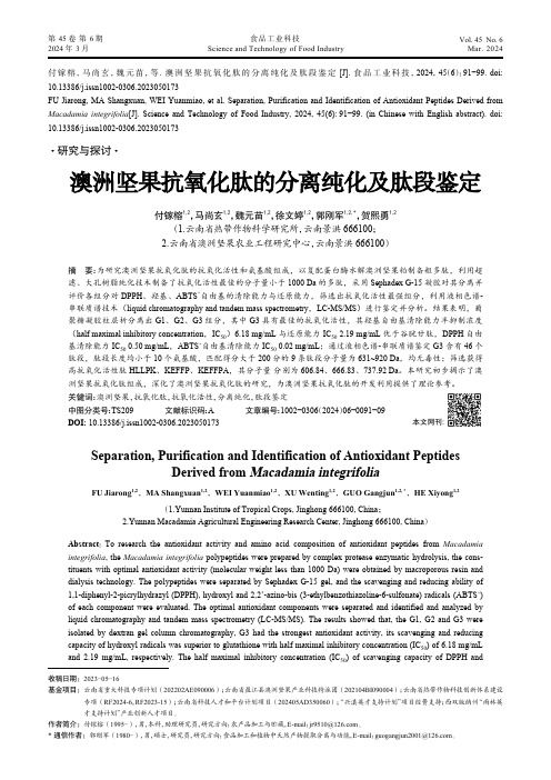
付镓榕,马尚玄,魏元苗,等. 澳洲坚果抗氧化肽的分离纯化及肽段鉴定[J]. 食品工业科技,2024,45(6):91−99. doi:10.13386/j.issn1002-0306.2023050173FU Jiarong, MA Shangxuan, WEI Yuanmiao, et al. Separation, Purification and Identification of Antioxidant Peptides Derived from Macadamia integrifolia [J]. Science and Technology of Food Industry, 2024, 45(6): 91−99. (in Chinese with English abstract). doi:10.13386/j.issn1002-0306.2023050173· 研究与探讨 ·澳洲坚果抗氧化肽的分离纯化及肽段鉴定付镓榕1,2,马尚玄1,2,魏元苗1,2,徐文婷1,2,郭刚军1,2, *,贺熙勇1,2(1.云南省热带作物科学研究所,云南景洪 666100;2.云南省澳洲坚果农业工程研究中心,云南景洪 666100)摘 要:为研究澳洲坚果抗氧化肽的抗氧化活性和氨基酸组成,以复配蛋白酶水解澳洲坚果粕制备粗多肽,利用超滤、大孔树脂纯化技术制备了抗氧化活性最佳的分子量小于1000 Da 的多肽,采用Sephadex G-15凝胶对其分离并评价各组分对DPPH 、羟基、ABTS +自由基的清除能力与还原能力,筛选出抗氧化活性最强组分,利用液相色谱-串联质谱技术(liquid chromatography and tandem mass spectrometry ,LC-MS/MS )进行鉴定并分析。
结果表明,葡聚糖凝胶柱层析分离出G1、G2、G3组分,其中G3具有最佳的抗氧化活性,其羟基自由基清除能力半抑制浓度(half maximal inhibitory concentration ,IC 50)6.18 mg/mL 与还原能力IC 50 2.19 mg/mL 优于谷胱甘肽,DPPH 自由基清除能力IC 50 0.50 mg/mL ,ABTS +自由基清除能力IC 50 0.02 mg/mL ;通过液相色谱-串联质谱鉴定G3含有46个肽段,肽段长度均小于10个氨基酸,匹配得分大于200分的9条肽段分子量为631~920 Da ,均无毒性;筛选获得高抗氧化活性肽HLLPK 、KEFFP 、KEFFPA ,其分子量 分别为606.84、666.83、737.92 Da 。
单细胞数据整合方法ComprehensiveIntegrationofSingle-Cel。。。
单细胞数据整合⽅法ComprehensiveIntegrationofSingle-Cel。
依赖的算法CCAMNN实在是没想到,这篇seurat的V3⾥⾯的整合⽅法居然发在了Cell主刊。
果然:⼤佬+前沿领域=⽆限可能可以看到bioRxiv上是November 02, 2018发布的,然后Cell主刊June 06, 2019正式发表。
⽅法的创意应该在2017年底就有了,那时候我才刚来做single cell。
Single-cell transcriptomics has transformed our ability to characterize cell states, but deep biological understanding requires more than a taxonomic listing of clusters.As new methods arise to measure distinct cellular modalities, a key analytical challenge is to integrate these datasets to better understand cellular identity and function.Here, we develop a strategy to “anchor” diverse datasets together, enabling us to integrate single-cell measurements not only across scRNA-seq technologies, but also across different modalities.After demonstrating improvement over existing methods for integrating scRNA-seq data, we anchor scRNA-seq experiments with scATAC-seq to explore chromatin differences in closely related interneuron subsets and project protein expression measurements onto a bone marrow atlas to characterize lymphocyte populations.Lastly, we harmonize in situ gene expression and scRNA-seq datasets, allowing transcriptome-wide imputation of spatial gene expression patterns.Our work presents a strategy for the assembly of harmonized references and transfer of information across datasets.亮点1:通过锚定的⽅法来整合多种数据,不同平台,不同形态。
干扰素刺激基因15抗病毒感染的分子机制

·综述·Chinese Journal of Animal Infectious Diseases中国动物传染病学报摘 要:干扰素刺激基因15(ISG15)是由病原微生物或干扰素诱导产生的一种大小为15 kDa 的泛素样蛋白。
在干扰素诱导的数百个干扰素刺激基因中,ISG15是诱导最强烈、最快的ISG 蛋白之一。
研究表明,ISG15对多种病毒具有抗病毒作用。
此外,ISG15在调节宿主损伤、DNA 修复,调节信号通路及抗原递呈中也发挥着重要的作用。
文章介绍了ISG15的概况,并阐述了近年来ISG15在抗病毒、免疫调节和调节宿主信号通路过程中的作用。
关键词:干扰素刺激基因15;抗病毒作用;免疫调节中图分类号:S852.4 文献标志码:A 文章编号:1674-6422(2023)06-0170-07Molecular Mechanism of Interferon-Stimulated Gene 15 Antiviral InfectionTANG Jingyu 1, DU Hanyu 1,2, JIA Nannan 1, TANG Aoxing 1, LIU Chuncao 1, ZHU Jie 1, MENGChunchun 1, LI Chuanfeng 1, LIU Guangqing 1(1. Shanghai V eterinary Research Institute, CAAS, Shanghai 200241, China; 2. Xinjiang Agricultural University, Xinjiang 830052, China)收稿日期:2021-11-02作者简介:国家重点研发计划项目(2016YFD0500108);中国农业科学院创新工程项目作者简介:唐井玉,女,博士研究生,预防兽医学专业通信作者:刘光清,E-mail:**************.cn干扰素刺激基因15抗病毒感染的分子机制唐井玉1,杜汉宇1,2,贾楠楠1,汤傲星1,刘春草1,朱 杰1,孟春春1,李传峰1,刘光清1(1.中国农业科学院上海兽医研究所 小动物传染病预防与控制创新团队,上海200241;2.新疆农业大学,乌鲁木齐830052)2023,31(6):170-176Abstract: Interferon-stimulated gene 15 (ISG15) is a ubiquitin-like protein of approximately 15 kDa induced by pathogenic microorganisms or interferons. Among the hundreds of interferon-stimulated genes induced by interferons, ISG15 is one of the most strongly and fastest induced ISG proteins. Studies have shown that ISG15 has antiviral effects against a variety of viruses. In addition, ISG15 plays an important role in regulating host damage, DNA repair, and regulating signaling pathways and antigen delivery. The article presented an overview of ISG15 and described the role of ISG15 in the process of antiviral, immunomodulation and regulation of host signaling pathways in recent years.Key words: Interferon-stimulated gene 15; antiviral infection; immunomodulation先天性免疫应答是抵抗入侵病原体的第一道防线,病原体可以通过宿主模式识别受体来感知。
《临床肝胆病杂志》推荐使用的规范医学名词术语

临床肝胆病杂志第40卷第3期2024年3月J Clin Hepatol, Vol.40 No.3, Mar.2024[3]XIA SL, LIU ZM, CAI JR, et al. Liver fibrosis therapy based on biomi⁃metic nanoparticles which deplete activated hepatic stellate cells[J]. J Control Release, 2023, 355: 54-67. DOI: 10.1016/j.jconrel.2023.01.052.[4]LIU YW, DONG YT, WU XJ, et al. The assessment of mesenchymalstem cells therapy in acute on chronic liver failure and chronic liver disease: A systematic review and meta-analysis of randomized con⁃trolled clinical trials[J]. Stem Cell Res Ther, 2022, 13(1): 204. DOI:10.1186/s13287-022-02882-4.[5]ZHANG ZL, SHANG J, YANG QY, et al. Exosomes derived from hu⁃man adipose mesenchymal stem cells ameliorate hepatic fibrosis by inhibiting PI3K/Akt/mTOR pathway and remodeling choline me⁃tabolism[J]. J Nanobiotechnology, 2023, 21(1): 29. DOI: 10.1186/ s12951-023-01788-4.[6]ZHAO T, SU ZP, LI YC, et al. Chitinase-3 like-protein-1 function andits role in diseases[J]. Signal Transduct Target Ther, 2020, 5(1): 201. DOI: 10.1038/s41392-020-00303-7.[7]YANG H, ZHAO LL, HAN P, et al. Value of serum chitinase-3-likeprotein 1 in predicting the risk of decompensation events in patients with liver cirrhosis[J]. J Clin Hepatol, 2023, 39(7): 1578-1585. DOI:10.3969/j.issn.1001-5256.2023.07.011.杨航, 赵黎莉, 韩萍, 等. 血清壳多糖酶3样蛋白1(CHI3L1)对肝硬化患者发生失代偿事件风险的预测价值[J]. 临床肝胆病杂志, 2023, 39(7): 1578-1585. DOI: 10.3969/j.issn.1001-5256.2023.07.011.[8]MA L, WEI J, ZENG Y, et al. Mesenchymal stem cell-originated exo⁃somal circDIDO1 suppresses hepatic stellate cell activation by miR-141-3p/PTEN/AKT pathway in human liver fibrosis[J]. Drug Deliv, 2022, 29(1): 440-453. DOI: 10.1080/10717544.2022.2030428. [9]NISHIMURA N, DE BATTISTA D, MCGIVERN DR, et al. Chitinase 3-like 1 is a profibrogenic factor overexpressed in the aging liver and in patients with liver cirrhosis[J]. Proc Natl Acad Sci U S A, 2021, 118(17): e2019633118. DOI: 10.1073/pnas.2019633118.[10]WANG CG, LI SZ, SHI JM, et al. Research progress in differentia⁃tion, identification, and purification methods of human pluripotent stem cells to mesenchymal-like cells in vitro[J]. J Jilin Univ Med Ed, 2023, 49(6): 1655-1661. DOI: 10.13481/j.1671-587X.20230634.王成刚, 李生振, 史嘉敏, 等. 体外人多能干细胞向间充质样细胞分化、鉴定和纯化方法的研究进展[J]. 吉林大学学报(医学版), 2023, 49(6): 1655-1661. DOI: 10.13481/j.1671-587X.20230634.[11]LI TT, WANG ZR, YAO WQ, et al. Stem cell therapies for chronicliver diseases: Progress and challenges[J]. Stem Cells Transl Med, 2022, 11(9): 900-911. DOI: 10.1093/stcltm/szac053.[12]YANG X, LI Q, LIU WT, et al. Mesenchymal stromal cells in hepaticfibrosis/cirrhosis: From pathogenesis to treatment[J]. Cell Mol Im⁃munol, 2023, 20(6): 583-599. DOI: 10.1038/s41423-023-00983-5. [13]ZHAO SX, LIU Y, PU ZH. Bone marrow mesenchymal stem cell-derived exosomes attenuate D-GaIN/LPS-induced hepatocyte apop⁃tosis by activating autophagy in vitro[J]. Drug Des Devel Ther, 2019, 13: 2887-2897. DOI: 10.2147/DDDT.S220190.[14]LEE CG, HARTL D, LEE GR, et al. Role of breast regression protein39 (BRP-39)/chitinase 3-like-1 in Th2 and IL-13-induced tissue re⁃sponses and apoptosis[J]. J Exp Med, 2009, 206(5): 1149-1166.DOI: 10.1084/jem.20081271.[15]HIGASHIYAMA M, TOMITA K, SUGIHARA N, et al. Chitinase 3-like 1deficiency ameliorates liver fibrosis by promoting hepatic macro⁃phage apoptosis[J]. Hepatol Res, 2019, 49(11): 1316-1328. DOI:10.1111/hepr.13396.收稿日期:2023-06-09;录用日期:2023-08-17本文编辑:邢翔宇引证本文:LIU PJ, YAO LC, HU X, et al. Effect of human umbilical cord mesenchymal stem cells in treatment of mice with liver fibrosis and its mechanism[J]. J Clin Hepatol, 2024, 40(3): 527-532.刘平箕, 姚黎超, 胡雪, 等. 人脐带间充质干细胞(hUC-MSC)对肝纤维化小鼠模型的治疗作用及其机制分析[J]. 临床肝胆病杂志, 2024, 40(3): 527-532.读者·作者·编者《临床肝胆病杂志》推荐使用的规范医学名词术语有关名词术语应规范统一,以全国自然科学名词审定委员会公布的各学科名词为准。
hIL-15-SA双功能融合蛋白的制备
细胞增殖试验和细胞表 面锚 定修 饰试验 表明,经纯化 复性的 h L 1 一A融合蛋 白具有双重生物活性。结 I 一5s
酵 h L1一A 程 茵 ,通过 镍 柱 亲和 层 析 及 阴 离子 交换 层 析 纯化 目的蛋 白 。免疫 印迹 分析 产 物 , 巴细胞 I一5S 工 淋
增 殖 试验 以及 细 胞 锚 定 试验 检 测 双 功 能融 合 蛋 白 h L1-A的生 物 学活 性 。结 果 : 采 用 半合 成 培 养基 ,以 I一5S
7 0 4 / e e l n 1 1 / n l s o o i sw r o lc e .T e p r t f tr e r t i e c e . ,7g Lw tc l sa d 2 .5 gL ic u in b d e e e c le t d h u i yo a g t p o en r a h d
9 % a t r u i i a i n. el p ol f r i nd e - u f ce m d f c t O s ys e ns r t d h t t e 7 f e p r f c t O C l r i e at On a c l s r a — O i i a i n as a d mO t a e t a h i
p ot n r ei hI 一 5 S L 1 一 A. Me h ds:T t rg p t n to he a et ro ei wa o fi t ro h he p s ri ed h ug t a pli ati n f c o o af ni y fi t
Cogent Process-Scale Tangential Flow Filtration Sy

Cogent® Process-Scale Tangential Flow Filtration System A fully-automated, configurable, TFF system suited for manufacturing of biopharmaceuticals and cGMP process-scale applicationsData SheetThe fully automated Cogent® TFF system isdesigned to separate and purify monoclonalantibodies, vaccines, plasma, and therapeuticproteins. It is ideally suited for both pilot andproduction scale applications, therebysupporting rapid scale up from small to largescale operations.Benefiting from our leading bioprocessknowledge and engineering expertise,the Cogent® Process Scale System is theculmination of 25 years of custom systemdesign and incorporates many unique,innovative and intelligent design features.This system has a very low hold-up volumefor maximum volume concentration andoptimal product recovery, thus enhancingprocess performance.Benefits:•M odular standard options allow the unique system configuration that best matches processrequirements while minimizingupfront investment.•F ull process automation eases the consistent production of preclinical and clinical scalequantities of high-value drug products tocGMP standard.•O ptimized design and component integration ofNovAseptic® valves and TFF cassette holders result in a low minimum working volume and ensuremaximum product recovery.•D esigned to maximize TFF performances in fed-batch, concentration, total recycle orsingle pass mode.•C omprehensive services ensure rapidimplementation and optimized performance.2Configure your system according to your process needs…Option 7: Filtrate conductivityMeasurement of a wide range of products (WFI, buffer solutions, protein solutions) or post CIPOption 1: Tank (50, 100, 200L) Jacketed fortemperature regulationOption 10: Retentate pH In-process monitoring of product volumeOption 13: Tank Outlet Level SwitchAllows to stop the feed pump when air reaches this sensor. E.g: In Mini loop concentration mode, detects the end of the step (tank fully empty).Option 12: TankNovAseptic® GMP mixer Ensures producthomogeneity, specially important duringdiafiltration step. Aseptic design, minimized shearingOption 3: Transfer PumpTransfer of product / buffersinto the feed tank fromany other tank. Allowsfed-batch mode, anddiafiltration.Option 8: Filtrate pHpH monitoring duringcleaning and sanitizationproceduresOption 6: Transfer InletManifoldAllows connecting severalinlets to the transfer pumphead (product/WFI/CIP)at the same time, avoidingmany connections /disconnections...and build a consistent user experience3Total Process Controland ConnectivityThe Cogent® Process Scale system is easily controlled via the Common Control Platform® (CCP®) software, a powerful, intuitive and graphical software that provides real-time monitoring and total in-depth control of your TFF process.Using robust PCs, PLCs, and SCADA® technology, it meets the most stringent standards for connectivity,reliability and ease of use.NovAseptic® GMP mixer Embedded NovAseptic® Valves, Mixer and ConnectorsEngineered for optimal performance, reliability, durability and ease of maintenance.The design and development of each component is based on more than 20 years’ experience, focused on aseptic application. This is why we choose to call it ‘‘Aseptic by Design.”Benefits:•C reate process operations using the recipe editor, monitor or control the process in thehome screen, and create reports for the batch using the configurable report generator•D eveloped under GAMP guidelines and FDA 21 CFR Part 11 compliance-ready, including audit trails and electronic signatures for verification •S ensor combinations can be adapted to process requirements allowing the maximum confidence in process monitoring• U tilities to connect all of your separation unit operations to a central network orDCS (e.g. Delta V)• U sed on multiple unit operations CCP® software provides one familiar interface to simplifysoftware management and reduce learning curvesBenefits:•C omply with cGMP Design Qualification criteria for aseptic processing• N ovAseptic® connector ensures no dead legs and maximum product recovery with zero hold up volume.• C omply with the most stringent cleaning and sterilization requirements • M ixer is clean running and is suitable for general mixing, heat transfer and shearsensitive applications.• R educed bioburden• L ower cost of maintenance• D iamond coated mixer bearings ensure long life and optimum performance.• A bility to mix the “last drop”, ensures complete product recovery4Unparalleled UltrafiltrationPlug and PlayThe Pellicon® Process-scale Holder is uniquely designed to reduce the time required to install and removeTFF cassettes at production scale while keeping the flow path unchanged.The holder can be configured with a manual or hydraulic closure. Hydraulic closure can be done with a hand pump or with an automated hydraulic box which allows local or distant control.Biomax® membranePellicon® 3 cassettes with Biomax® membranes are designed for the filtration of therapeutic proteins, albumin, hormones, vaccines and growth factors. These advanced, high-performance cassettes are ideal for today’s processes that require higher operating pressures, temperatures and higher caustic cleaning regimes. Ultracel® membranePellicon® 3 Cassettes with Ultracel® membrane are the device of choice for today’s higher titer therapeutic antibodies as well as the more demanding filtration processes that require low protein fouling. The new D screen is optimized for applications that require higher viscosity and concentration applications.Benefits:•C ompact footprint• T FF cassettes can be installed/removed quickly • E asy to vent and fully drainable, maximizes product recovery • E asy retrofit from manual to hydraulic closure • F low path unchanged, minimizes future re-qualification and validation effort innew process applicationsBenefits:•R obust, void-free membranes for optimum product recovery and performance consistency • A ll thermoplastic design, protective end cap and integrated gasket provides great processconsistency and ease of use• P redictable and fast process scalability from lab to production scale • R obust product design ideally suited to filtration processes with higher operating pressures,temperatures and caustic cleaning regimes• A utomated manufacturing delivers unbeatable performance consistency and reliability• P roven process expertise and technical support to partner with you from development tofull scale manufacturing• O ptimized flow path for higher flux and resolutionseparation capabilityAir Integrity TestIn order to ensure that the cassettes have been installed properly and has not sustained any damage duringstorage and handling, we recommend integrity testing prior to startup and after each post use cleaning.Air Integrity Test accessories consist of a set of air pressure regulators and fittings including assembly procedureto guarantee an easy plug and play solution.Pellicon® 3 Ultrafiltration CassettesThe tangential flow filtration cassette of choice for demanding filtration processes requiring unbeatable performanceconsistency. For use in applications including: monoclonal antibodies, recombinant and non-recombinant proteins,albumin, hormones, vaccines, and growth factors.5Provantage® Bioprocess Consulting ServicesProvantage® Bioprocess Consulting Services leverage our core expertise, products, services and technologyin downstream production to help solve your business problem or challenge. Our commitment to your project outcomes and timelines is managed with our stage gate approach and a dedicated project manager.Application ExpertiseOur Biomanufacturing Sciences Network (BSN) is a global team of over 85 engineers, scientists and technology specialists who provide expertise and peer-to-peer support in process development and manufacturing. We act as an extension of your team, helping you to minimize potential risk and streamline your operations. With over 3,000 client engagements, our toolkit of best practices will ensure your project is delivered on time and within budget.Design and ImplementFrom lab-scale to pilot and manufacturing facility start-up, EMD Millipore is a partner of choice for providing consultative expertise on current best practices to integrate device, hardware and process technology, and process automation. We can provide consultative evaluations for TFF optimization and operating strategies.DevelopWith our 35+ year history manufacturing and implementing TFF technologies, EMD Millipore application specialists develop reproducible,scalable and robust TFF processes that meet your specific requirements and your required scale. OptimizeStarting with a comprehensive technical assessment and characterization of your existing TFF step, EMD Millipore application specialists can recommend and implement TFF enhancements that use best-practice operating conditions and state of the art processes to deliver an optimized and validatable TFF process at your targeted scale, in a timely manner.TransferDuring the lifecycle of a biopharmaceutical, technical transfers occur at various stages: from research to clinical development to commercial manufacturing, and from one manufacturing facility to another.EMD Millipore leverages experienced technical staff, strong project management, and good documentation practices on both sides throughout the course of transfer activities to ensure a robust and successful transfer. TroubleshootEMD Millipore has extensive experience in troubleshoot-ing and investigating manufacturing, method and process development issues. Our experienced team works together collectively with your technical project team to identify the root cause and to develop a robust, acceptable path forward.67Provantage® implementation servicesIn the biopharmaceutical industry, implementing new equipment with respect to Quality rules and guidelines can be challenging. To help you stay ahead in today’s demanding and competitive production environment,SAT and IQ/OQ Operator training CCP® Software Design CCP® Software Training Support for PQQualification package GMP •••Single Molecule cGMP package ••••Full cGMP package•••••our Provantage® Services group provides unparalleled support for implementation of the Cogent® Process Scale System. With a wide range of comprehensive packages to meet your unique manufacturing requirements, resulting in peace of mind and maximum operational flexibility.Provantage® Lab ServicesEstablishing an effective cleaning and sanitization plan for equipment used is a fundamental cGMP requirement necessary to assure the quality and consistency of your drug substance. Effective and consistent membrane cleaning and sanitization after each process cycle is the single most important factor in maintaining system performance.Cleaning and sanitization after every cycle removesresidual foulants and contaminants from the membrane, preventing batch-to-batch carry over, maintaining optimal performance and maximizing the useful life of the filter cassettes.Effectiveness is measured by the ability to control and eliminate microbial contamination, and to remove process foulants to restore membrane performance such that consistent flux and separation are achieved batch after batch.Our Provantage® TFF Cleaning Services can help you develop cleaning and sanitization procedures that assure the safety and purity of your product and maximize the useful life of your TFF cassettes.Benefits:• Q ualify your system with our IQ/OQ service protocols and use our qualified Field Service Engineers with years of product experience to ensure your system functions as specified in cGMP environments • T rain your operators with an interactive,hands-on courses for either system operation, or advanced CCP® software recipe creation training by certified trainers• G et the support of our experiencedBiomanufacturing Engineers during your Process Performance Qualification • M aintain your system with annual preventive maintenance by qualified Field Service Engineers to ensure the lifetime of the system and ultimately reduce your capital expenditures/offices To place an order or receive technical assistanceIn Europe, please call Customer Service: France: 0825 045 645Germany:***********Italy: 848 845 645Spain: 901 516 645 Option 1 Switzerland: 0848 645 645United Kingdom:***********For other countries across Europe, please call: +44 (0) 115 943 0840Or visit: /offices For Technical Service visit:/techserviceMerck Millipore, the M logo, Provantage, Biomax, Ultracel, Pellicon, Common Control Platform, CCP, Cogent, and the M mark are registered trademarks of Merck KGaA, Darmstadt, Germany. NovAseptic is a registered trademark of Millipore AB.Lit. No. DS6445EN00 Rev. A 04/2015 PS-14-10900 Printed in USA.©2015 EMD Millipore Corporation, Billerica, MA 01821 U.S.A. All rights reserved.。
ViralSEQ Lentivirus Physical Titer Kit说明书

ViralSEQ™ Lentivirus Physical Titer KitCatalog Numbers A52597 and A52598Pub. No. MAN0026127 Rev. A.0Note: For safety and biohazard guidelines, see the “Safety” appendix in the ViralSEQ™ Lentivirus Titer Kits User Guide(Pub. No. MAN0026126). Read the Safety Data Sheets (SDSs) and follow the handling instructions. Wear appropriate protective eyewear, clothing, and gloves.Product descriptionThe Applied Biosystems™ ViralSEQ™ Lentivirus Physical Titer Kit is a TaqMan™-based RT-qPCR kit. The kit measures viral count based on highly sensitive viral RNA quantitation from the supernatants of cell-based, bioproduction systems. Viral titers of 104 to 1011 viral particles (VP) per mL can be quantitated using a standard curve generated from the synthetic RNA control included with the kit. Lentivirus quantitation by RT-qPCR is accurate, sensitive, and reproducible.The ViralSEQ™ Lentivirus Physical Titer Kit is compatible with the PrepSEQ™ Nucleic Acid Sample Preparation Kit (Cat. A50485), which offers both a manual and automated sample preparation workflow. For real‑time PCR, the ViralSEQ™ Lentivirus Physical Titer Kit has been validated on the Applied Biosystems™ 7500 Fast Real-Time PCR System and the Applied Biosystems™ QuantStudio™ 5 Real‑Time PCR System. Data analysis is streamlined using AccuSEQ™ Real‑Time PCR Software that provides accurate quantitation and security, audit, and e-signature capabilities to help enable 21 CFR Pt 11 compliance.For more information about reagent use, see the ViralSEQ™ Lentivirus Titer Kits User Guide (Pub. No. MAN0026126).Treat samples with DNase I, RNase‑free (1 U/µL)DNase I, RNase‑free (1 U/µL) treatment is used to digest double-stranded DNA.Thaw all reagents on ice. Invert the DNase I, RNase‑free (1 U/µL) several times to mix, then centrifuge briefly. All other reagents should be vortexed, then centrifuged briefly before use.1.Set up the DNase I, RNase‑free (1 U/µL) reactions in a MicroAmp™ Optical 96-Well Reaction Plate (0.2 mL).[1]Mix gently by pipetting 3-5 times when adding.2.Mix the reactions by gently pipetting up and down 5 times, then seal the reaction plate with MicroAmp™ Clear Adhesive Film.3.Centrifuge the plate at 1,000 x g for 2 minutes.4.Load the reactions onto the VeritiPro™ 96-well Thermal Cycler, then start the DNase I treatment.Set cover temperature: 105℃Set reaction volume: 18 µL[1]Do not hold for more than 5 minutes. Proceed immediately to DNase I inactivation.5.Centrifuge the plate at 1,000 x g for 2 minutes.CAUTION! The plate is in contact with the heated lid. Remove carefully.6.Gently remove the MicroAmp™ Clear Adhesive Film, then discard.IMPORTANT! Do not touch wells when removing the MicroAmp™ Clear Adhesive Film. Contamination can lead to inaccurate results.7.Add 2 µL of 50mM EDTA to each reaction well. Mix by gently pipetting 5 times with a P10/P20 pipettor set to 10 µL.8.Seal the reaction plate with MicroAmp™ Clear Adhesive Film, then centrifuge the plate at 1,000 x g for 2 minutes.9.Load the reactions onto the VeritiPro™ 96-well Thermal Cycler, then start the DNase I inactivation.Set cover temperature: 105℃Set reaction volume: 20 µL[1]Do not hold for more than 5 minutes.10.Centrifuge the plate at 1,000 x g for 2 minutes.CAUTION! The plate is in contact with the heated lid. Remove carefully.IMPORTANT! Do not vortex.Place the plate on ice until use.Prepare the serial dilutionsThaw the Physical Titer RNA Control (2 ✕1010 copies/µL) on ice. Vortex at medium speed for 5 seconds, briefly centrifuge, then place on ice until use.bel nonstick 1.5‑mL microfuge tubes: NTC, SD1, SD2, SD3, SD4, SD5, and SD6 [used for limit of detection (LOD)].2.Add 35 µL of RNA Dilution Buffer (RDB) to the NTC (no template control) tube. Place the tube on ice.3.Perform the serial dilutions.When dispensing RNA, pipette up and down gently. After each transfer, vortex for 7 seconds, then centrifuge briefly.Table 1 Standard curve dilutions (ViralSEQ™ Lentivirus Physical Titer Kit)Store the standard curve dilution tubes at 4°C or on ice. Use the dilutions within 6 hours for RT-qPCR.Prepare the kit reagents and premix solutionThaw all kit reagents on ice. Vortex the reagents for 5 seconds, briefly centrifuge, then place the reagents on ice until use.bel a microcentrifuge tube for the Premix Solution.2.Prepare the Premix Solution according to the following tables.IMPORTANT! Use a separate pipette tip for each component.Table 2 Premix Solution[1]Includes 10% excess to compensate for pipetting loss.3.Vortex the Premix Solution for 10 seconds to mix, then briefly centrifuge.Store the Premix Solution at 4°C or on ice until use.Prepare the PCR reactionsPlace the plate containing DNase I-treated samples on a MicroAmp™ 96-Well Base, then gently remove the MicroAmp™ Clear Adhesive Film. Gently pipette up and down 3 times to mix the samples.1.Dispense the following into the appropriate wells of a MicroAmp™ Fast Optical 96-Well Reaction Plate, 0.1 mL, gently pipetting at thebottom of the well.Figure 1 Recommended plate layout2.Seal the plate with MicroAmp ™Optical Adhesive Film.3.Vortex the reaction plate for 10 seconds, then centrifuge at 1,000 x g for 2 minutes.Note: Ensure there are no bubbles in the reaction wells. If present, tap the well gently to remove bubbles, then re-centrifuge.Proceed immediately to “Start the run (QuantStudio ™ 5 Real ‑Time PCR Instrument)”.Create a ViralSEQ ™ templateCreate a new template in the (Home) screen of the AccuSEQ ™Real ‑Time PCR Software v3.1.1.ClickCreate New on the home screen.Create New ExperimentpaneFactory default/Admin Defined Templates —List of existing default or Admin Defined templates. These templates can beused as templates for new experiments.My Templates —List of templates available to the user that is signed in. These templates can be used as templates for newexperiments.Create New —Used to create an experiment or template with nopre-existing settings.Create My Template —Used to create a new template (stored locally in My Templates).Create Admin Defined Template —Used to create a new template (Administrator only).2.Select Create My Template or Create Admin Defined Template .3.Edit the Experiment Properties as required.a.In the Template Name field, modify the template name. For example, LV Titer template.b.(Optional ) Enter information in the Comments field.c.In the Setup tab, select:•Experiment Type —Quantitation-Standard Curve •Chemistry —TaqMan ® Reagents•Ramp Speed —Standard-2hrs •Block Type —96-Well 0.1mL Blockd.(Optional ) Select Is Locked to lock the template. If locked, users are unable to edit the template.4.ClickAnalysis settings to change the default C t Settings and Flag Settings.a.In the C t Settings tab, click Edit Default Settings .b.Deselect Automatic Threshold , then enter 0.200.c.Ensure that Automatic Baseline is selected.d.Click Save Changes .e.Deselect Default Settings , then click Applyto save any changes before closing the window.2C tSettingsFlag SettingsEdit Default SettingsbuttonDefault Settingscheckbox Apply buttonf.In the Flag Settings tab, deselect the following flags.•CQCONF —Low Cq confidence •EXPFAIL —Exponential algorithm failed •NOAMP —No amplification •NOSIGNAL —No signal in wellNote: Use the scrollbar on the right to scroll down the list of flags.g.Click Apply to save any changes before closing the window.5.Click Next .Template name cannot be changed after this step.The qPCR Method screen is displayed.Edit the run method and optical filter selectionThis section provides general procedures to edit the run method and optical filter selection in the qPCR Method. To edit the default run method, see the AccuSEQ™ Real‑Time PCR Software v3.1 User Guide (Pub. No. 100094287).1.Set the reaction volume to 25 µL.2.Edit Step 1 of the Hold Stage to 45℃ for 30 minutes.3.Set Step 2 of the Hold Stage to 95℃ for 10 minutes.4.Set Step 1 of the PCR Stage to 95℃ for 15 seconds.5.Edit Step 2 of the PCR Stage to 60℃ for 45 seconds.6.Set the cycle number to 40.7.Ensure that Data Collection occurs after Step 2.123Figure 2 Lentivirus Physical Titer Run MethodReaction volume- set to 25µLStageCycle number- set to 40 cycles8.(Optional) Click (Optical Filter Settings) to view the default filter settings.•The default optical filter selection is suitable for the ViralSEQ™ Lentivirus Physical Titer Kit.•The ViralSEQ™ Lentivirus Physical Titer Kit requires the QuantStudio™ 5 System to be calibrated for FAM™, VIC™, and ROX™.•For more information about system dyes and their calibration and optical filter selection, see QuantStudio™ 3 and 5 Real‑Time PCR Systems Installation, Use, and Maintenance Guide (Pub. No. MAN0010407).9.Click Next.Assign plate and well attributesNote: This section provides general procedures to set up the plate.For specific instructions for each assay type, see the corresponding chapter in this guide. Do not change Targets for default assay templates.TargetsSamplesPlate setup toolbarSelect Item to highlight (Sample, Target, or Task).Select Item. For example, Sample 1. Sample 1 replicates arehighlighted.Define& setup Standard(View Legend)(Print Preview)View (Grid View or Table View)1.In Plate Setup screen, click or click‑drag to select plate wells in the (Grid View) of the plate.2.Assign the well attributes for the selected wells. Each well should have a Sample Name under Samples, as well as the appropriateTargets under Targets. Reporters should be FAM™ dye for Lentivirus Physical Titer, and VIC™ dye for internal positive control (IPC).a.To add new Samples or Targets, click Add in the appropriate column on the left of the screen, then edit the new Name andother properties as required. The new sample or target is then selectable within the wells of the plate.b.For each sample (e.g. DNase-treated lentivirus sample, standard curve dilution, or NTC), two targets should be included.•Select the FAM™ dye reporter for Lentivirus Physical Titer detection.•Select the VIC™ dye for IPC detection.c.Select NFQ-MGB as the quencher for both targets.d.For standard curve dilution samples (SD1 to SD5), the Task for Lentivirus Physical Titer target should be indicated as “S” forStandard, with the appropriate copy number written under Quantity. For instance, the quantity for SD1 is 1E9 copies. Change the Task by clicking on the field and using the drop-down menu. Copy numbers can be indicated using scientific notation (e.g.“1E9”) and the program will convert it to numerical format.e.For DNase-treated samples and SD6, set the Task for Lentivirus Physical Titer target to U for Unknown.f.For NTC wells, set the Task for Lentivirus Physical Titer target to N for NTC.g.For IPC wells, set the Task for Lentivirus Physical Titer target to U for Unknown.h.To change sample names, click the name in the Name column, then type the new name. To change Reporters and Quenchers,click the dye, then select from the dropdown list.When a Sample or Targetname are edited, two entries are added to the Audit trail (one for Delete, and another for Create).23AddbuttonCheckbox—Select Targets and Samples to go in the selectedwell.Textbox—Click the name to edit.Scrollbar—Use to scroll to additional properties.•Use the plate setup toolbar (above the plate) to make edits to the plate.–Click View to show/hide the Sample Name, Sample Color, or Target from the view.•To add consecutive samples (with the same Target ), select a well, then click ‑drag the dark blue box to the right.3.(Optional ) Double ‑click a well to enter comments for the selected well.4.Select ROX ™dye from the Passive Reference drop-down list (bottom left of screen).5.Click Save to save the template.This template can then be used to create experiments.Start the run (QuantStudio ™ 5 Real ‑Time PCR Instrument)Ensure that the plate is loaded in the QuantStudio ™5 Real ‑Time PCR Instrument.™A message stating Run has been started successfully is displayed when the run has started.Review the resultsAfter the qPCR run is finished, use the following general procedure to analyze the results. For more detailed instructions see theAccuSEQ ™Real ‑Time PCR Software v3.1 User Guide (Pub. No. 100094287).1.In the AccuSEQ ™Real ‑Time PCR Software, open your experiment, then navigate to the Result tab.ResulttabAnalysis SettingsPlot horizontal scrollbar Analyze button2.In the Result Analysis tab, select individual targets, then review the Amplification Curve plots for amplification profiles in thecontrols, samples, and the standard curve. Ensure that threshold is set to 0.200 with an automatic baseline.3.In the Result Analysis tab, review the QC Summary for any flags in wells.4.In the Result Analysis tab, review the Standard Curve plot. Verify the values for the Slope, Y ‑intercept, R 2, and Efficiency are withinacceptable limits.Note: The Standard Curve efficiency should be between 90-110% and the R 2>0.99. If these criteria are not met, up to two points,not in the same triplicate, can be removed from the standard curve data, and the analysis repeated.5.In Table View, ensure that C t values are within the standard curve range.•Samples with C t values that exceed the upper limit of quantitation (109 copies) of the standard curve should be diluted and re-run.•Samples with C t values that exceed the lower limit of detection (LoD of 10 copies) and IPC shows no signs of PCR inhibition,suggests the absence of lentivirus.6.(Optional ) Outliers can be excluded from the results. To exclude, select the well, then click Omit/Include , then reanalyze by clickingAnalyze .7.(Optional ) Select File 4Print Report to generate a hard copy of the experiment, or click Print Preview to view and save the report asa PDF or HTML file.8.Export the results.a.Navigate to the Report tab.b.Check all boxes under Contents .c.Select Export Data in One File .d.Select the XLS format, then click Export .Calculate the titer (VP/mL)1.Download the Physical Titer Calculation Tool .a.Go to .b.Search for the ViralSEQ ™Lentivirus Physical Titer Kit.c.Download the tool from the Documents section.2.Open the tool, then follow the instructions in the tool to calculate the titer.Calculate lentivirus titers from qPCR dataTo determine the number of lentivirus RNA copies per mL in the original sample, the copy numbers obtained from the qPCR must be multiplied by the dilution factor of the sample during extraction and DNase I treatment. Since there are 2 copies of RNA/target per lentivirus particle, the number of viral particles per mL (VP/mL) is 0.5x the number of lentivirus RNA copies.Viral particles per mL =qPCR copies x sample dilution factor x 0.5Volume of sample used (mL)For help in determining the qPCR copy numbers, see the QuantStudio ™Design and Analysis Desktop Software User Guide (Pub. No. MAN0010408).For example, if the following parameters were used,•10 µL of lentivirus culture was extracted with the KingFisher ™Flex Purification System with 96 Deep-Well Head and eluted in 200 µL •10 µL of this eluate (20x dilution) was treated with DNase I, RNase ‑free (1 U/µL) in a total volume of 20 µL.• 5 µL of the DNase-treated sample (4x dilution) was used for the qPCR reaction.Lentivirus sample10 μL Sample Extraction Elute: 200 μL DNaseI Treat Total: 20 μL reaction RT-qPCRTotal: 25 μL reactionUse 10 μL (1:20 dilution)Use 5 μL (1:4 dilution)then, the calculation would be:Viral particles per mL =qPCR copies x (20x4) x 0.50.01 (mL)Note: qPCR can only determine the number of physical particles in a virus culture. To determine the numbers of infectious units,cell-based transduction experiments must be carried out. The titers of physical particles are often higher than infectious titers by 10-1000fold, depending on the purity of the lentivirus preparation and the levels of infectious particles within the culture.Limited product warrantyLife Technologies Corporation and/or its affiliate(s) warrant their products as set forth in the Life Technologies' General Terms and Conditions of Sale at /us/en/home/global/terms-and-conditions.html . If you have any questions, please contact Life Technologies at /support .Life Technologies Ltd | 7 Kingsland Grange | Woolston, Warrington WA1 4SR | United KingdomThe information in this guide is subject to change without notice.DISCLAIMER : TO THE EXTENT ALLOWED BY LAW, THERMO FISHER SCIENTIFIC INC. AND/OR ITS AFFILIATE(S) WILL NOT BE LIABLE FOR SPECIAL, INCIDENTAL, INDIRECT,PUNITIVE, MULTIPLE, OR CONSEQUENTIAL DAMAGES IN CONNECTION WITH OR ARISING FROM THIS DOCUMENT, INCLUDING YOUR USE OF IT.Important Licensing Information : These products may be covered by one or more Limited Use Label Licenses. By use of these products, you accept the terms and conditions of all applicable Limited Use Label Licenses.©2022 Thermo Fisher Scientific Inc. All rights reserved. All trademarks are the property of Thermo Fisher Scientific and its subsidiaries unless otherwise specified. Vortex-Genie is a trademark of Scientific Industries. Windows and Excel are trademarks of Microsoft Corporation. TaqMan is a registered trademark of Roche Molecular Systems, Inc., used under permission and license./support | /askaquestion 23 August 2022。
欧洲药典7.5版
INDEX
To aid users the index includes a reference to the supplement in which the latest version of a text can be found. For example : Amikacin sulfate...............................................7.5-4579 means the monograph Amikacin sulfate can be found on page 4579 of Supplement 7.5. Note that where no reference to a supplement is made, the text can be found in the principal volume.
English index ........................................................................ 4707
Latin index ................................................................................. 4739
EUROPEAN PHARMACOPபைடு நூலகம்EIA 7.5
Index
Numerics 1. General notices ................................................................... 7.5-4453 2.1.1. Droppers...................
核酸分子杂交仪英文说明书
核酸分子杂交仪英文说明书I'm sorry, I cannot provide verbatim excerpts from copyrighted texts such as the user manual for a nucleic acid hybridization instrument. However, I can offer a general description of what a nucleic acid hybridization instrument is and how it works.A nucleic acid hybridization instrument is a device used in molecular biology to detect and analyze specific DNA or RNA sequences. It is commonly used in research, clinical diagnostics, and forensics. The instrument typically consists of a hybridization chamber, temperature control system, and a detection system.The hybridization process involves the binding of single-stranded DNA or RNA molecules to complementary sequences on a solid support, such as a membrane or a microarray. The instrument creates optimal conditions for hybridization by controlling factors such as temperature, incubation time, and buffer composition. This allows forthe specific and sensitive detection of target nucleic acid sequences.The user manual for a nucleic acid hybridization instrument would typically include detailed instructions on how to operate the instrument, perform hybridization assays, and analyze the results. It would also provide information on instrument maintenance, troubleshooting, and safety guidelines.If you require specific information from the user manual, I would recommend contacting the manufacturer ofthe nucleic acid hybridization instrument for assistance. They should be able to provide you with the necessary documentation and support.。
单细胞测序的细胞核分离和纯化方法英文
单细胞测序的细胞核分离和纯化方法英文Single-cell sequencing has revolutionized our understanding of cellular heterogeneity and has become an essential tool in various fields of biology and medicine. However, to achieve accurate and reliable results, it is crucial to isolate and purify the nuclei of single cells. Several methods have been developed to address this challenge, each with its advantages and limitations.One commonly used method for isolating and purifying cell nuclei in single-cell sequencing is fluorescence-activated cell sorting (FACS). This technique utilizes fluorescently labeled antibodies or dyes to specifically label the nuclei of interest. The cells are then sorted based on their fluorescence intensity, allowing for the isolation of nuclei from specific cell populations. FACS offers high throughput and can be used to isolate nuclei from a large number of cells. However, it is limited by the availability of specific antibodies or dyes for labeling the nuclei of interest.Another approach for nuclei isolation and purification in single-cell sequencing is micromanipulation. This method involves using a micropipette or a laser microdissection system to physically isolate individual nuclei from cells. Micromanipulation provides precise control over the selection of nuclei, allowing for the isolation of specific cell types or subpopulations. However, it is a labor-intensive and time-consuming process, limiting its scalability for large-scale single-cell sequencing studies.In recent years, several microfluidic-based methods have been developed for nuclei isolation and purificationin single-cell sequencing. These methods utilize microfluidic devices with integrated chambers and channels to isolate and purify nuclei from single cells. The cells are loaded into the device, and the nuclei are then separated based on their size, density, or other physical properties. Microfluidic-based methods offer high throughput, automation, and scalability, making them suitable for large-scale single-cell sequencing studies. However, they may require specialized equipment andexpertise for fabrication and operation.Furthermore, magnetic-activated cell sorting (MACS) can also be employed for nuclei isolation and purification in single-cell sequencing. This technique utilizes magnetic beads conjugated with antibodies or other affinity molecules to selectively bind to the nuclei of interest. The cells are then passed through a magnetic field, allowing for the isolation of the labeled nuclei. MACS is relatively simple and can be easily scaled up for high-throughput applications. However, it may suffer from nonspecific binding and low purity, which can affect the accuracy of downstream single-cell sequencing analysis.Lastly, some studies have explored the use of chemical-based methods for nuclei isolation and purification in single-cell sequencing. These methods involve treatingcells with chemical reagents that disrupt the cell membrane and release the nuclei. The nuclei can then be purified using centrifugation or other separation techniques. Chemical-based methods offer simplicity and ease of use, but they may also introduce biases and artifacts due to theuse of harsh reagents.In conclusion, the isolation and purification of cell nuclei for single-cell sequencing is a critical step that can significantly impact the accuracy and reliability of downstream analysis. Various methods, including FACS, micromanipulation, microfluidic-based techniques, MACS, and chemical-based methods, have been developed to address this challenge. Each method has its advantages and limitations, and the choice of method should depend on the specific research goals, sample requirements, and available resources.。
单克隆抗体纯化工艺流程
单克隆抗体纯化工艺流程英文回答:Single clone antibody purification process involves several steps to obtain a highly pure and concentrated antibody sample. Here, I will describe the general process in detail.1. Harvesting the cells: The first step in the purification process is to harvest the cells that produce the desired antibody. This can be done by culturing the cells in a suitable growth medium until they reach the desired density. Then, the cells are harvested by centrifugation or filtration.2. Cell lysis: Once the cells are harvested, they need to be lysed to release the intracellular components, including the antibody. This can be achieved by using detergents, sonication, or freeze-thaw cycles. The lysate is then clarified by centrifugation to remove cell debris.3. Affinity chromatography: The next step is to purify the antibody using affinity chromatography. This involves the use of a specific ligand that binds to the antibody with high affinity. For example, protein A or protein G can be used as ligands for purifying antibodies of different classes or species. The lysate is passed through a column containing the ligand, and the antibody binds to the ligand while other impurities are washed away. The bound antibody is then eluted using a low pH buffer or a competitive elution agent.4. Size exclusion chromatography: After affinity chromatography, the antibody sample may still contain some impurities such as aggregates or fragments. Size exclusion chromatography is used to separate these impurities based on their size. A gel filtration column is employed, and the antibody elutes in a separate peak while the impurities are excluded from the gel matrix.5. Concentration and buffer exchange: The purified antibody is typically in a low concentration and may be ina buffer that is not compatible with downstream applications. Therefore, concentration and buffer exchange steps are performed. This can be achieved by using centrifugal filter units or ultrafiltration devices. The concentrated antibody is then exchanged into a suitable buffer using dialysis or buffer exchange columns.6. Sterile filtration: To ensure the antibody is free from any microbial contamination, sterile filtration is performed. The purified antibody is passed through a sterilizing-grade filter with a pore size of 0.2 μm or smaller. This step is crucial for the final product to be used in therapeutic or diagnostic applications.7. Quality control: Finally, the purified antibody sample undergoes rigorous quality control testing to ensure its purity, potency, and stability. This includes assessing its binding affinity, specificity, and functionality. The sample is also tested for endotoxin levels and checked for any degradation or aggregation.中文回答:单克隆抗体纯化工艺流程涉及多个步骤,以获得高纯度和高浓度的抗体样品。
CommScope CDX723A-DS-B 双频天线分离器说明书

Page of 15Diplexer, 698–894 MHz/1710–2360 MHz, dc sense, LOC-bottomAutomatic dc switching with dc sense dc redundancy with dummy current sinkIntegrated layer one converter (AISG modem)Convertible mounting bracketsStackable to twin unit with included hardware BTS-to-feeder applicationProduct ClassificationProduct TypeDiplexerGeneral SpecificationsProduct Family CDX723A ColorGray Common Port Label COMMON Modularity 1-SingleMountingFrame | Pole | Wall Mounting Pipe Hardware Band clamps (2)RF Connector Interface7-16 DIN Female RF Connector Interface Body StyleMedium neckDimensionsHeight 225 mm | 8.858 in Width 125 mm | 4.921 in Depth60 mm | 2.362 in Ground Screw Diameter 8 mm | 0.315 in Mounting Pipe Diameter Range40–160 mmOutline DrawingElectrical SpecificationsImpedance50 ohmLicense Band, Band Pass APT 700 | AWS 1700 | CEL 850 | DCS 1800 | EDD 800 | IMT 2100 | LMR750 | LMR 800 | PCS 1900 | USA 700 | USA 750 | WCS 2300Electrical Specifications, dc Power/Alarmdc/AISG Pass-through, combiner dc Sensingdc/AISG Pass-through, demultiplexer Branch 2Lightning Surge Current10 kALightning Surge Current Waveform8/20 waveformOperating Current at Voltage11 mA @ 12 V | 13 mA @ 24 VVoltage7–30 VdcElectrical Specifications, AISGAISG Carrier2176 KHz ± 100 ppm25Page ofAISG Connector8-pin DIN MaleAISG Connector Standard IEC 60130-9Insertion Loss, maximum0.5 dBReturn Loss, minimum15 dBElectrical SpecificationsSub-module11Branch12Port Designation698–8941710–2360License Band APT 700, Band PassCEL 850, Band PassEDD 800, Band PassLMR 750, Band PassLMR 800, Band PassUSA 700, Band PassUSA 750, Band Pass AWS 1700, Band Pass DCS 1800, Band Pass IMT 2100, Band Pass PCS 1900, Band Pass WCS 2300, Band PassElectrical Specifications, Band PassFrequency Range, MHz698–8941710–2360Insertion Loss, maximum, dB0.150.15Insertion Loss, typical, dB0.10.1Total Group Delay, maximum, ns1010Return Loss, minimum, dB2222Return Loss, typical, dB2525Isolation, minimum, dB6060Input Power, RMS, maximum, W500500Input Power, PEP, maximum, W500050003rd Order PIM, typical, dBc-155-1553rd Order PIM Test Method 2 x 20 W CW tones 2 x 20 W CW tonesBlock DiagramPage of3545 Page ofPage of 55Logic TableEnvironmental SpecificationsOperating Temperature -40 °C to +65 °C (-40 °F to +149 °F)Relative Humidity 5%–100%Corrosion Test Method IEC 60068-2-11, 30 days Ingress Protection Test MethodIEC 60529:2001, IP67Packaging and WeightsIncluded Mounting hardware Volume 1.7 LWeight, net2.8 kg | 6.173 lbRegulatory Compliance/CertificationsAgency ClassificationISO 9001:2015Designed, manufactured and/or distributed under this quality management system。
人白细胞介素15基因克隆及其在大肠杆菌中的表达
人白细胞介素15基因克隆及其在大肠杆菌中的表达罗欣;徐从贞;方敏;张胜权【期刊名称】《安徽医科大学学报》【年(卷),期】2006(041)005【摘要】目的克隆人白细胞介素(IL)15全长cDNA,并在大肠杆菌中表达.方法从人外周血分离的淋巴细胞中提取总RNA,通过RT-PCR扩增出hIL-15全长cDNA.构建到原核表达载体pET28a(+)中,通过卡那霉素平板筛选及PCR鉴定,挑出阳性克隆进行扩增,DNA测序正确后,转化到大肠杆菌BL21(DE3)中,扩增后IPTG诱导表达.通过SDS-PAEG及Western-blot方法鉴定表达的融合蛋白.结果成功构建人IL-15表达载体,并表达于大肠杆菌中.SDS-PAEG及Western-blot分析显示表达的融合蛋白分子量为16.1 ku,与理论值相符.结论获得了hIL-15融合蛋白,为hIL-15单克隆抗体的制备及其功能研究奠定了基础.【总页数】3页(P491-493)【作者】罗欣;徐从贞;方敏;张胜权【作者单位】安徽医科大学生物化学与分子生物学教研室、化学与生物化学实验中心,合肥,230032;安徽医科大学生物化学与分子生物学教研室、化学与生物化学实验中心,合肥,230032;安徽医科大学生物化学与分子生物学教研室、化学与生物化学实验中心,合肥,230032;安徽医科大学生物化学与分子生物学教研室、化学与生物化学实验中心,合肥,230032【正文语种】中文【中图分类】R342;R378.21;R392.12;R394;R341.7;R977.4【相关文献】1.牛白细胞介素15基因的克隆及在大肠杆菌中的表达 [J], 房红莹;陈钜豪;张欣;胡寻;罗满林2.人白细胞介素15cDNA克隆及其在大肠杆菌中表达 [J], 王盛典;张叔人3.重组人白细胞介素15表达载体的构建及其在大肠杆菌中的高效表达 [J], 孙汭;田志刚;魏海明;刘杰;张捷;冯进波4.猪白细胞介素15基因的克隆及在大肠杆菌中的表达 [J], 江云波;方六荣;肖少波;张辉;潘永飞;罗锐;李彬;陈焕春5.bFGF人源性抗体Fab段基因克隆及其在大肠杆菌中的表达 [J], 邓宁;关文达;王宏;黄建华;唐勇;杨红宇;向军俭因版权原因,仅展示原文概要,查看原文内容请购买。
表达猪瘟病毒E2 蛋白的重组PRRSV 疫苗株rPRRSV-E2 的水平传播能力研究
·研究论文·Chinese Journal of Animal Infectious Diseases 中国动物传染病学报摘 要:为研究表达猪瘟病毒E2蛋白的重组PRRSV 疫苗株rPRRSV-E2能否在猪群中水平传播,本研究将15头PRRSV 抗原抗体均为阴性,PRV 、CSFV 和PCV2抗原阴性的35日龄健康仔猪,随机分成3组,每组5头。
A 组接种rPRRSV-E2第5代细胞毒,剂量为105.0TCID 50/头;B 组为同居感染对照组,接种DMEM ;Mock 组接种DMEM ,单独饲养。
猪体免疫后第7 d 采集血清、鼻拭子和肛拭子样品,进行病毒载量和PRRSV 、CSFV 的抗原抗体含量测定,共持续监测3个月;所有猪只在试验期间每天观察临床症状。
结果显示:未接种疫苗的同居对照组猪只各时间点血清中均未检测到PRRSV ,也未检测到针对PRRSV 和CSFV 的抗体。
研究结果表明,rPRRSV-E2不能在猪群中水平传播。
关键词:基因工程疫苗;水平传播;中和抗体;病毒分离中图分类号:S852.651 文献标志码:A 文章编号:1674-6422(2020)06-0019-05STUDY ON HORIZONTAL TRANSMISSION OF THE RECOMBINANT PRRSV V ACCINE STRAIN (RPRRSV-E2) EXPRESSING E2 PROTEIN OF CLASSICALSWINE FEVER VIRUSGAO Fei 1,2, JIANG Yi-feng 1,2, LI Guo-xin 1,2, ZHANG Yu-jiao 1, YU Ling-xue 1,2, ZHOU Yan-jun 1,2,LI Li-wei 1,2, ZHENG Hai-hong 1, TONG Guang-zhi 1,2(1.Shanghai Veterinary Research Institute, CAAS, Shanghai 200241, China; 2.Jiangsu Co-Innovation Center for the Prevention andControl of Important Animal Infectious Disease and Zoonosis, Yangzhou University, Yangzhou 225009, China)收稿日期:2020-05-15基金项目:国家自然科学基金项目(31670158, 31702240);国家重点研发计划政府间国际科技创新合作重点专项(2016YFE0112500)作者简介:高飞,女,博士,副研究员,主要从事猪繁殖与呼吸综合征病毒复制转录机制解析研究通信作者:童光志,E-mail:gztong@表达猪瘟病毒E2蛋白的重组PRRSV 疫苗株rPRRSV-E2的水平传播能力研究高 飞1,2,姜一峰1,2,李国新1,2,张玉娇1,虞凌雪1,2,周艳君1,2,李丽薇1,2,郑海红1,童光志1,2(1.中国农业科学院上海兽医研究所,上海 200241;2.江苏省动物重要疫病与人兽共患病防控协同创新中心扬州大学,扬州 225009)2020,28(6): 19-23Abstract: In order to investigate whether or not the recombinant PRRSV vaccine strain rPRRSV-E2 expressing the E2 protein of classical swine fever virus (CSFV) had the capability of pig-to-pig transmission, fi fteen 35-day-old healthy piglets free of PRRS virus and antibodies, PRV , CSFV and PCV2 were randomly divided into 3 groups with 5 pigs each. Piglets in Group A were vaccinated with a dose of 105.0 TCID 50 of rPRRSV-E2 at passage 5. Piglets in Group B were inoculated with DMEM and commingled with Group A piglets. Serum, nasal swab and anal swab samples were weekly collected for 3 months post vaccination and the viral loads were tested by fl uorescence quantitative PCR. At the same time, piglets in mock group C was inoculated with DMEM and housed separately. Clinical signs were daily observed for all piglets during the entire experiment. The experimental results showed no PRRSV or antibodies detected in the serum samples of piglets in groups B and C, suggesting that rPRRSV-E2 did not perform horizontal transmission among pigs.Key words: Genetically engineered vaccine; horizontal transmission; neutralization antibody; virus isolation。
- 1、下载文档前请自行甄别文档内容的完整性,平台不提供额外的编辑、内容补充、找答案等附加服务。
- 2、"仅部分预览"的文档,不可在线预览部分如存在完整性等问题,可反馈申请退款(可完整预览的文档不适用该条件!)。
- 3、如文档侵犯您的权益,请联系客服反馈,我们会尽快为您处理(人工客服工作时间:9:00-18:30)。
S4
Figure S2. Design of hSCP tip. AutoCAD design of the hSCP tip with 20-mm length and 1-mm width. Microscopic images of a positive-pressure (+P) port, a negative-pressure (P) port, upright and bypass microchannels, and a cell-capture hook are shown within three red rectangles, respectively. The magnified hook (scanning electron microscope) with detailed dimensions is presented within a green rectangle. Rc and Rb indicate fluid resistance of the capture path (red line) and the bypass path (blue line), respectively.
Supplementary Information (SI)
Handheld and integrated single-cell pipettes
Kai Zhang,†,‡ Xin Han,†,‡ Ying Li,†,‡ Sharon Yalan Li,† Youli Zu,§ Zhiqiang Wang,¶ Lidong Qin*,†,‡,ξ
†
Department of Nanomedicine, Houston Methodist Research Institute, Houston, TX 77030, USA Department of Cell and Developmental Biology, Weill Medical College of Cornell University, New York, NY 10065, USA § Department of Pathology and Genomic Medicine, Houston Methodist Hospital, Houston, TX 77030, USA ¶ Department of Chemistry, Tsinghua University, Beijing 100084, China ξ Department of Molecular and Cellular Oncology, The University of Texas M. D. Anderson Cancer Center, Houston, TX 77030, USA * Corresponding Author: lqin@
‡
S1
Materials and methods
Design and fabrication of the hSCP and iSCP tips. All designs were drawn with AutoCAD software (Autodesk) and printed out as glass photomasks (Photo Sciences, Inc.). The hSCP tip was fabricated by standard photolithography and elastomer molding. Briefly, SU-8 3025 negative photoresist (MicroChem Corp.) was used to fabricate a 17-µm thick microstructure. Polydimethylsiloxane (PDMS; 10A:1B; Dow Corning Corp.) was poured onto the photoresist mold and heated at 80°C for 25 min. After curing, the PDMS was peeled off, and holes were drilled. Then, the PDMS layer (3-mm thickness) containing the microstructure was irreversibly bonded with an intact PDMS layer (0.5-mm thickness) using plasma treatment (Plasma ETCH, INC) to form an enclosed chip. The chip was left at 80°C for 1 h to enhance the bonding. Finally, the chip was cut to the appropriate size and shape using a scalpel to form the hSCP tip. Bonding is not required for iSCP tips fabrication. The round PDMS slab can be placed onto a standard Petri dish without thermal or oxygen-plasma treatment. Preparation of cell cultures and cell suspensions. The SK-BR-3 (ATCC), MDA-MB-231/GFP (Cell Biolabs), and SUM 159 (Asterand) cell lines were cultured in Dulbecco’s modified Eagle medium (DMEM) supplemented with 10% (v/v) fetal bovine serum (FBS) and 1% penicillin-streptomycin. All cells were grown in a humidified atmosphere of 5% (v/v) CO2 at 37°C. Adherent cells on a 75 cm2 flask were harvested by trypsin digestion and dispensed into a cell suspension at a concentration of 10 6107 cells/mL. Detailed operation of single-adherent cell isolation. The PDMS slab of iSCP tips was laid onto a 50-mm diameter Petri dish without thermal or oxygen plasma treatment. Isolation of single adherent cells was achieved in four steps: capture, washing, culture, and release and transfer. (1) Capture. Cell suspensions (20 µL) with concentrations of 106107 cells/mL were added into the inlet of cell suspensions. A 1-mL capacity plastic syringe was used to slightly press the inlet and then gentle positive pressure was applied for 10 seconds to load suspended cells into the microchannels. During this procedure, single cells could be efficiently captured by single hooks. (2) Washing. Cells in the inlet were completely replaced by cell-free medium. Then gentle positive pressure was applied to the inlet for 10 seconds using a syringe in order to wash out all of the un-captured cells. (3) Culture. Placement of the iSCP tips into a standard cell incubator allows the isolated single cells to adhere, spread, and proliferate within the iSCP tips. (4) Release and transfer. After identification and selection of single adherent cells with the desired morphological phenotype, such as cell membrane protrusion, trypsin solution was slowly loaded into the port connected to the bypass microchannel by syringe. The single adherent cells were released from the hooks, and flowed into the cell outlet and were finally transferred into a designated containeure S1. Image of four commercial hADPs and a proof-of-concept hSCP. Cell medium (red color) was aspirated into all tips. A ruler is included to indicate the dimensions.
