6 Highly pH-sensitive polyurethane exhibiting shape memory and drug release
pH敏感的荧光探针及在活细胞中的应用
关键词 : p H敏感 ;荧光探针;小分子 ;活细胞 中图 分 类号 : Q 3 3 4
文 献标 志码 : A
文章编号 : 1 0 0 7  ̄1 4 6 ( 2 0 1 6 ) 0 6 - 0 4 9 1 — 1 0
pH - s e ns i t i v e Fl uo r e s c e n c e Pr o be s a nd The i r App l i c a t i o ns i n Li v i ng Ce l l s
o f S c i e n c e a n d T e c h n o l o g y,W u h a n 4 3 0 0 7 4,Hu b e i ,C h i n a ;2 . Mo E Ke y L a b o r a t o r y f 0 r Bi o me d i c a l P h o t o n i c s ,De p a r t me n t
t y ,h i g h s e l e c t i v i t y,s i mp l e o p e r a t i o n a n d b e i n g n o n —d e t ime r n t a 1 .T h i s r e v i e w h i g h l i g h t s t h e r e c e n t a d v a n c e s o f p H — s e n—
第2 5卷第 6期
2
V0 1 . 25 No . 6 De c . 2 O1 6
ACT A LAS ER BI OLOGY S I NI CA
d o i : 1 0 . 3 9 6 9 / j . i s s n . 1 0 0 7 - 7 1 4 6 . 2 0 1 6 . 0 6 . 0 0 2
敏感性高分子及水凝胶
敏感性高分子及水凝胶敏感性高分子及水凝胶摘要:本文介绍了几类敏感性高分子及其水凝胶。
主要包括pH 敏感水凝胶、温度敏感水凝胶、温度及pH 双重响应水凝胶、光响应水凝胶、磁场响应水凝胶等的性质及其研究进展。
简要介绍了敏感性高分子及其水凝胶的性质、制备方法、应用及其发展前景。
1 引言近年来,随着信息,生命,环境,航空航天等领域科学技术的飞速发展,人们对材料性能的要求越来越高。
因此,一批性能特异的新功能材料相继问世,敏感性材料就是其中的一类。
对环境具有可感知,可响应,并具有功能发现能力的高分子和水凝胶被称之为环境敏感性高分子(environment sensitive polymers)和环境敏感性水凝胶(environment sensitive hydro gels)[ 1]。
与传统的高分子和水凝胶不同,这类高分子和水凝胶的某些物理或化学性质可因环境条件的变化而发生突变。
因此,这类高分子也被称为“刺激响应性高分子(stimuli-responsive polymers)”、“灵巧性高分子(smart polymers)”或“智能性高分子(intelligent polymers)”,相应的水凝胶被称为“刺激响应性水凝胶(stimuli-responsive hydro gels)”、“灵巧性水凝胶(smart hydro gels)” 和“智能性水凝胶(intelligent hydro gels)”[2]。
与高分子不同,凝胶是一类可保持一定几何外形,同时具有固体和液体某些性质的胶体分散体系。
它是软物质(soft materials)存在的一种重要形式,是介于固体和液体之间的一种物质形态。
凝胶体系由胶凝剂(gelators)所形成的三维网络结构和固定于其中的大量溶剂组成。
敏感性水凝胶[3] 是一种亲水性高分子交联网络,它能够感知外界环境的微小变化(例如温度、pH、离子强度、光、电场和磁场等) ,并通过自身体积的膨胀和收缩来响应外界的刺激. 敏感性水凝胶的上述特点使其在药物控制释放、物质分离提纯、活性酶包埋和生物材料培养等方面有广泛应用前景。
综述-pH敏感双亲性聚合物
pH敏感双亲性聚合物的研究进展摘要:pH敏感双亲性聚合物由于具有多种潜在的用途而引起广泛关注。
本文综述了pH敏感双亲性聚合物的概念,组成,分类,合成方法以及在药物输送中的应用,并对其发展趋势进行了展望。
关键词:pH敏感;双亲性;聚合物;共聚物;胶束;脂质体;纳米粒两亲性聚合物是指同一高分子中同时具有对两种性质不同的相(如水相与油相,两种油相,两种不相容的固相等)皆有亲和性的聚合物。
pH敏感性聚合物是其溶液相态能随环境pH、离子强度变化的聚合物。
已有理论研究结果表明,聚合物分子内及分子间交联作用力可以分为以下几种:氢键、范德华力、静电作用和疏水作用力[1]。
在pH响应体系中四种作用力共同起作用引发pH敏感性,其中离子间作用力起主要作用,其它三种作用力起到相互影响、相互制约的作用。
一般来说,具有pH响应性的高分子中含有弱酸性(弱碱性)基团,随着介质pH值、离子强度改变,这些基团发生电离,造成聚合物内外离子浓度改变,并导致大分子链段间氢键的解离,引起体相分子构型或溶解度的改变。
1.pH敏感双亲性聚合物的分类pH敏感双亲性聚合物有两大类:一是聚合物中包含弱酸、弱碱基团和聚电解质的化合物;二是聚合物中有能在酸性条件下水解的连接段[2]。
1.1包含有可离子化的弱酸、弱碱基团的聚合物和聚电解质化合物羧基是典型的弱有机酸聚合物取代基。
这一类可在较低pH下接受质子并在中性和较高pH 下放出质子,如聚丙烯酸(PAA)或聚甲基丙烯酸(PMAA)。
弱有机碱聚合物如聚(4-乙烯基吡啶)在较高pH下接受质子,在较低pH下放质子,如聚[甲基丙烯酸-2-(N,N-二甲氨基)乙酯](PDMAEMA),侧基带有取代氨基,因而在中性或酸性条件下可获得质子[3,4]。
药物载体在酸性或碱性条件下,聚合物中pH敏感基团会水解断裂或极性发生变化,使得聚合物纳米粒子破裂,同时负载其中的药物会被释放出来[5-7],释放过程中没有药物和载体之间没有化学键的变化。
综述-pH敏感双亲性聚合物
pH敏感双亲性聚合物的研究进展摘要:pH敏感双亲性聚合物由于具有多种潜在的用途而引起广泛关注。
本文综述了pH 敏感双亲性聚合物的概念,组成,分类,合成方法以及在药物输送中的应用,并对其发展趋势进行了展望。
关键词:pH敏感;双亲性;聚合物;共聚物;胶束;脂质体;纳米粒两亲性聚合物是指同一高分子中同时具有对两种性质不同的相(如水相与油相,两种油相,两种不相容的固相等)皆有亲和性的聚合物。
pH敏感性聚合物是其溶液相态能随环境pH、离子强度变化的聚合物。
已有理论研究结果表明,聚合物分子内及分子间交联作用力可以分为以下几种:氢键、范德华力、静电作用和疏水作用力[1]。
在pH响应体系中四种作用力共同起作用引发pH敏感性,其中离子间作用力起主要作用,其它三种作用力起到相互影响、相互制约的作用。
一般来说,具有pH响应性的高分子中含有弱酸性(弱碱性)基团,随着介质pH 值、离子强度改变,这些基团发生电离,造成聚合物内外离子浓度改变,并导致大分子链段间氢键的解离,引起体相分子构型或溶解度的改变。
1.pH敏感双亲性聚合物的分类pH敏感双亲性聚合物有两大类:一是聚合物中包含弱酸、弱碱基团和聚电解质的化合物;二是聚合物中有能在酸性条件下水解的连接段[2]。
1.1包含有可离子化的弱酸、弱碱基团的聚合物和聚电解质化合物羧基是典型的弱有机酸聚合物取代基。
这一类可在较低pH下接受质子并在中性和较高pH下放出质子,如聚丙烯酸(PAA)或聚甲基丙烯酸(PMAA)。
弱有机碱聚合物如聚(4-乙烯基吡啶)在较高pH下接受质子,在较低pH下放质子,如聚[甲基丙烯酸-2-(N,N-二甲氨基)乙酯](PDMAEMA),侧基带有取代氨基,因而在中性或酸性条件下可获得质子[3,4]。
药物载体在酸性或碱性条件下,聚合物中pH敏感基团会水解断裂或极性发生变化,使得聚合物纳米粒子破裂,同时负载其中的药物会被释放出来[5-7],释放过程中没有药物和载体之间没有化学键的变化。
pH敏感性高分子材料汇总

2004.No.5 化学与生物工程 Chemistry&Bioengineering 综述专论1pH敏感性高分子材料胡晖,范晓东(西北工业大学应用化学系,陕西西安710072)摘要:综述了pH敏感性高分子材料的概念、机理、分类、最新研究成果及应用前景,对其发展趋势进行了展望。
关键词:pH敏感性;高分子材料;高分子凝胶;高分子复合物中图分类号:TQ427126 文献标识码:A 文章编号:1672-5425(2004)05-0001-03pH敏感性高分子材料可因pH值的变化而产生体积或形态改变。
这种变化是基于分子水平及大分子水平的刺激响应性,具有可重现特性。
由于它性质特殊,并具有广泛的用途,引起了国内外许多专家、学者的重视,并致力于开发这一类材料。
211 聚酸类pH敏感性高分子21111 丙烯酸类高分子在聚酸类pH敏感性高分子材料中,最典型的例子就是丙烯酸类聚合物。
丙烯酸类高分子含有可离子化的-COOH基团,是研究人员研究得较为成熟的一类pH敏感性高分子材料。
(1)丙烯酸类共聚物以甲基丙烯酸为基础共聚的阴离子型水凝胶、阳离子型水凝胶和两性水凝胶都具有pH敏感性,两性水凝胶在整个pH范围内都有一定的溶胀比,且在pH中性时,其溶胀速度要高于相应的阴离子型和阳离子型水凝胶。
聚(丙烯酸)-co-(丙烯腈)和聚(丙烯酸)-co-(N-异丙基丙烯酰胺聚合物)两种水凝胶都具有温度及pH双重敏感特性。
这种特性对于水凝胶在药物控制释放领域中的应用具有较大的意义(2)丙烯酸类接枝物[2]1 pH敏感性高分子材料的机理探索Tanaka把诱导凝胶体系发生相转变的分子间作用归纳为四类:疏水作用、范德华力、氢键、离子间作用力[1]。
这四种作用力被公认为是引发智能凝胶敏感响应的原动力。
在pH敏感的水凝胶中四种作用力共同起作用引发pH敏感性,其中离子间作用力起主要作用,其它三种作用力起到相互影响、相互制约的作用。
一般来说,具有pH响应性的高分子中含有弱酸性(弱碱性)基团,随着介质pH值、离子强度改变,这些基团发生电离,造成高分子内外离子浓度改变,并导致大分子链段间氢键的解离,引起不连续的溶胀体积变化或溶解度的改变。
具有PH和温度双重响应的新型高分子的研究

H2 C O
n
C CN
CH3
AIBN, NIPAM, 70oC,48h
O CH3 H N CH
m
C C O CH2 CH2 N
n
C CN
CH3
NIPAM=
HC C CH2
H 3C
CH3
图 1. PDMAEMA-b-PNIPAM 嵌段聚合物的合成示意图
1.4 复合胶束的制备
1.3 PDMAEMA-b-PNIPAM 嵌段聚合物的合成
将 PDMAEMA (0.4 g, 74 umol) , NIPAM (0.593 g, 5.2 mmol) , AIBN (4.0 mg, 24.4 umol) 加入两口瓶中,3 ml 1,4-二氧六环溶解。液氮冷冻,抽真空,充氮气,反复三次。70oC 反 应 48 小时,透析,冷冻干燥,得到聚合物样品 0.60 g (33%)。反应示意图 1
表 1 RAFT 聚合得到嵌段聚合物的结果
Sample PDMAEMA PDMAEMA-b-PNIPAM
nDMAEMAa 58 58
nNIPAMa 0 96
Mna 9300 20000
Mnb 5400 27000
PDIb 1.16 1.23
a 通过 1H NMR 计算得到的结果 b 通过凝胶色谱(GPC)得到的结果
CH3 H 2C C C O H2C CH2 N H 3C CH3 S C S H C C O NH CH3 H3C CH CH3 H2 C H3C CH3 H2 C O O S C S CH3 C C O CH2 CH2 N CH3 CH3 CH3
CDPB,AIBN, dioxane, 70oC,24h
ph敏感的聚丙烯酰胺水凝胶的制备方法英文、
ph敏感的聚丙烯酰胺水凝胶的制备方法英文、Preparation Method of pH-Sensitive Polyacrylamide HydrogelPolyacrylamide hydrogel is a versatile material that has many applications in biomedical engineering, drug delivery, and tissue engineering. In this study, we present a method for preparing pH-sensitive polyacrylamide hydrogel using a crosslinker and a pH-responsive monomer.Materials:- Acrylamide (AAm)- N,N'-methylenebisacrylamide (MBAA)- N,N,N',N'-tetramethylethylenediamine (TEMED)- Ammonium persulfate (APS)- N-(3-aminopropyl) methacrylamide hydrochloride (APMA) - Sodium hydroxide (NaOH)- Hydrochloric acid (HCl)Method:1. Preparation of monomer solution: Dissolve APMA in deionized water (DI water) at a concentration of 1 M.2. Preparation of crosslinker solution: Dissolve MBAA in DI water at a concentration of 1% (w/v).3. Preparation of initiator solution: Dissolve APS in DIwater at a concentration of 10% (w/v).4. Preparation of gelation buffer: Dissolve NaOH in DI water to obtain a 0.1 M solution. Adjust the pH to 7.4 using HCl.5. Preparation of hydrogel: Mix AAm, APMA, and MBAA at a ratio of 99:1:0.1 (w/w/w). Add TEMED and APS to the mixture to initiate polymerization. Pour the mixture into a mold and allow it to polymerize at room temperature for 2 hours.6. pH sensitivity test: Cut the hydrogel into small pieces and place them in different pH solutions (pH 2, 4, 6,7.4, 8, and 10). Observe the swelling behavior of the hydrogel at different pH values.Results:The resulting hydrogel exhibited pH sensitivity, as evidenced by its swelling behavior in different pH solutions. The hydrogel swelled more in alkaline solutions than in acidic solutions. This can be attributed to the presence of thepH-responsive monomer APMA, which undergoes protonation and deprotonation at different pH values.Conclusion:In conclusion, we have developed a simple method for preparing pH-sensitive polyacrylamide hydrogel using acrosslinker and a pH-responsive monomer. The resulting hydrogel exhibited pH sensitivity and may have potential applications in drug delivery and tissue engineering.。
pH敏感两亲性嵌段聚合物的合成及其对新型抗生物膜药物的控释研究
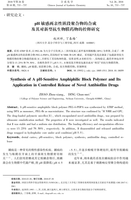
Synthesis of A pH ̄Sensitive Amphiphilic Block Polymer and Its Application in Controlled Release of Novel Antibiofilm Drugs
ZHAO Zhou ̄xiangꎬ DING Chun ̄6ꎬ 2018
Scheme 1
聚乙二醇单甲醚(PEGꎬ Mn = 2 000)ꎻ DPAꎬ纯 度 97% ꎻ聚乙烯醇(PVAꎬ Mw = 89 000 ~ 98 000ꎬ水 解度≥99% )ꎬ Sigma ̄Aldrich 公司ꎻ抗生物膜药物ꎬ Specs 公司ꎻ其余所用试剂均为分析纯或色谱纯ꎮ
( College of Polymer Science and Engineeringꎬ Sichuan Universityꎬ Chengdu 610065ꎬ China)
Abstract: A pH ̄sensitive amphiphilic block polymer PEG ̄b ̄PDPA was synthesized by ATRP methodꎬ using DPA as monomerꎬ PEG ̄Br as macroinitiator. The structure was confirmed by 1 H NMR and GPC. The drug ̄loaded polymeric micelles(1) ꎬ which encapsulated novel antibiofilm drugsꎬ was prepared by ultrasonic emulsification method. The properties of 1 were investigated as well. The results indicated that 1 was stable and had a uniform size distribution. The loading efficiency and encapsulation efficien ̄ cy were 13. 25% and 79. 50% ꎬ respectively. In additionꎬ 1 disassembled and released antibiofilm drugs wrapped in hydrophobic core under acid condition( pH 5. 5) . Keywords: dental cariesꎻ pH ̄sensitiveꎻ block polymerꎻ synthesisꎻ antibiofilm drugꎻ controlled re ̄ lease
【CN109824884A】一种pH敏感和活性氧增敏的普兰尼克聚合物及其制备方法和应用【专利】
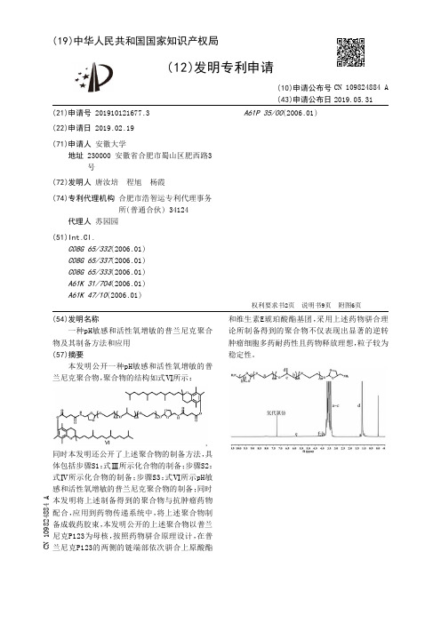
(19)中华人民共和国国家知识产权局(12)发明专利申请(10)申请公布号(43)申请公布日(21)申请号 201910121677.3(22)申请日 2019.02.19(71)申请人 安徽大学地址 230000 安徽省合肥市蜀山区肥西路3号(72)发明人 唐汝培 程旭 杨霞 (74)专利代理机构 合肥市浩智运专利代理事务所(普通合伙) 34124代理人 苏园园(51)Int.Cl.C08G65/332(2006.01)C08G65/337(2006.01)C08G65/333(2006.01)A61K31/704(2006.01)A61K47/10(2006.01)A61P35/00(2006.01)(54)发明名称一种pH敏感和活性氧增敏的普兰尼克聚合物及其制备方法和应用(57)摘要本发明公开一种pH敏感和活性氧增敏的普兰尼克聚合物,聚合物的结构如式Ⅵ所示:同时本发明还公开了上述聚合物的制备方法,具体包括步骤S1:式Ⅲ所示化合物的制备;步骤S2:式Ⅳ所示化合物的制备;步骤S3:式Ⅵ所示pH敏感和活性氧增敏的普兰尼克聚合物的制备;同时本发明将上述制备得到的聚合物与抗肿瘤药物配合,应用到药物传递系统中,将上述聚合物制备成载药胶束,本发明公开的上述聚合物以普兰尼克P123为母核,按照药物骈合原理设计,在普兰尼克P123的两侧的链端部依次骈合上原酸酯和维生素E琥珀酸酯基团,采用上述药物骈合理论所制备得到的聚合物不仅表现出显著的逆转肿瘤细胞多药耐药性且药物释放理想,粒子较为稳定性。
权利要求书2页 说明书9页 附图6页CN 109824884 A2019.05.31CN19824884A1.一种pH敏感和活性氧增敏的普兰尼克聚合物,其特征在于,聚合物的结构如式Ⅵ所示:2.一种制备如权利要求1所述的pH敏感和活性氧增敏的普兰尼克聚合物的方法,其特征在于,所述pH敏感和活性氧增敏的普兰尼克聚合物的合成路线如下:3.根据权利要求2所述的制备pH敏感和活性氧增敏的普兰尼克聚合物的方法,其特征在于,包括以下步骤:S1、式Ⅲ所示化合物的制备:权 利 要 求 书1/2页2CN 109824884 A。
以磁纳米粒为核的pH敏感型聚谷氨酸树状分子药物载体
磁性物质对磁纳 米 粒 的 包 裹,还 会 降 低 其 磁 饱 和 强 度 ,影 响 磁 纳 米 粒 的 磁 响 应 能 力 .
为了克服上述 问 题,必 须 采 用 新 的 材 料 和 方 法对磁纳米粒表面进行功能化. 利用多巴胺与高 温分解法制备的磁纳米粒表面的油酸通过配体交 换进行纳米粒的表面功能化已成为一种新表面改 性策略[8,9],然 而 由 于 多 巴 胺 分 子 上 功 能 团 数 量 的 限 制 ,要 提 高 改 性 磁 纳 米 粒 的 载 药 量 ,还 要 在 改 性后的磁纳米粒表面提供更多的能够键合药物分 子的官能团. 肽类树状大分子是近年来发展起来 的一类新型的、表 面 呈 现 高 密 度 功 能 团 的 生 物 医 用 高 分 子 材 料 ,它 具 有 类 似 蛋 白 的 球 状 结 构 、良 好 的生物相容性和水溶性并且可生物降解且降解产 物无毒性. 利用肽类树状分子对磁纳米粒表面进 行功能化,不仅可 以 通 过 提 高 树 状 分 子 的 代 数 获 得高的官能团密 度 从 而 提 高 载 药 量,而 且 表 面 功 能化后的磁性纳米粒的粒径也可以通过外围树状 分子的代数进行调节[10,11],利用 肽 类 树 状 分 子 外 围的功能团引入 pH 敏感的化学键键合抗肿瘤药 物 ,还 能 实 现 药 物 在 肿 瘤 部 位 的 智 能 释 放[12] .
pH敏感壳聚糖-海藻酸钠复合水凝胶的溶胀特性及应用

pH敏感壳聚糖-海藻酸钠复合水凝胶的溶胀特性及应用胡波;金仙华;谭天伟【期刊名称】《中国组织工程研究》【年(卷),期】2004(008)011【摘要】AIM: To study the swelling characteristic of compound sodium alginate-chitosan gel and its application in slow releasing ofdrugs.METHODS: The granules of compound sodium alginate-chitosan gel were made under mild conditions for studying its swelling kinetics in the pH 1.4environment of stomach and pH 7.4 of environment of small intestine and the effects of various factors on its swelling characteristic. Ketoprofen microcapsule was made according to the gel' s characteristic for detecting its slow releasing effect in the artificial gastric and intestinal fluids.RESULTS: Degree of swelling of the gel was lower in hydrochloric acid buffer of pH 1.4 and was much higher in hydrochloric acid buffer of pH 7.4. Ketoprofen microcapsules made of this kind of gel had significantly slow releasing effect of drugs.CONCLUSION: The effects of the contents of chitosan, sodium alginate and calcium ion content on the swelling characteristics of the gel was discussed and how to made ketoprofen microcapsules of ideal releasing effect according to the characteristics.%目的:研究pH敏感性壳聚糖海藻酸钠水凝胶体系的溶胀特性及在药物缓释中的应用.方法:在温和条件下制备壳聚糖-海藻酸钠复合水凝胶颗粒,研究其在胃的pH环境(pH=1.4)和肠道pH环境(pH=7.4)的溶胀动力学并考察各种因素对凝胶溶胀性能的影响,利用该凝胶的特性制备酮洛芬微囊,测定其在人工胃液和人工肠液中的缓释效果.结果:该凝胶颗粒在pH=1.4的盐酸缓冲溶液中溶胀度较小,而在pH=7.4的磷酸盐缓冲溶液中溶胀度很大,用它制备的酮洛芬微囊具有明显的缓释效果.结论:讨论了壳聚糖浓度,海藻酸钠浓度,钙离子浓度对凝胶溶胀性能的显著影响以及利用这些特性制备的酮洛芬微囊的缓释效果.【总页数】2页(P2162-2163)【作者】胡波;金仙华;谭天伟【作者单位】北京化工大学化学工程学院生物化工系,北京市,100029;北京化工大学化学工程学院生物化工系,北京市,100029;北京化工大学化学工程学院生物化工系,北京市,100029【正文语种】中文【中图分类】R318【相关文献】1.pH 敏感海藻酸钠/氧化石墨烯复合水凝胶球的制备及性能研究 [J], 刘翠云;高喜平;王旭;李东;汤克勇2.pH敏感型壳聚糖-地塞米松纳米粒的制备及体外特性 [J], 冯荣洁;陈广斌;许碧莲3.海藻酸钠与N-异丙基丙烯酰胺接枝共聚及其温度、pH敏感特性研究 [J], 许立新;王秀芬;张立群;刘力4.pH敏感型聚乙烯醇/壳聚糖互穿网络水凝胶的溶胀及载药释药性能研究 [J], 邵雪;梁天宇;丁克毅;刘军5.羧甲基壳聚糖/海藻酸钠水凝胶pH值对溶胀行为的影响 [J], 闻燕;杨志会因版权原因,仅展示原文概要,查看原文内容请购买。
Polyphenol oxidases in plants and fungi Going places A review
ReviewPolyphenol oxidases in plants and fungi:Going places?A reviewAlfred M.MayerDepartment of Plant and Environmental Sciences,The Hebrew University of Jerusalem,Jerusalem 91904,IsraelReceived 3June 2006;received in revised form 22July 2006Available online 14September 2006AbstractThe more recent reports on polyphenol oxidase in plants and fungi are reviewed.The main aspects considered are the structure,dis-tribution,location and properties of polyphenol oxidase (PPO)as well as newly discovered inhibitors of the enzyme.Particular stress is given to the possible function of the enzyme.The cloning and characterization of a large number of PPOs is surveyed.Although the active site of the enzyme is conserved,the amino acid sequence shows very considerable variability among species.Most plants and fungi PPO have multiple forms of PPO.Expression of the genes coding for the enzyme is tissue specific and also developmentally controlled.Many inhibitors of PPO have been described,which belong to very diverse chemical structures;however,their usefulness for controlling PPO activity remains in doubt.The function of PPO still remains enigmatic.In plants the positive correlation between levels of PPO and the resistance to pathogens and herbivores is frequently observed,but convincing proof of a causal relationship,in most cases,still has not been published.Evidence for the induction of PPO in plants,particularly under conditions of stress and pathogen attack is consid-ered,including the role of jasmonate in the induction process.A clear role of PPO in a least two biosynthetic processes has been clearly demonstrated.In both cases a very high degree of substrate specificity has been found.In fungi,the function of PPO is probably different from that in plants,but there is some evidence indicating that here too PPO has a role in defense against pathogens.PPO also may be a pathogenic factor during the attack of fungi on other organisms.Although many details about structure and probably function of PPO have been revealed in the period reviewed,some of the basic questions raised over the years remain to be answered.Ó2006Elsevier Ltd.All rights reserved.Keywords:Polyphenol oxidase;Structure;Genes coding;Multiplicity;Distribution;Induction;Pathogens;Herbivores;Inhibitors;Function of enzymeContents 1.Introduction ..............................................................................23192.Structure and molecular weight of PPO...........................................................23203.Distribution and expression ...................................................................23203.1.Plant PPO..........................................................................23203.2.Methyl jasmonate and PPO .............................................................23213.3.PPO in diverse genera .................................................................23223.4.Chromosomal location of PPO ...........................................................23223.5.Fungal PPO ........................................................................23224.Location and properties of PPO in plants and fungi ..................................................23234.1.Plant PPO..........................................................................23234.2.Fungal PPO ........................................................................23235.Inhibitors of PPO ..........................................................................23245.1.Inhibitors related to phenolic compounds....................................................23245.2.New classes of inhibitors ...............................................................23240031-9422/$-see front matter Ó2006Elsevier Ltd.All rights reserved.doi:10.1016/j.phytochem.2006.08.006E-mail address:mayer@vms.huji.ac.il/locate/phytochemPhytochemistry 67(2006)2318–2331PHYTOCHEMISTRY6.Function (2325)6.1.PPO in biosynthetic processes (2325)6.2.PPO in browning reactions (2325)6.3.Role of PPO in resistance of plants to stress and pathogens (2325)6.4.Role of PPO in defense against herbivores (2326)6.5.Role of PPO in fungal pathogenicity and fungal defense reactions (2327)7.Perspectives (2328)Acknowledgement (2328)References (2328)1.IntroductionPolyphenol oxidases or tyrosinases(PPO)are enzymes with a dinuclear copper centre,which are able to insert oxygen in a position ortho-to an existing hydroxyl group in an aromatic ring,followed by the oxidation of the diphe-nol to the corresponding quinone.Molecular oxygen is used in the reaction.The structure of the active site of the enzyme,in which copper is bound by six or seven his-tidine residues and a single cysteine residue is highly con-served.The enzyme seems to be of almost universal distribution in animals,plants,fungi and bacteria.Much is still unknown about its biological function,especially in plants,but also in fungi.Enzyme nomenclature differen-tiates between monophenol oxidase(tyrosinase,EC 1.14.18.1)and catechol oxidase or o-diphenol:oxygen oxi-doreductase(EC1.10.3.2),but in this review the general term polyphenol oxidase(PPO)will be used.The topic of PPO has been reviewed frequently,and among the more recent general reviews is that of Steffens et al.(1994).In addition reviews of specific aspects of the biochemistry of PPO have appeared.PPO in plants has been reviewed by Yoruk and Marshall(2003),but much of their review covers ground also stressed in other surveys. The mechanism of reaction of tyrosinase has been dis-cussed in great detail by Lerch(1995)and Sanchez-Ferrer et al.(1995),who emphasise the importance of the enzyme in melanogenesis.A survey of mushroom tyrosinase, including lists of inhibitors,the characteristics of the enzyme and its potential uses for clinical purposes has appeared(Seo et al.,2003).The browning of mushrooms, Agaricus bisporus is of major economic importance and the underlying mechanisms have been reviewed by Jolivet et al.(1998),with particular stress on the involvement of tyrosinase in the process.The most recent review of fungal tyrosinases and their applications in bioengineering and biotechnology is by Halalouili et al.(2006),who cover most aspects of this PPO in depth.The potential use of PPO in organic synthesis is reviewed by Burton(2003),although the emphasis in the review is on laccases rather than on PPOs.A comparative analysis of polyphenol oxidase from plants and fungal species,with particular emphasis on sec-ondary protein structure and similarities to hemocyanin was published very recently(Marusek et al.,2006),ampli-fying an earlier review(van Gelder et al.,1997).Their later review emphasizes the amino acid sequence of the enzyme from different sources and especially the N-and C-terminal domains of the enzyme.The review by Marusek et al. (2006)is especially important because it deals with aspects of PPO structure not previously discussed in detail elsewhere.Lastly it should be mentioned that the importance of PPO in browning reactions continues to occupy many researchers as indicated by an ACS Symposium(Lee and Whitaker,1995),and very many subsequent publications describe browning reactions in a variety of species and their tissues.Since the1994review hundreds of papers dealing with plant and fungal PPO have been published.The reason for this plethora of papers is probably the relative ease with which the enzyme activity can be assessed,despite the fact that there are many potential pitfalls in its assay.Many of the published papers report on correlations between levels of PPO activity and environmental factors,attacks by pathogens or changes during food processing or storage. Although useful contributions to the store of information they do not advance the basic understanding of the function of the enzyme and proof of causal relationships between observed phenomena and levels of PPO are mostly missing.It is clear from the perusal of the literature that PPOs are quite diverse in many of their properties,distribution and cellular location.It could therefore be asked whether it is justified to review such a very diverse group.Jaenicke and Decker(2003)write‘‘Probably there is no common tyrosinase:the enzymes found in animals,plants and fungi are different with respect to their sequences,size,glycosyl-ation and activation’’.Discussing the phylogenetic tree of PPO,Wichers et al.(2003),conclude that tyrosinases (PPOs)cluster in groups for higher plants,vertebrate ani-mals,fungi and bacteria.‘‘Homologies within such clusters are considerably higher than between them’’.However,the PPOs have at least one thing in common,they all have at their active site a dinuclear copper centre,in which type3 copper is bound to histidine residues,and this structure is highly conserved.Despite the huge variability of PPO it still seems justified to try and provide an overview of what is happening.The intention of this review is to attempt to provide such an overview for the period from1994untilA.M.Mayer/Phytochemistry67(2006)2318–23312319to today,so that the reader can see where the biochemistry of this group of enzymes is going.2.Structure and molecular weight of PPOThe crystal structure of one PPO in its active form,from Ipomoea batatas has been solved (Klabunde et al.,1998).No comparable data are available for the latent forms of PPO.The crystal structure of a tyrosinase from Streptomy-ces ,bound to a ‘‘caddie protein’’has been resolved.This tyrosinase (Fig.1)shows several features which differ from the plant catechol oxidase (Matoba et al.,2006).These authors ascribe the ability of this tyrosinase to act as a monophenolase as due to some of the observed structural differences.However,it must be remembered that many plant PPOs are able to both hydroxylate monophenols and oxidize dihydroxy phenols,so that monophenolase activity is not a unique characteristic of the Streptomyces enzyme.Indeed,the study of another bacterial tyrosinase,from Ralstonia has shown that possibly the unusually high ratio of hydroxylase/dopa oxidase activity of this particu-lar PPO was linked to the presence of a seventh histidine unit,binding Cu (Hernandez-Romero et al.,2006).The importance of the histidine residues of a fungal PPO,the tyrosinase from Aspergillus oryzyae ,expressed in Esche-richia coli ,has revealed the importance of a previously unrecorded histidine residue (Nakamura et al.,2000).These authors used site directed mutagenesis in their study of the enzyme.They propose that while CuA is linked to three histidine units and one cysteine,CuB is liganded by four histidines,including the newly described one.Thus new information about the detailed structure of PPOs is still being uncovered.No fungal PPO has yet been crystal-lized,either in its active or its latent form.Perhaps the pro-cedures for crystallization described by Matoba et al.(2006)will give an impetus to further attempts in thisdirection.From the structural studies it is also apparent that PPOs do have distinct features and that not only the amino acid sequences of PPOs differ,but that there are also some differences even at the highly conserved active site.The amino acid sequence of a considerable number of PPOs,on plants,fungi and other organisms derived from cloning of the enzyme,has now been published and many of the reports and reviews give such comparative information,e.g.van Gelder et al.(1997),Wichers et al.(2003),Cho et al.(2003),Marusek et al.(2006),Halaouili et al.(2006),Hernan-dez-Romero et al.(2006),Nakamura et al.(2000)and Matoba et al.(2006).As already stated,except for the active site,amino acid sequences show considerable variability and it seems to this reviewer that the salvation for understanding the role of PPO in plants and fungi will not come from the description of yet more amino acid sequences.Reports on the molecular weight of plant PPO are very diverse and variable.It must be assumed that part of this variability is due to partial proteolysis of the enzyme during its isolation.Furthermore,since there obviously is a family of genes coding for plant PPO,some multiplicity must be a result of genetic variability.This problem is addressed by Sommer et al.(1994)and van Gelder et al.(1997).3.Distribution and expression 3.1.Plant PPOWhile the list of species in which PPO have been described and at least partly characterized is growing steadily,the majority of the reports fill out details and do not add any new dimension to the subject.For this reason we will mention only a few of the newer reports,particularly those which also identify the genes coding for theenzyme.Fig.1.Overall structure of the tyrosinase from Streptomyces castaneoglobisporus ,complexed with a ‘‘caddie’’protein,ORF378.The tyrosinase is shown in red and the ORF 378in blue.Copper atoms are shown as green spheres (after Matoba et al.,2006,with permission).2320 A.M.Mayer /Phytochemistry 67(2006)2318–2331The gene coding for PPO in the moss Physcomitrella patens,the properties of the enzyme,and changes in the expression of the gene during growth of the protonema of the moss in liquid culture has been reported(Richter et al.,2005)and this is probably thefirst full report on a PPO in bryophyte.There appears to be only a single gene coding for PPO in this moss,which has one unusual fea-ture,the presence of an intron,absent in most plant PPO genes reported so far.However,in banana an intron is also thought to be present(Gooding et al.,2001),and banana tissues contain at least four distinct genes coding for PPO.The major progress in the description of PPO in plant tissues has been the research on the multiplicity of genes coding for PPO,their description and the characterization of the expression pattern of some of these genes.Some of this ground breaking work was by Steffens and his collab-orators(Newman et al.,1993),as partially described in the review by Steffens et al.(1994).Differential,tissue specific, expression of six genes coding for PPO in potatoes has been reported by Thygesen et al.(1995),and for seven genes in different tissues of tomatoes(Thipyapong et al.,1997). Other early contributions to this aspect are the observa-tions that apple PPO is encoded by a multiple gene family, whose expression is up-regulated by wounding of the tissue (Boss et al.,1994;Kim et al.,2001).The DNA coding for one of the PPOs from apple fruit was cloned and expressed in E.coli.The PPO contained a transit protein and was processed to a mature PPO,M r56kDa.Although the pro-tein expressed in E.coli,M r56kDa,was detected using antibodies,the gene product was enzymically inactive (Haruta et al.,1998).Two different genes are expressed at different stages of appleflower development,one gene cod-ing for PPO being expressed only at the post-anthesis stage, but the two genes had55%identity in their amino acid sequence(Kim et al.,2001).This multiplicity of genes,their differential expression in different parts of the plant and at different stages of development is one of the most impor-tant features of recent work on plant PPO.The sequences of PPO in any one species are highly conserved,but there is a lot of divergence in the sequences among different spe-cies(Thygesen et al.,1995).However,this divergence may not be greater than has been reported for other genes cod-ing for enzymes,and a comparison is difficult.Surprisingly, early work by Robinson and his co-workers indicate the presence of only a single PPO gene in grape vine(Dry and Robinson,1994).3.2.Methyl jasmonate and PPOThe response of expression of PPO to wounding has been shown in poplar(Constabel et al.,2000),who also showed that methyl jasmonate induced expression of PPO genes,a fairly general phenomenon(Constabel and Ryan,1998).However,not all species respond to methyl jasmonate by induction of PPO,so that although common, induction of PPO activity by methyl jasmonate,is by no means a universal response.It is also by now well estab-lished that methyl jasmonate induces formation of other proteins involved in the defense response of plants(Const-abel et al.,1995;Howe,2004).The presence of PPO in glandular trichomes of tomato and potato has been described by Steffens et al.(1994).The trapping of insects, mediated by PPO,was a groundbreaking result on the function of the enzyme in a clearly defined system.More and more examples of PPO induction by jasmonate or methyl jasmonate are appearing.In the leaves of Datura wrightii,PPO is induced in the trichomes,irrespective of whether the trichomes are glandular or non-glandular (Hare and Walling,2006).Other enzymes were not induced, e.g.peroxidase,nor was alkaloid production enhanced.However,acyl sugars were preferentially syn-thesised in one form of the trichomes,the glandular ones. Clearly the inductive effect of jasmonate is very complex. Such methyl jasmonate induced expression has also been demonstrated by Koussevitzky et al.(2004),who showed that import and processing of the PPO into chloroplasts from tomato was enhanced by pre-treatment with methyl jasmonate.This observation adds a new and important facet to the mechanisms which control PPO activity in plant tissues.The step most affected by methyl jasmonate seemed to be transport to the thylakoids.A localized effect of methyl jasmonate has been shown to exist in tomato seeds(Maki and Morohashi,2006).At the stage of radical protrusion,the level of PPO increased about fourfold at the micropylar end of the endosperm, but not in other parts of the endosperm.Wounding had a similar effect on the level of PPO activity.The wound induced PPO was distinct from the PPO in other parts of the endosperm.Treatment of half seeds of tomato with methyl jamonate,either ruptured or non-ruptured showed that methyl jasmonate only induced PPO activity in the micropylar part of the endosperm.The M r of PPO induced in ruptured and un-ruptured micropylar endosperm was different,but this could be the result of different processing. The formation during germination can probably be ascribed to wounding,which occurs during radicle protru-sion,but its role is by no means clear.It must be assumed that such a localized formation of PPO in a well defined and controlled developmental process is of biological sig-nificance,but its function requires further study.The ability of jasmonic acid,applied to the leaves of Physalis,to induce PPO activity was dependent on the time of year when it was applied,maximum induction being obtained in young plants during the summer(Doan et al.,2004).No data seem to be available whether different enantiomers of methyl jasmonate show differences in their ability to induce PPO activity.Jasmonate can have clear effects even underfield condi-tions.Tobacco plants grown in the vicinity of sagebrush (Artemisia tridentata)responded to clipping of the sage-brush by an increased formation of PPO,the response being mediated by methyl jasmonate(Karban et al., 2000).Moreover,the tobacco plants near the clipped sage-brush experienced reduced damage by grasshoppers andA.M.Mayer/Phytochemistry67(2006)2318–23312321cutworms.Obviously communication between plants plays a role in controlling PPO levels.Expression of the iso-forms of PPO is differential(Haruta et al.,2001;Wang and Constabel,2003).3.3.PPO in diverse generaThe presence of PPO has been described in a variety of plants,some unusual or exotic.In most cases the descriptions cover molecular weight and often multiplic-ity.The characteristics of the PPOs mostly show no spe-cial features,but a few instances will be mentioned.The PPO from the aerial roots of an orchid Aranda was found to be present in four iso-forms,which were par-tially characterized,including the N-terminal sequences of the iso-forms(Ho,1999).Since aerial roots contain chloroplasts,it is probable that these PPOs were located in the plastids.Two distinct PPOs are present in leaves and seeds of coffee(Mazzafera and Robinson,2000),in the parasitic plant Cuscuta(dodder)(Bar Nun and Mayer,1999), and in Chinese cabbage(Nagai and Suzuki,2001).The latter has been partially purified and appears to be an example of a PPO in the Cruciferae.Annona muricata (Bora et al.,2004),oregano(Dogan et al.,2005a),persim-mon(Ozen et al.,2004),artichoke(Dogan et al.,2005b), marula(Sclerocarya birrea)(Mduli,2005),loquat(Eriob-otrya japonica)(Selle´s-Marchart et al.,2006)and Uapaca kikiana fruit,a plant belonging to the Euphoriaceae (Muchuweti et al.,2006),all contain PPO.The PPO level in apricot fruits remains high even at a stage when it’s mRNA can no longer be detected,indicating that the pro-tein is stable for long periods(Chevalier et al.,1999).This raises the question,is the presence of mRNA a good indi-cator of function or importance of the enzyme in a given tissue.Most work characterizing PPO and its DNA and mRNA implicitly assume that levels of mRNA are directly related to function,but this may not be true in all cases.The PPO present in red clover,which has an important role during ensiling of leaves,has been cloned and charac-terized.At least three PPO genes were detected,which had a high degree of identity,and which were differen-tially expressed in different parts of the plant(Sullivan et al.,2004).One of these genes was successfully expressed in E.coli.The proteins encoded by these three genes all had sequences which predict that they would localize in chloroplast thylakoids.An unsuspected significance of this particular PPO is that silages prepared from clover forage with high PPO activity are of better quality than those with lower PPO activity(Lee et al.,2004).An important aspect the work by Sullivan et al.(2004)is that it is apparently the only report of recombinant expression of PPO with partial activity of the expressed protein.Obvi-ously this opens up many possibilities for further study of PPO.Although it is generally agreed that PPO is plas-tid located,the site at which it is present in potato tubers is not entirely clear.PPO in undamaged tissue was located to starch grains and the cytoplasm,but upon several hours after mechanical bruising the PPO becomes more generally distributed including in the vacuolar region (Partington et al.,1999).This is presumably due to break-down of membrane integrity and thus direct evidence of ‘‘leakage’’from defined sites was demonstrated,using immuno-gold localisation integrity appearing in the cytoplasm.3.4.Chromosomal location of PPOThe observation that hexaploid wheat kernels have six genes coding for PPO,of which at least three are expressed during development of the kernel(Jukanti et al.,2004)is worth noting.The deduced amino acid sequence and sim-ilarity with other PPOs has been recorded for at least one PPO from wheat(Demeke and Morris,2002).This is sig-nificant in view of the browning phenomena in cereal prod-ucts.Some variability in reports on purified wheat PPO is apparent.Kihara et al.(2005)report a M r of35kDa or 40kDa,depending on assay,for a homogeneous prepara-tion of PPO from Triticum aestivum and say that its amino acid sequence resembles that of other PPOs,but Anderson and Morris(2003)report a M r of67kDa and state that it resembles other PPOs as much as other wheat PPO.The purified PPO from wheat bran appeared to be the mature form and lacked the transit peptide locating it to the plas-tids(Anderson and Morris,2003).Perhaps these results are not surprising in view of the fact that six genes coding for PPO in wheat are known and perhaps forms differing in maturity were isolated.Genetic analysis of the location of PPO in wheat points to a complex situation.One gene from of T.turgidum coding for PPO was mapped to chro-mosome2D(Jimenez and Dubcosky,1999).Reports on QTLs for PPO in T.aestivum indicate that a number are present on different chromosomes(Demeke et al.,2001). Genetic mapping of PPO in wheat seems to be the most detailed reported for any PPO,and has implications for selection for wheat with low PPO activity.Vickers et al.(2005)manipulated the levels of PPO in transgenic sugar cane using constructs of sense and anti-sense to the native PPO gene and found that as a result the degree of browning of the juice could be changed. Over-expressing PPO led to enhance browning,the PPO content of the juice being elevated.3.5.Fungal PPOSince fungal tyrosinase has been reviewed in great detail recently(Halaouili et al.,2006;Seo et al.,2003),no attempt will be made here to report on its distribution, which in any case appears to be universal in fungi.Perhaps the isolation of the latent form of PPO in the ascocarp of Terfezia claveryl(a truffle),should be recalled(Perez-Gila-bert et al.,2001),the general behaviour of this PPO falling in line with that of other fungal PPOs.2322 A.M.Mayer/Phytochemistry67(2006)2318–23314.Location and properties of PPO in plants and fungi4.1.Plant PPOAn early report by Rathjen and Robinson(1992)sug-gested that PPO in grape berries could accumulate in what appeared to be an aberrant form,with a molecular weight of60kDa,and not the expected one of40kDa.They sug-gested that PPO in the variegated grapevine was synthesized as a precursor protein which was then processed to a lower molecular weight form.It was also shown that the PPO of broad bean,which is latent,can be activated by SDS,and can undergo proteolytic cleavage with out loss of activity (Robinson and Dry,1992).Sommer et al.(1994)investi-gated the pathway by which plant PPO reaches the chloro-plast.They studied in detail the synthesis,targeting and processing of ing an in vitro system and pea chlo-roplasts they showed that tomato PPO,coded by cDNA, was processed in pea chloroplasts in two steps during its import.The precursor PPO with M r67kDa was imported into the stroma of the chloroplasts by an ATP-dependent step.It was then processed into a62kDa form by a stroma peptidase.Subsequent transport into the lumen was light dependent and resulted in the mature59kDa form.Appar-ently such processing is a feature of all chloroplast-located PPOs.The precursor protein contains a transit peptide, which must be removed in order that the PPO reaches its site in the chloroplast.The processing is carried out by a stromal peptidase,which was purified and characterized (Koussevitzky et al.,1998).The import and processing did not require Cu2+,but import was inhibited by micromo-lar concentrations of Cu2+.Further studies revealed that this inhibition was probably due to inhibition of the stromal peptidase involved in the processing of the precursor pro-tein(Sommer et al.,1995).It is clear therefore,that the synthesis of PPO and its transport to its site in chloroplasts,where plant PPOs are thought to be located,is a complex process,but which has the general features of import of nuclear coded proteins into sub-cellular organelles.A curiousfinding on the possible ability of PPO to act as a protease has been reported(Sokolenko et al.,1995; Kuwabara,1995;Kuwabara et al.,1997;Kuwabara and Katoh,1990).According to these reports a protein said to be identical with PPO in structure and properties can under certain conditions oxidatively degrade a low molec-ular weight protein located in chloroplasts.The evidence for the identity of this PPO-like protein is still not totally convincing and it has not been cloned or fully characterized using the techniques of molecular biology.Whether this activity is of any physiological significance remains to be demonstrated.4.2.Fungal PPOThe location of fungal PPO is not entirely clear.Gener-ally it appears to be a cytoplasmic enzyme.However,a PPO from Pycnoporus over-produced in Aspergillus niger, could be targeted to the extracellular growth medium (Halaouili et al.,2006).An additional PPO is present in the mycelium of Pycnoporus saguineus,which has a very high tyrosinase activity and is able to convert coumaric acid to caffeic acid in vitro(Halaouili et al.,2005).This enzyme differs from other PPOs from Pycnoporus and existed in four iso-forms,with M r of45kDa.The enzyme was not N-glycosylated.At least in some cases fungal PPO is associated with the cell wall,occurring apparently in the extra cellular matrix (Rast et al.,2003).The molecular weights of fungal PPO show considerable diversity.Part of this diversity is genetic as is indicated by the clustering in the phylogenetic tree of the enzyme(Halaouili et al.,2006),and part can be ascribed to artifacts arising during isolation of the enzyme.This ques-tion is discussed by Halaouili et al.(2006).Fungal PPO,as plant PPO,can be present in latent form which is activated by SDS or proteolysis or acid shock.SDS activation in beet root PPO is said to be reversible(Perez-Gilabert et al.,2004), while trypsin activation is not.Gandia-Herrero et al. (2005b)suggest that a common peptide is involved in activa-tion both by SDS and trypsin.The evidence is not totally compelling and it should be remembered that very old work on sugar beet chloroplast PPO already showed that even peptides with M r of around10kDa still retain considerable PPO activity.This shows that by no means the entire protein is required for activity(Mayer,1966).Further insight into the activation of a latent PPO comes from the work of Kanade et al.(2006)on PPO from Dolichos lablab,which shows that both acid and SDS change the environment of a single glutamic residue,close to the di-copper active site. As a result the active site is unblocked or opened and enzyme activity enhanced.An intriguing suggestion by the authors,as yet unproven,is that wounding or methyl jasm-onate could cause localized acidification which results in conversion of a latent to an active enzyme.An interesting feature of two tyrosinases from Agaricus is that the proteins contain putative glycosylation and phosphorylation sites, although no glycosylation or phosphorylation has so far been reported for fungal PPO(Wichers et al.,2003).It is fairly clear that fungal PPOs also undergo some processing,by proteolysis,and that the synthesized form of the enzyme is trimmed.What is less clear at the present is what exactly happens during the conversion of latent to active forms of the fungal PPOs?Tyrosinases from crustaceans have been shown to occur in vivo as a hexamer,made up from a single subunit of molecular weight71kDa(Jaenicke and Decker,2003). This of course recalls the many older reports of associa-tions of plant and fungal PPO into aggregates,but in the case of the crustacean PPO,the hexamer has been shown to exist in vivo and its structure demonstrated by electron microscopy.The similarities of this structure with haemo-cyanin are apparent(Gerdemann et al.,2002).Nevertheless aggregation into a hexameric form,existing in vivo,has not been shown for any plant or fungal PPO.A.M.Mayer/Phytochemistry67(2006)2318–23312323。
pH敏感树枝状聚合物纳米粒子-载氟尿嘧啶聚合物纳米载体
pH敏感树枝状聚合物纳米粒子-载氟尿嘧啶聚合物纳米载体
一个树枝状聚合物子分散在水中形成1-10nm大小的粒子,它的最大特点是结构非常规整,内核有一疏水空腔,表面存在许多亲水的功能基团。
树枝状聚合物的毒性通常很低,因此是非常有发展前景的给药载体。
药物包裹于树枝状聚合物的内腔,致密的外表层控制着药物释放。
表面功能基团可以接上配体,为树枝状聚合物纳米粒子给药提供靶向性。
最近树枝状聚合物纳米粒子有用于抗生剂的系列报道。
但由于树枝状聚合物的制备比较复杂,短期内广泛应用受到限制。
用超支化聚合物代替树枝状聚合物也可实现药物靶向输送.
以表面接枝聚乙二醇链的聚酰胺胺树枝状聚合物(PEG-PAMAM)为纳米载体,在其内部空腔包覆金纳米粒子,在金纳米粒子表面连接硫辛酸改性的阿霉素(LA-DOX),从而间接实现了抗癌药物在PEG-PAMAM 内的高效负载.同时,LA-DOX中的酰腙键提供pH响应性,实现了药物的pH响应性释放.紫外可见(UV-Vis)光谱表明,包覆金纳米粒子的PEG-PAMAM纳米载体对LA-DOX的负载能力显著增强.体外细胞实验表明,负载LA-DOX的树枝状聚合物-金纳米粒子复合药物载体具有较强的抗肿瘤能力.。
pH敏感性羟乙基甲壳素/聚丙烯酸水凝胶的制备及其释药性能研究
pH敏感性羟乙基甲壳素/聚丙烯酸水凝胶的制备及其释药性能研究生物医学工程学杂志2OO6l23(2):338~341JBiomedEng…………pH敏感性羟乙基甲壳素/聚丙烯酸水凝胶的制备及其释药性能研究*赵育陈国华孙明昆晋治涛高从土皆h1(中国海洋大学化学与化工学院,青岛266003)2(国家海洋局水处理技术开发中心,杭州310012)摘要用氯乙醇对甲壳索进行醚化改性,得到水溶性羟乙基甲壳索(Hydroxyethylchitin,HECH),用化学交联法制备了由聚丙烯酸(PAA)和HECH复合的互穿网络(IPN)水凝胶.溶胀实验表明:该水凝胶在人工肠液(pH7.4,I=0.1)中的溶胀度远大于在人工胃液(pil1.4,I—o.1)中的溶胀度,凝胶的溶胀度随着温度的升高而增大;以该凝胶制备了双氯芬酸钾缓释体系,释放实验表明该凝胶具有较好的缓释性能.关键词羟乙基甲壳素聚丙烯酸双氯芬酸钾缓释水凝胶StudyonPreparationofthepHSensitiveHydroxyethyiChitin/Poly(AcrylicAcid)HydrogeiandItsDrugReleaseProperty ZhaoYuChenGuohuaSunMingkunJinZhitaoGaoCongjie'1(CollegeofChemistryandChemicalEngineering,OceanUniversityofChina,Qingdao266 003,China)2(DevelopmentCenterWaterTreatmentTechology,SOA,Hangzhou310012,China) AbstractHydroxyethylchitin(HECH)isawatersolublechitinderivativemadebyetherificati onofchitin, ethylenechlorohydrinwasusedasetherificationreagentinthisreaction.Anovelinterpenetratingpolymernetwork(IPN)composedofHECH/PAAwasprepared.TheIRspectraconfirmedthatHECH/PAAwa sformedthroughchemicalbondinteraction.ThesensitivityofthishydrogeltOtemperatureandpHwasstudied .Theswellingratio ofthishydrogelinartificialintestinaljuiceismuchgreaterthanthatinartificialgastricjuice.T heIPNhydrogelexhibitedatypicalpH-sensitivity,anditsdegreeofswellingratioincreasedwiththeincreaseo ftemperature.Thesustained-releasedrugsystemofDichlofenacpotassiumwaspreparedbyusingHECH/PAA asthedrugcarrier. Thereleaseexperimentshowedaperfectreleasebehaviorinartificialintestinaljuice.ThisIP NisexpectedtObe usedasagooddrugdeliverysystemofentericmedicine.' KeywordsHydroxyethylchitin(HECH)PAADichlofenacpotassiumDrugdeliverysystem Hydrogel1引言环境敏感性水凝胶是当前研究得非常广泛的一类水凝胶L1],这类水凝胶在生物医学领域和智能化药物缓释体系[2中的应用日益得到重视.卓仁禧等口对聚丙烯酸/聚N一异丙基丙烯酰胺互穿聚合物网络水凝胶的溶胀性能进行了研究,发现这种水凝胶在弱碱性条件下的溶胀度远大于酸性条件下的溶胀度.李文俊等[4制备了聚丙烯酸/壳聚*国家973计期资助课题(2oo3CB6157oo)△通讯作者***********************糖半互穿聚合物网络膜,考察了其对pH和离子的刺激响应.Fwu—longLs等用甲壳素/乙交酯丙交酯嵌段共聚物(Polylactide—CO—glycolide,PIGA)共混物制备了药物缓释微胶囊.由于甲壳素具有生物降解性和PIGA的水解特性,该胶囊能够在人体中发生降解以释放出包埋药物.研究表明,共混组分中甲壳素的含量越高,胶囊就降解的越快.羟乙基甲壳素是一种水溶性甲壳素衍生物,具有较好的生物相容性,其水凝胶的制备及性能未见文献报导.我们用羟乙基甲壳素与PAA复合,得到一种同时具有pH敏感性和生物相容性的互穿网络(IPN)水凝胶.由于聚合物问的互穿作用,使其溶胀性能不同于单独的第2期赵育等.pH敏感性羟乙基甲壳索/聚丙烯酸水凝胶的制备及其释药性能研究33g凝胶体系.双氯芬酸钾(Dichlofenacpotassium,DCFP)是一种邻氨基苯甲酸类解热,镇痛和抗炎药物,半衰期短,1d需3~-.4次给药,口服后吸收较快,对肠胃道有刺激,所以需开发其缓释材料.本文利用HECH/PAA作为双氯芬酸钾的释放材料,并对其缓释机理进行了初步的研究.2实验2.1仪器和药品甲壳素,青岛海汇生物有限公司;氯乙醇,分析纯,南翔试剂厂;双氯芬酸钾,苏州市立德化学有限公司;95%乙醇,分析纯,淄博化学试剂厂;甲醇,分析纯,济南试剂总厂;丙烯酸,化学纯,中国上海五联化工厂,N,N一亚甲基双丙烯酰胺(Bis),分析纯,Sigma公司,过硫酸钾,分析纯,宜兴市第二化学试剂厂;磷酸二氢钾,分析纯,上海化学试剂公司,磷酸氢二钠,分析纯,上海化学试剂公司.RE一52型旋转蒸发器,上海亚荣生化仪器厂;BS210S型电子天平,北京塞多利斯天平有限公司, 感量0.1mg;Aratar360型红外光谱仪,Nicolet公司;Spectrumlab52紫外分光光度计;DSHZ一300水浴恒温振荡箱,江苏太仓实验设备厂,日本电子JsM6700F型扫描电镜.2.2羟乙基甲壳素的制备参考国外文献i-6-1,并加以改进:将5g甲壳素粉末分散于100m150%NaOH水溶液中,常温减压(~20mmHg)碱化4h,过滤后,滤饼用35ml50 NaOH水溶液洗净,加入碎冰60g,高速搅拌30 min,得到黏稠的碱溶液,然后稀释成NaOH浓度为14的溶液,在冰浴中,搅拌下滴加36g氯乙醇,移去冰浴,搅拌过夜.然后在冰浴下用冰醋酸中和,过滤,将滤液减压蒸发浓缩,丙酮沉淀.再用80%乙醇多次脱盐,经硝酸银溶液检查至无白色沉淀生成, 40"C真空干燥得羟乙基甲壳素.将得到的HECH溶于200ml去离子水,过滤除去不溶物,再用丙酮沉淀,干燥,得到提纯产物.2.3HECH/PAA缓释凝胶的制备2.3.1HECH/PAA互穿网络(IPN)凝胶制备称取HECH1g溶解到盛有20g水的烧杯中,加入丙烯酸单体1g和交联剂N,N一亚甲基双丙烯酰胺0.02g,再加入过硫酸钾0.02g,搅拌至充分混合,在60℃的水浴中反应24h后,取出.切成1cm.小胶块,用去离子水多次洗涤,50~C真空干燥至恒重, 得到IPN凝胶颗粒.2.3.2HECH/PAA双氯芬酸钾缓释凝胶制备制备及处理过程同2.3.1,在加入上述各组分后,加入双氯芬酸钾药物颗粒500mg,然后搅拌至充分混合,处理后得到双氯芬酸钾缓释凝胶颗粒.2.4HECH/PAAIPN凝胶的红外光谱样品真空干燥过夜,粉碎,用KBr压片法进行红外表征.2.5HECH/PAA双氯芬酸钾缓释凝胶表面形态观察将包药的干凝胶表面喷金处理后利用扫描电镜照片(Scanelectronmicrograph,SEM)观察其表面形态.2.6HECH/PAA凝胶的溶胀性能准确称量一定量的干凝胶,将干凝胶浸泡在相同离子强度(I一0.1)不同pH值(pH一1.4,7.4)的缓冲溶液中,来模仿人工胃液和人工肠液环境[3],在一定温度下,平衡一定时间,纱布过滤,用滤纸吸干表面的水,称重,溶胀度按下式计算:SwellingRatio 一(Wt—Wo)/wo(Wt和Wo分别为吸水后和吸水前的重量).注:人工胃液:pH一1.4,I一0.1的盐酸与氯化钠溶液体系.人工肠液:pH一7.4,I一0.1的0.025M的KHzPO.和NaHPO.缓冲溶液.2.7标准曲线的绘制精密称取60~C真空干燥至恒重的双氯芬酸钾20mg,置于100ml容量瓶中,加甲醇溶解,并用甲醇稀释至刻度,摇匀.将溶液在200400nm波长扫描,选取最大吸收波长.扫描最大吸收波长为285 nm.精密量取0.5,1.0,1.5,2.0,2.5,3.0m1分别置于25ml容量瓶中,用甲醇稀释至刻度.以甲醇作空白,于285nm处测A值,得到标准曲线回归方程:A一0.00673+0.03376C(R一0.9992,7/一6)2.8体外释放实验体外释放实验[采用带有温控装置的振荡器(释放温度为37.5土0.5℃,振速30次/min).释放介质为人工肠液.准确称取干燥后的缓释凝胶0.1 g,分散于200m1人工肠液中.从释放开始至第1, 2,3,4,6,8,10,12h各取样一次,每次取样5ml,并补充相同体积的同种介质,以甲醇为空白,测定吸光度.对应回归方程计算药物释放浓度,累积释放率按下式计算:Q一(szG—l+200(7,,)/(取样量x药物含量)式中:Q为第i次取样后体系的累积释放率;C为第生物医学工程学杂志第23卷i次取样时药物的释放浓度;5为每次取样体积(m1);200为释放体系总体积(m1).3结果与讨论3.1红外光谱如图l所示:a.的2923cn1一处是CH的吸收峰,有典型的三个酰胺谱带,分别出现在l660 Cnl~.1559Cnl和I315cm..左右,其中I660cm是双重峰.b.的2920cm是饱和烃的吸收峰.17,50cm处是羧基的吸收峰从图中可以看出,12.的红外谱图恰好与羟乙基l=I|壳素f.a)聚而烯酸(b)的红外谱图之和相吻合,说明在形成互穿结构的过程中没有复杂的化学键形成.囤l羟乙基甲壳摩la).聚丙擂馥(1,】和互穿咄凝胶【c)的虹扑谱图FitIIREpecnCI"ch"Ln'a).PA^(b)wadlPt)3.2双氯芬酸钾缓释凝胶的表面形态由图2可见,IPN袭面形态为非连续性.说明PAA与HECH形成互穿聚合物网络结构,白色的药物颗粒分散在凝胶中.周2IPN水凝胺的扫描电镀j!}c片【放大5000倍jFig2Scan~|eclronmicrographorIPNhydrogel(magnification×5000)3.3凝胶的溶胀性研究本研究制备的HECH/PAA凝胶在人工胃液和人工肠渡中的溶胀度随温度的变化趋势如图3所示,水凝胶在人工肠液缓冲溶液的溶胀度远大于人工胃液的溶胀度.随着温度的提高,水凝胶的溶胀度增大,在弱碱性缓冲溶液中的溶胀度显着高于酸性缓冲溶液,具有明显的pH敏感性,而且其随着温度增加溶胀度增大趋势前者高于后者,是一种"热胀型"水凝胶而聚(丙烯酸)/聚(N一异丙基丙烯酰胺)凝胶0在酸性条件下,随着温度的于七高+溶胀率也随之逐渐上升,在弱碱性的条件下,当温度低于较低临界溶解温度(LCST)时,溶胀率也随着温度的上升而上升,当温度达到ICST时,凝胶的溶胀率突然急剧下降,并随着温度的逐渐上升而下降两者的不同主要是由于HECH对于温度的响应不同于聚(N一异丙基丙烯酰胺)所致3.2焉∞2兰1202530354045t(℃)囤3水疆肢的溶胜鹰在人工膏淮和人工舾液中瞄温度的变化酋缦Fig3Temperaturedepcndellceofswellingrallojnartificialjuicegastricandarli[irillin"Iinaliuice3.4体外释放曲线HECH/PAA缓释凝胶的释放曲线如图d,将1~12h的释放血线接近似线性方程拟台.释放方程为:Mt/Mmc』一17.29+7.42t(尺:0.9450+,2—8)式中:Mt为t时刻累计释放量;Mmax为药物含量.药物在第1h的累积释放率达到17.12h内的释放几乎是匀速的,释放接近零级动力学+累积释放率达到95.由于该聚合物的溶胀性能具有pH敏感的特睦.当口服缓释颗粒进人胃中,在酸性条件下颗粒的溶胀度较小,因此药物在胃液中基本不释放当颗粒进人人的肠道后,由于肠液呈弱碱性,缓释颗粒溶胀度明显增大,药物开始释放,该缓释体系可以连续在人体中释药长达12h,显示出优异的缓第2期赵育等.pH敏感性羟乙基甲壳素/聚丙烯酸水凝胶的制备及其释药性能研究341释性能.O24e8,0,2Time(h)圈4双氯芬酸钾缓释互穿凝胶在人工肠液中的释放曲线Fig4ReleaseCurveofDCFP—IPNinartificialintestinaljuice4结论(1)HECH/PAAIPN水凝胶在弱碱性介质人工肠液中的溶胀度大于在酸性介质人工胃液中的溶胀度,具有明显的pH敏感性,并且溶胀度随着温度的升高而增大,是一种"热胀型"的水凝胶.(2)以这种凝胶为骨架的双氯芬酸钾缓释制剂能够在12h匀速释放,适合作为肠溶型药物释放体系.参考文献1HuangGF,QingSB,LiSB.Theadvanceofpolysaccharide biomedicalmaterials—chitinandchitosan.PolymerBulletin,2001 (3)t43[黄光佛,卿胜波,李胜彪等.多糖类生物医用材料一甲234567壳素和壳聚糖的研究及应用.高分子通报,2001(3)t43]LiF,ZhaoF,Yin,YJ,eta1.Applicationsforchitosanbasedhydroge1.ChemistryBulletin,2001(3)l129[-~方,赵峰,尹玉姬等.壳聚糖基智能凝胶材料及其应用.化学通报,2001 (3):129"]ZhuoRX,ZhangXZ.Thesynthesisandcharacterizationof temperatureandpHsensitiveofPoly(acrylicacid)/Poly(N—isopropylacrylamide)IPNhydrolge[.ActaPolymericaSinica, 1998;(1):39[卓仁禧,张先正.温度及pH敏感聚(丙烯酸)/聚(N一异丙基丙烯酰胺)互穿聚合物网络水凝胶的合成及性能研究.高分子,1998(1)t39]LiwJ,WangHF,LuYH,eta1.Chitosan/polyacrylicacid complexformingsemi—interpenetratingpolymernetwork membraneanditsstimulatingresponseforpHandions.Acta PolymericaSinica,1997(1):106[李文俊.王汉夫,卢玉华等.壳聚糖/聚丙烯酸配合物半互穿聚合物网络膜及其对pH和离子的刺激响应.高分子,1997(1)t1o6JFwu—longMi,Yi-MeiLin,Yu-BeyWu,Chitin/PLGAblend microspheresasabiodegradabledrug—deliverysystem:phase—separation.degradationandreleasebehavior.Biomaterials,2002(23):3257HidennoriYamada,TaijiImoto.Aconvenientsynthesisof glycolchitin,asubstrateoflysozyme.CarbohydrateResearch,198l(92):16OHuangYW.LuoXG,ZhuoRX,Studyoncontrolledreleaseof aspirininthetemperatureandpHsensitivehydroge1.Polymer MaterialsScienceandEngineering,1998;14:141—147[黄月文,罗宣干,卓仁禧.包埋在温度及pH值敏感水凝胶中的阿司匹林的控制释放研究.高分子材料科学与工程,1998;I4t141-147-] (收稿l2004—02—04修回:2004—06—07)∞∞∞∞的∞∞{乏伯。
用于药物递送的pH敏感聚合物研究进展

用于药物递送的pH敏感聚合物研究进展
齐荣翔;崔汉钊;周海梅
【期刊名称】《山东化工》
【年(卷),期】2016(45)23
【摘要】本文综述了pH敏感聚合物(pH-sensitive polymer)载体靶向输送抗肿瘤药物的研究进展.肿瘤组织的细胞间质呈弱酸性(pH值<7),而肿瘤细胞内的溶酶体和内涵体具有更强的酸性(pH值4~6).pH敏感聚合物在酸性介质中能发生质子化或化学反应,使其物理或化学性质发生改变.使用pH敏感型聚合物做成的药物载体能够对肿瘤酸性微环境产生响应,实现药物的快速释放、激活载体的靶向传输功能、促进载体的细胞内吞.大量实验表明,pH敏感型药物载体输送抗肿瘤药物可以明显增加药物在作用部位的浓度,从而饱和肿瘤细胞的多种抗药机制,克服肿瘤细胞的耐药性,提高抗癌药物的治疗效果同时减少其毒副作用.
【总页数】4页(P72-75)
【作者】齐荣翔;崔汉钊;周海梅
【作者单位】河南科技大学法医学院,河南洛阳471023;河南科技大学法医学院,河南洛阳471023;河南科技大学法医学院,河南洛阳471023
【正文语种】中文
【中图分类】R94
【相关文献】
1.温度敏感型原位凝胶药物递送系统的研究进展 [J], 宋亚; 祁小乐; 沙康; 曾佳; 吴正红
2.pH响应性聚合物胶束在药物递送系统中的研究进展 [J], 李艳艳;毕韵梅
3.用于抗肿瘤药物递送的刺激敏感型聚合物载体的研究进展 [J], 李景果;冯华阳
4.聚合物纳米载体介导化疗药物口服递送的研究进展 [J], 乐志成;刘志佳;陈永明
5.集原位SERS传感及pH敏感于一体的CS/TiO2纳米药物递送系统 [J], 于东雪;江欣;杨立滨;赵冰
因版权原因,仅展示原文概要,查看原文内容请购买。
- 1、下载文档前请自行甄别文档内容的完整性,平台不提供额外的编辑、内容补充、找答案等附加服务。
- 2、"仅部分预览"的文档,不可在线预览部分如存在完整性等问题,可反馈申请退款(可完整预览的文档不适用该条件!)。
- 3、如文档侵犯您的权益,请联系客服反馈,我们会尽快为您处理(人工客服工作时间:9:00-18:30)。
PAPER
Published on 02 May 2014. Downloaded by University of North Carolina at Chapel Hill on 23/10/2014 15:01:40.
View Article Online
View Journal | View Issue
Cite this: Polym. Chem., 2014, 5, 5168
Highly pH-sensitive polyurethane exhibiting shape memory and drug release†
Hongmei Chen,ab Ying Li,a Ye Liu,a Tao Gong,a Lin Wanga and Shaobing Zhou*a
a
Key Laboratory of Advanced Technologies of Materials, Ministry of Education, School of Materials Science and Engineering, Southwest Jiaotong University, Chengdu, P. R. China. E-mail: shaobingzhou@; shaobingzhou@; Fax: +86-2887634649; Tel: +86-28-87634068 Department of Chemistry and Materials Science, Sichuan Normal University, Chengdu, P. R. China
Received 3rd April 2014 Accepted 1st May 2014 DOI: 10.1039/c4py00474d /polymers
Introduction
Stimulus-responsive materials can undergo conformational or phase changes in response to environmental signals.1 The materials represent one of the most exciting emerging areas of science2 due to their promising applications in elds such as actuator systems,3 tissue engineering4 and programmable delivery systems.5 Although some progress has been made in recent years, the design and engineering of a synthetic material with an abilitontrollable and predictable fashion remains a challenge. Among these responsive materials, shape memory polymers (SMPs) have attracted increasing attention for their potential medical applications.6,7 SMPs can change from their permanent shape into a temporary shape, and recover to their permanent shape upon application of an appropriate stimulus such as temperature,8,9 light,10,11 water,12,13 magnetic eld14 or chemicals.15 Thermally induced SMPs are the most widely studied, but their biomedical
1
applications are sometimes limited because the switching temperature should be in the range from room temperature to body temperature. For biomedical applications, an ideal stimulus must be chosen based on the physiological environment. Variations in physiological pH exist among different sites in the body including the gastrointestinal tract, vagina and blood vessels; on the other hand, a sharp pH gradient generally appears across biological systems on both the cellular and systemic levels in pathological states which differ from the physiological pH of 7.4.16 Thus, the pH stimulus is a good choice for the design of shape memory polymers with potential applications in medicine. Zhang et al. reported a pH-sensitive shape memory polymer prepared with b-cyclodextrin modied alginate and diethylenetriamine modied alginate.17 This material can be processed into a temporary shape at pH 11.5 and recover to its initial shape at pH 7. But as far as we know, SMPs stimulated by acidic conditions have not been reported. And inspired by research in drug delivery, pH stimuli can also be used as a reversible switch turning drug release on and off, thus we aim to produce a novel multi-functional shape memory material. Hydrogen bond interactions have been used to synthesise new shape memory polymer networks.18 The reversibility of hydrogen bond association endows polymers with an athermally induced shape memory function, with the rate of shape recovery being strongly dependent on temperature because weak hydrogen bond interactions are highly sensitive to heat.18 To date, little work has been published to describe the precise control of the shape memory function via the dissociation and
b
† Electronic supplementary information (ESI) available: FT-IR spectra (Fig. S1), H-NMR spectra (Fig. S2) and DSC curves (Fig. S3). The effects of H2O (Fig. S4) and H+ (Fig. S5) on the hydrogen bond between N–H and C]O of urethane groups and between N–H and the pyridine ring in the polymer. A video of the shape xity and recovery procedure under acidic and alkaline conditions (Movie S1). See DOI: 10.1039/c4py00474d
In this study, a highly pH-sensitive polymer is synthesised by introducing pyridine rings into the backbone of polyurethane. The chemical structures of the resulting materials are confirmed by FT-IR and 1H-NMR spectroscopy. To analyse the mechanism of the pH sensitivity of this polymer, its structural transformations under acidic and basic conditions are studied by FT-IR spectroscopy, theoretical calculations and 1H-NMR spectroscopy. We observe that the mechanism of pH responsiveness is the formation of a hydrogen bond interaction between the N atom of the pyridine ring and H–N of urethane in neutral or alkaline environments which is disrupted under acidic conditions due to the protonation of the pyridine ring. The pH-sensitivity is demonstrated by simply adjusting the pH value of the environment, which can act as a switch to control shape memory and drug release. Unlike other systems with thermally sensitive behaviour, the shape memory functionality of this material is independent of temperature, which is dependent only on the variation in the pH of the environment. This strategy provides a potent tool for the design of multifunctional materials based on the physiological environment to fulfil the complex requirements of drug delivery and tissue engineering systems.
