Fatal case of necrotizing fasciitis due to Myroides odoratus
会阴部坏死性筋膜炎7例临床诊治体会

会阴部坏死性筋膜炎7例临床诊治体会会阴部坏死性筋膜炎(necrotizing fasciitis of the perineum)是一种罕见但危重的感染性疾病,通常发生在男性糖尿病患者或长期使用尿管的患者身上。
在本文中,我将介绍临床上诊断和治疗这种疾病的体会。
1. 早期诊断和治疗是关键会阴部坏死性筋膜炎是一种迅速进展的感染性疾病,未经及时治疗,死亡率接近100%。
因此,早期诊断和治疗是关键。
对于有高危因素的糖尿病患者或长期使用尿管的患者,一旦发现会阴部疼痛、肿胀、红肿、皮肤发热等症状,应立即就医。
2. 必须采取手术治疗会阴部坏死性筋膜炎是一种感染性坏死性病变,必须采取手术治疗。
治疗手段包括肌肉切开、切除坏死组织和必要的固定和引流。
手术过程中要注意清除坏死组织、控制感染以及保护正常组织结构,以确保术后的伤口纵深愈合和功能的恢复。
3. 抗生素治疗同样重要抗生素治疗同样重要,但必须在手术治疗后立即使用。
选择抗生素应根据细菌培养结果及时进行调整。
常见的抗生素包括甲氧西林、头孢类药物、氨基糖苷类药物等。
4. 随访和康复治疗患者治愈后应进行一定的随访和康复治疗。
包括去除糖尿病、戒烟戒酒、饮食调整、定期检查等。
定期医学美容治疗是非常必要的,不仅可以改善局部色素沉着,还能调整局部组织结构使之较为贴近正常。
5. 注意预防对于高危人群,一些预防措施仍然非常必要。
这包括杜绝糖尿病、合理使用尿管、及时治疗泌尿系统感染、保持局部卫生等。
本文总结了我对会阴部坏死性筋膜炎的临床诊治体会。
早期诊断和治疗是关键,手术治疗以及适当的抗生素治疗是必要的。
加强随访和康复治疗,注意预防是非常必要的。
necrosis名词解释

necrosis名词解释necrosis是指细胞或组织的死亡现象。
这种现象通常是由于细胞遭受到严重的损害或释放出的有害物质导致的。
当细胞死亡和组织坏死发生时,常常伴随着炎症反应和细胞碎片的清除。
通常情况下,正常的细胞通过凋亡(apoptosis)方式进行死亡,这是一种程序性的死亡过程,是生命中维持平衡的一部分。
然而,当细胞受到严重的外界刺激,如感染、损伤、缺血等,就可能发生necrosis。
necrosis可以分为多种类型,包括凝固性坏死(coagulative necrosis)、液化性坏死(liquefactive necrosis)、干酪样坏死(caseous necrosis)、脂肪坏死(fat necrosis)和坏死性肝炎(necrotizing hepatitis)等。
每种类型的necrosis都有其独特的特点和形态学表现。
一种常见的necrosis类型是凝固性坏死。
这种坏死通常发生在缺血引起的组织死亡中,其中细胞和组织结构被保留,但细胞内的高度有机化合物,如蛋白质和酶,被破坏。
这使得细胞和组织失去了其特有的形态结构,并呈现出均质的玻璃样外观。
液化性坏死发生在组织受到感染或脑组织缺血时。
这种坏死特征是细胞和组织的溶解,形成液体状。
这是由于炎症反应引起的细胞被溶解的酶的释放,导致组织的流动和快速溶解。
干酪样坏死是一种特殊的坏死类型,常见于结核感染。
这种坏死表现为干酪样的外观,呈现橘黄色或奶酪样的质地。
干酪样坏死由于细胞内和细胞外的物质沉积,导致组织变得松散并形成类似干酪的质地。
脂肪坏死是指当脂肪组织受到急性损伤时发生的坏死。
这种坏死可以由于外伤、化学物质或炎症等多种原因引起。
脂肪坏死的特征是脂肪细胞的破裂和释放脂肪酸,导致周围炎症反应和纤维化。
坏死性肝炎是一种严重的肝脏疾病,主要由于肝脏受到病毒感染或其他损伤导致。
这种坏死表现为广泛的肝细胞死亡和组织坏死,严重影响肝脏的功能。
总之,necrosis是细胞或组织的死亡现象,通常由严重的损伤或有害物质引起。
《中国鲍曼不动杆菌感染诊治与防控专家共识》

万方数据万方数据万方数据万方数据万方数据万方数据万方数据万方数据万方数据万方数据中国鲍曼不动杆菌感染诊治与防控专家共识作者:陈佰义, 何礼贤, 胡必杰, 倪语星, 邱海波, 石岩, 施毅, 王辉, 王明贵, 杨毅, 张菁, 俞云松作者单位:陈佰义(中国医科大学第一附属医院, 沈阳,110001), 何礼贤,胡必杰(复旦大学附属中山医院), 倪语星(上海交通大学附属瑞金医院), 邱海波,杨毅(东南大学附属中大医院), 石岩(北京协和医院), 施毅(南京军区总医院), 王辉(北京大学人民医院), 王明贵,张菁(复旦大学附属华山医院), 俞云松(浙江大学医学院附属邵逸夫医院)刊名:中华医学杂志英文刊名:National Medical Journal of China年,卷(期):2012,92(2)被引用次数:31次1.Peleg AY;Seifert H;Paterson DL Acinetobacter baumannii:emergence of a successful pathogen[外文期刊] 2008(3)2.Falagas ME;Koletsi PK;Bliziotis IA The diversity of definitions of multidrug-resistant (MDR) and pandrug-resistant (PDR)Acinetobacter baumannii and Pseudomonas aeruginosa 20063.Paterson DL;Doi Y A step closer to extreme drug resistance (XDR) in gram-negative bacilli 20074.Falagas ME;Karageorgopoulos DE Pandrug resistance (PDR),extensive drug resistance (XDR),and multidrug resistance (MDR) among Gram-negative bacilli:need for international harmonization in terminology 20085.Zhou H;Yang Q;Yu YS Clonal Spread of Imipenemresistant Acinetobacter baumannii among different cities of China[外文期刊] 20076.Perez F;Hujer AM;Hujer KM Global challenge of multidrug-resistant Acinetobacter baumannii 20077.Munoz-Price LS;Robert AW Acinetobacter Infection 20088.Guardado AR;Blanco A;Asensi V Multidrug-resistant Acinetobacter meningitis in neurosurgical patients with intraventricular catheters:assessment of different treatments[外文期刊] 2008(4)9.Lenie D;Alexandr N;Harald S An increasing threat in hospitals:multidrug-resistant Acinetobacter baumannii 200710.杨启文;王辉;徐英春腹腔感染细菌流行病学调查 200911.Falagas ME;Karveli EA;Kelesidis I Community-acquired Acinetobacter infections[外文期刊] 200712.Galani I;Flora K;Maria S Colistin susceptibility testing by Etest and disk diffusion methods[外文期刊] 2008(5)13.Zhou H;Zhang T;Yu D Genomic analysis of the multidrug resistant Acinetobacter baumannii strain MDR-ZJ06 widely spread in China 201114.Bergogne E;Towner KJ Acinetobacter spp. as Nosocomial Pathogens: Microbiological, Clinical, and Epidemiological Features 1996 Scola B;Gundi VA;Khamis A Sequencing of the rpoB gene and flanking spacers for molecular identification of Acinetobacter species 200616.Turton JF;Woodford N;Glover J Identification of Acinetobacter baumannii by detection of the blaOXA-51-like carbapenemase gene intrinsic to this species[外文期刊] 200617.Ecker JA;Massire C;Hall TA Identification of Acinetobacter species and genotyping ofAcinetobacter baumannii by multilocus PCR and mass spectrometry 200618.Higgins PG;Wisplinghoff H;Krut O A PCR based method to differentiate between Acinetobacter baumannii and Acinetobacter genomic species 13TU[外文期刊] 2007(12)19.Clinical and Laboratory Standards Institute Performance Standards for Antimicrobial Susceptibility Testing, Sixteenth Informational Supplement 201120.Fishbain J;Peleg AY Treatment of Acinetobacter infections[外文期刊] 201021.Garnacho-Montero J;Amaya-Villar R Multiresistant Acinetobacter baumannii infections:epidemiology and management 201022.Neonakis IK;Spandidos DA;Petinaki E Confronting multidrugresistant Acinetobacter baumannii:a review[外文期刊] 201123.Jian Li;Craig RR;Roger LN Heteroresistance to Colistin in Multidrug-Resistant Acinetobacter baumannii 200624.Waites KB;Duffy LB;Dowzicky MJ Antimicrobial susceptibility among pathogens collected from hospitalized patients in the United States and in vitro activity of tigecycline,a new glycylcycline antimicrobial[外文期刊] 200625.Navon-Venezia S;Leavitt A;Carmeli Y High tigecycline resistance in multidrug-resistant Acinetobacter baumannii[外文期刊] 200726.Lee NY;Wang CL;Chuang YC Combination carbapenemsulbactam therapy for critically ill patients with multidrug-resistant Acinetobacter baumannii bacteremia:four case reports and an in vitro combination synergy study 200727.Kiffer CR;Sampaio JL;Sinto S In vitro synergy test of meropenem and sulbactam against clinical isolates of Acinetobacter baumannii 200528.Petrosillo N;Ioannidou E;Falagas ME Colistin monotherapy bination therapy:evidence from microbiological,animal and clinical studies[外文期刊] 2008(9)29.Sopirala MM;Mangino JE;Gebreyes WA Synergy testing by Etest,microdilution checkerboard,and time-kill methods for pan-drug-resistant Acinetobacter baumannii[外文期刊] 201030.Entenza JM;Moreillon P Tigecycline in combination with other antimicrobials:a review of in vitro, animal and case report studies[外文期刊] 200931.Petersen PJ;Labthavikul P;Jones CH In vitro antibacterial activities of tigecycline in combination with other antimicrobial agents determined by chequerboard and time-kill kinetic analysis 200632.Jennifer HM;Marina H;Paul H Colistin resistance in Acinetobacter baumannii is mediated by complete loss of lipopolysaccharide production 201033.Luna CM;Aruj PK Nosocomial Acinetobacter pneumonia[外文期刊] 2007(6)34.Towner KJ Acinetobacter:an old friend,but a new enemy 200935.Hilmar W;Michael B;Edmond MA Nosocomial bloodstream infections caused by Acinetobacter species in united states hospitals:clinical features,molecular epidemiology,and antimicrobial susecptibility 200036.Trottier V;Namias N;Pust DG Outcomes of Acinetobacter baumannii infection in critically ill surgical patients 200737.Rodriguez-Bano J;Cisneros JM;Fernandez-Cuenca F Clinical features and epidemiology of Acinetobacter baumannii colonization and infection in Spanish hospitals[外文期刊] 2004(10)38.桑福德;范宏伟热病:《桑福德抗微生物治疗指南》(新译第39版) 201139.Mermel LA;Allon M;Bouza E Clinical practice guidelines for the diagnosis and management of intravascular catheter-related infection:2009 update by the infectious diseases society of America 200940.中华医学会重症医学分会血管内导管相关感染的预防与治疗指南(2007) 200841.Lemuel LDent;Dana RM;Siddharth P Multidrug resistant Acinetobacter baumannii:a descriptive study in a city hospital[外文期刊] 201042.Solomkin JS;Mazuski JE;Bradley JS Diagnosis and management of complicated intra-abdominal infection in adults and children:guidelines by the Surgical Infection Society and the Infectious Diseases Society of America 201043.Akkoyun S;Kuloglu F;Tokuc B Etiologic agents and risk factors in nosocomial urinary tract infections 200844.Hooton TM;Bradley SF;Cardenas DD Diagnosis,prevention,and treatment of catheter-associated urinary tract infection in adults:2009 international clinical practice guidelines from the infectious diseases society of America[外文期刊] 201045.Tenke P;Kovacs B;Bjerklund Johansen TE European and Asian guidelines on management and prevention of catheterassociated urinary tract infections 200846.Petersen K;Riddle MS;Danko JR Trauma-related infections in battlefield casualties from Iraq[外文期刊] 200747.Murray CK Acinetobacter skin carriage among US army soldiers deployed in Iraq 200748.中国医师协会皮肤科分会皮肤软组织感染诊断和治疗共识 200949.Corradino B;Toia F;Di Lorenzo S A difficult case of necrotizing fasciitis caused by Acinetobacter baumannii[外文期刊] 201050.Duan X;Yang L;Xia P Septic arthritis of the knee caused by antibiotic-resistant Acinetobacter baumannii in a gout patient:a rare case report[外文期刊] 2010chanas E;Tomos P;Sfyras N Acinetobacter baumannii mediastinitis after cardiopulmonary bypass:case report and literature review 200852.Tekce Ay;Erbay A;Cabadak H Pan-resistant Acinetobacter baumannii mediastinitis treated successfully with tigecycline:acase report 201153.Schafer J;Mangino Je Multidrug-resistant Acinetobacter baumannii osteomyelitis from Iraq[外文期刊] 200854.Marioni G;Marchese-Ragona R;Boldrin C Deep neck absess due to Acinetobacter baumannii infection[外文期刊] 201055.Aivazova V;Kainer F;Friese K Acinetobacter baumaonii infection during pregnancy and puerperium[外文期刊] 201056.Boyce JM;Pittet D Guideline for Hand Hygiene in Health Care Settings. Recommendations of the Healthcare Infection Control Practices Advisory Committee and the HICPAC/SHEA/APIC/IDSA Hand Hygiene Task Force 200257.Pittet D;Allegranzi B;Boyce J The World Health Organization Guidelines on Hand Hygiene in Health Care and their consensus recommendations[外文期刊] 2009(7)1.骆松梅.廖彩霞.叶鸿斌.刘敏鲍氏不动杆菌耐药与抗菌药物使用强度的相关性分析[期刊论文]-中华医院感染学杂志 2013(13)2.瞿嵘.郭智.凌云.纪妍替加环素联合美罗培南治疗广泛耐药鲍曼不动杆菌感染的临床分析[期刊论文]-中国医药 2013(8)3.于国平神外病区多重耐药鲍曼不动杆菌医院感染暴发调查与控制[期刊论文]-中国消毒学杂志 2013(8)4.刘玉岭.张会平.丁真.宋红岩.史广鸿2006~2011年鲍曼不动杆菌耐药性变迁[期刊论文]-中华全科医学 2013(7)5.高世华.池细俤.陈家龙.李国玉.吕春燕CHROM-agar产色平板在MDRAB快速检测的应用研究[期刊论文]-中国实验诊断学 2013(5)6.于亮.王梅三种碳青霉烯类抗菌药物联用头孢哌酮舒巴坦对耐碳青霉烯鲍曼不动杆菌的抗菌活性分析[期刊论文]-中华实验和临床感染病杂志(电子版) 2013(6)7.池细?.高世华.陈家龙.李国玉.林蓉金.胡望平.张永平多重耐药鲍曼不动杆菌随机多态性DNA分型与院内感染相关性研究[期刊论文]-中国人兽共患病学报 2013(6)8.甘井山.刘秀书.隋洪飞我院住院患者抗菌药物使用情况调查分析[期刊论文]-天津药学 2013(1)9.李裕军.潘楚芝.赵子文.陈惠玲.赵祝香.卢伟波.叶惠芬鲍曼不动杆菌常用药物敏感性试验方法之间的差异比较[期刊论文]-实用医学杂志2013(1)10.屈振东.宛传丹常熟地区鲍曼不动杆菌临床感染分布及耐药性监测[期刊论文]-检验医学与临床 2013(18)11.于翠香.傅爱玲.王均玲.王英田.周玲多重耐药鲍曼不动杆菌耐药相关基因的样本聚类分析[期刊论文]-中国抗生素杂志 2013(4)12.凌玲.孙树梅.汪能平.向前.周浩.王茵茵鲍曼不动杆菌所致呼吸机相关性肺炎危险因素及疾病经济负担[期刊论文]-中国感染控制杂志2013(6)13.王飞.姚贝.张捷.贺蓓鲍氏不动杆菌及铜绿假单胞菌肺炎患者死亡相关危险因素分析[期刊论文]-中华医院感染学杂志 2013(6)14.林孟相.郭蕾.郭献阳.庄荣.金胜威.应斌宇替加环素联合头孢哌酮/舒巴坦治疗泛耐药鲍氏不动杆菌感染临床疗效观察[期刊论文]-中华医院感染学杂志 2013(8)15.于亮.王梅亚胺培南联合头孢哌酮/舒巴坦对耐碳青霉烯鲍曼不动杆菌的抗菌作用[期刊论文]-中华传染病杂志 2013(7)16.徐晓晓.林立.张慧玲.李昌崇174株鲍曼/醋酸钙不动杆菌复合体临床分布及耐药性分析[期刊论文]-医学研究杂志 2013(12)17.陈爱玲.陈建.张金兰临床药师参与药物治疗实践回顾与体会[期刊论文]-中国卫生产业 2013(24)18.黄芸耐碳青霉烯鲍曼不动杆菌的耐药分析[期刊论文]-上海预防医学 2013(4)19.林永棠临床药师参与胫骨平台骨折术后感染的治疗分析[期刊论文]-中国药物警戒 2013(9)20.戴燕鸣34例鲍曼不动杆菌阳性患者耐药及抗菌药物使用情况分析[期刊论文]-医药前沿 2012(15)21.周朝阳.周建英鲍曼不动杆菌呼吸机相关性肺炎的危险因素分析[期刊论文]-中国微生态学杂志 2013(6)22.黄何清.王琴.周祖模.黄富礼诸暨市人民医院鲍氏不动杆菌的临床分布与耐药分析[期刊论文]-国际流行病学传染病学杂志 2013(1)23.倪菊平.余跃天.沈国锋.刘春艳.李响.邓星奇呼吸机相关性肺炎患者鲍曼不动杆菌的分离率和耐药性变迁[期刊论文]-中国医师进修杂志2013(16)24.潘楚芝.李裕军.赵子文.陈惠玲.赵祝香.卢伟波.叶惠芬纸片扩散法与微量肉汤稀释法检测鲍曼不动杆菌对替加环素体外敏感性的比较[期刊论文]-广东医学 2013(14)25.于亮.王梅.姜梅杰.李玉臣.刘爱华药物对美罗培南抑制耐碳青霉烯类抗菌药物鲍曼不动杆菌的影响[期刊论文]-中华实验和临床感染病杂志(电子版) 2012(6)26.戴燕鸣.刘文娟6种特殊使用级抗菌药物用药频度和使用强度与医院内部分病原菌耐药率的相关性分析[期刊论文]-中国药业 2012(24)27.梁伟.邹明祥.邬靖敏.邬国军.李军.豆清娅.刘文恩长沙地区临床分离碳青霉烯类耐药鲍曼不动杆菌的分子流行病学特征[期刊论文]-中南大学学报(医学版) 2012(5)28.田泾.王学彬.钱皎.高申.王卓28例肾移植术后肺部感染患者的药物治疗分析[期刊论文]-药学服务与研究 2013(4)29.林筱琦.沈静.贾民2011-2012年重症医学科抗菌药物使用与细菌耐药性分析[期刊论文]-新疆医科大学学报 2013(7)30.徐玲.李晓华.徐红冰药师参与替加环素治疗耐多药鲍曼不动杆菌肺部感染2例的体会[期刊论文]-中国医院用药评价与分析 2013(11)31.李贤文.郑健临床药师参与1例老年社区获得性肺炎抗感染治疗的病例分析[期刊论文]-医学信息 2013(24)32.陈姝.戎光.杨志旭.李洁综合ICU病原菌的分布及耐药性分析[期刊论文]-南昌大学学报(医学版) 2012(7)33.公衍文.薛炼.李继霞碳青霉烯类耐药鲍曼不动杆菌对其他抗菌药物的非耐药模式[期刊论文]-国际检验医学杂志 2012(20)34.谭建龙.张卫东.柳威.刘志光.李文朴.江刚替加环素治疗多或泛耐药鲍曼不动杆菌肺炎的疗效观察[期刊论文]-中国呼吸与危重监护杂志2013(6)35.宁红.罗军.李红霞2012年绵阳市中心医院临床常见细菌耐药性监测[期刊论文]-中国医院用药评价与分析 2013(11)36.王实慧.曹力.余晓耕.钟海利.章新晶.梅竹君1例老年重症感染患者诊疗中的药学监护报告[期刊论文]-江西医药 2013(5)37.袁雅冬.宫小薇.于婧2012年呼吸病学的部分进展[期刊论文]-临床荟萃 2013(6)38.亓梅.韩其政.贾曰林替加环素治疗泛耐药鲍曼不动杆菌肺部感染1例[期刊论文]-中国感染与化疗杂志 2013(3)39.周朝阳.陈增瑞.郑孝敬.林美爱.章林华综合干预措施对鲍曼不动杆菌呼吸机相关性肺炎的影响[期刊论文]-全科医学临床与教育 2013(2)40.刘琳.董琳.徐月波.陈兆兴.樊节敏儿童侵袭性鲍曼不动杆菌感染的临床特征和耐药分析[期刊论文]-中国当代儿科杂志 2013(5)41.王晓东.王俊平.李争艳呼吸机相关性肺炎的病原菌综合感染分析[期刊论文]-中华医院感染学杂志 2013(8)42.张辉.冯明亮重症监护病房脑出血患者院内感染现状分析[期刊论文]-中国医师进修杂志 2013(17)43.童孜蓉.张程.宋燕波神经外科重症患者鲍曼不动杆菌感染的护理体会[期刊论文]-江苏医药 2013(14)44.张晓曼.罗小娜.娄萍.李艾帆.逄涛.许予明.张慧萍脑梗死机械通气患者呼吸机相关肺炎病原学及相关因素分析[期刊论文]-中华老年心脑血管病杂志 2013(11)45.王实慧.曹力.余晓耕.钟海利.章新晶.梅竹君1例老年重症感染患者诊疗中的药学监护报告[期刊论文]-江西医药 2013(5)46.鄂桂香.刘英.梁华鲍曼不动杆菌感染患者的药学监护[期刊论文]-抗感染药学 2013(2)47.王娟.王磊.李志奎.宋立强.张方琪.唐元元综合性医院ICU鲍曼不动杆菌分布及耐药性变迁分析[期刊论文]-中华肺部疾病杂志(电子版) 2013(5)48.范秀.潘海燕.汪得喜鲍曼不动杆菌耐消毒剂基因检测及同源性和耐药特征分析[期刊论文]-医学与哲学 2013(12)49.徐红冰.刘皋林.王晓平中枢神经系统感染的药物治疗现状[期刊论文]-世界临床药物 2013(1)50.乔莉.张劲松.梅亚宁.张华忠.苏成磊鲍曼不动杆菌血流感染预后的危险因素分析[期刊论文]-中华危重病急救医学 2013(8)51.吴春阳.钱雪峰.顾国浩多重耐药鲍曼不动杆菌碳青霉烯酶基因型研究[期刊论文]-国际检验医学杂志 2013(13)52.李茉莉.潘频华.胡成平呼吸ICU医院获得性肺炎的病原学分布与致病菌耐药性的变迁[期刊论文]-中南大学学报(医学版) 2013(3)53.石岩.徐英春.刘晔.杜微.芮曦.王瑶头孢哌酮-舒巴坦联合米诺环素治疗广泛耐药鲍曼不动杆菌感染[期刊论文]-中华医学杂志 2012(40)54.唐震海.胡钱红.麦菁芸.柳艳丽.俞丽君.彭慈爱.林振浪.林锦新生儿重症监护病房鲍曼不动杆菌感染的临床调查与耐药现状[期刊论文]-中国新生儿科杂志 2013(3)55.王霞.徐敏.陈淑君预防呼吸机相关性肺炎的循证护理实践[期刊论文]-医学信息 2013(4)56.陈志东实施《抗菌药物临床应用管理办法》对药师职业定位的思考[期刊论文]-上海医药 2012(15)57.中国医师协会急诊医师分会2013中国急诊急性胰腺炎临床实践指南[期刊论文]-中国急救医学 2013(12)58.中华医学会重症医学分会呼吸机相关性肺炎诊断、预防和治疗指南(2013)[期刊论文]-中华内科杂志 2013(6)本文链接:/Periodical_zhyx201202002.aspx。
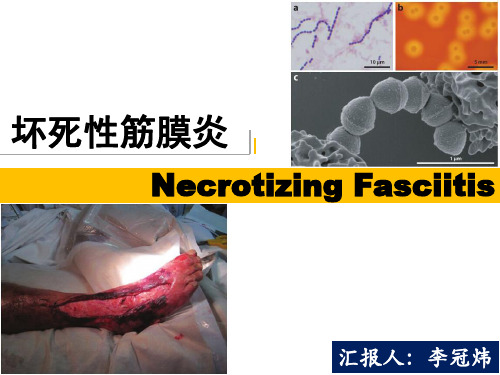
坏死性筋膜炎-Necrotizing-Fasciitis

Clinical Features
Noninvasive disease:链球菌性喉炎,脓疱病 Invasive disease:坏死性筋膜炎(NF)、链球菌中
毒性休克综合征(STSS)、蜂窝组织炎、菌血症、 肺炎、产后脓毒病 Nonsuppurative sequelae:急性风湿热、链球菌感 染后肾小球肾炎
➢ 食萄—球G肉-菌:菌、大真肠肠实球杆面菌菌目、为铜绿GA假S单,胞是菌非常普遍存在的细菌 ➢ N—F厌也氧早菌在:一拟杆个菌世属纪、前梭就菌见属、诸产报气道荚膜杆菌(罕见) ➢ 死对亡所率有取药决物于合耐并药症,情也况是媒体编造出来的事实
NF分型及病因
第二种类型:单细菌型。 20%-30%的NF属于此类型。多见于免疫功能正常的年轻人 多发生于四肢部位
细胞壁成分
透明质酸酶;
链激酶,又称链球菌溶纤维蛋白酶;
链道酶,又称脱氧核糖核酸酶; 链球菌溶血素,主要有“O”和“S”两种;
侵袭性酶
致热外毒素(SPE),又称红疹毒素或猩红热毒素 外毒素类
GAS分型
链球菌抗原类型
核蛋白抗原(p抗原):无特异性,各种链球菌均相 同,与葡萄球菌有交叉
定义
发Байду номын сангаас部位
—上肢(10%-48%) —下肢(28%) —会阴部(Fournier坏疽, 21%) —躯干(18%) —头颈(5%)
A—群—链食N球肉F菌菌分(G型ro及up病A S因treptococcus, GAS)
➢ 1994年5月,从英国格罗斯特郡开始爆发GAS感染导 致NF的病例
➢ 英国各大媒体纷纷头条报道:“Deadly virus baffles doctors”“Killer bug ate my face”
1例肛周坏死性筋膜炎外科清创后的伤口护理

1例肛周坏死性筋膜炎外科清创后的伤口护理发布时间:2021-11-25T15:44:16.311Z 来源:《医师在线》2021年7月13期作者:吕瑶[导读]吕瑶(新疆维吾尔自治区中医医院;新疆乌鲁木齐830000)摘要:坏死性筋膜炎 ( necrotizing,fasciitis,NF) 是一种较为少见的会威胁生命的进行性感染疾病,皮下组织和筋膜受到细菌的攻击时,造成的急性坏死性软组织感染,具有发病较为迅速、起病急、破坏力强、病亡率高等特点,死亡率大约 12% ~ 35%[1]。
本案例因医疗环境和患者身体因素影响,无法及时接受手术,只做了普通外科清创和换药处理,最后患者病情好转出院。
现将此案例的一些护理经验和体会记录下来,与各位同仁交流与共享。
关键字:坏死性筋膜炎;氧疗;伤口护理;中药湿敷中图分类号:R473.6文献标识码:A坏死性筋膜炎(necrotizing fasciitis, NF)作为一种临床罕见的重症感染性疾病,一般发生于腹部、会阴部等皮下软组织较为疏松的部位,如发于肛周,为肛周坏死性筋膜炎[2]。
及时行外科手术,广泛扩大清除所有坏死组织及可疑感染组织是坏死性筋膜炎的治疗原则[3],但在医疗环境和患者身体因素的局限下,只做了普通外科清创。
1病例资料赵某,男,44岁,因“肛周肿痛不适5天,加重1天”于 2019 年 8月 5日收治入院。
患者入院5天前无明显诱因出现肛周肿痛不适,自觉发热,曾来我院测体温为39.8℃,故发热门诊留观三天,予以金黄膏外敷肛门疼痛区,并口服乳果糖后自觉疼痛较前改善,体温正常后回家。
2019年8月4日患者如厕出现肛周破溃,破溃口流出较多脓血,患者自觉肛门疼痛加重,8月5日晨起肿痛延至阴囊,故再次来院求治,由急诊“肛周脓肿“收住入院。
患者入院后完成了相关常规检查:血便常规、尿液分析+尿沉渣、凝血+D2聚体、血型测定、肝功、肾功、电解质、心电图、肺部CT、甲乙丙肝艾滋梅毒、心脏彩超、睾丸及附睾彩超以了解病情。
坏死性筋膜炎常见并发症的预防及护理

坏死性筋膜炎常见并发症的预防及护理[摘要]目的观察坏死性筋膜炎常见并发症发生情况,探究其预防和护理措施。
方法选取2021年7月~2022年5月在我院进行治疗的56例坏死性筋膜炎患者展开本次实验研究,观察其发生感染、贫血等并发症的情况,分析总结预防并发症的对策和护理措施。
结果在治疗期间出现并发症的患者总计有44例,发生率高达78.57%,包括低蛋白血症、贫血等;在治疗和护理后,除5例患者病情恶化放弃治疗出院,其余51例患者均已痊愈出院。
结论坏死性筋膜炎有着较高的并发症发生率,应予以抗休克治疗和营养支持等有效并发症预防护理措施,达到预防、减少并发症目的,提高临床疗效。
[关键词]坏死性筋膜炎;并发症;预防;护理[Abstract] Objective To observe the common complications of necrotizing fasciitis and explore the prevention and nursing measures. Methods Fifty-six patients with necrotizing fasciitis treated in our hospital from July 2021 to May 2022 were selected for this experimental study. The incidence of complications such as infection and anemia was observed, and the countermeasures and nursing measures to prevent complications were analyzed and summarized. Results A total of 44 patients had complications during the treatment, the incidence rate was 78.57%, including hypoproteinemia, anemia, etc. After treatment and nursing, except for 5 patients who gave up treatment and discharged from hospital, the remaining 51 patients were cured and discharged. Conclusions Necrotizing fasciitis has a high incidence of complications. Effective preventive nursing measures such as anti-shock therapy and nutritional support should be taken to prevent and reduce complications and improve clinical efficacy.[Key words] Necrotizing fasciitis; Complications; Prevention; nursing坏死性筋膜炎指的是患者皮下组织、筋膜受到链球菌和金黄色葡萄球菌等多种细菌侵袭导致的软组织感染性疾病,皮肤损伤以及术后损伤等均可导致坏死性筋膜炎的发生,发病部位多为四肢、肛周等。
fatalcaseduetome...

Staphylococcus aureus causes acute and often fatal infections. Small colony variants (SCVs), which are subpopulations of S. aureus,are implicated in persistent and recurrent infections (in particular osteomyelitis, septic arthritis, respiratory tract infections in patients with cystic fibrosis, and deep-seated abscesses)(1-4). These phenotypic variants produce small,slow-growing, nonpigmented, nonhemolytic colo-nies on routine culture media, making correct identification difficult for clinical laboratories.Biochemical characterization of these variants suggests that they are deficient in electron transport activity (5). We report a fatal case of a persistent deep-seated hip abscess due to methicillin-resistant S. aureus SCVs that led to osteomyelitis and bloodstream infection in a patient with AIDS.Case ReportA 36-year-old man with AIDS came to the Cologne University Hospital, Cologne, Germany,in June 1997 with fever and progressive pain (of 6 weeks duration) in his right hip. HIV infection had been diagnosed in 1986. In 1994, his CD4 cell count was 250/µL, and oral zidovudine therapy was started. His medical history included Pneumocystis carinii pneumonia, pulmonary tuberculosis, and recurrent oral thrush; his medication included zidovudine, lamivudine,fluconazole, and trimethoprim/sulfamethoxazole.In September 1996, he was in a traffic accident and had severe cerebral trauma resulting in spastic hemiparesis with occasional seizures.After an intramuscular injection 2 months before admission, pus was surgically drained to treat recurrent abscesses of his right hip. Specimens for culture were not obtained.Physical examination found limited mobility of his right thigh and a tender, nondraining scar at the site of surgical drainage. Neither warmth nor swelling was observed over his right hip.Vital signs were temperature, 38.2°C; respira-tion rate, 28; and heart rate, 108. He was awake and alert and had spastic paresis in his right boratory studies performed on admission showed hemoglobin, 10.8 g/dL; leukocyte count,3,000/µL with a normal differential; CD4 cell count, 20/µL; platelet count, 131,000/µL;C-reactive protein, 184 mg/L; and alkaline phosphatase, 1490 U/L. Radiographs of the chest and a plain film of the pelvis were normal. A triple-phase bone scan showed an area of minor tracer accumulation in the acetabulum region of the right hip. Blood cultures were drawn, but antimicrobial therapy was withheld until culture results became available.On hospital day 2, one of two blood cultures drawn on admission yielded nonhemolytic staphylococci that were clumping factor negative. The organisms were initially misidentified as coagulase-negative staphylo-cocci and were considered contaminants.Fatal Case Due to Methicillin-ResistantStaphylococcus aureus Small ColonyVariants in an AIDS PatientHarald Seifert,* Christoph von Eiff, and Gerd Fätkenheuer *University of Cologne, Cologne, Germany; and Westfälische Wilhelms-Universität, Münster, GermanyAddress for correspondence: Harald Seifert, Institute of Medical Microbiology and Hygiene, University of Cologne,Goldenfelsstraße 19-21, 50935 Cologne, Germany; fax: 49-221-478-3081;e-mail:***************************.We describe the first known case of a fatal infection with small colony variants ofmethicillin-resistant Staphylococcus aureus in a patient with AIDS. Recovered fromthree blood cultures as well as from a deep hip abscess, these variants may haveresulted from long-term antimicrobial therapy with trimethoprim/sulfamethoxazole forprophylaxis of Pneumocystis carinii pneumonia.Figure 1. Staphylococcus aureus small colony variants (A) and S. aureus with a normal phenotype (B) cultured on sheep blood agar after 24 hours of incubation at 35°C. Staphylococci were identified by conventional methods (6) and with the ID 32 Staph system (bioMérieux, Marcy-L Etoile, France) follow-ing the instructions of the manufacturer. The tube-coagulase test was read after 24 hours. S. aureus isolates were characterized as small colony variants as described before (7-8). Auxotrophic requirements were evaluated with 10-µg hemin disks, 1.5-µg menadione disks, and 1.5-µg thymidine disks on Mueller-Hinton agar and on chemically defined medium (CDM) agar as well as on CDM agar supplemented with 1 µg/mL hemin, 100 µg/mL thymidine, and 1 µg/mL menadione, respectively.Empiric antistaphylococcal therapy with clindamycin (600 mg q8hr) was instituted. On hospital day 4, two sets of blood cultures obtained on hospital day 2 yielded phenotypically identical organisms, which on the basis of a positive tube coagulase test were identified as oxacillin-resistant S. aureus. The colony mor-phology was suggestive of an SCV of S. aureus .The patient was started on parenteral vancomy-cin treatment (1 g q12hr). However, his condition deteriorated rapidly, and he died of refractory septic shock 6 days after admission.Autopsy showed a large (12 x 10 x 8 cm),deep-seated abscess of the right hip and osteomyelitis of the ischial tuberosity. Both SCVs and typical large colony forms of S. aureus were cultured from postmortem specimens of the abscess and the bone.FindingsS. aureus SCVs were recovered from one of two blood culture sets obtained on admission and from two of four blood culture sets obtained on hospital day 2. Growth was not detected until the blood culture bottles had been incubated 24hours. S. aureus with a normal phenotype was recovered from nose and throat specimens but not from blood cultures, whereas both SCVs and typical S. aureus phenotypes were isolated from the deep hip abscess (Figure 1) before death, as well as from a postmortem specimen. All isolates were clumping factor negative but showed a delayed positive reaction in the tube-coagulase test at 24 hours. The results of the ID 32 staph test did not unambiguously identify SCVs as S. aureus because the tests for urease and trehalose were negative. Both the nuc gene and the coa gene were identified by polymerase chain reaction (PCR) amplification. Methicillin resis-tance was confirmed for both small and large colony forms by PCR amplification of the mecA gene.When cultured without supplementation, all SCVs were nonpigmented and nonhemolytic.Supplementation with hemin, thymidine, or menadione identified two SCVs showing thymi-dine auxotrophy and a combined thymidine and menadione auxotrophy, respectively. All SCVs were stable on repeated subculturing.Epidemiologic typing by PCR analysis of inter-IS256 spacer length polymorphisms (Fig-ure 2) and pulsed-field gel electrophoresis of genomic DNA (data not shown) showed identical banding patterns for both SCVs and large colony forms, which indicates that the phenotypically different S. aureus isolates represented a single strain. Antimicrobial susceptibility testing was performed by microbroth dilution, according to the National Committee for Clinical Laboratory Standards guidelines. Susceptibility to trimethoprim/sulfamethoxazole was tested with Etest (AB Biodisk, Solna, Sweden). In contrast to current standards, the MICs for SCVs were determined after 48 hours of incubation at 35°C.Susceptibility testing showed that all S. aureus isolates were resistant to penicillin (MIC, >8 µg/mL), ampicillin (MIC, >32 µg/mL), oxacillin (MIC, >8 µg/mL), erythromycin (MIC, >32 µg/mL),clindamycin (MIC, >32 µg/mL), ciprofloxacin (MIC, >8 µg/mL), gentamicin (MIC, >500 µg/mL),and trimethoprim/sulfamethoxazole (MIC,>32 µg/mL) and susceptible to vancomycinFigure 2. Fingerprint patterns obtained for Staphylococcus aureus small colony variants (lanes 3-5, bloodculture isolates; lanes 6 and 7, isolates from hip abscess; lane 8, postmortem specimen) and S.aureus isolates with a normal phenotype (lanes 10and 11, isolates from nose and throat; lanes 12 and 13, isolates from hip abscess and postmortem specimen) after polymerase chain reaction (PCR)analysis of inter-IS256 spacer length showing identical strains. Lane 1, 100-bp ladder; lanes 2, 9,and 16, methicillin-resistant S. aureus (MRSA)reference strain; lanes 14 and 15, epidemiologically unrelated MRSA strains. Strain relatedness of all isolates with different colony morphologies and from different sources was analyzed by PCR analysis of inter-IS256 spacer length polymorphisms (9) and pulsed-field gel electrophoresis after Sma I restric-tion (8). Minor modifications included the use of brain heart infusion broth instead of trypticase soy broth to obtain sufficient growth of S. aureus small colonyvariants.(MICs, 1-2 µg/mL), teicoplanin (MICs,0.5-1 µg/mL), and quinupristin/dalfopristin (MICs, 0.5-1 µg/mL). No differences in MICs were observed between S. aureus SCVs and S.aureus isolates with normal phenotype.To our knowledge, this case represents the first of a serious S. aureus infection in an AIDS patient in which all blood cultures yielded SCVs.The SCVs unusual morphologic appearance and slow growth delayed the correct identifica-tion of these organisms as S. aureus . The empiric antimicrobial regimen in our patient did not include a glycopeptide, because of the low rate of methicillin resistance in commu-nity-acquired S. aureus infection in Germany.Appropriate antistaphylococcal therapy was,therefore, not started until hospital day 4.Delayed antimicrobial therapy on day 4 rather than on day 2 may have contributed to the patient s death.Proctor and colleagues recently reported five cases in which SCVs of S. aureus were implicated in persistent and relapsing infections. They identified only a single case reported in the previous 17 years and ascribed this to insufficient ability of laboratories to identify these organisms (8). In most cases, patients had received antibiotics. Aminoglycoside treatment may have selected for S. aureus SCVs (10), and in cases of osteomyelitis or deep-seated abscesses,persistence of these variants in the intracellular milieu may have permitted evasion of host defenses and allowed for the development of resistance to antimicrobial therapy (7,11). Von Eiff and colleagues recently reported four cases of chronic osteomyelitis due to SCVs of S. aureus in patients who had received gentamicin beads as an adjunct to surgical therapy for osteomyeli-tis (2). Kahl et al. described persistent infection with S. aureus SCVs in patients with cystic fibrosis (4). All these patients had received long-term trimethoprim/sulfamethoxazole prophy-laxis. It may be tempting to speculate that administration of trimethoprim/sulfamethoxazole for prophylaxis against P. carinii pneumonia may have selected for SCVs within the patient s large hip abscess. Further prospective studies are needed to assess the role of S. aureus SCVs in HIV-infected patients on long-term antimicro-bial therapy.Dr . Seifert is assistant professor at the Institute of Medical Microbiology and Hygiene, University of Co-logne, Germany . His research interests include the mo-lecular epidemiology of nosocomial pathogens, in par-ticular Acinetobacter species, catheter-related infections,and antimicrobial resistance.References 1.Proctor RA, Balwit J M, Vesga O. Variant subpopulations of Staphylococcus aureus as cause of persistent and recurrent infections. Infectious Agents and Disease 1994;3:302-12. 2.von Eiff C, Bettin D, Proctor RA, Rolauffs B, Lindner N,Winkelmann W, et al. Recovery of small colony variants of Staphylococcus aureus following gentamicin bead placement for osteomyelitis. Clin Infect Dis 1997;25:1250-1. 3.Spearman P, Lakey D, J otte S, Chernowitz A, Claycomb S, Stratton C. Sternoclavicular joint septic arthritis with small colony variant Staphylococcus aureus .Diagn Microbiol Infect Dis 1996;26:13-5. 4.Kahl B, Herrmann M, Everding AS, Koch HG, Becker K, Harms E, et al. Persistent infection with small colony variant strains of Staphylococcus aureus in patients with cystic fibrosis. J Infect Dis 1998;177:1023-9.5.von Eiff C, Heilmann C, Proctor RA, Woltz C, Peters G,Götz F. A site-directed Staphylococcus aureus hemB mutant is a small colony variant which persists intracellularly. J Bacteriol 1997;179:4706-12.6.Kloos WE, Bannerman TL. Staphylococcus andmicrococcus. In: Murray PR, Baron EJ, Pfaller MA, Tenover FC, Yolken RH, editors. Manual of clinical microbiology. 6th ed. Washington: American Society for Microbiology; 1995. p. 282-98.7.Balwit JM, van Langevelde P, Vann JM, Proctor RA.Gentamicin-resistant menadione and hemin auxotrophic Staphylococcus aureus persist within cultured endothelial cells. J Infect Dis 1994;170:1033-7. 8.Proctor RA, van Langevelde P, Kristjansson M, MaslowJ N, Arbeit RD. Persistent and relapsing infections associated with small colony variants of Staphylococcus aureus. Clin Infect Dis 1995;20:95-102.9.Deplano A, Vaneechoutte M, Verschraegen G, StruelensMJ. Typing of Staphylococcus aureus and Staphylococcus epidermidis by PCR analysis of inter-IS256 spacer length polymorphisms. J Clin Microbiol 1997;35:2580-7.10.Pelletier LL J r, Richardson M, Feist M. Virulentgentamicin-induced small colony variants of Staphylococcus aureus. J Lab Clin Med 1979;94:324-34.11.Proctor RA, Kahl B, von Eiff C, Vaudaux PE, Lew DP,Peters G. Staphylococcal small colony variants have novel mechanisms for antibiotic resistance. Clin Infect Dis 1998;27 Suppl 1:S68-74.。
Necrotizing fasciitis

AT LAST I WANT TO TELL EVERYONE, STAY HOME AND HAVE A CALM HEART THANK FOR WATCHING!
EXAMPLE
PROTECTIVE MEASURES
• If you want to prevent necrotizing fasciitis, you should pay attention to your diet first. You must quit smoking and alcohol, and do not eat spicy food. Instead, you should give foods with high protein and cellulose for many times. You should eat more nutritious food. Only if we absorb enough nutrients, the body's immune ability will be better. At the same time, we should also pay attention to our rest conditions, make sure that we have enough rest time, and never let ourselves be too tired. Everyone's life should be regular, not too nervous, and we must keep an optimistic attitude. In addition, we must do more physical exercises and adjust our physical condition at ordinary times. Finally, we should pay attention to keeping warm at ordinary times and never let ourselves suffer from cold. If we do this, we are unlikely to suffer from necrotizing fasciitis.
大芬戈尔德菌致肱骨骨髓炎1例报道并文献复习

·论著·大芬戈尔德菌致肱骨骨髓炎1例报道并文献复习马寅舰, 陈翠竹, 纪 冰, 贾 龙, 王凤霞摘要: 目的 了解大芬戈尔德菌的特点及其引起疾病的临床特征,提高对该菌引起感染性疾病的认识。
方法 回顾性分析1例诊断为大芬戈尔德菌骨髓炎患者的临床资料,结合文献复习探讨大芬戈尔德菌感染性疾病的特征。
结果 该例患者临床表现和实验室检查符合大芬戈尔德菌感染性骨髓炎的特征,脓液和组织培养有大芬戈尔德菌生长,术后无复发。
复习、汇总文献及该例患者,共32例(男性13例,女性10例,9例未提及),其中骨和关节(包括假体)感染16例、感染性心内膜炎5例、蜂窝织炎3例、乳腺感染3例、其他5例;临床表现主要与受累组织系统相关。
31例经外科手术联合抗菌药物治疗后痊愈,1例血流感染患者死亡。
结论 骨关节感染,尤其假体植入后关节感染,应警惕大芬戈尔德菌所致,必要时给予经验性抗厌氧菌治疗。
关键词: 大芬戈尔德菌; 肱骨; 感染; E 试验中图分类号:R378.11 文献标识码:A 文章编号:1009-7708 ( 2021 ) 02-0189-05DOI: 10.16718/j.1009-7708.2021.02.012One case report of the humerus osteomyelitis caused by Finegoldia magna and literature reviewMA Yinjian, CHEN Cuizhu, JI Bing, Jia Long, WANG Fengxia (Department of Laboratory Medicine, Binzhou Medical University Hospital, Binzhou Shandong 256600, China )Abstract: Objective To examine the characteristics of Finegoldia magna infection for improving the awareness of this disease. Methods One case of humerus osteomyelitis caused by F. magna was reported in a male patient. Other infectious diseases caused by F . magna were also searched from literature and reviewed. Results The diagnosis was confirmed as F . magna humerus osteomyelitis based on clinical manifestations and laboratory tests, including positive culture of F . magna from pus and tissue. No recurrence was observed at follow-up. A total of 32 cases (13 males, 10 females, 9 unknown) of F . magna infection were reviewed (including this one). The specific site of infection included infective endocarditis (n =5), bone and joint including prosthesis (n =16), subcutaneous mass (n =3), breast (n =3), and others (n =5). Clinical symptoms varied with the organs involved. Majority of the patients (n =31) were cured by surgery combined with antimicrobial therapy. Only one patient died with bloodstream infection. Conclusions Clinicians should be aware of the possibility of F . magna infection in case of bone and joint infection, especially following prosthesis implantation. Empirical anti-anaerobic therapy can be prescribed if necessary.Keywords: Finegoldia magna , humerus, infection, Etest基金项目:滨州医学院科技计划项目(BY2017KJ18)。
关于病危动物的英语作文

关于病危动物的英语作文The issue of critically endangered animals is a pressing concern for the world. There are various species in danger of extinction due to a range of factors such as habitat loss, poaching, and climate change. For instance, the giant panda is one iconic and critically endangered animal facing the threat of extinction due to habitat destruction. To address this issue, conservation efforts are crucial to prevent the loss of these precious species and ensure their survival for future generations.关于病危动物的问题是全球关注的焦点。
由于栖息地的丧失、偷猎和气候变化等各种因素,许多物种处于濒临灭绝的危险之中。
以大熊猫为例,这一标志性的病危动物由于栖息地的破坏正面临着灭绝的威胁。
为了解决这一问题,保护努力至关重要,以防止这些珍贵物种的丧失,并确保它们生存下去,留给后代。
One of the greatest challenges in protecting critically endangered animals is the loss of habitat. As human populations continue to grow, the demand for land for agriculture, urban development, and infrastructure increases. This often leads to the destruction andfragmentation of natural habitats, leaving many species with limited space to survive. Without suitable habitats, these animals struggle to find food, reproduce, and maintain viable populations. Additionally, habitat loss forces animals to move into human-dominated landscapes, increasing the likelihood of conflicts and further endangering their survival.保护病危动物面临的最大挑战之一是栖息地的丧失。
肛周坏死性筋膜炎8例临床分析

㊃临床论著㊃[文章编号]1009-2188(2021)01-0040-06肛周坏死性筋膜炎8例临床分析萧绮莉,吴畑华,苏玉环,杜刚毅,郑少康,莫德龙,林晓聪(中山市中医院肛肠科,广东中山528400)㊀㊀[摘要]㊀目的㊀总结肛周坏死性筋膜炎的临床诊疗经验㊂㊀方法㊀回顾性分析2018年2月至2019年6月间我院收治的8例肛周坏死性筋膜炎患者的临床资料㊂患者均为男性,年龄26~60岁,均行急诊手术治疗,采取大切口㊁纵行切口㊁网状切口㊁保留皮桥㊁对口引流㊂其中1例切除睾丸,3例行二次清创,4例择期行阴囊成形术㊂㊀结果㊀本组8例患者均经手术㊁抗感染及综合治疗,住院时间1~103d㊂1例因感染性休克及多器官功能不全死亡,7名存活患者随访8~10个月无一例复发或出现肛门功能障碍㊂㊀结论㊀准确诊断㊁及早手术㊁规范抗感染治疗及生命支持治疗可提高肛周坏死性筋膜炎患者存活率,术后对伤口进行专门护理可为其康复提供重要保障㊂㊀㊀[关键词]肛周坏死性筋膜炎[中图分类号]R657.1㊀[文献标志码]B㊀DOI:10.16260/ki.1009-2188.2021.01.008Clinical Analysis of Perianal Necrotizing Fasciitis:8Cases Report㊀XIAO Qi-li,WU Tian-hua,SU Yu-huan,DU Gang-yi,ZHENG Shao-kang,MO De-long,LIN Xiao-cong㊀(Department of Proctology,ZhongshanHospital of Traditional Chinese Medicine,Zhongshan528400,Guangdong,China)Abstract:㊀Objective㊀To summary the treatment experiences of perianal necrotizing fasciitis.Methods㊀The clinical data of8male patients with perianal necrotizing fasciitis(aged from26to60years old)in our hospital from February2018to June2019were analyzed retrospectively.All cases were performed surger-y with open incision.Orchidectomy was carried out in one case,secondary debridement in two cases and selec-tive scrotoplasty in four cases.㊀Results㊀After surgery,antibiotics treatment and comprehensive therapies,one case died of infective shock and multiple organ dysfunction syndrome and7cases survived.The hospitalstays were one to103days.During8to10months follow-up,no recurrence and anus fecal incontinence wasfound.㊀Conclusions㊀Accurate diagnosis,early surgery,standardized anti-infective treatment and life support treatment can improve the survival rate of patients with perianal necrotizing fasciitis.Postoperative special nurs-ing of wounds can provide an important guarantee for their rehabilitation.㊀㊀Key words:perianal necrotizing fasciitis㊀㊀肛周坏死性筋膜炎(perianal necrotizing fasci-itis,PNF)是一种发生于肛周㊁会阴部的严重软组织感染性疾病,病理变化限于皮下及筋膜,其下肌肉大多正常[1]㊂该病具有早期诊断困难㊁进展迅速等特点,如误诊或处理不及时往往会导致脓毒血症及多器官衰竭,甚至死亡[2],因此肛周坏死性筋膜炎的临床治疗逐渐得到重视㊂目前其发病率㊀㊀[收稿日期]2020-10-12,[修回日期]2021-01-27㊀㊀[作者简介]萧绮莉,中山市中医院肛肠科主治医师㊂通信作者:杜刚毅,中山市中医院肛肠科主任医师㊂不详,我院既往亦鲜有收治该类病例,自2018年2月至2019年6月我科陆续接诊或参与手术治疗肛周坏死性筋膜炎患者共8例,现结合其临床表现㊁影像学特点㊁大体病理特点及治疗历程进行回顾性分析,以期为临床诊断及治疗肛周坏死性筋膜炎提供参考㊂1㊀临床资料1.1㊀一般资料2018年2月至2019年6月共收治肛周坏死性筋膜炎患者共8例,均为男性,平均年龄45.9(26~60)岁,发病至就诊时间为2~30d㊂其中,肛肠科首诊收治2例,泌尿科首诊收治3例,内分泌科首诊收治2例,ICU 首诊收治1例㊂发病诱因:除1例诉发病前曾食用过期食品,其余均未见明确诱因㊂临床均表现为肛周㊁会阴部肿痛甚至皮肤发黑(图1,2),病变部散发恶臭气味;伴发热3例,病灶处流脓7例,伴精神状态差4例㊂本组患者病例资料见表1㊂1.2㊀术前检查实验室检查:白细胞计数1.03ˑ109/L ~28.90ˑ109/L,中性粒细胞0.46ˑ109/L ~25.94ˑ109/L;降钙素原水平0.16~83.59ng /ml;随机血糖6.1~26mmol /L;术中取分泌物送培养,结果示肺炎克雷伯菌阳性3例,大肠埃希菌阳性2例,金黄色葡萄球菌阳性3例;详见表2,3㊂影像学检查:术前行CT 检查6例,可见感染组织呈现不同程度的炎症水肿伴 小气泡征 (图3,4)㊂表1㊀本组8例PNF 患者临床资料病例性别年龄(岁)发病至就诊时间(d)首诊科室合并症㊀㊀手术方式住院时间(d)治疗结果1男482ICU多脏器衰竭骨盆直肠窝脓肿切开引流术㊁肛周坏死性筋膜炎切开引流术1死亡2男605泌尿外科糖尿病(多年)阴囊坏疽清创术㊁肛周坏死性筋膜炎清创引流术,20d 后行阴囊成形缝合术30治愈出院3男3914泌尿外科肝硬化失代偿;糖尿病(本次诊出)阴囊探查㊁会阴及肛周坏死性筋膜炎切开引流术;6d 后行右侧腹股沟切开清创引流术;2月后行阴囊清创缝合㊁阴囊成形术103治愈出院4男577肛肠科高血压;糖耐量异常(本次诊出)骨盆直肠窝脓肿切开引流术㊁会阴部脓肿切开引流术;27d 后行阴囊清创㊁成形术29治愈出院5男268肛肠科糖尿病(本次诊出)坏死性筋膜炎切开引流术㊁会阴脓肿切开引流术17治愈出院6男4630内分泌科糖尿病(7年)肛周坏死性筋膜炎切开引流术20治愈出院7男5020内分泌科腔隙性脑梗塞史;糖尿病(本次诊出)阴囊清创切开减压术㊁双侧睾丸切除术;2d 后行肛周坏死性筋膜炎切开引流术25治愈出院8男479泌尿外科糖尿病(多年)阴囊坏疽清创术㊁肛周坏死性筋膜炎切开引流术;25d 后行会阴部创口清创缝合㊁阴囊成形术37治愈出院表2㊀本组8例PNF 患者实验室检查结果病例白细胞(ˑ109/L)中性粒细胞(ˑ109/L)降钙素原(ng /ml)随机血糖(mmol /L)1 1.030.4683.59 6.382 5.52 3.44 2.515.66312.24 6.31 3.768.548.84 6.49 3.17.51517.4715.69 1.9522623.7525.940.2117.63716.5814.560.726819.0614.010.1610.311.3㊀治疗1.3.1㊀手术治疗本组8例患者均于入院当天行急诊手术治疗,全麻或腰硬联合麻醉,取膀胱截石位或仰卧位㊂手术采取大切口㊁纵行切口㊁网状切口㊁保留皮桥㊁开放伤口㊁对口引流(图5,6),所有伤口均以双氧水㊁生理盐水冲洗后加以油纱引流㊂手术方式见表1,其中5例次与泌尿外科联合手术,1例切除睾丸,3例行二次清创,4例择期行阴囊成形术㊂1.3.2㊀药物治疗抗感染治疗方案根据指南[2]选取广谱抗生素联合甲硝唑治疗,随后根据药敏结果调整用药方案,见表3㊂中药汤剂:除2例未服用中药汤剂外,其余6例予仙方活命饮加减,1次/d,疗程7d㊂其他治疗包括控制血糖㊁营养支持㊁补充能量㊁维持水电解质平衡等㊂图1㊀肛周㊁会阴部红肿图2㊀局部皮肤发黑图3㊀CT可见 小气泡征图4㊀CT可见 小气泡征图5㊀大切口㊁纵行切口㊁网状切口图6㊀开放伤口,保留皮桥,对口引流1.4㊀术后处理创伤护理小组视伤口情况换药,2~3次/d,以碘伏棉球消毒创面周围,双氧水㊁生理盐水㊁甲硝唑注射液多次㊁反复冲洗脓腔,及时清除坏死组织,清洗完毕后再以碘伏棉球消毒创面,最后以碘伏油纱填塞脓腔以便引流通畅,并避免假性愈合㊂表3㊀本组患者抗生素给药方案病例分泌物培养结果㊀㊀㊀㊀首次给药㊀用药方案㊀㊀㊀㊀用药时长(d)调整给药㊀调整方案㊀用药时长(d)调整原因㊀㊀㊀㊀㊀1肺炎克雷伯菌亚胺培南西司他丁1--㊀㊀㊀㊀-2金黄色葡萄球菌头孢他啶30--㊀㊀㊀㊀-3肺炎克雷伯菌㊁鸟肠球菌亚胺培南西司他丁9美罗培南15检出鸟肠球菌,根据药敏调整4金黄色葡萄球菌亚胺培南西司他丁4克林霉素11检出金黄色葡萄球菌,根据药敏调整5大肠埃希菌头孢哌酮舒巴坦11--㊀㊀㊀㊀-6金黄色葡萄球菌美洛西林钠舒巴坦3阿米卡星4检出金黄色葡萄球菌,根据药敏调整7肺炎克雷伯菌亚胺培南西司他丁15--㊀㊀㊀㊀-8大肠埃希菌头孢他啶11亚胺培南西司他丁20检出大肠埃希菌,根据药敏结果调整㊀㊀注: - 为首次方案对致病菌有效,故未调整用药2㊀结㊀㊀果本组8例患者治疗结果见表1,其中7例(87.5%)治愈出院,1例死亡,住院时间平均32.75 (1~103)d㊂死亡的1例患者入院时已存在明显感染性休克及多器官功能不全,入院至手术时间仅2h,病变范围却从肛旁5cm扩大至肛旁10cm,并于术后6h死亡㊂其余7例患者随访8~10个月,未见肛门失禁或复发㊂3㊀讨㊀㊀论肛周坏死性筋膜炎为肛肠外科较为凶险的急重症[3]㊂1883年该病由Fournier首次报道,1924年Meleney提出了链球菌坏疽,1952年Wilson将其称为坏死性筋膜炎㊂本病一旦发病,进展迅速,死亡率高达12.8%~35%[4-5]㊂国外数据显示该病发病率较低,为(1.6~3.3)/100000[6],国内尚未见有关发病率的统计报道㊂我科在2018年2月至2019年6月期间共收治肛周坏死性筋膜炎患者8例,占同期患者的0.19%㊂肛周坏死性筋膜炎多见于男性[7],然而女性一旦患病,病情进展速度更快,更容易发生致死性腹膜炎[8]㊂有文献曾报道由于肛周坏死性筋膜炎位置的特殊性,炎症及坏死病变波及睾丸以致需要手术切除的案例㊂对于伴有睾丸坏死的患者,需要充分怀疑是否存在腹膜内或腹膜后的感染[9]㊂本组患者中,行双侧睾丸切除1例,但未发现腹腔感染㊂肛周坏死性筋膜炎为多种细菌混合感染所致,需氧菌和厌氧菌的协同作用在感染过程中扮演了主要角色[2]㊂常见细菌包括大肠杆菌㊁肺炎克雷伯菌㊁梭状芽胞杆菌㊁变形杆菌等,文献报道从肛周坏死性筋膜炎组织中培养出来的细菌已达70余种[9]㊂本组8患者分别发现肺炎克雷伯菌㊁大肠埃希菌㊁金黄色葡萄球菌等3种细菌,其中1例发现肺炎克雷伯菌合并鸟肠球菌,细菌种类与文献报道相近,但以单一菌种感染多见,可能与细菌培养方式对其他菌种未能覆盖有关㊂然而肛周坏死性筋膜炎患者的死亡往往不是局部组织病变所致,而是由于炎症带来的全身性影响,如严重的脓毒血症㊁凝血功能障碍㊁急性肾衰竭㊁糖尿病酮症酸中毒或多脏器衰竭等[10]㊂本组患者中死亡1例,考虑为感染性休克所致㊂虽然肛周坏死性筋膜炎最初被认为仅发生于健康年轻男性,但是最近几十年临床发现其更倾向于发生在患有基础疾病的中年患者身上,尤其是糖尿病患者,有文献指出约20%~70%的肛周坏死性筋膜炎患者患有糖尿病[11-12]㊂其他常见的易感因素有免疫抑制㊁外伤㊁营养状态低下㊁肥胖㊁肾功能衰竭等[2],在易感因素存在的条件下,致病菌带来的侵袭性损伤将产生极强的毒性及破坏力㊂Tuncel 等[13]发现代谢率㊁发病诱因和病变范围是影响预后的三个最为重要的因素㊂本组8例患者中有6例糖尿病患者,1例糖耐量异常患者,即血糖升高者占患者总数的87.5%,因此在整个治疗过程中,控制血糖成为临床极为重要的内容㊂实验室检查结果可以体现出肛周坏死性筋膜炎患者与一般肛周脓肿患者的不同㊂白细胞异常升高㊁血小板减少㊁高血糖㊁低蛋白血症㊁贫血等均提示脓毒血症开始出现或已经存在㊂LRINEC评分于2004年被提出[14],用于评估坏死性筋膜炎的感染风险,评价指标包括C反应蛋白㊁白细胞㊁血红蛋白㊁血清钠㊁血清肌酐㊁血浆葡萄糖等内容,积分与风险呈正相关㊂但观察本组病例的白细胞水平㊁贫血情况等并未与病情严重程度呈现相关性,反而降钙素原水平能较好地体现出患者的感染情况㊂及时㊁彻底的清创手术及引流是提高肛周坏死性筋膜炎患者存活率的唯一有效方法[15],必要时实施有计划的重复清创术,有利于评估病情进展[16]㊂手术可以去除失活组织,从而避免感染进一步发展,同时清除坏死组织㊁毒素和细菌对机体的影响[17]㊂外科重建可在感染得以控制㊁无须再行清创后开始实施,包括覆盖和缝合创面,多数病例适合自体游离皮肤移植,效果较为理想[18-19]㊂本组8例患者均于入院当天行急诊手术,其中3例曾行二次清创,4例择期行阴囊成形术㊂手术以彻底清创为原则,采取大切口(即切口大于病变范围约2cm)㊁纵行切口㊁网状切口㊁保留皮桥㊁对口引流等方式,清除腐坏组织至可见正常组织,术中注意尽量减少对括约肌的损伤㊂术后治疗重点转移至创伤管理㊂文献报道[20]多使用碘伏㊁双氧水㊁甲硝唑溶液等对伤口进行冲洗,可以保持伤口清洁,促进坏死组织脱落㊂使用一些抗菌敷料可为伤口愈合提供良好的环境,减少生物负荷及表面污染㊂另有文献报道称高压氧治疗及封闭式负压引流技术有助于伤口愈合[21]㊂本组7例患者术后换药采用了碘伏消毒伤口周围皮肤,双氧水㊁生理盐水㊁甲硝唑溶液对伤口进行有序冲洗,视情况换药2~3次/d以保持伤口的清洁,为新鲜肉芽生长创造条件,虽未使用高压氧或封闭式负压引流技术或抗菌辅料,但患者伤口生长恢复仍取得满意的效果㊂快速液体复苏和心肺功能维护在脓毒血症患者的管理中有着不容置疑的地位[22]㊂同时,使用广谱抗生素抗感染也应列为首选治疗之一㊂由于细菌培养结果具有滞后性,选用抗生素时应选取针对两种或以上病原菌有效的广谱抗生素,早期㊁联合㊁足量给药[23]㊂美国感染性疾病协作组2014年指南新增了单一使用碳青霉烯类抗生素治疗肛周坏死性筋膜炎的方案[16]㊂由于肛周坏死性筋膜炎全身症状显著㊁治疗过程长,患者很可能出现全身消耗性表现,因此充分的全身营养支持治疗可为本病的治疗提供重要保证[24]㊂通过本组8例患者的临床治疗,作者体会准确诊断㊁及早手术㊁规范抗感染治疗及生命支持治疗提高了肛周坏死性筋膜炎患者的存活率(本组为87.5%),创伤护理小组对伤口进行专门的护理则为其康复提供了重要保障㊂目前对于肛周坏死性筋膜炎患者的长期预后尚缺乏详细的临床证据,此类患者的生活质量有可能会因为瘘管复发㊁组织缺损㊁瘢痕等原因而下降㊂本组7例生存患者随访8~10个月无一例复发或出现肛门功能障碍,但其身心健康㊁泌尿生殖功能等情况尚有待在多学科协助下进行进一步随访[25]㊂本组8例患者发病至就诊时间为2~30d,跨度较大,因此很难界定坏死性筋膜炎从原发感染进展成爆发性坏疽的时间节点,因其多发生于患者就诊前,具体时间很难核实㊂当疾病进展缓慢时,患者往往不能记起症状出现的确切时间,或者只能提供一个比实际发病时间要晚的节点,且患者可能会尽量避免让医生形成他们低估了病情的严重性或者不愿意及时就诊的印象㊂另外,关于中医治疗和新型敷料在肛周坏死性筋膜炎的治疗过程中所起的作用,尚有待进一步研究加以论证㊂[参考文献][1]㊀吴肇汉,秦新裕,丁强,等.实用外科学[M].第4版.北京:人民卫生出版社,2017.45-46.[2]㊀中国医师协会肛肠医师分会临床指南工作委员会.肛周坏死性筋膜炎临床诊治中国专家共识(2019年版)[J].中华胃肠外科杂志,2019,22(7):689-693.doi:10.3760/cma.j.issn.1671-0274.2019.07.017.[3]㊀李梅岭,何洪芹,张秀艳.肛周急性坏死性筋膜炎18例临床分析[J].疑难病杂志,2018,17(3):300-302.doi:10.3969/j.issn.1671-6450.2018.03.020.[4]㊀Sorensen MD,Krieger JN.Fournier's gangrene:Epidemiology andoutcomes in the general US population[J].Urol Int,2016,97(3):249-259.doi:10.1159/000445695.[5]㊀Sartelli M,Malangoni MA,May AK,et al.World Society of E-mergency Surgery(WSES)guidelines for management of skin and soft tissue infections[J].World J Emerg Surg,2014,9(1):57.doi:10.1186/1749-7922-9-57.[6]㊀Vogel JD,Johnson EK,Morris AM,et al.Clinical practice guide-line for the management of anorectal abscess,fistula-in-ano,and rectovaginal fistula[J].Dis Colon Rectum,2016,59(12):1117-1133.doi:10.1097/DCR.0000000000000733.[7]㊀李大伟,赵刚,唐宇峰,等.多学科联合治疗多发伤合并坏死性筋膜炎7例体会[J].创伤外科杂志,2018,20(9):647-650.doi:10.3969/j.issn.1009-4237.2018.09.003.[8]㊀孙静,郝丹,李晓雪,等.Fournier坏疽17例患者治疗及转归分析[J].实用皮肤病学杂志,2019,12(1):23-25.doi:10.11786/sypfbxzz.1674-1293.20190107.[9]㊀Katsanos KH,Ignatiadou E,Sarandi M,et al.Fournier's gan-grene complicating ulcerative pancolitis[J].J Crohns Colitis, 2010,4(2):203206.doi:10.1016/j.crohns.2009.11.006.[10]Carrillo-Córdova LD,Aguilar-Aizcorbe S,Hernández-Farías MA,et al.Escherichia coli productora de betalactamasas de espectro extendido como agente causal de gangrena de Fournier de origen u-rogenital asociada a mayor mortalidad[J].Cir Cir,2018,86(4): 327-331.doi:10.24875/CIRU.M18000050.[11]Insua-Pereira I,Ferreira PC,Teixeira S,et al.Fournier's gan-grene:a review of reconstructive options[J].Cent European J Urol,2020,73(1):74-79.doi:10.5173/ceju.2020.0060.[12]潘亚红,曹志彬,王元天,等.Fournier坏疽诊治体会[J].中国男科学杂志,2015,29(10):68-69.doi:10.3969/j.issn.1008-0848.2015.10.017.[13]Tuncel A,Aydin O,Tekdogan U,et al.Fournier's gangrene:Three years of experience with20patients and validity of the Fournier's Gangrene Severity Index Score[J].Eur Urol,2006,50(4):838-843.doi:10.1016/j.eururo.2006.01.030. [14]Wong CH,Khin LW,Heng KS,et al.The LRINEC(LaboratoryRisk Indicator for Necrotizing Fasciitis)score:a tool for distin-guishing necrotizing fasciitis from other soft tissue infections[J].Crit Care Med,2004,32(7):1535-1541.doi:10.1097/01.ccm.0000129486.35458.7d.[15]Misiakos EP,Bagias G,Papadopoulos I,et al.Early diagnosisand surgical treatment for necrotizing fasciitis:A multicenterstudy[J].Front Surg,2017,4:5.doi:10.3389/fsurg.2017.00005.eCollection2017.[16]Stevens DL,Bisno AL,Chambers HF,et al.Practice guide-lines for the diagnosis and management of skin and soft tissue infections:2014update by the infectious diseases society of A-merica[J].Clin Infect Dis,2014,59(2):147-159.doi:10.1093/cid/ciu296.[17]Ahmad A,Brumble L,Maniaci M.Vibrio parahaemolyticus in-duced necrotizing fasciitis:An atypical organism causing an unusu-al presentation[J].Case Rep Infect Dis,2013,2013:216854.doi:10.1155/2013/216854.[18]Spyropoulou GA,Jeng SF,Demiri E,et al.Reconstruction ofperineoscrotal and vaginal defects with pedicled anterolateral thigh flap[J].Urology,2013,82(2):461-465.doi:10.1016/j.urol-ogy.2013.04.044.[19]Lee SH,Rah DK,Lee WJ.Penoscrotal reconstruction with graci-lis muscle flap and internal pudendal artery perforator flap transpo-sition[J].Urology,2012,79(6):1390-1394.doi:10.1016/j.urology.2012.01.073.[20]谢翔宇,林正军,林明慧.肛周㊁会阴部急性坏死性筋膜炎诊疗体会[J].中国现代医药杂志,2019,21(4):59-61.doi: CNKI:SUN:ZHTY.0.2019-04-017.[21]中华医学会烧伤外科学分会,‘中华烧伤杂志“编辑委员会.负压封闭引流技术在烧伤外科应用的全国专家共识(2017版)[J].中华烧伤杂志,2017,33(3):129-135.doi:10.3760/ cma.j.issn.1009-2587.2017.03.001.[22]万林骏,廖庚进,万晓红,等.严重脓毒症和感染性休克患者早期复苏时器官功能障碍的回顾性分析[J].中华危重病急救医学,2016,28(5):418-422.doi:10.3760/cma.j.issn.2095-4352.2016.05.008.[23]Maguire P,Azagrar JM,Carb A,et al.The successful use of neg-ative-pressure wound therapy in two cases of canine necrotizing fas-ciitis[J].J Am Anim Hosp Assoc,2015,51(1):43-48.doi:10.5326/JAAHA-MS-6033.[24]吴健,薛晓东,刘俊玲,等.坏死性筋膜炎的诊断与治疗(附16例分析)[J].吉林医学,2012,33(13):2709-2710.doi: CNKI:SUN:JLYX.0.2012-13-010.[25]Goh T,Goh LG,Ang CH,et al.Early diagnosis of necrotizingfasciitis[J].Br J Surg,2014,101(1):e119-25.doi:10.1002/ bjs.9371.。
病毒性筋膜炎作文英语

病毒性筋膜炎作文英语Necrotizing Fasciitis。
Necrotizing Fasciitis, also known as flesh-eating disease, is a rare but potentially life-threateningbacterial infection that affects the soft tissue beneaththe skin, including the fascia, muscles, and nerves. It is characterized by rapid and extensive tissue destruction, leading to necrosis, sepsis, and organ failure.The disease is caused by a group of bacteria, including Streptococcus pyogenes, Staphylococcus aureus, and Clostridium perfringens, which invade the body through breaks in the skin, such as cuts, burns, insect bites, or surgical wounds. Once inside, the bacteria release toxins that destroy the surrounding tissue and create an anaerobic environment that favors their growth and spread.The symptoms of Necrotizing Fasciitis may include fever, chills, swelling, redness, pain, and tenderness in theaffected area, as well as blisters, ulcers, and blackeningof the skin. The infection can spread rapidly to otherparts of the body, causing septic shock and multiple organ failure, and may require amputation or surgical debridement to remove the dead tissue.The diagnosis of Necrotizing Fasciitis is based on a combination of clinical signs, laboratory tests, andimaging studies, such as X-rays, CT scans, or MRI. Early recognition and treatment are crucial to prevent thedisease from progressing and reduce the risk of complications.The treatment of Necrotizing Fasciitis usually involves a combination of antibiotics, surgical debridement, and supportive care, such as fluid replacement, pain management, and nutritional support. The antibiotics should cover a broad spectrum of bacteria and be administeredintravenously for at least 2-4 weeks. The surgical debridement aims to remove all the necrotic tissue and create a clean wound bed for healing. In severe cases, amputation or skin grafting may be necessary.The prevention of Necrotizing Fasciitis involves good hygiene, proper wound care, and prompt medical attentionfor any signs of infection. People with weakened immune systems, such as diabetics, cancer patients, or HIV/AIDS patients, are at higher risk and should take extra precautions.In conclusion, Necrotizing Fasciitis is a rare but serious bacterial infection that can cause extensive tissue damage and organ failure. Early recognition and treatment are essential to prevent the disease from spreading and reduce the risk of complications. Good hygiene, proper wound care, and prompt medical attention are important for prevention.。
肛周脓肿致大面积急性坏死性筋膜炎及阴囊坏疽一例并文献复习

华中科技大学硕士学位论文肛周脓肿致大面积急性坏死性筋膜炎及阴囊坏疽一例并文献复习华中科技大学同济医学院附属协和医院急诊科 430022硕士研究生:涂志刚导师:蒋春舫教授中文摘要目的提高临床医生对急性坏死性筋膜炎及阴囊坏疽的认识。
方法结合肛周脓肿致大面积急性坏死性筋膜炎及阴囊坏疽一例的诊治过程,复习相关文献,对其病因、发病机制、临床表现及诊治特点进行分析。
结果急性坏死性筋膜炎及阴囊坏疽大多是由多因素致病,起病急,进展快,危害极大,处理相当困难的软组织感染性疾病。
多数可经多次及时手术切开清除坏死组织、创面冲洗换药及抗休克、抗感染,加强营养支持等对症治疗后治愈。
结论急性坏死性筋膜炎及阴囊坏疽是罕见病,但近年来国内外报道增多,且发病早期与其他软组织感染如丹毒、蜂窝织炎及气性坏疽等难以鉴别,尽管现代医学发展迅速,但误诊率仍较高,故早期诊断与及时适当治疗可明显减少病死率与术后并发症。
关键词坏死性筋膜炎;阴囊坏疽;肛周脓肿;清创华中科技大学硕士学位论文Perianorectal abscess lead to large area of acute necrotizing fasciitis and scrotum gangrene:a case report and review of theliteratureDepartment of Emergency, Union Hospital, Tongji Medical College, HuazhongUniversity of Science and TechnologyMaster candidate: Tu ZhigangSupervisor: Prof. Jiang ChunfangAbstractObjective To improve the awareness of acute necrotizing fasciitis and scrotum gangrene for clinicians.Method One case of large area of acute necrotizing fasciitis and scrotum gangrene was reported, and related literatures were reviewed to explore the etiology, pathogenesis, clinical presentations, and characters of diagnosis and treatment of acute necrotizing fasciitis and scrotum gangrene.Results Most of acute necrotizing fasciitis and scrotum gangrene are caused by multiple factors, which belong to the soft tissue of infectious that have disease urgent, rapidly spreading, potentially fatal and difficultly to deal with. Most patients can be cured after repeatedly treated via radical debridement and wound irrigation in time, also needed resistance to shock or infection and strengthen nutritional support.Conclusions Acute necrotizing fasciitis and scrotum gangrene are rare disease,but the reported increased at home and abroad in recent years. However, modern medicine technology development quickly, as it is hard to distinguish them from the other soft tissue infections such as erysipelas, cellulitis, and gas gangrene by the early clinical华中科技大学硕士学位论文manifestations of the disease, the misdiagnosis rate is still high. Therefore, early diagnosis and timely appropriate treatment can significantly reduce the mortality and postoperative complications.Key words: necrotizing fasciitis; scrotum gangrene; perianorectal abscess; debridement华中科技大学硕士学位论文前言肛周脓肿在临床上很常见,一般起源于肛窦炎或肛腺感染,导致直肠肛管周围间隙形成脓肿,可沿直肠纵肌向各周围间隙蔓延[1],其导致急性坏死性筋膜炎在临床上发病率并不高,但一旦发病,病情进展非常快,部分病人很快出现脓毒性休克,病情危重,如不及时正确处理,危及生命。
奇怪的疾病英语作文

奇怪的疾病英语作文Title: The Enigma of a Peculiar Disease。
In recent times, a mysterious ailment has surfaced, baffling medical professionals and leaving communities in a state of perplexity. This enigmatic malady presents a myriad of symptoms, defying conventional diagnosis and treatment. In this essay, we shall delve into the peculiarities of this strange disease, exploring its manifestations, potential causes, and the challenges it poses to the medical community.The first striking aspect of this peculiar disease is its diverse array of symptoms. Afflicted individuals have reported a range of manifestations, including but not limited to chronic fatigue, intermittent fevers, joint pains, cognitive impairment, and gastrointestinal disturbances. Moreover, these symptoms often fluctuate in intensity, further complicating diagnosis and management.Despite extensive investigations, the precise etiology of this mysterious ailment remains elusive. Speculations abound regarding potential triggers, including environmental factors, genetic predispositions, and infectious agents. Some researchers hypothesize that the disease may stem from a dysregulation of the immune system, leading to chronic inflammation and tissue damage. Others suggest a viral or bacterial origin, though no definitive pathogen has been identified thus far.One of the most formidable challenges posed by this peculiar disease is the lack of a standardized diagnostic criteria. In the absence of specific biomarkers or imaging findings, clinicians must rely on a combination of clinical history, physical examination, and laboratory tests to formulate a tentative diagnosis. However, this approach often yields inconclusive results, leading to delays in appropriate management and heightened patient distress.Furthermore, the management of this enigmatic malady remains largely symptomatic, focusing on alleviating individual manifestations rather than addressing theunderlying cause. Pain management, cognitive rehabilitation, and lifestyle modifications form the cornerstone of therapy, aimed at improving quality of life and functional status. Nonetheless, many patients continue to experiencepersistent symptoms, highlighting the urgent need for targeted interventions.In addition to its medical ramifications, this peculiar disease exerts a profound impact on the psychological well-being of affected individuals. The relentless cycle of symptoms exacerbates feelings of frustration, isolation,and hopelessness, undermining both resilience and coping mechanisms. Consequently, holistic support measures, including counseling, support groups, and communityoutreach programs, are essential to address thepsychosocial dimensions of this complex illness.In conclusion, the emergence of this peculiar disease represents a formidable challenge to the medical community, characterized by its diverse symptomatology, elusive etiology, and profound impact on patients' lives. Effortsto unravel its mysteries require interdisciplinarycollaboration, innovative research methodologies, and unwavering commitment to patient-centered care. Only through collective endeavor can we hope to conquer this enigmatic malady and restore health and vitality to those afflicted.。
遭受灭亡的危险英语作文

遭受灭亡的危险英语作文Title: The Peril of Annihilation: A Wake-Up Call。
In the current global landscape, numerous existential threats loom over humanity, casting a shadow of uncertainty and apprehension. Among these threats, the prospect of annihilation stands out as one of the most daunting challenges we face. Whether it be from the devastating impacts of climate change, the proliferation of nuclear weapons, or the emergence of deadly pandemics, the specter of annihilation lurks ominously, demanding our urgent attention and collective action.First and foremost, climate change poses an imminent danger to the survival of our planet and its inhabitants. The relentless emission of greenhouse gases has triggered a cascade of catastrophic events, including rising temperatures, extreme weather patterns, and the melting of polar ice caps. If left unchecked, these changes could unleash widespread devastation, leading to the displacementof millions, the extinction of countless species, and the collapse of ecosystems vital to our survival.Moreover, the proliferation of nuclear weapons presents a grave threat to global security and stability. With tensions simmering in various regions around the world, the risk of nuclear conflict has become increasingly palpable. The detonation of even a single nuclear warhead could unleash unfathomable destruction, causing incalculable loss of life and irreparable damage to the environment. The consequences of such an event would reverberate across generations, leaving a legacy of suffering and despair in its wake.In addition to these man-made perils, the emergence of deadly pandemics poses a significant threat to human civilization. The rapid spread of infectious diseases, fueled by factors such as urbanization, globalization, and antimicrobial resistance, has highlighted the fragility of our public health systems. The COVID-19 pandemic, in particular, has exposed critical weaknesses in our preparedness and response mechanisms, claiming millions oflives and wreaking havoc on economies worldwide. As pathogens continue to evolve and adapt, the risk of future pandemics looms large, posing a formidable challenge to our collective resilience and ingenuity.In the face of these existential threats, complacencyis not an option. We must recognize the urgency of the situation and take decisive action to mitigate the risksand safeguard our future. This requires a concerted effort on multiple fronts, including:1. Environmental Stewardship: Implementing robust measures to reduce greenhouse gas emissions, transition to renewable energy sources, and preserve biodiversity.2. Nuclear Disarmament: Pursuing diplomatic solutionsto nuclear proliferation, promoting arms control agreements, and fostering a culture of dialogue and cooperation among nations.3. Pandemic Preparedness: Strengthening global health systems, investing in research and development of vaccinesand treatments, and enhancing international cooperation in disease surveillance and response.4. Resilience and Adaptation: Building resilience atthe individual, community, and societal levels to withstand and recover from shocks and crises, whether natural or man-made.Furthermore, it is imperative that we engage in openand honest dialogue about the existential risks we face, fostering a culture of awareness, responsibility, and solidarity. By mobilizing collective action and harnessing the power of innovation and collaboration, we can confront the perils of annihilation and chart a course toward a safer, more sustainable future for generations to come.In conclusion, the specter of annihilation casts a long shadow over humanity, reminding us of the fragility of our existence and the imperative of collective action. By confronting existential threats with courage, determination, and solidarity, we can overcome adversity and build a moreresilient and prosperous world for all. The time to act is now.。
医学名词解释

一、极端的痛楚(EXCLUCIATION)椎间盘突出(DISK HERNIATION)神经炎、脑膜炎(NEURITIS、MENINGITIS)大面绩广泛性发炎(EXTENSIVE INFLAMMATION ANYWHERE)手术后大面绩广泛性发炎(POSTSURGICAL PAIN WITH EXTENSIVE INFLAMMATION)多处骨折及软组织的伤害(MULTIPLE FRACTURE AND SOFT-TISSUE INJURY)坏死性胰炎(NECROTIZING PANCREATITIS)坏死性胆囊炎(NECROTIZING CHOLECYSTITIS)严重的肠道扩张(SEVERE BOWEL DISTENTION)病理性骨折(PATHOLOGY FRACTURE)恶性骨癌(OSTEOSARCOMA)神经组织的钢钉植入(IMPLANTS IMPINGING ON NEURAL TISSUE)全耳道摘除(TOTAL EAR CANAL ABLATION)二、剧烈的疼痛(SEVERE)骨关节炎(OSTEOARTHRITIS)关节内的整形手术(INTRA-ARTICULAR ORTHOPEDIC SURGERY)骨折修复或截肢(FRACTURE REPAIR OR AMPUTATION)血栓症或局部缺血(THROMBOSIS OR ISCHEMIA:SADDLE THROMBUS)胆汁性腹膜炎及尿液性或败血性腹膜炎(PERITONITIS:ESPECIALLY BILE;ALSO URINE AND SEPTIC) 器官肿大造成包膜疼痛(ORGANOMEGALY,CAPSULAR PAIN)MODERATE DISTENTION OF HOLLOW VISCOUS肠胃道扭转、子宫扭转、睾丸扭转(TORSIONS:GASTROINTESTINAL,UTERINE,TESTICULAR)尿道阻塞(URETHRAL OBSTRUCTION)眼科状况:溃烂,青光眼,葡萄膜炎(OPHTHALMOLOGIC CONDITIONS:ULCER,GLUCOMA,UVETITIS)复杂的剖腹、开胸手术(LAPAROTOMY:INVOLVED,THORACOTOMY)分娩(PARTURITION)创伤(TRAUMA)癌症疼痛(CANCER PAIN)三、中度疼痛(MODERATE)简单的剖腹(SIMPLE LAPAROTOMY)赫尼亚的修补(HERNIA REPAIR)简单的关节外整形手术(SIMPLE,EXTRA-ARTICULAR ORTHOPEDIC PRECEDURES)非广泛性的肿瘤摘除(MASS REMOVALS:UNLESS EXTENSIVE)较复杂的(年老或肥胖)子宫卵巢切除术(COMPLICATED OVARIOHYSTERECTOMY:OLD OR OBESE) 去爪(DECLAW:ONYCHECTOMY)撕裂伤(MOST LACERATION)膀胱炎(CYSTITIS)耳炎(OTITIS)胸管引流(CHEST DRAINS)拔牙(DENTAL EXTRACTIONS)(RESOLVING CONDITIONS)四、轻度疼痛(MILD)雄性节扎(CASTRATION)小肿块移除(LUMP REMOVAL:SMALL)插管造成的喉炎(LARYNGEAL INFLAMMATION:INTUBATION) 食道炎(ESOPHAGITIS:ENDOSCOPY)轻微的肌炎(MYOSITIS:MILD,UNCOMFORTABLE POSITIONING) 多针的穿刺(MULTIPLE NEEDLE PUNCTURES)(EARLY OR RESOLVING CONDITIONS)。
1例肛周坏死性筋膜炎合并糖尿病酮症酸中毒患者的术后护理体会

2022年第8卷第9期Vol.8,No.9,2022中西医结合护理Chinese Journal of Integrative Nursing1例肛周坏死性筋膜炎合并糖尿病酮症酸中毒患者的术后护理体会李盼盼,袁爱民(北京市肛肠医院创面修复门诊,北京,100120)摘要:本文总结1例肛周坏死性筋膜炎(PNF )合并糖尿病酮症酸中毒(DKA )患者的术后护理经验。
通过负压引流(VSD )辅助治疗,以及中医化腐清创治疗、营养支持、情志护理以及健康宣教等多元化的护理配合,有助于减轻患者机体疼痛感,降低并发症发生率,加快创面愈合。
关键词:肛周坏死性筋膜炎;糖尿病酮症酸中毒;负压引流;感染;中医护理中图分类号:R 473.6文献标志码:A文章编号:2709-1961(2022)09-0155-04Postoperative nursing of a patient with perianal necrotizingfasciitis complicated with diabetic ketoacidosisLI Panpan ,YUAN Aimin(Wound Repair Clinic ,Beijing Rectum Hospital ,Beijing ,100120)ABSTRACT :This article summarized the postoperative nursing measures for a patient with peri⁃anal necrotizing fasciitis (PNF )complicated with diabetic ketoacidosis (DKA ).The given vacu⁃um sealing drainage (VAS )was adopted for wound repair ,and multi -modal nursing measures in⁃cluding Traditional Chinese Medicine debridement ,nutrition support emotional care and health ed⁃ucation were carried out to reduce the pain degree and the incidence of complications ,and im⁃prove the wound healing.KEY WORDS :perianal necrotizing fasciitis ;diabetic ketoacidosis ;postoperative care ;vacuum sealing drainage ;infection ;Traditional Chinese Medicine nursing 肛周坏死性筋膜炎(PNF )是因肛周、直肠及泌尿系细菌感染引起的,以肛周皮肤、浅筋膜坏死为病理表现的软组织感染性疾病[1]。
坏死性颈筋膜炎的临床诊治进展

收稿日期:2020 ̄03 ̄03通信作者:肖旭平ꎮE ̄mail:469784129@qq.comdoi:10.6040/j.issn.1673 ̄3770.0.2020.072综述坏死性颈筋膜炎的临床诊治进展袁康龙㊀综述肖旭平㊀审校湖南师范大学附属第一医院/湖南省人民医院耳鼻咽喉头颈外科ꎬ湖南长沙410005摘要:坏死性颈筋膜炎是由细菌混合感染引起的颈部严重软组织炎症ꎬ以颈部皮下组织和深浅筋膜坏死为特征ꎬ易感人群以老年人及合并慢性基础疾病(如糖尿病)者为主ꎮ临床上早期诊断率较低ꎬ病情进展迅速ꎬ易出现危及生命的并发症ꎬ如下行坏死性纵隔炎㊁脓毒性休克等ꎬ病死率极高ꎮ就坏死性颈筋膜炎的临床诊治做综述ꎬ为更好对其早期诊断及治疗提供帮助ꎮ关键词:筋膜炎ꎻ坏死ꎻ颈部ꎻ诊断ꎻ治疗中图分类号:R686㊀㊀㊀文献标志码:A㊀㊀㊀文章编号:1673 ̄3770(2020)06 ̄0135 ̄04引用格式:袁康龙ꎬ肖旭平.坏死性颈筋膜炎的临床诊治进展[J].山东大学耳鼻喉眼学报ꎬ2020ꎬ34(6):135 ̄138.YUANKan ̄glongꎬXIAOXuping.Progressintheclinicaldiagnosisandtreatmentofcervicalnecrotizingfasciitis[J].JournalofOtolaryngologyandOphthalmologyofShandongUniversityꎬ2020ꎬ34(6):135 ̄138.ProgressintheclinicaldiagnosisandtreatmentofcervicalnecrotizingfasciitisYUANKanglong㊀OverviewXIAOXuping㊀GuidanceDepartmentofOtolaryngology&HeadandNeckSurgeryꎬtheFirstAffiliatedHospitalofHunanNormalUniversity/HunanProvin ̄cialPeople̓sHospitalꎬChangsha410005ꎬHunanꎬChianAbstract:Cervicalnecrotizingfasciitis(CNF)isaseveresofttissueinflammationoftheneckcausedbymixedbacterialinfections.Itischaracterizedbynecrosisofnecksubcutaneoustissuewithdeepandshallowfasciitis.Elderlyindividualsandthosewithchroniccomorbiddiseases(suchasdiabetes)aremoresusceptibletoCNF.Theearlyclinicaldiagnosisrateislowandthediseaseprogressesrapidly.Life ̄threateningcomplicationsaresuchasdescendingnecrotizingmediastinitisandsepticshockarefrequentlyobserved.Themortalityrateisextremelyhigh.ThisarticlereviewstheclinicaldiagnosisandtreatmentofCNFtofacilitateearlydiagnosisandtreatment.Keywords:FasciitisꎻNecrotizingꎻNeckꎻDiagnosisꎻTreatment㊀㊀坏死性筋膜炎(NecrotizingfasciitisꎬNF)是一种发展非常迅速的感染性疾病ꎬ感染沿深浅筋膜播散ꎬ并在累及的血管内形成血栓ꎬ引起相应区域的皮肤㊁皮下组织及筋膜组织坏死ꎬ通常不累及肌层组织[1]ꎬ常伴有毒血症㊁败血症及多脏器功能衰竭等ꎮ病变多发生于四肢㊁腹部㊁会阴ꎬ其中四肢最为多见ꎬ发生于头颈部较为罕见ꎮ发生于颈部的坏死性筋膜炎又称为坏死性颈筋膜炎(cervicalnecrotizingfasci ̄itisꎬCNF)ꎬ是一种极为罕见的由混合细菌感染引起的颈部严重软组织炎症ꎬ大多是由牙源性感染蔓延而来ꎬ亦有咽源性感染或外伤引起ꎬ以颈部皮下组织和深浅筋膜坏死为特征[2]ꎮ由于颈部筋膜系统解剖结构复杂ꎬ各间隙相通ꎬ当感染初期未能准确识别ꎬ并未及时有效控制时ꎬ感染可迅速沿头颈部筋膜扩散[3]ꎬ并可能产生危及生命的并发症包括下行坏死性纵隔炎(descendingnecrotizingmediastinitisꎬDNM)㊁脓毒性血症㊁败血症㊁感染性休克㊁气道梗阻㊁呼吸衰竭和多器官功能衰竭等[4]ꎬ患者病死率极高ꎬ是临床上的危急重症ꎮ1㊀病㊀因㊀㊀CNF通常继发于慢性基础疾病ꎬ如糖尿病㊁肿瘤㊁肝肾功能衰竭㊁放化疗骨髓抑制㊁免疫抑制等ꎬ肥胖㊁长期吸烟饮酒也是本病的危险因素[5]ꎮ已经明确糖尿病是与CNF相关的最常见疾病[6]ꎮ坏死性颈筋膜炎感染来源以牙源性感染最多见ꎬ骨放射性坏死㊁扁桃体或咽部感染㊁外伤㊁异物㊁头颈手术等诱因导致CNF亦有报道ꎮ其常见的致病菌包括:链球菌㊁葡萄球菌㊁变形杆菌㊁产气荚膜梭菌㊁大肠埃希菌㊁金黄色假单胞菌㊁克雷伯菌ꎬ以及厌氧菌等ꎬ多由需氧菌㊁厌氧菌或兼性厌氧菌协同致病[7]ꎮA族链球菌(groupastreptococcusꎬGASꎬ又称化脓性链球菌)的免疫逃避是人类发生侵袭性感染如坏死性筋膜炎的关键ꎬ国外学者近年来对GAS蛋白质组学进行分析发现ꎬBrpA可能在化脓性链球菌的定植和黏附中起作用ꎬ假设蛋白A20_1756c在化脓性链球菌侵袭性感染的免疫逃避机制中起重要作用ꎬSpeB在化脓性链球菌侵袭性感染的过程中会导致大量的组织损伤[8]ꎮWen等[9]对坏死性筋膜炎动物模型的蛋白质组学进行研究ꎬ发现了一种新型的酸诱导毒力因子HtpAꎬ可以提高化脓性链球菌的吞噬能力以引起坏死性筋膜炎ꎮCNF的病理特征是筋膜爆发性坏死ꎬ微血管血栓形成ꎬ脉管炎伴动静脉壁的纤维素样坏死ꎬ一般不累及肌肉[10]ꎮ2㊀临床症状与体征㊀㊀大多患者表现为咽痛㊁颈痛㊁皮肤红肿㊁发热ꎬ随着疾病进展可出现吞咽困难㊁张口受限㊁呼吸困难㊁胸痛及感染性休克等ꎮ查体可见颈部皮肤弥漫性红肿㊁触痛㊁皮温升高㊁可有捻发感等ꎬ甚至局部皮肤坏死ꎮ早期患者颈部皮肤无明显红肿改变ꎬ仅表现为颈痛ꎬ是由于CNF侵犯部位较深ꎬ故疾病早期不易在体表扪及包块或波动感[11]ꎬ这也是早期诊断困难的原因之一ꎮ临床表现与体格检查若出现 症征不一 者ꎬ需警惕CNF的可能[12]ꎮ3㊀影像学检查㊀㊀计算机断层扫描(CT)是诊断CNF最有价值的检查手段[13]ꎮCNF的典型CT表现为筋膜平面广泛积液和积气[14]ꎬ还可出现皮肤和皮下组织弥漫性水肿增厚ꎬ皮下脂肪的网状强化ꎬ深筋膜的浅㊁中㊁深层不同程度受累ꎬ肌肉不对称强化ꎬ反应性淋巴结肿大ꎬ血管血栓形成等表现[15]ꎮCT对界定手术范围ꎬ监测坏死性筋膜炎的进展及转归至关重要ꎮ磁共振成像(MR)T2WI上颈部深筋膜增厚㊁积液是CNF的重要表现ꎬMR对于深筋膜积液显像优于CTꎬ故MR是鉴别坏死性筋膜炎与非坏死性筋膜炎最有效的方法[16]ꎮ超声检查可发现皮下气肿沿深筋膜扩散ꎬ皮下脂肪组织回声增强ꎬ与筋膜周围液体相对应的相互连接ꎬ可出现鹅卵石样改变[17]ꎮ因超声检查中软组织鹅卵石征象是非特异性的ꎬ故超声检查对CNF的诊断效能不如CT及MRIꎮ4㊀实验室检查㊀㊀Wong等[18]对坏死性筋膜炎患者实验室检查结果进行研究ꎬ提出LRINEC评分用来对坏死性筋膜炎的早期诊断起辅助作用ꎮLRINEC评分总分13分ꎬ具体根据:C反应蛋白ȡ150mg/L计4分ꎬ反之为0分ꎻ白细胞计数>25ˑ109/L计2分ꎬ<15ˑ109/L计0分ꎬ居中计1分ꎻ血红蛋白<110g计2分ꎬ>135g/L计0分ꎬ居中计1分ꎻ血钠<135mmol/L计2分ꎬ反之计0分ꎻ血肌酐>141μmol/L计2分ꎬ反之计0分ꎻ血糖>10mmol/L计1分ꎬ反之计0分ꎬ针对以上五项结果根据标准进行打分评估ꎬLRINEC评分>6分可作为NF的预测指标ꎮKato等[19]对比坏死性筋膜炎与蜂窝织炎患者CK值与PCT值发现ꎬ坏死性筋膜炎患者CK及PCT水平明显高于蜂窝织炎患者ꎬ坏死性筋膜炎患者经治疗后PCT较治疗前明显下降ꎮ5㊀诊断与鉴别诊断㊀㊀结合病史㊁临床表现和影像学检查是早期临床诊断CNF的关键ꎬ实验室检查LRINEC评分可提高早期诊断效能ꎮ手术探查及组织病理是诊断CNF的金标准ꎮ早期CNF需与蜂窝织炎㊁丹毒等皮肤软组织感染鉴别ꎬ蜂窝织炎及丹毒仅累及皮下组织ꎬ不侵犯筋膜ꎬ较少出现全身性不适ꎬMR可对其进行准确的鉴别[20]ꎮ6㊀治㊀疗6.1㊀维持生命体征㊀作为耳鼻咽喉头颈外科医师ꎬ必须对此疾病有深刻的认识ꎮ该疾病的特点是早期症状不明显ꎬ进展快ꎬ故患者入院后如高度怀疑CNFꎬ需要立即予以开放静脉通道ꎬ监测生命体征ꎬ患者应住入监护病房[21]ꎬ迅速完善相关检查ꎬ积极开展治疗ꎮ6.2㊀积极抗炎㊀临床诊断CNF的患者ꎬ必须立即予以积极的抗感染治疗ꎬ如第二代或第三代头孢菌素或碳青霉烯类㊁氨基糖苷类抗生素ꎬ考虑到大多合并厌氧菌感染ꎬ还需全身应用甲硝唑㊁替硝唑等抗厌氧菌药物治疗[22]ꎮ在经验性抗炎的同时ꎬ必须从病理分泌物和血液培养物中获得微生物培养物ꎬ从而根据敏感性试验来调整抗生素的治疗方案ꎬ避免长期盲目使用抗生素ꎬ增加真菌感染风险ꎮ6.3㊀激素应用㊀对于CNF患者中激素的应用目前尚有争议ꎮ陈伟军等[16]提出CNF治疗在足量㊁有效应用抗生素的前提下ꎬ需及早㊁短时间突击使用大剂量糖皮质激素ꎬ具体用法为甲强龙10~15mg/kgꎬ分1~2次/d静滴ꎬ加入0.9%生理盐水液100~250mL中ꎬ2h内滴完ꎬ冲击3dꎬ第4天后逐渐减量ꎬ视病情变化停用糖皮质激素ꎮ但Petitpas等[23]分析CNF患者并发症产生的影响因素时发现ꎬ糖皮质激素的使用可掩盖CNF的症状ꎬ也可增加CNF患者并发下行坏死性纵隔炎的风险ꎬ不提倡早期使用糖皮质激素来减轻疼痛㊁肿胀的症状ꎮ6.4㊀手术治疗㊀手术是CNF的治疗重点ꎬ也是提高预后的关键ꎬ高度怀疑或经影像学诊断CNF患者需尽早手术治疗ꎮ典型CNF患者术中可见坏死的筋膜呈灰白色ꎬ易于与包绕组织分离ꎬ分泌物呈恶臭㊁咖啡样或灰色稀薄带气泡液体[24]ꎮ易红良等[11]总结29例坏死性颈筋膜炎患者手术过程ꎬ手术原则是根据病变范围ꎬ切口要足够大ꎬ充分暴露ꎬ切开范围一定要到达病变的最边缘ꎬ彻底清除坏死组织ꎬ钝性分离并充分探查引流每个可能潜在感染间隙ꎮ术中反复用过氧化氢液㊁碘伏及生理盐水进行局部冲洗ꎬ才能阻断炎症的继续蔓延ꎮ手术分离时最好与主要的颈部血管平行ꎬ以避免损伤大血管ꎬ防止切开后颈部皮肤坏死ꎮ若并发下行坏死性纵隔炎ꎬ需联合胸外科医师及时行纵隔引流[6]ꎬ必要时可采取多次手术ꎮ对CNF患者合并有气道梗阻或合并急性会厌炎等其他疾病ꎬ导致其呼吸困难的患者ꎬ应准确评估后行气管切开术[25]ꎮ6.5㊀术后处理㊀术后持续负压引流及每日多次换药有利于感染物的排出和促进伤口愈合[26]ꎮ手术后进行高压氧治疗对厌氧菌有杀灭作用ꎬ它可使组织恢复正常的氧环境ꎬ有助于血管生成ꎬ并促进伤口愈合ꎬ可缩短住院时间ꎬ降低死亡率[27]ꎮ加强对原发病的治疗ꎬ加强CNF患者的营养支持ꎬ对张口受限或术后经口进食困难患者进行胃管鼻饲饮食ꎮ治疗过程中对肝肾功能进行监测ꎬ注意对症支持治疗及维持水电解质平衡也是术后治疗的要点ꎮ静脉注射免疫球蛋白可通过降低溶血性链球菌对细胞因子释放的超抗原活性ꎬ从而降低CNF的死亡率[28]ꎬ但其确切疗效尚无大样本报道ꎮ7㊀预㊀后㊀㊀Gunaratne等[29]回顾性分析1235例坏死性颈筋膜炎患者发现ꎬ未行手术治疗的患者死亡率接近100%ꎬ经综合治疗后的患者死亡率为13.36%ꎬ他们发现下行坏死性纵隔炎(DNM)与感染性休克是本病最主要和严重的并发症ꎬDNM的发展与死亡显著相关ꎬ延误超过24h未行清创手术治疗也被认为是造成患者死亡的重要因素ꎮ迅速识别㊁诊断和进行快速且有针对性的干预措施ꎬ是减少并发症产生和降低病死率的关键[1]ꎮ综上所述ꎬ坏死性颈筋膜炎是一种少见的进行性致死性软组织感染ꎬ由于早期患者症状与体征表现不一ꎬ目前仍缺乏早期诊断特异性指标ꎬ且疾病进展迅速ꎬ延误诊断可能会导致严重并发症的产生ꎬ造成较高的死亡率ꎬ临床上应以危急重症进行处理ꎮ对于其诊治要点ꎬ首先就是能根据临床症状㊁相关病史及检查资料尽早诊断ꎬ注重早期经验性抗生素及抗厌氧菌药物的联合使用ꎬ根据微生物培养基药敏试验及时调整用药方案ꎮ激素治疗尚存争议ꎮ临床诊断后24h内进行外科手术清创充分引流ꎬ必要时可多次手术清创ꎬ术后可予持续负压吸引及联合高压氧治疗ꎬ对全身条件较差的患者需多学科联合诊治ꎬ以提高临床治疗效果ꎬ减少并发症的产生ꎬ降低病死率ꎮ参考文献:[1]KulasegaranSꎬCribbBꎬVandalACꎬetal.Necrotizingfasciitis:11 ̄yearretrospectivecasereviewinSouthAuck ̄land[J].ANZJSurgꎬ2016ꎬ86(10):826 ̄830.doi:10.1111/ans.13232.[2]ElanderJꎬNekludovMꎬLarssonAꎬetal.Cervicalnecro ̄tizingfasciitis:descriptiveꎬretrospectiveanalysisof59casestreatedatasinglecenter[J].EurArchOtorhinolar ̄yngolꎬ2016ꎬ273(12):4461 ̄4467.doi:10.1007/s00405 ̄016 ̄4126 ̄y.[3]GausepohlJꎬWagnerJ.Survivalfromcervicalnecrotizingfasciitis[J].WestJEmergMedꎬ2015ꎬ16(1):172 ̄174.doi:10.5811/westjem.2014.12.21553.[4]AdekanyeAGꎬUmanaANꎬOffiongMEꎬetal.Cervicalnecrotizingfasciitis:managementchallengesinpoorresourceenvironment[J].EurArchOtorhinolaryngolꎬ2016ꎬ273(9):2779 ̄2784.doi:10.1007/s00405 ̄015 ̄3841 ̄0.[5]FerzliGꎬSukatoDCꎬMouradMꎬetal.Aggressivene ̄crotizingfasciitisoftheheadandneckresultinginmassivedefects[J].EarNoseThroatJꎬ2019ꎬ98(4):197 ̄200.doi:10.1177/0145561319839789.[6]Al ̄AliMAꎬHefnyAFꎬIdrisKMꎬetal.Cervicalnecro ̄tizingfasciitis:anoverlookeddiagnosisofafataldisease[J].ActaOtolaryngolꎬ2018ꎬ138(4):411 ̄414.doi:10.1080/00016489.2017.1393841.[7]AbdurrazaqTOꎬIbikunleAAꎬBraimahRO.Cervicalnecrotizingfasciitis:apotentiallyfataldiseasewithvariedetiology[J].AnnMedHealthSciResꎬ2016ꎬ6(4):251 ̄256.doi:10.4103/amhsr.amhsr_33_16.[8]LyATꎬNotoJPꎬWalwynOLꎬetal.DifferencesinSpeBproteaseactivityamonggroupAstreptococciassociatedwithsuperficialꎬinvasiveꎬandautoimmunedisease[J].PLoSOneꎬ2017ꎬ12(5):e0177784.doi:10.1371/jour ̄nal.pone.0177784.[9]WenYTꎬWangJSꎬTsaiSHꎬetal.Label ̄freeproteomicanalysisofenvironmentalacidification ̄influencedStrepto ̄coccuspyogenessecretomerevealsanovelacid ̄inducedproteinhistidinetriadproteinA(HtpA)involvedinnecrotizingfasciitis[J].JProteomicsꎬ2014ꎬ109:90 ̄103.doi:10.1016/j.jprot.2014.06.026.[10]NouguéHꎬLeMahoALꎬBoudiafMꎬetal.Clinicalandimagingfactorsassociatedwithseverecomplicationsofcervicalnecrotizingfasciitis[J].IntensiveCareMedꎬ2015ꎬ41(7):1256 ̄1263.doi:10.1007/s00134 ̄015 ̄3830 ̄1.[11]赵雅铭ꎬ易红良ꎬ关建ꎬ等.29例颈部坏死性筋膜炎临床分析[J].临床耳鼻咽喉头颈外科杂志ꎬ2014ꎬ28(7):490 ̄492.doi:10.13201/j.issn.1001 ̄1781.2014.07.017.ZHAOYamingꎬYIHongliangꎬGUANJianꎬetal.Clin ̄icalanalysisof29casesofcervicalnecrotizingfasciitis[J].JournalofClinicalOtorhinolaryngologyHeadandNeckSurgeryꎬ2014ꎬ28(7):490 ̄492.doi:10.13201/j.issn.1001 ̄1781.2014.07.017.[12]GohTꎬGohLGꎬAngCHꎬetal.Earlydiagnosisofnecrotizingfasciitis[J].BrJSurgꎬ2014ꎬ101(1):e119 ̄e125.doi:10.1002/bjs.9371.[13]EmmanuelKofiAmponsahꎬPaulFrimpongꎬMiYoungEoꎬetal.Clinicalclassificationofcervicalnecrotizingfasciitis[J].EurArchOtorhinolaryngolꎬ2018ꎬ275(12):3067 ̄3073.doi:10.1007/s00405 ̄018 ̄5155 ̄5. [14]SandnerAꎬMoritzSꎬUnverzagtSꎬetal.Cervicalnecrotizingfasciitis:thevalueofthelaboratoryriskIndi ̄catorfornecrotizingfasciitisscoreasanindicativepa ̄rameter[J].JOralMaxillofacSurgꎬ2015ꎬ73(12):2319 ̄2333.doi:10.1016/j.joms.2015.05.035. [15]肖恩华.坏死性筋膜炎临床和影像学表现[J].临床放射学杂志ꎬ2002ꎬ21(5):400 ̄402.doi:10.13437/j.cnki.jcr.2002.05.030.[16]陈伟军ꎬ胡建文ꎬ腾陈迪.面颈部坏死性筋膜炎的诊治[J].中国耳鼻咽喉头颈外科ꎬ2010ꎬ17(3):153 ̄154.doi:10.16066/j.1672 ̄7002.2010.03.024. [17]ShyyWꎬKnightRSꎬGoldsteinRꎬetal.Sonographicfindingsinnecrotizingfasciitis[J].JUltrasoundMedꎬ2016ꎬ35(10):2273 ̄2277.doi:10.7863/ultra.15.12068. [18]WongCHꎬKhinLWꎬHengKSꎬetal.TheLRINEC(LaboratoryRiskIndicatorforNecrotizingFasciitis)score:atoolfordistinguishingnecrotizingfasciitisfromothersofttissueinfections[J].CritCareMedꎬ2004ꎬ32(7):1535 ̄1541.doi:10.1097/01.ccm.0000129486.35458.7d.[19]KatoTꎬFujimotoNꎬHondaSꎬetal.Usefulnessofser ̄umprocalcitoninforearlydiscriminationbetweennecro ̄tizingfasciitisandcellulitis[J].ActaDermVenereolꎬ2017ꎬ97(1):141 ̄142.doi:10.2340/00015555 ̄2465. [20]HayeriMRꎬZiaiꎬShehataMLꎬetal.Soft ̄tissueinfec ̄tionsandtheirimagingmimics:fromcellulitistonecro ̄tizingfasciitis[J].Radiographicsꎬ2016ꎬ36(6):1888 ̄1910.doi:10.1148/rg.2016160068.[21]JavierMoragaꎬMauricioChangꎬMaríaRodríguez.Atypicalpresentationofcervicalnecrotizingfasciitis.Casereport[J].JournalofOralResearchꎬ2017ꎬ6(7):182 ̄185.doi:10.17126/joralres.2017.052.[22]MunirajaPꎬKavithaKꎬAneeshꎬetal.Theroleofem ̄piricalantibiotictherapyintreatingnecrotizingfasciitis:aretrospectiverecordanalysis[J].InternationalJournalofContemporaryMedicalResearchꎬ2016ꎬ3(5):1481 ̄1484.[23]PetitpasFꎬBlancalJPꎬMateoJꎬetal.Factorsassociatedwiththemediastinalspreadofcervicalnecrotizingfasci ̄itis[J].AnnThoracSurgꎬ2012ꎬ93(1):234 ̄238.doi:10.1016/j.athoracsur.2011.09.012.[24]InanCHꎬYenerHMꎬYilmazMꎬetal.Cervicalnecro ̄tizingfasciitisofodontogenicoriginandhyperbaricoxy ̄gentherapy[J].JCraniofacSurgꎬ2017ꎬ28(7):e691 ̄e692.doi:10.1097/SCS.0000000000003842. [25]陈秀梅ꎬ宋西成.颈部坏死性筋膜炎7例并文献复习[J].山东大学耳鼻喉眼学报ꎬ2016ꎬ30(3):65 ̄67ꎬ72.doi:10.6040/j.issn.1673 ̄3770.0.2015.305.CHENXiumeiꎬSONGXichengꎬDiagnosisandtreat ̄mentofcervicalnecrotizingfasciitis:areportof7casesandliteraturereview[J].JournalofOtolaryngologyandOphthalmologyofShandongUniversityꎬ2016ꎬ30(3):65 ̄67ꎬ72.doi:10.6040/j.issn.1673 ̄3770.0.2015.305. [26]FrankelJKꎬRezaeeRPꎬHarveyDJꎬetal.Useofnega ̄tivepressurewoundtherapywithinstillationintheman ̄agementofcervicalnecrotizingfasciitis[J].HeadNeckꎬ2015ꎬ37(11):E157 ̄E160.doi:10.1002/hed.24028. [27]JuncarMꎬBranSꎬJuncarRIꎬetal.Odontogeniccervi ̄calnecrotizingfasciitisꎬetiologicalaspects[J].NigerJClinPractꎬ2016ꎬ19(3):391 ̄396.doi:10.4103/1119 ̄3077.179278.[28]TarnutzerAꎬAndreoniFꎬKellerNꎬetal.HumanpolyspecificimmunoglobulinattenuatesgroupAstrepto ̄coccalvirulencefactoractivityandreducesdiseasesever ̄ityinamurinenecrotizingfasciitismodel[J].ClinMicrobiolInfectꎬ2019ꎬ25(4):512.e7 ̄512.e13.doi:10.1016/j.cmi.2018.07.007.[29]GunaratneDAꎬTserosEAꎬHasanZꎬetal.Cervicalnecrotizingfasciitis:Systematicreviewandanalysisof1235reportedcasesfromtheliterature[J].HeadNeckꎬ2018ꎬ40(9):2094 ̄2102.doi:10.1002/hed.25184.(编辑:李纬)。
- 1、下载文档前请自行甄别文档内容的完整性,平台不提供额外的编辑、内容补充、找答案等附加服务。
- 2、"仅部分预览"的文档,不可在线预览部分如存在完整性等问题,可反馈申请退款(可完整预览的文档不适用该条件!)。
- 3、如文档侵犯您的权益,请联系客服反馈,我们会尽快为您处理(人工客服工作时间:9:00-18:30)。
CASE REPORTFatal case of necrotizing fasciitis due to Myroides odoratusN.F.Crum-Cianflone •R.W.Matson •G.Ballon-LandaReceived:9April 2014/Accepted:21April 2014/Published online:8May2014ÓSpringer-Verlag Berlin Heidelberg 2014Keywords Myroides ÁLife-threatening infection ÁNecrotizing fasciitis ÁSeptic shock ÁCirrhosisIntroductionSevere skin and soft tissue infections (SSTIs),including necrotizing forms,that are monomicrobial in nature are most commonly caused by a Staphylococcus and Streptococcus species [1].Among those with underlying conditions,including cirrhosis,Gram-negative bacteriasuch as Vibrio and Aeromonas species may occasionally cause necrotizing infections [2,3].Other organisms are less commonly implicated in severe SSTIs,but their rec-ognition is important,especially when these organisms are associated with unique antimicrobial susceptibilities and potentially fatal outcomes.Flavobacterium odaratum ,reclassified in 1996based on genotypic and phenotypic data as Myroides odoratus [4],was described nearly a century ago as an avirulent organ-ism of unclear human significance [5].Since then,there are few data on the potential pathogenicity of this organism [6].Furthermore,the risk factors and clinical course of infections caused by this organism are poorly character-ized.Given the unique characteristics of this organism,including its resistance to a large number of antibiotics,further clinical data on Myroides infections are important.We present a case of rapidly fatal necrotizing fasciitis due to M.odoratus and provide a review of the literature.While the pathogenicity of Myroides sp.has previously been debated,this case and our review clearly demonstrate this organism’s virulence especially in the setting of risk factors such as cirrhosis and open wounds.Case reportA 55-year-old Caucasian female presented with leg pain and was found to be in septic shock.She had been hospi-talized a month previously for bilateral lower extremity cellulitis and open wounds,treated initially with cefazolin and subsequently with vancomycin after a wound culture showed methicillin-resistant Staphylococcus aureus (MRSA).She had previously received piperacillin-tazo-bactam and levofloxacin for a sacral wound infection.Her past medical history included cirrhosis (Child-Pugh scoreThe content and views expressed in this publication is the sole responsibility of the authors.N.F.Crum-Cianflone (&)ÁR.W.Matson ÁG.Ballon-Landa Scripps Mercy Hospital,4077Fifth Ave,San Diego,CA 92103,USA e-mail:nancy32red@Infection (2014)42:931–935DOI 10.1007/s15010-014-0626-0B)due to chronic hepatitis C virus,morbid obesity,and lymphedema.On presentation,vital signs included a blood pressure of 58/26mmHg and a temperature of34.3°C via Foley catheter measurement.Examination revealed a confused patient with multiple left lower extremity open wounds on the calf area,the largest being694cm;the right lower extremity had multiple smaller ulcerative lesions.There were initially no purpuric lesions or boratory values included a white blood count of1,100cells/mm3 with48%neutrophils and10%band forms,hemoglobin 9.0g/dL,platelets19,000/mm3,creatinine0.7mg/dL,AST 98units/L,ALT39units/L,total bilirubin3.2mg/dL,INR 3.6,positive DIC panel,and arterial lactic acid level 6.2mmol/L.The patient was intubated,received vaso-pressors,and empiric broad-spectrum antibiotics with vancomycin1g intravenously every12h,piperacillin-ta-zobactam4.5g intravenously every8h,and levofloxacin 750mg intravenously daily.Within6h of initial presentation,purpuric discoloration of her left lower extremity occurred beginning at the large open wound site.Within18h,the purpuric rash extended up to the left thigh and bullous lesions appeared,with similarfindings subsequently appearing on the right leg (Fig.1).The wounds were noteworthy for a fruity odor. Antibiotics were expanded to imipenem/cilastatin500mg intravenously every12h,daptomycin500mg intrave-nously every48h,and clindamycin900mg intravenously every8h(adjusted for a rising creatinine to1.3mg/dL), and an emergent left above the knee amputation was performed.Within12h,the right lower extremity purpuric lesions progressed and a right above the knee amputation was performed.Pathologic examination from both surger-ies revealed necrotizing fasciitis.Gram stains of blood and wound specimens revealed Gram-negative bacilli without other organisms.All blood (two sets)and wound cultures(n=3,including from the deep fascia)grew only an oxidase positive,non-fermenting Gram-negative bacillus.Antibiotics were modified to imi-penem/cilastatin500mg intravenously every8h(renally dosed)and doxycycline100mg intravenously every12h awaitingfinal identification of the organism.Vasopressors as well as mechanical ventilation were continued.Acute kidney injury due to acute tubular necrosis from sepsis worsened,and continuous hemodialysis was initiated. Despite maximum medical care,extension of the necro-tizing infection occurred into perineum and sacral/lumbar areas.The patient’s family opted for comfort care and the patient expired\48h after admission.Blood and wound culture isolates were identified as M. odoratus using manual(rapid NF Plus System TM:[99.9% probability)and automated(Becton–Dickinson Phoenix TM automated microbiology system:99%confidence)micro-biological methods.No other organisms were identified.Antibiotic susceptibility testing of the Myroides sp.inthe blood and wound cultures using the BD Phoenix auto-mated microdilution system showed resistance to amika-cin(MIC[32l g/mL),gentamicin and tobramycin(MIC[8l g/mL),aztreonam(MIC[16l g/mL),ceftri-axone(MIC[32l g/mL),ciprofloxacin(MIC[2l g/mL),tetracycline(MIC[8l g/mL),trimethoprim-sulfa-methoxazole(TMP/SMX,MIC[2/38l g/mL),and van-comycin(MIC[16l g/mL).Isolates had intermediatesusceptibility to piperacillin-tazobactam(MIC=64/4l g/mL),cefepime(MIC=16l g/mL),and imipenem/cila-statin(MIC=4–8l g/mL),and was only fully susceptibleto meropenem(MIC=2l g/mL).Review of the literatureA PubMed search of the English literature using the searchterms‘‘Flavobacterium’’or‘‘Myroides’’and‘‘necrotizingfasciitis’’,‘‘cellulitis’’,or‘‘soft tissue infection’’was con-ducted.Cases of Flavobacterium sp.were then limited tothe species of odoratum.We also utilized references ofarticles to search for additional cases.One additional case of necrotizing fasciitis and septicshock due to F.odoratum was identified[7].This case alsooccurred in a person with underlying cirrhosis,with thepatient surviving after amputation of the affected leg and aprolonged course of antibiotics[7].Six additional cases ofnon-necrotizing skin and soft tissue infections(SSTIs) Fig.1Purpuric rash on the bilateral lower extremeties due necro-tizing fasciitis caused by Myroides odoratus932N.F.Crum-Cianflone et al.were identified(Table1)[8–13].All cases(n=8)had concurrent bacteremia,except one which involved a localized soft tissue infection after a child sustained a pig bite.A second case of SSTI after pig bite due to a Fla-vobacterium sp.was identified,but the species was unknown,hence this case was not included[14].Cases predominantly occurred in adults(mean age63, range49–72years),with the most common underlying condition being cirrhosis.All infections involved the lower extremities,except two that occurred at upper body sites after trauma to these specific areas.Six of the eight cases, including our case,had a wound present before the onset of infection.Antimicrobial treatment of the Myroides sp. infection was based on antibiotic susceptibility patterns of the isolate,and most commonly utilized ciprofloxacin, imipenem/cilastatin,TMP-SMX,piperacillin or piperacil-lin/tazobactam.Most commonly a single agent was ulti-mately utilized for treatment,with the mean duration of antibiotics of25days(range14–40).The outcome was favorable in all cases except the current case. DiscussionMyroides sp.,formerly F.odoratum,was discovered in 1923within the intestines of patients,but without known pathogenicity[5].Subsequently,the organism was descri-bed as a potential‘‘opportunist pathogen’’[6],and was occasionally cultured from open,albeit uninfected,wounds [6,15].Our case demonstrates the potential explosive pathogenicity of Myroides sp.The presence of specific factors such as open wounds,underlying liver disease,and prior exposure to broad-spectrum antibiotics altering the normal gut and skinflora may be important for Myroides sp.to cause rapidly invasive infections.In summary,this report updates the older notion that Myroides is a non-pathogenic species,and may serve as an important alert regarding a potential change in the pathogenicity of the organism.Myroides sp.should be considered with other Gram-negative bacilli,such as Vibrio vulnificus and Aeromonas hydrophila,as a potential cause of necrotizing fasciitis, among persons with liver disease[2,3].Similar to these pathogens,Myroides sp.resides in water[8,12,16].The mechanism by which our patient was exposed to contam-inated water is unknown,but may have occurred within home or healthcare settings.A Myroides sp.infection has also been reported after trauma and water exposure during recent tsunamis[17].After exposure to this water-associ-ated organism,our patient may have become bacteremic with Myroides sp.from her gastrointestinal tract with subsequent seeding of her leg,or the organism may haveTable1Skin and soft tissue infections due to Myroides sp.,formerly Flavobacterium odoratumReferences Organism Diagnosis Age/sex UnderlyingconditionAntibiotic/surgical treatment OutcomeHsueh[7] F.odoratum Necrotizing fasciitis(LLE),bacteremia,septic shock 71/F Cirrhosis Amputation(above the knee),ciprofloxacin forapproximately28daysSurvivedBachman[8] F.odoratum Cellulitis(BUE),bacteremia63/M COPD,steroiduse,skinbreakdown Imipenem/cilastatin910days,then TMP-SMX914days;Recurrence wastreated with piperacillin910days,then TMP-SMXfor30daysSurvivedGreen[9M.odoratus Cellulitis(RLE),bacteremia69/M Heart disease TMP-SMX914days SurvivedMotwani[10]M.odoratus Cellulitis(RLE),bacteremia,septic shock 62/M Diabetes,openwound/ulcerCiprofloxacin and TMP-SMX(unknown duration)SurvivedBachmeyer [11]M.odoratimimus Cellulitis(RLE),bacteremia49/M Cirrhosis,traumaticwoundsImipenem/cilastatin andciprofloxacin.Ciprofloxacincontinued for21daysSurvivedBenedetti[12]M.odoratimimus Amputation site infection(UE/chest),pneumonia,bacteremia,septic shock 72/M Trauma withwoundPiperacillin/tazobactam andteicoplanin.28day antibioticcourseSurvivedMaraki[13]M.odoratimimus Cellulitis,osteolytic lesions(RLE)13/M Pig bite Ciprofloxacin920days SurvivedCurrent case M.odoratus Necrotizing fasciitis(BLE),bacteremia,septic shock55/F Cirrhosis,wound Imipenem DiedBLE bilateral lower extremity,BUE bilateral upper extremity,COPD chronic obstructive pulmonary disease,F female,LLE left lower extremity, M male,RLE right lower extremity,UE upper extremityFatal case of necrotizing fasciitis933entered through the open leg wound leading to a systemic infection.Gram-negative pathogens(e.g.,Vibrio sp.) among cirrhotic patients have been described as rapidly progressive and associated with a high mortality[2]—our case suggests that Myroide s sp.may have a similar presentation.The diagnosis of Myroides sp.is by culture.A recent study found that PCR techniques did not accelerate the diagnosis of the causative pathogen compared with cultures for SSTI cases;hence,culture remains the diagnostic test of choice[18].Microbiologic characteristics of Myroides sp.include being Gram-negative,yellow-pigmented,non-motile,obligate aerobic rods that are halotolerant,oxidase positive,and non-fermenters.Further,the organism exhibits a classic fruity smell,which was readily detected in our case both from the open leg wound and after plating on MacConkey agar.This genus was formerly F.odora-tum,but was redefined in1996by genotypic and pheno-typic criteria as M.odoratus,with a second related species of Myroides odoratimimus,a name chosen to signify the small differences between the two species[4].Given the increasing use of molecular typing/automated identification techniques and the rising number of compromised patients, the emergence of this pathogen as a cause of invasive infections may be on the horizon.Myroides sp.may not be initially considered in the differential diagnosis of SSTIs or necrotizing infections. However,early recognition is important given the need for specific antibiotic susceptibility testing and this organism’s resistance to a wide range of antibiotics[6].Broth micro-dilution is the antibiotic susceptibility of choice as disk diffusion methods are generally unreliable[19,20]. Myroides sp.are typically resistant tofirst-and second-generation cephalosporins,aminopenicillins,ampicillin/ sulbactam,aztreonam,ertapenem,colistin and polymixin B,making antibiotic treatment options challenging.Some have reported susceptibility toflouroquinolones and TMP-SMX[9,10],but this is variable and our case’s isolates were resistant.Of note,fluoroquinolone resistance has increased in the past several years and is now present in almost all bacterial species[21],including in Myroides sp. We utilized a carbapenem for treatment,but resistance has been previously described to this antibiotic in other cases due to the production of chromosome-encoded metallo-b-lactamases in both M.odoratus(TUS-1)and M.odora-timimus(MUS-1)isolates that hydrolyze penicillins, cephalosporins,axtreonam,and carbapenems[22].A KPC-2carbapenem-hydrolizing enzyme has been described[23].In summary,Myroides sp.is an emerging pathogen that can cause severe,life-threatening SSTIs,especially among adults with liver disease and open wounds.Although pre-viously described as a relatively avirulent organism,the pathogenic nature of this bacterium is demonstrated by this case of rapidly fatal necrotizing fasciitis and septic shock. Recent antibiotic usage may be an important predisposing factor due to this organism’s intrinsic resistance to most antibiotics and the concurrent loss of normalflora may allow for its unimpeded growth.Myroides sp.should be considered with SSTIs and necrotizing infections unresponsive to traditional antibiotics,and antimicrobial therapy including a carbapenem andfluoroquinolone con-sidered while awaiting identification and susceptibility results.Conflict of interest The authors have nofinancial interest in this work.All authors contributed to the content of the manuscript and concurred with the decision to submit it for publication. References1.Anaya DA,Dellinger EP.Necrotizing soft-tissue infection:diagnosis and management.Clin Infect Dis.2007;44:705–10. 2.Lee CC,Chi CH,Lee NY,Lee HC,Chen CL,Chen PL,ChangCM,Wu CJ,Ko NY,Tsai MC,Ko WC.Necrotizing fasciitis in patients with liver cirrhosis:predominance of monomicrobial Gram-negative bacillary infections.Diagn Microbiol Infect Dis.2008;62:219–25.3.Oliver JD.Wound infections caused by Vibrio vulnificus andother marine bacteria.Epidemiol Infect.2005;133:383–91.4.Vancanneyt M,Segers P,Torck U,Hoste B,Bernardet J-F,Vandamme P,Kersters K.Reclassification of Flavobacterium odoratum(Stutzer1929)Strains to a new genus,Myroides,as Myroides odoratus comb.nov.and Myroides odoratimimus sp.nov.Int J Syst Bacteriol.1996;46:926–32.5.Stutzer MJ.Zur frage uber die faulnisbakterien im darm.zen-tralblatt fur bakteriologie,parasitenkunde und infektionskran-kheiten.I Abt Originale.1923;91:87–90.6.Holmes B,Snell JJ,Lapage SP.Flavobacterium odoratum:aspecies resistant to a wide range of antimicrobial agents.J Clin Pathol.1979;32:73–7.7.Hsueh PR,Wu JJ,Hsiue TR,Hsieh WC.Bacteremic necrotizingfasciitis due to Flavobacterium odoratum.Clin Infect Dis.1995;21:1337–8.8.Bachman KH,Sewell DL,Strausbaugh LJ.Recurrent cellulitisand bacteremia caused by Flavobacterium odoratum.Clin Infect Dis.1996;22:1112–3.9.Green BT,Green K,Nolan PE.Myroides odoratus cellulitis andbacteremia:case report and review.Scand J Infect Dis.2001;33:932–4.10.Motwani B,Krezolek D,Symeonides S,Khayr W.Myroidesodoratum cellulitis and bacteremia.a case report.Infect Dis Clin Prac.2004;12:343–4.11.Bachmeyer C,Entressengle H,Khosrotehrani K,Goldman G,Delisle F,Arlet G,Grateau G.Cellulitis due to Myroides odo-ratimimus in a patient with alcoholic cirrhosis.Clin Exp Der-matol.2008;33:97–8.12.Benedetti P,Rassu M,Pavan G,Sefton A,Pellizzer G.Septicshock,pneumonia,and soft tissue infection due to Myroides odoratimimus:report of a case and review of Myroides infections.Infection.2011;39:161–5.13.Maraki S,Sarchianaki E,Barbagadakis S.Myroides odoratimi-mus soft tissue infection in an immunocompetent child followinga pig bite:case report and literature review.Braz J Infect Dis.2012;16:390–2.934N.F.Crum-Cianflone et al.14.Goldstein EJ,Citron DM,Merkin TE,Pickett MJ.Recovery of anunusual Flavobacterium group IIb-like isolate from a hand infection following pig bite.J Clin Microbiol.1990;28:1079–81.15.Davis JM,Peel MM,Gillians JA.Colonization of an amputationsite by Flavobacterium odoratum after gentamicin therapy.Med J Aust.1979;2:703–4.16.Douce RW,Zurita J,Sanchez O,Cardenas Aldaz P.Investigationof an outbreak of central venous catheter-associated bloodstream infection due to contaminated water.Infect Control Hosp Epi-demiol.2008;29:364–6.17.Ka¨llman O,Lundberg C,Wretlind B,Ortqvist A.Gram-nega-tive bacteria from patients seeking medical advice in Stockholm after the tsunami catastrophe.Scand J Infect Dis.2006;38: 448–50.18.Johnson KE,Kiyatkin DE,An AT,Riedel S,Melendez J,Zenilman JM.PCR offers no advantage over culture for micro-biologic diagnosis in cellulitis.Infection.2012;40:537–41.19.Aber RC,Wennersten C,Moellering RC Jr.Antimicrobial sus-ceptibility offlavobacteria.Antimicrob Agents Chemother.1978;14:483–7.20.Fraser SL,Jorgensen JH.Reappraisal of the antimicrobial sus-ceptibilities of Chryseobacterium and Flavobacterium species and methods for reliable susceptibility testing.Antimicrob Agents Chemother.1997;41:2738–41.21.Dalhoff A.Resistance surveillance studies:a multifaceted prob-lem–thefluoroquinolone example.Infection.2012;40:239–62. 22.Mammeri H,Bellais S,Nordmann P.Chromosome-encoded beta-lactamases TUS-1and MUS-1from Myroides odoratus and Myroides odoratimimus(formerly Flavobacterium odoratum), new members of the lineage of molecular subclass B1metallo-enzymes.Antimicrob Agents Chemother.2002;46:3561–7. 23.Kuai S,Huang L,Pei H,Chen Y,Liu J.Imipenem resistance dueto class A carbapenemase KPC-2in a Flavobacterium odoratum isolate.J Med Microbiol.2011;60:1408–9.Fatal case of necrotizing fasciitis935。
